WINDSOR UNIVERSITY SCHOOL OF MEDICINE Dr.Vishal Surender.MD. Medical Physiology-II

Medical Physiology-II. Organization of Nervous System.
Dec 18, 2015
Welcome message from author
This document is posted to help you gain knowledge. Please leave a comment to let me know what you think about it! Share it to your friends and learn new things together.
Transcript

WINDSOR UNIVERSITYSCHOOL OF MEDICINE
Dr.Vishal Surender.MD.
Medical Physiology-II

Organization of Nervous System

Nervous System…
• The nervous system regulates most body systems using direct connections called nerves. It enables you to sense and respond to stimuli
• The basic function of nervous system are:1. Receive sensory input internal or external 2. Integrate the input3. Responding to internal and external stimuli
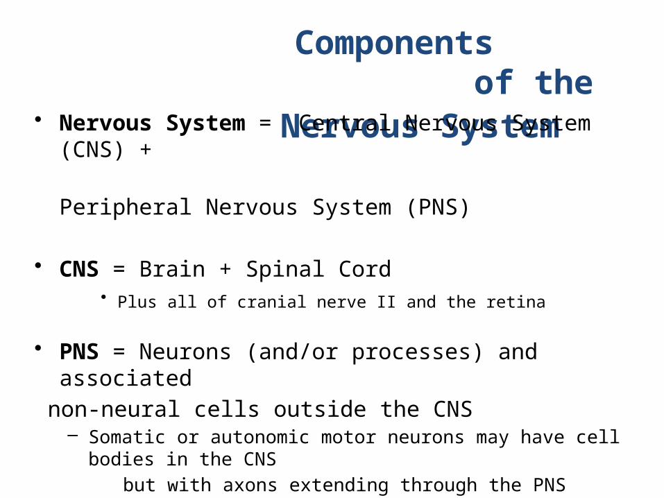
Components of the Nervous
System • Nervous System = Central Nervous System (CNS) +
Peripheral Nervous System (PNS)
• CNS = Brain + Spinal Cord
• Plus all of cranial nerve II and the retina
• PNS = Neurons (and/or processes) and associated
non-neural cells outside the CNS – Somatic or autonomic motor neurons may have cell bodies in the
CNS
but with axons extending through the PNS – Sensory cells may have receptive endings, axons and cell bodies
in
the PNS but restricted parts of their axons extending into the CNS – Many autonomic neurons dwell entirely in the periphery

Central Nervous System
Peripheral Nervous System

Central Nervous System• Five Parts of the Brain
– Telecephalon • Lateral ventricles
– Diencephalon • Third ventricle
– Mesencephalon • Cerebral aqueduct
– Metencephalon • Rostral fourth ventricle
– Myelencephalon • Caudal fourth ventricle and
medullary • central canal (continuous
with the• spinal central canal)

Central nervous system (CNS) Five parts of the brain
Midbrain = Mesencephalon Pons + cerebellum = Metencephalon Medulla oblongata = Myelencephalon
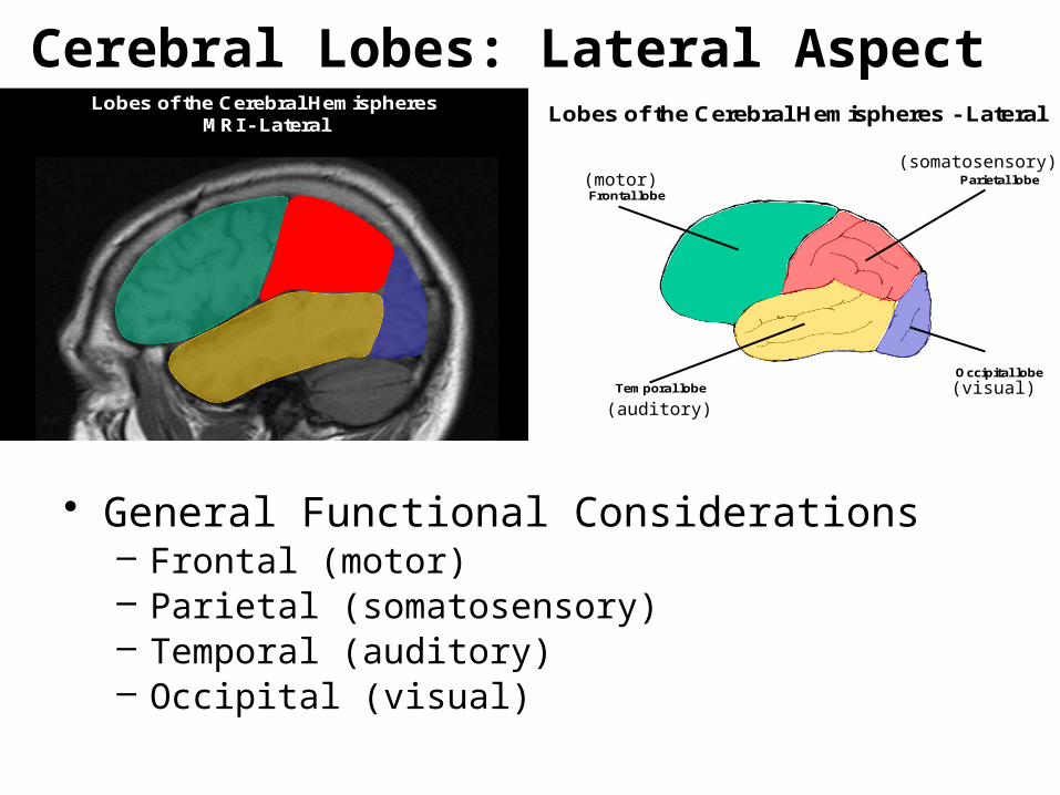
Lobes of the Cerebral Hemispheres MRI - Lateral Lobes of the Cerebral Hemispheres - Lateral
Frontal lobeParietal lobe
Temporal lobeOccipital lobe
Cerebral Lobes: Lateral Aspect
• General Functional Considerations – Frontal (motor) – Parietal (somatosensory) – Temporal (auditory) – Occipital (visual)
(motor)(somatosensory)
(visual)(auditory)

Lobes of the Cerebral Hemispheres –MRI - Medial
Lobes of the Cerebral Hemispheres - Medial
Parietal lobe
Frontal lobe
Occipital lobe
Temporal lobe
Limbic lobe
Cerebral Lobes: Medial Aspect
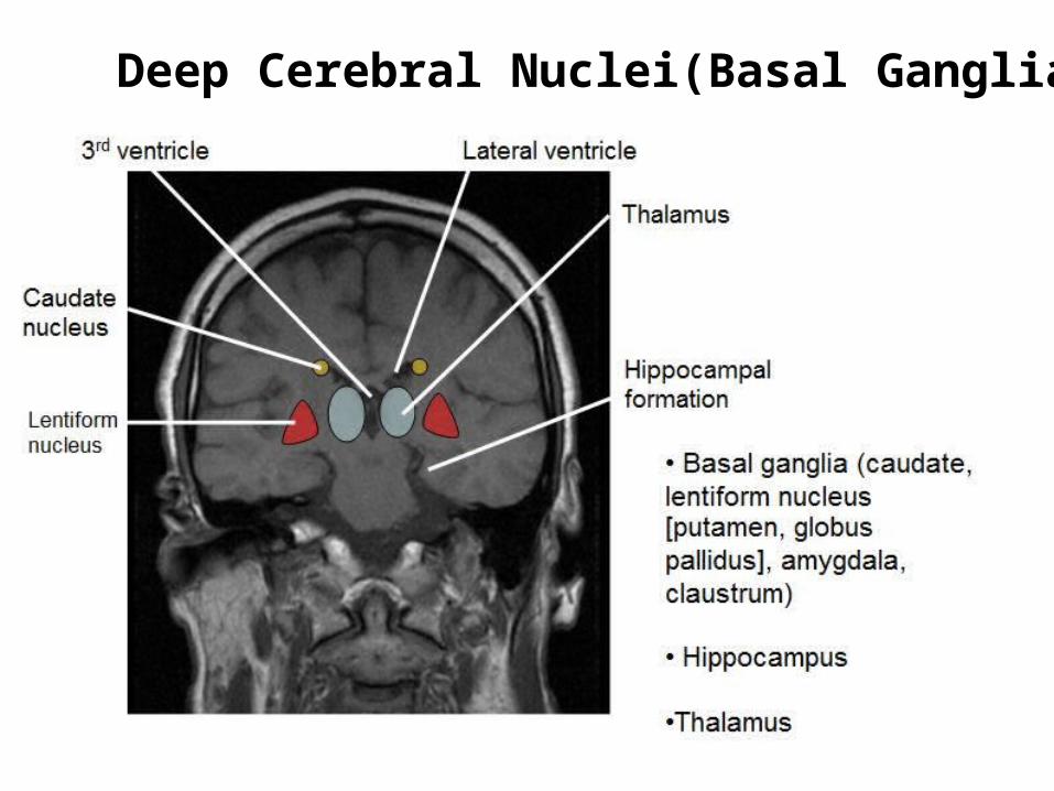
Deep Cerebral Nuclei(Basal Ganglia)
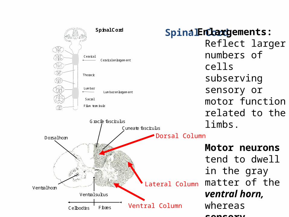
Spinal Cord • Enlargements: Reflect larger numbers of cells subserving sensory or motor function related to the limbs.
Motor neurons tend to dwell in the gray matter of the ventral horn, whereas sensory neurons tend to aggregate dorsally.
Spinal Cord
Cervical
Thoracic
Lumbar
Sacral
Filum terminale
Cervical enlargement
Lumbar enlargement
Ventral sulcus
FibresCellbodies
Cuneate fasciculus
Gracile fasciculus
Dorsal horn
Ventral horn
Dorsal Column
Lateral Column
Ventral Column

Spinal Nerves
• Dorsal and ventral nerve roots exit the cord, joining in the vertebral
canal. • Because there are 8 cervical
spinal segments but only 7 cervical vertebrae, the 1st spinal nerve emerges rostral to the 1st
cervical vertebra, whereas the 8th
cervical nerve emerges caudal to the 7th cervical vertebra and hence rostral to the first thoracic
vertebra.
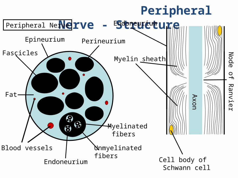
Peripheral Nerve - Structure
Fat
Blood vessels
Epineurium Perineurium
Endoneurium
Myelinated fibers
Unmyelinated fibers
Peripheral Nerve
Fascicles
Axon
No
de o
f Ra
nvie
r
Myelin sheath
Endoneurium
Cell body of Schwann cell

Important Sensory Pathways•Dorsal Column/Medial Lemniscus System - sensory
•Anterolateral System (spinothalamic tract) - sensory
Ventral sulcus
FibresCellbodies
Cuneate fasciculus
Gracile fasciculus
Dorsal horn
Ventral horn
Dorsal Column: Proprioception, Vibration, Tactile discrimination
Anterolateral System: Pain, Temperature, Pressure

Important Motor Pathways
• Corticospinal Tract - (lateral and anterior)
motor • Corticobulbar Tract - motor • Vestibulospinal (lateral and medial) Tract -
motor • Reticulospinal (lateral and medial) Tract -
motor • Rubrospinal Tract - motor

Your 43-yr-old female patient suffers a traumatic accident in which the dorsal columns of the spinal cord are damaged.
What function is lost? 1. Motor
2. Tactile discrimination
3. Nociception
Ventral sulcus
FibresCellbodies
Cuneate fasciculus
Gracile fasciculus
Dorsal horn
Ventral horn

Brodmann’s Areas - Left Hemisphere.

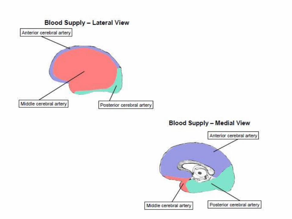
Vertebral and Basilar Artery
Picture taken from Ward Model
1 Anterior cerebral a.
2 Anterior communicating a.
3 Internal carotid a.
4 Middle cerebral a.
5 Posterior communicating a.
6 Posterior cerebral a.
7 Superior cerebellar a.
8 Basilar a.
9 Anterior inferior cerebellar a.
10 Vertebral a.
11 Posterior inferior cereballar a.
11
2
3 4
567
8
9
10
11
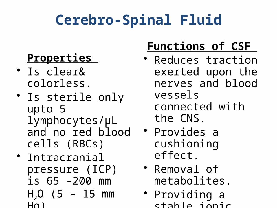
Cerebro-Spinal Fluid
Properties • Is clear& colorless. • Is sterile only upto 5
lymphocytes/µL and no red blood cells (RBCs)
• Intracranial pressure (ICP) is 65 -200 mm H2O (5 – 15 mm Hg)
Functions of CSF • Reduces traction exerted
upon the nerves and blood vessels connected with the CNS.
• Provides a cushioning effect.
• Removal of metabolites. • Providing a stable ionic
environment.

Circulation of the CSF
Essential Neuroscience, Updated 1st Edition Allan Siegel, Ph.D; Hreday N. Sapru PhD
1
2
3
4
5
6
7
8

Clinical Correlations
Hydrocephalus • Dilation of the ventricles (or hydrocephalus) occurs when
the circulation of CSF is blocked or its absorption is impeded, while the CSF formation continues to occur at a constant rate.
2 types
1. Non communicating (obstructive) Hydrocephalus
2. Communicating Hydrocephalus

Clinical Correlations • Transient Ischemic Attack (TIA). This is an acute loss of cerebral or monocular function with symptoms lasting under 24 hours. The origin is presumed to be a disorder of the cerebral circulation that leaves parts of the brain with an inadequate blood supply. Recovery of functions is likely. Predicted by stenosis if internal carotid artery and intermittent atrial fibrillation. Predicts stroke within a year. • Reversible Ischemic Neurological Deficit. This is an acute loss
of cerebral or monocular function with symptoms lasting longer than 24 hours due to inadequate blood supply of parts of the brain. Recovery of functions is likely.
• Stroke (Cerebrovascular Accident). This is a rapidly developing loss
of cerebral function due to cerebrovascular disturbance. The occurring
symptoms are irreversible or only partial reversible. In some patients the symptoms are global. The severity of loss ranges from partial recovery through permanent disability to coma and death.
Related Documents