Health Informatics - An International Journal (HIIJ) Vol.1, No.1, August 2012 27 MEDICAL IMAGES AUTHENTICATION THROUGH WATERMARKING PRESERVING ROI Sonika C. Rathi 1 and Vandana S. Inamdar 2 1 Department of Computer Engineering, College of Engineering Pune, Shivajinagar, Pune University, Maharashtra, India [email protected] 2 Department of Computer Engineering, College of Engineering Pune, Shivajinagar, Pune University, Maharashtra, India [email protected] ABSTRACT. Telemedicine is a well-known application where enormous amount of medical data need to be securely transferred over the public network and manipulate effectively. Medical image watermarking is an appropriate method used for enhancing security and authentication of medical data, which is crucial and used for further diagnosis and reference. This project focuses on the study of medical image watermarking methods for protecting and authenticating medical data. Additionally, it covers algorithm for application of water marking technique on Region of Non Interest (RONI) of the medical image preserving Region of Interest (ROI). The medical images can be transferred securely by embedding watermarks in RONI allowing verification of the legitimate changes at the receiving end without affecting ROI. Segmentation plays an important role in medical image processing for separating the ROI from medical image. The proposed system separate the ROI from medical image by GUI based approach, which works for all types of medical images. The experimental results show the satisfactory performance of the system to authenticate the medical images preserving ROI. KEYWORDS ROI & RONI, Segmentation, Authentication, security, medical confidentiality 1. INTRODUCTION Speedy development of internet in every field leads to availability of digital data to the public. Internet has been spread in many applications like telemedicine, online-banking, teleshopping etc. One of this application telemedicine is crucial one, where Internet is used to transfer or receive medical data by healthcare professional. Due to advancement in information and communication technologies, a new context of easier access, manipulation, and distribution of this digital data have been established [1]. The medical images can be readily shared via computer networks and easily used, processed, and transmitted by using great spread network [2, 3]. In the last decades, uses of advanced electronic and digital equipments in health care services are increased. In fact, in most of the hospitals physicians diagnose their patients by relying on the provided electronic and digital data (such as Ultrasonic, Computed Tomography (CT), Magnetic Resonance Imaging (MRI) and X-ray images). This results in generation of large number of electro digital data (i.e. medical images) continuously at various health care centers and hospitals around the world.

Welcome message from author
This document is posted to help you gain knowledge. Please leave a comment to let me know what you think about it! Share it to your friends and learn new things together.
Transcript

Health Informatics - An International Journal (HIIJ) Vol.1, No.1, August 2012
27
MEDICAL IMAGES AUTHENTICATION THROUGHWATERMARKING PRESERVING ROI
Sonika C. Rathi1 and Vandana S. Inamdar2
1Department of Computer Engineering, College of Engineering Pune, Shivajinagar,Pune University, Maharashtra, India
[email protected] of Computer Engineering, College of Engineering Pune, Shivajinagar,
Pune University, Maharashtra, [email protected]
ABSTRACT.
Telemedicine is a well-known application where enormous amount of medical data need to be securelytransferred over the public network and manipulate effectively. Medical image watermarking is anappropriate method used for enhancing security and authentication of medical data, which is crucial andused for further diagnosis and reference. This project focuses on the study of medical imagewatermarking methods for protecting and authenticating medical data. Additionally, it covers algorithmfor application of water marking technique on Region of Non Interest (RONI) of the medical imagepreserving Region of Interest (ROI). The medical images can be transferred securely by embeddingwatermarks in RONI allowing verification of the legitimate changes at the receiving end without affectingROI. Segmentation plays an important role in medical image processing for separating the ROI frommedical image. The proposed system separate the ROI from medical image by GUI based approach,which works for all types of medical images. The experimental results show the satisfactory performanceof the system to authenticate the medical images preserving ROI.
KEYWORDS
ROI & RONI, Segmentation, Authentication, security, medical confidentiality
1. INTRODUCTION
Speedy development of internet in every field leads to availability of digital data to the public.Internet has been spread in many applications like telemedicine, online-banking, teleshoppingetc. One of this application telemedicine is crucial one, where Internet is used to transfer orreceive medical data by healthcare professional. Due to advancement in information andcommunication technologies, a new context of easier access, manipulation, and distribution ofthis digital data have been established [1]. The medical images can be readily shared viacomputer networks and easily used, processed, and transmitted by using great spread network[2, 3].
In the last decades, uses of advanced electronic and digital equipments in health care servicesare increased. In fact, in most of the hospitals physicians diagnose their patients by relying onthe provided electronic and digital data (such as Ultrasonic, Computed Tomography (CT),Magnetic Resonance Imaging (MRI) and X-ray images). This results in generation of largenumber of electro digital data (i.e. medical images) continuously at various health care centersand hospitals around the world.

Health Informatics - An International Journal (HIIJ) Vol.1, No.1, August 2012
28
In number of medical applications, special safety and confidentiality is required for medicalimages, because critical judgment is done on medical images, which leads to the propertreatment. Therefore, it must not be changed in an illegitimate way; otherwise, an undesirableoutcome may results due to loss of decisive information. Therefore, there is a need to provide astrict security in medical images to ensure only occurrence of legitimate changes. Now-a-daysexchange of medical images between hospitals located in different geographical location is verycommon. Moreover, as this exchange of “medical reference data” done via unsecured opennetworks leads to the condition of changes to occur in medical images and creates a threatwhich results in undesirable outcome. Considering this fact, demand of security is gettinghigher due to easy reproduction of digitally created medical images.
For copyright protection and authentication of these medical images, digital watermarking is anemerging technique, which includes the embedding and extraction process. In embeddingprocess some secret information is embedded into medical images. Extraction process dealswith the extraction of secret message, which is embedded in the medical image. If failure occursin extraction process the physician would come to know that there has been some kind oftampering with that image, and he would take precaution of not making diagnosis based on thatimage. However, if the extraction process extracts the correct watermark, which generallyconsumes a few seconds, physician can continue with diagnosis.
Medical images hold decisive property and are very crucial and important part of medicalinformation. Such part of the medical image is called as Region of Interest (ROI). The ROI ishelpful in providing further diagnosis by the physician. A small bit of distortion in ROI maylead to undesirable treatment for patient. For securing medical images through watermarkingROI should be preserved and the watermarks can be applied on the remaining part of the imagecalled as Region of Non Interest (RONI). Therefore, application of watermarking in medicalimages can be considered as two-step process which includes:
1. Extracting ROI form the medical images
2. Applying watermarking on RONI
Different algorithms are available for segmentation of ROI on the different types of medicalimages. Additionally, there are different algorithms available for applying watermarking.
2. REGION OF INTEREST (ROI) SEGMENTATION
Segmentation plays an important role in medical image processing [4, 5]. In medical imageanalysis segmentation is the first step to be followed, to avoid distortion of ROI [4, 6]. Imagesegmentation deals with the process of partitioning an image into different regions by groupingtogether neighborhood pixels based on some predefined similarity criterion [7]. This similaritycriterion can be defined by specific properties of pixels in the image. Segmentation in medicalimaging is used for extracting the features, image display and for the measurement of image.The goal of segmentation is to divide entire medical image into sub regions i.e. (white and graymatter). In addition, this helps in classifying image pixels into anatomical regions (such asbones, muscles and blood vessels).
Defining the borders of ROI in medical image can simplify the procedure of segmentation. Inaddition, the step of defining borders of ROI is a crucial one, which helps in determining theresult of the application as entire analysis fully relies on the output from segmentation step.There are different approaches (for segmenting the image) defined for the different imagingtechnologies such as CT, MRI, US, colonoscopy etc. Segmentation is semi-automatic procedureand we need to define a seed point in an image. Therefore, the algorithm, which gives perfectresult for one application, might not even work for another. Figure 1 shows the ROI part ofmedical image, where physician performs the diagnosis.

Health Informatics - An International Journal (HIIJ) Vol.1, No.1, August 2012
29
Figure 1: Medical image indicating ROI
We have various existing medical imaging like Computed Tomography (CT), MagneticResonance Imaging (MRI), Ultrasound (US), and Positron Emission Tomography (PET) etc.Here, the most common imaging i.e. CT scan imaging is discussed in detail with their proposedalgorithms.
2.1. Computed Tomography (CT)
Computed Tomography (CT) scanning sometimes called Computed Axial Tomography (CAT)scanning [7], is a noninvasive medical test that helps physicians diagnose and treat medicalconditions. CT scanning combines special x-ray equipment with sophisticated computers toproduce multiple images or pictures of the inside of the body. These cross-sectional images ofthe area being studied can then be examined on a computer monitor, printed or transferred to aCD. CT scans of internal organs, bones, soft tissue and blood vessels provide greater clarity andreveal more details than regular x-ray exams. Using specialized equipment and expertise tocreate and interpret CT scans of the body, radiologists can more easily diagnose problems suchas cancers, cardiovascular disease, infectious disease, appendicitis, trauma, and musculoskeletaldisorders. Hence, the CT scan application is been widely used in medical domain. There aredifferent segmentation methods proposed considering CT scan of different body organs (such aslung, liver, kidney, etc.) This section covers the different segmentation algorithm for CT scanimages for protecting the distortion of diagnosis value.
The 2-D and 3-D segmentation of organs in medical application of image processing areclassified into model based and non-model based approaches. Non-model based approachesdepends on local information such as, texture, intensity, spatial correlation of 2-D organ imagein consecutive slices, and the location of the organ in the abdominal area with respect toneighboring structures, e.g., spine and ribs [8]. Various segmentation algorithms are developedusing non-model based approach. This section first covers the different segmentation algorithm,which uses non-model based approach. Susomboon et al. [9] presented texture features toperform region classification for extracting liver’s soft tissue. Seo et al. [10] employed amultimodal threshold method based on piecewise linear interpolation that used spine location asa reference point. Forouzan et al. [11] introduced a multilayer threshold technique, in which bystatistical analysis of the liver intensity it calculates the threshold value. Both these methods usethe local information of the liver’s relative position to the spine and ribs. Non-model basedmethods for organ segmentation leads to inaccuracies due to variation in imaging condition,because of occurrence of tumor inside the organ and noise. Dependencies on prior informationsuch as texture and image values could cause inaccuracies in segmentation process as featurecould change from one patient to another. Moreover, most of these methods are parameterdependent and hence for the best performance it often needs to adjust the parameters from oneCT volume to other. In recent years, model-based image segmentation algorithms developed forvarious medical applications. These methods aim to recover an organ based on statisticalinformation. State-of-the-art algorithms on model-based segmentation are based on active shape

Health Informatics - An International Journal (HIIJ) Vol.1, No.1, August 2012
30
and appearance models [8, 12]. Model-based techniques provide more accurate and robustalgorithm for segmenting the CT scan image. These techniques also deal with the missing imagefeatures via interpolation. The performance of these methods depends on the number and typeof training data. Moreover, if the shape to be segmented lies too far from the model space, thatmight not be detected by many those better methods which is not implemented by statisticalmodel-based approach.
Pan and Dawant [13] reported a geometrical-level set method for automatic segmentation of theliver in abdominal CT scans without relying on the prior knowledge of shape and size. Even ifthis method depends on a model-based technique, that outperforms threshold-based techniques,but it did not use prior knowledge of the liver shape. Lin et al. [14] presented the algorithm toperform segmentation of kidney, based on an adaptive region growing and an elliptical kidneyregion positioning that used spines as landmark. H. Badakhshannoory and P. Saeedi [15]incorporated a method for liver segmentation. Based on liver boundary edges to identify liverregions, nonrigid registration and a multilayer segmentation technique are combined in thisapproach. This method does not affected by the diversity of existing liver shapes, as it does notrely on any shape model. Samuel et al. [16] has proposed the use of Ball-Algorithm for thesegmentation of lungs. In this algorithm at the first stage, it applies the grey level thresholdingto the CT images to segment the thorax from background and then the lungs from the thorax.Then in the next step to avoid loss of juxtapleural nodules, this method performs the rolling ballalgorithm. Julian Ker [17] has presented the method of doing segmentation of lungs, which isnamed as TRACE method. Due to the possible presence of various disease processes, and thechange of the anatomy with vertical position results in variation of size, shape, texture of lungsCT image of different patients. Therefore, the boundary between lung and surrounding tissuescan vary from a smooth-edged, sharp-intensity transition to irregularly jagged edges with a lessdistinct intensity transition. The TRACE algorithm implemented with new perception of a non-approximating technique for edge detection. Shiying et al. [18] have introduced a fullyautomatic method for identifying lungs in 3D pulmonary X-Ray CT images. The methodfollows three main steps:
• Lung region is extracted from CT-Scan image by applying graylevel thresholding,
• By using a dynamic programming it identifies the anterior and posterior junction, toseparate left and right lungs and
• To smooth the irregularities of boundary along the mediastinum nodule, it implementssequence of morphological operations
Ayman El-Baz et al. [19] have employed a fully automatic Computer-Assisted Diagnosis(CAD) system for lung cancer screening using chest spiral CT scans. This paper presents asystem for detection of abnormalities, identification or classification of these abnormalities withrespect to specific diagnosis, and provides the visualization of the results over computernetworks. The process of detection of abnormalities, identification of these abnormalities canachieve by image analysis system for 3-D reconstruction of the lungs. Riccardo Boscolo et al.[20] proposed method that uses the novel segmentation technique that combines a knowledgebased segmentation system with a sophisticated active contour model. This method performsrobust segmentation of various anatomic structures. In this approach the user, need to provideinitial contour placement, and the required parameter optimization automatically determined bythe high-level process. Binsheng et al. [21] reported the algorithm, which used the method ofselecting the threshold value by analyzing the histogram. This algorithm initially separates thelung parenchyma from the other anatomical structures from the CT images by using thresholdvalue. By this algorithm structure in CT scan image with higher densities having some higherdensity nodules, can grouped into soft tissues and bones, leading to an incomplete extraction of

Health Informatics - An International Journal (HIIJ) Vol.1, No.1, August 2012
31
lung mask. For having complete hollow free lung mask, morphological closing is applied in thisapproach. Hossein B. et al. [8] has introduced the model-based segmentation algorithm. In thisapproach instead of using model information to direct the segmentation algorithm forsegmenting an organ of CT scan images, it uses this information to choose a segment withhighest fidelity to the organ.
After completing with the segmentation of ROI, needs to proceed with medical imagewatermarking technique to provide security, authentication and privacy of this medical data.The next section of this paper provides the survey of different available medical imagewatermarking approaches.
3. MEDICAL IMAGE WATERMARKING
There has been fair amount of work done in the area of medical image processing. Beforeproceeding with survey of medical image processing, this section covers the foundation ofdigital watermarking, types of domain and performance measurement. The typical blockdiagram for medical image watermarking is given in figure 2.
Encoder E embeds the watermark W in medical image to provide security and authentication.Decoder D extracts the watermark from watermarked image. By comparing the extractedwatermark with original watermark, one can affirm the tampering of medical image. Accordingto watermark embedding process, watermarking techniques are classified into two differentdomains.
• Spatial domain: The spatial-domain watermark insertion manipulates image pixels.However, the spatial-domain watermark insertion is simple and easy to implement, it isweak against various attacks and noise.
• Transform domain: The transform-domain watermark insertion is based on thetransform coefficients of cover image. It is more robust against attacks. Discrete CosineTransform (DCT), Discrete Wavelet Transform (DWT), and Discrete FourierTransform (DFT) are three popular methods in transform domain.
Figure 2: Block Diagram of Medical Image Watermarking
To ensure the reliability and quality of the watermarked image, the performance ofwatermarking is calculated, which measured in terms of perceptibility. There are two method ofcalculating the performance measure.
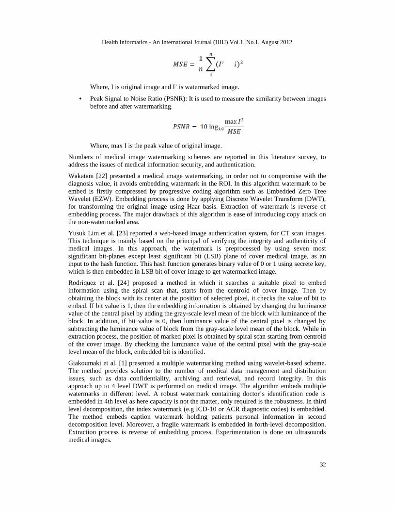
• Mean Square Error (MSE): It is simplest function to measure the perceptual distancebetween watermarked and original image. MSE can be defined as:
Health Informatics - An International Journal (HIIJ) Vol.1, No.1, August 2012
32
Where, I is original image and I’ is watermarked image.
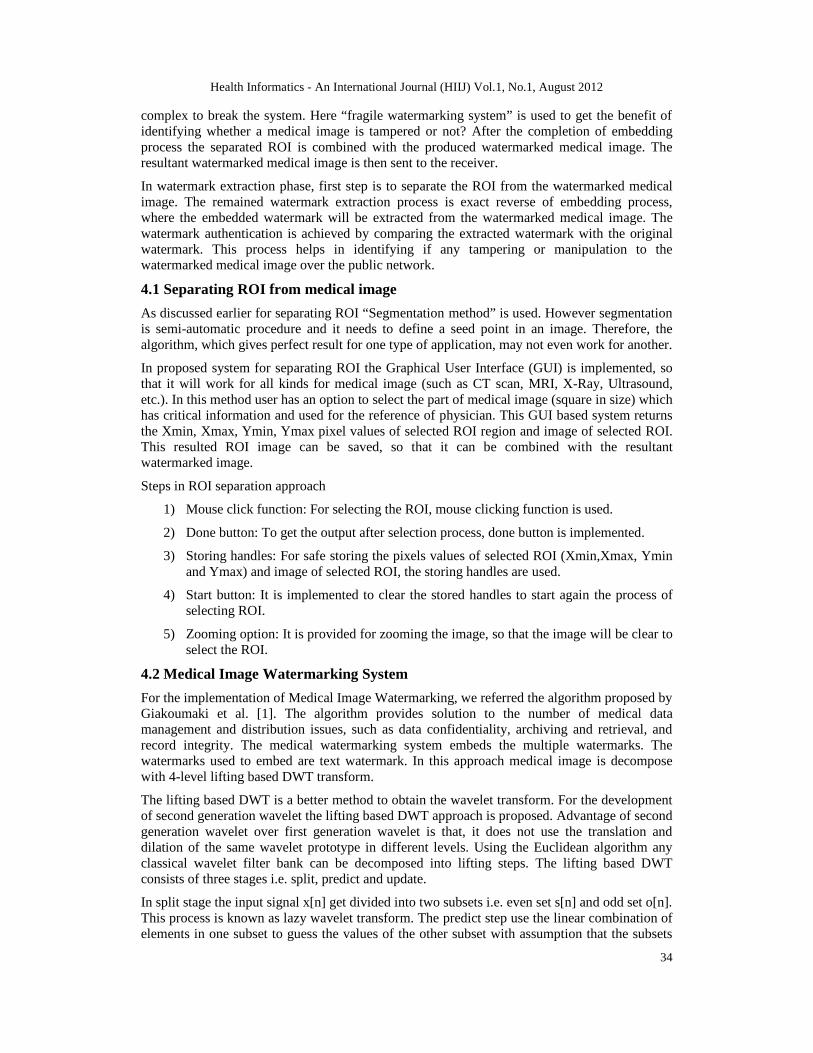
• Peak Signal to Noise Ratio (PSNR): It is used to measure the similarity between imagesbefore and after watermarking.
Where, max I is the peak value of original image.
Numbers of medical image watermarking schemes are reported in this literature survey, toaddress the issues of medical information security, and authentication.
Wakatani [22] presented a medical image watermarking, in order not to compromise with thediagnosis value, it avoids embedding watermark in the ROI. In this algorithm watermark to beembed is firstly compressed by progressive coding algorithm such as Embedded Zero TreeWavelet (EZW). Embedding process is done by applying Discrete Wavelet Transform (DWT),for transforming the original image using Haar basis. Extraction of watermark is reverse ofembedding process. The major drawback of this algorithm is ease of introducing copy attack onthe non-watermarked area.
Yusuk Lim et al. [23] reported a web-based image authentication system, for CT scan images.This technique is mainly based on the principal of verifying the integrity and authenticity ofmedical images. In this approach, the watermark is preprocessed by using seven mostsignificant bit-planes except least significant bit (LSB) plane of cover medical image, as aninput to the hash function. This hash function generates binary value of 0 or 1 using secrete key,which is then embedded in LSB bit of cover image to get watermarked image.
Rodriquez et al. [24] proposed a method in which it searches a suitable pixel to embedinformation using the spiral scan that, starts from the centroid of cover image. Then byobtaining the block with its center at the position of selected pixel, it checks the value of bit toembed. If bit value is 1, then the embedding information is obtained by changing the luminancevalue of the central pixel by adding the gray-scale level mean of the block with luminance of theblock. In addition, if bit value is 0, then luminance value of the central pixel is changed bysubtracting the luminance value of block from the gray-scale level mean of the block. While inextraction process, the position of marked pixel is obtained by spiral scan starting from centroidof the cover image. By checking the luminance value of the central pixel with the gray-scalelevel mean of the block, embedded bit is identified.
Giakoumaki et al. [1] presented a multiple watermarking method using wavelet-based scheme.The method provides solution to the number of medical data management and distributionissues, such as data confidentiality, archiving and retrieval, and record integrity. In thisapproach up to 4 level DWT is performed on medical image. The algorithm embeds multiplewatermarks in different level. A robust watermark containing doctor’s identification code isembedded in 4th level as here capacity is not the matter, only required is the robustness. In thirdlevel decomposition, the index watermark (e.g ICD-10 or ACR diagnostic codes) is embedded.The method embeds caption watermark holding patients personal information in seconddecomposition level. Moreover, a fragile watermark is embedded in forth-level decomposition.Extraction process is reverse of embedding process. Experimentation is done on ultrasoundsmedical images.

Health Informatics - An International Journal (HIIJ) Vol.1, No.1, August 2012
33
Hemin Golpira et al. [25] reported reversible blind watermarking. In this approach duringembedding process, firstly by applying Integer Wavelet Transform (IDWT) image isdecomposed into four subbands. By selecting two points, called thresholds, according to thecapacity required for the watermark data, watermark is embedded. To get watermarked imageInverse Integer Wavelet Transform (IIDWT) is applied. In the extraction process, all of thesestages are performed in reverse order to extract watermark as well as host image.
Nisar Ahmed Memonet et al. [26] presented fragile and robust watermarking technique formedical images. The method embeds two different watermarks, the robust watermark andfragile watermark in the medical images. The embedding process starts with separation of ROIand RONI from medical images. The robust watermark containing the electronic patient record(EPR), Doctor’s identification code (DIC) and 1st bit-plane of ROI by extracting the LSBs isencrypted by using pseudo random sequence generated by user defined key. Then this resultantwatermark is embedded in high frequency coefficient of IWT in RONI part of medical data. Theproposed method generates fragile watermark by creating the binary image in tiled fashion andthen this fragile watermark is cropped off by the same size as the ROI. The algorithm embedsthis fragile watermark into spatial domain of ROI part of medical image. The extraction processis reverse of embedding process.
4. PROPOSED SYSTEM
Our approach focuses on embedding watermark in RONI region of medical image by preservingROI. This approach helps in isolating ROI region i.e. not to distort the critical area of medicalimage, which will be referred by physician for the diagnosis. The system diagram for thisapproach is shown in figure 3.
Figure 3: Medical Image Watermarking Approach Preserving ROI
In first phase of system separating the ROI from the original medical image provides RONIregion for embedding watermark. This step isolates ROI from embedding process. In this phasemultiple watermarks are embedded into the RONI area of medical image. Embedding multiplewatermarks ensure high security of medical image as it carries high payload and it will be more
Health Informatics - An International Journal (HIIJ) Vol.1, No.1, August 2012
34
complex to break the system. Here “fragile watermarking system” is used to get the benefit ofidentifying whether a medical image is tampered or not? After the completion of embeddingprocess the separated ROI is combined with the produced watermarked medical image. Theresultant watermarked medical image is then sent to the receiver.
In watermark extraction phase, first step is to separate the ROI from the watermarked medicalimage. The remained watermark extraction process is exact reverse of embedding process,where the embedded watermark will be extracted from the watermarked medical image. Thewatermark authentication is achieved by comparing the extracted watermark with the originalwatermark. This process helps in identifying if any tampering or manipulation to thewatermarked medical image over the public network.
4.1 Separating ROI from medical image
As discussed earlier for separating ROI “Segmentation method” is used. However segmentationis semi-automatic procedure and it needs to define a seed point in an image. Therefore, thealgorithm, which gives perfect result for one type of application, may not even work for another.
In proposed system for separating ROI the Graphical User Interface (GUI) is implemented, sothat it will work for all kinds for medical image (such as CT scan, MRI, X-Ray, Ultrasound,etc.). In this method user has an option to select the part of medical image (square in size) whichhas critical information and used for the reference of physician. This GUI based system returnsthe Xmin, Xmax, Ymin, Ymax pixel values of selected ROI region and image of selected ROI.This resulted ROI image can be saved, so that it can be combined with the resultantwatermarked image.
Steps in ROI separation approach
1) Mouse click function: For selecting the ROI, mouse clicking function is used.
2) Done button: To get the output after selection process, done button is implemented.
3) Storing handles: For safe storing the pixels values of selected ROI (Xmin,Xmax, Yminand Ymax) and image of selected ROI, the storing handles are used.
4) Start button: It is implemented to clear the stored handles to start again the process ofselecting ROI.
5) Zooming option: It is provided for zooming the image, so that the image will be clear toselect the ROI.
4.2 Medical Image Watermarking System
For the implementation of Medical Image Watermarking, we referred the algorithm proposed byGiakoumaki et al. [1]. The algorithm provides solution to the number of medical datamanagement and distribution issues, such as data confidentiality, archiving and retrieval, andrecord integrity. The medical watermarking system embeds the multiple watermarks. Thewatermarks used to embed are text watermark. In this approach medical image is decomposewith 4-level lifting based DWT transform.
The lifting based DWT is a better method to obtain the wavelet transform. For the developmentof second generation wavelet the lifting based DWT approach is proposed. Advantage of secondgeneration wavelet over first generation wavelet is that, it does not use the translation anddilation of the same wavelet prototype in different levels. Using the Euclidean algorithm anyclassical wavelet filter bank can be decomposed into lifting steps. The lifting based DWTconsists of three stages i.e. split, predict and update.
In split stage the input signal x[n] get divided into two subsets i.e. even set s[n] and odd set o[n].This process is known as lazy wavelet transform. The predict step use the linear combination ofelements in one subset to guess the values of the other subset with assumption that the subsets

Health Informatics - An International Journal (HIIJ) Vol.1, No.1, August 2012
35
produced in the split stage are correlated with each other. The predicted values would be closeto the original values if the correlation between both the subset is high. Generally the linearcombination of the even subset elements are used to predict odd subset values. Although thereare chances of loss of properties of signal such as mean value in the predict step. The predictstep causes the loss of some basic properties of the signal like mean value, which needsto be preserved. The update step lifts the even sequence values using the linearcombination of the predicted odd sequence values so that the basic properties of theoriginal sequence is preserved [5].
4.2.1 Integer to Integer Transform:
It was observed that usually when wavelet transforms is performed on integer sequence it givesfloating point coefficients. As per Calderbank [6] wavelet transform which will map integers tointegers can be build with the help of lifting structure. This can be achieved by rounding off orupdating the filter in each lifting step before its addition or subtraction. The invert of the liftingsteps can be produced by following the exact reverse operation and flipping the signs.
4.2.2 Proposed Method:
The watermarks used in this approach:
1) Doctors identification code
2) Indexed watermark
3) Patients reference identification code
4) Patients diagnosis information
5) Patients treatment information
The listed watermarks used in this proposed watermarking scheme helps in addressing differentissues and concerns in healthcare management system, Such as confidentiality of medical data,recovering original image without any distortion, data integrity, authentication and efficient datamanagement.
Confidentiality of medical data is achieved by embedding watermark using Integer to IntegerDiscrete Wavelet Transform (IDWT), which confirms the imperceptibility property. Thisproperty ensures the embedded watermark will be invisible to the normal human eye and thewatermark can be extracted by the one who knows the embedding and extraction algorithmapplied in this system. By applying Inverse Discrete Wavelet Transform (IDWT) at the receiverend original image can be recovered without any distortion. Also the distortion to the ROI hasalready been avoided by separating the ROI before embedding the watermark into the medicalimage. Medical data integrity is achieved by using fragile watermarking system, so anymanipulation on medical image data leads in distortion of embedded watermark. For theauthentication purpose the included watermarks such as doctor’s identification code, patient’sidentification code will ensures the entitled users can access or modify the medical data. Toprovide efficient data management in this system the indexed watermark is embedded whichhelps in retrieving the image for the future reference if needed using database query.
The watermarks are inserted in different decomposition levels and sub-bands depending on theirtype. They can be independently embedded and retrieved without any intervention among them.By integrating this idea into different medical acquisition systems like Ultrasound, CT and MRIetc. This system can be applied in different applications such as e-diagnosis or medical imagesharing through picture archiving and communication.

Health Informatics - An International Journal (HIIJ) Vol.1, No.1, August 2012
36
4.2.3 Algorithm:
In this algorithm the multiple watermarks embedding technique is used. Where, depending onthe quantization of selected coefficients the multiple watermarks embedding procedure is used.This prevents any modification to the watermark bits by granting integer changes in spatialdomain of medical image. This can be achieved by applying 4-level haar wavelet transform todecompose the host medical image. Moreover it gives the output as coefficients, which are inthe form of dyadic rational numbers. These coefficients denominators are in powers of 2. Themultiple of 2^l (l is decomposition level) number adding or subtracting to the producedcoefficient value, assures that the inverse DWT provide integer pixel values. Wavelet transformgenerally provides the coefficients which are real numbers. By applying the quantizationfunction it assigns the binary number to every coefficient. This quantization function is definedas
Where s is a user-defined offset for increased security, f is frequency coefficient produced byhaar wavelet transform and Δ, the quantization parameter, is a positive real number. MoreoverΔ is defined as Δ =2^l.
The algorithm for embedding multiple watermarks is explained below:
Step 1: Separate the ROI region from the host medical data using GUI based mouse clickingapproach. Which results in image of RONI region, name it as original medical image.
Step 2: Save the removed ROI from medical image.
Step 3: The multiple watermarks to be embed into a original image is generated by reading thepatient’s information file from text document, and converting it into binary.
Step 4: Apply the 4-level Haar-lifting wavelet transform to original medical image, to obtain agross image approximation at the lowest resolution level and a sequence of detail imagescorresponding to the horizontal, vertical, and diagonal details at each of the four decompositionlevels.
Step 5: On each decomposition level the watermark bit wi is embedded into the key determinedcoefficient f, which is obtained by applying wavelet transform according to the followingcondition:
a) If Q(f) = wi, the coefficient is not modified.
b) Otherwise, the coefficient is modified so that Q(f) = wi, using the following equation:
f= f + ∆, if f <= 0
f = f - ∆, if f > 0
Step 6: The pre watermarked image is produced by performing the corresponding fourlevel inverse wavelet transform.
Step 7: The resultant watermarked image is obtained by combining the saved ROI with the prewatermarked image.
The watermark extraction process is similar to that of embedding one except that at thereceiving end extractor should have the knowledge of location of the embedded watermark. Thiscan achieve by the key-based embedding and detection. With this type of method access to the

Health Informatics - An International Journal (HIIJ) Vol.1, No.1, August 2012
37
watermark by unauthorized users is prevented. The algorithm for extraction process to recoverthe host medical image is explained below.
Step 1: Remove the ROI region from the received watermarked image with the help of Xmax,Xmin, Ymin and Ymax parameter provided with watermarked image.
Step 2: Apply the 4-level lifting-haar wavelet transform to the image which is created from step1, which results in a image approximation at level four and sequence of images correspondingto the horizontal, vertical, and diagonal details at each of the four decomposition levels.
Step 3: Identify the location of watermark by key-based detection.
Step 4: Extract the watermarks by applying quantization function defined in equation 5.3, whichrecovers the original coefficient. Convert the extracted binary watermark to text watermark.
Step 5: The pre output image is obtained by applying inverse 4-level haar wavelet transform.
Step 6: combine the separated ROI region to the pre output image to get the original hostmedical image.
5. EXPERIMENTS AND RESULTS
The proposed system has been applied against different type of medical image such as, CT scan,MRI, X-Ray and Ultrasound. We have tested the system over different size of medical imageslike 320 X 256, 384 X 384, and 512 X 512.
The applied watermark was consists of:
1. Doctor’s Identity: G123468
2. Indexing for database: 321-123.13
3. Patient's identification: sonika c rathi.190.85.04567851
4. Diagnosis Information: light.sugar healthy extra.spicy no.fats 12189.75.1
5. Treatment applied to the patient: painkiller.hgkkfgjklfd abcdefmglkh bkjdhkds.yeio
The results after applying the system against CT scan, MRI, X-Ray and Ultra-sound are shownbelow:
5.1 The experiments and results of the system without attacks
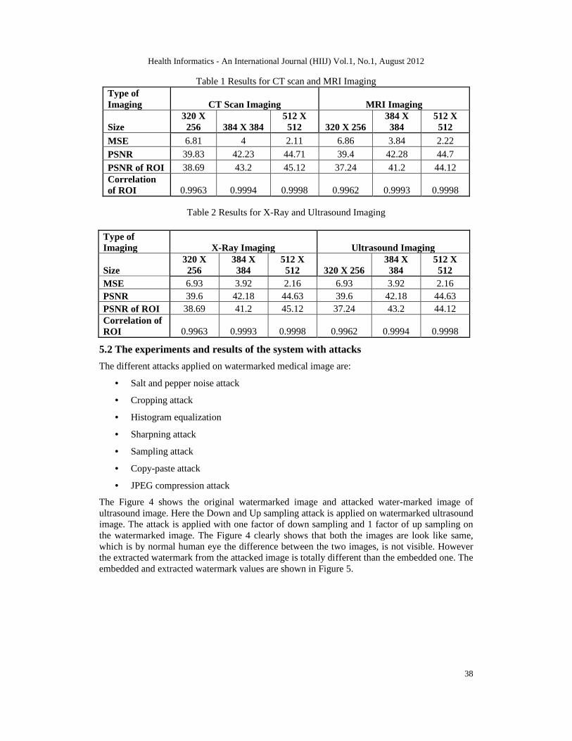
The system is applied on different images considering their image size and noted correspondingresults, which are shown in Table 1 and Table 2. The table shows the PSNR value for ROIextracted from host image and ROI extracted from watermarked image. As the corner pixelvalues of ROI image are changed the PSNR is not ∞ but there correlation is approximately 1.So, the selected ROI should be large enough to not compromise with the diagnosis value. Thetable also provides the PSNR value for embedded image and original image and their respectivemean square difference.
Health Informatics - An International Journal (HIIJ) Vol.1, No.1, August 2012
38
Table 1 Results for CT scan and MRI ImagingType ofImaging CT Scan Imaging MRI Imaging
Size320 X256 384 X 384
512 X512 320 X 256
384 X384
512 X512
MSE 6.81 4 2.11 6.86 3.84 2.22
PSNR 39.83 42.23 44.71 39.4 42.28 44.7
PSNR of ROI 38.69 43.2 45.12 37.24 41.2 44.12Correlationof ROI 0.9963 0.9994 0.9998 0.9962 0.9993 0.9998
Table 2 Results for X-Ray and Ultrasound Imaging
Type ofImaging X-Ray Imaging Ultrasound Imaging
Size320 X256
384 X384
512 X512 320 X 256
384 X384
512 X512
MSE 6.93 3.92 2.16 6.93 3.92 2.16PSNR 39.6 42.18 44.63 39.6 42.18 44.63PSNR of ROI 38.69 41.2 45.12 37.24 43.2 44.12Correlation ofROI 0.9963 0.9993 0.9998 0.9962 0.9994 0.9998
5.2 The experiments and results of the system with attacks
The different attacks applied on watermarked medical image are:
• Salt and pepper noise attack
• Cropping attack
• Histogram equalization
• Sharpning attack
• Sampling attack
• Copy-paste attack
• JPEG compression attack
The Figure 4 shows the original watermarked image and attacked water-marked image ofultrasound image. Here the Down and Up sampling attack is applied on watermarked ultrasoundimage. The attack is applied with one factor of down sampling and 1 factor of up sampling onthe watermarked image. The Figure 4 clearly shows that both the images are look like same,which is by normal human eye the difference between the two images, is not visible. Howeverthe extracted watermark from the attacked image is totally different than the embedded one. Theembedded and extracted watermark values are shown in Figure 5.

Health Informatics - An International Journal (HIIJ) Vol.1, No.1, August 2012
39
Figure 4: (a) The original watermarked Ultrasound image, (b) The image after up and down samplingattack
Figure 5 Embedded and extracted watermark values after down and up sampling attack on Ultrasoundimage
Health Informatics - An International Journal (HIIJ) Vol.1, No.1, August 2012
40
6. CONCLUSION AND FUTURE WORK
There exist various medical image watermarking algorithms which provide the confidentialityof medical data, recovering original image without any distortion, data integrity, authenticationand efficient data management. Also the different segmentation algorithms are in place, whichvary for the types of medical images such as MRI, CT scan, X-ray and Ultrasounds etc. Herethe proposed system used an algorithm to separate ROI from the host medical image that will beapplicable for all types of medical images. Separated ROI can be stored with xmin, xmax, ymin,and ymax value so that at the end of embedding process before transmitting watermarkedimage, the segmented ROI can be attached with watermarked image. So the ROI region whichis considered as a critical data and used as a reference by the physician for the treatment will besafe.
Proposed system uses DWT approach for embedding the watermark, instead of DWT use ofComplex Wavelet Transform (CWT) will make the system more robust and secure. The currentproposed system can further be extended to provide more secured system. This can be done byencrypting the watermark using secret key, before embedding it into medical images. Havingthe automated tool for separating the ROI from medical image will provide faster system andmore accurate system, which will be easier for end user. The watermark before embedding canbe compressed and then embedded. This will lead to more secured system. Also, it will takemore effort to break the system.
7. ACKNOWLEDGEMENTS
We take this opportunity to express my hearty thanks to all those who helped me in thecompletion of my project work. We are very grateful to the authors of various articles on theInternet, for helping us become aware of the research currently ongoing in this field.
8. REFERENCES
[1] Giakoumaki, Sotiris Pavlopoulos, and Dimitris Koutsouris, (Oct. 2006) “Multiple ImageWatermarking Applied to Health Information Management”, IEEE Trans. on informationtechnology in biomedicine, vol. 10, no. 4
[2] Imen Fourati Kallel, Mohamed Kallel, Mohamed Salim BOUHLEL, (2006 ) “A Secure fragileWatermarking Algorithm for medical Image Authentication in the DCT Domain”, IEEE
[3] M.S.Bouhlel, (Mars 2002) “Conception d'une banque d'images medicales sur INTERNET”,3eme Rencontres Institutionnelles: Rhones Alpes/ Tunisie (RIRAT'02). Tozeur, Tunisie, 21-22
[4] Preeti Aggarwal, Renu Vig, Sonali Bhadoria, and C.G.Dethe , (September 2011) “Role ofSegmentation in Medical Imaging: A Comparative Study”, International Journal of ComputerApplications (0975 – 8887), Volume 29– No.1
[5] Pradeep Singh; Sukhwinder Singh, Gurjinder Kaur, (2008) “A Study of Gaps in CBMIRusing Different Methods and Prospective, Proceedings of world academy of science,engineering and technology”, volume 36 , ISSN 2070-3740, pp. 492-496.
[6] Zhen Ma, Joao Manuel, R. S. Tavares, R. M. Natal Jorge, (2009) “A review on the currentsegmentation algorithms for medical images”, 1st International Conference on ImagingTheory and Applications (IMAGAPP), Lisboa, Portugal, INSTICC Press, pp. 135-140.
[7] Nisar Ahmed Memon, Anwar Majid Mirza, and S.A.M. Gilani, (2006) “Segmentation of Lungsfrom CT Scan Images for Early Diagnosis of Lung Cancer”, World Academy of Science,Engineering and Technology 20.
[8] Hossein Badakhshannoory and Parvaneh Saeedi, (September 2011) “A Model-Based ValidationScheme for Organ Segmentation in CT Scan Volumes”, IEEE Trans. on biomedical information,vol. 58, no. 9,

Health Informatics - An International Journal (HIIJ) Vol.1, No.1, August 2012
41
[9] R. Susomboon, D. Raicu, and J. Furst, (2007) “A hybrid approach for liver segmentation”, inProc. 3-D Segment. Clin.-MICCAI Grand Challenge
[10] K. Seo, L. C. Ludeman, S. Park, and J. Park, (2005) “Efficient liver segmentation based on thespine”, Adv. Inf. Syst., vol. 3261, pp. 400–409
[11] A. H. Forouzan, R. A. Zoroofi, M. Hori, and Y. Sato, (2009) “Liver segmentation by intensityanalysis and anatomical information in multislice CT images”, in Proc. Liver Segment. IntensityAnal Anatomical Inf. Multi-Slice CT Images, vol. 4, pp. 287–297
[12] T. F. Cootes, C. J. Taylor, and D. H. Cooper, (2001) “Statistical models of appearance formedical image analysis and computer vision,” Proc. SPIE, vol. 4322, pp. 236–248
[13] S. Pan and B. M. Dawant, (2001) “Automatic 3D segmentation of the liver from abdominal CTimages: A level-set approach,” Proc. SPIE, vol. 4322, pp. 128–138
[14] D. T. Lin, C. C. Lei, and S. W. Hung, (Jan 2006) “Computer-aided kidney segmentation onabdominal CT images”, IEEE Trans. Inf. Technol. Biomed.,vol. 10, no. 1, pp. 59–65
[15] H. Badakhshannoory and P. Saeedi, (2010) “Liver segmentation based on de-formableregistration and multilayer segmentation,” in Proc. IEEE Int. Conf. Image Process., pp. 2549–2552
[16] Samuel G. Armato III, Maryellen L. Giger and Catherine J. Moran, (1999) “ComputerizedDetection of Pulmonary Nodules on CT Scans”, RadioGraphics, vol. 19, pp. 1303-1311
[17] Julian Kerr, (May 2000) “The TRACE method for Segmentation of Lungs from Chest CTimages by Deterministic Edge Linking”, University of New South Wales, Department ofArtificial Intelligence, Australia
[18] Shiying Hu, Eric A.Huffman, and Joseph M. Reinhardt, (June 2001) “Automatic LungSegementation for Accurate Quantitiation of Volumetric X-Ray CT images”, IEEE Transactionson Medical Imaging, vol. 20, No. 6
[19] Ayman El-Baz, Aly A. Farag, Robert Falk, and Renato La Rocc, (Jan 2002) ” Detection,Visualization, and Identification of Lung Abnormalities in Chest Spiral CT Scans: Phase 1”,International Conference on Biomedical Engineering, Cairo, Egypt
[20] Riccardo Boscolo, Mathew S. Brown, Michael F. McNitt-Gray, (2002) “Medical ImageSegmentation with Knowledge-guided Robust Active Contours”, Radiographics, vol. 22, pp.437-448
[21] Binsheng Zhao, Gordon Gamsu, Michelle S. Ginsberg, (2003) “Automatic detection of smalllung nodules on CT utilizing a local density maximum algorithm”, Journal of Applied ClinicalMedical Physics, vol. 4, No. 3
[22] A. Wakatani, (Jan 2002) “Digital Watermarking for ROI Medical Images by Using CompressedSignature Image", Proceedings of the 35th International Conference on System Sciences,
[23] Yusuk Lim, Changsheng Xu, and David Dagan Feng, “Web based Image Authentication UsingInvisible Fragile Watermark”, Pan-Sydney Area Workshop on Visual Information Processing(VIP2001), Sydney, Australia
[24] C.R. Rodriguez, F. Uribe Claudia, T. Blas Gershom De J, (2007) “Data Hiding Scheme forMedical Images”, IEEE 17th International Conference on Electronics, communications andcomputers
[25] Hemin Golpira and Habibollah Danyali, (2009)“Reversible Blind Watermarking for MedicalImages Based on Wavelet Histogram Shifting”, IEEE
[26] Nisar Ahmed Memon, S.A.M. Gilani, and Shams Qayoom, (2009) “Multiple Watermarking ofMedical Images for Content Authentication and Recovery”, IEEE
Health Informatics - An International Journal (HIIJ) Vol.1, No.1, August 2012
42
Authors
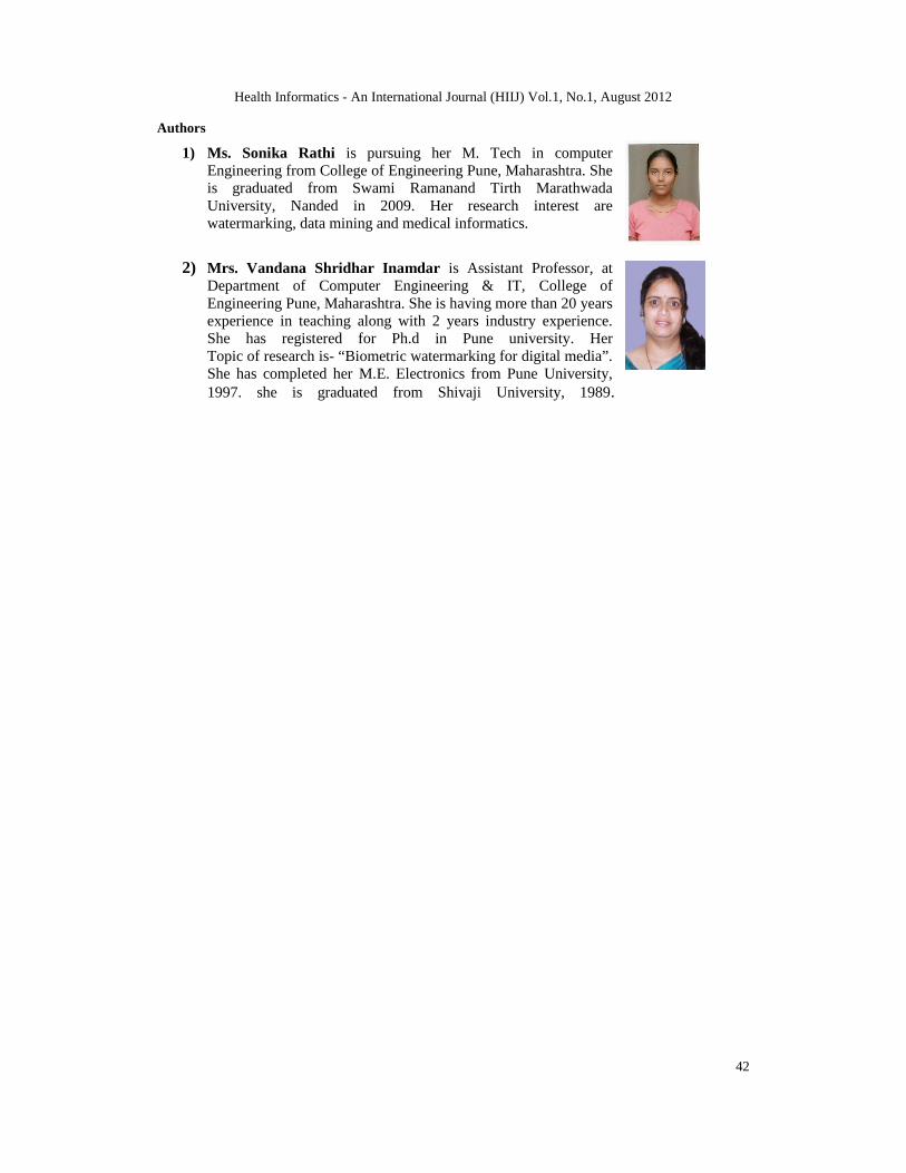
1) Ms. Sonika Rathi is pursuing her M. Tech in computerEngineering from College of Engineering Pune, Maharashtra. Sheis graduated from Swami Ramanand Tirth MarathwadaUniversity, Nanded in 2009. Her research interest arewatermarking, data mining and medical informatics.
2) Mrs. Vandana Shridhar Inamdar is Assistant Professor, atDepartment of Computer Engineering & IT, College ofEngineering Pune, Maharashtra. She is having more than 20 yearsexperience in teaching along with 2 years industry experience.She has registered for Ph.d in Pune university. HerTopic of research is- “Biometric watermarking for digital media”.She has completed her M.E. Electronics from Pune University,1997. she is graduated from Shivaji University, 1989.
Related Documents