Medical Image Analysis Medical Image Analysis Medical Imaging Modalities: X-Ray Imaging Figures come from the textbook: Medical Image Analysis, Second Edition, by Atam P. Dhawan, IEEE Press, 2011.

Welcome message from author
This document is posted to help you gain knowledge. Please leave a comment to let me know what you think about it! Share it to your friends and learn new things together.
Transcript
Medical Image AnalysisMedical Image AnalysisMedical Imaging Modalities: X-Ray Imaging
Figures come from the textbook: Medical Image Analysis, Second Edition, by Atam P. Dhawan, IEEE Press, 2011.
Anatomical or structural◦X-ray radiology, X-ray mammography,
X-ray CT, ultrasound, Magnetic Resonance Imaging
Functional or metabolic◦Functional MRI, (Single Photon
Emission Computed Tomography) SPECT, (Positron Emission Tomography) PET, fluorescence imaging
Figures come from the textbook: Medical Image Analysis, Second Edition, by Atam P. Dhawan, IEEE Press, 2011.
X-ray ImagingX-ray Imaging
Figures come from the textbook: Medical Image Analysis, Second Edition, by Atam P. Dhawan, IEEE Press, 2011.
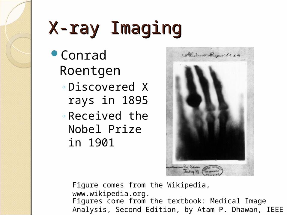
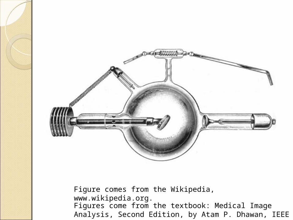
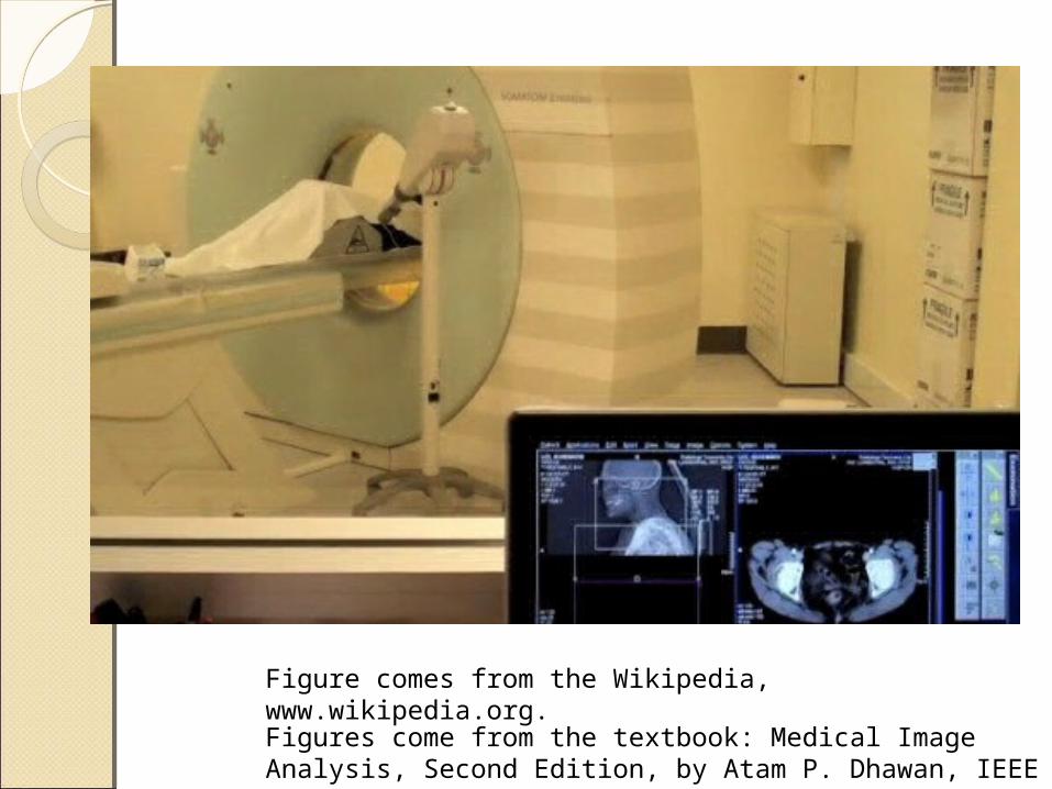
Figure comes from the Wikipedia, www.wikipedia.org.
Conrad Roentgen◦Discovered X
rays in 1895◦Received the
Nobel Prize in 1901
Soft X rays◦Wavelengths from 10 nm to 0.1 nm,
corresponding to 120eV to 12.3 KeVHard X rays
◦Wavelengths shorter than 0.1 nm up to 0.001 nm
Diagnostic◦12.3 KeV to 123 KeV
Figures come from the textbook: Medical Image Analysis, Second Edition, by Atam P. Dhawan, IEEE Press, 2011.
X-Ray GenerationX-Ray GenerationPrinciple
◦An accelerated electron loses energy in interaction with an atom and the loss of energy emits X-ray photons in a scattered direction
Figures come from the textbook: Medical Image Analysis, Second Edition, by Atam P. Dhawan, IEEE Press, 2011.
Figures come from the textbook: Medical Image Analysis, Second Edition, by Atam P. Dhawan, IEEE Press, 2011.
39 P50N K
LO
N
Ejected Electron
Incident Electron
X-ray Photon
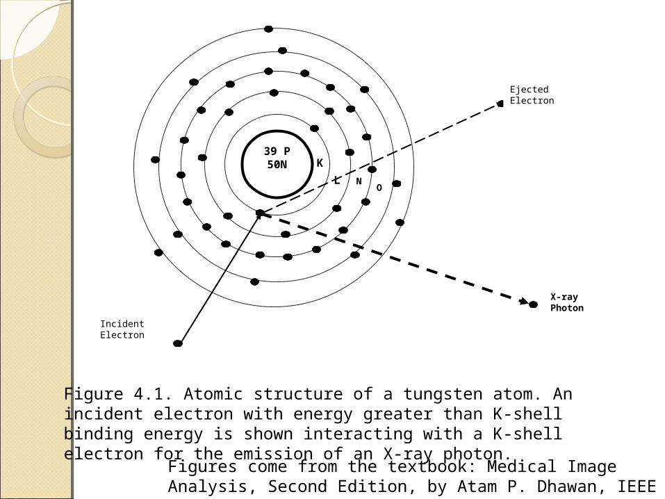
Figure 4.1. Atomic structure of a tungsten atom. An incident electron with energy greater than K-shell binding energy is shown interacting with a K-shell electron for the emission of an X-ray photon.
Tungsten◦K-shell binding energy level: 69.5 keV◦L-shell binding energy level: 10.2 keV◦An emission of X-ray photon of 59.3
keVX-ray generation
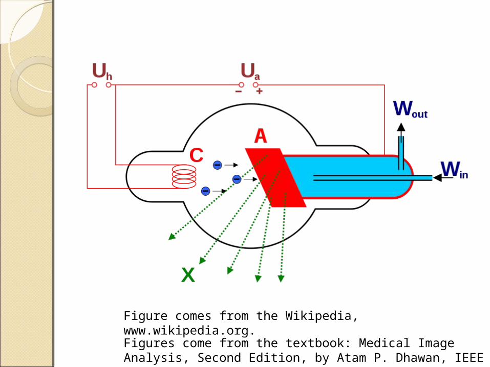
◦Electrons are released by the source cathode and are accelerated toward the target anode in a vacuum under the potential difference ranging from 20,000 to 150,000 volts
Figures come from the textbook: Medical Image Analysis, Second Edition, by Atam P. Dhawan, IEEE Press, 2011.
Figures come from the textbook: Medical Image Analysis, Second Edition, by Atam P. Dhawan, IEEE Press, 2011.
Figure comes from the Wikipedia, www.wikipedia.org.
Figures come from the textbook: Medical Image Analysis, Second Edition, by Atam P. Dhawan, IEEE Press, 2011.
Figure comes from the Wikipedia, www.wikipedia.org.
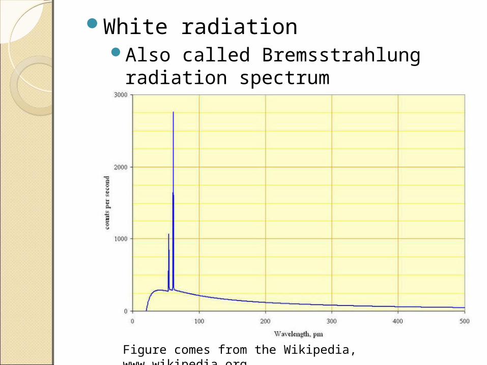
Figure comes from the Wikipedia, www.wikipedia.org.
White radiationAlso called Bremsstrahlung
radiation spectrum
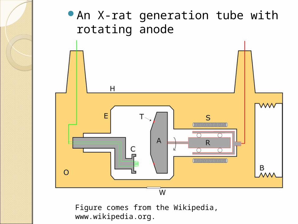
An X-rat generation tube with rotating anode
Figure comes from the Wikipedia, www.wikipedia.org.
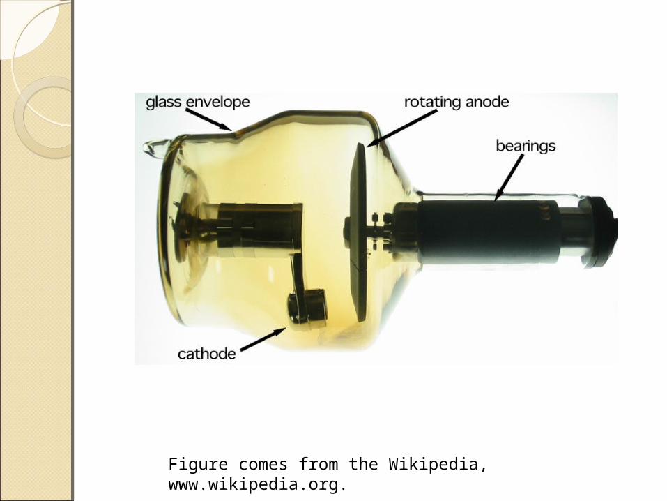
Figure comes from the Wikipedia, www.wikipedia.org.
X-ray 2-D Projection X-ray 2-D Projection ImagingImagingDiagnostic radiology
◦2-D projection of the three-dimensional anatomical structure of the human body
◦Localized sum of attenuation coefficients of material: air, blood, tissue, bone
◦Film or 2-D array of detectorsDigital radiographic system
◦Use scintillation crystals optically coupled with photomultiplier
Figures come from the textbook: Medical Image Analysis, Second Edition, by Atam P. Dhawan, IEEE Press, 2011.
Figures come from the textbook: Medical Image Analysis, Second Edition, by Atam P. Dhawan, IEEE Press, 2011.
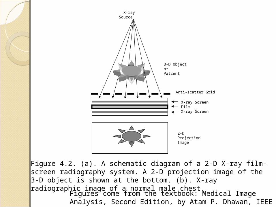
X-ray Source
X-ray ScreenFilmX-ray Screen
3-D Object orPatient
2-D ProjectionImage
Anti-scatter Grid
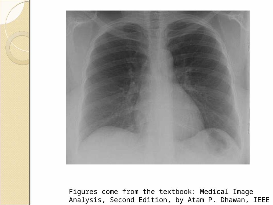
Figure 4.2. (a). A schematic diagram of a 2-D X-ray film-screen radiography system. A 2-D projection image of the 3-D object is shown at the bottom. (b). X-ray radiographic image of a normal male chest.
Figures come from the textbook: Medical Image Analysis, Second Edition, by Atam P. Dhawan, IEEE Press, 2011.
X-ray 2-D Projection X-ray 2-D Projection ImagingImagingScattering
◦Create artifacts and artificial structures
Reduce scattering◦Anti-scattered grids and collimators
X-ray intensifying screen
Figures come from the textbook: Medical Image Analysis, Second Edition, by Atam P. Dhawan, IEEE Press, 2011.
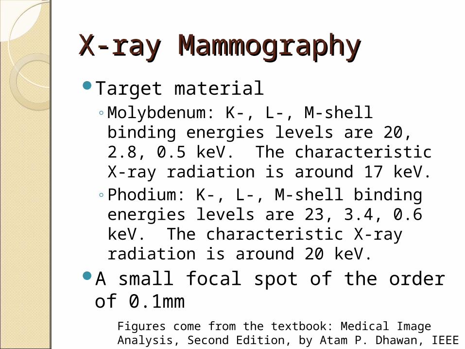
X-ray MammographyX-ray MammographyTarget material
◦Molybdenum: K-, L-, M-shell binding energies levels are 20, 2.8, 0.5 keV. The characteristic X-ray radiation is around 17 keV.
◦Phodium: K-, L-, M-shell binding energies levels are 23, 3.4, 0.6 keV. The characteristic X-ray radiation is around 20 keV.
A small focal spot of the order of 0.1mm
Figures come from the textbook: Medical Image Analysis, Second Edition, by Atam P. Dhawan, IEEE Press, 2011.
Figures come from the textbook: Medical Image Analysis, Second Edition, by Atam P. Dhawan, IEEE Press, 2011.
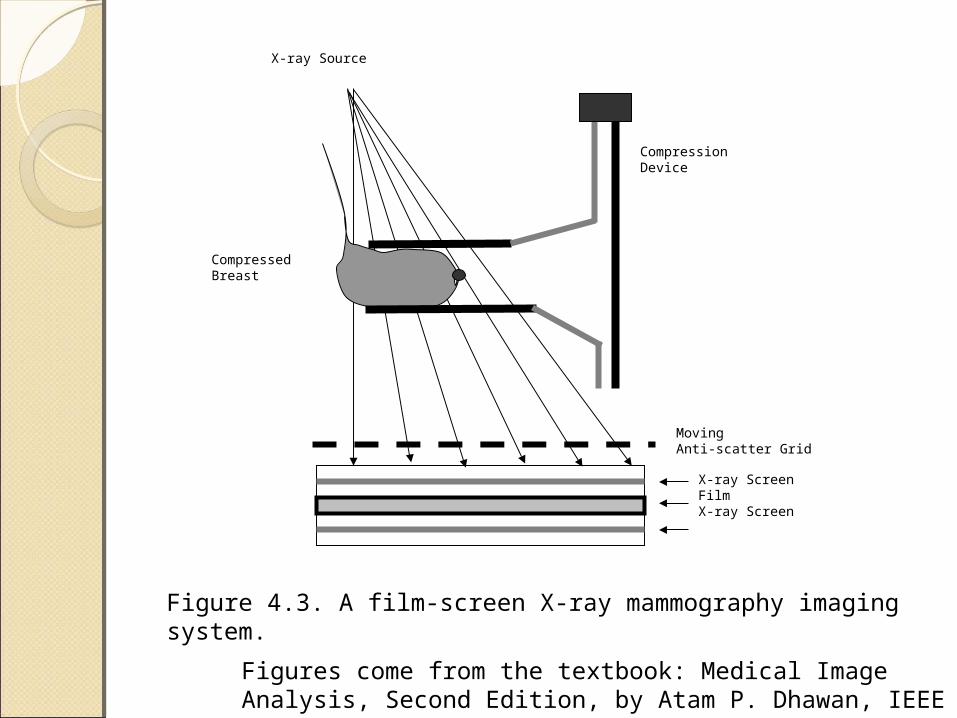
X-ray Source
X-ray ScreenFilmX-ray Screen
CompressedBreast
MovingAnti-scatter Grid
CompressionDevice
Figure 4.3. A film-screen X-ray mammography imaging system.
Figures come from the textbook: Medical Image Analysis, Second Edition, by Atam P. Dhawan, IEEE Press, 2011.
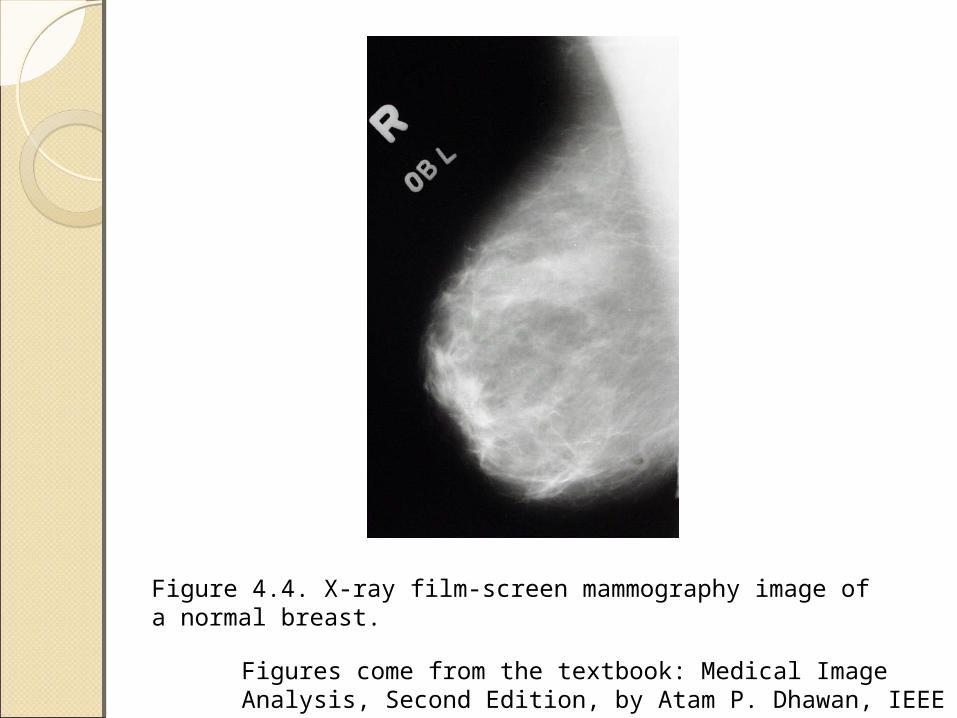
Figure 4.4. X-ray film-screen mammography image of a normal breast.
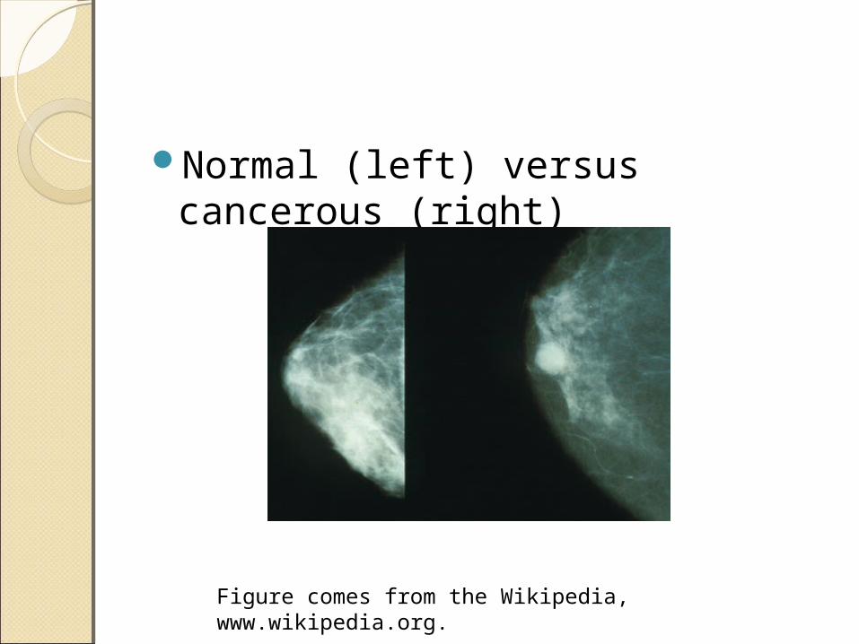
Normal (left) versus cancerous (right)
Figure comes from the Wikipedia, www.wikipedia.org.
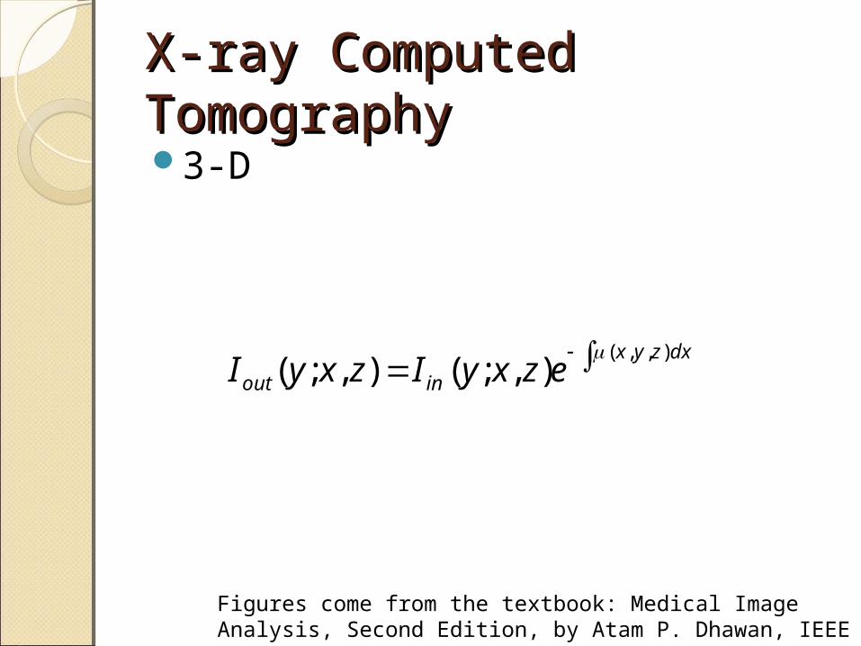
X-ray Computed X-ray Computed TomographyTomography3-D
Figures come from the textbook: Medical Image Analysis, Second Edition, by Atam P. Dhawan, IEEE Press, 2011.
dxzyx
inout ezxyIzxyI),,(
),;(),;(
Figures come from the textbook: Medical Image Analysis, Second Edition, by Atam P. Dhawan, IEEE Press, 2011.
Figure comes from the Wikipedia, www.wikipedia.org.
Figures come from the textbook: Medical Image Analysis, Second Edition, by Atam P. Dhawan, IEEE Press, 2011.
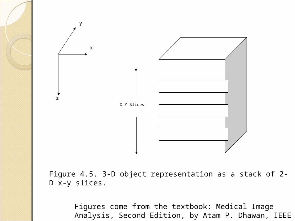
y
x
zX-Y Slices
Figure 4.5. 3-D object representation as a stack of 2-D x-y slices.
Figures come from the textbook: Medical Image Analysis, Second Edition, by Atam P. Dhawan, IEEE Press, 2011.
x
z
y
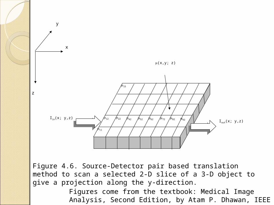
Iin(x; y,z)Iout(x; y,z)
(x,y; z)
11
22 92
15
12 42 52 62 72 82
Figure 4.6. Source-Detector pair based translation method to scan a selected 2-D slice of a 3-D object to give a projection along the y-direction.
Figures come from the textbook: Medical Image Analysis, Second Edition, by Atam P. Dhawan, IEEE Press, 2011.
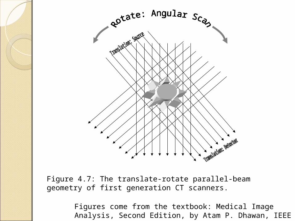
Figure 4.7: The translate-rotate parallel-beam geometry of first generation CT scanners.
X-ray Computed X-ray Computed TomographyTomographyGenerations
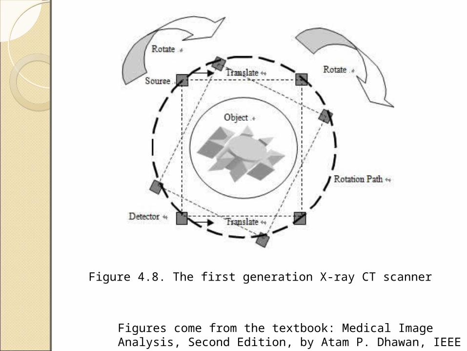
◦First: an X-ray source-detector pair that was translated in parallel-beam geometry
◦Second: a fan-beam geometry with a divergent X-ray source and a linear array of detectors. Use translation to cover the object and rotation to obtain additional views
Figures come from the textbook: Medical Image Analysis, Second Edition, by Atam P. Dhawan, IEEE Press, 2011.
Generations◦Third: a fan-beam geometry with a
divergent X-ray source and an arc of detectors. Without translation. Additional views are obtained by simultaneous rotation of the X-ray source and detector assembly. “Rotate only”
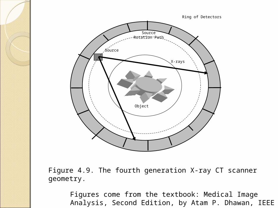
◦ Fourth: use a detector ring around the object. The X-ray source provides a divergent fan-beam of radiation to cover the object
Figures come from the textbook: Medical Image Analysis, Second Edition, by Atam P. Dhawan, IEEE Press, 2011.
Figures come from the textbook: Medical Image Analysis, Second Edition, by Atam P. Dhawan, IEEE Press, 2011.
Figure 4.8. The first generation X-ray CT scanner
Figures come from the textbook: Medical Image Analysis, Second Edition, by Atam P. Dhawan, IEEE Press, 2011.
Ring of Detectors
Source
SourceRotation Path
X-rays
Object
Figure 4.9. The fourth generation X-ray CT scanner geometry.
Figures come from the textbook: Medical Image Analysis, Second Edition, by Atam P. Dhawan, IEEE Press, 2011.
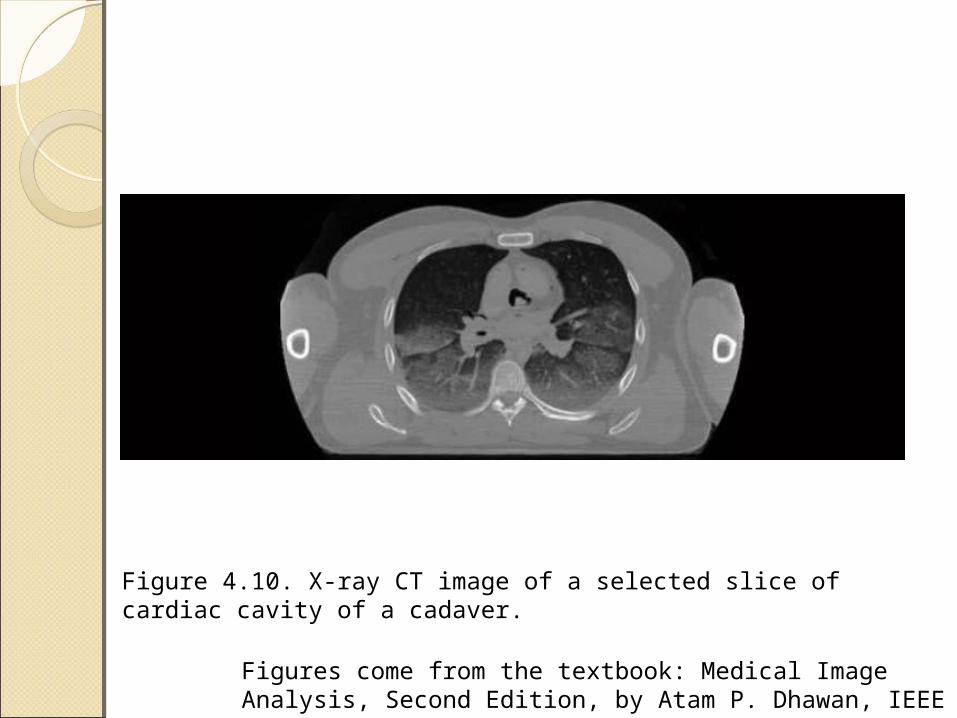
Figure 4.10. X-ray CT image of a selected slice of cardiac cavity of a cadaver.
Figures come from the textbook: Medical Image Analysis, Second Edition, by Atam P. Dhawan, IEEE Press, 2011.
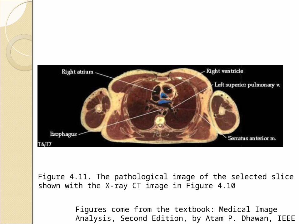
Figure 4.11. The pathological image of the selected slice shown with the X-ray CT image in Figure 4.10
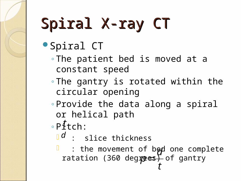
Spiral X-ray CTSpiral X-ray CTSpiral CT
◦The patient bed is moved at a constant speed
◦The gantry is rotated within the circular opening
◦Provide the data along a spiral or helical path
◦Pitch: : slice thickness : the movement of bed one complete
ratation (360 degrees) of gantryt
dp
td
Contrast Agent, Spatial Contrast Agent, Spatial Resolution, and SNRResolution, and SNRContrast agent
◦Barium sulfate, to enhance contrast in upper gastrointestinal (GI) tract imaging
◦Barium atom has a K-edge at 37.4KeV◦Iodine-based, used in angiography,
urography, and intra-arterial DSA to improve visibility of arteries and blood vessels
◦Iodine has a K-edge at 33.2KeV
Figures come from the textbook: Medical Image Analysis, Second Edition, by Atam P. Dhawan, IEEE Press, 2011.
Related Documents