Medical Image Analysis Medical Image Analysis Interaction of Electromagnetic Radiation with Matter in Medical Imaging Figures come from the textbook: Medical Image Analysis, by Atam P. Dhawan, IEEE Press, 2003.

Medical Image Analysis Interaction of Electromagnetic Radiation with Matter in Medical Imaging Figures come from the textbook: Medical Image Analysis,
Dec 13, 2015
Welcome message from author
This document is posted to help you gain knowledge. Please leave a comment to let me know what you think about it! Share it to your friends and learn new things together.
Transcript
Medical Image AnalysisMedical Image AnalysisInteraction of Electromagnetic Radiation with Matter in Medical Imaging
Figures come from the textbook: Medical Image Analysis, by Atam P. Dhawan, IEEE Press, 2003.
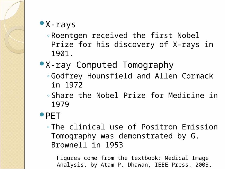
X-rays◦Roentgen received the first Nobel Prize
for his discovery of X-rays in 1901.X-ray Computed Tomography
◦Godfrey Hounsfield and Allen Cormack in 1972
◦Share the Nobel Prize for Medicine in 1979
PET◦The clinical use of Positron Emission
Tomography was demonstrated by G. Brownell in 1953
Figures come from the textbook: Medical Image Analysis, by Atam P. Dhawan, IEEE Press, 2003.
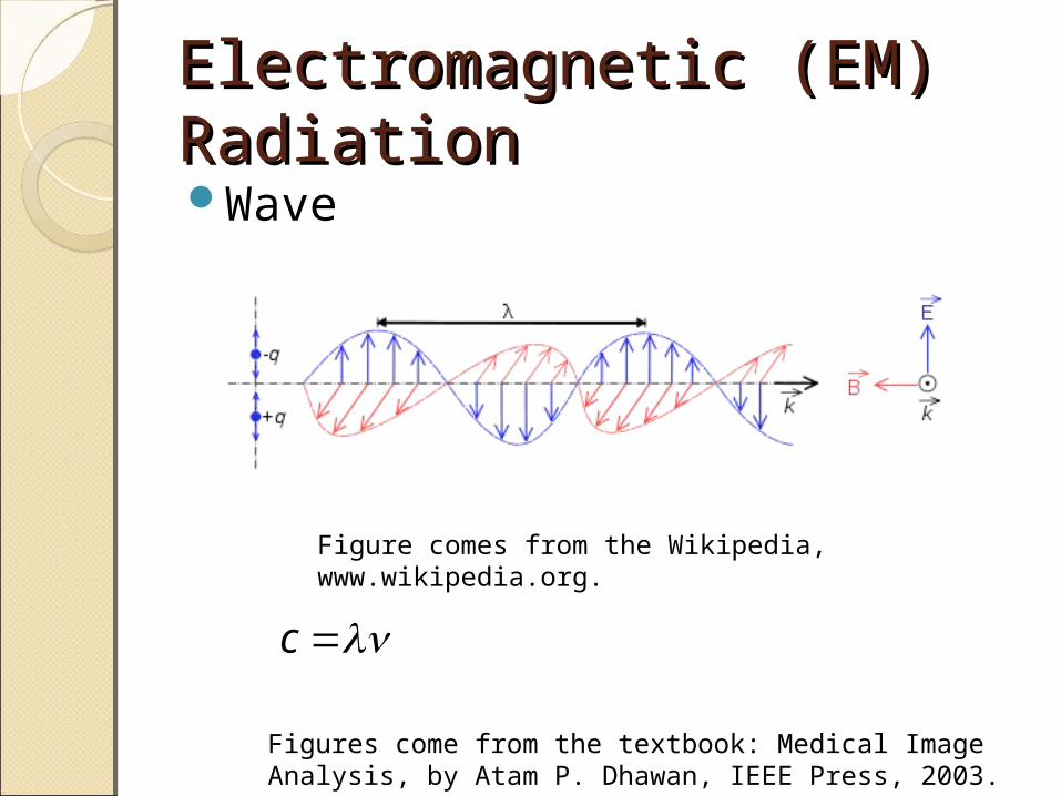
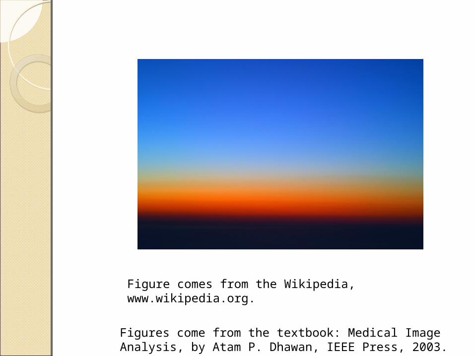
Electromagnetic (EM) Electromagnetic (EM) RadiationRadiationWave
Figures come from the textbook: Medical Image Analysis, by Atam P. Dhawan, IEEE Press, 2003.
Figure comes from the Wikipedia, www.wikipedia.org.
c
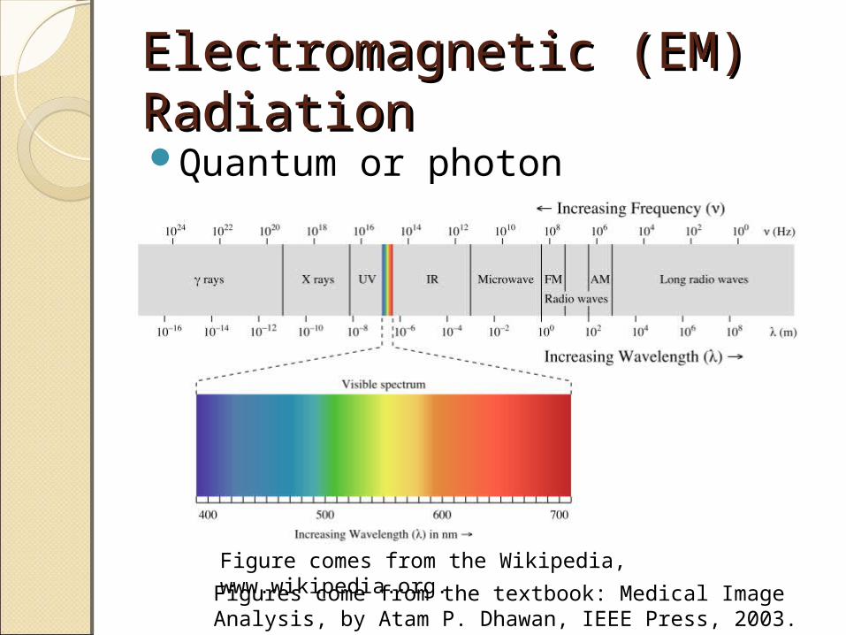
Electromagnetic (EM) Electromagnetic (EM) RadiationRadiationQuantum or photon
Figures come from the textbook: Medical Image Analysis, by Atam P. Dhawan, IEEE Press, 2003.
Figure comes from the Wikipedia, www.wikipedia.org.
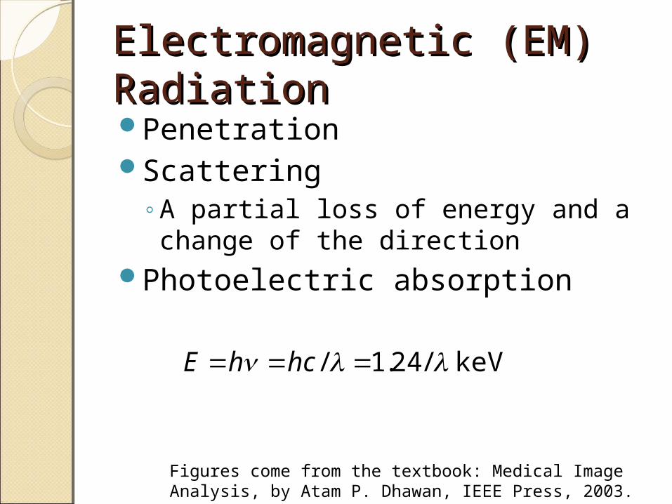
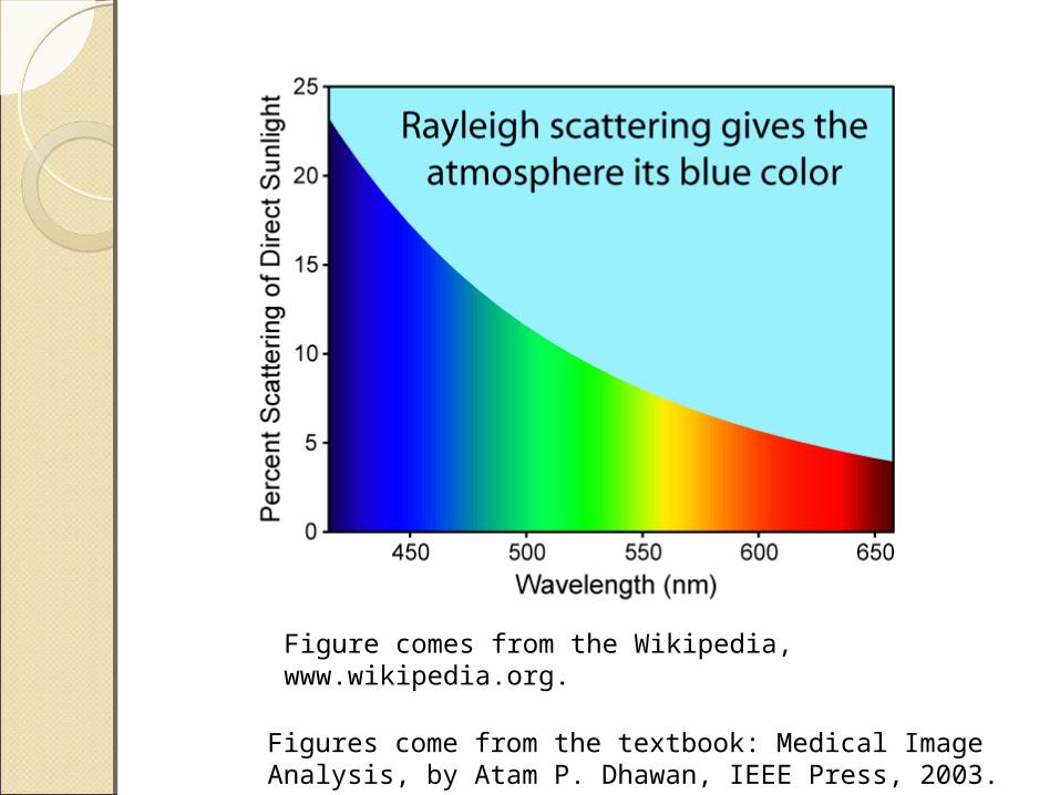
Electromagnetic (EM) Electromagnetic (EM) RadiationRadiationPenetrationScattering
◦A partial loss of energy and a change of the direction
Photoelectric absorption
Figures come from the textbook: Medical Image Analysis, by Atam P. Dhawan, IEEE Press, 2003.
keV /24.1/ hchE
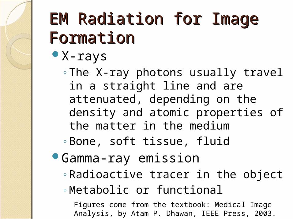
EM Radiation for Image EM Radiation for Image FormationFormationX-rays
◦The X-ray photons usually travel in a straight line and are attenuated, depending on the density and atomic properties of the matter in the medium
◦Bone, soft tissue, fluidGamma-ray emission
◦Radioactive tracer in the object◦Metabolic or functional
Figures come from the textbook: Medical Image Analysis, by Atam P. Dhawan, IEEE Press, 2003.
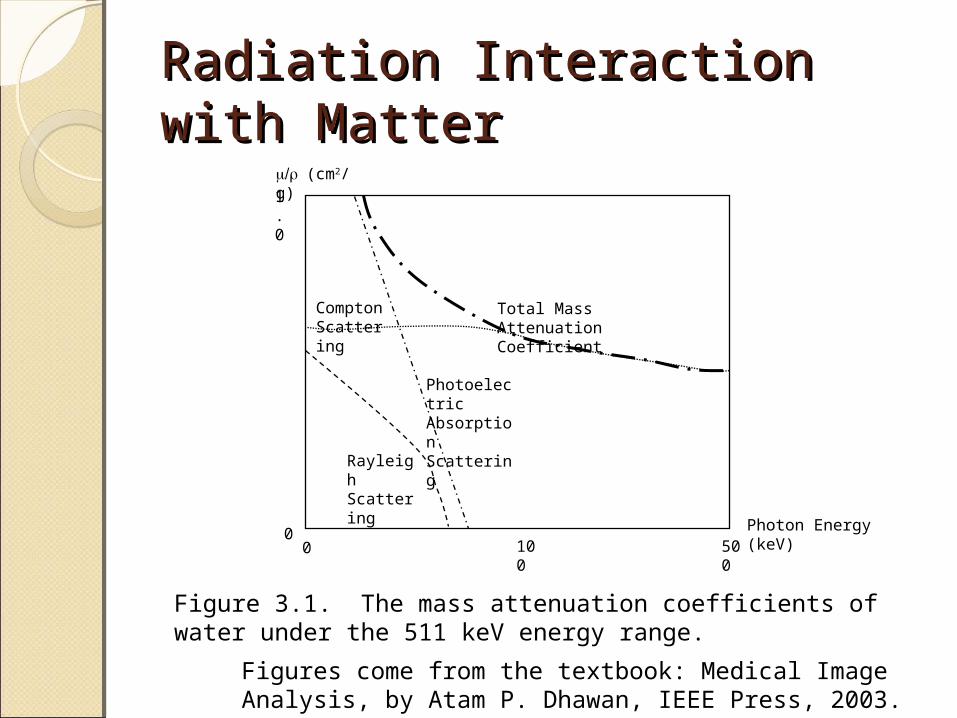
Radiation Interaction with Radiation Interaction with MatterMatter
Figures come from the textbook: Medical Image Analysis, by Atam P. Dhawan, IEEE Press, 2003.
Photon Energy (keV)0
0500
100
1.0
(cm2/g)
Rayleigh Scattering
Photoelectric Absorption Scattering
Compton Scattering
Total Mass Attenuation Coefficient
Figure 3.1. The mass attenuation coefficients of water under the 511 keV energy range.
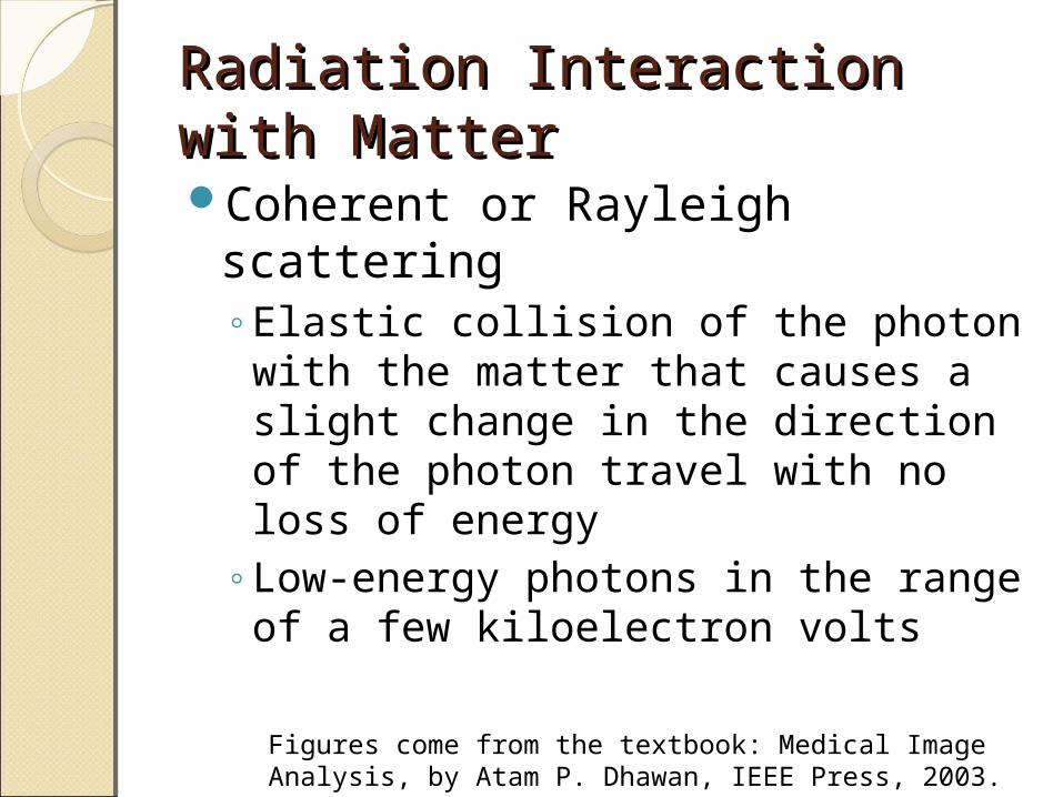
Radiation Interaction with Radiation Interaction with MatterMatterCoherent or Rayleigh scattering
◦Elastic collision of the photon with the matter that causes a slight change in the direction of the photon travel with no loss of energy
◦Low-energy photons in the range of a few kiloelectron volts
Figures come from the textbook: Medical Image Analysis, by Atam P. Dhawan, IEEE Press, 2003.
Figures come from the textbook: Medical Image Analysis, by Atam P. Dhawan, IEEE Press, 2003.
Figure comes from the Wikipedia, www.wikipedia.org.
Figures come from the textbook: Medical Image Analysis, by Atam P. Dhawan, IEEE Press, 2003.
Figure comes from the Wikipedia, www.wikipedia.org.
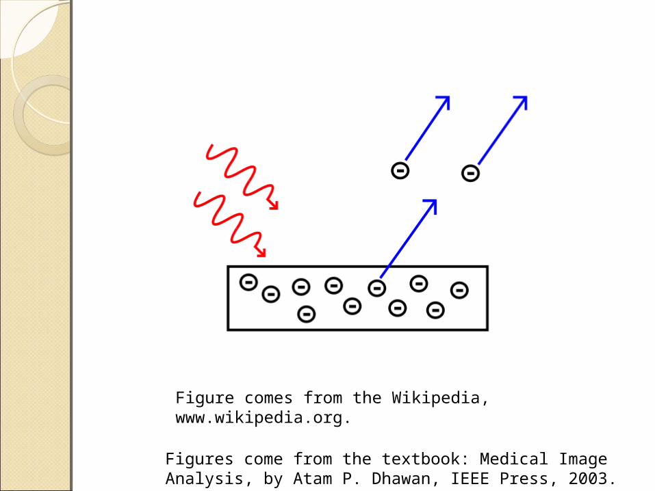
Radiation Interaction with Radiation Interaction with MatterMatterPhotoelectric absorption
◦A photon loses its energy by interacting with a tightly bound electron in the body matter, which is subsequently ejected from the atom due to the increased kinetic energy
◦Emission of a fluorescent radiation◦Low-energy photons are absorbed by M
and L shells of the atomic structure, while the high-energy photons are absorbed in the inner K-shell
Figures come from the textbook: Medical Image Analysis, by Atam P. Dhawan, IEEE Press, 2003.
Figures come from the textbook: Medical Image Analysis, by Atam P. Dhawan, IEEE Press, 2003.
Figure comes from the Wikipedia, www.wikipedia.org.
Radiation Interaction with Radiation Interaction with MatterMatterCompton scattering
◦Photon energies are comparable to the electron rest energy of 511 keV
Pair production◦Above 1.022 MeV
Figures come from the textbook: Medical Image Analysis, by Atam P. Dhawan, IEEE Press, 2003.
Figures come from the textbook: Medical Image Analysis, by Atam P. Dhawan, IEEE Press, 2003.
Figure comes from the Wikipedia, www.wikipedia.org.
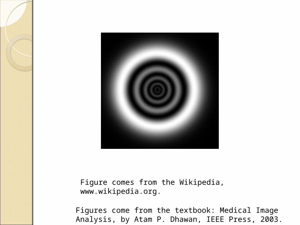
Radiation Interaction with Radiation Interaction with MatterMatterCompton scattering
◦An inelastic collision of a photon with an outer-shell electron with a negligible binding energy
◦After the collision, the photon with reduced energy is deflected while the electron with an increased energy is ejected from the atom
◦The deflections in scattering events cause uncertainties in photon localization as it becomes difficult to keep the desired radiation transmission path
Figures come from the textbook: Medical Image Analysis, by Atam P. Dhawan, IEEE Press, 2003.

Figures come from the textbook: Medical Image Analysis, by Atam P. Dhawan, IEEE Press, 2003.
Figure comes from the Wikipedia, www.wikipedia.org.

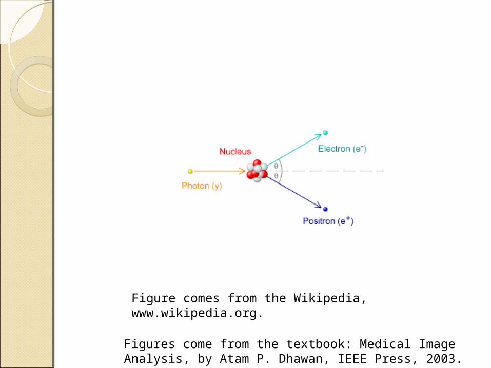
Radiation Interaction with Radiation Interaction with MatterMatterPair production
◦A high-energy photon of the order of 1 Mev interacts near the nucleus of an atom in a manner similar to the positron emission in a radioactive decay
◦Not used in diagnostic radiology
Figures come from the textbook: Medical Image Analysis, by Atam P. Dhawan, IEEE Press, 2003.
Figures come from the textbook: Medical Image Analysis, by Atam P. Dhawan, IEEE Press, 2003.
Figure comes from the Wikipedia, www.wikipedia.org.
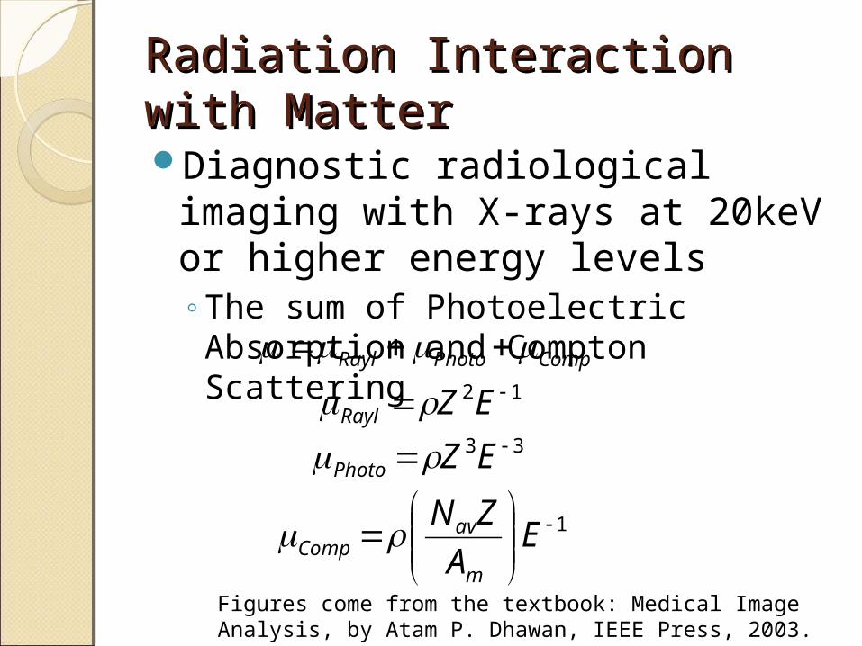
Radiation Interaction with Radiation Interaction with MatterMatterDiagnostic radiological imaging
with X-rays at 20keV or higher energy levels◦The sum of Photoelectric Absorption
and Compton Scattering
Figures come from the textbook: Medical Image Analysis, by Atam P. Dhawan, IEEE Press, 2003.
CompPhotoRayl 12 EZRayl 33 EZPhoto
1
E
A
ZN
m
avComp
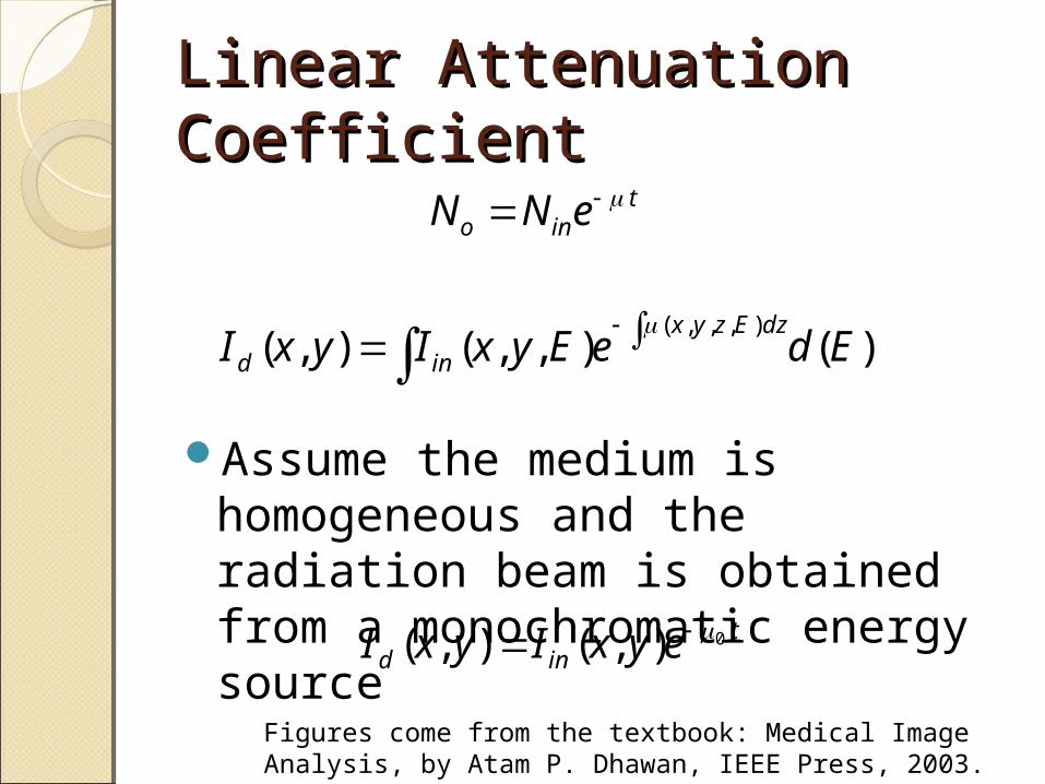
Linear Attenuation Linear Attenuation CoefficientCoefficient
Assume the medium is homogeneous and the radiation beam is obtained from a monochromatic energy source
Figures come from the textbook: Medical Image Analysis, by Atam P. Dhawan, IEEE Press, 2003.
tino eNN
)(),,(),(),,,(
EdeEyxIyxIdzEzyx
ind
tind eyxIyxI 0),(),(
Figures come from the textbook: Medical Image Analysis, by Atam P. Dhawan, IEEE Press, 2003.
Photon Energy (keV)0
0500
100
5.0
(cm2/g)
Fat
Compact Bone
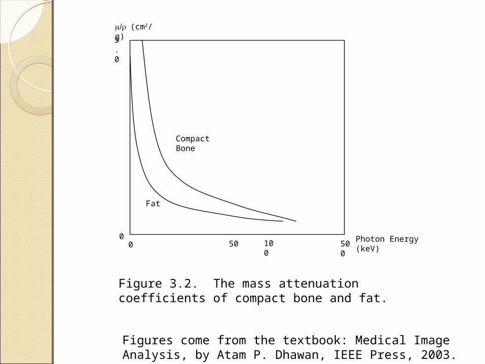
50
Figure 3.2. The mass attenuation coefficients of compact bone and fat.
Radiation DetectionRadiation DetectionSpectrometric detectors
◦Ionization and scintillation
Figures come from the textbook: Medical Image Analysis, by Atam P. Dhawan, IEEE Press, 2003.
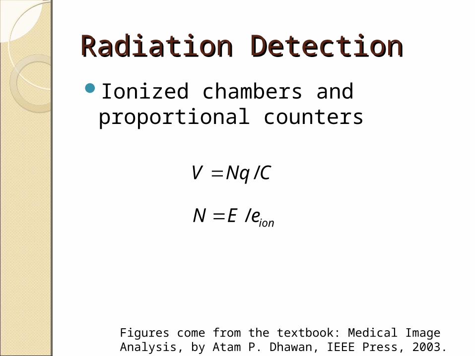
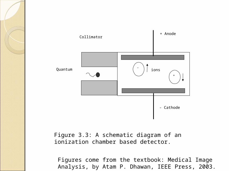
Radiation DetectionRadiation DetectionIonized chambers and
proportional counters
Figures come from the textbook: Medical Image Analysis, by Atam P. Dhawan, IEEE Press, 2003.
CNqV /
ioneEN /
Figures come from the textbook: Medical Image Analysis, by Atam P. Dhawan, IEEE Press, 2003.
+ Anode
- Cathode
Quantum
Collimator
+
-ions
Figure 3.3: A schematic diagram of an ionization chamber based detector.
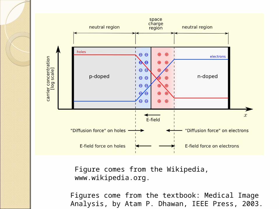
Radiation DetectionRadiation DetectionSemiconductor detectors
◦The particle energy is transformed into electric pulses at the junction region of the semiconductor material
◦Apply positive voltage to the n region and negative voltage to the p region
◦The depletion layer serves as an ionization chamber
◦A quantum interacting with the surface of the detector will create electron-hole pairs in the depletion layer
Figures come from the textbook: Medical Image Analysis, by Atam P. Dhawan, IEEE Press, 2003.
Figures come from the textbook: Medical Image Analysis, by Atam P. Dhawan, IEEE Press, 2003.
Figure comes from the Wikipedia, www.wikipedia.org.
Radiation DetectionRadiation DetectionAdvantages of semiconductor
detectors◦Fabricated with smaller e~3.6eV for
siliconE value becomes independent of
the mass and charge of the particleProvide small charge collection
time (<10ns)Cause very small recombination
losses due to fast charge collectionFigures come from the textbook: Medical Image Analysis, by Atam P. Dhawan, IEEE Press, 2003.

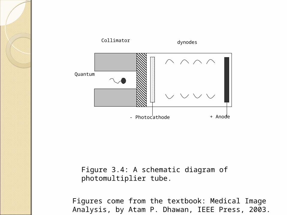
Radiation DetectionRadiation DetectionScintillation detectors
◦A scintillation phosphor and a photomultiplier tube
◦The charged particles interact with the scintillation material to excite molecules
◦The excited molecules emit optical photons during the relaxation process to return to the ground state
◦Photomultiplier tubes are used to amplify the optical photon intensity and produce voltages proportional to the energy of the particle creating scintillation
Figures come from the textbook: Medical Image Analysis, by Atam P. Dhawan, IEEE Press, 2003.
+ Anode- Photocathode
Quantum
Collimator dynodes
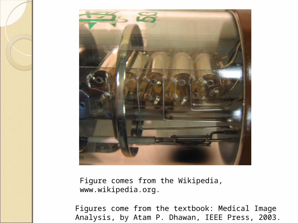
Figure 3.4: A schematic diagram of photomultiplier tube.
Figures come from the textbook: Medical Image Analysis, by Atam P. Dhawan, IEEE Press, 2003.
Figure comes from the Wikipedia, www.wikipedia.org.

Radiation DetectionRadiation DetectionThe number of electrons emitted
by the photocathode can be given as
Figures come from the textbook: Medical Image Analysis, by Atam P. Dhawan, IEEE Press, 2003.
phEwEkgN /00
Related Documents