10.1128/IAI.68.9.5167-5175.2000. 2000, 68(9):5167. DOI: Infect. Immun. Gerald T. Keusch and Honorine D. Ward H. Hamer, Barry Stein, Saul Tzipori, Miercio E. A. Pereira, Ana María Cevallos, Najma Bhat, Renaud Verdon, Davidson Monoclonal Antibody Glycoproteins Defined by a Neutralizing Infection In Vitro by Mucin-Like Cryptosporidium parvum Mediation of http://iai.asm.org/content/68/9/5167 Updated information and services can be found at: These include: REFERENCES http://iai.asm.org/content/68/9/5167#ref-list-1 at: This article cites 37 articles, 16 of which can be accessed free CONTENT ALERTS more» articles cite this article), Receive: RSS Feeds, eTOCs, free email alerts (when new http://journals.asm.org/site/misc/reprints.xhtml Information about commercial reprint orders: http://journals.asm.org/site/subscriptions/ To subscribe to to another ASM Journal go to: on September 22, 2014 by guest http://iai.asm.org/ Downloaded from on September 22, 2014 by guest http://iai.asm.org/ Downloaded from

Welcome message from author
This document is posted to help you gain knowledge. Please leave a comment to let me know what you think about it! Share it to your friends and learn new things together.
Transcript

10.1128/IAI.68.9.5167-5175.2000.
2000, 68(9):5167. DOI:Infect. Immun. Gerald T. Keusch and Honorine D. WardH. Hamer, Barry Stein, Saul Tzipori, Miercio E. A. Pereira, Ana María Cevallos, Najma Bhat, Renaud Verdon, Davidson Monoclonal AntibodyGlycoproteins Defined by a Neutralizing Infection In Vitro by Mucin-Like
Cryptosporidium parvumMediation of
http://iai.asm.org/content/68/9/5167Updated information and services can be found at:
These include:
REFERENCEShttp://iai.asm.org/content/68/9/5167#ref-list-1at:
This article cites 37 articles, 16 of which can be accessed free
CONTENT ALERTS more»articles cite this article),
Receive: RSS Feeds, eTOCs, free email alerts (when new
http://journals.asm.org/site/misc/reprints.xhtmlInformation about commercial reprint orders: http://journals.asm.org/site/subscriptions/To subscribe to to another ASM Journal go to:
on Septem
ber 22, 2014 by guesthttp://iai.asm
.org/D
ownloaded from
on S
eptember 22, 2014 by guest
http://iai.asm.org/
Dow
nloaded from

INFECTION AND IMMUNITY,0019-9567/00/$04.0010
Sept. 2000, p. 5167–5175 Vol. 68, No. 9
Copyright © 2000, American Society for Microbiology. All Rights Reserved.
Mediation of Cryptosporidium parvum Infection In Vitro byMucin-Like Glycoproteins Defined by a Neutralizing
Monoclonal AntibodyANA MARIA CEVALLOS,1 NAJMA BHAT,1 RENAUD VERDON,1† DAVIDSON H. HAMER,1
BARRY STEIN,2 SAUL TZIPORI,1,2 MIERCIO E. A. PEREIRA,1
GERALD T. KEUSCH,1 AND HONORINE D. WARD1,2*
Division of Geographic Medicine and Infectious Diseases, Tupper Research Institute, New England Medical Center,Tufts University School of Medicine, Boston, Massachusetts 02111,1 and Division of Infectious Diseases,
Tufts University School of Veterinary Medicine, North Grafton, Massachusetts 015362
Received 22 March 2000/Returned for modification 18 May 2000/Accepted 7 June 2000
The protozoan parasite Cryptosporidium parvum is a significant cause of diarrheal disease worldwide.Attachment to and invasion of host intestinal epithelial cells by C. parvum sporozoites are crucial steps in thepathogenesis of cryptosporidiosis. The molecular basis of these initial interactions is unknown. In order toidentify putative C. parvum adhesion- and invasion-specific proteins, we raised monoclonal antibodies (MAbs)to sporozoites and evaluated them for inhibition of attachment and invasion in vitro. Using this approach, weidentified two glycoproteins recognized by 4E9, a MAb which neutralized C. parvum infection and inhibitedsporozoite attachment to intestinal epithelial cells in vitro. 4E9 recognized a 40-kDa glycoprotein named gp40and a second, >220-kDa protein which was identified as GP900, a previously described mucin-like glycopro-tein. Glycoproteins recognized by 4E9 are localized to the surface and apical region of invasive stages and areshed in trails from the parasite during gliding motility. The epitope recognized by 4E9 contains a-N-acetyl-galactosamine residues, which are present in a mucin-type O-glycosidic linkage. Lectins specific for theseglycans bind to the surface and apical region of sporozoites and block attachment to host cells. The surface andapical localization of these glycoproteins and the neutralizing effect of the MAb and a-N-acetylgalactosamine-specific lectins strongly implicate these proteins and their glycotopes as playing a role in C. parvum-host cellinteractions.
Cryptosporidium parvum, an intestinal Apicomplexan para-site, is a significant cause of diarrheal disease worldwide (15,17). In immunocompetent individuals, the disease is usuallyself-limiting, but it may be chronic and life threatening inimmunocompromised patients such as those with AIDS. Re-cently, the parasite has gained notoriety as the causative agentof numerous outbreaks of waterborne diarrheal disease. Thereis currently no effective specific therapy approved for diseasecaused by this parasite.
Infection is initiated by ingestion of oocysts, which undergoexcystation to release sporozoites. Attachment of sporozoitesto epithelial cells and subsequent invasion of the host cellmembrane are crucial primary steps in the pathogenesis ofcryptosporidiosis. The ultrastructural aspects of attachmentand invasion have been characterized in detail (10, 24, 33, 34).Sporozoites attach to host cells by their anterior pole. Attach-ment is followed by invagination of the host cell plasma mem-brane, which extends along the surface of the sporozoite andeventually completely surrounds it, leading to formation of aparasitophorus vacuole where the parasite undergoes furtherdevelopment in a unique intracellular but extracytoplasmiclocation.
Using in vitro models of sporozoite attachment to epithelialcells, we previously showed that attachment was dose and timedependent and was influenced by pH, divalent cations, and thedegree of differentiation of host cells (20, 22). Further, attach-ment could be inhibited by polyclonal antibodies to C. parvumproteins as well as by glycoconjugates specific for a sporozoitesurface Gal/GalNAc-binding lectin which we had previouslydescribed (20–22). A recent study confirmed the role of Gal/GalNAc-specific lectin-carbohydrate interactions in attach-ment (4). Previous studies have also reported that C. parvuminfection in vitro can be inhibited by polyclonal or monoclonalantibodies to C. parvum proteins (5, 7, 9, 11, 23). In addition,sporozoite motility and invasion have been shown to be de-pendent on parasite and host cell cytoskeletal elements (4, 12,13).
Although ultrastructural details and various factors affectingattachment and invasion have been characterized, little isknown about the molecular basis of these initial host-parasiteinteractions or of specific parasite and host molecules whichmediate them (38). Knowledge of such molecules is crucial forunderstanding the pathogenic mechanisms involved in thehost-parasite interaction and for designing preventive and in-terventional strategies to combat cryptosporidiosis. The aim ofthis study was to identify and characterize specific parasiteproteins that may be involved in the initial C. parvum-host cellinteractions of attachment and invasion. In order to do this, weraised monoclonal antibodies (MAbs) to sporozoite surfaceproteins and screened them for inhibition of attachment andinfection in vitro. In this study, we describe two C. parvumglycoproteins identified by 4E9, a MAb to a carbohydrate
* Corresponding author. Mailing address: Division of GeographicMedicine and Infectious Diseases, New England Medical Center, Box041, 750 Washington St., Boston, MA 02111. Phone: (617) 636-7032.Fax: (617) 636-5292. E-mail: [email protected].
† Present address: Reanimation Medicale, Maladies Infectieses etTropicales, Centre Hospitalier Universitaire Cote-de-Nacre, Caen14033, France.
5167
on Septem
ber 22, 2014 by guesthttp://iai.asm
.org/D
ownloaded from

epitope present in multiple developmental stages of the para-site, which inhibits attachment and infection in vitro.
MATERIALS AND METHODS
Parasites. C. parvum oocytes of the GCH1 isolate (36) were treated with1.75% (vol/vol) sodium hypochlorite for 10 min on ice; washed with Dulbeccomodified Eagle medium (Life Technologies, Grand Island, N.Y.) containing 25mM HEPES, 100 U of penicillin per ml, and 100 mg of streptomycin per ml, andexcysted for 2 h at 37°C or for 1 h in the presence of 0.25% trypsin and/or 0.75%taurocholic acid. Sporozoites were purified by isopycnic Percoll gradient centrif-ugation (1) or by filtration through a 2.0-mm-pore-size Nucleopore polycarbon-ate filter (Costar Scientific Corporation, Cambridge, Mass.).
Shed proteins (SP) were obtained by excystation of oocysts in Dulbecco mod-ified Eagle medium for 2 h at 37°C, followed by centrifugation at 5000 3 g at 4°Cfor 10 min. Protease inhibitors (final concentrations of 2 mM phenylmethylsul-fonyl fluoride, 20 mM leupeptin, 10 mM E64, and 2 mM EDTA) were added tothe supernatant, which was concentrated 10-fold by ultrafiltration. The excysta-tion rate using this protocol ranged from 40 to 60% (depending on the age of theoocysts), compared to 60 to 80% when excystation was performed in the pres-ence of trypsin and/or taurocholic acid. This method was used to obtain SP inorder to avoid inclusion of proteins that may be released from the surface of theparasite by trypsin and/or taurocholic acid.
Other protozoan parasites were provided by A. Kane, Center for Gastroen-terology Research in Absorptive and Secretory Processes, New England MedicalCenter, Boston, Mass. (Giardia lamblia trophozoites and Entameba histolyticatrophozoites); M. E. A. Pereira, Tufts University School of Medicine, Boston,Mass. (Trypanosoma cruzi trypomastigotes and Leishmania major promastig-otes); and K. Kim, Albert Einstein School of Medicine, New York, N.Y. (Tox-oplasma gondii tachyzoites).
Cell culture. Caco-2A (human intestinal epithelial) cells were obtained fromthe cell culture core of the Center for Gastroenterology Research in Absorptiveand Secretory Processes at New England Medical Center and grown as describedpreviously (22).
MAbs. In order to obtain MAbs to surface epitopes, sporozoites were fixedwith 1% glutaraldehyde for 30 min on ice, residual aldehyde groups were blockedwith 0.1 M glycine, and sporozoites were washed with phosphate-buffered saline(PBS). BALB/c mice were immunized intraperitoneally with fixed sporozoites incomplete Freund’s adjuvant, followed by three intraperitoneal boosts with thesame preparation using incomplete Freund’s adjuvant. Spleen cells were fusedwith P3 3 63/Ag mouse myeloma cells and cloned in liquid medium. Clones werescreened for reactivity with the surface and/or apical region of sporozoites byimmunofluorescence (IF).
Clone 4E9, which showed the strongest reactivity by IF, was chosen for furtherstudy. This clone was further subcloned and determined to be of the immuno-globulin M (IgM) (kappa) isotype using an enzyme-linked immunosorbent assay(ELISA)-based isotyping kit (Southern Biotechnology Associates, Inc., Birming-ham, Ala.). Ascites fluid was obtained following injection of 4E9 hybridoma cellsinto Pristane-primed BALB/c mice. B9A4, an IgM control MAb against Brugiamalayi phosphorylcholine, was generously provided by C. Carlow, New EnglandBiolabs, Beverly, Mass. 7B3, an IgG1 MAb to GP900 (26), was obtained as a kindgift from C. Petersen, University of California, San Francisco. 4E9 and B9A4IgM were purified from ascites fluid using an E-Z-SEP purification kit (Phar-macia Biotech Inc., Piscataway, N.J.).
IF and FITC-lectin-binding assays. Purified oocysts were placed on poly-L-lysine (30 mg/ml in water)-coated slides. Purified sporozoites were allowed toglide on poly-L-lysine-coated slides for 30 min at room temperature (RT). Forintracellular stages, Caco-2A cells grown to confluence in 16-well chamber slideswere infected with oocysts for 24 h (37). Slides with oocysts and sporozoites werefixed with 4% paraformaldehyde for 10 min at RT. Slides with intracellular stageswere fixed and permeabilized with methanol. IF was performed as describedpreviously (20). Controls included culture medium and the irrelevant IgM MAbB9A4. Binding of the following fluorescein isothiocyanate (FITC)-conjugatedlectins (Sigma, St. Louis, Mo.) (2 mg/ml in PBS) was performed as describedearlier (39): Helix pomatia agglutinin (HPA), Maclura pomifera agglutinin(MPA), Artocarpus integrifolia agglutinin (AIA) (Jacalin), and Sambucus nigraagglutinin (SNA). Specificity of binding was determined by preincubation of thelectin with its cognate sugar inhibitor (as described for lectin blotting below).
Immunoelectron microscopy (IEM). Piglets and gamma interferon (IFN-g)-knockout mice were infected with C. parvum oocysts as described previously (16,35). Intestinal tissue, oocysts, and sporozoites were fixed in 0.25% glutaralde-hyde–4% paraformaldehyde in 0.1 M sodium phosphate buffer (pH 7.2) (PB), at4°C overnight. Oocysts and sporozoites were pelleted by centrifugation, and 2%agarose was added and allowed to solidify. Samples were washed with PB,dehydrated, and embedded in L. R. White Resin (Electron Microscopy Sciences,Fort Washington, Pa.). Silver-gold sections from polymerized blocks were placedon Formvar-coated 300-mesh nickel grids. Grids with sections were floated ondrops of 0.05 M glycine–0.5 M NaCl–1% Tween 20–0.05 M Tris (pH 7.5) (TBST)for 30 min, on 1% bovine serum albumin (BSA) in TBST for 10 min, and on 10%normal goat serum (NGS) in TBST for 10 min. Grids were then placed on 4E9IgM at 4°C for 1 h, followed by 1% BSA in TBST for 4 min and lastly on10-nm-diameter colloidal gold conjugated to goat anti-mouse Ig diluted in 5%
BSA–1% NGS in TBST for 30 min. Grids were washed, stained with 3% aqueousuranyl acetate, and viewed by transmission electron microscopy.
Immunoblotting and lectin blotting. Parasite proteins were separated by gra-dient sodium dodecyl sulfate-polyacrylamide gel electrophoresis (SDS-PAGE)(5 to 15% polyacrylamide) and transferred to nitrocellulose for 18 h at 395 mAat 4°C. Bound proteins were probed with MAb 4E9 and detected by chemilu-minescence or colormetric methods. For the chemiluminescence method, non-specific binding was blocked with 10% NGS in 10 mM Tris-HCl–150 mM sodiumchloride (pH 8.0) (TBS) for 1 h before incubation with MAb 4E9 in 5% NGS in0.1% Tween 20 in TBS (0.1% T-TBS) for 90 min at RT. After being washedthree times with 0.1% T-TBS, strips were incubated with horseradish peroxidase-conjugated goat anti-mouse antibody (Immunopure; Pierce) diluted in 5% NGS–0.1% T-TBS for 1 h at RT. The strips were washed, incubated in SuperSignalsubstrate (Pierce), exposed to film, and developed. For the colormetric method,nonspecific binding was blocked with 5% nonfat milk (NFM) in TBS for 1 hbefore incubation with MAb 4E9 in 1% NFM in TBS for 1 h at RT. After beingwashed with 0.05% T-TBS, strips were incubated with alkaline phosphatase-conjugated goat anti-mouse antibody (Promega, Madison, Wis.) in 1% NFM inTBS for 1 h at RT. The strips were washed and developed with nitrobluetetrazolium (NBT)–5-bromo-4-chloro-3-indolylphosphate (BCIP) substrate.Competitive inhibition of 4E9 binding by HPA was evaluated by incubating thestrips with HPA (50 mg/ml) for 10 min at RT before incubation with 4E9.
For lectin blotting, nonspecific binding was blocked with 0.1% Tween 20 inPBS (0.1% T-PBS) for 1 h at RT. Blots were incubated with the followingbiotinylated lectins (EY Laboratories, Inc., San Mateo, Calif.) diluted in 0.1%T-PBS (10 mg/ml) for 1 h at RT: HPA, AIA, MPA, SNA, Glycine max agglutinin(soybean agglutinin [SBA]), Dolichos biflorus agglutinin (DBA), Arachis hy-pogaea agglutinin (peanut agglutinin [PNA]), Sophora japonica agglutinin (SJA),Ulex europaeus agglutinin-1 (UEA-1), Maackia amurensis agglutinin (MAA),Canavalia ensiformis agglutinin (concanavalin A agglutinin [ConA]), and Triti-cum vulgare (wheat germ agglutinin [WGA]). To assess specificity of binding,lectins were preincubated with the following specific sugars (250 mM): N-acetyl-galactosamine (GalNAc) (for SBA, HPA, DBA, and SJA), b-lactose (for PNA,SNA, and MAA), N-acetylglucosamine (for WGA), melibiose (for AIA andMPA), methyl-a-mannopyranoside (for ConA), and fucose (for UEA-1). Blotswere washed with 0.1% T-PBS, incubated with an avidin-biotin-alkaline phos-phatase complex (ABC reagent; Vector, San Mateo, Calif.) for 1 h at RT,washed, and developed with NBT-BCIP substrate.
Immunoprecipitation. A mixture of sporozoites and oocysts (derived fromexcystation of 108 oocysts) in PBS containing protease inhibitors (describedabove) was lysed by five freeze-thaw cycles and detergent extraction with 1%Triton X-100. The lysate was centrifuged at 10,000 3 g for 30 min. Detergent-soluble material was incubated with MAb 7B3 overnight, followed by incubationwith protein G-Sepharose (Pharmacia Biotech Inc.) for 2 h at 4°C. After exten-sive washing with 20 mM sodium phosphate–0.5 M NaCl–0.5% Triton X-100–0.1% SDS–0.1% deoxycholate, immunoprecipitated proteins were analyzed bySDS-PAGE and immunoblotting with MAb 4E9.
Infection and attachment assays. The effect of MAb 4E9 on C. parvum infec-tion of Caco-2A cells was studied by a modification of an in vitro assay describedearlier (37). Briefly, oocysts (1 3 104/well) preincubated with 4E9 or B9A4 IgMfor 30 min at RT were incubated with Caco-2A cells (2 3 104/well) grown in96-well tissue culture plates for 24 h at 37°C with 5% CO2. Cells were fixed,permeabilized with methanol for 10 min at RT, and washed three times withTBS. Infection was quantified by ELISA (37) using a polyclonal rabbit anti-C.parvum antibody (20) that recognizes .30 proteins, ranging from 14 to .200kDa, present on sporozoites, merozoites, and intracellular stages but not oocysts.
The effect of MAb 4E9 or lectins on attachment of sporozoites to glutaralde-hyde-fixed Caco-2A cells was studied using an in vitro assay as described previ-ously (22). Briefly, Caco-2A cells grown to confluence in 96-well plates were fixedwith glutaraldehyde to prevent invasion. Sporozoites were preincubated with testor control antibodies or lectins for 30 min at 4°C. For attachment assays usinglectins, sporozoites were washed twice with assay buffer after preincubation withthe lectins. Controls (sporozoites in assay buffer) were treated in the same way.The sporozoites were then incubated with the glutaraldehyde-fixed Caco-2Amonolayers for 1 h at 37°C. Unbound parasites were washed off, and attachedsporozoites were quantified by ELISA using the same antibody as for the infec-tion assay.
Cytotoxicity assay. Caco-2A cells grown in 96-well plates were incubated withor without purified MAb IgM (100 mg/ml) for 24 h at 37°C with 5% CO2. Possiblecytotoxicity of the MAbs for the cells was assessed by measuring viability using aCellTiter96 AQueous kit (Promega).
Periodate oxidation and glycosidase digestion. SP were separated by SDS-PAGE and transferred to nitrocellulose. Strips were incubated with 50 mMsodium acetate buffer (pH 4.5) (SAB) alone (as a control) or with 10 mMperiodic acid in SAB in the dark for 1 h at RT. The strips were rinsed with SABand incubated with 50 mM sodium borohydride in PBS for 30 min at RT andthen probed with 4E9 as described above.
SP were treated with the following glycosidases (Oxford Glycosciences,Bedford, Mass.) at 37°C overnight, according to the manufacturer’s recommen-dations: peptide-N-glycosidase F (recombinant) (50 U/ml), endo-a-N-acetyl-galactosaminidase (from Streptococcus pneumoniae) (111 mU/ml), and a-N-acetylgalactosaminidase (from chicken liver) (300 mU/ml). As controls for the
5168 CEVALLOS ET AL. INFECT. IMMUN.
on Septem
ber 22, 2014 by guesthttp://iai.asm
.org/D
ownloaded from
various glycosidases, SP were incubated with buffer alone under identical con-ditions. Specificity of a-N-acetylgalactosaminidase activity was assessed by addi-tion of paranitrophenyl a-N-acetylgalactosaminide to a final concentration of 2.5mM.
Statistical methods. Attachment, infection, antigen-binding, and cytotoxicityassays were performed in three to six replicates, and the mean and standard error(SE) of the mean were determined. All assays were repeated at least three times.Results from representative assays are shown. Comparison of means was per-formed using a nonpaired Student t test. A P value of ,0.05 was consideredstatistically significant.
RESULTS
MAb 4E9 neutralizes C. parvum infection of intestinal epi-thelial cells and inhibits sporozoite attachment to these cellsin vitro. In order to identify putative C. parvum adhesion- andinvasion-specific proteins, we raised MAbs to sporozoites andscreened them by IF to identify clones reactive with the surfaceor anterior portion of sporozoites (suggestive of an apicalcomplex localization). Using this approach, we identified 4E9,an IgM MAb, which showed the strongest reactivity of all theclones by IF. To determine whether proteins recognized by4E9 were involved in initial host-parasite interactions, we eval-uated the effect of this MAb on C. parvum infection of intes-tinal epithelial cells using an in vitro assay (37). Compared toan irrelevant isotype-matched control MAb, 4E9 IgM neutral-ized infection of Caco-2A cells in a dose-dependent manner,with almost complete inhibition occurring at a concentration of100 mg/ml (Fig. 1A). The results obtained with this assay weresimilar to those obtained using a previously described IF-basedassay (37) in which intracellular stages are directly visualizedby phase-contrast and epifluorescence microscopy (R. Verdonand H. Ward, unpublished data). The inhibition of infectionwas not due to toxicity of 4E9 for host cells, since the MAb hadno effect on viability (data not shown). 4E9 IgM did not ag-glutinate sporozoites or oocysts at the concentrations used,indicating that the inhibitory effect was not due to agglutina-tion of the parasite.
In order to determine if the antibody inhibited infection byblocking adherence of sporozoites to intestinal cells, we stud-ied its effect on attachment using an in vitro assay in which cellsare fixed to prevent invasion (22). As seen in Fig. 1B, 4E9
inhibited sporozoite attachment to fixed Caco-2A cells in adose-dependent manner, with 50% inhibition occurring at 100mg/ml. The decrease in attachment measured by ELISA wasnot due to reduced binding of the detecting polyclonal anti-body to proteins recognized by 4E9, since similar results wereobtained using an IF-based assay (20) in which attached sporo-zoites are directly visualized by phase-contrast and epifluores-cence microscopy (D. Hamer and H. Ward, unpublished data).These results suggested that the neutralizing effect of the MAbis mediated at least in part by blocking of sporozoite attach-ment to host cells, resulting in subsequent inhibition of infec-tion.
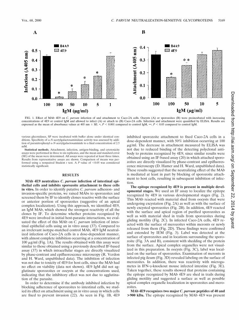
The epitope recognized by 4E9 is present in multiple devel-opmental stages. We used an IF assay to localize the epitoperecognized by 4E9 in various developmental stages (Fig. 2).This MAb reacted with material shed from oocysts that wereundergoing excystation (Fig. 2A) as well as with the surface ofnewly excysted sporozoites (Fig. 2B). In addition, 4E9 reactedwith the surface and apical region of purified sporozoites aswell as with material shed in trails from sporozoites duringgliding motility (Fig. 2C). In infected Caco-2A cells, 4E9 re-acted with the surface of merozoites within meronts or newlyreleased from them (Fig. 2D). These findings were confirmedand extended by IEM (Fig. 3). Label was detected at thesurface of sporozoites and in locations surrounding the sporo-zoite (Fig. 3A and B), consistent with shedding of the proteinfrom the surface. Apical complex organelles were not visual-ized in this preparation. In oocysts (Fig. 3C), label was local-ized on the surface of sporozoites. Examination of meronts ininfected pig ileum (Fig. 3D) revealed labeling on the surface ofmerozoites. In addition, there was reactivity with microga-metes in IFN-g-knockout mouse infected intestine (Fig. 3E).Taken together, these results showed that proteins containingthe epitope recognized by MAb 4E9 are shed in trails duringgliding motility and suggested a surface as well as possibleapical complex organelle localization in sporozoites and mero-zoites.
MAb 4E9 recognizes two major C. parvum peptides of 40 and>900 kDa. The epitope recognized by MAb 4E9 was present
FIG. 1. Effect of MAb 4E9 on C. parvum infection of and attachment to Caco-2A cells. Oocysts (A) or sporozoites (B) were preincubated with increasingconcentrations of 4E9 or control IgM and allowed to infect (A) or attach to (B) Caco-2A cells. Infection and attachment were quantified by ELISA. Results areexpressed as the mean of absorbance values at 405 nm 6 SE. p, P , 0.001 compared to control IgM, pp, P , 0.05 compared to control IgM.
VOL. 68, 2000 C. PARVUM NEUTRALIZATION-SENSITIVE GLYCOPROTEINS 5169
on Septem
ber 22, 2014 by guesthttp://iai.asm
.org/D
ownloaded from

on two major bands as determined by immunoblotting: a 40-kDa peptide and a very-high-molecular-mass peptide of .220kDa (Fig. 4A). The latter peptide comigrated with a previouslydescribed .900-kDa glycoprotein (GP900) recognized byMAb 7B3 (26) by immunoblotting (data not shown). Oocystand sporozoite proteins immunoprecipitated by MAb 7B3were recognized by 4E9 by immunoblotting, confirming thatthis peptide was GP900 (data not shown). The 40-kDa protein(designated gp40) and GP900 were present in oocysts andsporozoites. An additional 250-kDa band was also detectedonly in oocysts (Fig. 4A). There was no reactivity of the controlIgM MAb B4A9 (not shown) with any of these proteins, con-firming the specificity of 4E9. In addition, 4E9 did not reactwith proteins from other protozoa (Giardia lamblia, E. histo-lytica, Toxoplasma gondii, Trypanosoma cruzi, and L. major) inimmunoblotting (not shown), indicating that the epitope rec-ognized by MAb 4E9 is specific for Cryptosporidium.
Analysis of SP by SDS-PAGE and silver staining revealedthe presence of ;5 major bands of .900, 120, 55, 40, and 35kDa (Fig. 4B). Two of these, the .900- and 40-kDa bands,were recognized by MAb 4E9 by immunoblotting, confirming
the IF finding that these proteins are shed from the parasite(Fig. 4C). Since this preparation (SP) was enriched in thesetwo proteins, it was used for further characterization of theepitope recognized by 4E9.
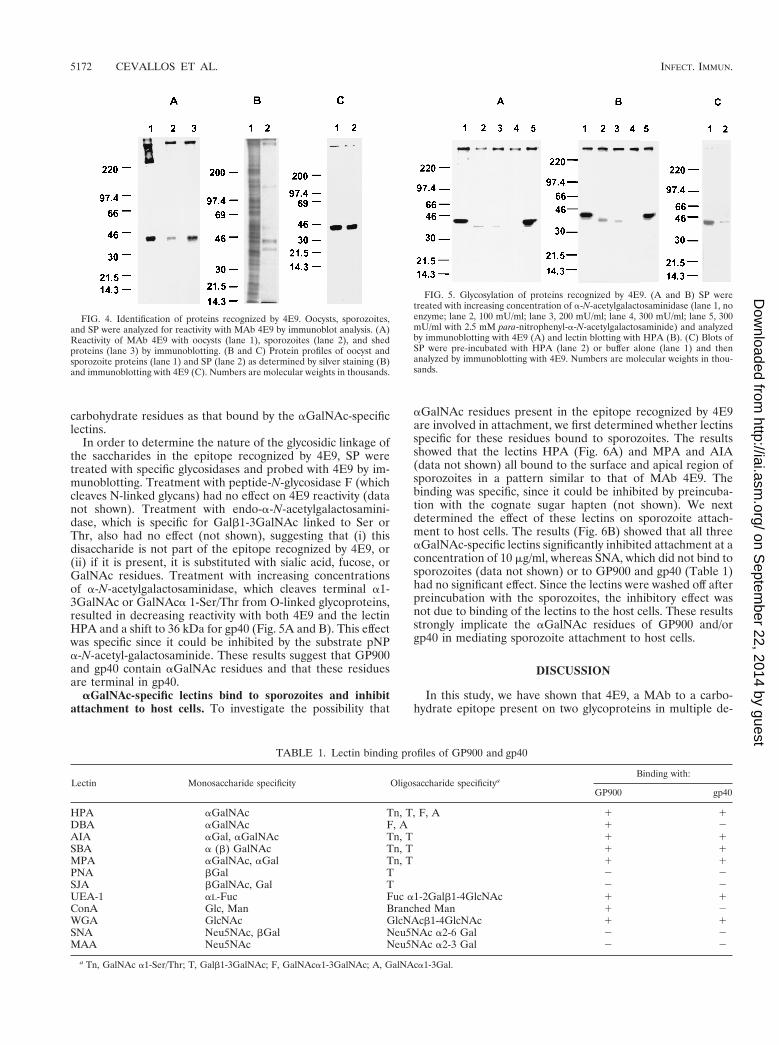
The epitope recognized by 4E9 is glycosylated and containsa-linked GalNAc residues. Reactivity of 4E9 with GP900 andgp40 was abolished by mild periodate treatment, suggestingthat the epitope is glycosylated (not shown). In order to de-termine the nature of the carbohydrates present, SP wereprobed with a panel of biotinylated lectins with various sugarspecificities (Table 1). Of these, HPA, SBA, AIA, MPA,UEA-1, and WGA bound to both GP900 and gp40, whereasConA and DBA bound to GP900 but not gp40. Binding wasspecific, since it could be inhibited by the appropriate cognatesugar hapten (not shown). Of note was the binding of a num-ber of aGalNAc-specific lectins. The patterns of reactivity ofaGalNAc-specific lectins such as HPA were identical to that of4E9 (Fig. 5A and B). In addition, reactivity of 4E9 could becompetitively inhibited by HPA (Fig. 5C). These results sug-gested that the epitope recognized by 4E9 contains the same
FIG. 2. Reactivity of MAb 4E9 with C. parvum stages as determined by IF. (A and B) Oocysts. Material shed from excysting oocysts (A) and the surface of newlyexcysted sporozoites (arrow) (B) is labeled. (C) Sporozoites. The surface and apical region of sporozoites and of material shed in trails (arrow) from sporozoites allowedto glide on poly-L-lysine coated slides are labeled. (D) Intracellular stages. Meronts and merozoites newly emerged from them (arrow) in infected Caco-2A cells arelabeled.
5170 CEVALLOS ET AL. INFECT. IMMUN.
on Septem
ber 22, 2014 by guesthttp://iai.asm
.org/D
ownloaded from
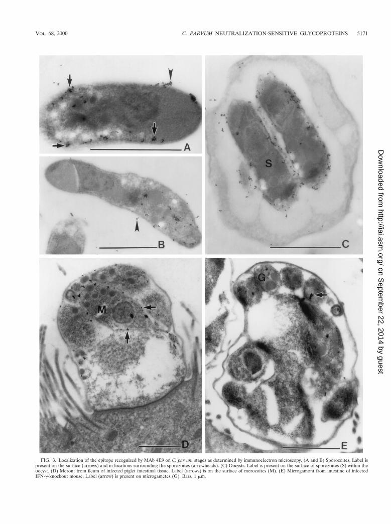
FIG. 3. Localization of the epitope recognized by MAb 4E9 on C. parvum stages as determined by immunoelectron microscopy. (A and B) Sporozoites. Label ispresent on the surface (arrows) and in locations surrounding the sporozoites (arrowheads). (C) Oocysts. Label is present on the surface of sporozoites (S) within theoocyst. (D) Meront from ileum of infected piglet intestinal tissue. Label (arrows) is on the surface of merozoites (M). (E) Microgamont from intestine of infectedIFN-g-knockout mouse. Label (arrow) is present on microgametes (G). Bars, 1 mm.
VOL. 68, 2000 C. PARVUM NEUTRALIZATION-SENSITIVE GLYCOPROTEINS 5171
on Septem
ber 22, 2014 by guesthttp://iai.asm
.org/D
ownloaded from
carbohydrate residues as that bound by the aGalNAc-specificlectins.
In order to determine the nature of the glycosidic linkage ofthe saccharides in the epitope recognized by 4E9, SP weretreated with specific glycosidases and probed with 4E9 by im-munoblotting. Treatment with peptide-N-glycosidase F (whichcleaves N-linked glycans) had no effect on 4E9 reactivity (datanot shown). Treatment with endo-a-N-acetylgalactosamini-dase, which is specific for Galb1-3GalNAc linked to Ser orThr, also had no effect (not shown), suggesting that (i) thisdisaccharide is not part of the epitope recognized by 4E9, or(ii) if it is present, it is substituted with sialic acid, fucose, orGalNAc residues. Treatment with increasing concentrationsof a-N-acetylgalactosaminidase, which cleaves terminal a1-3GalNAc or GalNAca 1-Ser/Thr from O-linked glycoproteins,resulted in decreasing reactivity with both 4E9 and the lectinHPA and a shift to 36 kDa for gp40 (Fig. 5A and B). This effectwas specific since it could be inhibited by the substrate pNPa-N-acetyl-galactosaminide. These results suggest that GP900and gp40 contain aGalNAc residues and that these residuesare terminal in gp40.
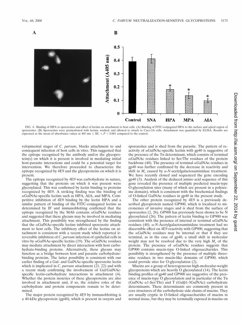
aGalNAc-specific lectins bind to sporozoites and inhibitattachment to host cells. To investigate the possibility that
aGalNAc residues present in the epitope recognized by 4E9are involved in attachment, we first determined whether lectinsspecific for these residues bound to sporozoites. The resultsshowed that the lectins HPA (Fig. 6A) and MPA and AIA(data not shown) all bound to the surface and apical region ofsporozoites in a pattern similar to that of MAb 4E9. Thebinding was specific, since it could be inhibited by preincuba-tion with the cognate sugar hapten (not shown). We nextdetermined the effect of these lectins on sporozoite attach-ment to host cells. The results (Fig. 6B) showed that all threeaGalNAc-specific lectins significantly inhibited attachment at aconcentration of 10 mg/ml, whereas SNA, which did not bind tosporozoites (data not shown) or to GP900 and gp40 (Table 1)had no significant effect. Since the lectins were washed off afterpreincubation with the sporozoites, the inhibitory effect wasnot due to binding of the lectins to the host cells. These resultsstrongly implicate the aGalNAc residues of GP900 and/orgp40 in mediating sporozoite attachment to host cells.
DISCUSSION
In this study, we have shown that 4E9, a MAb to a carbo-hydrate epitope present on two glycoproteins in multiple de-
FIG. 4. Identification of proteins recognized by 4E9. Oocysts, sporozoites,and SP were analyzed for reactivity with MAb 4E9 by immunoblot analysis. (A)Reactivity of MAb 4E9 with oocysts (lane 1), sporozoites (lane 2), and shedproteins (lane 3) by immunoblotting. (B and C) Protein profiles of oocyst andsporozoite proteins (lane 1) and SP (lane 2) as determined by silver staining (B)and immunoblotting with 4E9 (C). Numbers are molecular weights in thousands.
TABLE 1. Lectin binding profiles of GP900 and gp40
Lectin Monosaccharide specificity Oligosaccharide specificityaBinding with:
GP900 gp40
HPA aGalNAc Tn, T, F, A 1 1DBA aGalNAc F, A 1 2AIA aGal, aGalNAc Tn, T 1 1SBA a (b) GalNAc Tn, T 1 1MPA aGalNAc, aGal Tn, T 1 1PNA bGal T 2 2SJA bGalNAc, Gal T 2 2UEA-1 aL-Fuc Fuc a1-2Galb1-4GlcNAc 1 1ConA Glc, Man Branched Man 1 2WGA GlcNAc GlcNAcb1-4GlcNAc 1 1SNA Neu5NAc, bGal Neu5NAc a2-6 Gal 2 2MAA Neu5NAc Neu5NAc a2-3 Gal 2 2
a Tn, GalNAc a1-Ser/Thr; T, Galb1-3GalNAc; F, GalNAca1-3GalNAc; A, GalNAca1-3Gal.
FIG. 5. Glycosylation of proteins recognized by 4E9. (A and B) SP weretreated with increasing concentration of a-N-acetylgalactosaminidase (lane 1, noenzyme; lane 2, 100 mU/ml; lane 3, 200 mU/ml; lane 4, 300 mU/ml; lane 5, 300mU/ml with 2.5 mM para-nitrophenyl-a-N-acetylgalactosaminide) and analyzedby immunoblotting with 4E9 (A) and lectin blotting with HPA (B). (C) Blots ofSP were pre-incubated with HPA (lane 2) or buffer alone (lane 1) and thenanalyzed by immunoblotting with 4E9. Numbers are molecular weights in thou-sands.
5172 CEVALLOS ET AL. INFECT. IMMUN.
on Septem
ber 22, 2014 by guesthttp://iai.asm
.org/D
ownloaded from
velopmental stages of C. parvum, blocks attachment to andconsequent infection of host cells in vitro. This suggested thatthe epitope recognized by the antibody and/or the glycopro-tein(s) on which it is present is involved in mediating initialhost-parasite interactions and could be a potential target forintervention. We therefore proceeded to characterize theepitope recognized by 4E9 and the glycoproteins on which it ispresent.
The epitope recognized by 4E9 was carbohydrate in nature,suggesting that the proteins on which it was present wereglycosylated. This was confirmed by lectin binding to proteinsrecognized by 4E9. A striking finding was the binding ofaGalNAc-specific lectins such as HPA, AIA, and MPA. Com-petitive inhibition of 4E9 binding by the lectin HPA and asimilar pattern of binding of the FITC-conjugated lectins asdetermined by IF and immunoblotting confirmed that theepitope recognized by the MAb contains aGalNAc residuesand suggested that these glycans may be involved in mediatingattachment. This possibility was strengthened by the findingthat the aGalNAc-specific lectins inhibited sporozoite attach-ment to host cells. The inhibitory effect of the lectins on at-tachment is consistent with a recent study which reported ir-reversible inhibition of C. parvum infection of epithelial cells invitro by aGalNAc-specific lectins (19). The aGalNAc residuesmay mediate attachment by direct interaction with host carbo-hydrate-binding proteins. Alternatively, these glycans mayfunction as a bridge between host and parasite carbohydrate-binding proteins. The latter possibility is consistent with ourearlier finding of a Gal- and GalNAc-specific sporozoite lectinwhich is implicated in C. parvum attachment (21, 22) and witha recent study confirming the involvement of Gal/GalNAc-specific lectin-carbohydrate interactions in attachment (4).Whether the protein moieties of these glycoproteins are alsoinvolved in attachment and, if so, the relative roles of thecarbohydrate and protein components remain to be deter-mined.
The major protein recognized by 4E9 by immunoblotting isa 40-kDa glycoprotein (gp40), which is present in oocysts and
sporozoites and is shed from the parasite. The pattern of re-activity of aGalNAc-specific lectins with gp40 is suggestive ofthe presence of the Tn determinant, which consists of terminalaGalNAc residues linked to Ser/Thr residues of the proteinbackbone (40). The presence of terminal aGalNAc residues ingp40 was further confirmed by the decrease in reactivity andshift in Mr caused by a-N-acetylgalactosaminidase treatment.We have recently cloned and sequenced the gene encodinggp40 (3). Analysis of the deduced amino acid sequence of thisgene revealed the presence of multiple predicted mucin-typeO-glycosylation sites (many of which are present in a polyser-ine domain), which is consistent with the biochemical findingsof O-linked GalNAc residues in gp40 in the present study.
The other protein recognized by 4E9 is a previously de-scribed glycoprotein named GP900, which is localized to mi-cronemes of invasive stages and is shed from the surface ofsporozoites (2, 26). GP900 has previously been shown to be Nglycosylated (26). The pattern of lectin binding to GP900 wasconsistent with the presence of internal or terminal aGalNAcresidues (40). a-N-Acetylgalactosaminidase treatment had nodiscernible effect on 4E9 reactivity with GP900, suggesting thatthe aGalNAc residues may be internal or that if they areterminal, as in the case of gp40, a small shift in molecularweight may not be resolved due to the very high Mr of theprotein. The presence of aGalNAc residues suggests thatGP900 contains mucin-type O-linked oligosaccharides. Thispossibility is strengthened by the presence of multiple threo-nine residues in two mucin-like domains of GP900, whichcould provide sites for O-glycosylation (2).
Mucins are a group of heterogeneous high-molecular-weightglycoproteins which are heavily O glycosylated (14). The lectinbinding profiles of gp40 and GP900 are suggestive of the pres-ence of mucin-type O glycosylation and in particular of the Tn(GalNAc a1-Ser/Thr) and T (Galb1-3GalNAc) carbohydratedeterminants. These determinants are commonly present incore structures of the carbohydrate side chains of mucins. Theyare usually cryptic in O-linked oligosaccharides of mucins innormal tissue, but they may be terminally exposed in mucins on
FIG. 6. Binding of HPA to sporozoites and effect of lectins on attachment to host cells. (A) Binding of FITC-conjugated HPA to the surface and apical region ofsporozoites. (B) Sporozoites were preincubated with lectins, washed, and allowed to attach to Caco-2A cells. Attachment was quantified by ELISA. Results areexpressed as the mean of absorbance values at 405 nm 6 SE. p, P , 0.001 compared to the control.
VOL. 68, 2000 C. PARVUM NEUTRALIZATION-SENSITIVE GLYCOPROTEINS 5173
on Septem
ber 22, 2014 by guesthttp://iai.asm
.org/D
ownloaded from

tumor cells (40). In addition to GalNAc, N-acetylglucosamine(GlcNAc) and galactose (Gal) O-glycans of human intestinaland other mucins contain peripheral sugars, including sialicacid, which mask the core structures. However, unlike those ofother mucins, the O-glycans of gp40 and GP900 do not appearto contain sialic acid (based on lack of reactivity with the sialicacid-specific lectins SNA and MAA), which is consistent withthe presence of core structures such as the T and Tn determi-nants being terminally exposed. Binding of the lectin WGA(and lack of binding of the sialic-acid-specific lectins SNA andMAA) to both gp40 and GP900 is suggestive of the presence ofGlcNAc residues. These residues may be present on N-linkedoligosaccharides, since there are potential N-glycosylation sitesin the deduced amino acid sequences of both GP900 and gp40(2, 3). Alternatively, GlcNAc may be present in an O-glycosidiclinkage to serine and threonine residues. Interestingly, thistype of glycosylation is present in SREHP, a serine-rich surfacemembrane protein of the intestinal protozoan parasite Ent-ameba histolytica, which is also implicated in host-parasite in-teractions (31).
The O-glycosylated state of mucins contributes greatly totheir adhesive as well as protective functional properties. Themucin-type O-glycosylation profiles of both gp40 and GP900and the finding of mucin-like domains in the deduced aminoacid sequences of GP900 (2) and gp40 may therefore be con-sistent with a functional role for these proteins in mediatingadherence to host cells. Mucin-like proteins of other protozoahave also been implicated as playing a role in host-parasiteinteractions. Thus, Trypanosoma cruzi expresses an abundanceof mucin-like proteins on the surface, encoded by a large genefamily (6, 30). These proteins are shed from the surface andare believed to mediate invasion of host cells by the parasite.
Other investigators have described the presence of high-molecular-weight glycoproteins with features similar to GP900in C. parvum (reviewed in reference 38). Riggs et al. describeda 1,300-kDa glycoprotein, CSL, which was identified using aMAb to a carbohydrate epitope (28). This MAb neutralizedinfection in vitro, suggesting that the protein may be involvedin attachment and/or invasion. A recent study by that groupconfirmed that CSL contains a sporozoite ligand for attach-ment to intestinal epithelial cells (23). However, as those au-thors point out, CSL and GP900 appear to be different, sincethe former but not the latter is localized to dense granules (28).In addition, posterior translocation of CSL (but not GP900)occurs along the sporozoite pellicle by a cytoskeletal-depen-dent process (28). A high-molecular-weight glycoprotein iden-tified in association with 190- and 40-kDa bands by an IgMMAb, which reacted with sporozoites and intracellular stagesas determined by IF, was described by McDonald et al. (25).Robert et al. also described a high-molecular-weight glycopro-tein recognized by MAbs to a carbohydrate epitope, whichreacted with multiple developmental stages, as determined byIEM (29). The relationship, if any, between GP900, CSL, andthe other high-molecular-weight glycoproteins remains to bedetermined. However, as suggested by Langer and Riggs, thepresence of multiple distinct high-Mr glycoproteins in C. par-vum is consistent with their finding of several high-molecular-weight species of varying pI in silver-stained two-dimensionalgels (23).
Gut and Nelson described a 15-kDa glycoprotein which alsohad a lectin-binding profile suggestive of Tn (GalNAc a1-Ser/Thr) or T (Galb1-3GalNAc) carbohydrate determinants (18).This protein is also localized to the surface of sporozoites andis shed from them during gliding motility. Since the Tn and Tdeterminants have been implicated in adhesion in other sys-tems, those authors suggested that these determinants present
on C. parvum glycoproteins might be involved in attachmentand invasion and showed that C. parvum infection could beirreversibly inhibited by T- and Tn-specific lectins in vitro (19),a result consistent with our findings in the present study. Re-cently, the gene encoding this protein has been cloned andsequenced (3, 27, 32). Interestingly, we (3) and Strong et al.(32) have shown that this protein (named gp15) is encoded bythe same gene encoding gp40 and that both proteins are mostlikely proteolytic fragments of a precursor protein expressed inintracellular stages of the parasite. Although gp15 has alsobeen shown to display O-linked glycosylation, this protein ap-parently does not contain the epitope recognized by 4E9, sinceit is not recognized by this MAb on immunoblots.
Attachment and invasion of other Apicomplexans such asToxoplasma and Plasmodium have been shown to be mediatedby proteins exocytosed or shed from apical complex organelles(8). The proteins recognized by 4E9 have many features incommon with these other Apicomplexan proteins. They arepresent in the invasive stages of the parasite, have a surfaceand/or apical location, are shed from the parasite, and aredeposited in trails during gliding movement. Neutralization ofattachment and infection by MAb 4E9 and by lectins specificfor the epitope recognized by it implicates these proteinsand/or their glycotopes in these interactions. A previous studyshowed that native GP900, a cysteine-rich domain of the re-combinant protein, and antibodies to it competitively inhibitedC. parvum infection of MDCK cells in vitro, suggesting thatGP900 mediates attachment and invasion. We have recentlyshown that antibodies to gp40 block infection in vitro and thatnative gp40 binds specifically to intestinal epithelial cells, sug-gesting that this protein is also involved in attachment and/orinvasion (3). The relative contributions of GP900 and gp40 toattachment and invasion are currently under investigation.However, it is very likely, as is the case in other Apicomplexanswhere multiple parasite and host molecules are involved, thatboth may have a role in these processes. The involvement ofthese glycoproteins in the initial host-parasite interaction pro-vides a basis for devising strategies designed to inhibit thisinteraction and raises the possibility that they may serve astargets of potential specific preventive or therapeutic modali-ties.
ACKNOWLEDGMENTS
This work was supported by Public Health Service (PHS) grantsAI33384 and AI40344 and by the cell culture core of the GRASPDigestive Disease Center (PHS grant DK34928P30). Davidson Hamerwas supported by PHS training grant AI07389. Renaud Verdon wassupported by a fellowship from the Programme Lavoisier (Ministeredes Affaires Etrangeres, France).
We gratefully acknowledge Smitha Jaison for technical assistance,Carolyn Petersen and Clothilde Carlow for MAbs, Kami Kim andAnne Kane for parasites, and Carolyn Petersen, Ajit Varki, and ShivPillai for helpful discussions.
REFERENCES
1. Arrowood, M. J., and C. R. Sterling. 1987. Isolation of Cryptosporidiumoocysts and sporozoites using discontinuous sucrose and isopycnic Percollgradients. J. Parasitol. 73:314–319.
2. Barnes, D. A., A. Bonnin, J. X. Huang, L. Gousset, J. Wu, J. Gut, P. Doyle,J. F. Dubremetz, H. Ward, and C. Petersen. 1998. A novel multi-domainmucin-like glycoprotein of Cryptosporidium parvum mediates invasion. Mol.Biochem. Parasitol. 96:93–110.
3. Cevallos, A. M., X. Zhang, M. K. Waldor, S. Jaison, X. Zhou, S. Tzipori,M. R. Neutra, and H. D. Ward. 2000. Molecular cloning and expression of agene encoding Cryptosporidium parvum glycoproteins gp40 and gp15. Infect.Immun. 68:4108–4116.
4. Chen, X. M., and N. F. LaRusso. 2000. Mechanisms of attachment andinternalization of Cryptosporidium parvum to biliary and intestinal epithelialcells. Gastroenterology 118:368–379.
5174 CEVALLOS ET AL. INFECT. IMMUN.
on Septem
ber 22, 2014 by guesthttp://iai.asm
.org/D
ownloaded from

5. Crabb, J. H. 1998. Antibody-based immunotherapy of cryptosporidiosis.Adv. Parasitol. 40:121–149.
6. Di Noia, J. M., I. D’Orso, L. Aslund, D. O. Sanchez, and A. C. Frasch. 1998.The Trypanosoma cruzi mucin family is transcribed from hundreds of geneshaving hypervariable regions. J. Biol. Chem. 273:10843–10850.
7. Doyle, P. S., J. Crabb, and C. Petersen. 1993. Anti-Cryptosporidium parvumantibodies inhibit infectivity in vitro and in vivo. Infect. Immun. 61:4079–4084.
8. Dubremetz, J. F., N. Garcia-Reguet, V. Conseil, and M. N. Fourmaux. 1998.Apical organelles and host-cell invasion by Apicomplexa. Int. J. Parasitol.28:1007–1013.
9. Elliot, B. C., A. V. Wisnewski, J. Johnson, D. Fenwick-Smith, P. Wiest, D.Hamer, T. Kresina, and T. P. Flanigan. 1997. In vitro inhibition of Crypto-sporidium parvum infection by human monoclonal antibodies. Infect. Im-mun. 65:3933–3935.
10. Fayer, R., C. A. Speer, and J. P. Dubey. 1990. General biology of Cryptospo-ridium, p. 1–42. In R. Fayer (ed.), Cryptosporidiosis of man and animals.CRC Press Inc, Boca Raton, Fla.
11. Flanigan, T., R. Marshall, D. Redman, C. Kaetzel, and B. Ungar. 1991. Invitro screening of therapeutic agents against Cryptosporidium: hyperimmunecow colostrum is highly inhibitory. J. Protozool. 38:225S–227S.
12. Forney, J. R., D. B. DeWald, S. Yang, C. A. Speer, and M. C. Healey. 1999.A role for host phosphoinositide 3-kinase and cytoskeletal remodeling dur-ing Cryptosporidium parvum infection. Infect. Immun. 67:844–852.
13. Forney, J. R., D. K. Vaughan, S. Yang, and M. C. Healey. 1998. Actin-dependent motility in Cryptosporidium parvum sporozoites. J. Parasitol. 84:908–913.
14. Forstner, J. F., M. G. Oliver, and F. A. Sylvester. 1995. Production, structureand biologic relevance of gastrointestinal mucins, p. 71–88. In M. J. Blaser,P. D. Smith, J. I. Ravdin, H. B. Greenberg, and R. L. Guerrant (ed.),Infections of the gastrointestinal tract. Raven Press, New York, N.Y.
15. Griffiths, J. K. 1998. Human cryptosporidiosis: epidemiology, transmission,clinical disease, treatment, and diagnosis. Adv. Parasitol. 40:37–85.
16. Griffiths, J. K., C. Theodos, M. Paris, and S. Tzipori. 1998. The gammainterferon gene knockout mouse: a highly sensitive model for evaluation oftherapeutic agents against Cryptosporidium parvum. J. Clin. Microbiol. 36:2503–2508.
17. Guerrant, R. L. 1997. Cryptosporidiosis: an emerging, highly infectiousthreat. Emerg. Infect. Dis. 3:51–57.
18. Gut, J., and R. G. Nelson. 1994. Cryptosporidium parvum sporozoites deposittrails of 11A5 antigen during gliding locomotion and shed 11A5 antigenduring invasion of MDCK cells in vitro. J. Eukaryot. Microbiol. 41:42S–43S.
19. Gut, J., and R. G. Nelson. 1999. Cryptosporidium parvum: lectins mediateirreversible inhibition of sporozoite infectivity in vitro. J. Eukaryot. Micro-biol. 46:48S–49S.
20. Hamer, D. H., H. Ward, S. Tzipori, M. E. Pereira, J. P. Alroy, and G. T.Keusch. 1994. Attachment of Cryptosporidium parvum sporozoites to MDCKcells in vitro. Infect. Immun. 62:2208–2213.
21. Joe, A., D. H. Hamer, M. A. Kelley, M. E. Pereira, G. T. Keusch, S. Tzipori,and H. D. Ward. 1994. Role of a Gal/GalNAc-specific sporozoite surfacelectin in Cryptosporidium parvum-host cell interaction. J. Eukaryot. Micro-biol. 41:44S.
22. Joe, A., R. Verdon, S. Tzipori, G. T. Keusch, and H. D. Ward. 1998. Attach-ment of Cryptosporidium parvum sporozoites to human intestinal epithelialcells. Infect. Immun. 66:3429–3432.
23. Langer, R., and M. Riggs. 1999. Cryptosporidium parvum apical complex
glycoprotein CSL contains a sporozoite ligand for intestinal epithelial cells.Infect. Immun. 67:5282–5291.
24. Lumb, R., K. Smith, P. J. O’Donoghue, and J. A. Lanser. 1988. Ultrastruc-ture of the attachment of Cryptosporidium sporozoites to tissue culture cells.Parasitol. Res. 74:531–536.
25. McDonald, V., M. V. McCrossan, and F. Petry. 1995. Localization of parasiteantigens in Cryptosporidium parvum-infected epithelial cells using monoclo-nal antibodies. Parasitology 110:259–268.
26. Petersen, C., J. Gut, P. S. Doyle, J. H. Crabb, R. G. Nelson, and J. H. Leech.1992. Characterization of a .900,000-Mr Cryptosporidium parvum sporozoiteglycoprotein recognized by protective hyperimmune bovine colostral immu-noglobulin. Infect. Immun. 60:5132–5138.
27. Priest, J. W., J. P. Kwon, M. J. Arrowood, and P. J. Lammie. 2000. Cloningof the immunodominant 17-kDa antigen from Cryptosporidium parvum. Mol.Biochem. Parasitol. 106:261–271.
28. Riggs, M. W., A. L. Stone, P. A. Yount, R. C. Langer, M. J. Arrowood, andD. L. Bentley. 1997. Protective monoclonal antibody defines a circumsporo-zoite-like glycoprotein exoantigen of Cryptosporidium parvum sporozoitesand merozoites. J. Immunol. 158:1787–1795.
29. Robert, B., H. Antoine, F. Dreze, P. Coppe, and A. Collard. 1994. Charac-terization of a high molecular weight antigen of Cryptosporidium parvummicronemes possessing epitopes that are cross-reactive with all parasitic lifecycle stages. Vet. Res. 25:384–398.
30. Schenkman, S., M. A. Ferguson, N. Heise, M. L. de Almeida, R. A. Mortara,and N. Yoshida. 1993. Mucin-like glycoproteins linked to the membrane byglycosylphosphatidylinositol anchor are the major acceptors of sialic acid ina reaction catalyzed by trans-sialidase in metacyclic forms of Trypanosomacruzi. Mol. Biochem. Parasitol. 59:293–303.
31. Stanley, S. L., Jr., K. Tian, J. P. Koester, and E. Li. 1995. The serine-richEntamoeba histolytica protein is a phosphorylated membrane protein con-taining O-linked terminal N-acetylglucosamine residues. J. Biol. Chem. 270:4121–4126.
32. Strong, W. B., J. Gut, and R. G. Nelson. 2000. Cloning and sequence analysisof a highly polymorphic Cryptosporidium parvum gene encoding a 60-kilo-dalton glycoprotein and characterization of its 15- and 45-kilodalton zoitesurface antigen products. Infect. Immun. 68:4117–4134.
33. Tzipori, S. 1988. Cryptosporidiosis in perspective. Adv. Parasitol. 27:63–129.34. Tzipori, S., and J. K. Griffiths. 1998. Natural history and biology of Crypto-
sporidium parvum. Adv. Parasitol. 40:5–36.35. Tzipori, S., E. McCartney, G. H. Lawson, A. C. Rowland, and I. Campbell.
1981. Experimental infection of piglets with Cryptosporidium. Res. Vet. Sci.31:358–368.
36. Tzipori, S., W. Rand, J. Griffiths, G. Widmer, and J. Crabb. 1994. Evaluationof an animal model system for cryptosporidiosis: therapeutic efficacy ofparomomycin and hyperimmune bovine colostrum-immunoglobulin. Clin.Diagn. Lab. Immunol. 1:450–463.
37. Verdon, R., G. T. Keusch, S. Tzipori, S. A. Grubman, D. M. Jefferson, andH. D. Ward. 1997. An in vitro model of infection of human biliary epithelialcells by Cryptosporidium parvum. J. Infect. Dis. 175:1268–1272.
38. Ward, H., and A. M. Cevallos. 1998. Cryptosporidium: molecular basis ofhost-parasite interaction. Adv. Parasitol. 40:151–185.
39. Ward, H. D., J. Alroy, B. I. Lev, G. T. Keusch, and M. E. Pereira. 1988.Biology of Giardia lamblia. Detection of N-acetyl-D-glucosamine as the onlysurface saccharide moiety and identification of two distinct subsets of tro-phozoites by lectin binding. J. Exp. Med. 167:73–88.
40. Wu, A. M., and S. J. Sugii. 1988. Differential binding properties of Ga1NAcand/or Ga1 specific lectins. Adv. Exp. Med. Biol. 228:205–263.
Editor: W. A. Petri, Jr.
VOL. 68, 2000 C. PARVUM NEUTRALIZATION-SENSITIVE GLYCOPROTEINS 5175
on Septem
ber 22, 2014 by guesthttp://iai.asm
.org/D
ownloaded from
Related Documents