Mechanistic studies of Rab GTPase membrane targeting and cycling in cells Dissertation zur Erlangung des akademischen Grades eines Doktors der Naturwissenschaften (Dr. rer. nat.) eingereicht an der Fakultät für Chemie und Chemische Biologie an der Technischen Universität Dortmund vorgelegt von M. Sc. Fu Li Aus Kaifeng, China Dortmund, 2017

Welcome message from author
This document is posted to help you gain knowledge. Please leave a comment to let me know what you think about it! Share it to your friends and learn new things together.
Transcript

Mechanistic studies of Rab GTPase membrane
targeting and cycling in cells
Dissertation
zur Erlangung des akademischen Grades eines
Doktors der Naturwissenschaften
(Dr. rer. nat.)
eingereicht an
der Fakultät für Chemie und Chemische Biologie
an der Technischen Universität Dortmund
vorgelegt von
M. Sc. Fu Li
Aus Kaifeng, China
Dortmund, 2017


Declaration/Erklärung
1. Gutachter: Prof. Dr. Roger Goody
2. Gutachter: Prof. Dr. Philippe Bastiaens
Die vorliegende Arbeit wurde in
der Zeit von November 2011 bis
Oktober 2016 am Max-Plank-
Institut für Molekulare Physiologie
Dortmund unter der Anleitung von
Dr.Yaowen Wu durchgeführt.
Hiermit versichere ich an Eides
statt, dass ich die vorliegende
Arbeit selbstständing und nur mit
den angegebenen Hilfsmitteln
angefertigt habe.
The work described in this
Dissertation was performed from
November 2011 to October 2016 at
the Max Plank Institute of
Molecular Physiology Dortmund
under the guidance of Dr. Yaowen
Wu.
I hereby declare that I performed the
work independently and did not use
any other but the indicated aids.


Contents
Contents
Zusammenfassung .......................................................................................................................... I
Abstract ........................................................................................................................................ III
Abbreviations ................................................................................................................................. V
1 Introduction ............................................................................................................................ 1
Overview ...................................................................................................................................... 1
1.1 Rab GTPase ...................................................................................................................... 1
1.1.1 Rab GTPases and their discoveries .......................................................................... 1
1.1.2 The GTP-GDP cycle of Rab GTPase ....................................................................... 3
1.1.3 Regulation of Rab GTPase by GEFs, GAPs, REPs, GDIs, effectors ....................... 5
1.1.4 The prenylation of Rab GTPase ............................................................................. 12
1.1.5 The functions of Rab GTPases in vesicular traffic ................................................. 15
1.1.6 The localization of Rab GTPase in cells................................................................. 23
1.1.7 Membrane targeting of Rab GTPase in cells .......................................................... 25
1.1.8 Rab cascades and feed-back ................................................................................... 29
1.1.9 Rabs related diseases .............................................................................................. 31
1.2 Small GTPase Rab35 ...................................................................................................... 35
1.2.1 The discovery of Rab35 and its localization in cells .............................................. 35
1.2.2 The Rab35 GEFs, GAPs ......................................................................................... 36
1.2.3 The effectors of Rab35 and its functions ................................................................ 39
1.3 Lowe syndrome and OCRL1 .......................................................................................... 45
1.3.1 The OCRL1 domains .............................................................................................. 46
1.3.2 OCRL1 mutations and Lowe syndrome ................................................................. 49
2 Materials and methods ......................................................................................................... 51
2.1 Materials ......................................................................................................................... 51
2.1.1 Biochemistry ........................................................................................................... 51
2.1.2 Molecular biology .................................................................................................. 52
2.1.3 Cell biology ............................................................................................................ 53
2.1.4 Other materials ....................................................................................................... 53
2.1.5 Instruments ............................................................................................................. 53
2.1.6 Buffers and growth medium ................................................................................... 54
2.2 Methods .......................................................................................................................... 56
2.2.1 Molecular biology methods ........................................................................................ 56
2.2.1.1 Plasmids, bacterial strains and cell lines in this study ............................................ 56

Contents
2.2.1.2 Preparation of competent cells ............................................................................... 58
2.2.1.3 Preparative PCR ..................................................................................................... 59
2.2.1.4 Purification of PCR products by agarose gel electrophoresis................................. 59
2.2.1.5 Construction of vectors ........................................................................................... 60
2.2.1.6 Chemical transformation ........................................................................................ 61
2.2.1.7 Colony PCR screen ................................................................................................. 61
2.2.1.8 Preparation of plasmid DNA .................................................................................. 62
2.2.1.9 DNA sequencing .................................................................................................... 63
2.2.1.10 Transformation by electroporation ..................................................................... 64
2.2.1.11 Short hairpin (shRNA) construct generation ...................................................... 64
2.2.2 Protein expression, purification and modification methods ....................................... 66
2.2.2.1 Expression and purification of GFPRab1, 5, 7, 35-thioester proteins .................... 66
2.2.2.2 Universal C-terminal protein labeling with oxyamine ligation .............................. 68
2.2.2.3 In vitro prenylation ................................................................................................. 69
2.2.3 Analytical methods ..................................................................................................... 69
2.2.3.1 SDS-PAGE ............................................................................................................. 69
2.2.3.2 MALDI-TOF-mass spectrometry ........................................................................... 70
2.2.4 Microcsopy ................................................................................................................. 71
2.2.5 Mammalian Cell Culture and related works ............................................................... 73
2.2.5.1 Subculture of Mammalian Cells ............................................................................. 73
2.2.5.2 DNA transfection .................................................................................................... 73
2.2.5.3 siRNA transfection ................................................................................................. 74
2.2.5.4 Stable cell line generation....................................................................................... 74
2.2.5.5 Western Blotting ..................................................................................................... 75
2.2.5.6 Immunoprecipitation (pull down) ........................................................................... 76
2.2.5.7 Cell fixation and immunofluorescence (IF) ............................................................ 77
2.2.5.8 Microinjection of PEGylated Rab proteins............................................................. 77
2.2.5.9 Determination of the GTP/GDP ratio ..................................................................... 77
3 Aims of this work .................................................................................................................. 79
4 Results and discussion .......................................................................................................... 81
4.1 The role of the hypervariable C-terminal domain in Rab GTPases membrane targeting
81
4.1.1 Construction of PEGylated Rab Proteins ............................................................... 81
4.1.2 PEGylated Rab proteins undergo prenylation in vitro ............................................ 85

Contents
4.1.3 GFP-tagged PEGlytated Rab proteins for studying membrane targeting ............... 87
4.1.4 Mechanism of Rab protein membrane targeting .................................................... 97
4.1.5 Conclusion and discussion.................................................................................... 104
4.2 Cycling of Rab35 between the Golgi apparatus and the plasma membrane ................ 108
4.2.1 The polybasic region is essential for plasma membrane localization of Rab35 ... 108
4.2.2 Rab35 membrane targeting is not affected by Rab11 ........................................... 112
4.2.3 Rab35 cycles between the Golgi apparatus and the plasma membrane................ 115
4.2.4 PRA1 is important for plasma membrane localization of Rab35 ......................... 121
4.2.5 Nucleotide exchange regulates Rab35 localization at the plasma membrane ...... 124
4.2.6 OCRL1 is required for Rab35 plasma membrane localization and function ........ 128
4.2.7 Conclusion and discussion.................................................................................... 131
5 Appendices .......................................................................................................................... 137
6 References ........................................................................................................................... 146
Eidesstattliche Versicherung (Affidavit) .................................................................................. 175
Publications ................................................................................................................................. 179

Zusammenfassung
I
Zusammenfassung
Rab GTPasen spielen eine Schlüsselrolle in der Steuerung des vesikulären Transportes.
Rab Proteine (>60 bekannt im Menschen) lokalisieren in spezifischen intrazellulären
Membranen um dort diverse Membrantransportprozesse zu regulieren. Der GTPase-
Zyklus wird durch Guaninnukleotidaustauschfaktoren (GEFs) und GTPase-aktivierende
Proteine (GAPs) katalysiert. Im Zuge dieses Zyklus, wechselt Rab zwischen einer aktiven,
GTP-gebundenen und einer inaktiven, GDP-gebundenen inaktiven Form. GTP-
gebundenes Rab rekrutiert dabei Effektorproteine zu spezifischen Zellkompartimenten.
GDP-Dissoziationsinhibitor (GDI) extrahiert GDP-gebundenes Rab aus Membranen
indem es lösliche Komplexe bildet und so Rab ins Zytosol überführt. Es wird vermutete,
dass GDI displacement factors (GDF) die gezielte Membranlokalisierung vermitteln
indem sie die Dissoziation des Rab-GDI Komplexes an der Zielmembran katalysieren. Der
genaue Mechanismus der gezielten Rab Membranlokalisierung bleibt jedoch unklar.
In dieser Arbeit wurde der Mechanismus der Rab Membranlokalisierung untersucht.
Im Speziellen wurde die Rolle der hypervariablen C-terminale Domäne (HVD) und die
Faktoren, die zur Regulation der Rab35 Lokalisation beitragen, analysiert.
Hierzu wurde die HVD mit einem artifiziellen Polyethylenglykol-Linker ausgetauscht. Die
PEGylierten Rab-Proteine wurden weiterhin prenyliert, was die einzigartige Fähigkeit der
Rab Prenylierungsmaschinerie, vielfältige C-terminale Sequenzen prozessieren zu können,
hervorhebt.
Durch die Untersuchung von semisynthetischem PEGyliertem Rab1, Rab5, Rab7 und
Rab35, konnten wir die Rolle der HVD in der Rab Membranlokalisation ermitteln. Für
Rab1 und Rab5 scheint die HVD lediglich eine Funktion als Bindeglied zwischen der
GTPase-Domäne und der Membran zu erfüllen. Die N-terminalen Reste der HVD von
Rab7 vermitteln die Lokalisierung zur Membran später Endosomen und Lysosomen durch
ihre Interaktion mit dem Rab7-Effektor Rab-interacting lysosomal protein. Das C-
terminale polybasische Cluster (PBC) der Rab35 HVD ist essentiell für die Lokalisierung
des Proteins zur Plasmamembran (PM). Der Grund für diese Abhängigkeit sind vermutlich
die elektrostatischen Wechselwirkungen mit den negativ geladenen Lipiden der PM.
Um die Mechanismen der Membranlokalisierung von Rab35 zu ergründen,
untersuchten wir den dynamische Fluss von Rab35 in Zellen durch Fluorescence
Localization after Photoactivation (FLAP) und Fluorescence Recovery after

Zusammenfassung
II
Photobleaching (FRAP)-Experimente. Es konnte festgestellt werden, dass Rab35
zwischen der PM und dem Golgi zirkuliert. Der Transport von Rab35 vom Golgi zur PM
erfolgt schnell, vermutlich vermittelt durch GDI, wohingegen der Transport von der PM
zum Golgi durch den endozytotischen Membranverkehr erfolgt. Dies deutet darauf hin,
dass die Golgi-Membran als Zwischenstopp des Rab35 Zyklus fungiert. PRA1, ein GDF,
ist essentiell für den endozytotischen Membranverkehr und daher auch für den räumlichen
Rab35-Zyklus. Weiterhin sind sowohl DENND1A, ein Rab35 GEF, und OCRL1, ein
Rab35 Effektorprotein, wesentlich für die spezifische Lokalisation von Rab35 und den
Transfer zwischen PM und Golgi.
Unserer Ergebnisse deuten darauf hin, dass die gezielte Rab Membranlokalisierung
einem komplexen Mechanismus unterliegt, der in unterschiedlichem Maße von GEFs,
GAPs, GDIs, GDFs, Effektoren und den Interaktionen des C-Terminus sowie
möglicherweise weiteren Interaktionspartnern, wie zum Beispiel Phosphatidy-
linositolphosphaten, bestimmt wird.

Abstract
III
Abstract
Rab GTPases are the key regulators of vesicular transport. Rab proteins (>60 identified in
humans) localize at distinct intracellular membranes to regulate diverse membrane
trafficking events in the cell. The Rab GTPase cycle is catalyzed by guanine nucleotide
exchange factors (GEFs) and GTPase activating proteins (GAPs), converting Rab between
its GTP-bound active form and its GDP-bound inactive form.
The GTP-bound Rabs recruit effectors to the membranes of specific cellular
compartments. GDP-dissociation inhibitors (GDIs) extract GDP-bound Rabs from
membranes and form soluble complexes to maintain Rabs in the cytosol. GDI
displacement factors (GDFs) are proposed to catalyze the dissociation of the Rab-GDI
complexes at the destination for proper delivery to the target membrane. However, the
mechanism of Rab membrane targeting remains poorly understood. In this thesis, we
investigated the mechanism of Rab membrane targeting, the role of the Rab C-terminal
hypervariable domain (HVD) and the factors involved in regulation of Rab35 cycling.
To this end, we substituted the HVD with an unnatural polyethylenglycol (PEG) linker
to elucidate the function of the HVD. The PEGylated Rab proteins undergo normal
prenylation, underlining the unique ability of the Rab prenylation machinery to process the
diverse C-terminal sequences of the Rab family. By studying the behavior of
semisynthetic PEGylated Rab1, Rab5, Rab7, and Rab35 proteins, we were able to resolve
the role of the HVD of Rabs in membrane targeting.
The HVD of Rab1 and Rab5 is dispensable for membrane targeting and appears to
function simply as a linker between the GTPase domain and the membrane. The N-
terminal residues of the Rab7 HVD are important for late endosomal/lysosomal
localization due to their involvement in interaction with the Rab7 effector Rab interacting
lysosomal protein. The C-terminal polybasic cluster (PBC) of the Rab35 HVD is essential
for plasma membrane (PM) targeting, presumably because of the electrostatic interaction
with the negatively charged lipids on the PM.
To investigate the membrane targeting mechanism of Rab35, we examined its spatial
cycling in live cells using fluorescence localization after photoactivation (FLAP) and
fluorescence recovery after photobleaching (FRAP) techniques. We found that Rab35
cycles between the PM and the Golgi. The trafficking of Rab35 from Golgi to PM is a fast
process, probably mediated by GDI, while Rab35 traffics from the PM to the Golgi via the

Abstract
IV
endocytic pathway, indicating that the Golgi may serve as an intermediate stop of Rab35
cycling. The PRA1 (GDF) plays an important role in the endocytic pathway, and is
essential for Rab35 cycling. Both DENND1A (Rab35 GEF) and OCRL1 (Rab35 effector)
are crucial for Rab35 cycling and membrane targeting.
Altogether, Rab membrane targeting is dictated by a complex mechanism involving
GEFs, GAPs, GDIs, GDFs, effectors, the C-terminal interaction with membranes to
varying extents, and possibly other binding partners like phosphatidylinositol phosphates.

Abbreviations
V
Abbreviations
°C degree Celsius
Å Angstrom (1 Å = 10-10 m)
AA Amino acid
ATCC American type culture collection
BFP Blue fluorescent protein
BSA Bovine serum albumin
CBD Chitin-binding domain
CBR C-terminus binding region
CCV clatrin coated vesicle
CCP clatrin coated pit
CIM CBR binding motif
COPI und II coat protein complex I and II
COS7 CV-1 origin, SV-40, clonal isolate
C-terminal Carboxy-terminal
Da Dalton
DMEM Dulbecco's modified eagle medium
DNA deoxyribonucleic acid
DPBS Dulbecco’s phosphate buffered saline
DrrA/SidM defect in Rab recruitment/subtrate of Icm/Dot M
DTE 1,4-Dithioerythritol
EDTA Ethylenediaminetetraacetic acid
EE Early endosome
EEA1 early-endosomal autoantigen1
EGFP Enhanced green fluorescent protein
EPL Expressed protein ligation
ER Endoplasmic Reticulum
ESI-MS Electronspray ionization mass spectrometry
FBS Fetal bovine serum
FITC Fluorescein 5(6)-isothiocyanate
FLIP Fluorescence Loss In Photobleaching
FRAP Fluorescence recovery after photoactivation

Abbreviations
VI
FRET Fluorescence resonance energy transfer
FTase Farnesyltransferase
GAP GTPase activating protein
GDF GDI displacement factor
GDI GDP dissociation inhibitor
GDP Guaninediphosphate
GEF Guaninenucleotide exchange factor
GF Gel filtration
GFP Green fluorescent protein
GG Geranylgeranyl
GGPP Geranylgeranylpyrophosphate
GGTase Geranylgeranyltransferase
GTP Guaninetriphosphate
GTPase GTP-binding protein
HEPES 4-(2-Hydroxyethyl)piperazine-1-ethanesulfonic acid
HOPS Homotypic fusion and vacuole protein sorting
Hsp Heat shock protein
Icmt Isoprenylcysteine carboxyl methyltransferase
IP immunoprecipitation
IPTG Isopropyl--D-thiogalactoside
ITC Isothermal titration calorimetry
L liter
LAMP1&2 lysosomal-associated membrane protein 1& 2
LB lysogeny broth
LC-MS Liquid chromatography-mass spectrometry
LE Late endosome
MALDI-TOF-MS Matrix assisted laser desorption/ionization-time of flight mass
spectrometry
MCF-7 Michigan cancer foundation-7
MCS Multiple cloning site
MDCK Madin-darby canine kidney cells
MESNA 2-Mercaptoethanesulfonic acid
min minute(s)
MPR Mannose-6-phosphate receptor

Abbreviations
VII
NBD 7-Nitrobenz-2-oxa-1,3-diazol-4-yl
NBD-FPP NBD-farnesyl pyrophosphate
NCL Native chemical ligation
NEAA non-essential amino acids
NGF Nerve growth factor
NMR Nuclear magnetic resonance spectroscopy
NSF N-ethyl-maleimide sensitive fusion protein
NTA Nitrilotriacetic acid
OCRL1 Inositol polyphosphate 5-phosphatase OCRL-1
OD600 Optical density at 600 nm
PAGE polyacrylamide gel electrophoresis
PCC Pearson’s correlation coefficients
PCR Polymerase chain reaction
PEG Polyethylenglycol
PLAP Fluorescence Loss After Photoactivation
PM Plasma membrane
PMSF Phenylmethylsulfonyl fluoride
POI protein of interest
PRA1/Yip3 Prenylated Rab Acceptor/Ypt interacting protein 3
PtdIns(3,4,5)P3 Phosphatidylinositol 3,4,5-trisphosphate
PtdIns(4,5)P2 Phosphatidylinositol 4,5-bisphosphate
PtdInsP4 Phosphatidylinositol 4-phosphate
Rab Ras-like (protein) from Rat brain
Rabex-5 Rabaptin-5-associated exchange factor for Rab5
RabF Rab-Family
Rab-SF Rab-Subfamily
Ras Rat adeno sarcoma
RBP Rab binding platform
RBP Rab binding platform
RE recycling endosome
REP Rab escort protein
REP Rab Escort Protein
RNAi RNA interference

Abbreviations
VIII
ROI region of interest
RPE retinal pigment epithelium
RRF Rab recyling factor
RT Room temperature
ScrRNA Scrambled RNA
SDS Sodium dodecyl sulfate
shRNA Short hairpin RNA
siRNA short interfering RNA
SNAP Soluble NSF attachment protein
SNARE Soluble NSF attachment protein receptor
SNX sorting nexin
TEMED N,N,N′,N′-Tetramethylethylenediamin
TEV Tobacco Etch Virus
TGN trans-Golgi network
TLC Thin layer chromatography
TRAPPI&II transport protein particle I & II
Tris Tris(hydroxymethyl)-aminomethan
TTD (4,7,10)-Trioxa-1,13-tridecanediamine
U Unit
WT Wild type
Yip Ypt-interacting protein
Ypt Yeast protein transport
μl microliter

1. Introduction
1
1 Introduction
Overview
The plasma membrane (PM), a lipid bilayer-based biological membrane, separates
interior contents of a cell from its environment. The inner membranes of a cell constitute
various organelles such like endoplasmic reticulum (ER), Golgi apparatus, endosomes,
lysosomes and mitochondria in eukaryotic cells. Within these distinctly separated
organelles, many kinds of chemical reactions happen all the time. All above organelles
including vesicles which mediate transport among them to form a continuous membrane
group which called the endomembrane system. Such membrane system is the key for the
formation of morphologically and functionally distinguishable features, like vesicular
coats, tubules, and signaling platforms. In the endomembrane system, most proteins and
lipids could be carried to their destination correctly via vesicular transport. The specificity
of proteins and lipids transport is based on the selective packaging of the intended cargoes,
moving to the destination membranes along the microtubules or other cytoskeletons, and
finally fusion of the vesicle with the appropriate target compartment. Many players
including small GTPase Rabs, ARF, phosphoinositides, tethers, and Soluble NSF
Attachment protein REceptors (SNAREs) are involved in these critical processes.
In the past forty years, a central question in this field is how the organelles in a cell
remain distinct despite the constant flux of membrane and protein trafficking all the time.
In this thesis, I will discuss how Rab GTPases play their roles during membrane
trafficking with a particular focus on the mechanisms of Rab GTPase membrane targeting
and cycling in cells.
1.1 Rab GTPase
1.1.1 Rab GTPases and their discoveries
Ras superfamily contains five major kinds of small GTPases including Ras, Rho, Ran,
Rab and Arf (Colicelli, 2004). The GTPase proteins of each subfamily have similar
structures, sequences and functions. However, different family proteins play multiple and
divergent roles. The Ras family members mainly regulate signaling transduction, gene
expression, cell proliferation and differentiation. The Rho family members mainly
regulate cytoskeletal organization but also have an effect on gene expression. The Sar/Arf

1. Introduction
2
family control vesicle budding whereas the Ran family regulate nuclear transport as well
as microtubule organization during mitosis (Goitre et al., 2013). Rab (Ras-related in brain)
GTPase family is the biggest member of the Ras superfamily and is the key proteins to
control the vesicles trafficking. Ras superfamily proteins are versatile and are key
regulators of virtually all fundamental cellular processes. Therefore, it is not surprising
that their dysfunction leads to the pathogenesis of serious human diseases, including
cancer, neurodegeneration and other developmental syndromes (Wennerberg et al., 2005).
Rabs are monomeric GTPases with small sizes around 25 KDa and the regulation of
their functions dependents on association or dissociation of GTP. These so-called small
ʻGʼ proteins act as molecular switches inside cells. Their activities are regulated by
factors to bind and hydrolyze guanosine triphosphate (GTP) to guanosine diphosphate
(GDP). When they are bound to GTP or GDP, they are active (‘on’) or inactive ('off'),
respectively. Rab GTPases play roles in all steps of membrane trafficking including
budding, formation, motility, tethering, docking and fusion of vesicles (Segev, 2001;
Zerial and McBride, 2001; Stenmark, 2009).
The first identified functional Rab GTPase was Sec4 in Saccharomyces cerevisiae.
The story started before 1980 when Schekman and colleagues found some yeasts are
blocked in secretion and become dense than normal cells due to the accumulation of
dense secretory vesicles and other membranes. Hence, they discovered 23(sec1-sec23)
secretion mutants in yeast and Sec4 mutation that lead to the accumulation of TGN-
derived vesicles that are destined for the plasma membrane.
Before the identification of SEC4, YPT1 gene (Rab1 homology in mammalian cells)
was also discovered using genetic analysis methods that had high homology to Ras
(Gallwitz et al., 1983). Comparison of SEC4, YPT1 and Ras protein sequences showed
that SEC4 is rather close to YPT1 than to Ras. The Ras-like protein Sec4 and YPT1
performed a diverse set of functions indicating that they are essential for yeast growth
(Schmitt et al., 1986). However, they couldn’t rescue the Ras1/Ras2 double deletion
(Goud et al., 1988). The overexpression of SEC4 could suppress the phenotypes of many
of the late acting SEC mutants (Salminen et al., 1987).
Hence, Segev and colleagues found that the YPT1 conditional-lethal mutant cause
membranes and vesicles to accumulate within the yeast, which prevent complete

1. Introduction
3
glycosylation of invertase, and decrease its secretion (Segev et al., 1988). Ypt1 protein
was then shown to regulate secretion at the Golgi apparatus (Bacon et al., 1989).Through
the studies of SEC4 and YPT1, a novel family of GTPases were discovered which
controlled membrane dynamics in cells. The first Rab GTPase in mammalian cells was
identified during the process of searching for the Ras superfamily members by screening
rat cDNA library with oligonucleotide probes (Touchot et al., 1987). They identified four
genes which encoding proteins homologous to the yeast YPT proteins; these genes were
named Rab (Ras-like GTPase from rat brain)-1,-2,-3,-4. Later studies demonstrated these
genes also have homology to the yeast SEC4 protein (Zahouri et al., 1989). Soon after the
mouse Rab1 was found which could replace the Ypt1 in yeast and perform full functions.
This indicated that secretion as the other membrane trafficking events are controlled by
an evolutionarily conserved Rab GTPases system (Haubruck et al., 1989). These findings
led to people to ask if Rab GTPases dominate membrane transport in secretion (Bourne,
1988). In consistent with this idea, a large family of highly conserved Rab GTPases
contained in exocytic and endocytic compartments were discovered, each with a specific
subcellular localization (Chavrier et al., 1990). These findings initiated that the
mechanisms of Rab in regulating membrane protein transport.
Since the first homolog of Rab, Sec4 (Rab8 homolog in human) being found 30
years before, people have identified approximately 70 types of Rab GTpases in human,
29 types in C.elegans, 29 types in Drosophila melanogaster, 57 types in Arabidopsis
Thaliana and 13 types in yeasts (Pereira-Leal et al., 2001; Colicelli,2004; Yoshimura et
al., 2010).
1.1.2 The GTP-GDP cycle of Rab GTPase
Rab GTPases work as the key regulators of intracellular membrane trafficking which are
controlled by the cycling between GTP-bound active and GDP-bound inactive forms to
carry out their functions.
As depicted in Figure 1.1, the exchange of GDP to GTP is catalyzed by GEFs while
GAPs stimulate a Rabʼs intrinsic rate of GTP hydrolysis, thus inactivating the Rabs by
converting bound GTP to GDP. Therefore, Rab GTPase can be switched on and off. GDIs
extract GDP-Rabs from membranes and form soluble complexes to maintain Rabs in the
inactive state (Bobs et al., 2007; Stenmark, 2009).

1. Introduction
4
Figure 1-1. Rab GTPase GDP-GTP cycle and its circuitry. REP, Rab escort protein; GDI, Guanosine
nucleotide dissociation inhibitors; GDF, GDI displacement factor; GEF, Guanine nucleotide exchange
effector; GAP, GTPase-activating protein; GDP, guanosine diphosphate; GTP, Guanosine triphosphate.
(Adapted from: Stenmark, 2009)
The newly synthesized Rab GTPase, in the GDP-bound form, is presented by Rab
escort protein (REP) to Rab geranylgeranyl transferase (Rab GGTase). The Rab GGTase
transfers one or (usually) two geranylgeranyl groups to the cysteine(s) at the C-terminus
of Rab protein, which is known as prenylation. With the one or two hydrophobic
geranylgeranyl groups, Rab GTPases can reversibly associate with membranes to regulate
vesicular trafficking. Exchange of GDP for GTP is facilitated by guanine nucleotide
exchange factors (GEFs), which recognize specific residues in the Rab switch regions and
increase the dissociation rate of GDP by several orders of magnitude (Vetter and
Wittinghofer, 2001; Delprato et al., 2004). Considering the high concentration of GTP
(about 1 mM, GTP/GDP=10:1) in cytosol, GTP binds to Rabs as GDP is released from
Rab GTPases. Once activated, GTP-bound form Rab GTPases have the ability to interact
with effectors such as sorting adaptors, tethering factors, kinases, phosphatases and
motors, which are defined as those proteins binding tightly only to the 'ON'-state (Eathiraj,
et al.,2005).
Once the Rabs complete their functions, hydrolysis of the their bound GTP and
convert into GDP-bound form 'OFF'-state occurring, which are catalyzed by GTPase

1. Introduction
5
activating-proteins (GAPs) to accelerate the intrinsic GTPase activity of the Rab GTPases.
About 40 different yeast and human Rab GAPs with restricted specificities have been
identified, most of which contain TBC (Tre2/Bub2/Cdc16) domain (Albert and Gallwitz,
1999; Albert et al., 1999; Hass et al., 2007). The catalytic TBC domain crystal structures
of GAP-Rab complexes showed that the Rab GAP requires two conserved Arg and Gln
‘finger’ residues that accelerate the catalytic activity of the Rab GTPases. On the contrary,
Ras GAP needs only one conserved argine finger (Ahmadian, et al., 1997; Pan, et al.,
2006). The inactivated GDP-bound Rab is extracted by guanosine nucleotide dissociation
inhibitor (GDI) from membrane and then complete this round of GTPase cycle. Indeed, to
help the extraction of Rab from the high affinity Rab-GDI complex, membrane-localized
GDI displacement factor (GDF) has been postulated that might function to disrupt the
high affinity Rab–GDP–GDI complexes and to promote the release of Rabs (Sivars, et al.,
2003).
1.1.3 Regulation of Rab GTPase by GEFs, GAPs, REPs, GDIs, effectors
In the inactive (GDP-bound) conformation, accessory factors facilitate the targeting of
Rab GTPases to intracellular compartments (Sivars et al., 2003; Rak et al., 2003). After
nucleotide exchange to the active (GTP-bound) conformation, Rab GTPases interact with
functionally diverse effectors including lipid kinases, motor proteins and tethering
complexes.
All the functions of small GTPases are dependent on their structural conformation
and changing during variant interaction with GEF, GAP, GDI and other effectors. The
small GTPases-GEFs (-GDIs, -GAPs, -effectors) complexes structures give the clues of
their functions. Like other small GTPases, Rab GTPases have similar structure
information that consists of a six-stranded β sheets which flanked by five α helices. The
GTP-binding domain consists five (G1-G5) loops which are responsible for the
GDP/GTP and Mg2+
binding and GTP hydrolysis (Valencia et al., 1991; Bourne et al.,
1991).These G1-G5 domains contain the guanine nucleotide binding site which is
comprised of conserved motifs that recognize the guanine base (G4, N/TKxD motif) and
α-, β-phosphate and the magnesium ion (G1, P-loop with Gx4GKS/T sequences), and the
association of G3 motif [Dx2G (Q/H/T)] with Mg2+
and the γ-phosphate of GTP
(Wennerberg et al., 2005).

1. Introduction
6
Figure 1-2. Structure-based sequence alignment of representative small GTPases.
The P loop is black, switch 1 is deep red, the interswitch is green, G-domain is black and switch 2 is blue.
The glycine insertion in Arf, Rab, and Ran is highlighted in pink. The three residues that comprise the
aromatic triad in Arfs and Rabs are highlighted in yellow. Residues that are modified by lipid enzymes to
enable membrane attachment are highlighted in green. RabSF and RabF motifs are framed in orange. The c-
terminal hypervariable domain is marked in shallow blue. The prenylation sites of cysteine are green.
(Adapted from Khan AR and Ménétrey J, 2013)
In Ras GTPase family, there are two switch elements, termed Switch I and Switch II.
The two switch regions sense the status of the bound nucleotide and are involved in the
hydrolysis of GTP. However, the forms of nucleotide binding to the switch 1 and 2 are
different among small GTPases. In Ras, Rab, and Rho GTPases, the switch 1 interacts
with the guanine base and/or the sugar of both GDP and GTP. In the GTP-bound form,
switch I and II are bound to the γ-phosphate via the invariant Threonine (G2 motif) and
Glycine (G3 motif) residues. GTP hydrolysis causes the loss of the γ-phosphate and
allows the two switch regions to relax into the GDP-bound conformations. However, Arf,
Arf-like and Ran proteins demonstrate a large conformation change when GDP-bound is
exchange to GTP-bound (Vetter and Wittinghofer, 2001). Aside from the G1-G5 motifs,

1. Introduction
7
Pereira-Leal and Seabra identified five Rabs sequences, F1–F5 (F1, F3, and F4 are in
Switch domains, Figure 1-2). These motifs are conserved and are distinct from other Ras
superfamily members. The differences of these motifs were also considered as the
classification criteria of the Rab family. In addition, four conserved (RabSF1-RabSF4)
regions of Rab subfamily were identified and proposed to be effector-interaction motifs
(Pereira-Leal and Seabra, 2000). Interestingly, RabSF4 is located in the C-terminal
hypervariable domain, a region characterized by its sequence divergence.
In total, there are three Rab subfamily-conserved elements not in switch regions,
which define the Rab family, distinguish with the other Ras family members. More and
more crystal structures of Rabs and their effectors support the Rab classification model.
The Rab3a and its effector Rabphilin showed that effector must recognize switch domain
determinant and interact preferentially with the RabSF1, 3 and 4 motifs (Ostermeier and
Brunger, 1999). Another example is Rab7 with its effector RILP (Rab-interacting
lysosomal protein) which show that the RabSF1 and RabSF4 (hypervariable domain) are
important for their recognition and interaction (Wu et al., 2005).
In general, the release of GDP from GTPase is very slow but can be accelerated by
GEFs to yield effective activation in cells. The exchange reaction is initiated from a low-
affinity GTPase-GDP:GEF complex. Then, the complex is converted into a high-affinity
GTPase:GEF complex after the release of GDP. The loading of GTP to GTPase induces
the dissociation of GEF interaction and produce the active GTP-bound form of GTPase.
The first two crystal structures of nucleotide-free GTPases/GEFs were Ras/SOS complex
and Arf1-Gea2 complex (Boriack-Sjodin et al., 1998; Mossessova et al., 1998; Snyder et
al., 2002), giving us general pictures of how GEFs work with GTPases. Firstly, GEF is
localized close to the GTPase switch I motif because of the steric hindrance by the GDP
binding. Thus GEFs contact with the switch II of the GTPase extensively. GEFs contact
with switch I/II formation is important to stabilize the unstable nucleotide-free GTPase
and to avoid from unfolding. GEFs facilitate the dissociation of GDP by different means
for examples Ras-SOS, Cdc42-Dock9 or Arf1-Gea2. Dock9, one Cdc42 GEF, approach a
hydrophobic residue close to the Mg22+
-binding site which lowers its GDP affinity. The
Arf1 GEF of Gea2 inserts an acidic residue into the phosphate-binding site that
contributes repulsive electrostatic interactions to expel the bound nucleotide. By
combining the above two means, SOS remodel Ras switch II motif leading to a
1. Introduction
8
conserved alanine to put methyl group near the Mg2+
-binding site, thus forming a similar
hydrophobic repulsion to expel the GDP binding.
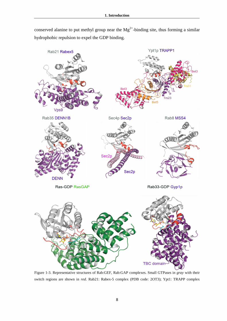
Figure 1-3. Representative structures of Rab:GEF, Rab:GAP complexes. Small GTPases in gray with their
switch regions are shown in red. Rab21: Rabex-5 complex (PDB code: 2OT3); Ypt1: TRAPP complex

1. Introduction
9
(PDB code: 3CUE); Rab35: DENN1B (PDB code: 3TW8); Sec4p: Sec2P complex (PDB code: 2OCY);
Rab8: MSS4 (PDB code: 1FU5). Ras: GAP complex (PDB code: 1WQ1); Rab33: Gyp1p complex (PDB
code: 2G77) (From Cherfils and Zeghouf, 2013).
After the GDP is released from GTPase:GEF complex, the nature of GTP loading to
the nucleotide-free complex was discovered by the structural dynamics of the GTPase and
the GEF. Several domains are contained in these processes, for examples, Sec7-domain
containing Arf/GEF (Renault et al., 2003), a Prone family RhoGEF (Thomas et al., 2007),
a DOCK family RhoGEF (Yang et al., 2009), and a Vps9 family RabGEF (Uejima et al.,
2010).
Rab GTPases GEFs can be subdivided into at least four types for their various
functional domains, probably because of the large members of different Rab subfamilies
(Hutagalung and Novick, 2011; Cherfils and Zeghouf, 2013). The GEF subfamilies
contain conserved catalytic domains DENN (Rab35 GEF) and Vps9 (Rab21/22 GEF)
motif with the surrounding of other domains (Figure 1-3, Delprato et al., 2004; Wu et al.,
2011). However, Sec2 (Sec4 GEF) and the TRAPP (Ypt1/Rab1 GEF) complex are the
unique subfamily which work as dimeric and pseudo-dimeric complexes, respectively
(Burton et al., 1993; Cai et al., 2007). Moreover, MSS4 in mammals, weakly stimulates
nucleotide exchange in a range of secretory Rab proteins (Nuoffer et al., 1997; Wixler et
al., 2011). However, biochemical studies and structure of nucleotide free Rab8 with
MSS4 indicate that MSS4 family members are just chaperones for nucleotide-free Rabs
but not actual GEFs (Itzen et al., 2006).
Although the mechanisms have not been fully understood, it has been known that
GEF proteins containing VPS motif and DENN motif can activate a group of even
overlapping Rab GTPases (Marat et al,. 2011; Carney et al,. 2006 ). TRAPP and
Sec2/Rabin8 are not like DENN domain containing GEF proteins; they have very specific
substrates, Ypt1/Rab1, Ypt31/32/Rab11 and Sec4/Rab8, in yeast and mammalian cells
respectively (Thomas and Fromme, 2016; Barrowman et al., 2010; Hutagalung and
Novick, 2011).
Once carried out their vesicle transport, GTPases dissociate from the membrane and
prepare for the next round cycle. GAPs accelerate the slow intrinsic GTPase activity to
exchange GTP-bound form to GDP-bound form. Similar with GTPase GEFs, the GAPs
are subfamilies specific which are verified by the structural information (Calmels et al.,

1. Introduction
10
1998). To date, most GAPs share a common conserved TBC (Tre-2/Cdc16/Bub2) domain
(Strom et al., 1993; Du et al., 1998; Albert et al, 1999; Vollmer et al., 1999; Eitzen et al.,
2000). The first discovered TBC domain GAP, GYP6 (GAP for Ypt6) was found in yeast
(Strom et al., 1993). Although more GAPs containing TBC domain were found later, the
GAPs number is still much less than the number of Rabs (Fukuda, 2011). This
phenomenon may be explained that one TBC-containing GAP is able to inactivate
multiple Rab GTPases (Frasa et al., 2012). The structure of Rab33-GDP: Gyp1 complex
demonstrates that the classical arginine finger (Arg343) and the glutamine finger (Gln378)
on the TBC domain of Gyp1 was faced with the switch II glutamine (Gln 92) of Rab33.
Both fingers from GAP protein contribute to stabilize the β-phosphate of GTP so that γ-
phsphate of GTP easily hydrolyze and dissociate from Rab (Figure 1-3) (Pan et al., 2006;
Rak et al., 2000). Indeed, some TBC domains lack of conserved glutamine or arginine
finger also perform as RabGAPs (Frasa et al., 2012).
Aside from GEF and GAP, two other proteins, GDI (GDP-dissociation inhibitor) and
REP (Rab escort protein) are also crucial for the function of Rabs. The structures of Rab-
GDI and Rab-REP show how these regulators associate with Rab proteins that mediate
membrane insertion. Although REP is similar to GDI and both of them are members of
the GDI superfamily, they have diverse functions in the Rab GTPase cycles. REP
associate with RabGGT to facilitate the addition of geranylgeranyl lipid moieties to the
C-termini of Rabs, and then interact with either prenylated or unprenylated Rabs.
However, GDI only tightly binds to the inactive Rab with its prenyl groups and thus to
extract prenylated Rabs from members (Pylypenko et al., 2006; Wu et al., 2007). The
structural of GDIs shows that they are highly conserved from yeast to human cells (Figure
1-4A). Both RabREP and RabGDI contain a two-site interface with Rab GTPases where
one domain recognizes the GDP-bound Rab RabF regions and the other domain interacts
with Rabs geranylgeranylated C-terminus (An et al., 2003; Rak et al., 2003; Rak et al.,
2004; Pylypenko et al., 2006). In addition, domain I also can interact with the binding
motif (CBR interacting motif, CIM) in the Rab hypervariable region.
The structures of the complexes between RabGGTase and REP-1, as well as
between Rab7 and REP-1 provided detailed biophysical information of REP1 and
RabGGTase working mechanisms (Pylypenko et al., 2003; Rak et al., 2004). Although
the structure of the catalytic ternary complex has not been solved, it was computationally

1. Introduction
11
modelled and biochemically validated by using structural information from the binary
complexes (Figure 1-4 C) (Wu et al., 2009). REP1 binds with high affinity to both
unprenylated Rab7 (Kd =0.22 nM) and even higher affinity with monoprenylated Rab7
(Kd=0.061 nM).
Figure 1-4. Structures of RabGDI, RasGDI and RabREP. (A)The Ypt1: RabGDI(PDB code: 2BCG)
complex between doubly geranylgeranylated Ypt1-GDP in gray, switch regions in red, C-terminal
extension in yellow with disordered regions in broken line, geranylgeranyl lipids in orange surface and
RabGDI in blue is shown. The location of the missense LP mutation found in X-linked mental retardation is
indicated. (B)The complex between PDEδ and farnesylated RheB-GDP(PDB code: 3T5G) in gray, with
switch regions in red, C-terminal extension in red with disordered regions in broken line, farnesyl lipid in
orange surface, and PDEδ in cyan. The β-sandwich of PDEδ is shown with the same orientation as the β-
sandwich of RhoGDI. The complex between PDEδ and Arl2-GTP (PDB code: 1KSH), the candidate GDF
for PDEδ, is overlaid. (This picture from Cherfils and Zeghouf, 2013) (C) Rab-GDP, REP-1 and GGTase-II
form a ternary complex (This picture from Wu et al., 2009). Prenyl groups are transferred in two
consecutive reactions from GGPP to Rab. Structure of the complex GGTase-II (α and β subunits are colored
gray and green, respectively), REP-1 (blue) and Rab7 (orange) based on structures of prenylated Rab7: REP
complex (PDB code, 1VG0) and REP-1: RabGGTase (PDB code, 1LTX). The farnesyl group is show in
stick representation in red, and Zn2+ as a turquoise ball in cyan.
Unlike REP1, GDI binds poorly to unprenylated Rab but with high affinity to mono-
or diprenylated Rab7. Combination of the structural and biochemical analysis suggests
that the second prenyl group may bind to the outside part of the pocket in REP and even
displace the first one to reduce its affinity. Although detailed structures showed the
interaction between prenylated Rabs with GDI or REP, is the mechanism still needs to be

1. Introduction
12
addressed that why GDI extracts Rab from the membranes with such high affinity. There
is a theory to explain how GDI removes a membrane bound Rab through masking its
hydrophobic prenyl tails from the aqueous environment (Wu et al., 2007). It was
proposed that the opposite reaction would require additional factors to efficiently break
the stable Rab-GDI interaction, such as a GDF or the molecular chaperone Hsp90 (Lee et
al., 2009; Goody et al., 2005; Ignatev et al., 2008 Chen and Balch, 2006). An integral
membrane protein Yip3/PRA1 was found as GDI-displacement factor (GDF), and it has
been shown to catalyze the dissociation of GDI from Rab9/Rab5 (Dirac-Svejstrup et al.,
1997, Sivars et al., 2003). However, it is unclear that how GDFs play roles in these
processes. It is difficult to get the structure of the Rab:GDI:GDF complex, probably due
to the low solubility of Yip3/PRA1.
A GDI-like solubilizing factor is PDEδ, which was considered as a GDI because it
can displace prenylated Rab13-GDP from the membrane (Marzesco et., 1998).
Subsequently, PDEδ was found to accelerate the dissociation of Ras family GTPase from
membrane and therefore it was classified as Ras GDI (Nancy et al., 2002). The structure
of PDEδ is similar to the β-sandwich lipid-binding domain of RhoGDIs (Hanzal-Bayer et
al., 2002; Nancy et al., 2002). The complex structure of farnesylated RheB (a member of
Ras family) with PDEδ shows that PDEδ uses its two β-sheets to accommodate the
farnesyl lipid of RheB, similar with that RhoGDI holds the geranylgeranyl lipid of Rho
protein (Figure 1-4B) (Ismail et al., 2011; Fansa et al., 2015). However, PDEδ does not
carry an additional GTPase-binding domain, and it recognizes RheB only by its
farnesylated C-terminus.
1.1.4 The prenylation of Rab GTPase
Except the 160-170 amino acids length core motif of Rab GTPase, the C-terminal
extension or C-terminal hypervariable domain (HVD) is also important for its functions
(Figure 1-2, Chavrier et al., 1991; Stenmark et al., 1994). The small GTPase Rab proteins
are post-translationally modified by geranylgeranyl-transferase II (RabGGTase) which
adds the geranylgeranyl moiety (ies) to one or (in most cases) two cysteines at the C-
terminus which secures the attachment of their active form to membrane (Casey et al.,
1996). The structures of Rab GTPases prenyl tails show these proteins have cysteine-
linked geranylgeranyl groups which come from soluble 20-carbon geranylgeranyl

1. Introduction
13
pyrophosphate (GGPP) (Farnsworth et al., 1990; Glomset et al., 1990; Swanson and Hohl,
2006).
RabGGTases prefer transfer the 20 carbon geranylgeranyl moieties to the C-terminal of
Rabs with CXC, CC, CCX, CCXX and CCXXX sequences, and some cases are mono
cysteine Rabs with CXXX such like Rab8 and Rab13 (Khosravifar et al., 1991; Kinsella
et al., 1992). In addition, Rab GTPases ending in CXC undergo carboxymethylation by
Isoprenylcysteine Carboxyl Methyltransferase (ICMT). The Methyl esterification
neutralizes the negative charge of the prenylcysteine and thereby increases membrane
affinity (Smeland et al., 1994; Dai et al., 1998).
RabGGTase contains α and β subunit which form heterodimer structures that has
delegated substrate recognition to Rab escort protein (REP) (Armstrong et al., 1993;
Zhang et al., 2000; Nguyen et al., 2010). REP interacts with the newly synthesized
unprenylated GDP-bound form of Rab protein (Alexandrov et al., 1998; Seabra, 1996b;
Sanford et al., 1993) and mediates its recognition by RabGGTase (Pylypenko et al., 2003;
Alexandrov et al., 1999; Anant et al., 1998). Upon Rab:REP:RabGGTase:GGPP complex
being formed, consecutive double prenylation without dissociation of the mono-
prenylated intermediate from GGTase-II (Thoma et al., 2001c). The mono-prenylated
Rab still tightly associate with REP to secure the complete di-prenylation of Rab (Shen
and Seabra, 1996). Complete the double prenylation, binding of the second GGPP
molecule triggers the release of Rab from Rab:REP complex to possible membrane
attachment (Thoma et al., 2001b). Hence, REP is released and prepared for another round
of Rab prenylation.
All the prenyltransferases require only Zn2+
but FTPase and RabGGTase also need
extra Mg2+
for their catalytic ability (Chen et al., 1993; Moomaw and Casey, 1992;
Seabra et al., 1992). The combinations of biochemical and spectroscopic methods that
utilize isoprenoid and protein-based fluorescent probes have illustrated the prenylation
mechanisms. In addition, the structural information come from the computationally model
of Rab7:RabGGTase:REP-1 (Figure 1-4 C) raises clues of prenylation of Rab GTPase
by RabGTTase (Wu et al., 2009). This model revealed that Rab switch I and II motifs
could interact with the Rab binding platform (RBP) of REP to facilitate their association.
Moreover, the determinant element is the interaction between the unprenylated Rab and

1. Introduction
14
the hydrophobic patch on the surface of REP, termed the C-terminal binding region
(CBR), with the so called CBR binding motif (CIM) which localize the Rab C terminus
(Rak et al., 2004; Wu et al., 2009). The CIM of most Rabs contains two large
hydrophobic residues surrounding a more polar residue. The catalytic ternary complex
model reveals that the RBP of REP first recognizes the Rab GTPase and thus assembles
Rab GTPase together, leading to a low- to intermediate-affinity complex (Nguyen et al.,
2010). With the association of CBR and CIM, the affinity of this complex is further
increased by an order of magnitude, which leads to the Rab C-terminus pointing to the
REP-associated RabGGTase.
The Rab C-terminus cysteines bind to the active site of RabGGTase through a series
of weak interactions in a step by step fashion. The weak interactions secure the protein
substrate specificity does not need to be encoded in the prenylatable C terminus,
facilitating the unspecific reorganization between GTPases and REP. This sequential
complex assembly with progressively weaker and smaller binding interfaces, and enables
cysteine residue(s) close to the C terminus to be prenylated by RabGGTase. This working
style also secures the multiple prenylation events on a single substrate being fulfilled
completely.
After double prenylation of the cysteines, new GGPP will be loaded to the REP1
making prenylated Rab being released from Rab:REP complex. The newly prenylated
Rabs will be delivered to some membranes and insert into the lipid bilayer with
hydrophobic isoprenoids tail(s) (Alexandrov et al., 1994). To date, only two REP proteins,
REP1 and REP2, are found in mammalian cells, while only one Mrs6p is found in yeast
(Cremers et al., 1994; Fujimura et al., 1994). The prenylation of Rab is crucial for its
cycle in cells and will induce diseases by causing the absence or mutations of REP
protein. A disease termed choroideremia is characterized by progressive atrophy of the
choroid, retinal pigment epithelium (RPE) and retina that lead to eventual blindness
(Seabra et al., 1993). Later analysis of tissue samples from patients with this disease
revealed that the unprenylated Rab27a lacks its normal function and accumulates in
retinal due to the mutation of REP-1. Intriguingly, the REP-2 can’t compensate the REP1
mutation and does not prenylate Rab27a in vivo (Cremers et al., 1994; Seabra et al.,
1995). The accumulation of non-functional Rab27a proteins induces a massive apoptosis
of retinal cells, which leads to a progressive degeneration of the retina.

1. Introduction
15
1.1.5 The functions of Rab GTPases in vesicular traffic
Rab proteins are the key regulators in vesicular traffic via interacting with various
effector proteins in respective pathways. The newly synthesized proteins and lipids are
transported to their destination via exotic pathway. Moreover, cells absorb nutrients,
molecules outside of plasma member and receptors on the cell surface are dependent on
endocytosis machinery. Both above pathways require various coated vesicles to transport
different contents in cells. At least three kinds of coated vesicles involved in the selective
cargo transport (Juan and Benjamin, 2004). Three kinds of coated vesicles, coat protein
complex-I (COPI) (Presley et al., 2002), coat protein complex-II (COPII) and clatrin
(Fotin et al., 2004a; Fotin et al., 2004b), are required for intracellular membrane
trafficking, corresponding to retrograde, anterograde(exotic pathway) and endocytic
pathways, respectively. The vesicular traffic contains several connective steps including
cargo selection, coated-vesicle formation, uncoating, directed vesicular movement, target
membrane recognition, and fusion. (Figure 1-5). During each step, a unique set of Rab
interacting proteins/effectors are required.
Figure 1-5. Steps of vesicles budding and fusion. (1) Initiation of coat assembly. The membrane-proximal
coat components (blue) are recruited to the donor compartment by binding to a membrane-associated

1. Introduction
16
GTPase (red) and/or to a specific phosphoinositide. Transmembrane cargo proteins and SNAREs begin to
gather at the assembling coat. (2) Budding. The membrane-distal coat components (green) are added and
polymerize into a mesh-like structure. Cargo becomes concentrated and membrane curvature increases. (3)
Scission. The neck between the vesicle and the donor compartment is severed either by direct action of the
coat or by accessory proteins such like dynamin. (4) Uncoating. The vesicle loses its coat due to various
events including inactivation of the small GTPase, phosphoinositide hydrolysis, and the action of uncoating
enzymes. Cytosolic coat proteins are then recycled for additional rounds of vesicle budding. (5) Movement
or transport. GTP-Rab proteins directly or via effectors recruit motor proteins to drive the movement of
vesicles along microtubules and actin filaments. (6, 7) Tethering, the vesicles are transported to the target
compartment. GTP-Rab proteins mediate recruitment of effectors, tethering factors, SNAREs, facilitating
tethering, docking and fusion of vesicles at the target membrane. The v- and t-SNAREs assemble into a
four-helix bundle. (8) This “trans-SNARE complex” promotes fusion of the vesicle and acceptor lipid
bilayers. Cargo is transferred to the acceptor compartment. (9, 10) A cis-SNARE complex in the fused
membrane α-SNAP binds to this complex and recruits NSF, which hydrolyzes ATP to dissociate the
complex. (11)The SNAREs are recycled to the donor membrane for next round cycle.
Effectors are defined as proteins that preferentially interact with their respective
GTP-bound form of Rabs. One notable exception is protrudin, which interact
preferentially with the GDP-bound form of Rab11 (Shirane and Nakayama, 2006).
In the COPII controlled anterograde process, the newly synthesized lipids and
proteins at ER are transported to their destination at the endosomes, plasma membrane or
outside of a cell (Gurkan et al., 2006). To balance the proteins and lipids within ER and
Golgi, COPI mediated retrograde pathway sends back those lost proteins and lipids from
Golgi to ER (Lippincott-Schwartz and Liu, 2006). The endocytic pathway is responsible
for nutrients uptake and internalization of various signal carriers, is carried out by clathrin
coat vesicle though some non-clatrin coats that works in this process (Edeling et al.,
2006).
The cargo selection and vesicle formation might be initiated at plasma membrane,
ER, Golgi and endosome and the vesicles bud from these donor membranes (Figure 1-5,
step1). The small GTPases Arf and Sar are the main participants in COPI and COPII
formation, respectively (Memon 2004; Barlowe et al., 1994). The active GTP-Sar1
recruits the Sec23:Sec24 to form the membrane-proximal layer, while Sec13:Sec31 forms
the second membrane-distal layer, step by step. During COPI Coat assembly, GTP-Arf,
simultaneously recruits the mem-cytobrane-proximal βγδζ, and the membrane-distalαβ’ε
sub-complexes (Hara-Kuge et al., 1994) at the same time which is different from the

1. Introduction
17
stepwise assembly of COPII by Sar1. Both Arf and Sar are also controlled by their GEFs
and GAPs in assembling and disassembling of COP coats. The clathrin coats are more
complicated than COPI and COPII. During the clathrin coats formation, Arf and/or
specific phosphoinositides such as PtdIns(4)P or PtdIns(4,5)P2 recruit a variety of adaptor
AP-1,-2,-3 heterotetrameric complexes and the monomeric GGA, Hrs, Epsin1 and ARH
proteins from cytosol to membrane (Bonifacino and Lippincott-Schwartz, 2003; Wang et
al., 2003; Bonifacino and Traub, 2003).
Apparently several Rabs are also involved in the coat budding process. For
example Rab9 directs the vesicle transport from the late endosome to trans-Golgi network
(TGN). GTP-bound Rab9 recruits its effector TIP47 that interacts with the mannose-6-
phosphate receptors (M6PRs) and transfer M6PRs from endosome to TGN. The
interaction between Rab9 and TIP47 enhances the affinity of TIP47 with M6PRs and
induces the richness of M6PRs in vesicles (Diaz and Pfeffer, 1998; Carroll et al., 2001;
Aivazian et al., 2006). Another well studied case is the cargo selection of retromer
complex. Retromer functions in conjunction with numerous associated proteins, including
select members of the sorting nexin (SNX) family (Seaman et al., 1998; Bonifacino and
Hurley., 2008). The retromer contains sorting nexins (SNXs) dimer which associated with
the Vps26-Vps29-Vps35 trimer (Horazdovsky et al., 1997; Seaman et al., 1998). The
SNXs are composed of a PX domain that interacts with phosphoinositides, and a Bar
domain which facilitates membrane curvature formation (Carlton et al., 2005; Frost et al.,
2008). GTP-bound Rab5 and GTP-bound Rab7 interact with the Vps26-Vps29-Vps35 in
a sequential manner (Rojas et al., 2008).The Vps26-Vps29-Vps35 interacts with Rab5
indirectly and is dependent on Rab5’s effector, phosphatidylinositol 3-kinase. As an
effector of Rab7, Vps26-Vps29-Vps35 can be recruited by Rab7 directly (Rojas et al.,
2008). Furthermore, Rab9 may play either a similar or complementary role in this process
(Carroll et al., 2001; Dong et al., 2013).
Some other GTPases have been involved in vesicles budding and fission (Figure 1-5,
step2). For example, at the beginning step of endocytosis, the scission and release of
clatrin coated vesicles (CCVs) are driven by dynamin, a vesicle invaginates. Around the
neck of the vesicle, dynamin forms a spiral circle which extends lengthwise and constricts
through GTP hydrolysis. Hence the vesicle neck breaks and results in the pinching off of
the vesicle from the parent membrane (Praefcke and McMahon, 2004). Recently

1. Introduction
18
identified Rab35 forms a tripartite complex with MICAL-L1 and ACAP2 to serve as a
scaffold for recruitment of EHD1 to endosomal recycling tubules (Kobayashi and Fukuda
2013; Kouranti et al. 2006). EHD1 is one member of Dynamin-like EHD family and
plays role in the process of the tubule scission.
To fuse the vesicle with acceptor membranes, it crucial to release the coats from
vesicles, a process that termed uncoating (Figure 1-5, step4). In addition to promote coats
formation, Rabs may also play a role in uncoating. For example, Ypt1/Rab1 has been
proposed to be involved in the ER-to-Golgi traffic pathway. It presumably recruits factors
that facilitate uncoating of COPII vesicles in the preparation for fusion (Jedd et al.1995;
Lian et al., 1994; Moyer et al., 2001; Pind et al., 1994). Rab5 regulates the early
endocytic pathway and is found on clathrin-coated vesicles (CCVs). Firstly, the assemble
clathrin adaptor AP-2 complex recruit clathrin to newly formed endocytic vesicles.
Meanwhile the AP-2 complex also bind another cargo such like transferrin receptor for
internalization, or clathrin triskelions to facilitate coat formation (Benmerah and Lamaze,
2007; Owen et al., 2004; Sorkin 2004). The reorganization of clahrin by AP-2 is
dependent on the phosphorylation of its μ2 subunit (Jackson et al., 2003). The μ2 kinase
can be recruited by Rab5 to AP-2 to phosphorylate μ2 subunit. With the action of the
Rab5 GAP GAPVD1, μ2 kinase was released from AP-2 to prevent it from
phosphorylating the μ2. PtdIns(4,5)P2 is also a significant component for recruiting AP-2
during clathrin-mediated endocytosis (Höning et al., 2005; Zoncu. et al., 2007).
Modulation of PtdIns(4,5)P2 levels by Rab5 may occur through recruitment of effectors
such as PtdIns(3)P kinases or PtdIns phosphatases (Christoforidis et al., 1999, Shin et al.,
2005).
Rab proteins are critical for vesicle movement often using motor proteins
(kinesins/dyneins and myosins) along actin- or microtubule-based cytoskeletal structures
(Figure 1-5, step5). There are several well studied examples of such Rabs and their
effectors in this process. To balance the receptors contents on plasma membranes, the
recycle transport are needed for sending back various receptors from cytosol. Rab11
interacts with myosin Vb (Myo5b) through its effector, Rab11 family interacting protein
2 (Rab11-FIP2), to regulate plasma membrane recycling (Hales et al., 2002). The
transport of melanin-containing melanosomes to the plasma membrane is regulated by
Rab27a which interacts with its effector melanophilin/Slac2-a that binds to the actin

1. Introduction
19
motor myosin Va (Myo5a) in melanocytes (Bahadoran et al., 2001; Hume et al., 2001;
Matesic et al., 2001; Stromet al., 2002; Wu et al., 2001; Wu et al., 2002). Mutation in any
one member of the Myo5a, Rab27a, and melanophilin tripartite complex leads to the rare
autosomal recessive disorder Griscelli syndrome (GS), the mouse mutants dilute, leaden,
and ashen (Myo5a, Rab27a, and melanophilin, respectively) (Van et al., 2009). These
patients display various symptoms ranging from hypopigmentation (GS3, melanophilin
mutation) and immunological defects (GS2, Rab27a mutations) to neurological
impairments (GS1, MyoVa mutations). In yeast, Ypt31p/Ypt32p facilitates the
recruitment of the Myosin V type motor Myo2p directly from Golgi to exocytic vesicles,
whereas the downstream GTPase Sec4p binds directly to Myo2p to coordinate the
transport of exocytic vesicles along the actin (Jin et al., 2011; Lipatova et al., 2008).
Aside from the above vesicle transports which are driven by actin, another major
membrane traffic pathway relies on microtubules in animal cells. Microtubules provide
high-speed, long-range transport, while actins usually facilitate slower and short-range
local transport events (Jordens et al., 2005). Rabs have been proposed to interact with
microtubule-based motors to regulate these pathways, either interacting with kinesins
(plus-end directed motors) or the dynein (minus-end directed motors) family. Dynein and
dynactin form a complex, which stimulate processive motility of vesicles along
microtubules (McGrail et al., 1995; Vaughan and Vallee, 1995). Rab6 localizes to the
Golgi and has been shown to be involved in exocytic traffic to the plasma membrane by
recruiting Rabkinesin-6 (kinesin family member 20A) directly to facilitate intra-Golgi
transport (White et al., 1999; Utskarpen et al., 2006; Echard et al., 1998; Martinez et al.,
1994). Rab6 also indirectly regulates microtubule motors through the effector proteins
Bicaudal D1/D2 that link Rab6-containing vesicles to the dynein-dynactin motor complex,
and it also links kinesin for exocytosis (Grigoriev et al., 2007; Hill et al., 2000; Matanis et
al., 2002; Young et al., 2005). Another well studied case is Rab7, which coordinates the
trafficking of late endosome and the lysosome or centrosome. Rab7 interacts with its
effector Rab-interacting lysosomal protein (RILP) to recruit the dynein-dynactin motor
complex to transport along microtubule (Johansson et al., 2007; Jordens et al., 2001).
Several intracellular pathogens manipulate Rab7-effector’s interaction for their growth or
replication after invasion host cells. The Salmonella secretes effector protein SifA can
hijack Rab7 that prevents the interaction between RILP and Rab7 to facilitate growth of

1. Introduction
20
the membrane-bound compartment in which the bacterium can replicate (Guignot et al.,
2004; Harrison et al., 2004). Heliobacter pylori secrets the VacA cytotoxin and causes the
formation of large vacuoles. These vacuoles contain bacteria and their surfaces are highly
enriched in Rab7 that can recruit RILP to direct endosmal traffic (Li et al., 2004;
Terebiznik et al., 2006).
Once the vesicle is closed to the acceptor membrane, it is critical to ensure the
fidelity of transport. A machinery termed tethering/docking has been addressed clearly
(Chia and Gleeson, 2014; Cai et al., 2007) (Figure 1-5, step6 and 7). The tethering factors
are classified into two types: One is long coiled-coil tethers and the other is multiprotein
complexes (Sztul and Lupashin, 2006). Both kinds of tethers are Rab effectors which
mean that Rab proteins also play roles in the tethering process. Rab effector tethering
factors include Uso1p/p115, the COG complex, Vac1/EEA1, the GARP complex, the
HOPS complex, and the CORVET complex. Coiled-coil tethers such as Golgins family
include p115/Uso1, giantin, GM 130, Golgin97, Golgin185, Golgin210 and so on, which
localize at the Golgi complex or closed to the endosomes (Short B et al., 2005).
Uso1/p115 was defined as an essential factor in ER to Golgi transport in yeast
(Sapperstein et al., 1995). GM130 and GRASP65 are Golgi peripheral membrane proteins
that play a key role in Golgi stacking and vesicle tethering (Puthenveedu et al., 2006;
Diao et al., 2008). Both GM130 and GRASP65 have been identified as effector of Rab1
(Barr et al., 1998; Moyer et al., 2001). Rab1 recruits GM130-GRASP65 complex and
interacts with p115 is thought to tether ER-derived COPII vesicles to the Golgi (Sztul and
Lupashin, 2006).
Multiprotein complexes such as TRAPPs were proposed to participate in the tethering
processes. The TRAPPI (7 subunits) and TRAPPII (10 subunits) complexes are
multisubunit tethers that regulate traffic in ER-to-Golgi, intra Golgi, and endosome-to-
late Golgi traffic, respectively (Cai et al., 2005; Cai et al., 2007; Sacher et al., 1998).
Unlike the above tethers, the TRAPP complexes do not work as Rab1/Ypt1 effector but
act as GEFs for Rab1 which active the GTP-bound form for interacting with effectors to
coordinate membrane traffic (Barrowman et al., 2010). The TRAPPI subunit Bet3 that
binds to the COPII subunit Sec23 (Cai et al., 2007; Yu et al., 2006) and Bet3 also has
genetic interactions with Bet1, Sed5, Sec22, and all SNARE proteins that function in ER-
to-Golgi traffic (Rossi et al., 1995; Sacher et al., 1998). Mammalian mBet3 can form the

1. Introduction
21
homotypic tethering of COPII-coated vesicles from vesiculotubular clusters, an
intermediate compartment between the ER and Golgi (Yu et al., 2006). The active
Rab1/Ypt1 recruits effectors such as Uso1/p115 and giantin, tether these intermediate
vesicles to the Golgi. In addition, TRAPP may interact with the COPI coat and exhibits
its function in regulating intra-Golgi and endosome-to- late Golgi traffic (Yamasaki et al.,
2009).
The final step of vesicular transport is fusion with the acceptor membrane.
Rothman and coworkers used Nethylmaleimide-sensitive factor (NSF)/α-
Nethylmaleimide-sensitive factor attachment protein(α-SNAP) as an affinity bait to
fractionate a brain lysate, and identified a set of three membrane-associated ‘SNAP
Receptors,’ or SNAREs (Söllner et al., 1993). SNAREs control membrane fusion in all
kinds of trafficking steps of the secretory pathway (Jahn and Scheller, 2006; Hong, 2005).
Most SNAREs are C-terminally anchored transmembrane proteins, with their functional
N-terminal domains facing toward the cytosol. Each type of transporting vesicle carries a
specific ‘vesicle associat (v)-SNARE’ that binds to a cognate ‘target associate (t)-SNARE’
on the target membrane (Rothman, 1994). Both v-SNARE and t-SNAREs contain a
heptad repeat ‘SNARE motif’ that can participate in coiled-coil formation (Bock et al.,
2001). Structural and biochemical studies showed that the SNARE complex generated by
the pairing of a cognate v- and t-SNARE is a very stable four-helix bundle, with one α
helix contributed by the monomeric v-SNARE and the other three α helices contributed
by the oligomeric t-SNARE (Fasshauer et al. 1997, Sutton et al. 1998). v-SNAREs and t-
SNAREs are also termed R-SNAREs and Q-SNAREs for at the characteristic position
within the SNARE motif, the v-SNAREs and t-SNAREs contain an Argine (R) and an
Glutamine (Q), respectively (Fasshauer et al., 1998.). The structural analysis shows that
SNARE complex composes v- and t-SNAREs pair in a parallel fashion (Hanson et al.,
1997, Lin and Scheller, 1997, Sutton et al., 1998). Therefore, the concept of trans-
SNARE complex means that v- and t-SNAREs are from separate membranes, while cis-
SNARE complex means v- and t-SNAREs are in the same membrane. A trans-SNARE
complex persists throughout the fusion reaction to become a cis-SNARE complex in the
fused membrane (Figure 1-5, step8). Hence α-SNAP binds along the edge of the SNARE
complex (Rice and Brunger, 1999) and recruits NSF (Figure 1-5, step9). ATP hydrolysis
by NSF untwists the four-helix bundle and dissociates the cis-SNARE complex (Figure 1-

1. Introduction
22
5, step10) (Mayer et al., 1996; May et al., 1999; Yu et al., 1999). Thus, the v-SNAREs
and t-SNAREs are recycled for another round of complex formation (Figure 1-5, step11).
Rabs regulate fusion process by interacting directly with SNARE proteins or
SNARE related proteins, such as SM or Lgl proteins, which can regulate SNARE
function. For example, Rab5 is found on early endosomes and plays a critical role in
endocytic pathway through the function of its numerous effectors. Rab5 effectors, EEA1
and rabenosyn-5, interact with the SNARE proteins, Syntaxin13, Syntaxin6 and the SM
protein VPS45, respectively (Nielsen et al., 2000; Simonsen et al., 1999). This interaction
is required to drive homotypic early endosome fusion (McBride et al., 1999).
Another example is the Rab7 effector, the Vici Syndrome protein EPG5, determines
the fusion specificity of autophagosomes with late endosomes/lysosomes (Wang et al.,
2016). Firstly, Rab7 and the late endosomal/lysosomal R-SNARE VAMP7/8 recruit
EPG5 to the late endosomes/lysosomes. In parallel, EPG5 is also recruited to LC3/LGG-1
(mammalian and C. elegans Atg8 homolog, respectively) and to assembled STX17-
SNAP29 Qabc SNARE complexes on autophagosomes. Therefore, EPG5 can stabilize
and facilitate the assembly of STX17-SNAP29-VAMP7/8 trans-SNARE complexes.
Moreover, EPG5 promotes STX17-SNAP29-VAMP7-mediated fusion of reconstituted
proteoliposomes. The depletion of SNAP25 can partially rescue the autophagy defect
caused by EPG5 knockdown (Wang et al., 2016).
In summary, by combining the regulation from above various factors, a clear map of
Rab GTPase vesicular transport and recycling turned out. Newly synthesized Rab proteins
are captured by Rab escort protein (REP) at the ER exit sites. REP acts as a molecular
chaperone of unprenylated Rab to make it soluble in cytosol. Then REP presents Rab
proteins to heterodimeric RabGGTase for being modified with (usually) two
geranylgeranyl moieties. Afterwards, the prenylated Rab proteins are delivered by REP to
their target membranes. The released REP recycles backs to cytosol to support additional
rounds of Rab prenylation. Prenylated Rab proteins associate with the membrane where
GEFs facilitate Rab-GDP exchange to Rab-GTP.GTP bound Rab proteins are active and
can bind to various effectors that are involved in vesicle budding and cargo selection.
GTP-Rab proteins directly, or via effectors, recruit motor proteins to drive the movement
of vesicles along microtubules or actin filaments. Once the vesicles are close to the target

1. Introduction
23
compartment, GTP-Rab proteins recruit effectors, tethering factors, SNAREs, facilitatete
thering, docking and fusion of vesicles at the target membrane. After completing the
vesicle transport, Rab GTPases undergoes hydrolysis of its bound GTP with the help of
GAPs. Related effectors are released from the inactive Rab-GDP and participate in a new
round of transport. GDIs extract Rab-GDP proteins from membranes and solubilize them
in cytosol. For the cycled Rab GTPases, GDI delivers them to the donor membrane
similar to REP. GDF may facilitates the release the prenylated Rab proteins from GDI on
endosome vesicles and perform GDP-GTP exchange by GEFs.
1.1.6 The localization of Rab GTPase in cells
Rab proteins constitute the largest branch of the Ras GTPase superfamily. To date, about
70 members in mammalian cells and 13 members in yeast have been found with various
functions and distinct localizations (Klopper et al. 2012; Steinet al. 2012). The much
larger number of Rabs in mammals meets the requirements of the higher complexity of
transport events in higher eukaryotes.
The factors controlling each step of vesicular transport are coordinated in time and
space. The molecular features and functional properties of Rabs that fit with such a
spatiotemporal coordination (Zerial and McBride 2001; Pfeffer 2013b; Barr 2013). Rab
GTPase localization is highly compartmentalized and organelles often have a unique
complement of Rab proteins (Figure 1-6) (Galvezet al. 2012; Hutagalung and Novick
2011; Stenmark 2009). In addition, Rab GTPases can shuttle between the cytosol and the
membrane in either an inactive GDP-bound or active GTP-bound conformation.

1. Introduction
24
Figure 1-6. The localizations and functional groups of Rabs in mammalian cell.
A typical cell shows the intracellular localization and associated vesicle transport pathway(s) of several
groups Rab GTPases. The secretion group including Rab1, Rab2, Rab24 and Rab30 regulate ER-Golgi
parts and Rab1, Rab2, Rab8, Rab10 and Rab18 are contained in intra-Golgi traffic and regulate biosynthetic
traffic from the trans-Golgi network (TGN) to the plasma membrane. While several secretory vesicles and
granules use Rab3, Rab12 and Rab26 regulate post-Golgi vesicle transport, secretory vesicles, finally
release molecular to the outside of cell environment. This group is highlighted in cyan. Rab2 is also
involved in recycling group which including other Rab proteins Ra4, Rab11, Rab14 and Rab35. The
recycling group is highlighted in shallow green. Rab6 regulates intra-Golgi traffic which is highlighted in
yellow. There are numerous Rabs associated with endosomal traffic, and the most active site of localization
is the early endosome. Most early endocytic steps rely on Rab5, which mediates fusion of endocytic
vesicles to form the early endosome and the other Rabs including Rab21, Rab22, and Rab24. This early
endosomal traffic is highlighted in orange. Traffic can be directed towards the lysosome for degradation,
which relies on action of several Rabs including Rab7, Rab9, and Rab23 are involved in late endosomal and
lysosomal trafficking. In this group, Rab27 is well-studied in the melanosome transport that also relies on
Rabs 32 and 38. This group is highlighted in purple. In addition, Rab24 and Rab33 mediate formation of
the preautophagosomal structure that engulfs cellular components to form the autophagosome that is
subsequently targeted to the lysosome/vacuole. Rab21 and Rab25 regulate transport of integrin to control
cell adhesion and cytokinesis. Rab13 directs traffic to and regulates formation of tight junctions in polarized
epithelial cells.
Rab GTPases are one of the main coordinators of membrane domain formation and

1. Introduction
25
dynamics. The first reported example is Rab5 which localizes on early endosome
(Chavrier et al., 1990), and later more cases showed that different Rabs localize at distinct
organelles or compartments (Zerial and McBride, 2001). Furthermore, a large amount of
evidences show that Rabs work as the organelles and compartments identity marker
(Marino and Heidi, 2001; Pfeffer, 2001; Barr, 2011). The works in Rab GTPases have
revealed molecular features and functional properties that fit with such a spatiotemporal
coordination (Zerial and McBride 2001; Pfeffer 2013b; Barr 2013). For example, Rab1 is
present on the Golgi (including ERGIC); Rab6 on the Golgi; Rab5 on early endosomes
and Rab7 on late endosomes (Zerial and McBride, 2001; Segev, 2001). More interestingly,
Rab GTPases are now known to collect integral and peripheral membrane proteins to
different organelles even on the same organelle with distinct domain (or scaffold), which
is referred to as Rab domains (Sonnichsen et al., 2000). Well studied cases are early
endosomes that compose separate domains enriched in Rab5 and Rab4 that are involved
in endosome fusion and endocytic recycling, respectively. The recycling endosome
contains domains that are enriched in Rab4 and Rab11, which are involved in vesicle
trafficking from the early endosome and to the plasma membrane, respectively. Another
example is Rab7 and Rab9 which localize at late endosomes (Barbero, et al., 2002).
1.1.7 Membrane targeting of Rab GTPase in cells
How Rabs associate their destination membrane or organelles by effectors is discussed
partially in above sections. More evidences indicate that regulators of Rab GTPases
include effectors, REP, GDIs, GDF, and GEFs which may contribute to their membrane
targeting.
A best case is Rab5 and its effectors. Biochemical and cellular studies show that it is
a key regulator of early endocytic trafficking (Zeigerer et al. 2012). Rab5 has a unique
complexity of regulators and provides important insights into the membrane
compartmentalization /targeting (Christoforidis et al. 1999a). Indeed, several of these
effector molecules act indirectly but cooperatively with other components of the transport
machinery. For example, the localized synthesis of phosphatidylinositol 3-phosphate
(PtdIns(3)P) by PtdIns(3)P kinases is regulated by Rab5. A positive feedback loop of
Rab5 recruitment and activation ensure the localized enrichment of Rab5 (Horiuchi et al.
1997; Stenmark et al. 1995). Rab5 interact with PtdIns(3)P kinases to regulate the

1. Introduction
26
generation of PtdIns(3)P (Christoforidis et al. 1999b; Murray et al. 2002). The
Rab5/PtdIns(3)P effectors oligomerize complexes that stabilize the Rab5 membrane
domain (McBride et al. 1999). Dynamics of these oligomers need free energy from the
form of GTP (Rab5) and ATP (through NSF). However, the molecular details of various
proteins contribute to the formation of the oligomers is to be addressed. These proteins
function sequentially or concomitantly with the recruitment by Rab5 membrane domain
where these activities can be amplified.
Along recruiting specific effectors to restricted membrane microdomains, the
GTPases might also confer membrane identity by controlling the local levels of
phosphatases (PtdInsPs). For example, OCRL1/INPP5F, the inositol polyphosphate 5-
phosphatase, is an effector of multiple Rabs including Rab1, Rab5, Rab6, Rab8 and
Rab35 (Noora et al., 2006; Dambournet et al., 2011).
RabGGTaseII transfer geranylgeranyl moieties to one or (in most cases) two cysteine
residues of Rab C-terminal end with the help of REP. This secures Rab for being able to
associate with membranes and fulfilling their proper functions. For the newly synthesized
Rabs, REP works as chaperon to solubilize prenylated Rab in cytosol and delivers it to the
targeted membrane. The prenylated Rabs insert into the membrane via their one, or most
cases two, hydrophobic geranylgeranyl moieties into the lipid bilayer (Shahinian and
Silvius, 1995). However, it still unclear that if other factors are also involved in the
delivery of prenylated Rab GTPase by REP. Some evidences indicate the attachment
ability of two moieties is stronger than one (Gomes et al., 2003) and may induce
unsuccessful membrane targeting/attachment. However, an exception is Rab1 with
CAAX terminal (Rab1-CLLL), which can be delivered to the ER/Golgi membrane and
functions well (Overmeyer et al., 2001). Several Rabs with CAAX box tail such as Rab8
(-CVLL), Rab13 (-CSLG) and Rab18 (-CSVL), can complete their membrane targeting
and perform their various functions with only one prenylated cysteine (Huber et al.1993;
Joberty et al., 1993). These CAAX box ends of Rabs are prenylated by RabGGTase but
not FTase nor GGTaseI which are responsible for the prenylation of Ras, Rho, Rac with
CAAx box C-terminal end.
The inactive Rab-GDP will be extracted by GDIs from membrane and becomes
soluble in the cytosol. GDIs carry the prenylated Rab-GDP to deliver to a target

1. Introduction
27
membrane. However, it is still unclear that how GDI and Rab-GDP complex associate
with its destination membrane. A hypothesis was proposed that there is one factor which
would catalyze the dissociation of Rab from GDI. Considering the tight affinity between
Rab and GDI (KD=4.5nM) (Wu et al., 2010), an assistance is required to release Rab
from its GDI. Pffefer and colleagues used screening methods and identified a protein
PRA1/Yip3 which works as GDI displacement factor (GDF) (Dirac-Svejstrup et al.,
1997). Prenylated Rab acceptor-1 (PRA1) and Ypt-interacting protein III (Yip3) feature
four transmembrane domains, with their N and C termini facing toward the cytosol while
the remaining bulky parts of their sequences being membrane embedded (Calero et
al.,2002; Lin et al., 2001) . PRA1/Yip3 localizes at Golgi and endosomes (Martincic et al.,
1997; Bucci et al., 1999) where it interacts with prenylated Rab proteins (Sivars et al.,
2003; Abdul-Ghani et al., 2001). Meanwhile, PRA1/Yip3 also weakly interacts with GDI
(Hutt et al., 2000).
Rab9-GDI complex can be dissociated by Yip3/PRA1 at pico-molar level, which is
indicted by the rate of 35S-GTPγS binding. Intriguingly, Yip3/PRA1 is not a common
GDF which only works for endosomal Rab proteins, such as Rab5, Rab7 and Rab9.
Mammalian Yip family contains 16 members in human and 14 members in mice. The
Yip1 can recruit Ypt1 on ER-Golgi Yip1 in vivo (Barrowman et al., 2003). The working
model of Yip3/PRA1 indicates several possible factors that preferably interact with Rab5,
Rab7 and Rab9 in vitro and in vivo.
Another explanation of membrane targeting mechanism is GEFs mediated Rabs
localization. GEFs have the ability to exchange Rab GDP-bound to Rab GTP-bound and
displace GDIs by lowering the affinity between Rabs and GDIs. Upon GTP binding, Rab
proteins undergo a conformational change. This protects them from being removed by
Rab GDI and allows for Rab effector protein binding. The first case of GEF displacing
GDI via GDP-GTP exchange is the identity of DrrA/Sidm. Legionella pneumophila’s
DrrA/Sidm, a virulence effector which plays a key role in hijacking the host vesicular
trafficking by recruiting Rab1 to the cytosolic face of the Legionella-containing vacuole
(LCVs). DrrA/Sidm acts as a GDP-GTP exchange factor (GEF) for the small GTPase
Rab1 (Rab1A, Rab1B), thereby converting Rab1 to an active GTP-bound state, leading to
the incorporation of Rab1 into LCVs (Schoebel et al., 2009; Wu et al., 2010; Murata et al.,
2006; Machner and Isberg, 2006). Interestingly, the Rab1 can even be recruited to plasma

1. Introduction
28
membrane with the ectopic expression of DrrA/Sidm at PM (Murata et al., 2006). A
similar case is the Rab3GEF which is necessary for recruiting Rab27 to melanosomes
(Tarafder et al., 2011).
A general function of GEFs is to provide the thermodynamic driving force for
Rab membrane targeting via nucleotide exchange (Wu et al., 2010). Therefore GEFs
might be sufficient to mediate specific targeting of Rabs to membranes. However, in
order to ensure that this mechanism works, GEFs need to be localized to particular
membranes. This hypothesis was proved by a more recent evidences that Rabex-5
(Rab5GEF), and Rabin8 (Rab8GEF) display the minimal targeting machinery for
recruiting Rabs from the cytosol to the correct membranes (Blümer et al., 2013).
GEFs are indeed recruited to or activated at cellular membranes on demand by
factors acting upstream, such as Rab cascades or feedbacks (Rivera-Molina and
Novick, 2009; Mizuno-Yamasaki et al., 2010; Poteryaev et al., 2010), and/or
dependence on changes in membrane phosphoinositide composition (Shin et
al.,2005 ; Christoforidis et al., 1999).
Rabs contain a conserved GTPase domain and a structurally flexible C-terminal
amino acid sequence of variable length (∼25- 40 amino acids), termed C-terminal
hypervariable domain (HVD or HVR) that bearing the one or two prenylated
cysteine residues at the very end. In contrast to the conservation of GTPase domain,
the C-terminal sequence is very divergent in sequence among Rab proteins. Zerial
and colleagues designed a series of chimeric Rabs such like Rab5 with Rab7’s HVR,
Rab2 with Rab5’s HVR or Rab7’s HVR. These chimera target Rabs to the
corresponding Rab’s HVR membranes. Compared to the divergence sequences of
hypervariable sequence of each Rab/Rab family, a hypothesis was proposed that
these domains contain the targeting information to associate a specific membrane
compartment. More recent work has shown similar results for GTPase
domain/hypervariable sequence Rab9/Rab5 and Rab9/Rab1 chimaeras (Aivazian et
al., 2006). All above cases showed that C-terminal hypervariable domain mediate
membrane targeting via its interaction with Rabs eggectors. On the contrary, the
presence of poly basic regions in the Rab35 and KRas C-terminus contributes to the
localization on negatively charged membranes (Heo et al., 2006). However,
subsequent works by Seabra and colleagues strongly suggest that multiple regions

1. Introduction
29
contribute to the correct Rab membrane targeting (Ali et al., 2004). They swapped the C-
terminal hypervariable domains of Rab1A, Rab2A, Rab5A, Rab7A and Rab27A, causing
these Rabs to localized to correct compartmental membranes. These evidences indicate
that the Rabs membrane targeting is determined by conserved GTPase domain but not the
C-terminal hypervariable domain (Ali et al., 2004; Tarafder et al., 2011). Therefore the
model that the C-terminal hypervariable domain determines Rabs membrane association
might not be universal. Thus, the function of the Rab HVD and a complete model for Rab
membrane targeting remain to be established.
In summary, the mechanisms of Rab membrane targeting via its prenylation tail(s),
its regulators are including GEF, GDF, and effectors. However, the mechanisms of
membrane targeting are quite complicated which need to be further elucidated.
1.1.8 Rab cascades and feed-back
As membrane flows from one organelle to another in a cell, it must transit through a
connective membrane units or Rab defined compartments. Rab acting as compartment
organizer is regulated by different factors including GEF, GDI, GAP, and effectors.
Therefore, a question is turned out that how the Rabs which localize at different
compartments membrane are active and inactive from one to another. To solve this
problem, a model of Rab cascade is proposed which address the compartment transitions
from an upstream Rab to a downstream Rab by recruiting effectors which work as the
GAP and the GEF for the upstream and downstream Rabs, respectively (Markgraf et al.,
2007; Rivera-Molina and Novick, 2009; Pfeffer, 2012; Novick , 2016).
For example, a cascade involved Ypt31/32(Rab11 homolog) and Sec4 (Rab8
homolog) play a role during membrane transit from late Golgi to plasma membrane via
secretary pathway (Benli et al., 1996; Goud et al., 1988; Jedd et al., 1997; Salminen et al.,
1987).
GTP-bound form Ypt31/32 recruits Sec2, the GEF of Sec4, which activates Sec4
that is associated with secretory vesicles. The loading of Sec4 into secretory vesicles
secures their delivery and fusion with the plasma membrane (Ortiz et al., 2002). In this
cascade, Sec4 can be involved in the correct pathway by association with secretory
vesicles which is dependent on its direct upstream Rab. Moreover, Sec2 also interacts

1. Introduction
30
with Sec15, an effector of Sec4, as a supplement to help recruit Sec4 on secretory
vesicles (Guo et al., 1999; Medkova et al., 2006; Salminen and Novick, 1989).
To date, the best well studied of Rab GEF cascade is the transition from early
endosome to late endosome which is directed by Rab5 and Rab7 (Rink et al., 2005;
Poteryaev et al., 2010). Endocytic cargo is initiated from Rab5-containing early
endosome that can undergo maturation to become Rab7-containing late endosome
for being targeted to lysosomes (Rink et al., 2005). The HOPS complex contains one
of its subunits, the Vps39 protein which is a GEF for Rab7. Herein, Rab7 also serves
as an effector of Rab5 (Cabrera et al., 2009; Rieder and Emr, 1997). In the
meanwhile, the HOPS complex is also an effector of Rab7 (Seals et al., 2000).
Therefore, Rab5-mediated recruitment of the HOPS complex in turn promotes the
association of Rab7 with this membrane and then initiates the maturation towards the
lysosome/vacuole. This process of Rab conversion appears to proceed in several
steps. Firstly, the active GTP-Rab5 associates with early endosomes. Secondly, the
association of Rab5 membranes progressively forms larger endosomal compartments
via homotypic fusion that moves from the cell periphery towards the cell center
along microtubules. Thirdly, a transient overlap Rab5 with Rab7 occurs, which is
mediated by the HOPS complex. Finally, Rab5 compartments convert to Rab7
compartments which are destined for the lysosome/vacuole. These results indicate a
maturation model where every transport compartment gains the necessary factors to
move forward while losing those that defines the upstream compartments (Rink et al.,
2005). Another support for the maturation model comes from the studies of the
Golgi in S.cerevisiae. Both studies show that specific Golgi cisternae transitions
from early Golgi to late Golgi through the secretory pathway (Losev et al., 2006;
Matsuura-Tokita et al., 2006).
The divalent effectors of Rab also can affect Rab conversion and target traffic
appropriately from a compartment that serves multiple pathways. In the early endocytic
pathway, another Rab5 effector, rabenosyn-5, has a binding site and is an effector of
Rab4 that is involved in targeting proteins to the Rab11-positive recycling endosome.
Functional study with the over expression of rabenosyn-5 in cells showed that the overlap
of Rab5 and Rab4 is prolonged (De Renzis et al., 2002).

1. Introduction
31
The GEF cascade mechanism explains how a downstream Rab can be recruited to a
membrane domain that is initially carries an upstream Rab. GAPs play key roles in this
process to avoid an extended period of overlap of Rab domains within a compartment; it
is also important to inactivate the upstream Rab once the downstream Rab has been
recruited and activated. For example, RabGAP-5(the GAP for Rab5) works in regulating
endosomal traffic; abnormal expression of RabGAP-5 in HeLa cells blocked trafficking
of substrates from early endosomes to the lysosome (Haas et al., 2005).
In short, the discoveries of Rab cascade indicates the nature of energy saving and
high efficiency work model in cells.
1.1.9 Rabs related diseases
1.1.9.1 Rab and cancer
Rab proteins work together with their cognate effectors, coordinate the dynamics of
trafficking pathway and determine the cargo proteins’ destination in cells. Aberrant Rab
GTPases functions by mutations or post-translational modifications will disrupt the
regulatory network of vesicle trafficking, which have implications in tumorigenesis,
Parkinson’s disease, Huntington’s disease and bacteria induced diseases via hijack Rab
and Rab regulators.
Many Rab GTPases have been proposed to be involved in the progression of
multiple cancer types. Membrane traffic plays a significant role in cancer biology,
primarily in the loss of cell polarity and in the metastatic transformation of tumor cells
(Mosesson et al., 2008). More evidences showed that a group of Rabs including Rab1,
Rab2A, Rab3, Rab4, Rab8, Rab11, Rab21, Rab23, Rab25, Rab27B, Rab35, Rab37 and
Rab38 promote tumor cell migration and invasion, and consequently exhibit their effects
on tumorigenesis and metastasis by interruption of intracellular signal transduction (Yoon
et al., 2005; Caswell et al.,2007; Bravo-Cordero et al., 2007; Yang et al.,2016; Luo et al.,
2015; Yang et al 2009; Tang et al.,2009; Hou et al., 2008; Cheng et al.,2004; Wheeler et
al., 2015). One of well characterized example Rab involved in cancer is Rab25, which
regulates apical endocytosis and transcytosis in epithelial cells (Casanova et al., 1999;
Wang et al., 2000). High expression level of Rab25 has been frequently found in poor
prognosis in breast and ovarian cancer patients due to amplification of the Rab25 gene.

1. Introduction
32
High expression level of Rab25 also promotes anti-apoptotic phosphoinositide 3-kinase
(PI3K)-Akt pathway and inhibits pro-apoptotic molecules expression such as BAK, and
thereby increasing aggressiveness of cancer cells (Cheng et al., 2004). Later studies
showed that Rab25 can interact with β1-integrin subunit and promote invasiveness of
tumor cells into a three-dimensional extracellular environment (Caswell et al., 2007).
Interestingly, Rab25 only facilitates tumor progression but does not initiate tumorigenesis.
Norman and co-workers found that Rab25 can maintain a pool of α5β1-integrin
heterodimers at the tip of the invasive pseudopod which facilitates efficient integrin
recycling and secures a stable association of the pseudopod within the extracellular
environment (Caswell et al., 2007). Similarly, Rab11 has been found to enhance cancer
cell invasion in breast cancer where it mediates α6β4 integrin trafficking (Yoon et al.,
2005). The oncogenic gene Rab1 was found to be over-expressed in some poor survival
cancer types (Bao et al., 2014; Thomas et al., 2014; Xu et al., 2015). Mechanistically,
over expressed Rab1A stimulates mTORC1 signalling and facilitates oncogenic growth
under amino acids stimulation, and therefore increases the invasion in colorectal cancer
(Thomas et al., 2014; Xu et al., 2015).
More recently, Rab35 gene has been identified to be an oncogene through two gain-
of function mutations in tumor cells. Constitutively active Rab35 mediates internalization
of platelet-derived growth factor receptor α (PDGFRα) to LAMP2-positive endosomal
membrane, where it drives the activation of oncogenic PI3K/Akt signaling (Wheeler et al.,
2015). This suggests that the cooperation between Rab-mediated vesicle dynamics and
oncogenic signaling leads to tumor progression.
1.1.9.2 Rab and neurological diseases
Recent discoveries showed that Rabs are related to several prevalent neurological
diseases. Some Rab proteins including Rab3, Rab11 and Rab23 are involved in synaptic
function, neurite outgrowth and nervous system developmental processes (Jenkinset al.,
2007). Membrane trafficking may perturb neurons via their unique polarized structure and
function.
Ferrer and colleagues got the first evidence that Rab is connect with
Parkinson’s disease (PD) in mouse (Dalfó et al., 2004). PD is the most prevalent
neurological disease which is characterized by disordered movement due to loss of

1. Introduction
33
dopaminergic nerve cells in the substantial nigra (Forno, 1996). The missense point
mutations of α-synuclein protein cause an autosomally dominant inherited form of PD
(Gasser, 2009). Lewy bodies are considered as the hallmark of PD that contains α-
Synuclein (α-syn) protein aggregates in neurons (Spillantini et al., 1998; Spillantini and
Goedert, 2000). Rab3a, Rab5, and Rab8 have been found that they can interact with point
mutation of A30P α-syn protein that could induce PD. However, the wild type α-syn
couldn’t be recruited by above Rabs. In addition, high copies of the α-syn gene can also
lead to PD (Ibáñez et al., 2004; Singleton et al., 2003). Furthermore, Rab1 has been found
to be involved in the process of α-syn proteins transport. The overexpression of α-syn
proteins disrupts ER-to-Golgi transport, which can be rescued by overexpression of
Ypt1/Rab1 (Cooper et al., 2006). Moreover, over expression of Rab1 can reduce α-
synuclein-induced toxicity in PD animal models and mammalian dopaminergic cells.
Subsequent data indicates that α-syn proteins may also affect Rab3 and Rab8 membrane
traffic pathways (Gitler et al., 2008). Above results may have given us a clue of PD
therapy via regulation of Rab GTPase expression level in brain.
Huntington’s disease (HD), which was known as Huntington’s chorea, is an inherited
or genetic disorder due to a trinucleotide repeat of huntingtin (htt) gene in the central
nervous system (Goedert et al., 1998). Huntington’s disease usually develops in both men
and women adulthood and can cause a very wide range of symptoms. The htt gene
mutation produces an N-terminal polyglutamine repeat with the length of the expansion,
and finally forms the polyQ repeat (Gil and Rego, 2008). The polyQ repeat associates
with membranes and plays a role in membrane trafficking though its unclear way that
how it produces a disease status (DiFiglia et al., 1995; Velier et al., 1998). Htt can interact
with two effectors of Rab8 optineurin protein and FIP-2 at the Golgi (Faber, et al., 1998;
Hattula and Peränen, 2000; Sahlender et al., 2005). Mutant of htt disrupts clathrin-
mediated traffic from post Golgi to lysosomes (Del Toro et al., 2009). The mutation
prevents Rab8 to recruit optineurin at the Golgi and leads to the reduction of AP-1 and
clathrin-dependent targeting of lysosomes. In addition, Rab8 recruits and maintains htt
proteins at the Golgi via the interaction with FIP-2, and the association of optineurin with
myosin VI (Myo6), respectively (Sahlender et al., 2005). Besides its interaction with
Rab8, Htt may also inhibit the catalytic ability of nucleotide exchange of a GEF for
Rab11 (Li et al., 2008; Li et al., 2009). Overexpression of Rab11S25N (dominant-

1. Introduction
34
negative mutant, DN) in normal adult brains induces neurodegeneration that is similar to
the HD mutant mouse model. The possibility of this phenotype is Rab11DN mutant
which delays the recycling of transferrin to the plasma membrane from recycling
endosomes (Li et al., 2009). It is still unclear that how the interplay between Rab8 and
Rab11 target proteins to the plasma membrane for localizing both of them to the recycling
endosome (RE) (Ang et al., 2003; Ang et al.,2004; Ullrich et al., 1996). More Rabs were
observed to be involved in pathophysiology of Huntington disease. Gunawardena and co-
workers found Htt influences the motility of various Rabs containing vesicles, including
Rab2, Rab3, Rab7 and Rab19, and Rab-mediated functions in the neurons of fruit fly
larvae (White II et al., 2015).
1.1.9.3 Rab and pathogenic microorganism induced diseases
After long term of natural evolution, microorganisms got the ability of
manipulating different Rabs, escape from the host cell degradation, and obtain
nutrients and building blocks to multiply. The majority of intracellular pathogens
hijack Rabs involved in the endocytic pathway, while the causative agent of
Legionnaire’s disease uses a bifunctional protein to capture Rab1. Legionella
pneumophila protein SidM/DrrA was first characterized as both a GDF and a GEF for
Rab1, cause the pneumonia which is known as Legionnaire’s disease (Machner M et
al., 2006; Machner, et al., 2007; Murata et al., 2006). The crystal structure of
SidM/DrrA and Rab1 complex illustrated that the GDF activity is mediated by the
region of SidM/DrrA that mediates GEF activity on Rab1. The high affinity of
SidM/DrrA for GDP-bound Rab1 may account for its GDF activity (Suh et al., 2010).
The N-terminal domain of SidM/DrrA mediates adenosine monophosphorylation
(AMPylation) of Rab1 at switch II region, and Rab1-GTP is the preferred substrate
for SidM/DrrA-mediated AMPylation (Muller et al., 2010). In mammalian cells, the
AMPylation activity of SidM/DrrA causes cytotoxicity and the release of Rab1 from
the host effector protein MICAL-3 but not the bacterially encoded effector LidA
(Müller et al., 2010). Legionella hijacks Rab1 through manipulating the SidM/DrrA
and forms vacuolar-like compartment which is destined for Golgi complex.
Some most recent cases include that intracellular uropathogenic E.Coli (UPEC)
leads to infections in urinary tract and Rab35 facilitates UPEC survival within

1. Introduction
35
vacuoles in bladder epithelial cells (Dikshit et al., 2015). Indeed, UPEC enhances the
expression of both Rab35 and TfR, and recruits these proteins to UPEC-containing
vacuoles, thereby enhancing iron delivery into the vacuole. Moreover, Rab35 helps UPEC
to escape lysosomal degradation, which further promotes intracellular survival of UPEC.
The compartment occupied by UPEC reassembles that of Anaplasma phagocytophilum
and also recruits Rab35, which perhaps to promote iron delivery to the bacteria at the
same time (Huang et al., 2010).
1.2 Small GTPase Rab35
In recent ten years, Rab35 became one of the most studied Rabs since it was identified as
an important player in endocytic recycling and cytokinesis. Increasing evidences shown
that it is involved in multiple cellular processes, including endosomal trafficking,
exosome release, phagocytosis, cell migration, immunological synapse formation, neurite
outgrowth, and even tumorigenesis.
1.2.1 The discovery of Rab35 and its localization in cells
Flier and coworkers first cloned gene of H-ray, the primary name of Rab35, from human
skeletal muscle and found that it is ubiquitously expressed in various tissues (Zhu et al.,
1994). Later the Rab35 gene was called Rab1c for its high sequence similarity to Rab1a
and Rab1b. Systematic sequencing revealed that Rab35 is conserved in all animal
metazoans and seems to even predate the rise of metazoans (Klopper et al., 2012).
Rab1a/1b and Rab35 have strong homology in the GTPase domain and switch regions,
but they clearly differ in the last C-terminal 30 amino acids, which is called C-terminal
hypervariable domain (HVD). Therefore, Rab1a/1b and Rab35 have distinct membrane
localizations as well as different cellular functions. Rab1a/1b regulates ER-to-Golgi
vesicular traffic and therefore they localize to the endoplasmic reticulum (ER) and Golgi
(Stenmark, 2009). However, Rab35 localizes to both endosomes and the plasma
membrane. The evolutionarily conserved C-terminal polybasic domain distinguishes
Rab35 from other Rab proteins. Recent evidences have confirmed that the plasma
membrane localization of GTP-bound Rab35 is dependent on its polybasic region in HVD
that direct binds to the negatively charged phosphoinositides PtdIns(4,5)P2 and
PtdIns(3,4,5)P3 (Heo et al., 2006; Gavriljuk et al., 2013; Li et al., 2014).

1. Introduction
36
1.2.2 The Rab35 GEFs, GAPs
The roles of Rab35 protein are dependent on its GTP-GDP cycle wherein that active
GTP-bound forms recruit their downstream effectors in various biological processes.
Several Rab35 GEFs such as DENND1A/B/C, Folliculin and GAPs, for example
EPI64A/B/C, have been identified (Chaineau et al., 2013).
1.2.2.1 Rab35 GEFs: DENN1 family and Folliculin
All the known Rab35 GEFs contain DENN (differentially expressed in normal and
neoplastic cells) domain which is an evolutionarily conserved motif that exits all
eukaryotes from yeast to human (Marat et al., 2011; Zhang et al., 2012). The DENN
domain was first identified as a Rab-binding module in the Rab6/Rab11 interacting
protein Rab6ip1 (Janoueix-Lerosey et al., 1995; Miserey-Lenkei et al., 2007). More data
showed that all DENN domain proteins have GEF activity with all members of any given
family targeting one or two Rabs (Yoshimura et al., 2010). The first evidence of DENN1
working as GEF of Rab35 was found in C.elegnas (Sato et al., 2008). RME4 (DENND1A
homology in C. elegans) catalyzes the GDP-GTP exchange of REM5 (Rab35 homology
in C. elegans) to regulate receptor-mediated endocytosis of yolk proteins. RME4 interacts
specifically with GDP-bound RME. It was identified that Rab35 plays a fundamental and
conserved role after cargo internalization. In mammal cells, all three members of
DENND1A family including DENND1A/connecdenn 1, DENND1B/connecdenn 2 and
DENND1C/connecdenn 3 showed GEF ability for Rab35 in different pathways (Marat et
al., 2010, Allaire et al., 2010). The structure of DENND1B:Rab35 complex revealed that
DENND1B composes an N-terminal lobe and a C-terminal lobe (See Figure 1-4) (Wu et
al., 2011). The C-terminal lobe has a core β-sheet flanked by α-helices which interacts
with the switch I and II of Rab35 that surround the nucleotide-binding pocket. Once
binding to the DENN domain, switch I changes its conformation and lowers the affinity
of Rab35 for GDP, facilitating GDP dissociation and allowing GTP to bind.
DENND1A was shown as a major GEF for Rab35 in the endocytic pathway (Marat
et al., 2011). Disrupting DENND1A function impairs trafficking through endosomes
which is similar with Rab35 depletion, showing that it is a real Rab35 GEF with function.
Before the identification of DENND1A as Rab35 GEF, DENND1A was found to interact
directly with clathrin, clathrin adaptor AP-2, intersectin, endophilin A1 and NECAP to

1. Introduction
37
form clathrin-coated vesicle (CCV) components (Allaire et al., 2006; Ritter et al., 2007).
In addition, in vitro experiment also showed that DENND1A and Rab35 are required for
the CCVs formation (Allaire et al., 2010; Kulasekaran et al., 2015). More recently,
Echard and coworkers using TIRF-microscopy revealed that DENND1A is loaded onto
CCVs just after the scission of CCPs from the plasma membrane and a few seconds
before Rab35 loading on (Cauvin et al., 2016). However, the mechanism of how
DENND1A is loaded into CCV still needs to be addressed. DENND1A is a ubiquitously
expressed protein but is particular expressed at high levels in neurons and enriched in
presynaptic nerve terminals. The fact that knock down of DENND1A impairs synaptic
vesicle endocytosis in cultured hippocampal neurons may indicates the functions of
Rab35 in synapse formation (Allaire et al., 2006).
DENND1B is widely expressed and its DENN domain shares high identity with that
of DENND1A (Marat et al., 2010). On CCVs, DENND1B has a strong GEF activity for
Rab35 via peptide motifs in its C-terminal region to interact directly with clathrin heavy
chain and AP-2. Depletion of DENND1B induces enlargement and perturbs the
localization of early endosomes (Marat and McPherson, 2010), which similar to knock
down of DENND1A or Rab35 (Allaire et al., 2010), indicating a role for DENND1B in
the regulation of Rab35 activity at early endosomes. Moreover, Knock down of
DENND1B induces the blocking of megalin transport that from early endosomes to
recycling endosomes and finally back to cell surface. Meanwhile, this fast recycling
pathway of megalin is dependent on Rab35 indicating the role of DENND1B as a GEF
for Rab35 (Shah et al., 2013).
Similar with DENND1A/B, DENND1C is found in many tissue types and also
functions as a GEF for Rab35, though with much lower enzymatic activity (Marat and
McPherson, 2010; Yoshimura et al., 2010). The C-terminal region of DENND1C contains
a unique actin interacting motif which is not present in DENND1A/B proteins.
DENND1C mediates this motif to interact with actin and a pool of the protein co-
localizes with actin filaments, targeting Rab35 to actin and to control actin dynamics
(Marat et al., 2012; Shim et al., 2010).
Folliculin is a renal tumor suppressor and most loss-of-function mutations cause
benign skin tumors and renal-cell carcinoma (Nickerson et al., 2002). The crystal

1. Introduction
38
structure of Folliculin reveals a strong structural similarity to the DENN domain of
DENND1B and therefore folliculin may functions as a GEF for Rab35 (Nookala et
al., 2012). Depletion of folliculin leads to a reduction of E-cadherin and a decrease
in adherens junctions, indicating that it regulates the formation of adherens junctions
(Nookala et al., 2012). Folliculin is also localized at the midbody of mitotic cells and
loss of folliculin induces more multi-nucleated cells, suggesting that it is necessary
for cytokinesis (Nahorski et al., 2012). Interestingly, Rab35 is essential for the
termination step of cytokinesis and the knock down of Rab35 increases the number
of multi-nucleated cells. This may be a reflection of the role of folliculin in
regulating Rab35 via catalyzes its GDP-GTP exchange (Kouranti et al., 2006;
Chesneau et al., 2012). However, more data are needed to demonstrate whether
folliculin is a functional Rab35 GEF or not in cells.
1.2.2.2 Rab35 GAPs
To date, there are five TBC domain proteins that have been identified as GAPs for Rab35,
including TBC1D10A-C/EPI64A-C, TBC1D13 and TBC1D14. TBC1D10C was first
identified as a GAP for Rab35 that regulates receptor recycling and immunological
synapse formation in T cells (Patino-Lopez et al., 2008). Subsequently, all TBC1D10
family members including TBC1D10A, B and C were demonstrated to show GAP
abilities for Rab35 and regulate exosomes release pathway that is mediated by
multivesicular bodies (MVBs) (Hsu et al., 2010). Overexpression of TBC1D10B inhibits
the formation of recycling carriers from endosomes and prevents Rab35 in mediating
cytokinesis (Kouranti et al., 2006; Chesneau et al., 2012).
In adipocytes, Rab35 regulates the process of the glucose transport which is carried
out by the glucose transporter GLUT4 under the insulin stimulation. Overexpression of
the Rab35 GAP TBC1D13 prevents the translocation of GLUT4 upon insulin stimulation,
and reduces Rab35 activation in cells (Egami et al., 2011). In addition, TBC1D13 showed
specific GAP ability for Rab35 in vitro and constitutively active mutant Rab35 Q67L can
rescue the defects that are induced by TBC1D13 overexpression (Davey et al., 2012).
In Drosophila neuronal system, the mutants of one TBC domain-bearing protein
skywalker (TBC1D24 homology) lead to enhanced synaptic transmission and
constitutively active Rab35, indicating that skywalker works as Rab35 GAP

1. Introduction
39
(Uytterhoeven et al., 2011). Moreover, skywalker shows GAP activity toward Rab35 in
vitro. Loss-of-function of skywalker mutation increases the Rab35 dependence on
synaptic vesicles transport and finally boosts neurotransmission. On the contrary, the
overexpression of dominant negative Rab35 exhibits fewer functional synaptic vesicles,
which is similar to the phenotypes in hippocampal neurons following knock down of the
Rab35 GEF DENND1A (Allaire et al., 2006). However, it is still unknown that if
TBC1D24 works as Rab35 GAP only in neurons or in ubiquitous systems.
1.2.3 The effectors of Rab35 and its functions
GEFs and GAPs control the Rab35 GDP-GTP cycles and Rab35 GTP bound form and
plays roles via the interaction with its various effectors in different pathways.
1.2.3.1 Rab35 and cytokinesis
Rab35 was proposed to play crucial roles in the cytokinesis via recruiting its effector
OCRL1, an inositol polyphosphate 5-phosphatase, which converts PtdIns(4,5)P2 to
PtdIns(4)P to dismiss the F-actin at the Intercellular bridge and thus completes a
successful abscission. The working model of Rab35 for cytokinesis is described in Figure
1-7. In consist with above mentioned discoveries, the deletion of Rab35 in Drosophila S2
cell or mammalian cells induces cytokinesis defects and the formation of binucleated cells
(Echard et al., 2004; Dambournet et al., 2011). In addition, Rab35 is also involved in the
formation and maintenance of microtubule-based meiotic spindles in mice oocytes, an
earlier step of cell division (Wang et al., 2016). Depletion of Rab35 in oocytes impairs
normal spindle formation and decreases polar body extrusion.
Figure 1-7. Working model of Rab35 in cytokinesis.

1. Introduction
40
1.2.3.2 Rab35 and endocytic recycling
Another important function of Rab35 is to promote endocytic recycling. Deletion of
Rab35 induces cytokinesis failure but also with the presence of large intracellular
vacuoles in cells that indicates a trafficking block. Later investigations found that Rab35
controls a fast endocytic recycling pathway from endosomes to the plasma membrane
(Kouranti et al., 2006). Subsequently, it was found thatthe dominant-negative mutant
Rab35S22N or deletion of Rab35 led to an accumulation of endocytic carriers such as
transferrin (Tf) and Tf receptor (TfR) at some compartment membrane of intracellular
vacuoles (Patino-Lopez et al., 2008). Consequent evidences show that the deletion of
Rab35 induces TfR recycling defects in cells (Dikshit et al., 2015). More evidences show
the Rab35-dependent endocytic recycling back to the plasma membrane. For example,
Rab35 controls the trafficking of major histocompatibility complex class-II (MHC-II)-
peptides in HeLa-CIITA cells (Walseng et al., 2008), MHC-I in COS-7 cells and HeLa
cells (Allaire et al., 2010; Allaire et al., 2013), T-cell receptor (TCR) in Jurkat T cells
(Patino-Lopez et al., 2008 ), Megalin in L2 rat yolk sac cells when autosomal recessive
hypercholesterolemia (ARH) is depleted (Shah et al.,2013), GLUT4 in adipocytes (Davey
et al., 2013), M- and N-Cadherin in C2C12 and COS-7 cells (Charrasse et al., 2013) and
β1-integrin in COS-7 and HeLa cells (Argenzio et al., 2014, Allaire et al., 2013). More
important, Rab35 also transports internalized toxin Shiga and CI-mannose-6-phosphate
receptors (CI-M6PR) from the endosomes to the trans-Golgi network (TGN) (Fuchs et al.,
2007; Cauvin et al., 2016).
Rab35 proteins work with its effectors MICAL-L1 and centaurin β2/ACAP2, an
ARF6 GAP in the process of transporting ARF6 positive recycling endosomes (Rahajeng
et al., 2012; Kanno et al., 2010).
Aside from the functions in cytokinesis, OCRL1 also participate in the trafficking
from endosomes to the TGN (Choudhury et al., 2005, Vicinanza et al., 2011). Rab35
recruits OCRL1 to facilitate the hydrolysis of PtdIns(4,5)P2 and further decreases the
density of F-actin. The depletion of either Rab35 or OCRL1 leads to accumulation of
PtdIns(4,5)P2- and F-actin-binding proteins on enlarged peripheral endosomes, and delays
trafficking of internalized M6PR to the TGN. Meanwhile, Rab35 depletion also impair
the transport of N- and M- cadherin via transferrin-, clathrin- and AP2-positive
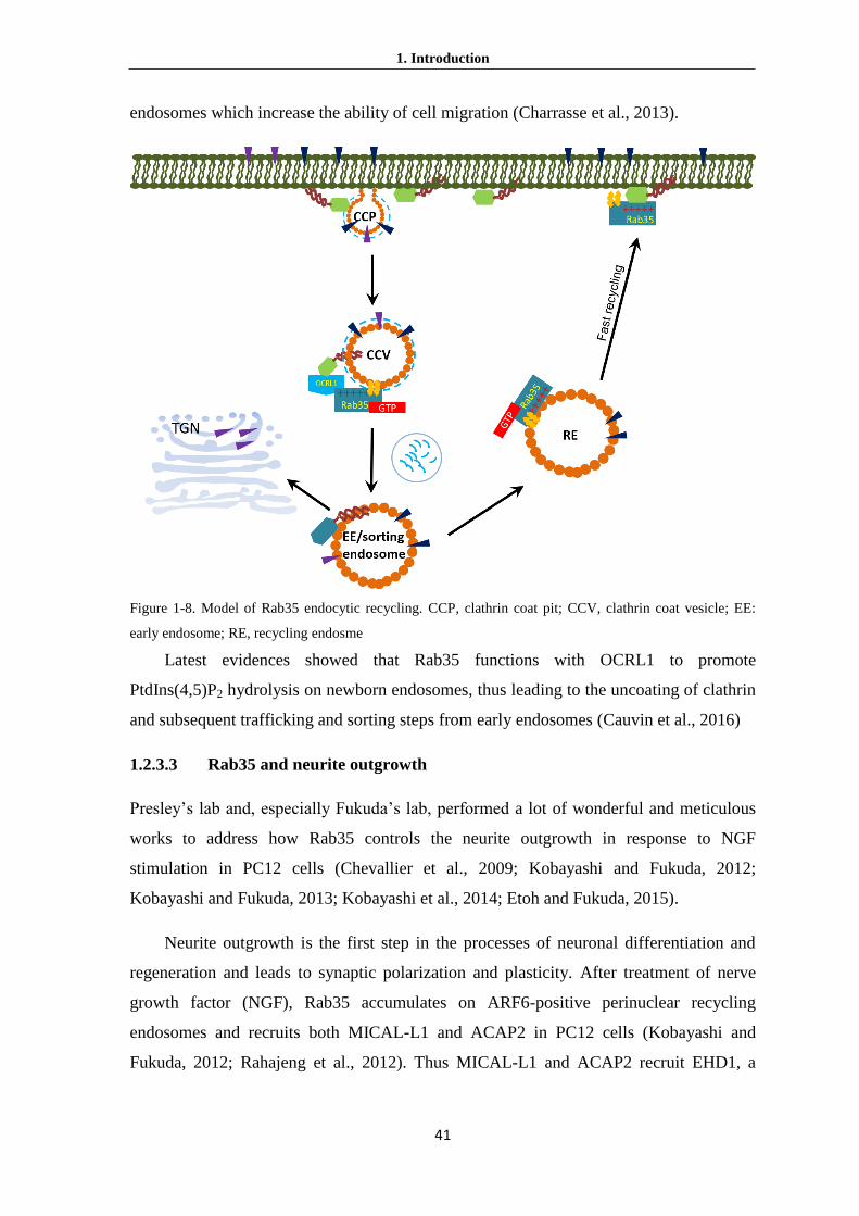
1. Introduction
41
endosomes which increase the ability of cell migration (Charrasse et al., 2013).
Figure 1-8. Model of Rab35 endocytic recycling. CCP, clathrin coat pit; CCV, clathrin coat vesicle; EE:
early endosome; RE, recycling endosme
Latest evidences showed that Rab35 functions with OCRL1 to promote
PtdIns(4,5)P2 hydrolysis on newborn endosomes, thus leading to the uncoating of clathrin
and subsequent trafficking and sorting steps from early endosomes (Cauvin et al., 2016)
1.2.3.3 Rab35 and neurite outgrowth
Presley’s lab and, especially Fukuda’s lab, performed a lot of wonderful and meticulous
works to address how Rab35 controls the neurite outgrowth in response to NGF
stimulation in PC12 cells (Chevallier et al., 2009; Kobayashi and Fukuda, 2012;
Kobayashi and Fukuda, 2013; Kobayashi et al., 2014; Etoh and Fukuda, 2015).
Neurite outgrowth is the first step in the processes of neuronal differentiation and
regeneration and leads to synaptic polarization and plasticity. After treatment of nerve
growth factor (NGF), Rab35 accumulates on ARF6-positive perinuclear recycling
endosomes and recruits both MICAL-L1 and ACAP2 in PC12 cells (Kobayashi and
Fukuda, 2012; Rahajeng et al., 2012). Thus MICAL-L1 and ACAP2 recruit EHD1, a

1. Introduction
42
protein inducing membrane fission due to its similarities to dynamin (Kobayashi and
Fukuda, 2013; Kobayashi et al., 2014; Giridharan et al., 2013). MICAL-L1 interacts
directly with EHD1 while ACAP2 recruits EHD1 indirectly through inactivation of ARF6
and thereby maintaining the scaffold factor for EHD1, PtdIns4P (Kobayashi and Fukuda,
2013). Finally, Rab35 positive endosomes are translocated to the neurite tips and facilitate
neurite outgrowth (Kobayashi et al., 2014). Previous findings provided similar cases that
Rab35 is required for EHD1 recruitment on recycling endosomes (Allaire et al., 2010).
Rab35-Q67L (constitutive active mutant) promotes neurite outgrowth in PC12 cells and
in N1E-115 cells (Langemeyer et al., 2014). Interestingly, ARF6-T27N (constitutively
active mutant) inhibits neurite outgrowth and endocytic recycling (Grant and Donaldson.,
2009; Kobayashi and Fukuda, 2012; Radhakrishna et al., 1997; Dutta and Donaldson,
2015). It is consistent with the evidences that Rab35 and ARF6 are antagonistic by
recruiting effector as the other’s GAP (Miyamoto et al., 2014).
During the neurite outgrowth, Rab35-GTP inhibits ARF6 activity by recruiting its
effector ACAP2 which works as Arf6 GAP. Meanwhile, ARF6-GTP also inhibits Rab35
activation by recruiting its effector EPI64A-C (TBC1D10A-C) as GAP for Rab35
(Hanono et al., 2006). Indeed, ARF6-GTP could interact with EPI64A-C which
constitutes a family of functional GAPs for Rab35 (Patino-Lopez et al., 2008; Chesneau
et al., 2012; Fuchs et al., 2007; Hsu et al., 2010). More data of overexpression of ARF6
Q67L,EPI64, or Rab35 S22N all lead to the formation of PtdIns(4,5)P2-rich vacuoles that
block endocytic cargo recycling and inhibit cytokinesis (Patino-Lopez et al., 2008;
Chesneau et al., 2012). Interestingly, ARF6 recruits EPI64B and thus preventing the
activation of Rab35 on clathrin-coated pits (CCP) but not on CCVs (Chesneau et al., 2012,
Montagnac et al., 2011; Cauvin et al., 2016). The precise spatial and temporal inactivation
and activation of Rab35 by its GAP EPI64B and its GEF DENND1A lead to the
production of new born clathrin-coated endosome from plasma membrane.
The crosstalk between Rab35 and Arf-6 ensures the precision of CCPs scission.
ARF6 interacts with a PtdIns(4)P 5-kinase and maintains PtdIns(4,5)P2 production level
and hence is essential for recruiting key CCP proteins. Once CCPs scission complete,
Rab35 recruits OCRL1 in order to hydrolyze PtdIns(4,5)P2 and promotes clathrin
uncoating (Cauvin et al., 2016). In conclusion, the balance between Rab35 and Arf6 is
predicted that it is either Rab35-GTP/ARF6-GDP, or Rab35-GDP/ARF6-GTP at a

1. Introduction
43
membrane, which could be important to control local phosphoinositide levels and F-actin
dynamics.
Figure 1-9. Functional model of Rab35 in a successful neurite outgrowth.
Altogether, there have been clear evidences that Rab35 always works in the
presence of proteins which are related to phosphatidylinositol metabolism, such as Arf6
and OCRL1. These data give us a strong clue that Rab35 may associate with negatively
charged phosphoinositide PtdIns(4)P, PtdIns(4,5)P2 and PtdIns(3,4,5)P3 via polybasic
HVD domain during its regulation of neurite outgrowth and endocytic recycling.
1.2.3.4 Rab35 and Drosophila bristles development
Besides its functions at cellular level, Rab35 also also play roles in Drosophila
development. For example, Drosophila bristles are mechanosensory organs found on the
cuticle and their shape and growth depends on actin bundles. Rab35 was identified in a
screen for Rab GTPases that influence bristle formation and the GTPase regulates bristle
development by recruiting fascin (Zhang et al., 2009). The direct binding of Fascin to
GTP-bound form of Rab35, i.e. the activated form of Rab35, specifically by
DENND1C/connecdenn 3, is necessary to recruit fascin to actin (Marat et al., 2012).
Fascin is an actin-crosslinking protein that assembles F-actin filaments into tightly packed
parallel bundles. It contains two actin-binding sites, one on each of its N- and C-termini
that facilitate bundling of adjacent actin filaments (Adams, 2004). Fascin has been linked
to many biological functions requiring actin regulation. Fascin also contributes to the
formation of actin structures in mammalian cells including filopodia, lamellipodia and
dendritic spines (Jayo and Parsons, 2010). Rab35 induces actin-rich protrusions in PC12

1. Introduction
44
cells and regulates lamellipodia and filopodia formation in Drosophila (Shim et al., 2010;
Chevallier et al., 2009). It is thus possible that many of fascin’s functions lie to the
downstream of Rab35.
1.2.3.5 Rab35 and diseases
Rab35 was presumed associated with tumorigenesis for depletion of Rab35 decreased cell
adhesion and increased cell migration, which is often observed in cancer cells (Allaire et
al., 2013).
Figure 1-10. The model of Rab35 works as an oncogene.
Studies from Sabatini lab and Sawyers lab provided direct evidences that Rab35
link to cancer through its regulatory roles in the phosphatidylinositol-3-kinase (PI3K)/
AKT signaling pathway (Wheeler et al., 2015). Rab35 knock down suppresses
phosphorylation-induced AKT activation in different cell types, whereas expression of
Rab35Q67L mutant induces constitutive AKT signaling. Expression of active Rab35 is
sufficient to drive PDGFR-α into Lamp2-positive endosomes even without ligands and
lead to AKT phosphorylation. Interestingly, AKT-dependent phosphorylation enhances
the ability of DENND1A GEF for Rab35 and promotes Rab35 activation in a positive
feedback fashion (Kulasekaran et al., 2015). The most important thing is that two somatic

1. Introduction
45
Rab35 mutations were found to constitutively activate PI3K/AKT signaling in cancer
patients. Both of two mutations lead to the expression of GTP-locked Rab35, suppressing
apoptosis and promoting cellular transformation in HeLa cells. Altogether, Rab35 is a
pivotal protein which plays roles in several biological processes including cell migration,
phagocytosis, immune synapse and exosome release.
1.3 Lowe syndrome and OCRL1
Lowe syndrome patients suffer primarily from congenital cataracts, neonatal hypotonia,
intellectual disability (with distinct behaviors) and renal proximal tubule dysfunction
(Fanconi syndrome). Lowe syndrome is estimated to be prevalent in the general
population of approximately 1 in 500,000. The mutation of OCRL1 (Oculocerebrorenal
syndrome of Lowe) gene which localizes at Xq26.1, causes Lowe syndrome, an x-linked
disorder (Charnas and Gahl, 1991).
OCRL1 is the major 5-phosphatase which converts PtdIns(4,5)P2 to PtdIns(4)P by
hydrolyzing the 5-phosphate (Suchy et al., 1995; Zhang et al., 1995). Cell extracts from
fibroblasts cultured from Lowe syndrome patients universally exhibit a markedly reduced
ability to dephosphorylate PtdIns(4,5)P2 (Su et al.,1999; Lin et al., 1997). In addition,
OCRL1 appears to be the major PtdIns(4,5)P2-hydrolyzing enzyme in human kidney
proximal tubule cells, and kidney cells derived from Lowe syndrome patients have
roughly double the normal cellular contingent of PtdIns(4,5)P2 (Zhang et al., 1998). Thus,
the OCRL1 phenotype correlates well with the loss of phosphatase activity.
Lowe syndrome patients almost universally have renal Fanconi syndromes,
including acidosis, amino aciduria, phosphaturia, and proteinuria (Charnas and Gahl,
1991). The defect in protein reabsorption has been suggested to be resulted from improper
function or trafficking of the cell surface receptor megalin (Lowe, 2005; Norden et al.,
2002). Megalin, a member of the LDL receptor family, is a 600-kDa transmembrane
protein that recycles at the apical domain of polarized epithelial cells (Christensen and
Birn, 2002). Megalin binds to numerous protein ligands that dissociate from the receptor
after internalization and are targeted to lysosomes for degradation. In patients with
Fanconi syndrome, ligand handling is somehow compromised, resulting in excess
secretion of filtered proteins into the urine. However, OCRL1 does not directly modulate
endocytosis or postendocytic membrane traffic and that the renal manifestations observed

1. Introduction
46
in Lowe syndrome patients are the downstream consequences of the loss of OCRL1
function (Cui et al., 2010). Almost all affected males have some degree of intellectual
disability; 10%-25% function in the low-normal or borderline range, approximately 25%
in the mild-to-moderate range, and 50%-65% in the severe-to-profound range of
intellectual disability. However, the reason of mental retardation among these patients is
still unclear.
The localization of OCRL1 is primarily at the trans-Golgi network (TGN) and is
also associated with a subset of endosomes , plasma membrane and clathrin-coated pits,
suggesting a potential function of this enzyme in membrane traffic through these
compartments (Choudhury et al., 2005; Choudhury et al., 2009; Erdmann et al., 2007;
Ungewickell et al., 2004; Dressman et al., 2000). This broad distribution is mediated by
its many interactions (Figure 2-8B). OCRL1 interacts with clathrin, AP-2 (clathrin
adaptor), 16 Rab GTPases, and the endocytic proteins APPL1 (adapter protein containing
PH domain, PTB domain and leucine zipper motif 1) or Ses1/2 (sesquipedalian 1 and 2)
via competitive manner. The broad intracellular distribution of OCRL1 indicates that the
physiological importance of phosphoinositides in endocytic trafficking (Ben El Kadhi et
al, 2011; Vicinanza et al., 2011; Erdmann et al., 2007), actin polymerization(Suchy et al.,
2002) establishment of cell polarity (Grieve et al., 2011) and cytokinesis (Dambournet et
al., 2011). Lack of OCRL1 leads to an accumulation of intracellular PtdIns(4,5)P2 and/or
PtdIns(3,4,5)P3, and therefore some pathological phenotypes are considered as the
consequence of these changes (Zhang et al.,1998)
1.3.1 The OCRL1 domains
The OCRL1 domain structure comprises an N-terminal PH (pleckstrin homology)
domain, a 5-phosphatase domain, an ASH (ASPM–SPD2–Hydin) domain and a C-
terminus RhoGAP (Rho GTPase activating) domain (Figure 2-8A) (Pirruccello and
Camilli, 2012). A loop in the PH domain contains an unusual clathrin box which mediates
the interaction of OCRL1 with clathrin heavy chain (Mao et al., 2009; Lafer, 2002). The
second clathrin box is presented in RhoGAP domain enhances the OCRL1 binding to
clathrin (Erdmann et al., 2007; Ungewickell et al., 2004). OCRL1 is directed to late-stage
endocytic clathrin-coated pits, and to endosomal/Golgi coats via these two clathrin-
binding motifs and one clathrin adaptors binding motif which exits between PH and

1. Introduction
47
inositol 5-phosphatase domains (Ungewickell et al., 2004; Choudhury et al., 2009). The
inositol 5-phosphatase domain is the core motif to hydrolyze PtdIns(4,5)P2, forming
PtdIns(4)P. The membranes targeting of OCRL1 is crucially dependent on the ASH
domain interactions with GTP-bound Rab proteins (Hyvola et al., 2006). OCRL1 interacts
with at least 16 different Rabs, for examples, Rab5 (endosomes), Rab6 (Golgi and
endosomes) and Rab35 (fast recycling route), by functional and colocalization studies
(Dambournet et al., 2011; Hyvola et al., 2006). However, the structure of OCRL1-Rab8a
complex reveals the interaction between Rab8 and OCRL1 via the Rab interaction surface
on the ASH domain and C-terminal catalytic domain helix of OCRL1 (Hou et al., 2011).
This unusual binding mode of OCRL1 for a Rab is different from the typical model that
Rab effectors recognize a hydrophobic triad in the GTP-bound Rab protein, which
incorporates residues from its switch and interswitch regions. The F668 is contained in
the Rab-binding interface of OCRL1, an IgG-like β-strand structure of the ASH domain.
Both F668A and F668V mutants impair the OCRL1 interaction with Rab8a due to the
lower hydrophobicity of alanine compared with phenylalanine (Hou et al., 2011).
The RhoGAP domain of OCRL1 interacts with Rac and GTP-bund form of Cdc42,
both of which belong to the Rho GTPases family (Erdmann et al., 2007; Faucherre et al.,
2003). The interaction with Rac may due to the localization of OCRL1 at membrane
ruffles (Faucherre et al., 2005). In addition, OCRL1 also interacts with two regulators of
5-kinases Arf1 and Arf6, and a mutation in the RhoGAP domain will disrupt their
interactions and leads to Lowe syndrome (Lichter-Konecki, 2006). Three endocytic
proteins APPL1 (adaptor protein, phosphotyrosine interacting with PH domain and
leucine zipper 1), Ses1 and Ses2 interact with the RhoGAP domain of OCRL1 via their
F&H motif, which contains a 12-13-amino-acid sequence forming an amphipathic helix
that recognizes an evolutionarily conserved surface on the RhoGAP domain of OCRL1
(Erdmann et al., 2007; Swan et al., 2010; Pirruccello et al., 2011). APPL1 is an adaptor
protein with multiple functional domains including the BAR (Bin1/amphiphysin/rvs167)
domain, PH domain and PBT (phosphotyrosine binding) domain. APPL1 interacts with
Rab5 via BAR domain and binds receptors (for example growth factor receptors) through
its PTB domain (Miaczynska et al., 2004; Mao et al., 2006; Lin 2006). APPL1 and its
homologous protein APPL2 (without F&H motif) form APPL heterodimer, which binds
AKT (a PI(3,4,5)P3 effector) and associate dynamically with endosomal membranes.

1. Introduction
48
Therefore, they are proposed to function in endosome-mediated signaling pathways
linking the cell surface to the cell nucleus (Chial et al., 2008; Miaczynska et al., 2005).
Figure 2-8. The functional domains strutures of OCRL1. (A) Scheme of OCRL1 protein domains. (B)
Combinational structure of OCRL1 derived from individual structures of its domains (orange= PH domain,
PDB code: 2KIE; green=5-phosphatase domain, PDB code: 3MTC; red = ASH domain, PDB code: 3QIS;
and blue =RhoGAP domain, PDB code: 2QV2). (Adapted from Pirruccello and Camilli, 2012). PH,
pleckstrin homology; ASH, ASPM–SPD2–Hydin domain; RhoGAP, Rho GTPase activating domain.
OCRL1 may also play a role in phagocytosis due to the interaction with APPL1 and
APLL2 heterodimer to dephosphorylate PtdIns(3,4,5)P3 (Bohdanowicz et al., 2012). As
endosomes mature and acquire PtdIns(3)P, APPL proteins will be displaced by Ses1 and
Ses2 oligomers from the F&H binding surface (Swan et al., 2010). Both Ses1 and Ses2
proteins contain a PH domain which induces their oligomerization and a C-terminal F&H
motif that mediates the interaction with OCRL1 (Swan et al., 2010; Noakes et al., 2011).
Interestingly, OCRL1 associates with endocytic vesicles at both the APPL and Ses stage
possibly via its Rab-dependent interactions. Moreover, phosphorylation of APPL1 on two
serine residues in the F&H domain forces the OCRL1 dissociation (Erdmann et al., 2007).
Therefore, the localization of OCRL1 on the endosomal membrane induces lipid turnover
with sorting events which is possibly controlled by this precise regulation.

1. Introduction
49
1.3.2 OCRL1 mutations and Lowe syndrome
Complete or partial deletion, frameshift and nonsense mutations of OCRL1 gene cause
OCRL1 losing its interactions, misfolding, or catalytic inactivity and result in Lowe
syndrome. Non-sense and frameshift mutations in PH domain were found in Dent disease
but rarely in Lowe syndrome patients (Hichri et al., 2010). Case studies showed that the
amount of missense mutations found in Lowe syndrome patients affect conserved
residues in 5-phosphatases, directly affecting OCRL1 proteins either folding, substrate
binding or catalytic activity (Tsujishita et al., 2001; Hichri et al., 2011). The majority of
mutations in the hydrophobic core of OCRL1 and several missense mutations clustered
around the active site lead to Lowe syndromes, indicating that the catalytic activity of
OCRL1 is crucial for diseases.
The missense mutations in ASH–RhoGAP motif induce the destabilization of
OCRL1 protein, which is evidenced by these mutations containing patient cell lines
express low quantity of OCRL1 protein (Hichri et al., 2010). In addition, overexpression
of OCRL1 missense mutants leads to their cytosolic localization in cells (McCrea et al.,
2008). The mutations in the F&H binding surface on the RhoGAP domain of OCRL1
disrupt the integrations with Rho and Rab GTPases which also lead to the Lowe
syndrome in human (Pirruccello et al., 2011).
Altogether, the mutations through the whole OCRL1 970 amid acids including
missense, frameshift, or nonsense were found in Lowe syndrome patients. However, how
the importance of OCRL1 interactions in its physiological function still needs to be
elucidated.

50

2. Materials and methods
51
2 Materials and methods
2.1 Materials
2.1.1 Biochemistry
Chemicals Supplier Acetic acid Merck
Acetonitrile JT Baker
Acetone JT Baker
Acrylamid/Bisacrylamide (37.5:1, 30 % w/v) Applichem
Ammonium sulfate Applichem
Ammonium persulfate (APS) Merck
Ampicillin (Amp) Serva
Bradford reagent Bio-Rad
Bromphenol blue Serva
Bovine serum albumine (BSA) Sigma
Chloroform JT Baker
Complete Mini EDTA-free protease inhibitor tablets Roche
Coomassie Brilliant Blue G250+R250 Serva
Deoxycholic acid sodium salt (DCS) Serva
Dimethylsulfoxide(DMSO) Sigma
Disodium hydrogenphosphate Roth
Dithioerythitol (DTE) Gerber
Disodium hydrogen phosphate (Na2HPO4) Merk
Dithiothreitol (DTT) Gerbu
Ethylenediaminetetraacetic adic (EDTA) Gerbu
Ethanethiol Sigma
Ethanol JT Baker
Ethidium bromide Sigma
Gernylgernyl pyrophosphate (GGPP) Sigma
Glycerol Gerbu
Glycine Roth
Guanosine diphophate (GDP) Pharma Waldhof
4-(2-Hydroxyethyl)piperazine-1-ethanesulfonic acid
(HEPES)
Roth
Hydrochloric acid JT Baker
Imidazol Gerbu
Isopropyl-β-D-thiogalactopyranoside (IPTG) Gaiberg
Kanamycin Boehringer
NBD FPP By Kui Hong Tan
Magnesium chloride Merk
Methanol Applichem
β-Mercaptoethanol Serva
Nocodazole Sigma
phenylmethylsulfonylfluoride (PMSF) Sigma
Phosphatase Inhibitor Cocktail 1 Sima
2. Materials and methods
52
Potassium dihydrogenphosphate Roth
Potassium hydroxide JT Baker
2-Propanol JT Baker
Sodium chloride(NaCl) Sigma
Sodium dodecylsulphate (SDS) Roth
Sodium dihydrogenphosphate(NaH2PO3) JT Baker
Sodium hydroxide(NaOH) JT Baker
Sodium 2-mercaptoehanesulfonate (MESNA) Sigma
N,N,N’,N’-tetramethylethylendiamin (TEMED) Roth
Trihydroxymethylaminomethane (TRIS) Roth
Tris-HCl JT Baker
Triton X-100 Serva
Tween 20 SERVA
Urea JT Baker
UltraPure Agarose Invitrogen
2.1.2 Molecular biology
Materials Supplier
100 mM dNTP mix Invitrogen
Anti-HA Abcam
Anti-OCRL Thermofisher
Anti-PRA1 Abcam
Anti-RILP Sigma-Aldrich
Anti-β-Actin Sigma-Aldrich
BigDye Applied Biosystems
Calf Intestinal Phosphatase NEB
DNA ladder Fermentas
DpnI Fermentas
DyeEx 2.0 Spin Kit QIAGEN
Dynabeads Protein A Invetrogen
FastDigest Age I Fermentas
FastDigest BamHI Fermentas
FastDigest EcoRI Fermentas
FastDigest HindIII Fermentas
FastDigest NdeI Fermentas
FastDigest SapI Fermentas
FastDigest SpeI Fermentas
FastDigest XhoI Fermentas
Gel Extraction kit QIAGEN
GelPilot loading dye, 5x QIAGEN
Low molecular weight marker Amersham Biosciences
PCR Purification kit QIAGEN
Phusion master mix FINNZYMES
Prestain Protein marker NEB
Rapid T4 ligase Fermentas
Red mix VWR
Spin Midiprep kit QIAGEN
Spin Miniprep kit QIAGEN
T4 DNA Ligase Fermentas

2. Materials and methods
53
2.1.3 Cell biology
Materials Supplier
[32P] orthophosphate PerkinElmer
Alexa Fluor® 594 goat anti-rabbit IgG Thermo
DMEM without phosphates Invitrogen
DMEM/F12 medium Sigma
DPBS Sigma
Dulbecco’s Modified Eagle’s Medium (DMEM) Sigma
Fetal bovine serum (FBS) Invitrogen
Fugene® 6 transfection reagent Roche
L-Glutamine Invitrogen
Lipofectamine 2000 Invitrogen
Lipofectamine 3000 Invitrogen
NEAA Invitrogen
Penicillin/streptomycin Invitrogen
Sodium pyruvate Invitrogen
Trypsin/EDTA Sigma
2.1.4 Other materials
Materials Supplier Amicon Ultra-4,15 (10K, 30K) Concentrator Milipore
BioSep-SEC-2000 gel filtration Phenomenex
Chitin beads NEB
Electroporation cuvettes Bio-Rad
Eppendorf tube (0.5 mL, 1.5 mL, 2.0 mL) Eppendorf
Falcon Tube (50 mL, 15 mL) Stradat
Glutathione Sepharose GE Healthcare
HiTrap Ni-NTA column Pharmacia
NAP-5 desalting column Amersham
Nitrocellulose membrane GE
Nitrocellulose paper Schleicher&Schuell
Quartz cuvette (1cm) Hellma Optik
Superdex 75/200 Gel filtration Pharmacia Biotech
Whatman FP 30/0.2, 0.4 μm cellulose filter Schleicher&Schuell
X-film Fuji
ZapCap filter Nalgene
2.1.5 Instruments Instruments supplier
1.0 mm 10-well combs BioRad
1.0 mm 15-well combs BioRad
1.0 mm cassettes for western blots Invitrogen
Argon Laser LGK 7872 ML05 (458/488/514 nm) Lasos

2. Materials and methods
54
Äkta prime system with REC112 recorder Pharmacia Biotech Biorad PE 9700 thermocycler Applied Biosystems Centrifuge 5415R Eppendorf
Centrifuge 5810R Eppendorf
Centrifuge Allegra X-22R Beckman Coulter Centrifuge Avanti J20-XP Beckman Coulter Centrifuge Optima L-70K Ultracentrifuge Beckman Coulter Electroporation device E. coli Pulser Bio-Rad Eppendorf Micromanipulators 5171 Eppendorf
Eppendorf Transinjector 5246 Eppendorf
FLA-5000 (Fluorescence image reader) Fujifilm Fluoroskan Ascent Fl type 374 Thermo BioAnalysis Gel Imaging Station BioRad
Incubator Shaker Series I26 New Brunswick
Isothermal titration calorimeter MicroCal
Leica TCS SP2 Leica Microsystems
Leica TCS SP5 Leica Microsystems
MALDI-TOF-MS Applied Biosystem
Microfluidizer Microfluidics
PCR-Cycler Eppendorf
SDS-PAGE Mini-Protean II system Bio-Rad
Shaker Infors
Thermomixer 5436 (1.5 mL) Eppendorf
Thermomixer 5436 (2 mL) Eppendorf
UV/Visible Spectrometer DU 640 Beckman Coulter
2.1.6 Buffers and growth medium
LB medium LB agar plates
5 g/L yeast extract 15 g/L Bacto agar
10 g/L Tryptone 50 mg/L Ampicillin
10 g/L NaCl
Antibiotics SDS-PAGE running buffer(10x)
125 mg/L Ampicillin 0.25 M HCl
34 mg/L Chloramphenicol 2 M Glycine
125 mg/L Ampicillin 1%(W/V) SDS
SDS-PAGE stacking gel buffer (4X) SDS-PAGE resolving gel buffer(4X) 0.5 M Tris-HCl, pH 6.8 1.5 M Tris-HCl, pH 8.8
0.4 % (w/v) SDS 0.4 % (w/v) SDS
SDS-PAGE loading buffer (2x) Coomassie staining solution 62.3 mM Tris-HCl, pH 6.8 10 % (v/v)
2 % (w/v) 2 % (w/v) 40 % (v/v)
10 % (v/v) glycerol 0.1 % (w/v) Coomassie Brilliant
5 % (v/v) β-Mercaptoethanol Blue R250
0.001 % (w/v)bromophenol blue
Distaining solution TAE buffer (1x) 10 % (v/v) acetic acid 40 mM Tris acetate, pH 8.5

2. Materials and methods
55
2 mM EDTA
20 mM Glacial acetic acid
PBS (10x) DNA loading buffer (5x) 80 g/L NaCl 30 %(w/v) Sucrose
2 g/L KCl 20 % (v/v) glycerol
14.4 g/L Na2HPO4⋅2 H2O 0.2 % (w/v) orange G
2.4 g/L KH2PO4
Bug buffer Buffer A
50 mM NaH2PO4, pH 8.0 50 mM NaH2PO4, pH 8.0
0.3 M NaCl 0.3 M NaCl
2 mM β-mercaptoethanol
Buffer B Breaking
Buffer
50 mM NaH2PO4, pH 8.0 25 mM NaH2PO4, pH 7.5
0.3 M NaCl 0.5 M NaCl
2 mM β-mercaptoethanol 2 mM MgCl2
0.5 M Imidazole 10 μM GDP
4% PFA RIPA lysis buffer
4 g paraformaldehyde 1x PBS (pH 7.4) 50mM Tris pH 7.5
150 mM NaCl
1 mM EGTA
Transfer buffer 1 mM EDTA
25 mM Tris-base 1% (W/V) IGEPAL
192 mM glycine 0.25% (W/V) Na deoxycholate
20 % methanol 2.5 mM Na pyrophosphate
Frozen medium 1 mM β-glycerophosphate
10% DMSO 0.1 mM PMSF
20% FBS TBS buffer(10x) (pH 7.6)
70% DMEM 0.2M Tris base
Annealing Buffer 1.5M NaCl
100mM K-acetate
30mM HEPES-KOH pH 7.4
2mM Mg-acetate

2. Materials and methods
56
2.2 Methods
2.2.1 Molecular biology methods
2.2.1.1 Plasmids, bacterial strains and cell lines in this study
Plasmids
Plasmid Name Insert Resistance marker
DENND1A-mCherry DENND1A Kanamycin
Dynamin2-K44A-mCherry Dynamin2-K44A Kanamycin
Dynamin2-mCherry Dynamin2 Kanamycin
paGFP-Rab35a-PBC-1M Rab35a-PBC-1M Kanamycin
paGFP-Rab35a-PBC-2M Rab35a-PBC-2M Kanamycin
paGFP-Rab35a-PBC-3M Rab35a-PBC-3M Kanamycin
paGFP-Rab35a-PBC-4M Rab35a-PBC-4M Kanamycin
paGFP-Rab35a-PBC-5M Rab35a-PBC-5M Kanamycin
paGFP-Rab5a Rab5a Kanamycin
paGFP-Rab7a Rab7a Kanamycin
pEGFP- RILP RILP Kanamycin
pEGFP-Rab11a Rab11a Kanamycin
pEGFP-Rab35a Rab35a Kanamycin
pEGFP-Rab35a-C201A Rab35a-C201A Kanamycin
pEGFP-Rab35a-PBC-1M Rab35a-PBC-1M Kanamycin
pEGFP-Rab35a-PBC-2M Rab35a-PBC-2M Kanamycin
pEGFP-Rab35a-PBC-3M Rab35a-PBC-3M Kanamycin
pEGFP-Rab35a-PBC-4M Rab35a-PBC-4M Kanamycin
pEGFP-Rab35a-PBC-5M Rab35a-PBC-5M Kanamycin
pEGFP-Rab35a-PBC-5M-C201A Rab35a-PBC-5M-C201A Kanamycin
pEGFP-Rab35aQ67L-PBC-5M Rab35aQ67L-PBC-5M Kanamycin
pEGFP-Rab35a-S150A Rab35a-S150A Kanamycin
pEGFP-Rab35a-S22N Rab35a-S22N Kanamycin
pEGFP-Rab35aS22N-PBC-5M Rab35aS22N-PBC-5M Kanamycin
pEGFP-Rab35a-S67L Rab35a-S67L Kanamycin
pEGFP-Rab5aD35Q79L-CSC Rab5aD35Q79L-CSC Kanamycin
pEGFP-Rab5aS34N Rab5aS34N Kanamycin
pEGFP-Rab5aS79L Rab5aS79L Kanamycin
pEGFP-Rab7a Rab7a Kanamycin
pEGFP-Rab7a-D24CC Rab7a-D24CC Kanamycin
pEGFP-Rab7a-D24CSC Rab7a-D24CC Kanamycin
pEGFP-Rab7a-D27CC Rab7a-D27CC Kanamycin
pEGFP-Rab7a-D27CSC Rab7a-D27CC Kanamycin
pEGFP-Rab7a-D34CC Rab7a-D3CC Kanamycin
pEGFP-Rab7a-D34CSC Rab7a-D3CC Kanamycin
pEGFP-Rab7a-V180A Rab7a-V180A Kanamycin

2. Materials and methods
57
pEGFP-Rab8a Rab8a Kanamycin
pEGFP-RILP RILP Kanamycin
pHA-Rab35 Rab35 Kanamycin
pHA-Rab7 Rab7 Kanamycin
pHA-Rab7Q67L Rab7Q67L Kanamycin
pHA-Rab7Δ24 Rab7Δ24Q67L Kanamycin
pHA-Rab7Δ24Q67L Rab7Δ24Q67L Kanamycin
pHA-Rab7Δ27 Rab7Δ27 Kanamycin
pHA-Rab7Δ27Q67L Rab7Δ27Q67L Kanamycin
pHA-Rab7Δ34 Rab7Δ34 Kanamycin
pHA-Rab7Δ34Q67L Rab7Δ34Q67L Kanamycin
PLL3.7-shRNA-OCRL-3 shRNA-OCRL-3 Ampicillin
PLL3.7-shRNA-OCRL-4 shRNA-OCRL-4 Ampicillin
PLL3.7-shRNA-PRA1 shRNA-PRA1 Ampicillin
PLL3.7-shRNA-Rab35-1 shRNA-Rab35-1 Ampicillin
PLL3.7-shRNA-Rab35-3 shRNA-Rab35-3 Ampicillin
PLL3.7-shRNA-Rab35-4 shRNA-Rab35-4 Ampicillin
PLL3.7-shRNA-RILP-1 shRNA-RILP1 Ampicillin
pMAL-MBP-RILP RILP Ampicillin
pmCherry-Rab35a Rab35a Kanamycin
pmCherry-RILP RILP Kanamycin
pRFP-C-RS DENND1A shRNA-DENND1A Ampicillin
pTWIN1-EGFP- Rab1Δ34-GGS EGFP- Rab1Δ34-GGS Ampicillin
pTWIN1-EGFP- Rab35Δ11 Rab35Δ11 Kanamycin
pTWIN1-EGFP- Rab7Δ34-GGS EGFP- Rab7Δ35-GGS Ampicillin
pTWIN1-EGFP-Rab1Δ13 EGFP-Rab1Δ13 Ampicillin
pTWIN1-EGFP-Rab5-GGS-Δ35 EGFP-Rab5-GGS-Δ35 Ampicillin
pTWIN1-EGFP-Rab5Q79L-GGS-
Δ35
EGFP-Rab5Q79L-GGS-
Δ35
Ampicillin
pTWIN1-EGFP-Rab5-Q79L-Δ14 EGFP-Rab5-Q79L-Δ14 Ampicillin
pTWIN1-EGFP-Rab5-Δ14 EGFP-Rab5-Δ14 Ampicillin
pTWIN1-EGFP-Rab7Δ15 EGFP-Rab7Δ15 Ampicillin
pTWIN1-EGFP-Rab8Δ11 Rab8Δ11 Kanamycin
pTWIN1-EGFP-Rab8Δ16 Rab8Δ16 Kanamycin
pTWIN1-Rab7Δ34 Rab7Δ34 Kanamycin
Empty pTWIN vector was obtained from New England Biolabs (Beverly, MA, USA).Empty pEGFP and
pmCherry vectors from Clontech Laboratories.
Bacterial strains
Bacterial strains Genotypes XL1 Blue (Stratagene) recA1, endA1, gyrA96, thi-1, hsdR17, supE44, relA1,
lac [F´, proAB, lacIqZΔM15, Tn10 (Tetr)]
BL21(DE3)(Stratagene) F-, ompT, lon, hsdS (rB-, mB-), dcm, gal, λ(DE3)
Top10(Thermo Fisher) F– mcrA Δ(mrr-hsdRMS-mcrBC) Φ80lacZΔM15
ΔlacX74 recA1 araD139 Δ(ara leu) 7697 galU galK
rpsL (StrR) endA1 nupG

2. Materials and methods
58
BL21(DE3) Codon Plus RIL
(Stratagene)
F-, ompT, lon, hsdS (rB-, mB-), dcm, gal, λ(DE3),
endA, Hte [argU ileY leuW CamR]
DH5α(Thermo Fisher) F– Φ80lacZΔM15 Δ(lacZYA-argF) U169 recA1
endA1 hsdR17 (rK–, mK+) phoA supE44 λ– thi-1
gyrA96 relA1
Mammalian Cell lines
Cell Line Origin Supplier
COS-7 African green monkey kidney fibroblast ATCC
HeLa Human cervical adenocarcinoma ATCC
MDCK Madin-darby canine kidney cells ATCC
SH-SY5Y Human neuroblastoma cell ATCC
2.2.1.2 Preparation of competent cells
1 L of LB medium was inoculated with 10 mL of an overnight-grown culture of the
desired E.coli strain. The culture was incubated at 37°C, 220rpm on a shaker, until the
OD600 reached about 0.5 (ca. 4 h). The culture was cooled on ice for 20 min, transferred
to sterile centrifugation vessels and centrifuged for 10 min at 4°C at 2000 g. The
supernatant was discarded.
Electro-competent cells
The bacterial cell pellet was gently resuspended in 5 mL of ice-cold sterile GYT
(0.125% (w/v) yeast extract, 0.25% (w/v) tryptone, 10% (v/v) glycerol) and centrifuged
twice as described above. Cell pellet were resuspended in 1 mL GYT, dispensed in 50 μl
aliquots, shock frozen in liquid nitrogen and stored at -80°C.
Chemical-competent cells
The pellet was gently resuspended in 20 mL of ice-cold sterile 100 mM CaCl2 solution
and incubated on ice for 30 min. The cells were centrifuged at 2000 g for 5 min at 4°C
and were resuspended in 1 - 5 mL of TFBII buffer (10 mM MOPS, pH 7.0, 75 mM CaCl2,
10 mM NaCl, 15 % glycerol). Aliquots of 100 μl were shock frozen in liquid nitrogen and
stored frozen at -80°C.

2. Materials and methods
59
2.2.1.3 Preparative PCR
The appropriate 5’- and 3’-primers were designed and synthesized by Eurofins Genomics.
The PCR reaction mixture was prepared in steps shown in in a PCR tube. The mixture
was incubated in Biorad PE 9700 thermocycler (Applied Biosystems) using PCR program
as below.
High fidelity PCR for sub-cloning
Step Content Volume (μL)
1 Forward primer(con.10μM) 2
2 Revers primer(con.10μM) 2
3 Template DNA(10~50ng) 1
4 Phusion master mix 25
5 ddH2O 20
Total 50
PCR program
Step Temp(°C) Time
1 98 30 s
2: 36 cycles 98 8 s
3: 36 cycles 50~65 20 s
4: 36 cycles 72 15 s-30 s/kb
5 72 7 min
6 8 Hold
2.2.1.4 Purification of PCR products by agarose gel electrophoresis
Depending on the size of the DNA fragment, the agarose concentration was between 0.8
and 2 % (w/v).
1. Dissolve the required amount of agarose by heating in TAE buffer.
2. Add Red safe to a final concentration of 0.5 μg/mL
3. Pour the solution into the gel casting equipment and allowed to polymerize.
4. Mix the samples with 5X DNA loading buffer and load into the wells. A 1 kb DNA
ladder (Fermentas) was used as a molecular weight standard.
5. Run the gels horizontally at 10 V/cm immersed in TAE-buffer until fragment
separation was complete.

2. Materials and methods
60
6. Excise the bands of interest and extract the DNA from the gel using a gel extraction
kit from Qiagen according to the instructions of the manufacturer.We also used PCR
kit to retake the PCR product (Qiagen DNA or Omiga clean up kit).
2.2.1.5 Construction of vectors
Restriction enzyme digestion
Both vector and purified PCR product were digested by the appropriate restriction
enzymes. Digestions of DNA fragments were performed as recommended by the
manufacturer at 37°C for 3 h. The reaction was stopped by addition of DNA loading
buffer. Fragments produced by restriction enzyme digestion were purified using agarose
gel electrophoresis. An example is shown below.
Step Contents Vector volume (μL) PCR product Volume (μL)
1 Buffer(10x) 3 4
2 DNA 2 (total 1μg) 30
3 BamHI 1(20U/μl) 1
4 XhoI 1 (20U/μl) 1
5 ddH2O 23 4
Total 30 40
Dephosphorylation of 5’-Phosphorylated DNA fragments
An essential step during cloning and before the ligation of two DNA fragments is the
dephosphorylation of 5’-end of destination vector to prevent its self-ligation. Self-ligation
of cut vector will prevent fragment insertion and produce empty vectors. For
dephosphorylation, 0.5 U/μg DNA of alkaline phosphatase (calf intestinal phosphatase
CIP) is added directly on the restriction mix and reaction tubes are incubated for 1 h at
37°C. Stop the reaction for 15min at 70°C.
Ligation
For ligation 1-10 ƒmol of linear plasmid DNA was mixed with a 3 fold molar excess of
insert fragment. Ligation was performed in ligation buffer in a volume of 20 μl, using 5
units of T4 DNA ligase for 1 h at 22°C (Rapid ligation, for short DNA fragment) or 1
units of T4 DNA ligase for 12~14 h at 16°C (long DNA fragment). An example is shown
below.

2. Materials and methods
61
Step Contents Volume(μL)
1 Linear vector 3
2 Insert 8
3 Rapid ligation buffer 5x or Ligation buffer 10x 4 or 2
4 Rapid T4 ligase 5 U/μL or T4 ligase 1 U/μL 1
5 ddH2O 4 or 6
Total 20
2.2.1.6 Chemical transformation
1. Add the ligation mixture containing approximately 1 ng of the desired plasmid DNA
to 50~100 μL chemical competent cells. The mixture was incubated on ice for 25~ 30
min without shaking.
2. Heat the cells at 42°C for 45 s and immediately cooled on ice for 2 min.
3. Spread on LB plate directly (Ampicillin resistance) or add 600 μL of LB medium into
the cells EP tube.
4. Incubate at 37°C for 50 min for Kanamycin resistance on a shaker.
5. Spread 100 μL cells on agar plates supplemented with the corresponding antibiotics.
6. Invert plates and incubate overnight (12-16 h) at 37°C.
2.2.1.7 Colony PCR screen
1. Pick a single bacterial colony growing on LB agar plate with the corresponding
antibiotics using a sterile yellow tip.
2. Inoculate the colony into 4 mL of LB medium with the corresponding antibiotics.
3. After 3 hours growth, withdraw 1 μL culture in a PCR test tube using sterile tips.
4. Perform PCR as shown below.
Cloning PCR for vector identification
Step Content Volume (μL)
1 Forward primer(con.10μM) 1
2 Revers primer(con.10μM) 1
3 LB medium mixture of cell 1
4 Red mix 10
5 ddH2O 7
Total 20

2. Materials and methods
62
PCR program
Step Temp(°C) Time
1 94 4min
2: 36 cycles 94 45 s
3: 36 cycles 50~65 45 s
4: 36 cycles 72 1 min/kb
5 72 10 min
6 8 Hold
5. Analyze the PCR products by agarose gel electrophoresis. The one showing a band of
PCR product at the expected molecular weight suggests a successful cloning.
Agarose concentration in % (w/v) Size of DNA fragments
2.5 <100bp
2.0 0.1 - 1.0 kb
1.8 0.2 – 2.0 kb
1.5 0.3 – 3.0 kb
1.2 0.5 – 5.0 kb
1.0 0.5 – 7.0 kb
0.8 0.8 – 12.0 kb
0.5 30.0 kb
2.2.1.8 Preparation of plasmid DNA
Plasmid DNA was prepared using the plasmid mini-prep kit (Qiagen) as follows:
1. Pick a single bacterial colony to seed 4 mL of LB medium containing the appropriate
antibiotics.
2. Grow the culture overnight (12~14 h) at 37°C,220rpm on shaker.
3. Harvest cells by centrifugation (16,000 g, 1 min). Discard the supernatant.
4. Resuspend the cell pellet in 250 μL Solution I with RNase A by vortex.
5. Add 250 μL Solution II and mix gently until the solution becomes clear.
6. Add 350 μL Solution III into clear lysate and mix gently to precipitate chromosomal
DNA, lipids, and proteins.
7. Remove the precipitates by centrifugation (14000rpm, 10 min).

2. Materials and methods
63
8. Load the supernatant on a silica spin column. Centrifuge the column for 1 min at
14,000 rpm.
9. Wash the column with 500 μL HB buffer, 700 μL wash buffer (with ethanol) twice by
centrifugation (14,000 rpm, 1 min).
10. Centrifuge the column for 1.5 min at 14,000 rpm to get rid of ethanol.
11. Elute the plasmid DNA with 70 μL EB buffer by centrifugation (14,000 rpm, 1.5 min).
2.2.1.9 DNA sequencing
For DNA sequencing, the cloned plasmid was subjected to PCR using the BigDyeDesoxy
terminator cycle sequencing kit. The sequencing reactions were prepared and PCR
program was set as shown below.
Sequencing PCR
Step Content Volume (μL)
1 Bigdye 2
2 5x Sequencing buffer 2
3 primer(con.10μM) 1
4 Plasmid(200~500ng/μL) 1
5 ddH2O 4
Total 10
PCR program
Step Temp(°C) Time
1 94 4min
2: 25 cycles 94 20 s
3: 25 cycles 50 30 s
4: 25 cycles 60 2.5 min
5 8 hold
Ethanol precipitation
1. Dilute the PCR product with 10 μL H2O.
2. Add 2 μL 3M sodium acetate (pH 5.5) to the sample tube.
3. Add 2 μL 0.1M EDTA to the sample tube.
4. Add 50μL 100% ethanol to precipitate DNA and incubate for 15 min at room
temperature.
5. Centrifuge the sample at 14,000 rpm for 20 min at room temperature. Remove the
supernatant.
6. Wash the precipitate once with 200 μL 70 % ethanol. Centrifuge the sample at 14000
rpm for 15 min at room temperature.

2. Materials and methods
64
7. Dry the sample under vacuum.
8. The sample was analyzed by the in house sequencing facility.
2.2.1.10 Transformation by electroporation
1. Add ca. 1 ng of plasmid DNA in a volume of 0.5 μL to 50 μL electro-competent cell
suspension in a chilled electroporation cuvette (0.2 μm path length).
2. Tap the cuvette to exclude air bubbles.
3. Apply a high voltage pulse using an E.coli Pulser from Biorad (conditions: 25 μF, 200
Ω, 1800V).
4. Immediately add 600 μL of LB medium to the cuvette after pulsing.
5. Grow the culture at 37°C, 220 rpm for 50 min on a shaker.
6. Spread 100 μL cell suspensions on agar plates containing the appropriate antibiotics.
7. Invert plates and incubate overnight (12-16 h) at 37°C.
2.2.1.11 Short hairpin (shRNA) construct generation
The principle of shRNA construct design is shown in Figure 2-1. Firstly, we need search
for target sequences located throughout the mRNA. The consensus sequence should
correspond to: AAGN18TT. A 5’ guanine is required due to the constraints of the U6
promoter. Normally we test 4 targets for each gene of interest.
Figure 2-1. The principle of shRNA constructs design.

2. Materials and methods
65
1. Oligo Design: A multiple cloning site immediately following the U6 promoter. An
HpaI site leaves a blunt end prior to the –1 position in the promoter. The oligo design
must incorporate a 5’ T in order to reconstitute the –1 nucleotide of U6. An XhoI site
cuts downstream of the U6 start site.
2. Oligo format: Sense oligo, 5’T-(GN18)-(TTCAAGAGA)-(81NC)-TTTTTTC;
antisense oligo, compliment of sense but with additional nucleotides at 5’ end to
generate XhoI overhang. The loop sequence (TTCAAGAGA) is based upon previous
report (Rummelkamp et al., 2002). Order oligos through Eurofins with 5’ phosphates
and PAGE purified.
3. Annealing oligos by below steps
Step Contents Volume (μL)
1 Sense oligo(100pmol/μL) 1
2 Antisense oligo(100pmol/μL) 1
3 Annealing Buffer 48
Total 50
4. Annealing by PCR program below
Step Temperature(°C) Time (min)
1 95 4
2 70 10
3 4 (decrease temperature slowly:0.1°C/min) 180
4 4 10
5. Digest PLL 3.7 vector 1 μg with XhoI and HpaI for 3h at 37°C then treats with CIP
for 1h at 37°C.
6. Ligate linearized product and annealed oligos at 60fmol of each component in a final
concentration of 10μL.
7. Transformat the ligation product into XL1 competent cells.
8. Spread the transformation product on LB plate with Ampicillin antibiotic.
9. Invert plates and incubate overnight (12-16 h) at 37°C.

2. Materials and methods
66
2.2.2 Protein expression, purification and modification methods
2.2.2.1 Expression and purification of GFPRab1, 5, 7, 35-thioester proteins
Expression of GFP-Rab5a(Q79L)Δ14 in E. coli
1. Transform plasmid pTWIN-His-EGFP-Rab5(Q79L)Δ14 into BL21(DE3) cells and
select on a LB agar plate containing 100 mg/L Carbenicillin (similar function as
Ampicillin).
2. The second day, pick a single colony from the agar plate using a sterile yellow tip and
inoculate it into 100 mL of LB-medium containing 125 mg/L ampicillin.
3. Incubate at 37°C at 170 rpm on a rotatory shaker overnight.
4. Inoculate this pre-culture into 5 L of LB-medium containing 125 mg/L ampicillin.
5. Grow cells in five 5 L flasks at 37°C on a shaker (170 rpm) until the absorbance at
600 nm (OD600) reached 0.5-0.7 (ca. 4 h). Take a sample of 40μL from the cell
suspension; mix it with 20 μL 4×SDS sample buffer, boile at 99°C for 5 min as a
control for step 9.
6. Put the flasks in the cold room to reduce the temperature of the culture to 20°C.
7. For induction of the protein expression, add IPTG to the culture to a final
concentration of 0.2 mM.
8. Incubate at 20°C on a shaker (170 rpm) overnight (18 h).
9. Before harvesting cells, take a sample of 30 μL from the cell suspension, mix it with
15 μL 4×SDS sample buffer, boile at 99°C for 5 min and run a denaturing SDS-PAGE
to estimate the protein expression level.
10. Harvest cells by centrifugation in the centrifuge Avanti J20-XP (Beckman Coulter) at
6000 rpm, 4°C for 15 min.
11. Discard the supernatant carefully and wash the cells with PBS by centrifugation in the
centrifuge Eppendorf 5804 (Eppendorf) at 5000 rpm, 4°C for 10 min. The weight of
the cells about 35g.
Here the cells can be frozen in liquid nitrogen and stored in -80°C.
Lysis of E. coli cells
12. Resuspend the cell pellet in 25 mL ice-cold Breaking Buffer (30 mM NaH2PO4, pH
7.5, 0.3 M NaCl, 1 mM MgCl2) freshly supplemented with 1 mM PMSF, 10 μM GDP.

2. Materials and methods
67
Note: The PMSF and GDP should be added freshly. Don’t add any reducing
substances.
13. Pass the cell suspensions through the chilled pressure chamber of Microfluidizer 3
times at 40,000 psi.
14. Add new portion of PMSF to 0.5 mM and TritonX-100 to 1% (v/v). Note: The
TritonX-100 should be added after breaking the cells.
15. Centrifuge the cell lysate in the ultracentrifuge Optima L-70K (Beckman Coulter) at
35,000 rpm, 4°C for 30 min.
16. Filter the supernatant through a 0.2 μm ZapCap filter (Nalgene).
Purification of EGFP-Rab5a(Q79L)Δ14 α-thioester
17. Load the filtrate with a flow rate of 2 mL/min on a 5 mL Hi-Trap Ni-NTA column
that has been equilibrated with the Buffer A (50 mM NaH2PO4, pH 8.0, 0.3 M NaCl, 2
mM β-mercaptoethanol).
18. Wash the column with Buffer A with a flow rate of 5 mL/min until the absorbance
reached the baseline.
19. Wash the column with 2% Buffer B (50 mM NaH2PO4, pH 8.0, 0.3 M NaCl, 2 mM β-
mercaptoethanol, 0.5 M imidazole) with a flow rate of 5 mL/min until the absorbance
reached the baseline.
20. Elute the column with a gradient of 2-100% Buffer B with a flow rate of 5 mL/min for
250 mL. Collect 5 mL/fraction.
21. Run SDS-PAGE and identify the fractions of interest.
22. Wash the column with 2 mM NaN3.
23. Collect the fractions of interest and dialyze the sample against buffer for (25 mM
Tris-HCl, pH 8.0, 100 mM NaCl, 4 mM sodium citrate, 2 mM β-mercaptoethanol).
24. Concentrate the protein to 5ml and Run gel filtration on the Superdex-75 column
using buffer (25mM NaH2PO4, pH7.5, 30 mM NaCl, 1 mM MgCl2, 20 μM GDP).
Note: Prepare fresh solution, filter the buffer through a 0.2 μm membrane filter
(Whatman) and degas on a vacuum-membrane pump (ILM/VAC GmbH) by stirring
for 0.5 h at room temperature (23°C).
25. Concentrate the protein and add sodium 2-mercaptoehanesulfonate (MESNA) powder
into the protein and a concentration of 0.5 M. Incubate overnight on a rotating wheel
at 23°C.

2. Materials and methods
68
26. Run SDS-PAGE and test the EGFP-Rab5a(Q79L)Δ14 α-thioester efficiency.
27. Concentrate the sample to at least 10 mg/mL, separate them in aliquots and froze them
flashily in liquid nitrogen. Store the protein in -80°C.
2.2.2.2 Universal C-terminal protein labeling with oxyamine
ligation
1. Dilute EGFP-Rab5a(Q79L)Δ14 α-thioester solution with 5-fold volume of Buffer A .
Note: the MESNA concentration is not lower 50mM but not higher than 100mM.
2. Load EGFP-Rab5a (Q79L)Δ14 α-thioester onto a Ni-NTA column equilibrated with
Buffer A containing 100 mM MESNA. Collect flow-through.
3. Wash column with 2-5% Buffer B containing 100 mM MESNA. Collect and pool
flow-through and concentrate protein.
4. Run a gel filtration on a Superdex 60 column using Elution Buffer (30 mM NaH2PO4,
pH 7.5, 50 mM NaCl, 100 mM MESNA). Note: Prepare fresh solution, filter buffer
through a 0.2 µm filter and degas for 30 min at room temperature.
5. Identify and collect fractions of interest by SDS-PAGE. Concentrate protein and snap-
freeze in liquid nitrogen. Store protein at -80 °C.
6. Incubate 200 μl protein-thioester (5-25 mg/mL) with 100 μl bis(oxyamine) (1 M stock
solution in reaction buffer, final 333 mM) in Reaction Buffer (30 mM NaH2PO4, pH
7.5, 50 mM NaCl) on ice overnight. The reaction product EGFP-Rab5a(Q79L)Δ14-
ONH2 is monitored by ESI-MS.
7. Dialyze protein twice against 1 L Dialysis Buffer (30 mM NaH2PO4 pH 7.5, 50 mM
NaCl, 2 mM DTE) at 4 °C.
8. Incubate 50 μM EGFP-Rab5a(Q79L)Δ14-ONH2 with 0.5 mM Keto-PEG1 for 20 h or
overnight on ice in the presence of 100 mM Aniline in Incubation Buffer (30 mM
NaH2PO4, pH 7.0, 50 mM NaCl, 2 mM DTE).
9. Remove unreacted small molecules by passing the reaction mixture over a NAP-5
desalting column pre-equilibrated with 30 mM sodium phosphate (pH 7.5, 50 mM
NaCl, 1 mM MgCl2, 10 μM GDP, and 5 mM DTE).
10. Add MESNA powder to final concentration 500 mM into the protein solution and
incubate on ice for 2 h.
11. The reaction is monitored by ESI-MS check the two StBu groups were totally
removed.

2. Materials and methods
69
12. Perform gel filtration to purify the ligated protein in prenylation buffer (50 mM
HEPES, pH 7.2, 50 mM NaCl, 5 mM DTE, 2 mM MgCl2, 10 μM GDP).
13. Concentrate the protein fractions to about 1-5 mg/mL for further prenylation and
microinjection studies.
2.2.2.3 In vitro prenylation
4 μM PEGylated Rab or wild type or mutants, 6 μM REP, 6 μM RabGGTase were
incubated in prenylation buffer (50 mM HEPES, pH 7.2, 50 mM NaCl, 5 mM DTE, 2
mM MgCl2, 10 μM GDP) at 25 °C, and 50 μM NBD-FPP was added to initiate the
reaction. At defined time intervals, 10 μl samples were withdrawn and quenched by
addition of 10 μl of 2 × SDS-PAGE sample buffer. For an end-point assay, 6 μM
PEGylated Rab or wild type proteins, 10 μM REP, and 6 μM RabGGTase were mixed
with 40 μM NBD-FPP in prenylation buffer and incubated for 1.5 h at 25 °C, followed by
quenching with 2 × SDSPAGE sample buffer. The samples were boiled at 95 °C for 3
min and were loaded onto 15% SDS-PAGE. The fluorescence bands corresponding to the
NBD-farnesylated protein were visualized in the gel using a Fluorescent Image Reader
FLA-5000 (Fuji, excitation laser: 473 nm, cut-off filter: 510 nm) followed by staining
with Coomassie Blue and scanning. The fluorescence intensities of the bands were
quantitatively analyzed using AIDA densitometry software. The traces were fitted to a
single exponential equation.
2.2.3 Analytical methods
2.2.3.1 SDS-PAGE
Preparation of SDS-Polyacrylamide Gel Electrophoresis (PAGE) gels
1. Pour the solution into a Biorad Multi-casting apparatus (9 gels). Add 70% ethanol on
the top of the resolving gel. Incubate at room temperature until the resolving gel was
polymerized (ca. 30 min).
2. Remove the ethanol and add the stacking gel solution prepared as below. Insert combs
immediately.

2. Materials and methods
70
3. Incubate at room temperature until the stacking gel was polymerized (ca. 20 min). The
gels can be stored in 4°C in a wet paper packet which is contained in a closed plastic
bag.
Sequence 1 2 3 4 5 6
Type of gel
(%)
Acrylamide/
bisacrylamide
(29:1, 30 %)
Mili-Q
H2O
Seperating
gel buffer
(4X)
Stacking
gel buffer
(4X)
10 %
APS
TEMED
Resolving gel (60 mL)
10 % 20 ml 25ml 15ml - 300 μl 30 μl
15% 30 ml 15ml 15ml - 300 μl 30 μl
Stacking gel (30 mL)
5 % 5 mL 17.5 mL - 7.5 mL 240 μl 30 μl
SDS-Gel electrophoresis
4. Prepare protein samples by adding an half amount of SDS-PAGE sample buffer (4x)
and heat them for 5 min at 99°C.
5. Run gels electrophoresis at 100V until the bromphenol blue front entered the buffer
solution.
6. Visualize fluorescent proteins by exposing the unstained SDS-PAGE gel to UV light
or fluorescent image reader.
7. Stain the proteins by heating the gels in the Coomassie staining solution in a
microwave. Incubate on a shaker at room temperature for 10 min.
8. Distain the protein by heating the gels in the distaining solution in a microwave.
Incubate on a shaker at room temperature.
2.2.3.2 MALDI-TOF-mass spectrometry
Matrix Assisted Laser Desorbtion Ionization - Time Of Flight (MALDI-TOF) spectra
were recorded on a Voyager-DE Pro Biospectrometry workstation from Applied
Biosystems (Weiterstadt, Germany). Protein samples were desalted using small GF spin
columns (DyeEx 2.0 Spin Kit, Qiagen) and mixed with an equal volume of matrice
(saturated sinapinic acid solution in 0.3 % TFA/acetonitrile (2:1 v/v)). The mixture was
quickly spotted on a MALDI sample plate, air-dried. Spectra were measured with the
following instrument settings: acceleration voltage = 25 kV, grid voltage = 93 %,
extraction delay time = 750 ns and guide wire = 0.3 %. The laser intensity was manually
adjusted during the measurements in order to obtain optimal signal to noise ratios.
Calibrations were carried out using a protein mixture of defined molecular mass (Sigma).

2. Materials and methods
71
Spectra recording and data evaluation was performed using the supplied Voyager
software package. The accuracy of the method for proteins within the molecular weight
range of 20-30 kDa is ca. ± 20 Da.
Electronspray ionization mass spectrometry (ESI-MS)
Liquid Chromatography-Electrospray Ionization-Mass Spectrometry (LC-ESIMS)
analysis was performed on an Agilent 1100 series chromatography system (Hewlett
Packard) equipped with an LCQ ESI mass spectrometer (Finnigan) using Jupiter C4
columns (5 μm, 15 x 0.46 cm, 300 Å pore-size) from Phenomenex (Aschaffenburg) for
proteins and 125/21 NUCLEODUR 5 μm C18 columns for peptides. For LC separation a
gradient of buffer B (0.1 % formic acid in acetonitrile) in buffer A (0.1 % formic acid in
water) with a constant flow-rate of 1 mL/min was applied. Mass spectra data evaluation
and deconvolution was performed using the Xcalibur software package. The accuracy of
the method for proteins within the molecular weight range of 20 kDa is ca. ± 1-2 Da and
70 kDa is ca. ± 15 Da.
2.2.4 Microcsopy
Laser Scanning Confocal Microscopy (LSCM)
Confocal images of live and fixed cells were obtained with Leica SP2 and Leica SP5
confocal laser-scanning microscope. TagBFP and pa-GFP was excited with 405 nm,
EGFP and mCitrine with the 488 line of a multiline Argon laser. mCherry and dsRed was
excited with the 561 nm line of a DPSS laser. Excitation light was focused into the
sample by a 60x/1.2 NA oil objective or a 40x/0.9 NA air objective using either the
DM405/488/561/633 or the DM458/515 dichroic mirror.
Detection of fluorescence was done by an acousto-optic tunable filter (AOTF) and
SIM scanner. Blue fluorescence (Alexa 405 or TagBFP) was detected in a channel with
bandwidth 425-478 nm and through a SDM490 emission beam splitter. Green and Citrine
FPs were collected between 498-551 nm and through a SDM 510 emission beam splitter
or SDM 560 if sequential imaging with the 561 nm laser was used. mCherry fluorescence
was detected by collecting photons within the range of 575-675 nm. Live cell imaging
was performed in an incubation chamber adjusted to 37°C, while fixed cells experiments
were performed at RT (~ 25°C). The process of LSCM is shown in Figure 2-2 as below.

2. Materials and methods
72
Figure 2-2. The process of Laser Scanning Confocal Microscopy (LSCM).
Fluorescence Loss After Photoactivation (FLAP)
FLAP experiments were carried out at 37°C on a Leica SP5 confocal microscope in
imaging medium. Transiently transfected 15- 24 hrs post-transfection, cells were allowed
to equilibrate in the incubation chamber on the microscope and imaged with laser setting
adjusted to minimize photobleaching (typically 488 and 561 lasers used at 10%). The
fluorescence was collected via a 63x/1.4 NA oil objective, with a custom routine that
followed through the three stps of FRAP/FLAP analysis: (1) Pre-bleach imaging (2
images with 488 and 561 lasers only). (2) Bleaching/Photoactivation (2 times with the
405 laser only at 80%) in a pre-defined region of interest (ROI). (3) Post-bleach imaging
(150 frames at 10 sec interval using the 488 and 561 lasers only). After standard image
processing, mean intensities of photoactivatable PA-GFPRab35, PA-GFPRab5, PA-
GFPRab7 in the photoactivated ROI yielded fluorescence loss curves, which were
normalized to mKate2-Giantin to account for changes in the structure and intensity in the
ROI resulting from the dynamic nature of live cells. Another ROI processed and
normalized in the same manner was chosen at the cell periphery to investigate vesicular
transport form the recycling endosome to the plasma membrane. For measuring half time

2. Materials and methods
73
of plasma membrane gain or recycling endosome loss of Rab35, Rab5, Rab7 fluorescence
in the selected ROIs, normalized fluorescence decay cures were averaged and average
curve was then fitted to a single exponential function in Igor Pro. I = I0+ A exp (-t/τ)
Where (I) is the intensity, (A) exponential constant, (t) is time; (τ) is the exponential time-
constant.
2.2.5 Mammalian Cell Culture and related works
2.2.5.1 Subculture of Mammalian Cells
Cells were maintained in Dulbecco’s Modified Eagle’s Medium (DMEM) supplemented
with 10% fetal bovine serum (FBS), 1% L-glutamine and 1% nonessential amino acids
(NEAA) and 1% sodium pyruvate grown at 37°C in a 90% humidified incubator with 5%
CO2. For starving, phenolred free Earle's Balanced Salt Solution (EBSS) was used.
Exponentially growing cells become confluent and reach a density where all available
substrate of the culture dish is occupied. When this happens, cell growth is usually
inhibited by contact inhibition and cells need to be subcultured to new dishes with much
less density to allow for further cell growth. When cultured cells reached 80-90%
confluency, cells were washed once with PBS, covered with 1 ml trypsin (10cm petra
dish) for 4 min at 37°C (MDCK cells required longer time ~ 12 min). Detached cells were
washed with 3 ml complete growth media to inactivate any remaining trypsin and cell
suspension transferred to a falcon tube and pipetted gently up and down to separate cells
to a single cell suspension. If cell number and/or viability were to be determined, 10 μL
of cell suspension in a total of 3ml media were used in Cell counter (BioRad) cell
viability analyzer and the desired number of cells required for seeding into different cell
culture dishes was calculated. For maintenance of cells lines, cells were subcultured at a
ratio of 1:10 into new culture dishes.
2.2.5.2 DNA transfection
For confocal microscopy, 2.0×10⁵ cells were cultured on 35 mm glass bottom dishes
(MatTek, Ashland, MA) for 20 hours prior to transfection. Transient plasmid expression
was achieved by overnight transfection with X-treme GENE HP DNA transfection
reagent (06366244001, Roche). For nocodazole treatment, cells were treated with 20 μM
nocodazole for 1.5 h followed by confocal fluorescence microscopy.

2. Materials and methods
74
2.2.5.3 siRNA transfection
siRNAs were transfected into Hela cells in 35mm MaTTeK dish with lipofectamine 2000
(Invitrogen) based on its protocol.
1. Add a cell suspension containing 4 x 10⁵ cells in 2ml of growth medium with serum
but without antibiotics.
2. Dilute 3 μg of siRNA into 100 μl of Opti-MEM.
3. Dilute 3 μl of Lipofectamine™ 2000 into 100 μl Opti-MEM Medium and incubate for
5 min at room temperature. Note: Once the Lipofectamine™ 2000 is diluted, combine
it with the siRNA within 30 min. Long time incubation may result in decreased
activity. This dilution can be prepared in bulk for multiple wells.
4. Combine the diluted siRNA from step 2 with the diluted Lipofectamine™ 2000 from
step 3. Incubate at room temperature for 20 min to allow DNA-Lipofectamine™2000
complexes to form.
5. Add the DNA-Lipofectamine™ 2000 complexes from step 4 (200 μl) directly to each
well containing cells and mix gently by rocking the plate back and forth.
6. Change with fresh medium after incubation for 4 h at 37°C in a CO2 incubator.
7. The lipofectimin™ 2000 usage can be calculated as below ratio.
Culture
Vessel 96-well 48-well 24-well 12-well 6-well 35-mm 60-mm 100-mm
Surface
Area
(cm2)
0.3 0.7 2 4 10 10 20 60
2.2.5.4 Stable cell line generation
In order to establish stable cell lines that constitutively express GFP-tagged proteins or
small-hairpin RNAs. We used the protocol is described as below.
1. Designed and construct your interested gene vectors such as mCherry/GFP-tagged or
shRNA plasmids. The above vector encodes kanamycin for selection in bacteria and
neomycin (G418) for selection in mammalian cells. For shRNA knock-down
construct, we choose pLL3.7 vector from Addgene #11795 as backbone, which
encodes ampicillin for selection in bacteria and puromycin for selection in
mammalian cells.

2. Materials and methods
75
2. Prepare low-passage cells (not higher 20 passages).
3. Transfect the mCherry/GFP-tagged or shRNA constructs into HeLa or MDCK cells.
3μg of plasmid DNA and 4.5μL Xtreme GENE HP DNA transfection reagent in
300μL OptiMEM per 6x10⁵ HeLa cells in to 100mm dishes.
4. The following day, replace the standard media with media containing 600μg/mL
G418 or 1μg/mL puromycin for shRNA knockdown cells.
5. Carefully change the media in the dish every one or two days, taking care not to
pipette directly onto the cells.
6. Over time (2-3 weeks) this will select the cells that have stably recombinant with the
GFP plasmid or shRNA into their genomic DNA.
7. Using sterile yellow tips to pick up the single colony of cells with 10X objective lens
under microscope, and culture the cells in 6-well-plate.
8. When the wells are confluent, rinse with PBS and trypsinize with trypsin-EDTA. Split
into one well of a 6-well plate (for passaging) and one well of your choice for imaging
or western blot in order to screen the colonies and decide which are worth keeping
and which should be discarded.
9. Using 300μg/mL G418 or 0.5μg/mL puromycin to maintenance the identified cells.
10. Freeze the cells as passage 1 and further passage from the 6-well plate.
2.2.5.5 Western Blotting
Western blotting is one of immuno-blotting which can detect proteins that were separated
electrophoretically are transferred to a nitrocellulose or PVDF membrane to enable
immunological protein detection with antibodies.
Firstly loading the interest protein into SDS-gel and run electrophoresis. After
electrophoresis blots were assembled in semidry bot modules or wet transferring
according to the manufacturer’s manual. Nitrocellulose membranes were activated for 5
min in methanol, and then left to equilibrate in 1 x transfer buffer together with the gel
and two pieces of (Whatmann) filter paper. The blot sandwich was assembled in the
following sequence from bottom-up: filter paper, NC membrane, gel and the second filter
paper. For a mini-gel semidry transfer was done at 40V for 20 min or wet transferring at
100V for 40min. Following transfer, NC membrane was transferred to incubation box for
the remaining binding sites of the membrane blocked with 5% milk or BSA for 1 hour on
a shaker at RT. Membranes were then incubated with primary antibodies diluted in 1 ml 3%

2. Materials and methods
76
milk 1x TBST solution. NC membrane was sealed in a plastic bag after discarding air
bubbles and incubated overnight at 4 °C. Next day, blots were transferred again to
incubation boxes, washed 3 times, 5 min each with 0.1% TBST. Blots were then
incubated with the appropriate secondary antibodies diluted in 10 ml TBS for 1 hour at
room temperature. Finally, blots were washed 5 times, 5 min each with 0.1% TBST
solution and detected with HRP X-film system.
2.2.5.6 Immunoprecipitation (pull down)
HeLa cells were transfected with HA-Rab7(Q67L), HA-Rab7(Q67L)Δ24-CSC, HA-
Rab7(Q67L)Δ27-CSC, or HA-Rab7(Q67L)Δ34-CSC. After 24 hours, the cells were lysed
by adding 100μl IP buffer (50 mM Tris, pH7.5, 15mM EDTA, 100mM NaCl, 0.1% TX-
100, 1 mM DTT, protease inhibitor cocktail). 30 μl amylose beads were washed with 5 ml
binding buffer (50mM HEPES, pH7.5, 30mM NaCl, 0.2mM β-ME) and were
subsequently incubated with 60 μl (10mg/ml) recombinant MBP-RILP or MBP proteins
for 1 hour with mild shaking. The beads were washed again with 5 ml binding buffer and
5 ml PBS. The beads were blocked with 10% BSA for 1 hour on ice and were rinsed with
PBS. MBP-RILP or MBP beads were incubated with HA-Rab7Q67L, HA-
Rab7Q67LΔ24-CSC, HARab7Q67LΔ27- CSC, or HA-Rab7Q67LΔ34-CSC cell lysates
for 3 h on ice with mild shaking. The beads were washed using cold washing buffer
(20mM Tris, pH7.9, 100mM NaCl, 0.1% NP40) for three times. The bound proteins were
eluted from the beads using 20 μl hot denaturing SDS-sample buffer (with 6M urea). The
samples were subject to 15% SDS-PAGE and western-blotting. The process of pull down
assay is desribled in Figure 2-3.
Figure 2-3. The process of pull down assay.

2. Materials and methods
77
2.2.5.7 Cell fixation and immunofluorescence (IF)
Cells were fixed with 4% paraformaldehyde in PBS for 30 min at RT, rinsed three times
(3X) with TBS and permeabilized with 0.5% Triton-X100 for 30 min at room temperature.
For saponin extraction, 0.5% saponin was added into the medium at this step. After
blocking with 5% FBS/0.1M Glycine/PBS for 1h, cells were incubated with anti-RILP
(Sigma-Aldrich) antibody (1μg/ml) in 5% FBS/PBS for 1h, and subsequently treated with
the secondary antibody (Alexa Fluor® 594 goat anti-rabbit IgG(H+L)) at a 1/500 dilution
for 1h.
2.2.5.8 Microinjection of PEGylated Rab proteins
For each experiment, about 50 HeLa cells were injected with the purified PEGylated Rab
proteins at concentrations of 5-7 mg/ml in prenylation buffer (50 mM HEPES, pH 7.2, 50
mM NaCl, 5 mM DTE, 2 mM MgCl2). The microinjection was performed with
Eppendorf Transinjector 5246 and Eppendorf micromanipulators 5171.
2.2.5.9 Determination of the GTP/GDP ratio
The analysis was performed essentially as described before (Colombo et al., 1998). HeLa
cells were transfected with HA-Rab7, HA-Rab7Δ24-CSC, HARab7Δ27-CSC, or HA-
Rab7Δ34-CSC (and the corresponding Q67L mutants). After 16 h, the cells were washed
with PBS for 3 times and were changed to the phosphate-free Dulbecco's modified Eagles
medium containing 0.5mCi/ml [32
P] orthophosphate (PerkinElmer Life Sciences). After 4
h incubation, the cells were washed 3 times with cold PBS and then lysed in lysis buffer
(50mM HEPES, pH 7.4, 1% Triton X-100, 100 mM NaCl, 5 mM MgCl2,mM PMSF, 0.1
mM GTP, 1 mM ATP and 10mM Na-phosphate). Nuclei and debris were removed by
centrifugation at 15 000 g for 2 min, and the supernatant was subjected to
immunoprecipitation with 1μl Anti-HA tag antibody (Abcam) on 20μl Dynabeads protein
ASepharose CL-4B ( Invitrogen) for 10 min at room temperature. The beads were then
washed three times with wash buffer (50 mM HEPES, pH 7.4, 500 mM NaCl, 5 mM
MgCl2, 1% TritonX-100) and then three times with PBS containing 0.02% Tween-20.
The bound nucleotides were then eluted in 20μl elution buffer (2 mM EDTA, 2 mM DTT,
0.2% SDS, 5 mM GDP and 5 mM GTP) for 10 min at 70°C. A volume of 5 μl of the
samples were spotted onto 0.1 mm PEI -cellulose TLC plates (Merck) which were

2. Materials and methods
78
developed for 40 min in developing buffer (1.2M ammonium formate, 0.8M HCl). The
plates were dried and placed in autoradiography cassettes containing intensifying screens.
For visualization of the [32
P]-labeled GTP and GDP, Fuji films were exposed at -80°C for
4 days. The processes of GTP/GDP ratio is described in Figure 2-4.
Figure 2-4. The principle of GTP/GDP ratio assay.

3. Aims of this work
79
3 Aims of this work
As the key regulator of vesicular transport, Rab has to be localized at correct and specific
membrane where it plays function properly. However, we still don’t know what
determine the spatial cycling and localization of Rabs. This work has used a combination
of chemical biology, biochemistry, cell biology and biophysical methods to address the
mechanism of Rab membrane targeting.
To further understand the significance of the C-terminal hypervariable domain
(HVD) in Rab membrane targeting and prenylation, new methods are needed to
manipulate the structure of Rab C terminus. In this work, we semisynthesized a series of
Rab probes, where the C-terminal hypervariable region and prenylatable cysteine residues
are replaced by PEG linkers and thiols, respectively. In order to further understand the
mechanism of Rab prenylation, we want to know whether there is nothing required for
Rab C-terminal sequence and to what extent the Rab prenylation machinery could tolerate
the replacement in the Rab C-terminal sequence. We also use chemical-biological tools to
probe whether the HVD of Rabs contains a general targeting signal for Rab localization.
By combining this semisynthetic strategy with cell imaging, we aim to elucidate the role
of the HVD, GEFs, GAPs and effectors in the subcellular Rab targeting.
To determine the cycling of Rab35 and to reveal its dynamics in the cell, we employ
fluorescence localization after photoactivation (FLAP) and fluorescence recovery after
photobleaching (FRAP) techniques to reach our aims. In this work, we knock down
PRA1, a potential GDF of Rab35, to exame its function in the cycling of Rab35.
Moreover, we aim to find out the detailed function of Rab35 GEF DENND1A for Rab35
membrane targeting. An effector of Rab35, OCRL1 is also considered a factor to affect
the Rab35 membrane targeting.
Taken together, this study aim to discovery the contributions of the different
regulators including GEFs, GDIs, GAPs, GDF, effectors and PtdInsPs for Rab membrane
targeting and trafficking in cell.

80
4.1 The role of the HVD in Rabs membrane targeting
81
4 Results and discussion
4.1 The role of the hypervariable C-terminal domain in Rab
GTPases membrane targeting
Intracellular membrane trafficking requires correct and specific localization of Rab
GTPases. The hypervariable C-terminal domain (HVD) of Rabs is posttranslationally
modified by isoprenyl moieties that enable membrane association. A model asserting
HVD directed targeting has been contested in previous studies, but the role of the Rab
HVD and the mechanism of Rab membrane targeting remain elusive (Chavrier et al.,
1991). In addition, the geranylgeranyl moieties(C-20 isoprenyl) are attached onto one or
two (most cases) cysteines at their C terminal end by Rab geranylgeranyl transferase
(RabGGTase). To fulfill the function of prenylation, RabGGTase along with REP, Rab
forms of a ternary catalytic Rab:REP:RabGGTase complex (Pylypenko et al., 2003; Rak
et al., 2004; Wu et al., 2009). Structural and biochemical analysis showed that key
bindings of the ternary complex involve the GTPase domain and the C-terminal
interacting motif (CIM) of Rab with REP and RabGGTase with REP (Guo et al., 2008;
Wu et al., 2009). However, it remains elusive how the single Rab prenylation machinery
can process the whole Rab family with diverse C termini.
To elucidate the function of the HVD, we will substitute this region with an unnatural
polyethylenglycol (PEG) linker by using oxime ligation. In this section, we will discuss
the HVD functions in Rab prenylation and membrane targeting. Through localization
studies and functional analyses of semisynthetic PEGylated Rab1, Rab5, Rab7, and
Rab35 proteins, we demonstrate that the role of the HVD of Rabs in membrane targeting
is more complex than previously understood. Our findings suggest that Rab membrane
targeting is dictated by a complex mechanism involving GEFs, GAPs, effectors, and C-
terminal interaction with membranes to varying extents, and possibly other binding
partners.
4.1.1 Construction of PEGylated Rab Proteins
In this study, we chose small GTPase Rab7 as the model protein to test the functions of
Rab HVD in prenylation and membrane targeting. Firstly, we substitute the peptide which
downstream of the essential CIM motif by a PEG linker (synthesized by Long Yi).

4.1 The role of the HVD in Rabs membrane targeting
82
According to previous report, the length of the linker is critical for Rab prenylation (Wu
et al., 2009) that we generate a C-terminal-length wild type like PEGylated Rab7 protein
conjugate. We constructed pTWIN-Rab7 vectors which contain gene sequences of
truncated the amino acid residues after the CIM motif of Rab7 protein. pTWIN vectors
are designed for protein purification or for the isolation of proteins with an N-terminal
cysteine and/or a C-terminal thioester (Evans et al., 1999). Expression of the fusion
proteins is under the control of the T7 promotor/Lac operator and is regulated by IPTG
due to the presence of a Lac repressor gene (see the protocol in appendices). The cloning
and expression strategies are shown in figure 4-1-1.
pTWIN-Rab7Δ15-intein fusion vector is transformed into BL21 (DE3) E.Coli cells
to express the target truncated Rab7 protein. As shown in figure 4-1-1, the expressed Rab-
intein-CBD fusion protein was purified from crude cell lysate using chitin agarose.
Rab7Δ15-thioester protein was generated by intein-mediated protein splicing using 0.5M
MESNA (2-mercaptoethanesulfonate) as the thiol reagent. Subsequently, Rab7Δ15-
thioester was treated with bis(oxyamine) at pH 7.5 on ice for 4 h to achieve oxyamine-
modified protein, Rab7Δ15-ONH2, which is competent for oxime ligation with a
chemical linker containing a ketone moiety (Figure 4-1-2A) .
Figure 4-1-1. Production of Rab7 thioesters using IMPACT™ system.
(A) The construct map of Rab7d15-intein fusion gene. (B) The route of Rab7d15-thioester production.
MESNA: Sodium 2-mercaptoethanesulfonate; CBD: chitin binding domain.
Figure 4-1-2 shows a chemical PEG linker (PEG1) that containing a ketone group

4.1 The role of the HVD in Rabs membrane targeting
83
for oxime ligation and two prenylatable cysteines. The PEG-1 linker contains two
cysteines protected with StBu groups, which can be removed by the treatment with
reducing reagents after protein ligation. The final product Rab7-PEG-1 was obtained after
incubation of PEG1-linker with Rab7Δ15-ONH2 on ice overnight. We confirmed the
reaction results by SDS-PAGE and ESI-MASS data (Figure 4-1-2 and table 4-1-1).
Furthermore, we asked whether the prenylation reaction is affected by a single
cysteine containing at the Rab C-terminal end. We designed and synthesized a chemical
linker PEG-2 that contains a (PEG)8 linker with a ketone group for protein ligation and
only one prenylatable thiol linked by disulfide bond, which can be readily cleaved by
using 5 mM DTE during protein ligation. With the similar chemical reaction processes
like Rab7-PEG-CC, we got the Rab7-PEG-SH and monitored by LC-MS (Figure 4-1-2 C
and D).
Figure 4-1-2. Routines of Rab7-PEG-CC and Rab7-PEG-SH production.
(A) Production of the Rab7d15-ONH2. (B) Production of Rab7-PEG-CC and Rab7-PEG-SH which are
different with wild type Rab7 at the C-terminal end.
We have known the C-terminal hypervariable domain (HVD) of about 40 amino
acids shows high level of sequence divergence among Rab family members. To

4.1 The role of the HVD in Rabs membrane targeting
84
investigate the role of HVD, a long PEGylated chemical linker PEG-3 was synthesized
(by Yi Long) to replace the whole HVD of 34 amino acids in Rab7 (Figure 4-1-3A). As
the CIM motif is necessary for the Rab:REP interaction, it was kept in the linker. Figure
4-1-3B shows the Rab7 C-terminal sequence that was replaced by the PEG3 fragment.
The CIM (IKL) was replaced by VKL.
Figure 4-1-3. The replacement strategy of the Rab7 HVD sequence.
(A)The construct without the HVD of Rab7d34 and it is ligated with PEG3 linker. (B) A modified strategy
to get the Rab7-(GGS)n-PEG-CC. The amino acid sequence before the CIM domain is replaced by GGS
repeats.
However, the oxime ligation between Rab7Δ34-ONH2 and PEG3 cannot be achieved
based on the establishment ligation conditions. We then sought to use a non-specific
sequence linker GGS repeats ((GGS)n) (Pedersen, et al., 1998) to replace the C-terminal
hypervariable region before the CIM. As shown in Figure 4-1-3C, a (GGS)n-VKL-PEG1-
CC was used to replace the C-terminal 34 amino acids of Rab7. The successful
semisynthesis of the protein conjugate Rab7Δ34-(GGS)n-VKL-PEG1-CC is indicated by
LC-MS and SDS-PAGE.

4.1 The role of the HVD in Rabs membrane targeting
85
Figure 4-1-4. The modifications of Ra7Δ15-PEG-CC. (A) SDS-PAGE analysis of Rab7Δ15- thioester (lane
1), Rab7Δ15-ONH2 (lane 2) and Rab7-PEG-CC (lane 3). (B) ESI-MS spectra of Rab7Δ15-thioester (red)
and its reaction product with bis(oxyamine), Rab7Δ15-ONH2 (black). (C) ESI-MS spectrum of the oxime
ligation product Rab7Δ15-PEG-CC(Mcal = 22613 Da). (D) ESI-MS spectrum of the oxime ligation product
Rab7Δ15-PEG-SH (Mcal=22504Da). (E) ESI-MS spectrum of the oxime ligation product Rab7Δ15-
(GGS)n-PEG-CC( Mcal=21772Da).(These data from Long Yi).
4.1.2 PEGylated Rab proteins undergo prenylation in vitro
Based on EPL method, we have successfully got the PEGylated Rab proteins including
Rab7-PEG-CC, Rab7-PEG-SH and Rab7-(GGS)n-PEG-CC. These PEGylated Rab7
proteins were firstly tested by prenylation assay using NBD-FPP. NBD-FPP is a
fluorescent analog of the lipid substrate GGPP, with an NBD group coupling to a C-15
farnesyl moiety, mimicking the length of a C-20 native lipid substrate (Wu et al., 2006).
As shown in Figure4-1-4, Rab7-PEG-CC displays nearly identical prenylation efficiency
to that of the wild type Rab7 protein. The prenylation kinetics at room temperature further
supported that Rab7-PEG-CC can be prenylated as efficient as that of wide-type Rab7.
Next, we further examined whether intact cysteine residues are indispensable for
prenylation. Interestingly, Rab7-PEG-SH undergoes prenylation with an observed rate
that is ca. 2-fold higher than that of wild-type protein (Figure 4-1-5B). These findings
indicate that Rabs do not require specific sequences in the HVD for prenylation, except
for the CIM and an SH group as an isoprenyl acceptor. Rab7Δ34-GGS-PEG-CC
displayed a little faster prenylation efficiency than that of the wild type Rab7. To confirm
the native posttranslational modification, prenylation of PEGylated Rab7 proteins were
performed using GGPP as the substrate. Prenylation of Rab7-PEG-CC using GGPP also

4.1 The role of the HVD in Rabs membrane targeting
86
led to the doubly geranylgeranylated protein as shown by ESI-MS. The thiol group in
Rab7-PEG-SH can be prenylated as efficient as cysteines at the Rab C-terminus,
indicating flexibility within RabGGTase prenylation machinery. Prenylation of Rab7Δ34-
(GGS)n-VKL-PEG1-CC using GGPP led to the double-geranylgeranylated protein as
indicated by ESI-MS result (Figure 4.14C,D and E). These results demonstrate that the
whole HVD of the Rab, except for the CIM, does not encode the prenylation specificity.
The amino acids downstream of the CIM can be replaced by a nonpeptidic PEG linker
containing cysteines or thiol groups without perturbing Rab prenylation in vitro.
In vitro prenylation analysis of the PEGylated Rab proteins probes provides insights
into the mechanism of Rab protein prenylation. This study together with previous reports
suggests that the specificity of the Rab prenylation machinery is conferred by three
binding interfaces, involving the GTPase domain, the CIM, REP, and RabGGTase
(Pylypenko et al., 2003; Pak et al., 2004). The HVD of Rabs, with the exception of the
CIM, does not contribute specificity to the assembly of the ternary protein complex. Once
the ternary protein complex is established, the Rab C terminus is concentrated within the
microenvironment of the complex, thus enhancing the probability of C-terminal cysteines
reaching the active site of RabGGTase. As a consequence, the protein substrate
specificity of RabGGTase does not need to be encoded in the Rab C terminus, in contrast
to that of CaaX protein prenyltransferases.
Figure 4-1-5. In vitro prenylation of PEGylated Rab proteins.

4.1 The role of the HVD in Rabs membrane targeting
87
(A) Incorporation of fluorescent isoprenoid (NBD-farnesyl) into Rab7 protein and conjugates. The reaction
mixtures were resolved by SDS-PAGE (upper panel: coomassie blue staining; lower panel: fluorescent laser
scan of NBD). (B) Reaction kinetics of prenylation from the SDS/PAGE assay using the fluorescent analog
of GGPP, NBD-FPP. The solid lines show single exponential fits for Rab7 wild type (kobs = 0.021/min),
Rab7-PEG-CC (kobs = 0.031/min), Rab7-(GGS)n-PEG-CC (kobs = 0.018/min), and Rab7-PEG-SH (kobs =
0.050/min).(C) ESI-MS spectra of the Rab7-PEG-CC after prenylation with GGPP (Mcal = 22780 Da). (D)
ESI-MS spectra of the Rab7-PEG-SH after prenylation with GGPP (Mcal=22780 Da). (E) ESI-MS spectra of
the Rab7-(GGS)n-PEG-CC after prenylation with GGPP(Mcal=22316). Reaction condition: 6 µM PEGylated
Rab7, 10 µM REP, and 6 µM RabGGTase were mixed with 100 µM GGPP in prenylation buffer and
incubated for 1 h at 37 °C.
The model is consistent with the fact that RabGGTase has essentially no sequence
preference for the context of the prenylatable cysteines, and the C-terminal sequences
occurring in Rab GTPases include CC, CXC, CCX, CCXX, CCXXX, and CXXX. Hence,
any cysteine- or thiol-containing fragment that can be properly presented to the active site
of RabGGTase is able to undergo prenylation. This property allows RabGGTase to
process all members of the family of more than 60 Rab proteins with hypervariable C-
terminal sequences, a feature that is uncommon in protein-modifying enzymes. This
unique property of the Rab prenylation machinery also enables it to process Rab proteins
with unnatural C-terminal moieties.
4.1.3 GFP-tagged PEGlytated Rab proteins for studying membrane
targeting
4.1.3.1 Preparation of PEGylated Rab Proteins
To gain insights into the role of the Rab C terminus in membrane targeting, we use
GFP (green fluorescent protein) tag as a fluorescent indicator, which does not interfere
with Rab localization and function (Ali et al., 2004). We cloned the truncated Rab1, Rab5,
Rab7 genes into the modified pTWIN-eGFP vectors with 10xHis tag at the C-terminal of
sequence. The fusion proteins were purified by Ni-NTA chromatography. We prepared
PEGylated EGFP-Rab1, EGFPRab5, EGFP-Rab7, and EGFP-Rab35 proteins by ligating
Keto-PEG-CC to the truncated EGFP-Rab and EGFP-Rab-(GGS)n proteins, as done for
the PEGylated Rab7 conjugates. All of semi-synthetic proteins were characterized by LC-
MS spectra (Table 4-1), SDS-PAGE and in vitro prenylation assay.

4.1 The role of the HVD in Rabs membrane targeting
88
Table 4-1. LCMS results of Rab protein probes used in this study. (Calculated mass)
Protein (P) P-COSR P-ONH2 P-PEG1 P-PEG1-
CC
P-PEG1-
C(GG)C(GG)
P-PEG2-SH P-PEG2-
S(GG)
Rab7Δ15
21974 (21972)
21926 (21922)
22792 (22789)
22617 (22613)
23162 (23157)
22508 (22500)
22777 (22772)
GFP-Rab1 Δ13 49732
(49752)
49683
(49702)
50553
(50569)
50378
(50381)
50923
(50937)
50271
(50280)
50540
(50538)
GFP-Rab5 Δ14 50001 (50019)
49954 (49969)
n.d n.d n.d 50545 (50547)
50814 (50809)
GFP-Rab5-Q79L-Δ14 49988
(49989)
49943
(49945)
50798
(50821)
50623
(50621)
51168
(51216)
50520
(50522)
50789
(50790)
GFP-Rab7 Δ15 50889 (50905)
50836 (50855)
51707 (51722)
51532 (51536)
52077 (52096)
51459 (51433)
51818 (51816)
GFP-Rab8 Δ13 49659
(49652)
49602
(49602)
50168
(50180)
50437
(50435)
GFP-Rab35 Δ11 50527 (50542)
50481 (50492)
51346 (51359)
51170 (51183)
51175 (51163)
Rab7Δ34-(GGS)n-GG
21135
(21131)
21086
(21081)
21954
(21948)
21779
(21772)
22321
(22316)
GFP-Rab1Δ31-GGS)n-GG
49122 (49134)
49078 (49084)
49938 (49951)
49775 (49975)
50320 (50319)
GFP-Rab5Δ35-Q79L-
(GGS)n-GG
49186
(49191)
49150
(49140)
50007
(50007)
49834
(49839)
50379
(50384)
GFP-Rab7Δ34-(GGS)n -GG
49025 (49037)
48977 (48987)
49841 (49855)
49678 (49673)
50223 (50224)
n.d stands for not done.
4.1.3.2 Prenylation of PEGylated GFP-Rab Proteins in vitro
These protein conjugates were subjected to in vitro prenylation. As shown in Figure 4-1-6,
Figure 4-1-6. In vitro prenylation of PEGylated GFP-Rab proteins.
Prenylation reaction through incorporation of NBD-FPP to Rab proteins was resolved by SDS-PAGE
(upper: Coomassie blue staining; lower: fluorescent scan for NBD fluorescence). M, molecular marker. (A)
Prenylation of GFP-Rab-PEG-CC proteins. Left lanes of Rab1, Rab7 and Rab35 indicate EGFP-Rab-PEG-
CC proteins alone as negative controls, while the left lane of Rab5Q79L indicates a mixture of prenylation

4.1 The role of the HVD in Rabs membrane targeting
89
enzymes with GFP-Rab5Q79L-PEG-CC in the absence of NBD-FPP. Right lanes indicate prenylation
reaction. Wide type Rab7 was used as a positive control for prenylation reaction. (B) Prenylation of GFP-
Rab-(GGS)n-PEGCC proteins. Left lanes of Rab1, Rab5Q79L and Rab7 indate GFP-Rab-(GGS)n-PEG-CC
proteins alone as negative controls. Right lanes indication prenylation reaction. Wide type Rab7 was used
as a positive control for prenylation reaction. (C) Prenylation of GFP-Rab- PEG-SH proteins in the presence
of and the absence of lipid substrate NBD-FPP.
all PEGylated Rab proteins undergo prenylation in vitro. Furthermore, we also performed
the prenylation with GGPP, the natural substrates of RabGGTase. The prenylation results
were monitored by ESI-MS which is shown in Figure 4-1-7. These results imply that Rab
probes were efficiently prenylated in vitro, and again indicate that the RabGGTase
machinery is indeed very flexible for protein prenylation.
Figure 4-1-7. In vitro prenylation of PEGylated GFP-Rab proteins. ESI-MS spectra of the Rab1-PEG-CC
(A), Rab5Q79L-PEG-CC (B), Rab7-PEG-CC (C), Rab1-(GGS)nPEG-CC (D), Rab5Q79L-(GGS)n-PEG-
CC (E), Rab7-(GGS)nPEG-CC (F), and Rab35-PEG (G) before and after prenylation with GGPP. Reaction
condition: 6 μM PEGylated Rab7, 10 μM REP, and 6 μM RabGGTase were mixed with 100 μM GGPP in
prenylation buffer and incubated for 1 h at 37 °C.
4.1.3.3 The C-terminal sequence downstream from the CIM in HVD is not
crucial for Rab 1/5/7 membrane targeting

4.1 The role of the HVD in Rabs membrane targeting
90
I have summarized current models of membrane targeting in section 2.1.7. However, the
function of HVD in membrane targeting is still not clear. Here, we use PEGylated Rab1,
Rab5, Rab7, and Rab35 as probes to investigate this classical but still not yet resolved
question.
Firstly, we microinjected the GFP-tagged PEGylated Rab proteins into cells without
expressing respective mCherry-Rab wild types as a control. After one night incubation
after microinjection, the localization of Rab protein conjugates was confirmed by
confocal microscopy. We checked the membrane targeting of GFP-Rab1-PEG-CC, GFP-
Rab5-Q79L-PEG-CC and GFP-Rab7-PEG-CC proteins. The results showed that the
PEGylated Rab proteins with a dicysteine motif such as Rab1-PEG-CC, Rab7-PEG-CC
and Rab35-PEG-CC localize to intracellular structures suggesting that PEGylated Rab
proteins are prenylated in the cells (Figure 4-1-8).
Figure 4-1-8. Subcellular localization of EGFP-Rab1, 7, 35-PEG-CC and EGFP-Rab-(GGS)n-PEG-CC
proteins in non-transfected HeLa cells. Scale bar, 10 μm.
To investigate the localizations of these PEGylated Rab proteins in cells, we
microinjected them into HeLa or MDCK cells expressing Cherry-Rab wild-type

4.1 The role of the HVD in Rabs membrane targeting
91
proteins as a marker for their subcellular localizations. We foundthat both Rab1-PEG-CC
and Rab7-PEG-CC colocalize with the wild-type Rab1 and Rab7 on the Golgi apparatus
and late endosomes/lysosomes, respectively (Figure 4-1-9 A and C). To demonstrate that
the PEGylated Rab proteins are functional, we prepared Rab5Q79L mutants that are
deficient in GTPase activity and therefore are constitutively in the active GTP-bound state.
Constitutively active Rab5 promotes the formation of enlarged endosomes, in line with its
function in homotypic endosome fusion (Stenmark et al., 1994). Likewise, microinjection
of Rab5Q79L-PEG-CC into the cell leads to the formation of enlarged vesicles (Figure 4-
1-9 D).
Moreover, Rab5Q79L-PEG-CC colocalized with the Rab5Q79L mutant on the
enlarged early endosomes when it was introduced into cells expressing Cherry-
Rab5Q79L (Figure 4-1-9 B). These results suggest that PEGylated Rab1-PEG-CC, Rab5-
PEG-CC and Rab7-PEG-CC proteins are correctly targeted and functional, indicating the
downstream sequence behind of CIM in hypervariable domain is not essential for their
membrane targeting and function, in these Rab proteins.
Figure 4-1-9. Subcellular localization of GFP-Rab-PEG-CC.
(A) GFP-Rab1-PEG-CCcolocalize with mCherry-Rab1 wild-type protein at the Golgi apparatus. (B) GFP-
Rab5Q79L-PEG-CC colocalize with mCherry-Rab5Q79L on enlarged endosomes. (C) GFP-Rab7-PEG-CC
colocalize with mCherry-Rab7 wild-type protein. (D) GFP-Rab5Q79L-PEG-CC induces formation of
enlarged vesicles. (E) Pearson’s colocalization coefficient (PCC) analysis of chimeric Rab proteins with
wild-type Rab proteins. Scale bar, 10μm.
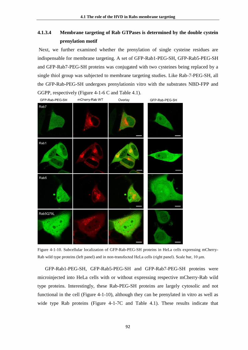
4.1 The role of the HVD in Rabs membrane targeting
92
4.1.3.4 Membrane targeting of Rab GTPases is determined by the double cystein
prenylation motif
Next, we further examined whether the prenylation of single cysteine residues are
indispensable for membrane targeting. A set of GFP-Rab1-PEG-SH, GFP-Rab5-PEG-SH
and GFP-Rab7-PEG-SH proteins was conjugated with two cysteines being replaced by a
single thiol group was subjected to membrane targeting studies. Like Rab-7-PEG-SH, all
the GFP-Rab-PEG-SH undergoes prenylationin vitro with the substrates NBD-FPP and
GGPP, respectively (Figure 4-1-6 C and Table 4.1).
Figure 4-1-10. Subcellular localization of GFP-Rab-PEG-SH proteins in HeLa cells expressing mCherry-
Rab wild type proteins (left panel) and in non-transfected HeLa cells (right panel). Scale bar, 10 μm.
GFP-Rab1-PEG-SH, GFP-Rab5-PEG-SH and GFP-Rab7-PEG-SH proteins were
microinjected into HeLa cells with or without expressing respective mCherry-Rab wild
type proteins. Interestingly, these Rab-PEG-SH proteins are largely cytosolic and not
functional in the cell (Figure 4-1-10), although they can be prenylated in vitro as well as
wide type Rab proteins (Figure 4-1-7C and Table 4.1). These results indicate that

4.1 The role of the HVD in Rabs membrane targeting
93
digeranylgeranylation rather than the sequence of the prenylation motif is essential for the
correct subcellular localization and function of Rab proteins, in keeping with a previous
report on Rab5 membrane localization (Gomes et al., 2003). One exception that C-
terminal prenylation is not required is Rab13, which associates with vesicles via protein-
protein interactions in its GDP-bound form (Ioannou et al., 2016).
At the same time, we also constructed vectors which express single cysteine Rab-C
protein via site mutation method. Rab-C is largely cytosolic, similar to that of Rab-PEG-
SH proteins in cells, albeit Rab1-C still localizes on the Golgi body partially. These
results indicate that the prenylation of single cysteine residue is not sufficient for correct
membrane targeting in digeranylgeranylation Rabs which have double cysteines end such
like -CC, -CXC, CCX, -CCXX, -CCXXX.
4.1.3.5 The localization of Rab-(GGS)n-PEG-CC proteins in cells
Since the C-terminal sequence downstream from the CIM is not essential for
membrane targeting of Rab1, 5, 7 proteins in cells, we asked if the amino acid sequence
before CIM is crucial for membrane association. To answer this question, we
microinjected the Rab-(GGS)n-PEG-CC proteins into HeLa cells expressing mCherry-
tagged wide-type Rab protein.
As depicted in Figure 4-1-11, both Rab1-(GGS)n-PEG-CC and Rab5Q79L-(GGS)n-
PEG-CC colocalize with the wild-type Rab1 and Rab5Q79L on the Golgi apparatus and
enlarged early endosomes, respectively. These results showed that the replacement of the
whole HVD of Rab1 and Rab5 does not interfere with the correct membrane targeting and
function of Rab proteins in cells. Together with the data shown in section 4.1.3.2, we can
conclude that the HVDs of Rab1 and Rab5 are not essential for their membrane targeting
and function.

4.1 The role of the HVD in Rabs membrane targeting
94
Figure 4-1-11. Subcellular localization of GFP-Rab-(GGS)n-PEG-CC proteins in Hela cells.(A) GFP-Rab1-
(GGS)n-PEG-CC colocalize with mCherry-Rab1 wild-type protein at the Golgi apparatus.(B) GFP-
Rab5Q79L-(GGS)n-PEG-CC colocalize with mCherry-Rab5Q79L on enlarged endosomes.(C) GFP-Rab7-
(GGS)n-PEG-CC does not colocalize with mCherry-Rab7 wild-type protein.(D) single microinjection of
GFP-Rab5Q79L-(GGS)n-PEG-CC induce formation of enlarged vesicles.(E) Pearson’s colocalization
coefficient analysis of chimeric Rab proteins with wild-type Rab1,5,7 proteins, respectively. Scale bars, 10
μm.
To examine the effects of carboxyl methylation on Rab protein localization, two
kinds of prenylation motifs, CXC and CC were included. After prenylation, Rab proteins
with C-terminal CXC undergo carboxyl methylation by isoprenylcysteine carboxy-
methyltransferase (Icmt) at the ER membrane, whereas those having CC and CCXX
sequences in their C termini are not carboxymethylated (Bergo et al., 2001; Li and Stahl,
1993; Smeland et al., 1994). To further confirm our ideas, we generate a set of constructs
to substitute the hypervariable domain of Rab1 and Rab5 with a (GGS)n-VKL-(GGS)n-
CC fragment, Rab1Δ31-(GGS)n-CC, Rab5Δ35-(GGS)n-CC and Rab5Q79LΔ35-(GGS)n-
CC for functional study. Rab1Δ31-(GGS)n-CSC, and Rab5Δ35-(GGS)n-CSC and
Rab5Q79LΔ35-(GGS)n-CSC constructs were also designed and generated. In these
chimeric proteins, only the CIM and the prenylation motif were retained while the rest of
the HVD was replaced with flexible GGS repeats. Analogous scenarios were observed in
the cells expressing Rab1 and Rab5 chimeric proteins, correct membrane localization and
induction of the enlarged early endosomes by Rab5Q79LΔ35-CSC or Rab5Q79LΔ35-CC

4.1 The role of the HVD in Rabs membrane targeting
95
(Figure 4-1-12).
Figure 4-1-12. Subcellular localization of Rab proteins with C-terminal substitutesions. The HVD of Rab1,
Rab5 is completely substituted with a (GGS)n-VKL-(GGS)n-CSC or (GGS)n-VKL-(GGS)n-CC fragment.
The HVD is highlighted in cyan, the CIM motif is highlighted in yellow, and prenylatable cysteines are in
red. (A, B) GFP-Rab1Δ31-CSC/CC proteins colocalize with mCherry-Rab1 wild type protein on the Golgi
body. (C, D) GFPRab5Δ35- CSC/CC proteins colocalize with mCherry-Rab5 wild type protein on the early
endosomes. (E, F) GFP-Rab5Q79LΔ35-CSC/CC proteins colocalize with mCherry-Rab5 on the enlarged
endosomes. (G, H) Expression of EGFP-Rab5Q79Δ35-CSC/CC proteins induces formation of enlarged
endosomes. (I) Pearson’s colocalization coefficient analyses of the experiments were performed in (A-F).
Measurements are performed in HeLa cells. Scale bar, 10 μm.
However, an interesting scenario is observed that the membrane localization of
GFP-Rab7-(GGS)n-PEG-CC with the substitution of the whole hypervariable domain is
different from that of the wild-type Rab7 protein (Figure 4-1-13E). Furthermore, we
found that Rab7-(GGS)n-PEG-CC colocalizes with the Golgi marker, Giantin (Figure 4-1-

4.1 The role of the HVD in Rabs membrane targeting
96
13F). In order to confirm these results, we also constructed the Rab7aΔ34-(GGS)n-CSC
and Rab7a34-(GGS)n-CC conjugates to check their membrane targeting. Both Rab7aΔ34-
(GGS)n-CSC and Rab7a34-(GGS)n-CC proteins do not localize to late endosome and
lysosome but mistargeting to the Golgi apparatus, similar to Rab7-(GGS)n-PEG-CC
proteins.
Figure 4-1-13. Subcellular localization of Rab proteins with C-terminal substitutesions and PEGylated Rab.
The HVD of Rab7 is completely substituted with a (GGS)n-VKL-(GGS)n-CSC or (GGS)n-VKL-(GGS)n-CC
fragment. The HVD is highlighted in cyan, the CIM motif is highlighted in orange, and prenylatable
cysteines are in red. (A, C) GFP-Rab7Δ34-CSC/CC proteins are not colocalize with mCherry-Rab7 wild
type protein on the late endosomes and lysosomes. (B, D) GFPRab7Δ35- CSC/CC proteins colocalize with
Giantin on the Golgi body. (E, F) GFP-Rab7-(GGS)n-PEG-CC does not colocalize with mCherry-Rab7
wild-type protein (E) but colocalizes with mKate2-Giantin at the Golgi apparatus (F). (G) Pearson’s
colocalization coefficient analysis of the experiments performed in (A-F). Measurements are performed in
HeLa cells. Scale bar, 10 μm.
To confirm whether the localization of Rab7-(GGS)n-PEG-CC and Rab7Δ34-
(GGS)n-CSC on the Golgi body, the cells were treated with 20μM nocodazole for
1hour. The nocodazole disrupts the microtubule network, leading to the formation of

4.1 The role of the HVD in Rabs membrane targeting
97
Golgi fragments that are distributed throughout the cell (Lippincott-Schwartz, 1998).
With the treatment of nocodaole, Rab7-(GGS)n-PEG-CC was found to colocalize
extensively with Giantin on the Golgi fragments (Figure 4-1-14). These results indicate
that the HVD of Rab7 upstream from the CIM is essential for membrane targeting, and
mutation in this region leads to the mistargeting to the Golgi apparatus.
Figure 4-1-14. Localization of EGFP-Rab7-(GGS)n-PEG at the Golgi apparatus. (A) EGFP-Rab7-(GGS)n-
PEG colocalize with mKate2-Giantin on the Golgi body. (B) EGFP-Rab7-(GGS)n-PEG distributes on the
disturbed Golgi structures in the presence of 20μM Nocodazole, (C) Pearsons colocalization coefficient
analysis of the experiments performed in (A) and (B), respectively. Scale bar, 10 μm.
4.1.4 Mechanism of Rab protein membrane targeting
4.1.4.1 Binding to RILP is essential for Rab7 membrane targeting
We sought to find out what kind of factor plays a role in Rab7 membrane targeting to the
late endosome and lysosome. To map the region that is essential for Rab7 targeting, we
substituted the Rab7 HVD partially or completely with (GGS)n-VKL-(GGS)n-CSC or
(GGS)n-VKL-(GGS)n-CC fragments and generate EGFP-Rab7Δ24-(GGS)n-CSC/CC,
EGFP-Rab7Δ27-(GGS)n-CSC/CC and EGFP-Rab7Δ34-(GGS)n-CSC/CC constructs. The
chimeric Rab7 proteins were expressed in HeLa cells, and their subcellular localization

4.1 The role of the HVD in Rabs membrane targeting
98
was examined. Rab7Δ24-CSC colocalizes with the wild-type protein Rab7, whereas
partial colocalization and no significant colocalization but rather mislocalization to the
Golgi apparatus were observed in Rab7Δ27-CSC and Rab7Δ34-CSC/CC proteins,
respectively (Figure 4-1-15).
Figure 4-1-15. Colocalization of EGFP-Rab7Δ-CSC and wild type Cherry-Rab7 proteins. The HVD of
Rab7 is partly or completely substituted. The HVD is highlighted in cyan, the CIM is highlighted in yellow,
and prenylatable cysteines are in red. EGFPRab7 wild-type protein and EGFP-Rab7Δ-CSC proteins are co-
expressed with mCherry-Rab7 (A–D) in HeLa cells. The arrowheads indicate Rab7 vesicles that do not
contain Rab7Δ27-CSC, whereas the arrows indicate Rab7Δ27-CSC vesicles that do not contain Rab7. (E)
Pearson’s correlation coefficients (PCC) for colocalization with Rab7. Scale bars, 10 μm.
Above results suggest that residues (174–183) of the HVD are involved in Rab7
membrane targeting. These residues are known to be involved in binding to the Rab7
effector, Rab-interacting lysosomal protein (RILP). In particular, V180, L182, and
Y183 are involved in hydrophobic interactions with RILP (Wu et al., 2005). Since
we have known Rab7 interact with RILP via the upstream of CIM in HVD region,
the colocalization of Cherry-RILP protein with Rab7, Rab7Δ24-(GGS)n-CSC/CC,

4.1 The role of the HVD in Rabs membrane targeting
99
Rab7Δ27-(GGS)n-CSC/CC and Rab7Δ34-(GGS)n-CSC/CC proteins were checked,
respectively. Colocalization of RILP with Rab7 wild-type, Rab7Δ24-CSC, Rab7Δ27-CSC,
and Rab7Δ34-CSC/CC proteins at the clustered late endosomes/lysosomes decreases
sequentially (Figure 4-1-16).
Figure 4-1-16.Colocalization of EGFP-Rab7Δ-CSC and mCherry-RILP proteins. The HVD of Rab7 is
partly or completely substituted. The HVD is highlighted in cyan, the CIM is highlighted in yellow, and
prenylatable cysteines are in red. EGFP-Rab7 wild-type protein and EGFP-Rab7Δ-CSC proteins are
coexpressed with mCherry-RILP (A–D) in HeLa cells. (Scale bars: 10 μm.) Pearson’s correlation
coefficients (PCC) analysis for colocalization with RILP performed in experiment (E). Scale bars, 10 μm.
Next, we confirm the interaction between Rab7 and RILP in vitro. Firstly, we
constructed pGATEV-GST-RILP241-320 plasmid and purified GST tagged truncated
RILP241-320 protein. On the other hand, we also got purified MBP-Rab7 full length, MBP-
Rab7Δ24, MBP-Rab7Δ27 and MBP-Rab7Δ34 protein. The assay of interaction between
Rab7 proteins and RILP was performed with ITC. Unfortunately, the aggregation of coil-
coiled RILP241-320 during titration deteriorates the read-out. We then used pull down assay.
RILP proteins were loaded on affinity matrix (Amylose Resin) to pull down Rab7

4.1 The role of the HVD in Rabs membrane targeting
100
proteins from cell lysate. A HA-tag was conjugated with Rab7aQ67L Rab7aΔ24Q67L,
Rab7aΔ27Q67L and Rab7aΔ34Q67L, respectively. At the same time, MBP protein was
used as a negative control. The results showed that binding of Rab7 full-length, Rab7Δ24-
CSC, Rab7Δ27-CSC, and Rab7Δ34-CSC to RILP declines sequentially (Figure 4-1-17),
in line with the colocalization results of Rab7 with RILP in cells (Figure 4-1-16).
Figure 4-1-17. RILP pull down HA-Rab7. (A) Binding of Rab7Δ-CSC proteins with RILP. HeLa cells were
transfected with full-length and truncated (C-terminally replaced) HARab7Q67L (constitutively active)
proteins. Proteins were pulled down from cell lysate with MBP-RILP or MBP (control)-coated beads.
Beads were subjected to SDS/PAGE and Western blotting with anti-HA antibody (Upper).The protein level
of cell lysate, MBP-RILP and MBP on the beads was examined by Coomassie blue staining (Lower). C,
control; L, cell lysate; P, pull down.(B) Quantitation of Rab7 proteins bound to RILP as shown in B.
Measurements were performed in triplicate.
To exclude the possibility of C terminal truncation might affect the nucleotide
exchange, we examined the GTP/GDP ratio of Rab7 and Rab7Q67L (GTPase deficient)
proteins in HeLa cells. We checked nucleotides status and found that the truncated Rab7
chimera proteins undergo normal nucleotide exchange in the cell (Figure 4-1-18). Taken
together, these results indicate that residues of Rab7 HVD, particularly amino acids 174–
183, which are indispensable for the Rab7–RILP interaction, are also essential for correct
targeting. Therefore, the interaction with RILP appears to be essential for Rab7
membrane targeting. Indeed, colocalization of RILP with Rab7 wild-type, Rab7Δ24- CSC,
Rab7Δ27-CSC, and Rab7Δ34-CSC/CC proteins in the clustered late
endosomes/lysosomes decreases sequentially (Figure. 4.1.15A-E), in line with the
Rab7WT colocalization and the RILP pull-down results (Figure. 4-1-17B). These results
also explained the idea that HVD may be involved in membrane targeting for Rab7
(Chavrier et al., 1991; Ali et al., 2004).
To further investigate the role of RILP in Rab7 membrane targeting, we knocked

4.1 The role of the HVD in Rabs membrane targeting
101
down RILP in HeLa cells by siRNA. As negative control, a stable cell line expressing of
scrambled shRNA was used. The level of RILP knock-down was determined by western
blot using anti-RILP antibody. We got very nice knock down (KD) effect in cells (Figure
4-1-19H).
Figure 4-1-18. GTP: GDP ratio of Rab7 and mutants in cells. (A) Hela cells were transfected with HA-Rab7
wild type (Lane1), HA-Rab7Δ24-CSC (Lane3), HA-Rab7Δ27-CSC (Lane5), HA-Rab7Δ34-CSC (Lane7),
and the corresponding GTPase-deficient Q67L mutants (Lane 2, 4, 6, 8). The bound nucleotides were
determined by thin-layer chromatography (TLC). (B) Quantification of GTP/GDP ratio shown in (A).
We microinjected GFP-Rab7-PEG-CC and GFP-Rab7-(GGS)n-PEG-CC proteins
into both scrambled cells and RILP KD cells. After about 24 hours, these cells were fixed
and performed immunofluorescence experiment to confirm the endogenous RILP knock
down. As shown in the Figure 4-1-19, Rab7 wild type and Rab7-PEG-CC are largely
cytosolic in RILP knock-down cells. The subcellular localization of these proteins is
normal in scrambled cells (Figure 4-1-19 A, B, and C). In contrast, Rab7-(GGS)n-PEG-
CC and Rab7Δ34- CSC lack of RILP binding mistargeted on the Golgi apparatus. The
membrane localizations are not affected by RILP knock-down (Figure 4-1-19 C, G and F).
These results demonstrate that RILP plays an essential role in Rab7 membrane targeting.
We also checked the puncta which stand for the membrane-associated level of Rab7 at
endosomes. Knockdown of RILP significantly reduces the membrane binding of Rab7
(Figure 4-1-19 D and I). Therefore, RILP appears to be a targeting factor for Rab7 and
determines the steady-state distribution of Rab7 on subcellular compartments.

4.1 The role of the HVD in Rabs membrane targeting
102
Figure 4-1-19. Effect of RILP RNAi on Rab7 localization. (A–C) Subcellular localization of EGFP-Rab7
WT (A), EGFP-Rab7-PEG-CC (B), and EGFP-Rab7-(GGS)n-PEG-CC (C) in control siRNA cells. (D-G)
Subcellular localization of EGFP-Rab7 WT (D), EGFP-Rab7-PEG-CC (E), EGFP-Rab7Δ34-CSC (F), and
EGFP-Rab7-(GGS)n-PEG-CC (G) in RILP siRNA cells. Endogens RILP was detected by
immunofluorescence. (H) Knockdown of endogenous RILP in HeLa cells, detected by Western blot using
anti-RILP antibody. Scrambled siRNA (scr) cells were used as a control. (I) Quantification of membrane
localization of Rab7 WT and Rab7-PEG-CC in siRNA or control cells by counting Rab7-positive vesicles
in the cell. ***P < 0.001. Scale bars, 10 μm.
4.1.4.2 The C-terminal polybasic cluster is important for Rab35 localization to
the plasma membrane
For the Rab membrane targeting, a very distinct example is Rab35. Rab35 was initially
called H-Ray and later Rab1C due to its high sequence similarity to Rab1A and Rab1B.
However, although strong homology of Rab35 with Rab1A/B in the GTPase domain and
switch regions, they clearly differ in the C terminal hypervariable domain. The distinct
feature of Rab35 is its evolutionarily conserved polybasic C-terminal extremity.
Interestingly, the plasma membrane localization of Rab35 is GTP-dependent and may
involves this region through its direct binding to the PtdIns(4,5)P2 and PtdIns(3,4,5)P3
(Heo et al.,2006; Gavriljuk et al., 2013). To test whether down stream of CIM in HVD of

4.1 The role of the HVD in Rabs membrane targeting
103
Rab35 is crucial for its membrane targeting, we generated Rab35-PEG-CC which C-
terminal polybasic cluster substituted by the PEG linker.
GFP-Rab35-PEG-CC was microinjected into Hela cells with or without mCherry-
Rab35 protein expression. The results showed that Rab35-PEG-CC does not localize on
the plasma membrane (PM) but mislocalizes on the Golgi apparatus (Figure 4.1.19 A, B,
C). Next, we examined the localization of Rab35 constructs with the HVD being replaced
by (GGS)n-VKL-(GGS)n-CSC or (GGS)n- VKL-(GGS)n-CC fragment. Not surpringly,
both proteins mistarget to the Golgi apparatus (Figure 4-1-20 D and E).
Figure 4-1-20. Subcellular localization of EGFP-Rab35-PEG-CC, Rab35-(GGS)n-CSC/CC. (A) The EGFP-
Rab35 wild-type protein in Hela cells. (B) EGFP-Rab35-PEG-CC localizes in the perinuclear region but not
the plasma membrane. (C) EGFP-Rab35-PEG-CC colocalizes with mKate2-Giantin at the Golgi apparatus.
(D, E) EGFP-Rab35Δ28-CSC/CC proteins colocalize with mKate2-Giantin on the Golgi body. (F)
Pearson’s colocalization coefficient analysis of chimeric Rab proteins with Giantin. Scale bar, 10μm.
To confirm its Golgi localization, cells were also treated with 20μM nocodazole for 1.5
hours and Rab35-PEG-CC was found to colocalize extensively with Giantin on the
fragmented Golgi structures in the nocodazole-treated cells (Figure 4-1-21B). These

4.1 The role of the HVD in Rabs membrane targeting
104
results demonstrate that the polybasic region is essential for the PM targeting and/or PM
membrane affinity, presumably because of interaction with negatively charged
phosphatidylinositol phosphate lipids (Heo et al., 2006). It is not clear whether Rab35
initially targets to the Golgi apparatus and subsequently redistributes to the PM. This
question will be addressed in section 4.2.
Figure 4-1-21. Localization of EGFP-Rab35-PEG-CC at the Golgi apparatus. (A) EGFP-Rab35-PEG-CC
colocalize with mKate2-Giantin on the Golgi body. (B) With 20μM Nocodazole treatment, EGFP-Rab35-
PEG-CC distributes on the disturbed Golgi structures. (C) Pearsons’ colocalization coefficient analysis of
the experiments performed in (A) and (B), respectively. Scale bar, 10 μm.
4.1.5 Conclusion and discussion
In the present study, we have combined synthetic chemistry, bioorthogonal chemistry,
and protein engineering to introduce unnatural C-terminal fragments into Rab proteins.
This method resolves inherent problems of the traditional HVD swapping approach in the
analysis of Rab membrane targeting. Because the HVD may be only a partial determinant
for membrane targeting in some Rabs, investigation of chimeric Rab proteins may lead to
ambiguous results (Chavrier et al., 1991; Aivazian et al., 2006; Ali et al., 2004).
Analysis of subcellular localization and function of the PEGylated Rab proteins
probes facilitates elucidation of Rab membrane targeting and the role of the hypervariable
domain in this process. Our findings suggest that the HVDs of individual Rab proteins

4.1 The role of the HVD in Rabs membrane targeting
105
play distinct roles in membrane targeting. In the case of Rab1 and Rab5, the HVD is not
required for membrane targeting, probably because it is not involved in binding with
potential targeting factors, including effectors and GEFs (Aivazian et al., 2006, Cai et
al.,2008; Delprato and Lambright, 2007; Mishra et al., 2010). It serves rather as an
anchoring chain that physically connects the functional GTPase domain with the
membrane. The HVD can be substituted by a nonnative PEG linker of sufficient length.
The digeranylgeranyl lipid anchor is important for membrane association, whereas the
sequence of the prenylation motif is not essential for correct membrane targeting.
However, natively monogeranylgeranylated Rabs (e.g., Rab8, Rab13, Rab18, Rab23, and
Rab28) obviously do not require diprenylation for appropriate targeting to subcellular
membranes. In contrast, some of the N-terminal residues of the Rab7 HVD are involved
in binding with the Rab7 effector RILP, and the results presented here suggest that this
interaction stabilizes Rab7 association with late endosomes/lysosomes. Because residues
of the Rab7 HVD are one of the elements involved in the interaction with RILP (binding
to the GTPase domain is also required), a Rab chimeric protein with the Rab7 HVD
would bind to RILP less efficiently (Wu et al., 2005). In keeping with this result,
replacing the hypervariable domain of Rab5 with that of Rab7 only led to partial is
localization from early endosomes (Ali et al., 2004). Moreover, knockdown of RILP
renders Rab7 largely cytosolic in cells. Therefore, RILP appears to be a targeting factor
for Rab7 and determines the steady-state distribution of Rab7 on subcellular
compartments. Evidence has been presented that the Rab9 effector TIP47 might interact
with the Rab9 HVD, and this interaction has been implicated in Rab9 localization
(Aivazian et al., 2006). The C-terminal polybasic cluster of Rab35 HVD appears to play a
key role for PM targeting, probably due to the electrostatic interaction with negatively
charged lipids on the PM. However, we cannot rule out the possibility that other
unidentified.
Rab35 effectors that associate with the C terminus of Rab35 may play a role in
enhancing its interaction with the PM. It is not clear how and why those Rabs lacking in
affinity to effectors or lipids are mistargeted to the Golgi apparatus. There might be two
hypotheses for the role of the Golgi membranes as the initial site for Rab attachment or as
the default localization of mislocalized Rabs. In the former case, newly synthesized
prenylated Rabs would be delivered to the Golgi membranes and be subsequently sorted
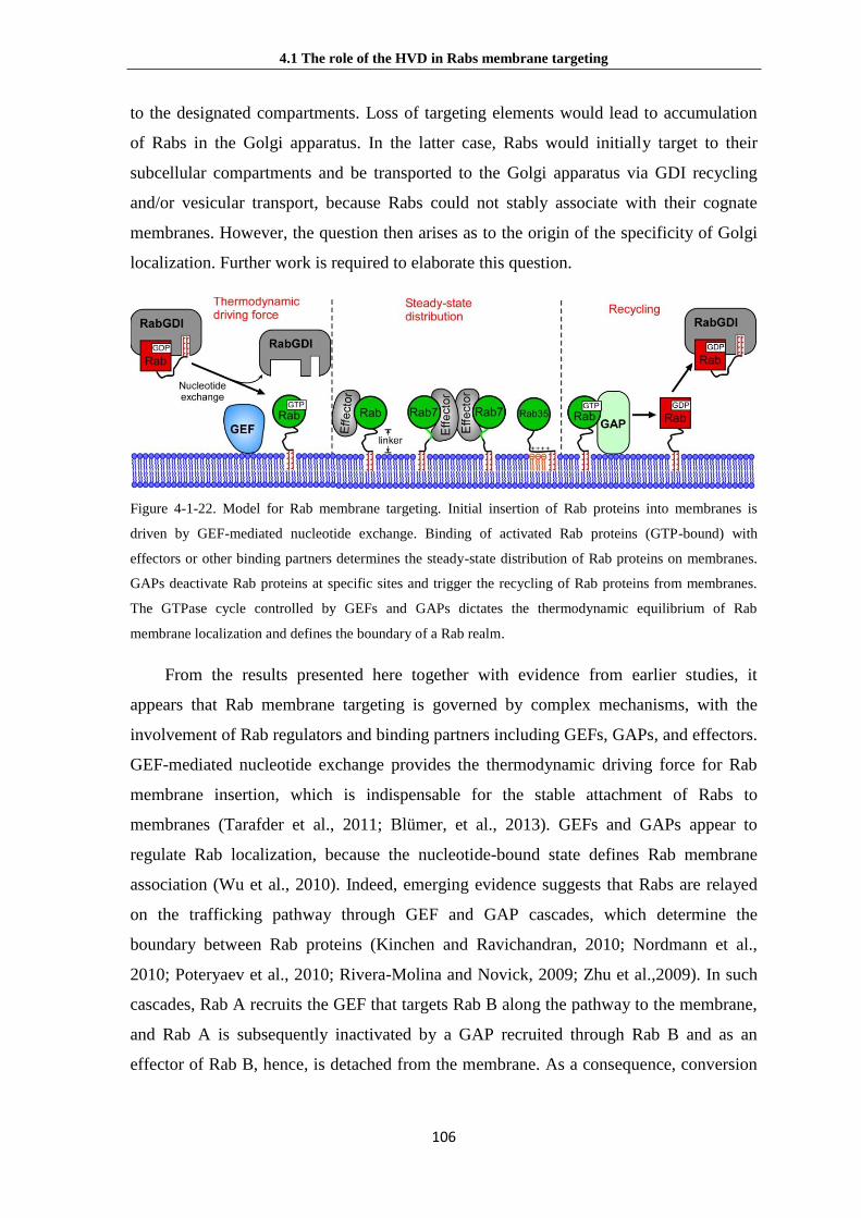
4.1 The role of the HVD in Rabs membrane targeting
106
to the designated compartments. Loss of targeting elements would lead to accumulation
of Rabs in the Golgi apparatus. In the latter case, Rabs would initially target to their
subcellular compartments and be transported to the Golgi apparatus via GDI recycling
and/or vesicular transport, because Rabs could not stably associate with their cognate
membranes. However, the question then arises as to the origin of the specificity of Golgi
localization. Further work is required to elaborate this question.
Figure 4-1-22. Model for Rab membrane targeting. Initial insertion of Rab proteins into membranes is
driven by GEF-mediated nucleotide exchange. Binding of activated Rab proteins (GTP-bound) with
effectors or other binding partners determines the steady-state distribution of Rab proteins on membranes.
GAPs deactivate Rab proteins at specific sites and trigger the recycling of Rab proteins from membranes.
The GTPase cycle controlled by GEFs and GAPs dictates the thermodynamic equilibrium of Rab
membrane localization and defines the boundary of a Rab realm.
From the results presented here together with evidence from earlier studies, it
appears that Rab membrane targeting is governed by complex mechanisms, with the
involvement of Rab regulators and binding partners including GEFs, GAPs, and effectors.
GEF-mediated nucleotide exchange provides the thermodynamic driving force for Rab
membrane insertion, which is indispensable for the stable attachment of Rabs to
membranes (Tarafder et al., 2011; Blümer, et al., 2013). GEFs and GAPs appear to
regulate Rab localization, because the nucleotide-bound state defines Rab membrane
association (Wu et al., 2010). Indeed, emerging evidence suggests that Rabs are relayed
on the trafficking pathway through GEF and GAP cascades, which determine the
boundary between Rab proteins (Kinchen and Ravichandran, 2010; Nordmann et al.,
2010; Poteryaev et al., 2010; Rivera-Molina and Novick, 2009; Zhu et al.,2009). In such
cascades, Rab A recruits the GEF that targets Rab B along the pathway to the membrane,
and Rab A is subsequently inactivated by a GAP recruited through Rab B and as an
effector of Rab B, hence, is detached from the membrane. As a consequence, conversion

4.1 The role of the HVD in Rabs membrane targeting
107
of a Rab A to a Rab B membrane is achieved. Once Rab proteins are activated and
stabilized on the membrane (GTP-bound), the steady-state Rab localization might be
dictated by binding partners, including effectors and lipids (Figure 4-1-22).
Such a stabilization of Rabs on their cognate membranes by effectors appears to
play an essential role in Rab membrane targeting, because depletion of effectors (RILP
knockdown) or loss of binding to effectors (Rab HVD replacement) leads to
mislocalization of Rab proteins. Some effectors associate with a specific membrane
independently of Rabs (Houghton et al., 2009). For those effectors that are recruited to
membranes in a Rab-dependent manner (Stenmark, 2009), there must be a synergy
between Rabs and effectors in membrane localization. For example, Rabenosyn-5 has two
distinct binding sites for Rab4 and Rab5, suggesting its coordinative role in Rab4 and
Rab5 localization and function (Eathiraj et al., 2005). Based on the above observations,
we propose a mechanistic model of the HVD roles for Rabs membrane targeting is shown
as Figure 4-1-22.

4.2 Cycling of Rab35 between the Golgi and the PM
108
4.2 Cycling of Rab35 between the Golgi apparatus and the
plasma membrane
Correct intracelluar localization of Rab GTPases is essential for their functions in
vesicular transport and signaling transduction. The mistargeting of Rabs may cause
diseases, for example, when they are hijacked by pathogenic effectors. Rab35 plays
various roles, including endosomal trafficking, exosome release, phagocytosis, cell
migration, immunological synapse formation and neurite outgrowth (Klinkert and Echard,
2016). However, membrane targeting mechanism of Rab35 is still unclear.
In this study, we combine the biochemistry, microinjection, RNA interference,
fluorescence recovery after photobleaching (FRAP), fluorescence localization after
photobleaching (FLAP) and cell biological techniques to investigate the mechanism of
Rab35 plasma membrane targeting. We reveal how GEF, effector and GDF of Rab35
work together to regulate the spatial cycling of Rab35. In addition, we show that the
depletion of OCRL1 induces the disruption of the Rab35 plasma membrane localization,
suggesting a link of Rab35 to the Lowe syndrome.
4.2.1 The polybasic region is essential for plasma membrane
localization of Rab35
In section 4.1.2.2, we have shown that the polybasic cluster (PBC) is essential for the
association of Rab35 with the plasma membrane, probably through binding to the
negatively charged phosphoinositide PtdIns(4,5)P2 and PtdIns(3,4,5)P3 (Li et al., 2014;
Heo et al., 2006).
To illuminate the function of the PBC, we sequentially mutate the positively charged
amino acids (lysine and arginine) to glutamine (Q), leading to Rab35-PBC-1M to -5M
constructs (Figure 4-2-1). We express these Rab35 mutant proteins in HeLa or Cos-7 cells
and check their intracellular localization. Quantification of the enrichment of Rab35 wild
type and its PBC mutants at the PM shows that with sequential increase in the number of
mutation residues in the PBC region, Rab35 gradually reduces its PM-colocalization and
mistargets to the perinuclear region (Figure 4-2-3A). The PM/cytosol ratio of PBC-2M
drops down dramatically (Figure 4-2-3B). We, therefore, conclude that at least four

4.2 Cycling of Rab35 between the Golgi and the PM
109
positively charged amino acids are required for the Rab35 PM localization. Additionally,
although we speculate that the cluster of six glutamine residues (6Q) may affect the
localization of Rab35, no obvious localization changes were found when they are
substituted by (GGS)2 repeat amino acids (Figure 4-2-1C, Rab35-6Q_M).
Figure 4-2-1. Subcellular localization of Rab35 and its PBC mutants. (A) The membrane targeting of Rab35
wild type, Rab35-PBC-1M, -2M,-3M,-4M and -5M. (B) The fold enrichment at PM of Rab35 wide type
and its mutants. (C) The subcellular localization of Rab35-6Q_M. The polybasic region is highlighted in
red, the CIM is highlighted in yellow, and the mutation sites are highlighted in green. *P < 0.05; ***P <
0.001. Scale bar, 10μm.
The association of Rab proteins with the membrane is generally mediated by
hydrophobic interactions via the C-terminal geranylgeranyl moieties attached to one or
two cysteines. To investigate the role of C-terminal prenylation motif in Rab35
localization, we generated Rab constructs with single C-terminal cystein including
Rab1b-C, Rab5a-C, Rab5a-Q79L-C, Rab35-C and Rab35-5M-C (Figure 4-2-2). The
results show that Rab1-C, Rab5-C and Rab5-Q79L-C are almost cytosolic or on ER rather
than localized at their respective targeting membranes, in keeping with previous report
(Gomes et al., 2003). However, natively monogeranylgeranylated Rabs (e.g., Rab8,
Rab13, and Rab27) obviously do not require diprenylation for appropriate targeting to
subcellular membranes. For these double cysteines Rab proteins, it is probably due to the
weak binding force of a single geranylgeranyl with membrane, which is not sufficient to

4.2 Cycling of Rab35 between the Golgi and the PM
110
ensure these Rab proteins’ membrane localization. Surprisingly, considerable amount of
Rab35-C molecules still localize at the plasma membrane, although some portion of
Rab35-C protein is found in the cytosol. The Rab35-PBC-5M-C protein, by contrast, is
largely cytosolic and loses its localization at perinuclear region.
Figure 4-2-2. Subcellular localizations of Rab1b, 5a, 5aQ79L, 35a, 35a-PBC-5M wild type and Rab-C
mutant. (A) The membrane localizations of Rab1b/-C, Rab5a/-C, Rab5aQ79L/-C, Rab35a/-C and Rab35a-
PBC-5M/-C. (B) Quantification of GFP-Rab35a enrichment at the PM relative to cytosol GFP-Rab35
intensity under these conditions. The prenylated cysteine(s) is highlighted in red, the CIM is highlighted in
orange, and the mutation sites are highlighted in green. **P < 0.01. Scale bar, 10μm.
These results indicate that the PBC of Rab35 is an essential element for PM
localization. This cellular localization is not only limited in PM targeting. A very similar
scenario is found in K-Ras4B protein, which also has a polybasic lysine-rich sequence at
the C-terminus that leads to the PM enrichment (Hancock et al., 1990; Choy et al., 1999;
Jang et al., 2015). Next, we investigate the membrane localization of the Rab35 PBC
mutant proteins in cells. Rab35-PEG-CC, Rab35Δ28-(GGS)n-CC and Rab35Δ28-(GGS)n-
CSC proteins have been shown to localize at the Golgi apparatus (Section 4.1.2.2). It
seems that Rab35 mistargets to the Golgi when it is lack of the PBC domain. In contrast
to the enrichment at the PM, the degrees of the localization at the Golgi apparatus of
Rab35 PBC mutants increase gradually with the increase of PBC mutation number
(Figure 4-2-3A). Therefore, Rab35 PBC domain is crucial for its proper membrane
localization. Rab35 may cycle between the PM and the Golgi apparatus.

4.2 Cycling of Rab35 between the Golgi and the PM
111
Figure 4-2-3. Subcellular localization of GFP-Rab35 wide type and PBC mutant proteins at the Golgi
apparatus. (A) The membrane targeting of Rab35-WT,Rab35-PBC-1M, Rab35-PBC-2M, Rab35-PBC-3M,
Rab35-PBC-4M and Rab35-PBC-5M, with more Rab35 PBC mutant mistargeting on Golgi apparatus. (B)
Pearsons colocalization coefficient analysis of the experiments performed in (A). Scale bar, 10μm.
To confirm the Golgi localization of Rab35 PBC mutants, we treated the cells with
5μM nocodazole for 1 hour, and find that both Rab35-PBC-4M and Rab35-PBC-5M
colocalize extensively with Giantin on the Golgi fragments (Figure 4-2-4).

4.2 Cycling of Rab35 between the Golgi and the PM
112
Figure 4-2-4. Colocalization of GFP-Rab35 PBC mutants with the Golgi marker Giantin in the presence of
nocodazole. (A, B) GFP-Rab35-PBC-4M colocalizes with the Golgi marker Giantin with (B) or without
nocodazole (A) treatment. (C, D) GFP-Rab35-PBC-5M colocalizes with the Golgi marker Giantin with (D)
or without nocodazole (C) treatment. (E) Pearsons colocalization coefficient (PCC) analysis of the
experiments performed in (A), (B), (C) and (D). Scale bar, 10μm.
4.2.2 Rab35 membrane targeting is not affected by Rab11
Previous studies have revealed that Rab35 is involved in fast recycling pathways, which
send various receptors back to the PM from the cytosol or Golgi (Ilektra et al., 2006;
Patino-Lopez et al, 2008; Barth and Julie, 2009). In addition, Rab11 is known to associate
primarily with perinuclear recycling endosomes and regulates recycling of endocytosed
cargos. The different localization profiles of Rab11 in diverse cell types have complicated
the assessment of its precise function in intracellular membrane transport. In non-
polarized cells Rab11 was found to be associated with both Golgi and recycling
endosomes (Ullrich et al., 1996; Ren et al., 1998). In polarized and regulated secretory
cells Rab11 localized to the Golgi as well as a variety of specialized membrane
compartments (Urbe et al., 1993; Deretic et al., 1996; Goldenring et al., 1996; Goldenring
et al., 1997; Sheehan et al., 1996; Calhoun and Goldenring, 1997). More data have shown

4.2 Cycling of Rab35 between the Golgi and the PM
113
that Rab11 regulates vesicular trafficking from the trans-Golgi network (TGN) and
recycling endosomes to the plasma membrane (Chen et al., 1998; Prekeris, 2003;
Takahashi et al., 2012; Andreas et al., 2010). To examine whether the trafficking of
Rab35 is affected by Rab11 in recycling pathway, we examined the localization of Rab35
and Rab35-PBC-5M with the co-expression of Rab11 in HeLa cells, respectively. Figure
4-2-5 shows that Rab35 and PBC-5M mutant colocalize with Rab11 in the cells.
Figure 4-2-5. Colocalization of Rab35 wild type and its PBC mutant proteins with Giantin and Rab11. (A)
Colocalization of Rab35 and Rab11 in Hela cells. (B) Colocalization of Rab11, Rab35-PBC-5M and
Giantin. (C) Pearson’s correlation coefficients (PCC) analysis of the experiments performed in experiments
(A) and (B). Scale bar, 10μm.
Bastiaens and coworkers found Rab11 plays roles in trafficking of KRas-4B to the
PM via the endocytic recycling (Malte et al., 2014). Interestingly, KRas-4B also contains
a polybasic cluster at the C-terminal tail. We ask if the alterations of Rab11 function
would perturb the membrane targeting of Rab35 in cells. The depletion of Rab11 was first
performed by using RNA interference with Rab11 siRNA in HeLa cells. We examined
the membrane association of Rab35 in Rab11 knock-down cells and found that the PM
enrichment of Rab35 only slightly decreases compared to the control (Figure 4-2-6 B and
A). In contrast to previous results that Rab11 regulates the localization of KRas-4B in
cells (Figure 4-2-6G, Schmick et al., 2014), the PM targeting of Rab35 shows only a little
change with the expression of Rab11, Rab11DN (the dominant negative form), or
Rab11QL (dominant active form) conditions (Figure 4-2-6D, E, and F). These results
indicate that Rab11-associated recycling endosomes are not involved in the recycling of

4.2 Cycling of Rab35 between the Golgi and the PM
114
Rab35 to the PM in cells.
Figure 4-2-6. The effects of Rab11 knock down and mutations for the localization of Rab35. (A-F) The
subcellular localization of GFP-Rab35 wide type (A) in the scrambled shRNA cells. The subcellular
localization of GFP-Rab35 (B) and GFP-Rab35-PBC-5M (C) in Rab11 knock-down cells. The subcellular
localization of GFP-Rab35 wide type in the co-expression of Rab11 wild type (D), Rab11DN (dominant
negative, E) and Rab11QL (dominant active, F) cells. (G) The localization of mCitrine-KRas-4B is
regulated by Rab11 protein (Schmick et al., 2014). (H) Quantification of GFP-Rab35 enrichment at the PM
relative to the cytosolic GFP-Rab35 under these conditions (A-F). NS, not significant. Scale bar, 10μm.

4.2 Cycling of Rab35 between the Golgi and the PM
115
4.2.3 Rab35 cycles between the Golgi apparatus and the plasma
membrane
4.2.3.1 Rab35 trafficks from the Golgi apparatus to the PM via a non-vesicular
pathway
The Golgi localization of Rab35 PBC mutants raises the question whether Rab35 cycles
between the PM and the Golgi. The photoactivatable green fluorescence protein (paGFP)
was employed to study the dynamics of Rab35 trafficking in HeLa and in Cos-7 cells.
Wild type GFP exists in two forms, which give rise to a major and a minor
absorbance peak at 397nm and 475nm, respectively. Intense illumination at 400 nm shifts
the population to the 475nm form, thereby increasing the absorbance of the minor peak.
This photoconversion feature led to the development of photoactivatable green
fluorescence protein (paGFP) (George et al., 2000). By selecting a form of GFP with a
negligible 475nm peak, photoconversion will produce a much greater proportional
increase in the absorbance at 475nm compared to the standard GFP and therefore
increases contrast. paGFP exhibits very low green emission (max 517nm) with 488nm
excitation, which can be increased to 100-fold by stimulation with 405nm light. This
means that, paGFP activated by photo-irradiation will yield bright signals over a dark
background. The advent of paGFP offers new possibilities to study biomolecules, such as
pulse-chase labeling and single molecule localizations. We generated a panel of paGFP-
Rab35 and its PBC mutant constructs for studying their dynamics by confocal microscopy.
We co-expressed paGFP-Rab35 and mKate2-Giantin as Golgi marker in HeLa cells.
Photo-activation of paGFP-Rab35 at the Golgi region, which is indicated by mKate2-
Giantin, led to transient observation of Rab35 accumulation at the Golgi, followed by
rapid decrease of the fluorescence at the Golgi with the concurrent increase at the PM.
Figure 4-2-7 shows the two processes of the FLAP measurments: (1) dissociation of
Rab35 from the Golgi membrane; (2) association of Rab35 to the PM. The half times (t1/2)
of Golgi dissociation (t1/2=19.4±5.7s) and PM association (t1/2=28.9±14.9s) are similar,
suggesting that Rab35 trafficks from the Golgi to the PM.
The trafficking of Rab35 from the Golgi to the PM may be a fast process. Sevral
reports have defined the rapid/fast recycling from endosomes to the PM with half- times

4.2 Cycling of Rab35 between the Golgi and the PM
116
of 50 seconds to 2 minutes, depending on the internalized lipid analogs. The half-time of a
vesicular recycling is typically 4~5 minutes and up to 9~10 minutes in different cell lines
(Hao and Maxfield, 2000). So we speculated that the trafficking of Rab35 from the Golgi
to the PM may not undergo a vesicular transport process, which is cytoskeleton dependent.
Figure 4-2-7. The trafficking of paGFP-Rab35 from the Golgi apparatus to the PM. (A) Scheme of the
photoactivation process of a paGFP protein in a cell. (B) Representative time-lapse sequences of
fluorescence distribution of paGFP-Rab35 photoactivated at the Golgi (upper row, the red circle is active
site) and of mKate2-Giantin (lower row). (C, D) Quantification of Rab35 dissociation kinetics from the
Golgi and association kinetics to the PM. Scale bar, 10μm.
To confirm our hypothesis, the cells were treated with 5μM nocodazole for 5
minutes to disrupt microtube and therefore vesicular transport. We obtained the t1/2s of
disassociation from the Golgi and association to the PM for 21.6±6.4 seconds and
19.4±9.1 seconds, respectively which are similar to the t1/2s without nocodazole treatment
(Figure 4-2-8C). Taken together, we conclude that Rab35 trafficks from the Golgi to the
PM in a non-vesicullar transport pathway.

4.2 Cycling of Rab35 between the Golgi and the PM
117
Figure 4-2-8. The trafficking of paGFP-Rab35 from the Golgi apparatus to the PM in the presence of
nocodazole. (A) Representative time-lapse sequences of fluorescence distribution of paGFP-Rab35
photoactivated at the Golgi body (upper row, the red circle is active site) and of mKate2-Giantin (lower row)
in the presence of 5 μM nocodazole. (B) Quantification of Rab35 dissociation kinetics from the Golgi and
association kinetics to the PM. (C) Half-times of fluorescence decay and improve for paGFP-Rab35 with or
without 5µM nocodazole treatment. NS, not significant. Scale bar, 10μm.
FRAP (Fluorescence recovery after photobleaching) has been used to characterize
the mobility of cellular molecules (Vikstrom et al., 1992), by observing the movement of
intracellular materials through photobleaching of the fluorescence. Several images using a
low light level are acquired to determine the initial fluorescence, and then a high level of
light for a short time inside a region of interest (ROI) is used to bleach the fluorescence in
the ROI. The recovery of fluorescence at the ROI after photobleaching is followed by
fluorescence microscopy. In this study, we performed the FRAP experiments at the Golgi
region to measure the kinetics for trafficking of Rab35 mutant proteins to the Golgi
(Table 4-2-1). These data also indicates that Rab35 is highly dynamic in cells.
Although we have confirmed Rab35 trafficking from the Golgi to the PM, it is not
clear that whether these Rab35 molecules originated from the PM/cytosol pool or from
newly synthesized proteins. Therefore, we can not exclude that the photoactivated
paGFP-Rab35 at the Golgi may involve the newly synthesized molecules but is not from
the cytosol, the endosome membranes or the PM.

4.2 Cycling of Rab35 between the Golgi and the PM
118
To test our hypothesis, we used the protein synthesis inhibitor cycloheximide (CHX)
to inhibit generation of new Rab35 proteins. CHX exerts its effect by interfering with the
translocation step during the protein synthesis (movement of two tRNA molecules and the
mRNA in relation to the ribosome), thus blocking translational elongation (Schneider-
Poetsch et al., 2010). After the treatment of the cells, co-expressing paGFP-Rab35 and
mKate2-Giantin with 10μg/ml cycloheximide for 4 hours, FLAP experiments were
performed. Under these conditions, significant amount of Rab35 molecules transiently
accumnulate at the Golgi followed by a decay with t1/2=18.27 seconds, which is similar to
that without CHX (Figure 4-2-9E).
Figure 4-2-9. Activation of paGFP-Rab35 at the Golgi in the presence of cycloheximide. (A, B and C)
Time-lapse sequences of fluorescence distribution of paGFP-Rab35 are induced by three times
photoactivation at the Golgi in the round A, B and round C (Upper row). (D) Fluorescence curves of
consecutive activation cycles in (A), (B), and (C). (E) The overlay of normalized curves in round A, B and
C. Scale bar, 10μm.
Moreover, we performed three consecutive activation cycles in the FLAP
measurement. All of them display identical t1/2s (t1/2=17.41±0.8 seconds) (Figure 4-2-9 A-
D). These results suggest that Rab35 cycles between the Golgi and the PM.

4.2 Cycling of Rab35 between the Golgi and the PM
119
4.2.3.2 Rab35 trafficks from the PM to the Golgi apparatus in an endocytic
pathway
To investigate how Rab35 trafficks from the PM to the Golgi apparatus, the edge of the
cell was photoactivated in HeLa cells, which co-express paGFP-Rab35 and mKate2-
Giantin proteins. Figure 4-2-10 shows that Rab35 diffuses in the PM due to the lateral
diffusion, while trafficking within the cytoplasm. Many Rab35 vesicles were found to
accumulate near the Golgi region, indicating that vesicular pathway may be involved in
PM-to-Golgi trafficking of Rab35. The half-time of the association to the Golgi region is
157 seconds, which is much longer than the dissociation half-time from Golgi apparatus.
Figure 4-2-10. The trafficking of paGFP-Rab35 after photoactivation at the PM in HeLa cell. (A)
Representative time-lapse sequences of fluorescence distribution of paGFP-Rab35 photoactivated At the
PM and partial of cytosol (upper row) and of mKate2-Giantin (lower row). The arrows indicate Rab35
vesicles. (B) Quantification of Rab35 association kinetics at Golgi region from the PM and the cytosol.
Scale bar, 10μm.
It has been shown before that Rab35 is involved in clathrin-dependent endocytosis
(Dutta and Donaldson, 2015; Cauvin et al., 2016). We speculate that Rab35 is involved in
the clathrin-dependent endocytosis pathway. Dynamin is a small GTPase involved in
clathrin coated vesicles (CCVs) mediated endocytosis in the eukaryotic cell (Henley et al.,
1999). The role of Dynamin is responsible for the scission of newly formed CCVs from
the PM, the Golgi and the endosomal membranes (Praefcke and McMahon, 2004).

4.2 Cycling of Rab35 between the Golgi and the PM
120
Moreover, dynamin plays a role in many processes including division of organelles,
cytokinesis and microbial pathogen resistance (Hinshaw, 2000; Thoms and Erdmann,
2005). There are three dynamin members in human cells. Dynamin1 is mainly expressed
in neurons while dynamin2 is expressed in most cell types. Dynamin3 is strongly
expressed in the testis, but is also present in heart, brain and lung tissues.
Figure 4-2-11. Rab35 is involved in clathrin coated vesicle (CCV) mediated endocytosis. (A) The
subcellular localization of Rab35 with the expression of dynamine2 in HeLa cells. (B) The subcellular
localization of Rab35 with the expression of dynamine2K44A mutant in cells. (C) Quantification of GFP-
Rab35 enrichment at the PM relative to cytosol GFP-Rab35 intensity in (A and B). Dy2-K44A MT:
dynamine2-K44A mutant. ***P < 0.001. Scale bar, 10 μm.
Next, we co-expressed GFP-Rab35 and dynamine2-Cherry in HeLa cells. The
localization of Rab35 was not affected (Figure 4-2-11A). However, when Rab35 was co-
expressed with dynamin2-K44A (dominant negative mutant), Rab35 loses its enrichment
at PM and becomes largely cytosolic (Figure 4-2-11B). In addition, Rab35 tubulation is
found which crosses the entire cell.
Altogether above results, we conclude that the Rab35 trafficks from the Golgi to the
PM via a non-vesicular transport, while from the PM to the Golgi via an endocytic
pathway.

4.2 Cycling of Rab35 between the Golgi and the PM
121
4.2.4 PRA1 is important for plasma membrane localization of Rab35
Next, we sought to understand which protein mediates the non-vesicular pathway of
Rab35 trafficking from the Golgi to the PM. GDP-dissociation inhibitor (GDI) is a
possible candidate, which recycles prenylated GDP-bound form Rabs between
membranes and cytosol (Regazzi et al., 1992; Pylypenko et al., 2006; Wu et al., 2007).
PDE6δ is a GDI-like solubilizing factor (GSF) for prenylated Ras proteins (Chandra et al.,
2011). However, the PM localization of Rab35 doesn’t change in the presence of
Deltarasin, a PDE6δ inhibitor, in contrast to that of Ras proteins (Figure4-2-12).
Figure 4-2-12. Subcellular localization of GFP-Rab35 in the presence of Deltarasin. (A) Subcellular
localization of GFP-Rab35 without the treatment of Deltarasin. (B) Subcellular localization of GFP-Rab35
in the presence of Deltarasin. (C) Quantification of GFPRab35 enrichment at the PM relative to cytosol
GFP-Rab35 intensity in (A and B). NS, not significant. Scale bar, 10 μm.
Figure 4-2-13. Subcellular localization of GFP-PRA1. Colocalizations of GFP-PRA1 with the Golgi marker
mKate2-Giantin (A) and with the late endosome/lysosome marker LAMP1-Cherry (B).

4.2 Cycling of Rab35 between the Golgi and the PM
122
Prenylated Rab acceptor (PRA1/YIP3) is an integral membrane protein which was
proposed as GDI-displacement factor (GDF). It dislodges Rabs from GDI to facilitate
GDI-mediates Rab cycles. It was reported to localize at the Golgi and endosomes (Dirac-
Svejstrup et al., 1997, Sivars et al., 2003). We found that PRA1 mostly localized on Golgi
body while only a few localized on the late endosomal membranes (Figure 4-2-13B).
Biochemistry data showed that PRA1/Yip3 functions on the prenylation of Rabs,
especially for endosomal Rab GTPases including Rab9, Rab5 and Rab7. The possible
function of GDI involved in the Golgi-to-PM recycling of Rab35 prompt us to investigate
the role of PRA1 (GDF) in Rab membrane targeting the function of PRA1 for membrane
targeting of Rab proteins. We depleted PRA1 expression by RNA interference (RNAi).
We generated PRA1 shRNA constructs and knock-down the endogenous expression of
PRA1 (Figure 4-2-14H).
Figure 4-2-14. Subcellular localization of Rab35 in the PRA1 knock-down cells. (A-C) Subcellular
localization of GFP-Rab35 wide type (A), GFP-Rab35Q67L mutant (B) and GFP-Rab35-PBC-5M (C) in
scrambled shRNA cells. (D-F) Subcellular localization of GFP-Rab35 wide type (D), GFP-Rab35Q67L
mutant (E) and GFP-Rab35-PBC-5M (F) in PRA1 knock-down cells. (G) Subcellular localizaition of GFP-
Rab35 with the co-expression of Cherry-PRA1 in PRA1 knock-down cells. (H) Knock-down of endogenous
PRA1 in HeLa cells, detected by western blot using anti-PRA1 antibody. Scrambled siRNA (scr) cell was
used as a control. (I) Quantification of GFP-Rab35 enrichment at the PM relative to the cytosol GFP-Rab35
intensity under these conditions (A-F).***P < 0.001. Scale bar, 10μm.
The PRA1 depletion impairs the PM localization of Rab35 (Figure 4-2-14D) and
even the Rab35Q67L dominant active mutant (Figure 4-2-14E). The cytosolic phenotype

4.2 Cycling of Rab35 between the Golgi and the PM
123
of Rab35 wide type can be rescued by co-transfected with mCherry-PRA1, which is
resistant to the siRNA (Figure 4-2-14G).
Interestingly, the localization of Rab35-PBC-5M at the Golgi body is not affected by
PRA1 knock-down (Figure 4-2-14F). To check the dynamics of Rab35, FRAP
(Fluorescence recovery after photobleaching) of Rab35 and its mutant proteins were
performed in both the scrambled siRNA and PRA1 knock-down cells (Table 4-2-1). In
control cells, the t1/2 of Rab35-PBC-5M is 15.7 seconds while it is 16.02 seconds in PRA1
knock-down cells. Therefore, the fact that the PRA1 depletion does not slow down the
Rab35-PBC-5M dynamics dramatically may due to its localization at the Golgi but not on
endosome. When Rab35 mutant does not undergo the Golgi-PM cycling, its subcellular
localization is not regulated by PRA1.
Table 4-2-1. Summary of Rab35and its PBC mutant proteins delivery kinetics in cell.
t1/2 FRAP at the Golgi (single exponential fit) FRAP at the Golgi (PRA1 KD cells)
GFPRab35 WT ND 14.23s
GFP-Rab35-PBC-1M ND 21.90
GFP-Rab35-PBC-2M 8.2s 17.54s
GFP-Rab35-PBC-3M 21.5s 13.37s
GFP-Rab35-PBC-4M 18.6s N.D
GFP-Rab35-PBC-5M 15.7s 16.08s
N.D stands for not done.
These results suggest that PRA1 plays an essential role in the Golgi-PM cycling of
Rab35 and hence its PM localization. Therefore, GDI is the key recycling factor involved
in the Golgi-to-PM trafficking.
Since PRA1 has been proposed to function as a GDF for sevral endosomal Rab
proteins (Dirac-Svejstrup et al., 1997, Sivars et al., 2003), the localization of several Rabs
were examined.
We expressed GFP-Rab1, GFP-Rab5 and its dominant active mutant GFP-
Rab5Q79L, GFP-Rab7, GFP-Rab8, GFP-Rab9 and GFP-Rab11 proteins in PRA1 knock-
down cells. The localization of GFP-Rab1 remains at the Golgi apparatus in PRA1 knock-
down cells (Figure 4-2-15A and B). However, the membrane localizations of endosomal
Rabs including Rab5, Rab5Q79L, Rab7 and Rab11 are affected dramatically (Figure 4-2-
15B). Rab5 and Rab5Q79L vesicles form clusters in the cell. In contrast, Rab7 localizes
on enlarged vesicles (Figure 4-2-15B). The localization of Rab11 is very similar to its

4.2 Cycling of Rab35 between the Golgi and the PM
124
S25N dominant negative mutant, which localizes in the perinuclear region (Figure 4-2-
15C). However, the localizations of Rab8 and Rab9 are not altered in the PRA1 KD cells.
Surprisingly, the depletion of PRA1 does not change the localization of Rab9, which is
also an endosomal Rab. Previous result has shown that the membrane association of Rab9
is decreased dramatically in PRA1/YIP3 KD cells (Sivars et al., 2003).
Figure 4-2-15. Rab GTPases subcellular localization in PRA1 knock-down cells. (A)Subcellular
localization of GFP-Rab1, GFP-Rab5, GFP-Rab5Q79L, GFP-Rab7, GFP-Rab8 GFP-Rab9 and GFP-Rab11
in scrambled shRNA cells. (B) Subcellular localization of GFP-Rab1, GFP-Rab5, GFP-Rab5Q79L, GFP-
Rab7, GFP-Rab8, GFP-Rab9 and GFP-Rab11 in in PRA1 knockdown cells. (C) Subcellular localization of
GFP-Rab11S25N. Scale bar, 10 μm.
Altogether, we found that PRA1 is crucial for plasma membrane localization of
Rab35. Additionally, PRA1 plays a role in Rab5, Rab7 and Rab11 membrane trafficking.
4.2.5 Nucleotide exchange regulates Rab35 localization at the plasma
membrane
Guanine exchange factors (GEFs) mediated the GDP exchange to GTP for the activation
of the Rab GTPases. In the GTP-bound form, Rabs recruit downstream effectors to fulfill
their functions. Moreover, GEFs serve as the determinant for the membrane targeting of
Rabs (Wu et al., 2010; Blümer et al., 2013). Since PRA1 depletion impairs the membrane
targeting of Rab35, we asked if Rab35 GEFs affect the correct membrane localization of
Rab35. The DENN domain has been found to display Rab GEF activity for Rab35
(Allaire et al., 2010) and DENND1A (connecdenn 1) has been shown to be the major
GEF for Rab35 in the endocytic pathway (Marat et al., 2011).

4.2 Cycling of Rab35 between the Golgi and the PM
125
Firstly, the localizations of Rab35S22N and Rab35Q67L were examined in HeLa
cells. Less Rab35Q67L (dominant active form) proteins are found on PM and most of
them on endosomal membranes compared to that of Rab35 wide type (Figure 4-2-16A).
Interestingly, Rab35S22N (dominant negative form) mutant proteins loss their PM
localization, and a consideration portion of them are localized on endosomes. These
results suggest that the GTPase cycle regulated by GEF and GAP is essential for Rab35
spational cycles and therefore membrane targeting.
Figure 4-2-16. Subcellular localization of GFP-Rab35 mutant proteins in HeLa cells. (A) The localization
of GFP-Rab35Q67L mutant in cell. (B) The localization of GFP-Rab35S22N mutant on endosomes. (C)
The localization of GFP-Rab35Q67L-PBC-5M at the Golgi in cell. (D) The localization of GFP-
Rab35S22N-PBC-5M in cell. (E) Quantification of GFP-Rab35Q67L enrichment at the PM relative to
cytosol. (F) Quantification of GFP-Rab35 enrichment at the Golgi relative to cytosol GFP-Rab35 intensity
under these conditions (C, D). Scale bar, 10μm.
Rab35Q67L-PBC-5M and Rab35-PBC-5M mutant protein were found still localize
on the Golgi apparatus (Golgi marker did not shown). For Rab35S22N-PBC-5M mutant
protein, some of them are still localized at the Golgi although show that they can
dissociate from Golgi and become more cytosolic (Figure 4-2-16D) and their
localizations at the Golgi were measured (Figure 4-2-16F). The localizations of Rab35

4.2 Cycling of Rab35 between the Golgi and the PM
126
and Rab35-PBC-5M mutant proteins suggest that the membrane targeting of Rab35 is
partially contributed by its GEFs.
To examine the precise localization of Rab35S22N mutant proteins, we co-expressed
Rab35S22N and EEA1, an early endosome marker or LAMP1, a late endosome/lysosome
marker in HeLa cells. Figure 4-2-17 shows most Rab35S22N are localized on early
endosome and late endosome/lysosomes. The dominant negative mutant may present
intermediate endocytic localization en route to the Golgi. It is known that Rab35 recruits
OCRL1 to newborn endosomes, where OCRL1 converts PtdIns(4,5)P2 to PtdIns(4)P leads
to the uncoating of clathrin coated vesicles(CCVs) (Martin, 2001). Rab35S22N mutant
impairs the recruitment of OCRL1 to CCVs and causes the accumulation of PtdIns(4,5)P2
on CCVs, early endosomes and late endosomes. The localization of Rab35S22N protein
on endosomal membranes suggests that Rab35 may associate on endosome via the
binding between the PBC and PtdIns(4,5)P2 or other PtdInPs. This electrostatic
interaction may enhance the affinity of Rab35S22N with the negative charged lipids on
CCPs, early endosomes and late endosomes. Therefore, mutation of the PBC
(Rab35S22N-5M) led to more cytosolic localization of the protein (Figure 4-2-16D).
These data suggest Rab35 GEFs may be involved in its membrane targeting.
Figure 4-2-17. Subcellular localization of GFP-Rab35S22N mutant in HeLa cells. (A) The colocalizatio of
GFP-Rab35S22N with Cherry-EEA1. (B) The colocalizatio of GFP-Rab35S22N with LAMP1-Cherry. (C)
Pearson’s correlation coefficients (PCC) for colocalization (A and B), respectively. Scale bar, 10μm.

4.2 Cycling of Rab35 between the Golgi and the PM
127
To further identify the function of GEFs for Rab35 membrane targeting, we
generated cherry-tagged shRNA constructs to knock down the Rab35 GEF DENND1A in
HeLa cells. DENND1A depletion leads to Rab35 being largely cytosolic, suggesting that
DENND1A is crucial for Rab35 PM-targeting. By carefully examining the phenotype
resulted from DENND1A depletion, we found that the density of Rab35 is higher in the
proximity area of PM, which may localize in endsomes (Figure 4-2-18A). This phenotype
was also observed in PRA1 depletion cells (Figure 4-2-14D). Interestingly, the depletion
of DENND1A did not alter the localization of Rab35-PBC-5M (Figure 4-2-18B), which
still localizes at the Golgi apparatus. This result is probably attributed to several reasons.
One possible reason is that Rab35-PBC-5M can be actived by another GEF such like
DENND1B, DENND1C, Folliculin (FLCN) or an unknown GEF (Allaire et al., 2008;
Allaire et al., 2006;Cauvin et al., 2016). Another possibility is that the membrane
localization of Rab35-PBC-5M is independent on the GDP or GTP bound form, which
means that Rab35-PBC-5M can be recruited by a protein localizing at the Golgi.
Figure 4-2-18.Subcelluar localization of Rab35 and PBC mutant in DENND1A knock-down HeLa cells. (A)
The localization of Rab35 in DENND1A knock-down cells. (B) The localization of Rab35-PBC-5M in
DENND1A knock-down cells. (C) Quantification of GFP-Rab35 (A) and GFP-Rab35-PBC-5M enrichment
at the PM relative to cytosol intensity. ***P < 0.001. Scale bar, 10μm.
Therefore, the GTPase cycle is essential for the membrane targeting of Rab35 in
cells. DENND1A, the Rab35 GEF, plays a crucial role in Rab35 GTPase cycle and
thereby Rab35 membrane targeting. It is conceivable that without activation of Rab35 on
the endosomes and the endocytic trafficking of Rab35 is disrupted. As a consequence, the
cycling between the Golgi and the PM is perturbed. Thus, Rab35 loses its correct
membrane targeting in the cell.

4.2 Cycling of Rab35 between the Golgi and the PM
128
4.2.6 OCRL1 is required for Rab35 plasma membrane localization and
function
OCRL1, the effector of Rab35, shows important roles in the processes of the cytokinesis
and the uncoating of CCVs (Hanono et al., 2006; Cauvin et al., 2016). In addition, OCRL
depletion perturbs endocytic recycling and trafficking from endosomes to the TGN
(Choudhury et al., 2005; Vicinanza et al., 2011). OCRL1 is a inositol polyphosphate 5-
phosphatase(INPP5F), which catalyzes the production of PtdIns(4)P and PtdIns(3,4)P2
from PtdIns(4,5)P2 and PtdIns(3,4,5)P3. An interesting result showed that an OCRL1 pool
localizes in the trans-Golgi network (Hanono et al., 2006), where OCRL may play a role
in the metabolism of phosphatidylinositides.
Figure 4-2-19. Subcellular localizaition of Rab35 in OCRL1 knock-down HeLa cells. (A) The
colocalization of GFP-Rab35 and mChery-OCRL1 in scrambled shRNA cells. (B) The localization of GFP-
Rab35 in OCRL1 knock- down cell. (C) The localization of GFP-Rab35-PBC-5M in OCRL1 knock-down
cells. (D) The localization of GFP-Rab7Δ34-(GGS)n-VKL-CSC in OCRL1 knock-down cells. (E)
Subcellular localizaition of Rab35 with the co-expression of Cherry-OCRL1 in OCRL1 knock-down cells.
(F) Subcellular localizaition of Rab35 with the co-expression of Cherry-OCRL1F668A mutant in OCRL1
knock-down cells. (G) Knockdown of endogenous OCRL1 in HeLa cells, detected by Western blot using

4.2 Cycling of Rab35 between the Golgi and the PM
129
anti-OCRL1 antibody. Scrambled siRNA (scr) cell was used as a control. (H) Quantification of GFP-Rab35
enrichment at the PM relative to cytosol GFP-Rab35 intensity under these conditions (B, E, F). Arrow head:
two nucleuses in a binucleate cell. ***P < 0.001; **P < 0.01. Scale bar, 10μm.
To investigate the role of OCRL in Rab35 membrane targeting, OCRL1 depletion
was performed by shRNA in HeLa cells. The subcellular localization of Rab35 is largely
cytosolic in OCRL knock-down cells (Figure 4-2-19B), while the localization of Rab35-
PBC-5M at Golgi apparatus is not altered (Figure 4-2-19C). The cytosolic phenotype of
Rab35 in OCRL1 depletion cells can be rescued by the expression of mCherry-OCRL1,
which is resistant to siRNA (Figure4-2-19E). Intriguingly, the Lowe syndrome mutant
OCRL1-F668A, could not rescue the cytosolic phenotype of Rab35 in OCRL1 knock-
down cell (Figure4-2-19F). The crystal structure OCRL1-Rab8a complex revealed that
F668 is included in the Rab-binding interface of OCRL1, an IgG-like β-strand structure of
the ASPM-SPD-2-Hydin domain. F668A substitution impairs the interaction with Rab8a
due to the lower hydrophobicity of alanine compared to phenylalanine (Hou et al., 2011).
It is possible that the F668A mutation also disrupts the Rab35 interaction to OCRL1.
However, more direct evidences are needed that can be provided by precise methods such
as ITC or fluorescence polarization. The results suggest that the OCRL binding is
essential for Rab35 membrane targeting.
Figure 4-2-20. Subcellular localizations of Rab5, Rab7, Rab8 and Rab1 in OCRL1 knock-down HeLa cells.
(A, C, E, G) The localization of GFP-Rab5 (A), GFP-Rab7 (C), GFP-Rab8 (E) and GFP-Rab1 (G) in
scrambled siRNA cells. (B, D, F, H) The localization of GFP-Rab5 (B), GFP-Rab7 (D), GFP-Rab8 (F) and
GFP-Rab1 (H) in OCRL1 siRNA cells. Scale bar, 10μm.

4.2 Cycling of Rab35 between the Golgi and the PM
130
Previous evidences revealed that OCRL works as an effector of more than ten Rab
GTPases including Rab1, Rab5, Rab8, Rab13, Rab35, Rab36, and so on (Fukata et al.,
2008). Therefore, we suspect whether the membrane targeting the other Rabs are
disturbed which similar with the case of Rab35 in OCRL1 depletion cells. We chose
Rab1, Rab5, Rab7 and Rab8 to test the effect of OCRL1 depletion. Figure 4-2-20 clearly
shows the enlargement of GFP-Rab5 vesicles (Figure 4-2-20B). Interestingly, although
OCRL1 is not an effector of Rab7, the enlarged Rab7 vesicles were observed in OCRL1
knock-down cell (Figure 4-2-20D). Both Rab5 and Rab7 are involved in endocytic
pathway, which is now impaired due to the loss-of-function of Rab35 in the absence of
OCRL1. Thus, the enlarged Rab7 vesicles may be not induced by OCRL1 depletion
directly but the result of endocytic pathway blocking. In contrast, knock-down of OCRL1
does not perturb the membrane targeting of Rab1 and Rab8, though OCRL1 was shown
to bind these Rabs (Fukata et al., 2008). The results indicate OCRL1 is involved in
endocytic pathway.
Altogether, as an effector of Rab35, OCRL1 depletion leads to the formation of
cytosolic of Rab35, which is very similar to the effect of RILP on Rab7 (Li et al., 2014).
This is a new evidence of Rab effector playing role in the Rab membrane targeting
process. OCRL1 may regulate Rab35 membrane targeting via its 5-phosphatase activity
involved in the metabolism of phosphoinositides, which are involved in electrostatic
interactions with Rab35. On the other hand, OCRL1 plays an important role in the
endocytic trafficking pathway, thereby regulating the Golgi-PM cycling of Rab35.

4.2 Cycling of Rab35 between the Golgi and the PM
131
4.2.7 Conclusion and discussion
In this study, we combined biochemistry, cell biology, molecular biology and confocal
microscopy techniques to study the mechanism of Rab35 membrane targeting. The
polybasic region is crucial for the PM localization of Rab35 and its divergent functions.
The polybasic cluster (PBC) depletion leads to the mistargeting of Rab35 to Golgi. The
Golgi apparatus may work as a stop of Rab35 trafficking. Furthermore, the trafficking of
Rab35 from Golgi to PM is via a fast non-vesicular pathway and may involve GDI. The
trafficking of Rab35 from the PM to the Golgi apparatus is largely mediated by an
endocytic pathway.
4.2.7.1 PRA1 affects the membrane localization of Rab35
PRA1 was proposed as a Rab GDF which dislodges the endosomal Rabs from GDI and
facilitates the subsequent nucleotide exchange on the membrane (Sivars et al., 2003). The
PRA1 depletion disrupts the PM localization of Rab35 and renders Rab35 largely
cytosolic. Moreover, the depletion of PRA1 leads to clusters of Rab5 vesicles and
enlargement of Rab7 vesicles. These phenomena suggest the important role of PRA1 in
endocytic pathway. Although Rab9 was proposed as a model protein to address the GDF
functions of PRA1, surprisingly, the membrane localization of Rab9 is not altered
obviously in the PRA1 knock-down cells. The localization of Rab35-PBC-5M at the
Golgi apparatus means that Rab35-PBC-5M may function similarly to Rab1 and is not
invovled in endocytic pathway trafficking. Therefore, the localization and dynamics at the
Golgi are not affected by PRA1 depletion.
The aggregation of Rab5 vesicles and the enlargement of Rab7 vesicles suggest that
PRA1 pertubes the endocytic pathway.
4.2.7.2 The function of Rab35GEF in the membrane localization of Rab35
Rab35GEF is another regulator for the membrane targeting of Rab35. The depletion of
the major GEF for Rab35, DENND1A, renders Rab35 largely cytosolic. This shows that
the nucleotide exchange is crucial for Rab35 membrane targeting and function. However,
the membrane localization of Rab35-PBC-5M is not affected by the depletion of
DENND1A, which indicates that another protein may work as GEF for Rab35-PBC-5M
at the Golgi. Rab35 and Rab1a/1b share a high homology in the GTPase domai, whereas

4.2 Cycling of Rab35 between the Golgi and the PM
132
they have different HVDs. Since the most important characteristics of Rab35, the PBC
region in the HVD, are eliminated in PBC-5M, Rab35-PBC-5M becomes similar to Rab1,
which is localized at the Golgi apparatus. Furthermore, the GDP/GTP exchange of
Rab35-PBC-5M is probably also executed by Rab1GEF TRAPPII complex. Some results
have shown that TRAPPII is not only the Rab1GEF but also work as a GEF for Rab11
and Rab18 (Thomas and Fromme, 2016; Li et al., 2016). There is one method that can be
used to identity whether TRAPP works as a GEF for Rab35-PBC-5M or not in cell. We
can knock-down the core subunit of TRAPP complex, such as Trs23. In this way, the
GEF function of TRAPP should be impaired.
Additionally, the membrane localization of the dominant negative mutant of Rab35
(Rab35S22N) suggests that Rab35 may be involved in the endocytic pathway, en route to
the Golgi apparatus.
4.2.7.3 The Function of the effector OCRL1 in the membrane localization of
Rab35
The Rab35 effector, OCRL1, controls the membrane targeting of Rab35. The most
important finding of this project is that the depletion of OCRL1 impairs the PM
localization of Rab35. OCRL1 depletion also changes the phenotype of Rab5 and Rab7,
represented by the enlarged Rab5/Rab7 vesicles in cells. This indicates that OCRL
depletion disturbs the whole endocytic pathway. The depletion or mutation of OCRL1
leads to the Lowe syndrome or Dent disease (Charnas and Gahl, 1991, Pirruccello and De
Camilli, 2012). However, it still remains elusive how the loss of OCRL1 function results
in Lowe syndrome. One of the characteristics of Lowe syndrome is intellectual disability
which may be induced by the undifferentiated neurons. Neurite outgrowth is the first step
in the processes of neuronal differentiation which leads to synaptic polarization and
plasticity. Rab35 controls the neurite outgrowth in response to NGF stimulation in PC12
cells (Chevallier et al., 2009; Kobayashi and Fukuda, 2012; Kobayashi and Fukuda, 2013;
Kobayashi et al., 2014; Etoh and Fukuda, 2015). Our results indicate that the depletion or
mutation of OCRL1 leads to disruption of Rab35 PM localization, which is probably
important for its function in the neurite outgrowth in embryonic and young child stages.
This idea can be verified by studying the neurite outgrowth status in OCRL1-depleted
SH-SY5Y cells. More direct evidences could be obtained by examination of the

4.2 Cycling of Rab35 between the Golgi and the PM
133
phenotype of Rab35 in the cells from Lowe syndrome patients. If the Rab35 is cytosolic
and looks similar to that of in OCRL1 depleted cells, it would indicate that the OCRL1-
Rab35 cascade indeed causes the intellectual disability.
The depletion of PBC domain leads to the mislocalization of Rab35 to the Golgi.
This suggests that the endocytic pathway could have been disrupted by the absence of the
interaction involved in the Rab35 PBC region. We have known that the PM localization
of Rab35 could be due to the direct binding to the negatively charged phosphoinositide
PtdIns(4,5)P2 and PtdIns(3,4,5)P3 ( Li et al., 2014; Heo et al., 2006). We also know that,
Rab35-GTP recruits OCRL1 on the newly born endosomes to hydrolyze PtdIns(4,5)P2
and PtdIns(3,4,5)P3 to PtdIns4P and PtdIns(3,4)P2, and to induce uncoating of the CCVs
(Cauvin et al., 2016). Therefore, Rab35 may bind to the negative charged lipids PtdInPs
on the sorting endosomes where it subsequently trafficks to late endosome/lysosome, and
then is delivered to the Golgi apparatus.
Phosphoinositides play important roles in defining localized membrane properties
and modulates various subcellular biological processes, including cell signaling, protein
and membrane trafficking, cytoskeleton organization, and gene expression (Schramp et
al., 2012). Recent reports showed that both PtdIns(4,5)P2 and its primary precursor
PtdIns(4)P, have been found at multiple subcellular membrane structures including the
Golgi complex (D’Angelo et al., 2008). Indeed, in the trans-Golgi/plasma
membrane/endocytic membranes, the tight packing of saturated and negatively charged
lipids would favor electrostatic interactions (Leventis and Grinstein 2010; Bigay and
Antonny 2012). Moreover, electron microscopy (EM) studies using the pleckstrin
homology domain of phospholipase C-δ (PLCδ-PH) as a specific probe (Lemmon and
Ferguson, 2000) have revealed that a substantial pool of PtdIns(4,5)P2 is localized at the
nucleus, the Golgi complex, ER, mitochondria, recycling endosomes, and the limiting
membrane and intraluminal vesicles of multivesicular endosomes, although the majority
of PtdIns(4,5)P2 is still found at the plasma membrane (Vicinanza et al., 2011; Watt et al.,
2002). It’s not clear yet whether Rab35 is involved in the phosphoinositides metabolism.
The receptor signaling is dependent on endocytic pathway which starts from PM
internalization, and then forms CCPs and CCVs, early endosomes, late endosomes and
end at the lysosomes for degradation. A key step during early-to-late endosome

4.2 Cycling of Rab35 between the Golgi and the PM
134
maturation is the formation of intraluminal vesicles (ILVs), which is partially driven by
the endosomal sorting complexes required for transport (ESCRT) machinery (Henne et al.,
2011; Hurley et al., 2010). Together with endosomal maturation, the ESCRT machinery
also controls the intraluminal sorting of many cell-surface receptors, which serves as a
mechanism to target receptor cargos for lysosomal degradation (Eden et al., 2009).
However, recent evidences also pointed to a another role of endosomal PtdIns(4,5)P2 in
the regulation of ESCRT functions in the endolysosomal sorting of receptor cargos (Tan
et al., 2015). Although presented at much lower levels at endosomes compared to at the
plasma membrane, PtdIns(4,5)P2 appears to add an additional layer of control over
membrane and/or protein trafficking at endosomes and lysosomes. All above evidences
show us a possible role of Rab35 PBC domain in interacting with PtdIns(4,5)P2.
Altogether, GDF (PRA1), GEF (DENND1A) and the effector (OCRL1) play
important but distinct roles during the Rab35 membrane trafficking.
4.2.7.4 The model of Rab35 membrane targeting
Based on previous evidences and our discoveries, I may propose a working model for
Rab35 membrane trafficking in cell (Figure 4-2-21). The newly synthesized Rab35
GTPase is first captured by REP1 which presents GDP-bound Rab35 to Rab GGTase for
prenylation at the C-terminal dual cysteines. The prenylated Rab35 is carried by GDI in
the cytosol, while the PBC domain associates with PtdInsPs (e.g. PtdIns(4,5)P2). PRA1
dislodges Rab35 from REP/GDI and presents Rab35 to its GEF (DENND1A) for
GDP/GTP exchange. The GTP-bound Rab35 recruits its effector OCRL1 to the Golgi
apparatus and the endosomes, where OCRL1 hydrolyses PtdIns(4,5)P2 to PtdIns(4)P. The
GDP-bound form of Rab35 is extracted from the Golgi membranes by GDI. The
association of Rab35 with PM is dependent on its PBC domain’s positive charges, which
can bind to the negative charged phosphoinositides at the PM. At the same time the
phosphoinositides may work as a scaffold for Rab35 nucleotide exchange, which is
catalyzed by GEFs for example DENND1A at the PM.

4.2 Cycling of Rab35 between the Golgi and the PM
135
Figure 4-2-21. Model of Rab35 membrane trafficking in cell. CCP, clathrin coat pit; CCV, clathrin coat
vesicle; EE, early endosome; LE, late endosome; Lyso, lysosome; RE, recycling endosome.
Once the endocytic events have happened, the formation of CCPs, which contains
clathrin coat and AP2, that recruits DENND1A to the CCPs membrane and further fuses
into CCVs. The DENN domain of DENND1A contains a lipid-binding module which is
sufficient for the stable membrane association with uncoated CCVs (Allaire et al., 2010),
where DENND1A recruits Rab35 to CCVs and exchanges the GDP to GTP. The
activated GTP-Rab35 recruits its effector OCRL1 to the CCVs to hydrolyze PtdInsPs,
which promotes clathrin uncoating. The affinity of PtdInsPs with PBC domain of Rab35
keeps Rab35 on these vesicles such as early endosomes, late endosomes and lysosomes.
Finally, Rab35 is routed to the Golgi and enters the next round of trafficking.
Interestingly, the nucleotide exchange of Rab35-PBC-5M mutant may be regulated
by another GEF as the TRAPP II complex at the Golgi. The Rab35-PBC-5M could not
associate with the PM because of the absence of the positively charged PBC domain.
Therefore, Rab35-PBC-5M proteins recycled back to the Golgi apparatus. This

4.2 Cycling of Rab35 between the Golgi and the PM
136
hypothesis can also explain the phenomena that depletions of PRA1, DENND1A and
OCRL1 render Rab35 cytosolic but not the Rab35-PBC-5M mutant. However, more work
are still needed to further prove the above hypotheses.

5. Appendices
137
5 Appendices
1. ESI-MS spectra of proteins in the project of PEGylated Rab
proteins

5. Appendices
138

5. Appendices
139

5. Appendices
140

5. Appendices
141

5. Appendices
142
2. Kinetics of PEGylated proteins in cells
2-1. GFP-Rab1Δ31-GGS-PEG-CC

5. Appendices
143
2-2. GFP-Rab5Q79LΔ35-(GGS)n-PEG-CC

5. Appendices
144
2-3. GFP-Rab7Δ34-(GGS)n-PEG-CC

5. Appendices
145
2-3. GFP-Rab7Δ34-(GGS)n-PEG-CC

6. Refferences
146
6 References:
Abdul-Ghani M, Gougeon P, Prosser D, Da-Silva L, Ngsee J. (2001). PRA isoforms are targeted
to distinct membrane compartments. J Biol Chem 276, 6225–6233.
Ahmadian M, Stege P, Scheffzek K, Wittinghofer A. (1997). Confirmation of the arginine-finger
hypothesis for the GAP-stimulated GTP-hydrolysis reaction of Ras. Nat Struct Biol 4, 686-689.
Aivazian D, Serrano R, Pfeffer S. (2006). TIP47 is a key effector for Rab9 localization. J Cell
Biol 173,917–926.
Albert S and Gallwitz D. (1999). Two new members of a family of Ypt/Rab GTPase activating
proteins. J Biol Chem 274, 33186-33189.
Albert S, Will E, Gallwitz D. (1999). Identification of the catalytic domains and their functionally
critical arginine residues of two yeast GTPase-activating proteins specific for Ypt/Rab transport
GTPases. EMBO J 18, 5216-5225.
Alexandrov K, Simon I, Iakovenko A, Holz B, Goody R, Scheidig A. (1998). Moderate
discrimination of REP-1 between Rab7 x GDP and Rab7 x GTP arises from a difference of an
order of magnitude in dissociation rates. FEBS Lett 425, 460-464.
Alexandrov K, Simon I, Yurchenko V, Iakovenko A, Rostkova E, Scheidig A, Goody,R. (1999).
Characterization of the ternary complex between Rab7, REP-1 and Rab geranylgeranyl
transferase. Eur J Biochem 265, 160-170.
Ali, B.R., Wasmeier, C., Lamoreux, L., Strom, M. and Seabra, M.C. (2004).Multiple regions
contribute to membrane targeting of Rab GTPases.J Cell Sci 117, 6401–6412.
Allaire PD, Marat AL, Dall’Armi C, Di Paolo G, McPherson PS, Ritter B. (2010). The
Connecdenn DENN domain: a GEF for Rab35 mediating cargo-specific exit from early
endosomes. Mol Cell 37:370–382.
Allaire PD, Ritter B, Thomas S, Burman JL, Denisov AY, Legendre-Guillemin V, Harper SQ,
Davidson BL, Gehring K, McPherson PS. (2006). Connecdenn, a novel DENN domain-
containing protein of neuronal clathrin-coated vesicles functioning in synaptic vesicle endocytosis.
J Neurosci 26:13202–13212.
Allaire PD, Seyed Sadr M, Chaineau M, Seyed Sadr E, Konefal S, Fotouhi M, Maret D, Ritter B,
Del Maestro RF, McPherson PS. (2013). Interplay between Rab35 and Arf6 controls cargo
recycling to coordinate cell adhesion and migration. J Cell Sci 126:722–731.
Anant J, Desnoyers L, Machius M, Demeler B, Hansen J, Westover K, Deisenhofer J, Seabra M.
(1998). Mechanism of Rab geranylgeranylation: formation of the catalytic ternary complex.
Biochemistry 37, 12559-12568.
Ang A, Fölsch H, Koivisto U, Pypaert M, Mellman I. (2003). The Rab8 GTPase selectively
regulates AP-1B-dependent basolateral transport in polarized Madin-Darby canine kidney cells. J
Cell Biol 163:339–350.
Ang A, Taguchi T, Francis S, Fölsch H, Murrells L, Pypaert M,Warren G, Mellman I. (2004).
Recycling endosomes can serve as intermediates during transport from the Golgi to the plasma

6. Refferences
147
membrane of MDCK cells. J Cell Biol 167, 531–543.
Argenzio E, Margadant C, Leyton-Puig D, Janssen H, Jalink K, Sonnenberg A, Moolenaar WH.
(2014). CLIC4 regulates cell adhesion and beta1 integrin trafficking. J Cell Sci 127:5189–5203.
Ayuko Sakane, Kazufumi Honda, and Takuya Sasaki. (2010). Rab13 Regulates Neurite
Outgrowth in PC12 Cells through Its Effector Protein, JRAB/MICAL-L2. Mol Cell Biol
30(4):1077-87
Bacon R, Salminen A, Ruohola H, Novick P, Ferro-Novick S. (1989). The GTP-binding protein
Ypt1 is required for transport in vitro: the Golgi apparatus is defective in ypt1 mutants. J Cell Biol
109, 1015–1022.
Bahadoran P, Aberdam E, Mantoux F, Busca` R, Bille K, Yalman N, de Saint-Basile G, Casaroli-
Marano R, Ortonne J, Ballotti R. (2001). Rab27a: a key to melanosome transport in human
melanocytes. J Cell Biol 152: 843–850.
Barlowe C, Orci L, Yeung T, Hosobuchi M, Hamamoto S, Salama N, Rexach MF, Ravazzola M,
Amherdt M, Schekman R. (1994). COPII: a membrane coat formed by Sec proteins that drive
vesicle budding from the endoplasmic reticulum. Cell 77(6):895-907.
Barr F, Nakamura N, Warren G. (1998). Mapping the interaction between GRASP65 and GM130,
components of a protein complex involved in the stacking of Golgi cisternae. EMBO J 17: 3258–
3268.
Barr F. (2013). Rab GTPases and membrane identity: causal or inconsequential? J Cell Biol 202,
191–199.
Barrowman J, Bhandari D, Reinisch K, Ferro-Novick S. (2010). TRAPP complexes in membrane
traffic: convergence through a common Rab. Nat Rev Mol Cell Biol 11: 759–763.
Barrowman J, Wang W, Zhang Y Ferro-Novick S. (2003). The Yip1p–Yif1p complex is required
for the fusion competence of endoplasmic reticulum-derived vesicles. J Biol Chem 278, 19878-
19884.
Bayer M, Venkitaraman AR, Wittinghofer A, Bastiaens PI. (2011). The GDI-like solubilizing
factor PDEδ sustains the spatial organization and signalling of Ras family proteins. Nat Cell Biol
14(2):148-58.
Ben El Kadhi K, Roubinet C, Solinet S, Emery G, Carréno S. (2011). The inositol 5-phosphatase
OCRL controls PI(4,5)P2 homeostasis and is necessary for cytokinesis. Curr Biol 21, 1074–1079.
Benli M, Döring F, Robinson D, Yang X, Gallwitz D. (1996). Two GTPase isoforms, Ypt31p and
Ypt32p, are essential for Golgi function in yeast. EMBO J 15: 6460–6475.
Benmerah A, Lamaze C. (2007). Clathrin-coated pits: vive la difference? Traffic 8: 970–982.
Beranger F, Paterson H, Powers S, de Gunzburg J, Hancock JF. (1994). The effector domain of
Rab6, plus a highly hydrophobic C terminus, is required for Golgi apparatus localization. Mol
Cell Biol 14(1):744–758.
Bergo MO, Leung GK, Ambroziak P, Otto JC, Casey PJ, Gomes AQ, Seabra MC, Young SG..
(2001) Isoprenylcysteine carboxyl methyltransferase deficiency in mice. J Biol Chem

6. Refferences
148
276(8):5841–5845.
Blümer J, Rey J, Dehmelt L, Mazel T, Wu YW, Bastiaens P, Goody RS, Itzen A.(2013).
RabGEFs are a major determinant for specific Rab membrane targeting.J Cell Biol 200(3):287-
300.
Bock, J.B, Matern, H.T, Peden, A.A, and Scheller, R.H. (2001). A genomic perspective on
membrane compartment organization. Nature 409: 839–841.
Bonifacino J, Glick B. (2004).The mechanisms of vesicle budding and fusion. 116:153-166.
Bonifacino J, Lippincott-Schwartz J. (2003). Coat proteins: shaping membrane transport. Nat Rev
Mol Cell Biol 4, 409–414.
Bonifacino J, Traub L. (2003). Signals for sorting of transmembrane proteins to endosomes and
lysosomes. Annu Rev Biochem 72, 395–447.
Boriack-Sjodin P, Margarit S, Bar-Sagi D, Kuriyan J. (1998). The structural basis of the
activation of Ras by Sos. Nature 394, 337–343.
Bos J, Rehmann H, Wittinghofer A. (2007). GEFs and GAPs: Critical Elements in the Control of
Small G Proteins. Cell 29, 865-877.
Bourne H, Sanders D, and McCormick F. (1991). The GTPase Superfamily-Conserved
Structureand Molecular Mechanism. Nature 349, 117-127.
Bravo-Cordero J, Marrero-Diaz R, Megías D, Genís L, García-Grande A, García M. (2007).
MT1-MMP proinvasive activity is regulated by a novel Rab8-dependent exocytic pathway.
EMBO J 26, 1499–510.
Bucci, C., Chiariello, M., Lattero, D., Maiorano, M. & Bruni, C. B. (1999). Interaction cloning
and characterization of the cDNA encoding the human prenylated Rab acceptor (PRA1). Biochem
Biophys Res Commun 258(3):657-62.
Burton J, Roberts D, Montaldi M, Novick P, De Camilli P. (1993). A mammalian guanine
nucleotide-releasing protein enhances function of yeast secretory protein Sec4. Nature 361: 464–
467.
Cabrera M, Ostrowicz C, Mari M, LaGrassa T, Reggiori F,Ungermann C. (2009). Vps41
phosphorylation and the Rab Ypt7 control the targeting of the HOPS complex to endosome-
vacuole fusion sites. Mol Biol Cell 20, 1937–1948.
Cai Y, Chin HF, Lazarova D, Menon S, Fu C, Cai H, Sclafani A, Rodgers DW, De La Cruz EM,
Ferro-Novick S, Reinisch KM. (2008). The structural basis for activation of the Rab Ypt1p by the
TRAPP membrane-tethering complexes. Cell 133(7):1202-13.
Caldwell K, Caldwell G, Marsischky G, Kolodner R, Labaer J, Rochet J, Bonini N,Lindquist S.
(2006). α-synuclein blocks ER-Golgi traffic and Rab1 rescues neuron loss in Parkinson’s models.
Science 313: 324–328.
Calero, M., Winand, N. J. & Collins, R. N. (2002). Identification of the novel proteins Yip4p and
Yip5p as Rab GTPase interacting factors. FEBS Lett 515, 89–98.
Calmels TP, Callebaut I, Leger I, Durand P, Bril A, Mornon JP, Souchet M. (1998). Sequence and

6. Refferences
149
3D structural relationships between mammalian Ras- and Rho-specific GTPase activating proteins
(GAPs): the cradle fold. FEBS Lett 426: 205–211.
Carlton J, Bujny M, Rutherford A, Cullen P. (2005). Sorting nexins–unifying trends and new
perspectives. Traffic 6: 75–82.
Carney DS, Davies BA, Horazdovsky BF. (2006). Vps9 domain-containing proteins: activators of
Rab5 GTPases from yeast to neurons. Trends Cell Biol 16: 27–35.
Carroll KS, Hanna J, Simon I, Krise J, Barbero P, Pfeffer SR. (2001). Role of Rab9 GTPase in
facilitating receptor recruitment by TIP47. Science 292(5520):1373–1376.
Casanova J, Wang X, Kumar R, Bhartur S, Navarre J, Woodrum J, Altschuler Y, Ray G,
Goldenring J. (1999). Association of Rab25 and Rab11a with the apical recycling system of
polarized Madin-Darby canine kidney cells. Mol Biol Cell 10, 47–61.
Casey P, Seabra M. (1996). Protein prenyltransferases. J Biol Chem 271, 5289-5292.
Caswell P, Spence H, Parsons M, White D, Clark K, Cheng K, Mills G, Humphries M, Messent A,
Anderson K, McCaffrey M, Ozanne B, Norman J. (2007). Rab25 associates with alpha5beta1
integrin to promote invasive migration in 3D microenvironments. Dev Cell 13, 496–510.
Cauvin C, Rosendale M, Gupta-Rossi N, Rocancourt M, Larraufie P, Salomon R, Perrais D,
Echard A. (2016).Rab35 GTPase triggers switch-like recruitment of the Lowe syndrome lipid
phosphatase OCRL on newborn endosomes. Curr Biol 26:120–128.
Chandra A, Grecco HE, Pisupati V, Perera D, Cassidy L, Skoulidis F, Ismail SA, Hedberg C,
Hanzal-
Charnas LR, Gahl WA. (1991). The oculocerebrorenal syndrome of Lowe. Adv Pediatr 38: 75–
107.
Charrasse S, Comunale F, De Rossi S, Echard A, Gauthier-Rouviere C. (2013). Rab35 regulates
cadherin-mediated adherens junction formation and myoblast fusion. Mol Biol Cell 24:234–245.
Chavrier, P., J.P. Gorvel, E. Stelzer, K. Simons, J. Gruenberg, and M. Zerial. (1991).
Hypervariable C-terminal domain of Rab proteins acts as a targeting signal. Nature 353:769–772.
Chavrier, P., R.G. Parton, H.P. Hauri, K. Simons, and M. Zerial. (1990). Localization of low
molecular weight GTP binding proteins to exocytic and endocytic compartments. Cell 62:317–
329.
Chen C, Balch W. (2006). The Hsp90 chaperone complex regulates GDI dependent Rab recycling.
Mol Biol Cell 17: 3494–3507.
Chen W, Moomaw J, Overton, Kost T, Casey P. (1993). High level expression of mammalian
protein farnesyltransferase in a baculovirus system. J Biol Chem 268, 9675-9680.
Cheng K, Lahad J, Kuo W, Lapuk A, Yamada K, Auersperg N,Liu J, Smith-McCune K, Lu K,
Fishman D, Gray J, Mills G. (2004). The RAB25 small GTPase determines aggressiveness of
ovarian and breast cancers. Nat Med 10, 1251–1256.
Cherfils J, Zeghouf M. (2013). Regulation of small GTPases by GEFs, GAPs, and GDIs. Physiol
Rev 93(1):269-309.

6. Refferences
150
Chesneau L, Dambournet D, Machicoane M, Kouranti I, Fukuda M, Goud B, Echard A. (2012).
An ARF6/Rab35 GTPase cascade for endocytic recycling and successful cytokinesis. Curr Biol,
22:147-153.
Chevallier J, Koop C, Srivastava A, Petrie RJ, Lamarche-Vane N, Presley JF. (2009). Rab35
regulates neurite outgrowth and cell shape. FEBS Lett 583:1096–1101.
Chia PZ, Gleeson PA. (2014). Membrane tethering. F1000Prime Rep, 6:74.
Chial HJ, Wu R, Ustach CV, McPhail LC, Mobley WC, Chen YQ. (2008). Membrane targeting
by APPL1 and APPL2: dynamic scaffolds that oligomerize and bind phosphoinositides.Traffic
9(2):215-29.
Chong S, Mersha FB, Comb DG, Scott ME, Landry D, Vence LM, Perler FB, Benner J, Kucera
RB, Hirvonen CA, Pelletier JJ, Paulus H, Xu MQ. (1997). Single-column purification of free
recombinant proteins using a self-cleavable affinity tag derived from a protein splicing element.
Gene 192, 271-281.
Choudhury R, Diao A, Zhang F, Eisenberg E, Saint-Pol A, Williams C, Konstantakopoulos A,
Lucocq J, Johannes L, Rabouille C, Greene LE, Lowe M. (2005). Lowe syndrome protein OCRL1
interacts with clathrin and regulates protein trafficking between endosomes and the trans-Golgi
network. Mol Biol Cell 16:3467–3479.
Choudhury R, Noakes CJ, McKenzie E, Kox C, Lowe M. (2009). Differential clathrin binding
and subcellular localization of OCRL1 splice isoforms. J Biol Chem 284: 9965–9973.
Christensen EI, Birn H. (2002). Megalin and cubilin: multifunctional endocytic receptors. Nat
Rev Mol Cell Biol 3: 256–266.
Christoforidis S, McBride HM, Burgoyne RD, Zerial M. (1999a). The Rab5 effector EEA1 is a
core component of endosome docking. Nature 397(6720):621–625.
Christoforidis S, Miaczynska M, Ashman K, Wilm M, Zhao L,Yip S, Waterfield M, Backer J,
Zerial M. (1999). Phosphatidylinositol-3-OH kinases are Rab5 effectors. Nat Cell Biol 1: 249–
252.
Constantinescu, A. T., Rak, A., Alexandrov, K., Esters, H., Goody, R. S., and Scheidig, A. J.
(2002). Rab-subfamily-specific regions of Ypt7p are structurally different from other
RabGTPases. Structure 10, 569–579.
Cooper A, Gitler A, Cashikar A, Haynes C, Hill K, Bhullar B,Liu K, Xu K, Strathearn K, Liu F,
Cao S,
Cremers, F.P.M., Armstrong, S.A., Seabra, M.C., Brown, M.S., and Goldstein, J.L. (1994). Rep-2,
a Rab Escort Protein encoded by the choroideremia-like gene. J Biol Chem 269, 2111-2117.
Cui S , Guerriero CJ, Szalinski CM, Kinlough CL, Hughey RP, Weisz OA. (2010). OCRL1
function in renal epithelial membrane traffic.Am J Physiol Renal Physiol 298(2):F335-45.
Dalfó E, Gómez-Isla T, Rosa J, Nieto Bodelón M, Cuadrado Tejedor M, Barrachina M, Ambrosio
S, Ferrer I. (2004). Abnormal alpha-synuclein interactions with Rab proteins in alpha-synuclein
A30P transgenic mice. J Neuropathol Exp Neurol 63: 302–313.

6. Refferences
151
Dambournet D, Machicoane M, Chesneau L, Sachse M, Rocancourt M, El Marjou A,
Formstecher E, Salomon R, Goud B, Echard A. (2011). Rab35 GTPase and OCRL phosphatase
remodel lipids and F-actin for successful cytokinesis. Nat Cell Biol 13:981–988.
Davey JR, Humphrey SJ, Junutula JR, Mishra AK, Lambright DG, James DE, Stockli J. (2012).
TBC1D13 is a Rab35 specific gap that plays an important role in GLUT4 trafficking in
adipocytes. Traffic13:1429-1441.
De Renzis S, Sönnichsen B, Zerial M. (2002). Divalent Rab effectors regulate the sub-
compartmental organization and sorting of early endosomes. Nat Cell Biol 4, 124–133.
Deepa SS, Dong LQ. (2009). APPL1: role in adiponectin signaling and beyond. Am J Physiol
Endocrinol Metab, 296(1):E22-36.
Del Toro D, Alberch J, Lázaro-Diéguez F, Martín-Ibáñez R, Xifró X, Egea G, Canals J. (2009).
Mutant huntingtin impairs post-Golgi trafficking to lysosomes by delocalizing optineurin/Rab8
complex from the Golgi apparatus. Mol Biol Cell 20: 1478–1492.
Delprato, A., Merithew, E. & Lambright, D. G. (2004). Structure, exchange determinants, and
family-wide Rab specificity of the tandem helical bundle and Vps9 domains of Rabex-5. Cell 118,
607–617.
Diao A, Frost L, Morohashi Y, Lowe M. (2008). Coordination of golgin tethering and SNARE
assembly: GM130 binds syntaxin 5 in a p115-regulated manner. J Biol Chem. 283(11):6957-67.
Díaz E, Pfeffer S. (1998). TIP47: a cargo selection device for mannose 6-phosphate receptor
trafficking. Cell 93: 433–443.
Diekmann Y, Seixas E, Gouw M, Tavares-Cadete F, Seabra MC, Pereira-Leal JB.(2011).
Thousands of rab GTPases for the cell biologist.PLoS Comput Biol 2011, 7(10):e1002217.
DiFiglia M, Sapp E, Chase K, Schwarz C, Meloni A, Young C, Martin E, Vonsattel J, Carraway
R, Reeves S. (1995). Huntingtin is a cytoplasmic protein associated with vesicles in human and
rat brain neurons. Neuron 14: 1075–1081.
Dikshit N, Bist P, Fenlon SN, Pulloor NK, Chua CE, Scidmore MA, Carlyon JA, Tang BL, Chen
SL, Sukumaran B. (2015). Intracellular uropathogenic E. coli exploits host Rab35 for iron
acquisition and survival within urinary bladder cells. PLoS Pathog 11:e1005083.
Dirac-Svejstrup A, Sumizawa T, Pfeffer S. (1997). Identification of a GDI displacement factor
that releases endosomal Rab GTPases from Rab-GDI. EMBO J 16: 465–472.
Dong B, Kakihara K, Otani T, Wada H, Hayashi S. (2013). Rab9 and retromer regulate retrograde
trafficking of luminal protein required for epithelial tube length control. Nat Commun 4:1358.
Dressman MA, Olivos-Glander IM, Nussbaum RL, Suchy SF. (2000). Ocrl1, a PtdIns(4,5)P2 5-
phosphatase, is localized to the trans-Golgi network of fibroblasts and epithelial cells. J
Histochem Cytochem 48: 179–190.
Dutta D, Donaldson JG. (2015). Sorting of undependent cargo proteins depends on Rab35
delivered by clathrin-mediated endocytosis. Traffic 16:994–1009.

6. Refferences
152
Eathiraj S, Pan X, Ritacco C, Lambright DG. (2005). Structural basis of family-wide Rab GTPase
recognition by rabenosyn-5. Nature 436(7049):415–419.
Echard A, Hickson GR, Foley E, O’Farrell PH. (2004). Terminal cytokinesis events uncovered
after an RNAi screen. Curr Biol 14:1685–1693.
Echard A, Jollivet F, Martinez O, Lacapère J, Rousselet A, Janoueix-Lerosey I, Goud B. (1998).
Interaction of a Golgi-associated kinesin-like protein with Rab6. Science 279: 580–585.
Edeling, M. A., Smith, C., and Owen, D. (2006). Life of a clathrin coat: insights from clathrin and
AP structures. Nat. Rev. Mol. Cell Biol. 7, 32-44.
Egami Y, Fukuda M, Araki N. (2011). Rab35 regulates phagosome formation through recruitment
of ACAP2 in macrophages during FcgammaR-mediated phagocytosis. J Cell Sci 124: 3557–3567.
Erdmann KS, Mao Y, McCrea HJ, Zoncu R, Lee S, Paradise S, Modregger J, Biemesderfer D,
Toomre D, De Camilli P.( 2007). A role of the Lowe syndrome protein OCRL in early steps of
the endocytic pathway. Dev Cell 13: 377–390.
Etoh K, Fukuda M. (2015). Structure-function analyses of the small GTPase Rab35 and its
effector protein Centaurin- eta2/ACAP2 during neurite outgrowth of PC12 cells. J Biol Chem
290:9064–9074.
Evans T, Benner J, Xu M. (1999). The cyclization and polymerization of bacterially expressed
proteins using modified self-splicing inteins. J Biol Chem 274, 18359-18363.
Faber P, Barnes G, Srinidhi J, Chen J, Gusella J, MacDonald M. (1998). Huntingtin interacts with
a family of WW domain proteins. Hum Mol Genet 7: 1463–1474.
Farnsworth C, Gelb M, Glomset J. (1990). Identification of geranylgeranyl-modified proteins in
HeLa cells. Science 247, 320-322.
Farnsworth C, Wolda S, Gelb M, Glomset J. (1989). Human lamin B contains a farnesylated
cysteine residue. J Biol. Chem 264, 20422-20429.
Fasshauer D, Bruns D, Shen B, Jahn R, Brunger A. (1997). A structural change occurs upon
binding of syntaxin to SNAP-25. J Biol Chem 272, 4582-4590.
Fasshauer D, Sutton R, Brunger, A, Jahn R. (1998). Conserved structural features of the synaptic
fusion complex (SNARE proteins reclassified as Q- and R-SNAREs) .Proc Natl Acad Sci 95,
15781–15786.
Faucherre A, Desbois P, Satre V, Lunardi J, Dorseuil O, Gacon G. (2003). Lowe syndrome
protein OCRL1 interacts with Rac GTPase in the trans-Golgi network. Hum Mol Genet,
12(19):2449-56.
Forno L. Neuropathology of Parkinson’s disease. (1996). J Neuropathol Exp Neurol 55, 259–272.
Fotin A, Cheng Y, Grigorieff N, Walz T, Harrison SC, Kirchhausen T.(2004). Structure of an
auxilin-bound clathrin coat and its implications for the mechanism of uncoating.Nature
432(7017):649-53.
Fotin, A., Cheng, Y.F., Sliz, P., Grigorieff, N., Harrison, S. C., Kirchhausen, T., and Walz, T.

6. Refferences
153
(2004b). Molecular model for a complete clathrin lattice from electron cryomicroscopy. Nature
432, 573-579.
Frasa MA, Koessmeier KT, Ahmadian MR, Braga VM. (2012). Illuminating the functional and
structural repertoire of human TBC/RABGAPs. Nat Rev Mol Cell Biol 13(2), 67-73.
Frost A, Perera R, Roux A, Spasov K, Destaing O, Egelman E,De Camilli P, Unger V. (2008).
Structural basis of membrane invagination by F-BAR domains. Cell 132, 807–817.
Fuchs E, Haas AK, Spooner RA, Yoshimura S, Lord JM, Barr FA. (2007). Specific Rab GTPase-
activating proteins define the Shiga toxin and epidermal growth factor uptake pathways. J Cell
Biol 177:1133–1143.
Fujimura, K., Tanaka, K., Nakano, A., and Tohe, A. (1994). The saccharomyces-cerevisiae Msi4
gene encodes the yeast counterpart of component-a of Rab geranylgeranyltransferase. J Biol
Chem 269, 9205-9212.
Fukuda M. (2011). TBC proteins: GAPs for mammalian small GTPase Rab? Biosci Rep 31:159–
168.
Galvez T, Gilleron J, Zerial M, O’Sullivan GA. (2012). SnapShot: mammalian Rab proteins in
endocytic trafficking. Cell 151, 234–234.
Gao Y, Balut CM, Bailey MA, Patino-Lopez G, Shaw S, Devor DC. (2010). Recycling of the
Ca2+-activated K+ channel, KCa2.3, is dependent upon RME-1, Rab35/EPI64C, and an N-
terminal domain.
Gasser T. (2009). Molecular pathogenesis of Parkinson disease: insights from genetic studies.
Expert Rev Mol Med 11: e22.
Gavriljuk K, Itzen A, Goody RS, Gerwert K, Kotting C. (2013). Membrane extraction of Rab
proteins by GDP dissociation inhibitor characterized using attenuated total reflection infrared
spectroscopy. Proc Natl Acad Sci 110:13380–13385.
Patterson GH, Lippincott-Schwartz J. (2002). A photoactivatable GFP for selective photolabeling
of proteins and cells. Science 297(5588):1873-7.
Gil J, Rego A. (2008). Mechanisms of neurodegeneration in Huntington’s disease. Eur J Neurosci
27: 2803–2820.
Girard M, Allaire PD, McPherson PS, Blondeau F. (2005). Non-stoichiometric relationship
between clathrin heavy and light chains revealed by quantitative comparative proteomics of
clathrin-coated vesicles from brain and liver. Mol Cell Proteomics 4:1145–1154.
Giridharan SS, Caplan S. (2013). MICAL-family proteins: complex regulators of the actin
cytoskeleton. Antioxid Redox Signal 20:2059–2073.
Gitler A, Bevis B, Shorter J, Strathearn K, Hamamichi S, Su L, Caldwell K, Caldwell G, Rochet J,
Glomset J A, Gelb M H, Farnsworth C C. (1990). Prenyl proteins in eukaryotic cells: a new type
of membrane anchor. Trends Biochem Sci 15, 139-142.
Goedert M. (1998). Neurofibrillary pathology of Alzheimer’s disease and other tauopathies. Prog
Brain Res 117: 287–306.

6. Refferences
154
Goitre L, Trapani E, Trabalzini L, Retta SF. (2014). The Ras superfamily of small GTPases: the
unlocked secrets. Methods Mol Biol 1120:1-18.
Goldenring J, Shen K, Vaughan H, Modlin I. (1993). Identification of a small GTP-binding
protein, Rab25, expressed in the gastrointestinal mucosa, kidney, lung. J Biol Chem 268, 18419–
18422.
Gomes AQ, Ali BR, Ramalho JS, Godfrey RF, Barral DC, Hume AN, Seabra MC. (2003).
Membrane targeting of Rab GTPases is influenced by the prenylation motif. Mol Biol Cell
14(5):1882–1899.
Goody R, Rak A, Alexandrov K. (2005). The structural and mechanistic basis for recycling of
Rab proteins between membrane compartments. Cell Mol Life Sci 62: 1657–1670.
Goud B, Salminen A, Walworth N, Novick P. (1988). A GTP-binding protein required for
secretion rapidly associates with secretory vesicles and the plasma membrane in yeast. Cell 53:
753–768.
Grant BD, Donaldson JG. Pathways and mechanisms of endocytic recycling. (2009). Nat Rev
Mol Cell Biol 10:597–608.
Grieve AG, Daniels RD, Sanchez-Heras E, Hayes MJ, Moss SE, Matter K, Lowe M, Levine TP.
(2011). Lowe Syndrome protein OCRL1 supports maturation of polarized epithelial cells. PLoS
One 6(8): e24044.
Grigoriev I, Splinter D, Keijzer N, Wulf P, Demmers J, Ohtsuka T, Modesti M, Maly I, Grosveld
F, Hoogenraad C, Akhmanova A. (2007). Rab6 regulates transport and targeting of exocytotic
carriers. Dev Cell 13: 305–314.
Guignot J, Caron E, Beuzón C, Bucci C, Kagan J, Roy C, Holden D. (2004). Microtubule motors
control membrane dynamics of Salmonella-containing vacuoles. J Cell Sci 117: 1033–1045.
Guo W, Roth D, Walch-Solimena C, Novick P. (1999). The exocyst is an effector for Sec4p,
targeting secretory vesicles to sites of exocytosis. EMBO J 18: 1071–1080.
Gurkan C, Stagg S, LaPointe P, Balch W. (2006). The COPII cage: unifying principles of vesicle
coat assembly. Nat. Rev. Mol Cell Biol 7, 727-738.
Haas A, Fuchs E, Kopajtich R, Barr F. (2005). A GTPase-activating protein controls Rab5
function in endocytic trafficking. Nat Cell Biol 7, 887–893.
Hales C, Vaerman J, Goldenring J. (2002). Rab11 family interacting protein 2 associates with
Myosin Vb and regulates plasma membrane recycling. J Biol Chem 277: 50415–50421.
Hanono A, Garbett D, Reczek D, Chambers DN, Bretscher A. (2006). EPI64 regulates microvillar
subdomains and structure. J Cell Biol 175:803–813.
Hanson P, Roth R, Morisaki H, Jahn R, Heuser J. (1997). Structure and conformational changes
in NSF and its membrane receptor complexes visualized by quick-freeze/deep-etch electron
microscopy. Cell 90, 523–535.
Hanzal-Bayer M, Renault L, Roversi P, Wittinghofer A, Hillig RC. (2002). The complex of Arl2-
GTP and PDE delta: from structure to function. EMBO J 21: 2095–2106.
https://www.ncbi.nlm.nih.gov/pubmed/?term=Ramalho%20JS%5BAuthor%5D&cauthor=true&cauthor_uid=12802062

6. Refferences
155
Hanzal-Bayer M, Venkitaraman AR, Wittinghofer A, Bastiaens PI. (2011). The GDI like
solubilizing factor PDEdelta sustains the spatial organization and signalling of Ras family
proteins. Nat Cell Biol 14(2):148-58.
Harrison R, Brumell J, Khandani A, Bucci C, Scott C, Jiang X, Finlay B, Grinstein S. (2004).
Salmonella impairs RILP recruitment to Rab7 during maturation of invasion vacuoles. Mol Biol
Cell 15:3146–154.
Hartman H, Hicks K, Fierke C. (2005). Peptide specificity of protein prenyltransferases is
determined mainly by reactivity rather than binding affinity. Biochemistry 44, 15314-15324.
Hattula K, Peränen J. (2000). FIP-2, a coiled-coil protein, links Huntingtin to Rab8 and modulates
cellular morphogenesis. Curr Biol 10, 1603–1606.
Haubruck H, Prange R, Vorgias C, Gallwitz D. (1989). The Ras-related mouse ypt1 protein can
functionally replace the YPT1 gene product in yeast. EMBO J 8(5):1427-32.
Hendrix A, Maynard D, Pauwels P, Braems G, Denys H, Van den Broecke R,Lambert J, Van
Belle S, Cocquyt V, Gespach C, Bracke M, Seabra M, Gahl W, De Wever O, Westbroek W.
(2010). Effect of the secretory small GTPase Rab27B on breast cancer growth, invasion, and
metastasis. J Natl Cancer Inst 102, 866–80.
Henley JR, Cao H, McNiven MA. (1999). Participation of dynamin in the biogenesis of
cytoplasmic vesicles. FASEB J.13: S243-7.
Heo WD, Inoue T, Park WS, Kim ML, Park BO, Wandless TJ, Meyer T. (2006). PI(3,4,5)P3 and
PI(4,5)P2 lipids target proteins with polybasic clusters to the plasma membrane. Science
314:1458–1461.
Hichri H, Rendu J, Monnier N, Coutton C, Dorseuil O, Poussou RV, Baujat G, Blanchard A,
Nobili F, Ranchin B, Remesy M, Salomon R, Satre V, Lunardi J. (2011). From Lowe syndrome to
Dent disease: correlations between mutations of the OCRL1 gene and clinical and biochemical
phenotypes. Hum Mutat 32(4):379-88.
Hill E, Clarke M, Barr F. (2000). The Rab6-binding kinesin, Rab6-KIFL, is required for
cytokinesis. EMBO J 19: 5711–5719.
Ho L, Carmichael J, Swartz J, Wyttenbach A, Rankin J, Rubinsztein D. (2001). The molecular
biology of Huntington’s disease. Psychol Med 31, 3-14.
Hong W. (2005). SNAREs and traffic. Biochim Biophys Acta 1744, 120-144.
Höning S, Ricotta D, Krauss M, Späte K, Spolaore B, Motley A, Robinson M, Robinson C,
Haucke V, Owen D. (2005). Phosphatidylinositol-(4,5)-bisphosphate regulates sorting signal
recognition by the clathrin-associated adaptor complex AP2. Mol Cell 18: 519–531.
Horazdovsky B, Davies B, Seaman M, McLaughlin S, Yoon S, Emr S. (1997). A sorting nexin-1
homologue, Vps5p, forms a complex withVps17p and is required for recycling the vacuolar
protein-sorting receptor. Mol Biol Cell 8, 1529–1541.
Horiuchi H, Lippe’ R, McBride HM, Rubino M, Woodman P, Stenmark H, Rybin V, Wilm M,
Ashman K, Mann M, Zerial M. (1997). A novel Rab5 GDP/GTP exchange factor complexed
toRabaptin-5 links nucleotide exchange to effector recruitment and function. Cell 90, 1149–1159.

6. Refferences
156
Hou Q, Wu Y, Grabsch H, Zhu Y, Leong S, Ganesan K, Cross D, Tan L, Tao J, Gopalakrishnan
V, Tang B, Kon O, Tan P. (2008). Integrative genomics identifies RAB23 as an invasion mediator
gene in diffuse-type gastric cancer. Cancer Res 68, 4623–30.
Hsu C, Morohashi Y, Yoshimura S, Manrique-Hoyos N, Jung S, Lauterbach MA, Bakhti M,
Gronborg M, Mobius W, Rhee J, Barr FA. (2010). Simons M. Regulation of exosome secretion
by Rab35 and its GTPase-activating proteins TBC1D10A-C. J Cell Biol 189:223–232.
Huang B, Hubber A, McDonough JA, Roy CR, Scidmore MA, Carlyon JA. (2010). The
Anaplasma phagocytophilum-occupied vacuole selectively recruits Rab-GTPases that are
predominantly associated with recycling endosomes. Cell Microbiol 12:1292–1307.
Huber LA, Pimplikar S, Parton RG, Virta H, Zerial M, Simons K. (1993). Rab8, a small GTPase
involved in vesicular traffic between the TGN and the basolateral plasma membrane. J Cell Biol
123, 35-45.
Hume A, Collinson L, Rapak A, Gomes A, Hopkins C, Seabra M. (2001). Rab27a regulates the
peripheral distribution of melanosomes in melanocytes. J Cell Biol 152, 795–808.
Hutagalung AH, Novick P. (2011). Role of Rab GTPases in membrane traffic and cell physiology.
Physiol Rev 91,119–149.
Hutt D, Da-Silva L, Chang L, Prosser D, Ngsee J. (2000). PRA1 inhibits the extraction of
membranebound Rab GTPase by GDI1. J Biol Chem 275, 18511–18519.
Ibáñez P, Bonnet A, Débarges B, Lohmann E, Tison F, Pollak P, Agid Y, Dürr A, Brice A. (2004).
Causal relation between alphasynuclein gene duplication and familial Parkinson’s disease. Lancet
364, 1169–1171.
Ignatev A, Kravchenko S, Rak A, Goody R, Pylypenko O. ( 2008). A structural model of the GDP
dissociation inhibitor rab membrane extraction mechanism. J Biol Chem 283, 18377–18384.
Ioannou MS, Girard M, McPherson PS. (2016). Rab13 Traffics on Vesicles Independent of
Prenylation. J Biol Chem 291(20):10726-35.
Ismail SA, Chen YX, Rusinova A, Chandra A, Bierbaum M, Gremer L, Triola G, Waldmann H,
Bastiaens PI, Wittinghofer A. (2011). Arl2-GTP and Arl3-GTP regulate a GDIlike transport
system for farnesylated cargo. Nat Chem Biol 7,942-9.
Itzen A, Pylypenko O, Goody RS, Alexandrov K, Rak A. (2006). Nucleotide exchange via local
protein unfolding: structure of Rab8 in complex with MSS4. EMBO J 25, 1445–1455.
J Biol Chem 285:17938–17953.
Jackson A, Flett A, Smythe C, Hufton L, Wettey F, Smythe E. (2003). Clathrin promotes
incorporation of cargo into coated pits by activation activation of the AP2 adaptor micro2 kinase.
J Cell Biol 163, 231–236.
Jahn R. and Scheller R.H. (2006). SNAREs--engines for membrane fusion. Nat. Rev. Mol. Cell
Biol. 7,631-643.
Janoueix-Lerosey I, Jollivet F, Camonis J, Marche PN, Goud B. (1995). Two-hybrid system
screen with the small GTP-binding protein Rab6. Identification of a novel mouse GDP
dissociation inhibitor isoform and two other potential partners of Rab6. J Biol Chem 270:14801–

6. Refferences
157
14808.
Jedd G, Mulholland J, Segev N. (1997). Two new Ypt GTPases are required for exit from the
yeast trans-Golgi compartment. J Cell Biol 137: 563–580.
Jedd G, Richardson C, Litt R, Segev N. (1995). The Ypt1 GTPase is essential for the first two
steps of the yeast secretory pathway. J Cell Biol 131: 583–590.
Jenkins D, Seelow D, Jehee F, Perlyn C, Alonso L, Bueno D,Donnai D, Josifova D, Josifiova D,
Mathijssen I, Morton J, Orstavik K, Sweeney E, Wall S, Marsh J, Nurnberg P, Passos-Bueno M,
Wilkie A. (2007). RAB23 mutations in Carpenter syndrome imply an unexpected role for
hedgehog signaling in cranial-suture development and obesity. Am J Hum Genet 80, 1162-170.
Jin Y, Sultana A, Gandhi P, Franklin E, Hamamoto S, Khan AR, Munson M, Schekman
R,Weisman LS. (2011). Myosin V transports secretory vesicles via a Rab GTPase cascade and
interaction with the exocyst complex. Dev Cell 21, 1156–1170.
Joberty G, Tavitian A, Zahraoui A. (1993). Isoprenylation of Rab proteins possessing a C-
terminal CaaX motif. FEBS Lett 330(3):323-8.
Johansson M, Rocha N, Zwart W, Jordens I, Janssen L, Kuijl C, Olkkonen V, Neefjes J. (2007).
Activation of endosomal dynein motors by stepwise assembly of Rab7-RILP-p150Glued, ORP1L,
the receptor betalll spectrin. J Cell Biol 176, 459–471.
John Colicelli. (2004). Human RAS Superfamily Proteins and Related GTPases. Sci STKE 2004
(250):RE13.
Jordens I, Fernandez-Borja M, Marsman M, Dusseljee S, Janssen L, Calafat J, Janssen H,
Wubbolts R, Neefjes J. (2001). The Rab7 effector protein RILP controls lysosomal transport by
inducing the recruitment of dynein-dynactin motors. Curr Biol 11: 1680–1685.
Jordens I, Marsman M, Kuijl C, and Neefjes J. (2005). Rab proteins, connecting transport and
vesicle fusion. Traffic 6, 1070-1077.
Juan S. Bonifacino, Benjamin S. Glick. (2004). The Mechanisms of Vesicle Budding and Fusion.
Cell 116,153–166.
Kanno E, Ishibashi K, Kobayashi H, Matsui T, Ohbayashi N, Fukuda M. (2010). Comprehensive
screening for novel rab-binding proteins by GST pull-down assay using 60 different mammalian
Rabs. Traffic 11:491–507.
Karki S, Holzbaur E. (1995). Affinity chromatography demonstrates a direct binding between
cytoplasmic dynein and the dynactin complex. J Biol Chem 270, 28806–28811.
Khosravifar R, Clark G J,Abe K, Cox A D, McLain T,Lutz R J, Sinensky M, and Der C J. (1992).
Ras (CXXX) and Rab (CC/CXC) prenylation signal sequences are unique and functionally
distinct. J Biol Chem 267, 24363-24368.
Khosravifar R, Lutz R J, Cox A D, Conroy L, Bourne J R, Sinensky M, Balch W E, Buss J E, Der
C J. (1991). Isoprenoid modification of Rab proteins terminating in CC or CXC motifs. Proc Natl
Acad Sci 88, 6264-6268.
Kinchen JM, Ravichandran KS. (2010). Identification of two evolutionarily conserved genes
regulating processing of engulfed apoptotic cells. Nature 464(7289):778–782.

6. Refferences
158
Klinkert K, Echard A. (2016). Rab35 GTPase: a central regulator of phosphoinositides and F-
actin in endocytic recycling and beyond. Traffic 17(10):1063-77.
Klopper T, Kienle N, Fasshauer D, Munro S. (2012). Untangling the evolution of Rab G proteins:
implications of a comprehensive genomic analysis. BMC Biol 10: 71.
Kobayashi H, Etoh K, Fukuda M. (2014). Rab35 is translocated from Arf6-positive perinuclear
recycling endosomes to neurite tips during neurite outgrowth. Small GTPases 5:e29290.
Kobayashi H, Etoh K, Ohbayashi N, Fukuda M. (2014). Rab35 promotes the recruitment of Rab8,
Rab13 and Rab36 to recycling endosomes through MICAL-L1 during neurite outgrowth. Biol
Open 3:803–814.
Kobayashi H, Fukuda M. (2012). Rab35 regulates Arf6 activity through centaurin-beta2 (ACAP2)
during neurite outgrowth. J Cell Sci 125:2235–2243.
Kobayashi H, Fukuda M. (2013). Rab35 establishes the EHD1-association site by coordinating
two distinct effectors during neurite outgrowth. J Cell Sci 126, 2424–2435.
Kouranti I, Sachse M, Arouche N, Goud B, Echard A. (2006). Rab35 regulates an endocytic
recycling pathway essential for the terminal steps of cytokinesis. Curr Biol 16:1719–1725.
Kulasekaran G, Nossova N, Marat AL, Lund I, Cremer C, Ioannou MS, McPherson PS. (2015).
Phosphorylation-dependent regulation of connecdenn/DENND1 guanine nucleotide exchange
factors. J Biol Chem 290, 17999–18008.
Lafer EM. (2002). Clathrin–protein interactions. Traffic 3,513–520
Langemeyer L, Nunes Bastos R, Cai Y, Itzen A, Reinisch KM, Barr FA. (2014). Diversity and
plasticity in Rab GTPase nucleotide release mechanism has consequences for Rab activation and
inactivation. Elife 3:e01623.
Lee M, Mishra A, Lambright D. (2009). Structural mechanisms for regulation of membrane
traffic by Rab GTPases. Traffic 10, 1377–1389.
Li C, Luo X, Zhao S, Siu GK, Liang Y, Chan HC, Satoh A, Yu SS. (2016). COPI-TRAPPII
activates Rab18 and regulates its lipid droplet association. EMBO J, pii: e201694866.
Li F, Yi L, Zhao L, Itzen A, Goody RS, Wu YW. (2014). The role of the hypervariable C-
terminal domain in Rab GTPases membrane targeting. Proc Natl Acad Sci 18; 111(7):2572-7.
Li G, Stahl PD. (1993). Post-translational processing and membrane association of the two early
endosome-associated rab GTP-binding proteins (rab4 and rab5). Arch Biochem Biophys 304(2),
471–478.
Li X, Sapp E, Chase K, Comer-Tierney L, Masso N, Alexander J, Reeves P, Kegel K, Valencia A,
Esteves M, Aronin N, Difiglia M. (2009). Disruption of Rab11 activity in a knock-in mouse
model of Huntington’s disease. Neurobiol Dis 36, 374–383.
Li X, Sapp E, Valencia A, Kegel K, Qin Z, Alexander J, Masso N, Reeves P, Ritch J, Zeitlin S,
Aronin N, Difiglia M. (2008). A function of huntingtin in guanine nucleotide exchange on Rab11.

6. Refferences
159
Neuroreport 19, 1643–1647.
Li X, Standley C,Sapp E, Valencia A, Qin Z, Kegel K, Yoder J,Comer-Tierney L, Esteves M,
Chase K, Alexander J, Masso N,Sobin L, Bellve K, Tuft R, Lifshitz L, Fogarty K, Aronin
N,DiFiglia M. (2009). Mutant huntingtin impairs vesicle formation from recycling endosomes by
interfering with Rab11 activity. Mol Cell Biol 29, 6106–6116.
Li Y, Wandinger-Ness A, Goldenring J, Cover T. (2004). Clustering and redistribution of late
endocytic compartments in response to Helicobacter icobacter pylori vacuolating toxin. Mol Biol
Cell 15, 1946–1959.
Lian J, Stone S, Jiang Y, Lyons P, Ferro-Novick S. (1994). Ypt1p implicated in v-SNARE
activation. Nature 372, 698–701.
Lin DC, Quevedo C, Brewer NE, Bell A, Testa JR, Grimes ML, Miller FD, Kaplan DR. (2006).
APPL1 associates with TrkA and GIPC1 and is required for nerve growth factor-mediated signal
transduction. Mol Cell Biol 26(23):8928-41.
Lin T, Orrison BM, Leahey AM, Suchy SF, Bernard DJ, Lewis RA, Nussbaum RL. (1997).
Spectrum of mutations in the OCRL1 gene in the Lowe oculocerebrorenal syndrome. Am J Hum
Genet 60, 1384-1388.
Lin X, Yang T, Wang S, Wang Z, Yun Y, Sun L, Zhou Y, Xu X, Akazawa C, Hong W, Wang T.
(2014). RILP interacts with HOPS complex via VPS41 subunit to regulate endocytic trafficking.
Sci Rep 4, 7282.
Lin J, Liang Z, Zhang Z, Li G. (2001). Membrane topography and topogenesis of prenylated Rab
acceptor (PRA1). J Biol Chem 276(45), 41733-41.
Lin, R.C and Scheller, R.H. (1997). Structural organization of the synaptic exocytosis core
complex. Neuron 19, 1087–1094.
Lipatova Z, Tokarev AA, Jin Y, Mulholland J, Weisman LS, Segev N. (2008). Direct interaction
between a myosin V motor and the Rab GTPases Ypt31/32 is required for polarized secretion.
Mol Biol Cell 19(10):4177-4187.
Lippincott-Schwartz J. (1998). Cytoskeletal proteins and Golgi dynamics. Curr Opin Cell Biol
10(1):52–59.
Losev E, Reinke C, Jellen J, Strongin D, Bevis B, Glick B. (2006). Golgi maturation visualized in
living yeast. Nature 441, 1002–1006.
Lowe M. (2005). Structure and function of the Lowe syndrome protein OCRL1. Traffic 6, 711–
719.
Luo M, Gong C, Chen C, Hu H, Huang P, Zheng M, Yao Y, Wei S, Wulf G,Lieberman J, Zhou X,
Song E, Lu K. (2015). The Rab2A GTPase promotes breast cancer stem cells and tumorigenesis
via Erk signaling activation. Cell Rep 11, 111–24.
Lynch-Day M, Bhandari D, Menon S, Huang J, Cai H, Bartholomew C, Brumell J, Ferro-Novick
S, Klionsky D. (2010). Trs85 directs a Ypt1 GEF, TRAPPIII, to the phagophore to promote
autophagy. Proc Natl Acad Sci 107, 7811–7816.

6. Refferences
160
Machner M, Isberg R. (2006). Targeting of host Rab GTPase function by the intravacuolar
pathogen Legionella pneumophila. Dev Cell 11, 47–56.
Machner M, Isberg R. (2007). A bifunctional bacterial protein links GDI displacement to Rab1
activation. Science 318, 974–977.
Machner, M.P. and Isberg, R.R. (2006). Targeting of host Rab GTPase function by the
intravacuolar pathogen Legionella pneumophila. Dev Cell 11, 47–56.
Mao X, Kikani CK, Riojas RA, Langlais P, Wang L, Ramos FJ, Fang Q, Christ-Roberts CY,
Hong JY, Kim RY, Liu F, Dong LQ. (2006). APPL1 binds to adiponectin receptors and mediates
adiponectin signalling and function. Nat Cell Biol, 8(5), 516-23.
Mao Y, Balkin DM, Zoncu R, Erdmann KS, Tomasini L, Hu F, Jin MM, Hodsdon ME, De
Camilli P. (2009). A PH domain within OCRL bridges clathrin-mediated membrane trafficking to
phosphoinositide metabolism. EMBO J 28(13), 1831-42.
Marat AL, Dokainish H, McPherson PS. (2011). DENN domain proteins: regulators of Rab
GTPases. J Biol Chem 286:13791–13800.
Marat AL, Dokainish H, McPherson PS. (2011). DENN domain proteins: regulators of Rab
GTPases. J Biol Chem 286: 13791–13800.
Marat AL, McPherson PS. (2010). The connecdenn family, Rab35 guanine nucleotide exchange
factors interfacing with the clathrin machinery. J Biol Chem 285:10627–10637.
Markgraf DF, Peplowska K, Ungermann C. (2007). Rab cascades and tethering factors in the
endomembrane system. FEBS Lett 581(11):2125-30.
Martincic I, Peralta M, Ngsee J. (1997). Isolation and characterization of a dual prenylated Rab
and VAMP2 receptor. J Biol Chem 272, 26991–26998.
Martinez O, Schmidt A, Salaméro J, Hoflack B, Roa M, Goud B. (1994). The small GTP-binding
protein rab6 functions in intra-Golgi transport. J Cell Biol 127, 1575–1588.
Marzesco AM, Galli T, Louvard D, Zahraoui A. (1998). The rod cGMP phosphodiesterase delta
subunit dissociates the small GTPase Rab13 from membranes. J Biol Chem 273, 22340–22345.
Matanis T, Akhmanova A, Wulf P, Del Nery E, Weide T, Stepanova T, Galjart N, Grosveld F,
Goud B, De Zeeuw C, Barnekow A, Hoogenraad C. (2002). Bicaudal-D regulates COPI-
independent Golgi-ER transport by recruiting the dynein-dynactin motor complex. Nat Cell Biol 4,
986–992.
Matesic L, Yip R, Reuss A, Swing D, O’Sullivan T, Fletcher C, Copeland N, Jenkins N. (2001).
Mutations in Mlph, encoding a member of the Rab effector family, cause the melanosome
transport defects observed in leaden mice. Proc Natl Acad Sci 98: 10238–10243.
Matsuura-Tokita K, Takeuchi M, Ichihara A, Mikuriya K, NakanoA. (2006). Live imaging of
yeast Golgi cisternal maturation. Nature 441, 1007–1010.
May A, Misura K, Whiteheart S, Weis W. (1999). Crystal structure of the amino-terminal domain
of N-ethylmaleimide-sensitive fusion protein. Nat Cell Biol 1, 175–182.
https://www.ncbi.nlm.nih.gov/pubmed/?term=Erdmann%20KS%5BAuthor%5D&cauthor=true&cauthor_uid=19536138
https://www.ncbi.nlm.nih.gov/pubmed/?term=Tomasini%20L%5BAuthor%5D&cauthor=true&cauthor_uid=19536138

6. Refferences
161
Mayer A, Wickner W, Haas A. (1996). Sec18p (NSF)-driven release of Sec17p (alpha-SNAP) can
precede docking and fusion of yeast vacuoles. Cell 85, 83–94.
McBride HM, Rybin V, Murphy C, Giner A, Teasdale R, Zerial M. (1999). Oligomeric
complexes link Rab5 effectors with NSF and drive membrane fusion via interactions between
EEA1 and syntaxin 13. Cell 98, 377-386.
McCaffery J, Barlowe C, Lindquist S. (2008). The Parkinson’s disease protein alpha-synuclein
disrupts cellular Rab homeostasis. Proc Natl Acad Sci 105, 145–150.
McCaffrey M, Ozanne B, Norman J. (2007). Rab25 associates with alpha5beta1 integrin to
promote invasive migration in 3D microenvironments. Dev Cell 13, 496–510.
McGrail M, Gepner J, Silvanovich A, Ludmann S, Serr M,Hays T. (1995). Regulation of
cytoplasmic dynein function in vivo by the Drosophila Glued complex. J Cell Biol 131, 411–425.
Medkova M, France Y, Coleman J, Novick P. (2006). The rab exchange factor Sec2p reversibly
associates with the exocyst. Mol Biol Cell 17, 2757–2769.
Memon A. (2004). The role of ADP-ribosylation factor and SARI in vesicular trafficking in plants.
Biochimica Biophysica Acta 1664, 9-30.
Menon S, Cai H, Lu H, Dong G, Cai Y, Reinisch K, Ferro-Novick S. (2006). mBET3 is required
for the organization of the TRAPP complexes. Biochem Biophys Res Commun 350: 669–677.
Miaczynska M, Christoforidis S, Giner A, Shevchenko A, Uttenweiler-Joseph S, Habermann B,
Wilm M, Parton RG, Zerial M. (2004). APPL proteins link Rab5 to nuclear signal transduction
via an endosomal compartment.Cell 116(3):445-56.
Mierzwa B, Gerlich DW. (2014). Cytokinetic abscission: molecular mechanisms and temporal
control. Dev Cell 31:525–538.
Mills I, Jones A, Clague M. (1998). Involvement of the endosomal autoantigen EEA1 in
homotypic fusion of early endosomes. Curr Biol 8, 881–884.
Miserey-Lenkei S, Waharte F, Boulet A, Cuif MH, Tenza D, El Marjou A, Raposo G, Salamero J,
Heliot L, Goud B, Monier S. (2007). Rab6- interacting protein 1 links Rab6 and Rab11 function.
Traffic 8:1385–1403.
Miyamoto Y, Yamamori N, Torii T, Tanoue A, Yamauchi J. (2014). Rab35, acting through
ACAP2 switching off Arf6, negatively regulates oligodendrocyte differentiation and myelination.
Mol Biol Cell 25:1532–1542.
Mizuno-Yamasaki E, Medkova M, Coleman J Novick P. (2010). Phosphatidylinositol 4-
phosphate controls both membrane recruitment and a regulatory switch of the Rab GEF Sec2p.
Dev Cell 18, 828–840.
Montagnac G, de Forges H, Smythe E, Gueudry C, Romao M, Salamero J, Chavrier P. (2011).
Decoupling of activation and effector binding underlies ARF6 priming of fast endocytic recycling.
Curr Biol 21, 574–579.
Moomaw J, Casey P. (1992). Mammalian protein geranylgeranyltransferase. Subunit composition
and metal requirements. J Biol Chem 267, 17438-17443.

6. Refferences
162
Morimoto S , Nishimura N, Terai T, Manabe S, Yamamoto Y, Shinahara W, Miyake H, Tashiro S,
Shimada M, Sasaki T. (2005). Rab13 mediates the continuous endocytic recycling of occludin to
the cell surface. J Biol Chem 280 (3), 2220–2228.
Mosesson Y, Mills G, Yarden Y. (2008). Derailed endocytosis: an emerging feature of cancer.
Nat Rev Cancer 8, 835–850.
Mossessova E, Gulbis JM, Goldberg J. (1998). Structure of the guanine nucleotide exchange
factor Sec7 domain of human arno and analysis of the interaction with ARF GTPase. Cell 92:
415–423.
Moyer B, Allan B, Balch W. (2001). Rab1 interaction with a GM130 effector complex regulates
COPII vesicle cis-Golgi tethering. Traffic 2, 268–276.
Müller M, Peters H, Blümer J, Blankenfeldt W, Goody R, Itzen A. (2010). The Legionella
effector protein DrrA AMPylates the membrane traffic regulator Rab1b. Science 329, 946–949.
Murata T, Delprato A, Ingmundson A, Toomre D, Lambright D, Roy C. (2006). The Legionella
pneumophila effector protein DrrA is a Rab1 guanine nucleotide-exchange factor. Nat Cell Biol 8,
971–977.
Murray JT, Panaretou C, Stenmark H, Miaczynska M, Backer JM. (2002). Role of Rab5 in the
recruitment of hVps34/p150 to the early endosome. Traffic 3, 416–427.
Nahorski MS, Seabra L, Straatman-Iwanowska A, Wingenfeld A, Reiman A, Lu X, Klomp JA,
Teh BT, Hatzfeld M, Gissen P, Maher ER. (2012). Folliculin interacts with p0071 (plakophilin-4)
and deficiency is associated with disordered RhoA signaling, epithelial polarization and
cytokinesis. Hum Mol Genet 21:5268–5279.
Nancy V, Callebaut I, El Marjou A, de Gunzburg J. (2002). The delta subunit of retinal rod cGMP
phosphodiesterase regulates the membrane association of Ras and Rap GTPases.J Biol Chem 277:
15076–15084.
Nava Segev. (2001). Ypt and Rab GTPases: insight into functions through novel interactions.
Curr Opin Cell Biol 13, 500-11.
Nickerson ML, Warren MB, Toro JR, Matrosova V, Glenn G, Turner ML, Duray P, Merino M,
Choyke P, Pavlovich CP, Sharma N, Walther M, Munroe D, Hill R, Maher E, Greenberg C,
Lerman MI, Linehan WM, Zbar B, Schmidt LS. (2002). Mutations in a novel gene lead to kidney
tumors, lung wall defects, and benign tumors of the hair follicle in patients with the Birt-Hogg-
Dubé syndrome. Cancer Cell 2(2):157-64.
Nielsen E, Christoforidis S, Uttenweiler-Joseph S, Miaczynska M, Dewitte F, Wilm M, Hoflack B,
Zerial M. (2000). Rabenosyn-5, a novel Rab5 effector, is complexed with hVPS45 and recruited
to endosomes through a FYVE finger domain. J Cell Biol 151, 601–612.
Noakes CJ, Lee G, Lowe M. (2011). The PH domain proteins IPIP27A and B link OCRL1 to
receptor recycling in the endocytic pathway. Mol Biol Cell 22(5):606-23.
Nokes RL, Fields IC, Collins RN, Fölsch H. (2008). Rab13 regulates membrane trafficking
between TGN and recycling endosomes in polarized epithelial cells. J Cell Biol 182(5):845-53.

6. Refferences
163
Nookala RK, Langemeyer L, Pacitto A, Ochoa-Montano B, Donaldson JC, Blaszczyk BK,
Chirgadze DY, Barr FA, Bazan JF, Blundell TL. (2012). Crystal structure of folliculin reveals a
hidDENN function in genetically inherited renal cancer. Open Biol 2:120071.
Norden AG, Lapsley M, Igarashi T, Kelleher CL, Lee PJ, Matsuyama T, Scheinman SJ, Shiraga
H, Sundin DP, Thakker RV, Unwin RJ, Verroust P, Moestrup SK. (2002). Urinary megalin
deficiency implicates abnormal tubular endocytic function in Fanconi syndrome. J Am Soc
Nephrol 13: 125–133.
Nordmann M, Cabrera M, Perz A, Bröcker C, Ostrowicz C, Engelbrecht-Vandré S, Ungermann C.
(2010). The Mon1-Ccz1 complex is the GEF of the late endosomal Rab7 homolog Ypt7. Curr
Biol 20(18):1654-9.
Noren CJ, Wang J, Perler FB. (2000). Dissecting the chemistry of protein splicing and its
applications. Angew Chem Int Ed Engl 39,450–66.
Novick P, Field C, Schekman R. (1980). Identification of 23 complementation groups required for
post-translational events in the yeast secretory pathway. Cell 21, 205-15.
Novick P. (2016). Regulation of membrane traffic by Rab GEF and GAP cascades. Small
GTPases 7(4):252-256.
Nuoffer C, Wu SK, Dascher C, Balch WE. (1997). Mss4 does not function as an exchange factor
for Rab in endoplasmic reticulum to Golgi transport. Mol Biol Cell 8, 1305–1316.
Ortiz D, Medkova M, Walch-Solimena C, Novick P. (2002). Ypt32 recruits the Sec4p guanine
nucleotide exchange factor, Sec2p, to secretory vesicles: evidence for a Rab cascade in yeast. J
Cell Biol 157: 1005–1015.
Owen D, Collins B, Evans P. (2004). Adaptors for clathrin coats: structure and function. Annu
Rev Cell Dev Biol 20: 153–191.
Pan X, Eathiraj S, Munson M, Lambright D. (2006). TBC-domain GAPs for Rab GTPases
accelerate GTP hydrolysis by a dual-finger mechanism. Nature 442, 303–306.
Pasqualato S, Senic-Matuglia F, Renault L, Goud B, Salamero J, Cherfils J. (2004). The
Structural GDP/GTP Cycle of Rab11 Reveals a Novel Interface Involved in the Dynamics of
Recycling Endosomes. J Biol Chem 279, 11480–11488.
Patino-Lopez G, Dong X, Ben-Aissa K, Bernot KM, Itoh T, Fukuda M, Kruhlak MJ, Samelson
LE, Shaw S. (2008). Rab35 and its GAP EPI64C in T cells regulate receptor recycling and
immunological synapse formation. J Biol Chem 283, 18323–18330.
Pedersen H, Hölder S, Sutherlin D, Schwitter U, King D, Schultz P. (1998). A method for
directed evolution and functional cloning of enzymes. Proc Natl Acad Sci 95, 10523-10528.
Pereira-Leal, J.B., and Seabra, M.C. (2000). The mammalian Rab family of small GTPases:
definition of family and subfamily sequence motifs suggests a mechanism for functional
specificity in the Ras superfamily. J Mol Biol 301, 1077–1087.

6. Refferences
164
Pfeffer SR. (2012). Rab GTPase localization and Rab cascades in Golgi transport. Biochem Soc
Trans 40(6), 1373-1377.
Pfeffer SR. (2013b). Rab GTPase regulation of membrane identity. Curr Opin Cell Biol 25(4),
414–419.
Pind S, Nuoffer C, McCaffery J, Plutner H, Davidson H, Farquhar M, Balch W. (1994). Rab1 and
Ca2+
are required for the fusion of carrier vesicles mediating endoplasmic reticulum to Golgi
transport. J Cell Biol 125, 239–252.
Pirruccello M, Swan LE, Folta-Stogniew E, De Camilli P. (2011). Recognition of the F&H motif
by the Lowe syndrome protein OCRL. Nat Struct Mol Biol 18(7), 789-95.
Poteryaev D, Datta S, Ackema K, Zerial M, Spang A. (2010). Identification of the switch in early-
to-late endosome transition. Cell 141(3), 497–508.
Praefcke GJ, McMahon HT. (2004). The dynamin superfamily: universal membrane tubulation
and fission molecules? Nat Rev Mol Cell Biol 5(2):133-47.
Puthenveedu MA, Bachert C, Puri S, Lanni F, Linstedt AD. (2006). GM130 and GRASP65-
dependent lateral cisternal fusion allows uniform Golgi-enzyme distribution. Nat Cell Biol
8(3):238-48.
Pylypenko O, Rak A, Durek T, Kushnir S, Dursina B, Thomae N, Constantinescu A, Brunsveld L,
Watzke A, Waldmann H, Goody R, Alexandrov K. (2006). Structure of doubly prenylated
Ypt1:GDI complex and the mechanism of GDI-mediated Rab recycling. EMBO J 25: 13–23.
Pylypenko O, Rak A, Reents R, Niculae A, Sidorovitch V, Cioaca MD, Bessolitsyna E, Thomä
NH, Waldmann H, Schlichting I, Goody RS, Alexandrov K. (2003). Structure of Rab escort
protein-1 in complex with Rab geranylgeranyltransferase. Mol Cell 11(2), 483–494.
Radhakrishna H, Donaldson JG. (1997). ADP-ribosylation factor 6 regulates a novel plasma
membrane recycling pathway. J Cell Biol 139:49–61.
Rahajeng J, Giridharan SS, Cai B, Naslavsky N, Caplan S. (2012). MICAL-L1 is a tubular
endosomal membrane hub that connects Rab35 and Arf6 with Rab8a. Traffic13:82–93.
Rak A, Fedorov R, Alexandrov K, Albert S, Goody R, Gallwitz D, Scheidig A. (2000). Crystal
structure of the GAP domain of Gyp1p: first insights into interaction with Ypt/Rab proteins.
EMBO J 19: 5105–5113.
Rak A, Pylypenko O, Durek T, Watzke A, Kushnir S, Brunsveld L, Waldmann H, Goody R,
Alexandrov K. (2003). Structure of Rab GDP-dissociation inhibitor in complex with prenylated
YPT1 GTPase. Science 302, 646–650.
Rak A, Pylypenko O, Niculae A, Pyatkov K, Goody RS, Alexandrov K. (2004). Structure of the
Rab7:REP-1 complex: Insights into the mechanism of Rab prenylation and choroideremia disease.
Cell 117(6):749–760.
Raman N , Weir E
, Müller S. (2010). The Mon1-Ccz1 complex is the GEF of the late endosomal
Rab7 homolog Ypt7. Curr Biol 20(18):1654–1659.
Renault L, Guibert B, Cherfils J. (2003). Structural snapshots of the mechanism and inhibition of

6. Refferences
165
a guanine nucleotide exchange factor. Nature 426: 525–530.
Rice, LM and Brunger, AT. (1999). Crystal structure of the vesicular transport protein Sec17. Mol
Cell 4: 85–95.
Richardson P, Zon L. (1995). Molecular cloning of a cDNA with a novel domain present in the
tre-2 oncogene and the yeast cell cycle regulators BUB2 and cdc16. Oncogene 11, 1139–1148.
Rieder S, Emr S. (1997). A novel RING finger protein complex essential for a late step in protein
transport to the yeast vacuole. Mol Biol Cell 8, 2307–2327.
Rink J, Ghigo E, Kalaidzidis Y, Zerial M. (2005). Rab conversion as a mechanism of progression
from early to late endosomes. Cell 122, 735–749.
Rink J, Ghigo E, Kalaidzidis Y, Zerial M. (2005). Rab Conversion as a Mechanism of Progression
from Early to Late Endosomes. Cell 122:735–749.
Ritter B, Denisov AY, Philie J, Allaire PD, Legendre-Guillemin V, Zylbergold P, Gehring K,
McPherson PS. (2007). The NECAP PHear domain increases clathrin accessory protein binding
potential. EMBO J 26:4066–4077.
Rivera-Molina F, Novick P. (2009). A Rab GAP cascade defines the boundary between two Rab
GTPases on the secretory pathway. Proc Natl Acad Sci 106, 14408–14413.
Rojas R, van Vlijmen T, Mardones G, Prabhu Y, Rojas A, Mohammed S, Heck A, Raposo G, van
der Sluijs P, Bonifacino J. (2008). Regulation of retromer recruitment to endosomes by sequential
action of Rab5 and Rab7. J Cell Biol 183, 513–526.
Roskoski R, and Ritchie P. (1998). Role of the carboxyterminal residue in peptide binding to
protein farnesyltransferase and protein geranylgeranyltransferase. Arch. Biochem. Biophys 356,
167-176.
Rossi G, Kolstad K, Stone S, Palluault F, Ferro-Novick S. (1995). BET3 encodes a novel
hydrophilic protein that acts in conjunction with yeast SNAREs. Mol Biol Cell 6, 1769–1780.
Rothman, J.E. (1994). Mechanisms of intracellular protein transport. Nature 372, 55–63.
Sacher M, Jiang Y, Barrowman J, Scarpa A, Burston J, Zhang L, Schieltz D, Yates Abeliovich H
Jr, Ferro-Novick S. (1998). TRAPP, a highly conserved novel complex on the cis-Golgi that
mediates vesicle docking and fusion. EMBO J 17, 2494–2503.
Sahlender D, Roberts R, Arden S, Spudich G, Taylor M, Luzio J, Kendrick-Jones J, Buss F.
(2005). Optineurin links myosin VI to the Golgi complex and is involved in Golgi organization
and exocytosis. J Cell Biol 169, 285–295.
Salminen A, Novick P. (1987). A ras-like protein is required for a post-Golgi event in yeast
secretion. Cell 49: 527–538.
Salminen A, Novick P. (1989). The Sec15 protein responds to the function of the GTP binding
protein, Sec4, to control vesicular traffic in yeast. J Cell Biol 109, 1023–1036.
Sanford J, Pan Y, Wessling-Resnick M. (1993). Prenylation of Rab5 is dependent on guanine
nucleotide binding. J Biol Chem 268, 23773-23776.

6. Refferences
166
Sapperstein S, Walter D, Grosvenor A, Heuser J, Waters M. (1995). p115 is a general vesicular
transport factor related to the yeast endoplasmic reticulum to Golgi transport factor Uso1p. Proc
Natl Acad Sci 92, 522–526.
Sato M, Sato K, Liou W, Pant S, Harada A, Grant BD. (2008). Regulation of endocytic recycling
by C. elegans Rab35 and its regulator RME-4, a coated-pit protein. EMBO J27:1183–1196.
Schmick M, Vartak N, Papke B, Kovacevic M, Truxius DC, Rossmannek L, Bastiaens PI. (2014).
KRas localizes to the plasma membrane by spatial cycles of solubilization, trapping and vesicular
transport. Cell 157(2):459-71.
Schneider-Poetsch T, Ju J, Eyler DE, Dang Y, Bhat S, Merrick WC, Green R, Shen B, Liu JO.
(2010). Inhibition of eukaryotic translation elongation by cycloheximide and lactimidomycin. Nat
Chem Biol 6(3): 209-217.
Schoebe, S., Oesterlin, L.K., Blankenfeldt, W., Goody, R.S. and Itzen, A. (2009). RabGDI
displacement by DrrA from Legionella is a consequence of its guanine nucleotide exchange
activity. Mol. Cell 36, 1060–1072.
Seabra M, Brown M, Goldstein J. (1993). Retinal degeneration in choroideremia: deficiency of
rab eranylgeranyl transferase. Science 259: 377–381.
Seabra M, Brown M, Slaughter C, Sudhof T, GoldsteinJ. (1992a). Purification of component A of
Rab geranylgeranyl transferase: possible identity with the choroideremia gene product. Cell 70,
1049-1057.
Seabra M, Ho Y, Anant J. (1995). Deficient geranylgeranylation of Ram/Rab27 in choroideremia.
J Biol Chem 270: 24420–24427.
Seabra M. (1996b). Nucleotide dependence of Rab geranylgeranylation. Rab escort protein
interacts preferentially with GDP-bound Rab. J Bio Chem 271, 14398-14404.
Seals D, Eitzen G, Margolis N, Wickner W, Price A. (2000). A Ypt/Rab effector complex
containing the Sec1 homolog Vps33p is required for homotypic vacuole fusion. Proc Natl Acad
Sci 97, 9402–9407.
Seaman M, McCaffery J, Emr S. (1998). A membrane coat complex essential for endosome-to-
Golgi retrograde transport in yeast. J Cell Biol 142, 665–681.
Sebti S. (2005). Protein farnesylation: implications for normal physiology, malignant
transformation, and cancer therapy. Cancer Cell 7, 297-300.
Segev N. (1991). Mediation of the attachment or fusion step in vesicular transport by the GTP-
binding Ypt1 protein. Science 252, 1553–1556.
Shah M, Baterina OY Jr, Taupin V, Farquhar MG. (2013). ARH directs megalin to the endocytic
recycling compartment to regulate its proteolysis and gene expression. J Cell Biol 202:113–127.
Shao,Y. and Paulus,H. (1997). Protein splicing: estimation of the rate of O-N and S-N acyl
rearrangements, the last step of the splicing process. J Pept Res 50, 193-198.
Shen F, Seabra M. (1996). Mechanism of digeranylgeranylation of Rab proteins. Formation of a
complex between monogeranylgeranyl-Rab and Rab escort protein. J Bio Chem 271, 3692-3698.

6. Refferences
167
Shim J, Lee SM, Lee MS, Yoon J, Kweon HS, Kim YJ. (2010). Rab35 mediates transport of
Cdc42 and Rac1 to the plasma membrane during phagocytosis. Mol Cell Biol 30:1421–1433.
Shin H, Hayashi M, Christoforidis S, Lacas-Gervais S, Hoepfner S, Wenk M, Modregger J,
Uttenweiler-Joseph S, Wilm M, Nystuen A, Frankel W, Solimena M, De Camilli P, Zerial M.
(2005). An enzymatic cascade of Rab5 effectors regulates phosphoinositide turnover in the
endocytic pathway. J Cell Biol 170, 607–618.
Shirane M, Nakayama K. (2006). Protrudin induces neurite formation by directional membrane
trafficking. Science 314, 818–821.
Short B, Haas A, Barr F. (2005). Golgins and GTPases, giving identity and structure to the Golgi
apparatus. Biochim Biophys Acta 1744, 383–395.
Simonsen A, Gaullier J, D’Arrigo A, Stenmark H. (1999). The Rab5 effector EEA1 interacts
directly with syntaxin-6. J Biol Chem 274, 28857–28860.
Simonsen A, Lippé R, Christoforidis S, Gaullier J, Brech A, Callaghan J, Toh B, Murphy C,
Zerial M, Stenmark H. (1998). EEA1 links PI(3)K function to Rab5 regulation of endosome
fusion. Nature 394, 494–498.
Singleton A, Farrer M, Johnson J, Singleton A, Hague S, Kachergus J, Hulihan M, Peuralinna T,
Dutra A, Nussbaum R,Lincoln S, Crawley A, Hanson M, Maraganore D, Adler C, Cookson M,
Muenter M, Baptista M, Miller D, Blancato J, Hardy J, Gwinn-Hardy K. (2003). α-Synuclein
locus triplication causes Parkinson’s disease. Science 302: 841.
Sivars U, Aivazian D, Pfeffer S. (2003). Yip3 catalyses the dissociation of endosomal Rab–GDI
complexes. Nature 425, 856–859.
Smeland TE, Seabra MC, Goldstein JL, Brown MS. (1994). Geranylgeranylated Rab proteins
terminating in Cys-Ala-Cys, but not Cys-Cys, are carboxyl-methylated by bovine brain
membranes in vitro. Proc Natl Acad Sci 91(22):10712–10716.
Snyder J, Worthylake D, Rossman K, Betts L, Pruitt W, Siderovski D, Der C,Sondek J. (2002).
Structural basis for the selective activation of Rho GTPases by Dbl exchange factors. Nature
Struct Biol 9, 468–475.
Söllner T, Whiteheart S, Brunner M, Erdjument-Bromage H, Geromanos S, Tempst P, Rothman J.
(1993). SNAP receptors implicated in vesicle targeting and fusion.Nature 362,318–324.
Sorkin A. (2004). Cargo recognition during clathrin-mediated endocytosis: a team effort. Curr
Opin Cell Biol 16, 392–399.
Spillantini M, Crowther R, Jakes R, Hasegawa M, Goedert M. (1998). α-Synuclein in filamentous
inclusions of Lewy bodies from Parkinson’s disease and dementia with Lewy bodies. Proc Natl
Acad Sci 95, 6469–6473.
Spillantini M, Goedert M. (2000). The alpha-synucleinopathies: Parkinson's disease, dementia
with Lewy bodies, and multiple system atrophy. Ann NY Acad Sci 920: 16–27.
Stein M, Pilli M, Bernauer S, Habermann B, Zerial M, Wade R. (2012). The interaction properties
of the human Rab GTPase family-comparative analysis reveals determinants of molecular binding
selectivity. PLoS One 7, e34870.

6. Refferences
168
Stenmark H, Valencia A, Martinez O, Ullrich O, Goud B, Zerial M. (1994). Distinct structural
elements of Rab5 define its functional specificity. EMBO J 13, 575-583.
Stenmark, H, Vitale G, Ullrich O, Zerial M. (1995). Rabaptin-5 is a direct effector of the small
GTPase Rab5 in endocytic membrane fusion. Cell 83, 423–432.
Strom M, Hume A, Tarafder A, Barkagianni E, Seabra M. (2002). A family of Rab27-binding
proteins. Melanophilin links Rab27a and myosin Vα function in melanosome transport. J Biol
Chem 277:25423–25430.
Strom M, Vollmer P, Tan TJ, Gallwitz D. (1993). A yeast GTPase-activating protein that interacts
specifically with a member of the Ypt/Rab family. Nature 361, 736–739.
Su T, Cariappa R, Stanley K. (1999). N-glycans are not a universal signal for apical sorting of
secretory proteins. FEBS Lett 453: 391–394.
Suchy SF, Nussbaum RL. (2002). The deficiency of PIP2 5-phosphatase in Lowe syndrome
affects actin polymerization. Am J Hum Genet, 71: 1420-1427.
Suchy SF, Olivos-Glander IM, Nussbaum RL. (1995). Lowe syndrome, a deficiency of
phosphatidylinositol 4,5-bisphosphate 5-phosphatase in the Golgi apparatus. Hum Mol Genet 4:
2245–2250.
Suh H, Lee D, Lee K, Ku B, Choi S, Woo J, Kim Y, Oh B. (2010). Structural insights into the
dual nucleotide exchange and GDI displacement activity of SidM/DrrA. EMBO J 29, 496–504.
Sun Y, Bilan PJ, Liu Z, Klip A. (2010). Rab8A and Rab13 are activated by insulin and regulate
GLUT4 translocation in muscle cells.Proc Natl Acad Sci 107(46):19909-14.
Sun Y, Jaldin-Fincati J, Liu Z, Bilan PJ, Klip A. (2016). A complex of Rab13 with MICAL-L2
and α-actinin-4 is essential for insulin-dependent GLUT4 exocytosis. Mol Biol Cell 27(1):75-89.
Sutton R, Fasshauer D, Jahn R, Brunger A. (1998). Crystal structure of a SNARE complex
involved in synaptic exocytosis at 2.4 Å resolution. Nature 395, 347–353.
Swan LE, Tomasini L, Pirruccello M, Lunardi J, De Camilli P. (2010). Two closely related
endocytic proteins that share a common OCRL-binding motif with APPL1. Proc Natl Acad Sci
107(8):3511-6.
Swanson K and Hohl R. (2006). Anti-cancer therapy: targeting the mevalonate pathway. Curr
Cancer Drug Targets 6, 15-37.
Sztul E and Lupashin V. (2006). Role of tethering factors in secretory membrane traffic. Am J
Physiol Cell Physiol 290, C11- 26.
Tan X, Sun Y, Thapa N, Liao Y, Hedman AC, Anderson RA. (2015). LAPTM4B is a
PtdIns(4,5)P2 effector that regulates EGFR signaling, lysosomal sorting, and degradation. EMBO
J, 34(4):475-90.

6. Refferences
169
Tang B, Ng E. (2009). Rabs and cancer cell motility. Cell Motil Cytoskeleton 66,365–70.
Tarafder A, Wasmeier C, Figueiredo A, Booth A, Orihara A, Ramalho J, Hume A, SeabraM.
(2011). Rab27a targeting to melanosomes requires nucleotide exchange but not effector binding.
Traffic 12, 1056–1066.
Terebiznik M, Vazquez C, Torbicki K, Banks D, Wang T, Hong W, Blanke S, Colombo M, Jones
N. (2006). Helicobacter pylori VacAtoxin promotes bacterial intracellular survival in gastric
epithelial cells. Infect Immun 74, 6599–6614.
Thoma N, Iakovenko A, Kalinin A, Waldmann Hm, Goody R, Alexandrov K. (2001). Allosteric
regulation of substrate binding and product release in geranylgeranyltransferase type II.
Biochemistry 40, 268-274.
Thoma N, Niculae A, Goody R, Alexandrov K. (2001). Double prenylation by RabGGTase can
proceed without dissociation of the mono-prenylated intermediate. J Biol Chem 276, 48631-
48636.
Thomas C, Fricke I, Scrima A, Berken A, Wittinghofer A. (2007). Structural evidence for a
common intermediate in small G protein-GEF reactions. Mol Cell 25, 141–149.
Thoms S, Erdmann R. (2005). Dynamin-related proteins and Pex11 proteins in peroxisome
division and proliferation. FEBS J 272 (20), 5169–81.
Tobias H, Nickias K, Dirk F, Sean M. (2012). Untangling the evolution of Rab G proteins:
implications of a comprehensive genomic analysis. BMC Bio 10, 71.
Touchot N, Chardin P, Tavitian A. (1987). Four additional members of the ras gene superfamily
isolated by an oligonucleotide strategy: Molecular cloning of YPT-related cDNAs from a rat brain
library.Proc Natl Acad Sci 84, 8210–8214.
Uejima T, Ihara K, Goh T, Ito E, Sunada M, Ueda T, Nakano A, Wakatsuki S. (2010). GDP-
bound and nucleotide-free intermediates of the guanine nucleotide exchange in theRab5-Vps9
system. J Biol Chem 285, 36689–36697.
Ullrich O, Reinsch S, Urbé S, Zerial M, Parton R. (1996). Rab11 regulates recycling through the
pericentriolar recycling endosome. J Cell Biol 135, 913–924.
Ungewickell A, Ward ME, Ungewickell E, Majerus PW. (2004). The inositol polyphosphate 5-
phosphatase Ocrl associates with endosomes that are partially coated with clathrin. Proc Natl
Acad Sci 101, 13501–13506.
Utskarpen A, Slagsvold H, Iversen T, Wälchli S, Sandvig K. (2006). Transport of ricin from
endosomes to the Golgi apparatus is regulated by Rab6A and Rab6A. Traffic 7, 663–672.
Valencia A, Chardin P, Wittinghofer A, Sander C. (1991). The Ras protein family – evolutionary

6. Refferences
170
tree and role of conserved amino-acids. Biochem 30, 4637-4648.
Van Gele M, Dynoodt P, Lambert J. (2009). Griscelli syndrome: a model system to study
vesicular trafficking. Pigment Cell Melanoma Res 22: 268–282.
Vaughan K, Vallee R. (1995). Cytoplasmic dynein binds dynactin through a direct interaction
between the intermediate chains and p150Glued. J Cell Biol 131, 1507–1516.
Velier J, Kim M, Schwarz C, Kim T, Sapp E, Chase K, Aronin N, DiFiglia M. (1998). Wild-type
and mutant huntingtins function in vesicle trafficking in the secretory and endocytic pathways.
Exp Neurol 152, 34–40.
Vetter IR, Wittinghofer A. (2001). The guanine nucleotide-binding switch in three dimensions.
Science 294, 1299–1304.
Vicinanza M, Di Campli A, Polishchuk E, Santoro M, Di Tullio G, Godi A, Levtchenko E, De
Leo MG, Polishchuk R, Sandoval L, Marzolo M, De Matteis MA.(2011). OCRL controls
trafficking through early endosomes via PtdIns4,5P(2)-dependent regulation of endosomal actin.
EMBO J 30:4970–4985.
Walseng E, Bakke O, Roche PA. (2008). Major histocompatibility complex class II-peptide
complexes internalize using a clathrin- and dynamin-independent endocytosis pathway. J Biol
Chem 283:14717–14727.
Wang HH, Cui Q, Zhang T, Wang ZB, Ouyang YC, Shen W, Ma JY, Schatten H, Sun QY. (2016).
Rab3A, Rab27A, and Rab35 regulate different events during mouse oocyte meiotic maturation
and activation. Histochem Cell Biol 145:647–657.
Wang X, Kumar R, Navarre J, Casanova J, Goldenring J. (2000). Regulation of vesicle trafficking
in Madin-Darby canine kidney cells by Rab11a and Rab25. J Biol Chem 275, 29138–29146.
Wang Y, Wang J, Sun H, Martinez M, Sun Y, Macia E, Kirchhausen T, Albanesi J, Roth M, Yin
H. (2003). Phosphatidylinositol 4 phosphate regulates targeting of clathrin adaptor AP-1
complexes to the Golgi. Cell 114, 299-310.
Watanabe, T., Ito, Y., Yamada, T., Hashimoto, M., Sekine, S. and Tanaka, H. (1994). The role of
the C-terminal domain and type III domains of chitinase A1 from Bacillus circulans WL-12 in
chitin degradation. J Bacteriol 176, 4465-4472.
Waterman-Storer C, Karki S, Holzbaur E. (1995). The p150 Glued component of the dynactin
complex binds to both microtubules and the actin-related protein centractin (Arp-1). Proc Natl
Acad Sci USA 92, 1634-1638.
Wennerberg K, Rossman K, Der C. (2005). The Ras superfamily at a glance. J Cell Sci 118, 843-
846.
Wheeler D, Zoncu R, Root D, Sabatini D, Sawyers C. (2015). Identification of an oncogenic RAB
protein. Science 350, 211-7.
White J, Johannes L, Mallard F, Girod A, Grill S, Reinsch S,Keller P, Tzschaschel B, Echard A,
Goud B, Stelzer E.(1999). Rab6 coordinates a novel Golgi to ER retrograde transport pathway in
live cells. J Cell Biol 147, 743-760.
Wickner W, Schekman R. (2008). Membrane fusion. Nat Struct Mol Biol 15:658-664.

6. Refferences
171
Wixler V, Wixler L, Altenfeld A, Ludwig S, Goody R, Itzen A. (2011). Identification and
characterisation of novel Mss4-binding Rab GTPases. Biol Chem 392, 239–248.
Wu M, Wang T, Loh E, Hong W, Song H. (2005). Structural basis for recruitment of RILP by
small GTPase Rab7. EMBO J 24(8):1491–1501.
Wu X, Bradley M, Cai Y, Kummel D, De La Cruz E, Barr F, Reinisch K. (2011). Insights
regarding guanine nucleotide exchange from the structure of a DENN-domain protein complexed
with its Rab GTPase substrate. Proc Natl Acad Sci USA 108, 18672-77.
Wu X, Rao K, Bowers M, Copeland N, Jenkins N, Hammer J.(2001). Rab27a enables myosin Va-
dependent melanosome capture by recruiting the myosin to the organelle. J Cell Sci 114, 1091–
1100.
Wu X, Rao K, Zhang H, Wang F, Sellers J, Matesic L, Copeland N, Jenkins N, Hammer J.(2002).
Identification of an organelle receptor for myosin-Va. Nat Cell Biol 4, 271–278.
Wu Y, Oesterlin L, Tan K, Waldmann H, Alexandrov K Goody R. (2010). Membrane targeting
mechanism of Rab GTPases elucidated by semisynthetic protein probes. Nat Chem Biol 6, 534–
540.
Wu Y, Tan K, Waldmann H, Goody R, Alexandrov K. (2007). Interaction analysis of prenylated
Rab GTPase with Rab escort protein and GDP dissociation inhibitor explains the need for both
regulators. Proc Natl Acad Sci USA 104(30):12294–12299.
Wu YW, Goody RS, Abagyan R, Alexandrov K. (2009). Structure of the disordered C terminus of
Rab7 GTPase induced by binding to the Rab geranylgeranyl transferase catalytic complex reveals
the mechanism of Rab prenylation. J Biol Chem 284(19): 13185–13192.
Wu YW, Waldmann H, Reents R, Ebetino FH, Goody RS, Alexandrov K. (2006). A protein
fluorescence amplifier: Continuous fluorometric assay for rab geranylgeranyltransferase.
ChemBioChem 7(12):1859–1861.
Yamamura R, Nishimura N, Nakatsuji H, Arase S, Sasaki T. (2008). The interaction of
JRAB/MICAL-L2 with Rab8 and Rab13 coordinates the assembly of tight junctions and adherens
junctions. Mol Biol Cell 19(3):971-83.
Yamasaki A, Menon S, Yu S, Barrowman J, Meerloo T, Oorschot V, Klumperman J, Satoh A,
Ferro-Novick S. (2009). mTrs130 is a component of a mammalian TRAPPII complex, a Rab1
GEF that binds to COPI coated vesicles. Mol Biol Cell 20, 4205–4215.
Yang J, Liu W, Lu X, Fu Y, Li L, Luo Y. (2015). High expression of small GTPase Rab3D
promotes cancer progression and metastasis. Oncotarget 6, 11125–38.
Yang J, Zhang Z, Roe SM, Marshall CJ, Barford D. (2009). Activation of Rho GTPases by
DOCK exchange factors is mediated by a nucleotide sensor. Science 325, 1398–1402.
Yang X, Li X, Zhang Y, Rodriguez-Rodriguez L, Xiang M, Wang H,Zheng X. (2016). Rab1 in
cell signaling, cancer and other diseases. Oncogene 35 (44), 5699-5704.
Yokoyama K, Goodwin G W, Ghomashchi F, Glomset J, Gelb M. (1991). A protein
geranylgeranyltransferase from bovine brain: implications for protein prenylation specificity. Proc
https://www.ncbi.nlm.nih.gov/pubmed/?term=Waldmann%20H%5BAuthor%5D&cauthor=true&cauthor_uid=17086561

6. Refferences
172
Natl Acad Sci 88, 5302-5306.
Yoon S, Shin S, Mercurio A. (2005). Hypoxia stimulates carcinoma invasion by stabilizing
microtubules and promoting the Rab11 trafficking of the alpha6beta4 integrin. Cancer Res 65,
2761–9.
Yoon SO, Shin S, Mercurio AM. (2005). Hypoxia stimulates carcinoma invasion by stabilizing
microtubules and promoting the Rab11 trafficking of the α6β4 integrin. Cancer Res, 65(7):2761–9.
Young J, Stauber T, del Nery E, Vernos I, Pepperkok R, Nilsson T. (2005). Regulation of
microtubule-dependent recycling at the trans-Golgi network by Rab6A and Rab6A. Mol Biol Cell
16, 162–177.
Yu R, Jahn R, Brunger A. (1999). NSF N-terminal domain crystal structure (models of NSF
function). Mol Cell 4, 97-107.
Yu S, Satoh A, Pypaert M, Mullen K, Hay J, Ferro-Novick S. (2006). mBet3p is required for
homotypic COPII vesicle tethering in mammalian cells. J Cell Biol 174: 359–368.
Zeigerer A, Gilleron J, Bogorad RL, Marsico G, Nonaka H, Seifert S, Epstein-Barash H,
Kuchimanchi S, Peng CG, Ruda VM, Del Conte-Zerial P, Hengstler JG, Kalaidzidis
Y,Koteliansky V, Zerial M . (2012). Rab5 is necessary for the biogenesis of the endolysosomal
system in vivo. Nature 485, 465–470.
Zerial M, McBride H. (2001). Rab proteins as membrane organizers. Nat Rev Mol Cell Biol
2(2):107-17.
Zhang D, Iyer LM, He F, Aravind L. (2012). Discovery of novel DENN proteins: implications for
the evolution of eukaryotic intracellular membrane structures and human disease. Front Genet
3:283.
Zhang X, Hartz PA, Philip E, Racusen LC, Majerus PW. (1998). Cell lines from kidney proximal
tubules of a patient with Lowe syndrome lack OCRL inositol polyphosphate 5-phosphatase and
accumulate phosphatidylinositol 4,5-bisphosphate. J Biol Chem 273: 1574–1582.
Zhang X, Jefferson AB, Auethavekiat V, Majerus PW. (1995). The protein deficient in Lowe
syndrome is a phosphatidylinositol-4,5-bisphosphate 5-phosphatase. Proc Natl Acad Sci USA 92:
4853–4856.
Zhu AX, Zhao Y, Flier JS. (1994). Molecular cloning of two small GTP-binding proteins from
human skeletal muscle. Biochem Biophys Res Commun 205:1875–1882.
Zhu G, Zhai P, Liu J, Terzyan S, Li G, Zhang X. (2004). Structural basis of Rab5-Rabaptin5
interaction in endocytosis. Nat Struct Mol Biol 11, 975–983.
Zhu H, Liang Z, Li G. (2009). Rabex-5 is a Rab22 effector and mediates a Rab22-Rab5 signaling
cascade in endocytosis. Mol Biol Cell 20(22):4720–4729.
Zoncu R, Perera R, Sebastian R, Nakatsu F, Chen H, Balla T,Ayala G, Toomre D, De Camilli P.
(2007). Loss of endocytic clathrincoated pits upon acute depletion of phosphatidylinositol 4,5-
bisphosphate. Proc Natl Acad Sci 104, 3793–3798.

6. Refferences
173
Zoncu R, Perera RM, Balkin DM, Pirruccello M, Toomre D, De Camilli P. (2009). A
phosphoinositide switch controls the maturation and signaling properties of APPL endosomes.
Cell 136(6):1110-21.

174

Eidesstattliche Versicherung (Affidavit)
175
Eidesstattliche Versicherung (Affidavit)
Fu Li____________________ 155084________________________
Name, Vorname Matrikel-Nr. (Surname, first name) (Enrollment bunmer)
______________________________ ____________________________
Ort, Datum Unterschrift (Place, date) (Signature)
Titel der Dissertation: (Title of the thesis):
______________________________________________________________________________________
______________________________________________________________________________________
____________________________ ____________________________
Ort, Datum Unterschrift (Place, date) (Signature)
Belehrung:
Wer vorsätzlich gegen eine die Täuschung über
Prüfungsleistungen betreffende Regelung einer Hochschulprüfungsordnung verstößt, handelt ordnungswidrig.
Die Ordnungswidrigkeit kann mit einer Geldbuße von bis zu
50.000,00 € geahndet werden. Zuständige Verwaltungsbehörde für die Verfolgung und Ahndung von Ordnungswidrigkeiten ist
der Kanzler/die Kanzlerin der Technischen Universität
Dortmund. Im Falle eines mehrfachen oder sonstigen schwerwiegenden Täuschungsversuches kann der Prüfling
zudem exmatrikuliert werden, §63 Abs. 5 Hochschulgesetz
NRW.
Die Abgabe einer falschen Versicherung an Eides statt ist
strafbar.
Wer vorsätzlich eine falsche Versicherung an Eides statt abgibt,
kann mit einer Freiheitsstrafe bis zu drei Jahren oder mit Geldstrafe bestraft werden, § 156 StGB. Die fahrlässige
Abgabe einer falschen Versicherung an Eides statt kann mit
einer Freiheitsstrafe bis zu einem Jahr oder Geldstrafe bestraft werden, § 161 StGB.
Official notification:
Any person who intentionally breaches any regulation of
university examination regulations relating to deception in examination performance is acting improperly. This offence
can be punished with a fine of up to EUR 50,000.00. The
competent administrative authority for the pursuit and prosecution of offences of this type is the chancellor of the TU
Dortmund University. In the case of multiple or other serious
attempts at deception, the candidate can also be unenrolled, Section 63, paragraph 5 of the Universities Act of North Rhine-
Westphalia.
The submission of a false affidavit is punishable.
Any person who intentionally submits a false affidavit can be punished with a prison sentence of up to three years or a fine,
Section 156 of the Criminal Code. The negligent submission of
a false affidavit can be punished with a prison sentence of up to one year or a fine, Section 161 of the Criminal Code.
I have taken note of the above official notification.
Ich versichere hiermit an Eides statt, dass ich die
vorliegende Dissertation mit dem Titel selbstständig und
ohne unzulässige fremde Hilfe angefertigt habe. Ich habe keine anderen als die angegebenen Quellen und Hilfsmittel
benutzt sowie wörtliche und sinngemäße Zitate kenntlich
gemacht. Die Arbeit hat in gegenwärtiger oder in einer anderen Fassung weder der TU Dortmund noch einer
anderen Hochschule im Zusammenhang mit einer
staatlichen oder akademischen Prüfung vorgelegen.
I hereby swear that I have completed the present dissertation independently and without inadmissible
external support. I have not used any sources or tools other
than those indicated and have identified literal and analogous quotations. The thesis in its current version or
another version has not been presented to the TU
Dortmund University or another university in connection
with a state or academic examination.

176

Acknowledgements
177
Acknowledgements
After more than five years study at Max Planck Institute, the completion of my
dissertation and subsequent Ph.D. has been a long journey. I would like to thank all
people during my PhD training time.
First of all, I am deeply grateful to my research supervisor Dr. Yaowen Wu for
giving me an opportunity of working in his lab. I could never have come this far without
his patient help and support. He’s been motivating, encouraging, and enlightening. Dr.
Wu is not only a mentor, but a friend. I have learned so much from him: how to think
about science, as importantly, how to communicate the results. I also thank him for the
critical reading and revision of my draft thesis.
I want to express my gratitude to Prof. Dr. Roger Goody, our previous director and
the first examiner of my PhD thesis. He is always extremely generous with his time,
knowledge to support and help me. I am grateful for his gentle encouragement all the time.
In addition, I would like to express my gratefulness to Prof. Dr. Philippe Bastiaens, the
second examiner of my PhD thesis. I thank him for his constructive suggestions which
light my ideas throughout my studying time.
Next, I want to express my thanks to the past and present members of the Wu Lab. I
am glad that I have met my lovely colleagues not only for cooperation, but for the help,
motivation, inspiration, and an excellent research environment in our group! I would like
to especially give my sincere thanks to the former members of our lab, Drs. Long Yi and
Wei Liu for the corporation of several projects, helpful discussions and continuous
encouragement. I want to thank Stephanie Voß for her nice microscopy techniques and
scientific discussion of the ongoing project. Big thanks to Drs. Xi Chen, Supansa
Pantoom, Lei Zhao, Aimin Yang, Georgios Konstantinidis and Laura Klewer, Simone
Brand for scientific and non-scientific discussion. I am not only learned scientific
knowledge, but also got a lot of fun from all of you. I love you all. I want to say a special
thanks to Dr. Xi Chen for my thesis proof reading.
Thirdly, I also acknowledge members from Goody group, especially Nathalie
Bleimling, for her constructive suggestions to solve my problem during my study and
providing many useful plasmids. In addition, I would like thank to Drs. Matthias Müller

Acknowledgements
178
and Emerich Mihai Gazdag for their scientific discussion and warmly encouragements.
Special thanks go to Dr. Ola Sabet and Dr. Malte Schmick for their helpful suggestion for
my study. I have to say many thanks to Dr. Sven Muller, for his kind support and help in
microscope using.
My sincere gratitude gave to Christa Hornemann for her kind support and help in
official and life matters.
Last, but certainly not least, I must acknowledge with tremendous and deep thanks
my family especially my parents, who have always encouraged me to study. Thank you
for sharing with my frustrations and happiness. Without you, I could not be here to
complete my PhD thesis from a small village.

Publications
179
Publications
"Chemical labeling of intracellular proteins via affinity conjugation and strain-promoted
cycloadditions in live cells."
Chen X, Li F, Wu YW.
Chem Commun (Camb). 2015, 51(92):16537-40.
"Locking GTPases covalently in their functional states."
Wiegandt D, Vieweg S, Hofmann F, Koch D, Li F, Wu YW, Itzen A, Müller MP, Goody
RS.
Nat Commun. 2015, 6:7773.
"A bioorthogonal small-molecule-switch system for controlling protein function in live
cells."
Liu P*, Calderon A*, Konstantinidis G*, Hou J*, Voss S, Chen X, Li F, Banerjee S,
Hoffmann JE, Theiss C, Dehmelt L, Wu YW.
Angew Chem Int Ed Engl. 2014, 53(38):10049-55. (*equal contribution)
“A rapid and fluorogenic TMP-AcBOPDIPY probe for covalent labeling of proteins in
live cells.”
Liu W*, Li F*, Chen X*, Hou J*, Yi L, Wu YW.
J Am Chem Soc. 2014, 136(12):4468-71. (*equal contribution)
“The role of the hypervariable C-terminal domain in Rab GTPases membrane targeting.”
Li F, Yi L, Zhao L, Itzen A, Goody RS, Wu YW.
Proc Natl Acad Sci U S A. 2014, 111(7):2572-7.
Related Documents