REVIEW Mechanisms that Underlie Co-variation of the Brain and Face Ralph S. Marcucio, 1 * Nathan M. Young, 1 Diane Hu, 1 and Benedikt Hallgrimsson 2 1 University of California, San Francisco, Orthopaedic Trauma Institute, Department of Orthopaedic Surgery, UCSF, San Francisco General Hospital, San Francisco, California 94110 2 Department of Cell Biology and Anatomy, University of Calgary, Calgary, AB, T2N 4N1, Canada Received 9 November 2010; Revised 15 December 2010; Accepted 23 December 2010 Summary: The effect of the brain on the morphology of the face has long been recognized in both evolutionary biology and clinical medicine. In this work, we describe factors that are active between the development of the brain and face and how these might impact craniofacial variation. First, there is the physical influence of the brain, which contributes to overall growth and morphol- ogy of the face through direct structural interactions. Second, there is the molecular influence of the brain, which signals to facial tissues to establish signaling centers that regulate patterned growth. Importantly, subtle alterations to these physical or molecular inter- actions may contribute to both normal and abnormal variation. These interactions are therefore critical to our understanding of how a diversity of facial morphologies can be generated both within species and across evolutionary time. genesis 49:177–189, 2011. V V C 2011 Wiley-Liss, Inc. Key words: Shh; Fgf8; Bmp; neural crest; FEZ; evolution; disease; craniofacial INTRODUCTION Development of the vertebrate face occurs through the growth and fusion of distinct primordia into an inte- grated structure. Facial primordia are composed of a mesodermal core surrounded by neural crest mesen- chyme and encased in epithelia derived from ectoderm or ectoderm and endoderm. Each primordium contrib- utes to a distinct region of the adult face: the lower jaw is derived primarily from the paired mandibular proc- esses, while the lateral part of the upper jaw is formed from the paired maxillary and lateral nasal processes, and the middle part of the upper jaw and mid-face are derived from the median nasal processes and the fronto- nasal process (FNP). Morphogenetic events within each region are governed by signaling interactions among ad- jacent tissues. For instance, in the mandibular processes and in the other pharyngeal arches, endoderm, neural crest mesenchyme, paraxial mesoderm, and overlying surface ectoderm interact to control the patterned growth of the arch derivatives [Reviewed in Chai and Maxson (2006), Graham et al. (2005), Richman and Lee (2003), Szabo-Rogers et al. (2009b)]. Similarly, in the maxillary, lateral nasal, median nasal, and FNPs interac- tions among the composite tissues produce the distinct morphologies of the upper jaw and midface and are likely to contribute to morphologic differences observed among and within populations of vertebrates. In this arti- cle, we review the current understanding of the role of the forebrain, neural crest mesenchyme, and surface ce- phalic ectoderm during patterning and growth of the upper jaw and mid-face and their contribution to varia- tion in morphology by focusing primarily on our own work defining the role of one important signaling center known as the frontonasal ectodermal zone (FEZ). * Correspondence to: Ralph S. Marcucio, University of California, San Francisco, Orthopaedic Trauma Institute, Department of Orthopaedic Sur- gery, UCSF, San Francisco General Hospital, Bldg. 9, Room 342, 2550 23rd Street, San Francisco, CA 94110, USA. E-mail: [email protected] Contract grant sponsor: NIH, Contract grant number: F32DE018596, Contract grant sponsor: NIH/NIDCR, Contract grant number: R01DE018234, Contract grant number: R01DE019638 Published online 3 January 2011 in Wiley Online Library (wileyonlinelibrary.com). DOI: 10.1002/dvg.20710 ' 2011 Wiley-Liss, Inc. genesis 49:177–189 (2011)

Welcome message from author
This document is posted to help you gain knowledge. Please leave a comment to let me know what you think about it! Share it to your friends and learn new things together.
Transcript

REVIEW
Mechanisms that Underlie Co-variationof the Brain and Face
Ralph S. Marcucio,1* Nathan M. Young,1 Diane Hu,1 and Benedikt Hallgrimsson2
1University of California, San Francisco, Orthopaedic Trauma Institute, Department of Orthopaedic Surgery,UCSF, San Francisco General Hospital, San Francisco, California 94110
2Department of Cell Biology and Anatomy, University of Calgary, Calgary, AB, T2N 4N1, Canada
Received 9 November 2010; Revised 15 December 2010; Accepted 23 December 2010
Summary: The effect of the brain on the morphology ofthe face has long been recognized in both evolutionarybiology and clinical medicine. In this work, we describefactors that are active between the development of thebrain and face and how these might impact craniofacialvariation. First, there is the physical influence of thebrain, which contributes to overall growth and morphol-ogy of the face through direct structural interactions.Second, there is the molecular influence of the brain,which signals to facial tissues to establish signalingcenters that regulate patterned growth. Importantly,subtle alterations to these physical or molecular inter-actions may contribute to both normal and abnormalvariation. These interactions are therefore critical to ourunderstanding of how a diversity of facial morphologiescan be generated both within species and acrossevolutionary time. genesis 49:177–189, 2011. VVC 2011
Wiley-Liss, Inc.
Key words: Shh; Fgf8; Bmp; neural crest; FEZ; evolution;disease; craniofacial
INTRODUCTION
Development of the vertebrate face occurs through thegrowth and fusion of distinct primordia into an inte-grated structure. Facial primordia are composed of amesodermal core surrounded by neural crest mesen-chyme and encased in epithelia derived from ectodermor ectoderm and endoderm. Each primordium contrib-utes to a distinct region of the adult face: the lower jawis derived primarily from the paired mandibular proc-esses, while the lateral part of the upper jaw is formedfrom the paired maxillary and lateral nasal processes,
and the middle part of the upper jaw and mid-face arederived from the median nasal processes and the fronto-nasal process (FNP). Morphogenetic events within eachregion are governed by signaling interactions among ad-jacent tissues. For instance, in the mandibular processesand in the other pharyngeal arches, endoderm, neuralcrest mesenchyme, paraxial mesoderm, and overlyingsurface ectoderm interact to control the patternedgrowth of the arch derivatives [Reviewed in Chai andMaxson (2006), Graham et al. (2005), Richman and Lee(2003), Szabo-Rogers et al. (2009b)]. Similarly, in themaxillary, lateral nasal, median nasal, and FNPs interac-tions among the composite tissues produce the distinctmorphologies of the upper jaw and midface and arelikely to contribute to morphologic differences observedamong and within populations of vertebrates. In this arti-cle, we review the current understanding of the role ofthe forebrain, neural crest mesenchyme, and surface ce-phalic ectoderm during patterning and growth of theupper jaw and mid-face and their contribution to varia-tion in morphology by focusing primarily on our ownwork defining the role of one important signaling centerknown as the frontonasal ectodermal zone (FEZ).
* Correspondence to: Ralph S. Marcucio, University of California, San
Francisco, Orthopaedic Trauma Institute, Department of Orthopaedic Sur-
gery, UCSF, San Francisco General Hospital, Bldg. 9, Room 342, 2550 23rd
Street, San Francisco, CA 94110, USA.
E-mail: [email protected]
Contract grant sponsor: NIH, Contract grant number: F32DE018596,
Contract grant sponsor: NIH/NIDCR, Contract grant number:
R01DE018234, Contract grant number: R01DE019638
Published online 3 January 2011 in
Wiley Online Library (wileyonlinelibrary.com).
DOI: 10.1002/dvg.20710
' 2011 Wiley-Liss, Inc. genesis 49:177–189 (2011)

THE BRAIN PHYSICALLY SHAPES THE FACE
Physical interactions between the brain and face beginduring the initial formation of the face and continuethroughout the fetal and early postnatal periods (seeFig. 1). Initially, the prominences are situated on a baseformed by the anterior neural tube and outgrowth andfusion of the facial prominences occur in close associa-tion with the rapidly developing brain. During earlyface formation, the cranial neural tube is large relativeto the face, and, as development proceeds, the neuraltube also grows. Hence the rate of neural tube expan-sion relative to facial prominence outgrowth can haveimportant effects. Based on the analyses of 3D recon-structions and medial plane sections of humanembryos, Diewert et al. (1993a, 1993b) have shownthat brain development impacts the positioning of thefacial prominences relative to each other, the brain, andthe eyes. We have further examined the effect of braingrowth on facial morphogenesis using a wide range ofgenetically varied strains of mice that differ in rates ofbrain and face growth. Using 3D morphometrics to ana-lyze the embryonic shape of one of these strains, Crf4,we discovered that reduction in brain growth producesan earlier developing and more prognathic (longer) face(Boughner et al., 2008). These results indicate thatwhen the brain is smaller, the face forms on a smallerplatform, and thus the same amount of facial outgrowthwill produce a more prognathic and developmentallyadvanced face (Fig. 1B).
Although this work clearly demonstrates the effect ofbrain growth on early facial development, how thisphysical interaction influences facial morphology atlater development time points is more poorly under-stood. For instance, despite having a long face at embry-onic time points, the Crf4 mice have surprisinglyshorter faces compared to controls as adults, suggestingother factors may come into play that are not necessar-ily driven by brain-face interactions. Diewert suggeststhat once the cartilaginous elements of the face are
formed, the brain-face interaction is less important,because morphogenesis in the skull is being driven pri-marily by intrinsic cartilaginous growth (Diewert, 1983,1985). This issue has not been explored experimentally,but there is morphometric evidence that throughoutthe course of brain growth, correlated variation is pro-duced between the neurocranium and surroundingbony structures, including the face (Aldridge et al.,2005; Hallgrimsson et al., 2007a; Richtsmeier and Del-eon, 2009; Richtsmeier et al., 2006). In humans, there iscorrelated brain and facial variation in dysmorphologiessuch as cleft lip and palate (Weinberg et al., 2009).However, there is no definitive answer as to when dur-ing the course of fetal and postnatal growth, correlatedmorphological variation between the brain and faceappears. Clearly though, understanding these continuedinteractions will be essential for a complete understand-ing of mechanisms that produce the ultimate adult facialform.
THE ROLE OF EPITHELIAL–MESENCHYMALINTERACTIONS IN FACIAL DEVELOPMENT
Epithelial–mesenchymal interactions are a hallmark ofvertebrate development and regulate a wide array ofhisto- and morphogenetic processes. In the face, aseries of iterative signaling interactions between the sur-face cephalic ectoderm and the underlying neural crestcells control facial morphogenesis. These types of inter-actions have been proposed to co-ordinate the develop-ment of the upper and lower jaw (Depew and Compag-nucci, 2008). Our work has defined an important signal-ing center located in the surface cephalic ectoderm ofthe upper jaw in mammals and avians (Hu and Marcu-cio, 2009b; Hu et al., 2003) that may contribute to thisco-ordinated growth by regulating morphogenesis ofthe upper jaw. This signaling center, which we namedthe FEZ [Hu et al. (2003)], is defined by the presence ofa boundary between Sonic hedgehog (Shh) and Fibro-
blast growth factor 8 (Fgf8) expressing cells locatedwithin the ectoderm covering the FNP of stage 20(Hamburger and Hamilton, 1951) chick embryos (seeFig. 2). When transplanted to ectopic regions of theupper or lower jaw, the FEZ induces duplications of theunderlying skeleton. Additionally, when the tissue issimply rotated rather than transplanted ectopically, thedorsoventral polarity of the upper jaw is altered due tothe presence of multiple Shh/Fgf8 boundaries. Fromthese results, we concluded that the FEZ not only indu-ces skeletogenesis, but also specifies the pattern of theunderlying mesenchymal cells and controls morphogen-esis of the upper jaw. Subsequently, we have deter-mined that the FEZ is present in mouse embryos(Hu and Marcucio, 2009b), and Shh expression inhuman embryos suggests that these embryos also havea FEZ (Odent et al., 1999). Thus, the FEZ appears to be
FIG. 1. Physical interactions between the brain and the face. A:Schematic showing the facial shape changes that occur with varia-tion in brain size during face formation. B: The actual shapechanges that occur in Crf mice in association with reduced braingrowth shown as wireframes.
178 MARCUCIO ET AL.

a universal signaling center that controls growth andpatterning of the distal portion of the upper jaw in ver-tebrates. Interestingly, when the FEZ was transplantedto the hyoid arch, which is filled with cells that expressHomeobox genes, no effect on the underlying mesen-chyme was observed. These results indicate that theunderlying mesenchyme must be capable of respondingto signals from the FEZ, and the HOX code may helprestrict this potential to anterior regions of the skull.
Defining the phenomenon associated with FEZ activ-ity has been relatively straightforward, but identifyingthe molecular mechanisms that underlie the ability ofthe FEZ to regulate patterned growth has been morechallenging. In our initial experiments, we showed thatthe FEZ induces expression of Ptc and Gli1, two down-stream targets of Shh signaling (Dahmane et al., 1997;Goodrich and Scott, 1998; Ruiz i Altaba, 1997), in themesenchyme, suggesting that Shh signals from the FEZto the mesenchyme may be important for the pattern-ing activity of the FEZ. Mechanistic understanding ofthe importance of Shh signaling within neural crestcells of the jaw have come from a number of differentstudies. Removing the ability of neural crest cells totransduce a Hh signal by conditional deletion ofSmoothened (Smo) in developing neural crest cellsreduces proliferation of the mesenchymal cells and pro-duces severe malformations of the upper jaw (Jeonget al., 2004). Additionally, Shh from the ruggae of theepithelium covering the secondary palate appears to
direct growth and may produce variation within thesecondary palate (Welsh and O’Brien, 2009). In thelower jaw, ectopic Shh from the pharyngeal endodermis required for the survival of neural crest cells and man-dibular morphogenesis (Ahlgren and Bronner-Fraser,1999; Brito et al., 2006). Furthermore, activation of Shhsignaling induces mirror image duplications of the man-dible by regulating the expression patterns of varioussignaling molecules that act to control growth and pat-terning of the mandibular process (Brito et al., 2008).Together, these data illustrate the importance of Shh sig-naling during facial development and that Shh from theFEZ acts to control growth centers that control morpho-genesis of the facial structures. More recently, we haveshown that transplantation of the FEZ induces expres-sion of Bmp2, Bmp4, and Bmp7 in the underlying mes-enchyme (Hu and Marcucio, 2009b). Additionally, byusing retroviral mediated gene transfer, Azbhanov et al.
(2004) experimentally created multiple Shh/Fgf8 boun-daries in the head of chick embryos and determinedthat each boundary was associated with ectopic expres-sion of Bmp4 and induction of chondrogenesis in theunderlying mesenchyme. Hence, regulating the spatialpattern of expression of various Bmps in the mesen-chyme may be a key component underlying FEZ func-tion, because Bmps regulate growth zones that distin-guish the faces of various avian species (Abzhanovet al., 2004; Wu et al., 2004, 2006) and are key regula-tors of chondrogenesis (Rosen, 2006). In the head
FIG. 2. Ontogeny of the FEZ and the role of signaling molecules. Shh expression (red) in the neuroectoderm of the diencephalon prior tostage 17 (1) turns on a similar zone just anterior to the optic recess in the telencephalon (2) at HH 17 that in turn establishes competency ofthe facial ectoderm to express Shh. Emigrating neural crest cells (circles) are required to initiate Shh expression in the facial ectoderm (3),which in turn establishes growth zones in the facial mesenchyme (4). Dorsoventral polarity and outgrowth of the face is established at theboundary between Fgf8 (green) and Shh expression in the facial ectoderm. Activity of Shh-signaling in the mesenchyme, illustrated by Gli(yellow) activity, drives upregulation of cell-cycle-related genes causing enhanced proliferation in affected neural crest cells (black) and out-growth of the FNP.
179MECHANISMS THAT UNDERLIE COVARIATION OF BRAIN AND FACE

blockade of Bmp signaling inhibits chondrogenesis andosteogenesis (Abzhanov et al., 2007; Ashique et al.,2002b; Hu et al., 2008), while activation of the Bmppathway leads to ectopic cartilage formation and con-verts what would normally be dermal bone into carti-lage (Abzhanov et al., 2007; Hu et al., 2008).
Although our work has focused on the role of Shhand Fgf8 in mediating FEZ activity, other molecules arealso expressed by the FEZ and are likely to participatein FEZ function (Ashique et al., 2002a; Foppiano et al.,2007; Francis-West et al., 1998; Geetha-Loganathanet al., 2009). Bmp2, Bmp4, and Bmp7 all have uniquespatial expression patterns within the FEZ (Foppianoet al., 2007; Francis-West et al., 1994). Blocking Bmpsignaling within the developing upper jaw decreasescell proliferation, alters gene expression patterns, cre-ates defects in growth of the upper jaw anlagen, andleads to cleft lip and palate in chick embryos (Ashiqueet al., 2002a; Foppiano et al., 2007). Ectopic activationof the Bmp pathway alters morphology of the develop-ing jaw (Barlow and Francis-West, 1997). Changes ingene expression in response to ectopic Bmp2 or Bmp4were correlated with bifurcations of the palatine bonein the upper jaw, which suggests that Bmp signalingmay participate in the patterning activity of the FEZ byhelping to regulate bone growth. Additionally, genesencoding canonical Wnt ligands are expressed in vari-ous regions of the surface ectoderm and may signalwithin the plane of the epithelium (Geetha-Loganathanet al., 2009; Hu and Marcucio, 2009a) and/or to themesenchyme (Geetha-Loganathan et al., 2009) to con-trol facial morphogenesis.
In addition to the FEZ, another signaling center thatparticipates in facial morphogenesis is present in the fa-cial ectoderm. Szabo-Rogers et al. (2008, 2009a) havedetermined that molecular signals including Fgf8 fromthe nasal pit are necessary for patterning the proximaland lateral portion of the upper jaw. The nasal pitappears to work in a fashion similar to the FEZ by regu-lating expression of key molecules in the mesenchymeand establishing gene expression domains in the ecto-derm (Firnberg and Neubuser, 2002; Szabo-Rogerset al., 2009a). Blocking Fgf signaling from the nasal pitcreates malformations of the upper jaw due todecreased cell survival and proliferation and altered pat-terns of gene expression (Hu and Marcucio, 2009a;Szabo-Rogers et al., 2008).
ONTOGENY OF GENE EXPRESSION IN THE FEZSUGGESTS MULTIPLE MODES OF REGULATION
Our initial description of the FEZ was based upon theanatomical boundary between Fgf8 and Shh expressingcells in the surface cephalic ectoderm beginning atstage 20 in chick embryos. However, before this stage,the boundary between these genes is not apparent.
Fgf8 transcripts are expressed in this ectoderm fromvery early stages of development. Before the anteriorneuropore closes at stage 10, Fgf8 is expressed in a con-tinuous domain that spans from within the anteriorforebrain through the closing neuropore to the surfacecephalic ectoderm. When the neuropore closes, theFgf8 domain becomes segregated into distinct forebrainand surface ectodermal domains (Ohkubo et al., 2002).Similarly, Bmp2, Bmp4, and Bmp7 are expressed in thesurface cephalic ectoderm from early stages of develop-ment. In contrast, Shh expression does not begin untilthe neural crest cells arrive in the FNP beginningaround stages 19–20. At this time, Shh expression isinduced in the ectoderm, and the boundary betweenFgf8 and Shh-expressing cells is apparent (Marcucioet al., 2005). Given the importance of Shh signalingfrom the FEZ for morphogenesis of the upper jaw(Jeong et al., 2004), understanding how Shh expressionis induced in the FEZ is essential for understanding mor-phogenesis in this region of the head.
The molecular network that controls the establish-ment of the FEZ and, in particular, Shh expression isnot known. Studies of the murine Shh locus haverevealed a complex regulatory network involving multi-ple tissue-specific enhancers and repressors that appearto control Shh expression (Epstein et al., 1999, 2000;Geng et al., 2008; Jeong and Epstein, 2003; Jeong et al.,2006, 2008). However, none of this work has yet identi-fied a FEZ-specific enhancer region. In our laboratory,we screened �110 kb of genomic DNA surrounding theavian Shh locus for a FEZ-specific enhancer region,because, within this region, a series of DNA elementsthat are highly conserved among a variety of vertebratesare present (Fig. 3A) and may be indicative of regionsthat regulate gene expression (Ahituv et al., 2004).About 5 kb intervals of genomic DNA that spanned 55kb upstream and downstream of the Shh start site werecloned into a vector containing a minimal thymidine ki-nase promoter, or the avian Shh promoter, thatexpresses b-galactosidase in the presence of novelenhancers (Tk-p-b-Gal). This vector was then electropo-rated into the FEZ of chick embryos just before theonset of Shh expression (Fig. 3B,C), and chicks werecollected 24 h later. In no case did we observe expres-sion of b-galacotsidase in these embryos. As a positivecontrol, DNA containing a set of forebrain enhancer ele-ments that are located in the second intron of the Shh
gene (Fig. 3D) was cloned into the Tk-p-b-Gal vectorand electroporated into the brain. Beta-galactosdiaseactivity appeared nearly indistinguishable from Shh
expression (Fig. 3E–H). Thus, these data suggest thatFEZ-specific enhancers reside farther than 55kB up- ordownstream of the Shh start site, but further work isrequired to define the FEZ-specific enhancer regionwithin the genome and to identify the mechanisms under-lying transcriptional regulation of Shh in this tissue.
180 MARCUCIO ET AL.

FIG. 3. Enhancer analysis within the Shh locus. A: The avian Shh gene is located on the end of chromosome 2. A red box indicates theposition of the Shh locus on the chromosome and two dotted lines are used to indicate the magnified area of the genome. The coding regionof the avian Shh gene is composed of three exons (red boxes) and two introns. This same organization is true for other species as well.Within 110 kilobases surrounding the transcription start site located in exon 1, a number of conserved regions among chick, human, chimp,mouse, zebrafish, and fugu are present. The location of each of these conserved regions is indicated as black boxes beneath the diagramof the chicken Shh locus. B: Twenty-four hours after electroporation into the stomodeal ectoderm at HH St. 20 b-gal activity (arrow) isobserved throughout the Shh expression domain in the stomodeal ectoderm. Expression of b-gal from the HSP-LacZ construct does notrequire additional enhancer elements and can be used to monitor gene transfer in control embryos (n 5 6/6, arrow). C: Sections through theembryo shown in (B) illustrate that transfer of the transgene is restricted to the ectoderm (arrow). D: Diagram of a 3.9-kb fragment corre-sponding to part of intron 2 of the avian Shh gene. The location of conserved consensus sequences for foxa2, t-box, foxh1, and homeodo-main proteins between mouse, zebrafish, and chick are shown (red boxes). E: Electroporation of HSP-LacZ was used to optimize and visu-alize the extent of electroporation within the neural tube of HH St. 12 embryos. Widespread b-gal expression is observed throughout thebrain (n 5 11/11). F: Embryos electroporated with TK-p-b-gal demonstrate that this reporter construct exhibits no basal transcriptional ac-tivity (n 5 6/6). G: After electroporation of TK-p-b-gal-intron 2 into the neural tube, b-gal activity was restricted to the ventral forebrain (n 58/9, arrow). This pattern confirms the presence of enhancers that direct gene expression to the ventral neural tube as previously describedfor mouse and zebrafish embryos. H: Whole-mount in situ hybridization shows that expression of Shh in the ventral neural tube (arrow) cor-responds to the location of enhancer activity observed in embryo in G.
181MECHANISMS THAT UNDERLIE COVARIATION OF BRAIN AND FACE
Although the exact molecular mechanisms that regu-late Shh expression are not known, the tissue interac-tions that induce Shh in the FEZ are better elucidated.The onset of Shh expression in the FEZ occurs concom-itantly with the arrival of neural crest cells into the FNP.This observation suggests that neural crest cells may beinvolved in inducing Shh expression in the FEZ, andindeed there is ample evidence that the neural crestcells are key regulators of FEZ formation. In birds, trans-plantation of neural crest cells from quail embryos intoducks changes facial morphology. In part, this change isassociated with altered Shh expression. Quail are afaster developing species than duck, and in the pres-ence of the quail neural crest cells, the duck ectodermexpresses Shh prematurely in response to the quail neu-ral crest (Schneider and Helms, 2003). Likewise, inzebrafish, Shh expression in the roof of the stomodeum,which may likely be homologous to the FEZ, requiresthe presence of neural crest cells (Eberhart et al.,2008).
The data in these two works clearly demonstrate thatthe neural crest cells are required for the onset of Shhexpression in the FEZ, but the molecular signals fromthe neural crest that induce Shh expression in the FEZare not known. One set of signals, the Bmps, are likelyto be involved in activation or maintenance of Shh
expression in the FEZ. Bmp2, Bmp4, and Bmp7 areexpressed in unique domains in the ingressing neuralcrest cells, and Bmp receptors are present on the neuralcrest, the neuroepithelium, and the FEZ (Ashique et al.,2002a; Bennett et al., 1995; Foppiano et al., 2007; Fran-cis-West et al., 1994). To test the involvement of Bmpsignaling in FEZ formation, we blocked Bmp signalingin the FNP by exogenous expression of Noggin. Theseembryos exhibited severe facial malformations and hadsignificant reductions in Shh expression in the FEZ(Foppiano et al., 2007). In support of these data, previ-ous investigations have shown that application of Bmp-soaked beads to the developing maxillary process led toan extension of Shh expression in the surface cephalicectoderm (Barlow and Francis-West, 1997). However,whether Bmps act directly on the ectoderm to stimulateShh expression or operate within the mesenchyme tocontrol neural crest gene expression patterns is notknown.
Wnt signaling may also be intimately involved in regu-lating Shh expression in the FEZ. Conditional ablationof the b-catenin gene from the surface ectoderm ofmouse embryos reduces Shh expression in the FEZ andcreates severe facial malformations, while activating theWnt pathway in the ectoderm expands the Shh expres-sion domain, induces Fgf4 and Fgf8 expression, andleads to hypertrophy of the median and lateral nasalprocesses (Reid, in press).
The brain also produces molecular signals that directfacial development. Activation of Bmp signaling in the
forebrain of chick embryos leads to massive apoptosisin the basal portion of the brain and creates facialdefects suggesting that signals from the brain partici-pate in facial development (Golden et al., 1999). Withinthe ventral forebrain, Shh exhibits a dynamic expressionpattern. Initially, Shh is expressed in the basal dience-phalon and then is induced in the ventral telencepha-lon. Blockade of Shh in the brain inhibits this inductionsequence, dorsalizes the forebrain, and creates severefacial malformations (Marcucio et al., 2005). In particu-lar, the upper jaw does not undergo mediolateral orproximodistal extension due to decreased cell prolifera-tion. In these and other studies (Cordero et al., 2004),we did not observe significant amounts of apoptosis inthe FNP mesenchyme. This is in contrast to apoptosisthat is observed in the mandible after blocking Shh sig-naling (Ahlgren and Bronner-Fraser, 1999; Brito et al.,2006; Cordero et al., 2004) and suggests that the role ofShh in the FNP may be slightly different from that in thepharyngeal arches. With this in mind, we have shownthat restoring Shh to the FNP after blocking Shh in thebrain is able to restore more normal growth of the facialcomplex by stimulating Shh expression in the FEZ. Inexperimental studies on zebrafish embryos, an earlyShh signal from the brain to the stomodeal ectoderm(zebrafish homologue of the FEZ) was shown to berequired for gene expression within the stomodeal ecto-derm and for the condensation of neural crest cells onthe roof of the mouth (Eberhart et al., 2006). Collec-tively, these results indicate that Shh signaling from thebrain to the ectoderm is required for establishing thesignaling properties of the FEZ, which then patternsoutgrowth of the upper jaw (see Fig. 2).
MECHANISMS UNDERLYING PRODUCTION OFUNIQUE MORPHOLOGIES
The physical influence of the growth of the brain onthe shape of the surrounding cranial structures includ-ing those of the face has been the subject of much spec-ulation in evolutionary biology. DeBeer (1937), forexample, speculated that variation in the size of thebrain relative to the rest of the skull is a key contributorto large scale trends in the changing morphology of thevertebrate skull. In the context of human evolution, Bie-gert (1963) formalized this idea into the ‘‘spatial pack-ing’’ hypothesis, which holds that increasing brain sizecauses a predictable series of morphological changes inthe skull. The most important of these is flexion of thecranial base, which occurs as the enlarged brain isaccommodated on the cranial base (Fig. 4A). Impor-tantly, the cranial base angle describes the relative posi-tioning of the face and braincase. Thus, Biegert wasarguing that the human face, which is unusual in thatit is positioned below instead of anterior to thefrontal lobes of the brain, is primarily a by-product of
182 MARCUCIO ET AL.

expansion of the brain. The spatial packing model hasbeen tested on comparative data in primates, whichreveal that the ratio of brain size to basicranial lengthexplains a significant proportion of the interspecific var-iation in the cranial base angle (Ross and Henneberg,1995; Ross and Ravosa, 1993). We have shown thatmutations in mice that increase brain size or brain sizerelative to basicranial size similarly produce changesconsistent with Biegert’s model (Hallgrimsson and Lie-berman, 2008; Hallgrimsson et al., 2007a; Liebermanet al., 2008; Fig. 4B). Although much remains poorlyunderstood about how the brain and face interact physi-cally during development, these studies demonstratethat the structural relationship between developmentof the brain and face is important for producing the ulti-mate craniofacial morphology.
Similarly, important molecular interactions betweenthe forebrain, neural crest, and facial ectoderm (i.e., theFEZ) regulate development of the upper jaw (Abzhanovand Tabin, 2004; Hu et al., 2003; Marcucio et al., 2005;Schneider et al., 2001) and may occur in many, if notall, vertebrates (Hu and Marcucio, 2009b). Alterationsto the molecular interactions that control formation ofthe FEZ therefore are likely to contribute to differencesin facial morphology, but how might these variationsoriginate, and what interactions are most important forpopulation and species level variation?
Work by Schneider and Helms (Schneider and Helms,2003) demonstrated that neural crest cells regulate spe-cies-specific facial form in part by regulating the onsetof Shh expression in the FEZ, and our unpublished datasuggest that neural crest cells participate along with theforebrain in establishing the spatial organization of theFEZ. This evidence strongly suggests that the brain and
neural crest cells act together to establish unique FEZorganization within the developing upper jaw of diversespecies. Changes in the organization of the brain orsignaling from neural crest cells may therefore lead toalterations in these centers and thereby generate mor-phological variation that is relevant to evolutionary dif-ferences. Neural crest cells may also directly influencedevelopment of the brain. A recent body of work hasrevealed a series of signaling interactions between theneural crest and the brain that appear to regulate sizeand growth of the anterior part of the neural tube(Creuzet, 2009a,b; Creuzet et al., 2006; Le Douarinet al., 2007). Thus, co-ordinated development of thesetwo structures would be predicted to contribute to thevariation produced in both the brain and the face viaevolutionary processes. This has not been examined ingreat detail as of yet.
As discussed earlier, Shh plays an essential role in theepithelial–mesenchymal interactions that control proxi-modistal extension and dorsoventral polarity of the ver-tebrate upper jaw. Our own work shows that despitethe divergent facial morphologies that characterizebirds and mammals, both have a functional FEZ, albeitslightly different in size and organization. In chicks,the FEZ is a single continuous band of expression, butin mice the FEZ is broken into two regions associatedwith the median nasal prominences (Hu and Marcucio,2009b). Interestingly, when Shh signaling is overacti-vated in the chicken, the mouse FEZ condition is phe-nocopied. This suggests that the FEZ may be importantto macroevolutionary differences, but is there any evi-dence to suggest that these interactions contribute tofiner scaled differences, such as at the populationlevel?
FIG. 4. Brain size contributes to variation in facial morphology. A: The angle (red dashed line) between the cranial base (green) and brain(blue) is altered by brain size (e.g., between species with varying relative brain volume), which in turn affects facial shape. B: Comparisonsamong mouse strains reveals that almost all variation (87%) in the cranial base angle can be explained by three-dimensional neural and fa-cial packing, illustrated here by mice at extremes of brain size. This suggests that physical interactions between brain, face, and craniuminfluence the final shape of the skull in ways that are unpredictable by genetics alone.
183MECHANISMS THAT UNDERLIE COVARIATION OF BRAIN AND FACE
We recently performed a series of Shh gain- and loss-of-function experiments in chickens to test this ques-tion. We found that reducing Shh-signaling in the braincaused a continuous structural narrowing of the FNP,progressive hypotelorism, and medial maxillary rota-tion, while increasing Shh-signaling in the brain causedmidfacial widening, frontonasal hypoplasia/bifurcation,and lateral divergence of the maxillaries (Young et al.,2010). These changes in shape were further associatedwith gene expression changes in the facial epithelia(e.g., the size of the FEZ was directly related to thetreatment dosage in the brain), mesenchymal mitoticactivity, and Shh expression in the brain as measured byqRT-PCR. Furthermore, at 13 days posttreatment, phe-notypic outcomes ranged from a progressive narrowingand shortening of the midfacial skeleton to progres-sively wider midfaces with median clefts, consistentwith the direction of midfacial growth in the embryonicshape analysis. Together, these results demonstrated thataltering Shh activity in the brain has a predictable effecton variation in midfacial growth, shape, and size, particu-larly on the width of the presumptive avian midface.
Given the broad conservation of FEZ function in bothavians and mammals, we speculate that variation in Shhligand-associated concentration parameters might playan important role in both normal and abnormal popula-tion level variation in facial shape and thus could be atarget of selection. For example, variation might exist inthe concentration of Shh ligand in the facial mesen-chyme or in the sensitivity of mesenchymal cells to thisconcentration. In the former, gradient formation may beaffected by mutations that alter the ability of Shh to sig-nal to adjacent cells by affecting the ligand or any of themultitude of factors that are required for Shh activation,secretion, transport, or accumulation (e.g., Shh, Disp1,Cdo, Boc, and Gas1; Allen et al., 2007; Etheridge et al.,2010; Saha and Schaffer, 2006; Seppala et al., 2007; Ten-zen et al., 2006; Tian et al., 2004, 2005; Zhang et al.,2006). Alternatively, mutations in Ptc, Smo, or genesinvolved in primary cilia formation or function may alterthe response of a cell responding to Shh due to changesin the ability to sense and transduce the signal (Inghamand McMahon, 2001; Tobin et al., 2008). Variation inany of these parameters would be predicted to have asimilar phenotypic effect by altering the location andactivity of midfacial growth zones. Supporting this idea,allelic variation in Shh pathway genes is known to yielda range of midfacial phenotypes in mice that qualita-tively parallel those we found in avians, and the relativeeffect appears to be proportional to the gene’s functionwithin the pathway. Thus, while removing Cdo has asmall effect on phentoype due to its affect on gradientformation, Shh heterozygosity contributes to midfacialvariation by reducing ligand production (Tenzen et al.,2006). Although this experimental outcome shows thepotential of variation in the Shh pathway to contribute
to variation in midfacial shape, is there evidence thatthis pathway outside of the lab is relevant or importantto normal (i.e., intraspecific) and/or evolutionary (i.e.,interspecific) variation?
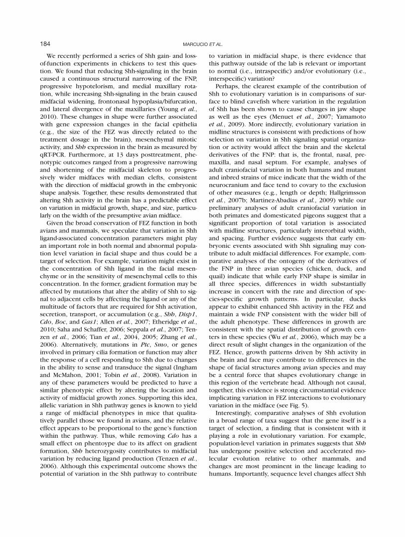
Perhaps, the clearest example of the contribution ofShh to evolutionary variation is in comparisons of sur-face to blind cavefish where variation in the regulationof Shh has been shown to cause changes in jaw shapeas well as the eyes (Menuet et al., 2007; Yamamotoet al., 2009). More indirectly, evolutionary variation inmidline structures is consistent with predictions of howselection on variation in Shh signaling spatial organiza-tion or activity would affect the brain and the skeletalderivatives of the FNP: that is, the frontal, nasal, pre-maxilla, and nasal septum. For example, analyses ofadult craniofacial variation in both humans and mutantand inbred strains of mice indicate that the width of theneurocranium and face tend to covary to the exclusionof other measures (e.g., length or depth; Hallgrimssonet al., 2007b; Martinez-Abadias et al., 2009) while ourpreliminary analyses of adult craniofacial variation inboth primates and domesticated pigeons suggest that asignificant proportion of total variation is associatedwith midline structures, particularly interorbital width,and spacing. Further evidence suggests that early em-bryonic events associated with Shh signaling may con-tribute to adult midfacial differences. For example, com-parative analyses of the ontogeny of the derivatives ofthe FNP in three avian species (chicken, duck, andquail) indicate that while early FNP shape is similar inall three species, differences in width substantiallyincrease in concert with the rate and direction of spe-cies-specific growth patterns. In particular, ducksappear to exhibit enhanced Shh activity in the FEZ andmaintain a wide FNP consistent with the wider bill ofthe adult phenotype. These differences in growth areconsistent with the spatial distribution of growth cen-ters in these species (Wu et al., 2006), which may be adirect result of slight changes in the organization of theFEZ. Hence, growth patterns driven by Shh activity inthe brain and face may contribute to differences in theshape of facial structures among avian species and maybe a central force that shapes evolutionary change inthis region of the vertebrate head. Although not causal,together, this evidence is strong circumstantial evidenceimplicating variation in FEZ interactions to evolutionaryvariation in the midface (see Fig. 5).
Interestingly, comparative analyses of Shh evolutionin a broad range of taxa suggest that the gene itself is atarget of selection, a finding that is consistent with itplaying a role in evolutionary variation. For example,population-level variation in primates suggests that Shhhas undergone positive selection and accelerated mo-lecular evolution relative to other mammals, andchanges are most prominent in the lineage leading tohumans. Importantly, sequence level changes affect Shh
184 MARCUCIO ET AL.
at the protein level and suggest that apes and humanshave evolved more complex posttranslational regulationthat may be related to the dramatic evolution of thebrain, the skeleton of the face, or both, in this lineage(Dorus et al., 2006). Similarly, recent analysis of theNeandertal genome suggests that DISP1, a key compo-nent of Shh signaling, also experienced positive selec-tion in modern humans that may reflect similar selec-tion on the brain and/or midface (Green et al.). Furtheranalyses are needed to determine how these lineageand species-specific changes affect developmental pa-rameters associated with the brain and face.
DISEASES OF THE BRAIN-FACE COMPLEX
Just as interactions between the brain and face mayserve as a source of normal or evolutionary variation,when these same processes are disrupted they may alsolead to disease phenotypes. Often facial malformationsare accompanied by underlying brain defects (DeMyer,
1964). We have described two fundamental ways bywhich development of the brain and face are interre-lated. First, the brain serves as an architectural founda-tion upon which the face develops. Thus, as the founda-tion expands, the face accommodates and developsaccordingly, and perturbations to the frame can lead tofacial defects. Second, molecular signaling between thebrain and face regulates morphogenesis. Hence, disrupt-ing this molecular dialogue can create malformations inboth tissues. This is exemplified in humans whereextremes of Shh signaling produce disease phenotypes(Muenke and Cohen, 2000). In holoprosencephaly, Shhsignaling is reduced, the brain can be severely mal-formed, and facial malformations range from hypotelor-ism and midfacial hypoplasia to complete cyclopia(Muenke and Beachy, 2000), while in Greig cephalopo-lysyndactyly and Gorlin syndrome, mutations in GLI3 orPTC increase Shh-signaling activity and phenotypesrange from hypertelorism to medial clefts of the face(Balk and Biesecker, 2008; see Fig. 5). Importantly, in
FIG. 5. Hypothetical relationship of Shh-signaling activity in the brain and FEZ to both population and evolutionary level variation in midfa-cial shape. A: Normal levels of Shh-signaling activity in the brain establish the FEZ, which establishes growth zones that help to regulate theshape and size of midfacial structures, such as the frontal, premaxilla, and nasal septum. Extremes of signaling due to mutational effects,environmental factors, or a combination of both can cause progressive loss of midline structures when signaling is abnormally low (e.g., inHoloprosencephaly) or expansion of midline structures when signaling is abnormally high. More subtle differences in signaling introduce var-iation in the midline facial structures of normal individuals that can serve as a target of natural selection. B: Individual species exhibit a rangeof intraspecific variation in midfacial shape as well as interspecific or evolutionary variation between species. In the case of hominoid apes,interorbital width varies from narrower in the orangutan to wider in gorillas (tree illustrates phylogenetic relationships of living and recent spe-cies as well as facial skeletons of potential ancestral species). Variation in Shh signaling, in combination with variation in associated cranio-facial structures, is hypothesized to have contributed to the evolution of the midface in living apes and humans as well as other vertebratespecies.
185MECHANISMS THAT UNDERLIE COVARIATION OF BRAIN AND FACE

each of these diseases, family members of affected indi-viduals may manifest less-severe midfacial phenotypessuch as relatively narrower or wider faces (Balk andBiesecker, 2008; Muenke and Beachy, 2000), suggestingthat variation in Shh signaling between the brain andface contributes to normal variation in midfacial shapeand size.
CONCLUSIONS
In this work, we focused on the physical interactionsbetween the brain and face, the molecular level epithe-lial–mesenchymal interactions among the brain, theFEZ, and the neural crest mesenchyme, and their contri-bution to facial variation. However, it is important toremember that these comprise only a small number ofpotential factors contributing to facial morphogenesisand that craniofacial morphology is the result of a seriesof overlapping developmental events, each contributingto the final outcome (i.e., the adult phenotype) (Atchleyand Hall, 1991; Hallgrı́msson, 2009). For example, theshape of the bones and cartilages of the jaw, which areunique in that they are neural crest derivatives(Donoghue et al., 2008; Evans and Noden, 2006), arethe result of multiple inputs from neural crest formationand migration to general skeletal growth (e.g., Atchleyand Hall, 1991), each with a relative contribution thatmay differ in both magnitude and direction. Early eventslike neural crest migration may be masked by variationin later events such as somatic growth, or early eventsmay have a large effect that constrains later events toparticular outcomes. This model of development sug-gests that the adult phenotype, much like a Medieval‘‘palimpsest,’’ which holds the entire contents of itshistory in opaque terms, makes simple genotype–phe-notype correlations for complex traits unlikely (Hall-grı́msson, 2009). Ultimately, to understand how adultphenotypic diversity is generated, we must betterunderstand how genetic variation impacts individualdevelopmental events and how these contribute toadult phenotypic outcomes, overall population varia-tion, and ultimately evolutionary and disease variation.
ACKNOWLEDGMENTS
We thank the members of the Marcucio and Hallgrims-son laboratories as well as the many colleagues whohave discussed this work with us.
LITERATURE CITED
Abzhanov A, Protas M, Grant BR, Grant PR, Tabin CJ.2004. Bmp4 and morphological variation of beaksin Darwin’s finches. Science 305:1462–1465.
Abzhanov A, Rodda SJ, McMahon AP, Tabin CJ. 2007.Regulation of skeletogenic differentiation in cranialdermal bone. Development 134:3133–3144.
Abzhanov A, Tabin CJ. 2004. Shh and Fgf8 act synergisti-cally to drive cartilage outgrowth during cranial de-velopment. Dev Biol 273:134–148.
Ahituv N, Rubin EM, Nobrega MA. 2004. Exploitinghuman–fish genome comparisons for decipheringgene regulation. Hum Mol Genet 13:R261–R266.
Ahlgren SC, Bronner-Fraser M. 1999. Inhibition of sonichedgehog signaling in vivo results in craniofacialneural crest cell death. Curr Biol 9:1304–1314.
Aldridge K, Kane AA, Marsh JL, Yan P, Govier D, Richts-meier JT. 2005. Relationship of brain and skull inpre- and postoperative sagittal synostosis. J Anat206:373–385.
Allen BL, Tenzen T, McMahon AP. 2007. The Hedgehog-binding proteins Gas1 and Cdo cooperate topositively regulate Shh signaling during mousedevelopment. Genes Dev 21:1244–1257.
Ashique AM, Fu K, Richman JM. 2002a. Endogenousbone morphogenetic proteins regulate outgrowthand epithelial survival during avian lip fusion. Devel-opment 129:4647–4660.
Ashique AM, Fu K, Richman JM. 2002b. Signallingvia type IA and type IB bone morphogenetic proteinreceptors (BMPR) regulates intramembranousbone formation, chondrogenesis and feather forma-tion in the chicken embryo. Int J Dev Biol 46:243–253.
Atchley WR, Hall BK. 1991. A model for developmentand evolution of complex morphological structures.Biol Rev Camb Philos Soc 66:101–157.
Balk K, Biesecker LG. 2008. The clinical atlas of Greigcephalopolysyndactyly syndrome. Am J Med GenetA 146:548–557.
Barlow AJ, Francis-West PH. 1997. Ectopic applicationof recombinant BMP-2 and BMP-4 can change pat-terning of developing chick facial primordia. Devel-opment 124:391–398.
Bennett JH, Hunt P, Thorogood P. 1995. Bone morpho-genetic protein-2 and -4 expression during murineorofacial development. Arch Oral Biol 40:847–854.
Biegert J. 1963. The evaluation of characteristics ofthe skull, hands and feet for primate taxonomy. InWashburn SL, editor. Classification and humanevolution. Chicago: Aldine. pp 116–145.
Boughner JC, Wat S, Diewert VM, Young NM, BrowderLW, Hallgrimsson B. 2008. Short-faced mice anddevelopmental interactions between the brain andthe face. J Anat 213:646–662.
Brito JM, Teillet MA, Le Douarin NM. 2006. An early rolefor sonic hedgehog from foregut endoderm in jawdevelopment: Ensuring neural crest cell survival.Proc Natl Acad Sci USA 103:11607–11612.
Brito JM, Teillet MA, Le Douarin NM. 2008. Induction ofmirror-image supernumerary jaws in chicken man-dibular mesenchyme by Sonic Hedgehog-producingcells. Development 135:2311–2319.
186 MARCUCIO ET AL.

Chai Y, Maxson RE Jr. 2006. Recent advances in cranio-facial morphogenesis. Dev Dyn 235:2353–2375.
Cordero D, Marcucio R, Hu D, Gaffield W, Tapadia M,Helms JA. 2004. Temporal perturbations in sonichedgehog signaling elicit the spectrum of holopro-sencephaly phenotypes. J Clin Invest 114:485–494.
Creuzet SE. 2009a. Neural crest contribution to fore-brain development. Semin Cell Dev Biol 20:751–759.
Creuzet SE. 2009b. Regulation of pre-otic brain develop-ment by the cephalic neural crest. Proc Natl AcadSci USA 106:15774–15779.
Creuzet SE, Martinez S, Le Douarin NM. 2006. Thecephalic neural crest exerts a critical effect on fore-brain and midbrain development. Proc Natl Acad SciUSA 103:14033–14038.
Dahmane N, Lee J, Robins P, Heller P, Ruiz I, Altaba A.1997. Activation of the transcription factor Gli 1and the Sonic hedgehog signalling pathway in skintumors. Nature 389:876–881.
DeBeer G. 1937. The development of the vertebrateskull. London: Oxford University Press.
DeMyer W. 1964. The face predicts the brain: Diagnos-tic significance of median facial anomialies forholoprosencephaly (arhinencephay). Pediatrics 34:256–263.
Depew MJ, Compagnucci C. 2008. Tweaking the hingeand caps: Testing a model of the organization ofjaws. J Exp Zool B Mol Dev Evol 310:315–335.
Diewert VM. 1983. A morphometric analysis of craniofa-cial growth and changes in spatial relations duringsecondary palatal development in human embryosand fetuses. Am J Anat 167:495–522.
Diewert VM. 1985. Growth movements during prenataldevelopment of human facial morphology. Prog ClinBiol Res 187:57–66.
Diewert VM, Lozanoff S. 1993a. A morphometric analy-sis of human embryonic craniofacial growth in themedian plane during primary palate formation.J Craniofac Genet Dev Biol 13:147–161.
Diewert VM, Lozanoff S, Choy V. 1993b. Computerreconstructions of human embryonic craniofacialmorphology showing changes in relations betweenthe face and brain during primary palate formation.J Craniofac Genet Dev Biol 13:193–201.
Donoghue PC, Graham A, Kelsh RN. 2008. The originand evolution of the neural crest. Bioessays 30:530–541.
Dorus S, Anderson JR, Vallender EJ, Gilbert SL, Zhang L,Chemnick LG, Ryder OA, Li W, Lahn BT. 2006. SonicHedgehog, a key development gene, experiencedintensified molecular evolution in primates. HumMol Genet 15:2031–2037.
Eberhart JK, He X, Swartz ME, Yan YL, Song H, BolingTC, Kunerth AK, Walker MB, Kimmel CB, Post-lethwait JH. 2008. MicroRNA Mirn140 modulates
Pdgf signaling during palatogenesis. Nat Genet40:290–298.
Eberhart JK, Swartz ME, Crump JG, Kimmel CB. 2006.Early Hedgehog signaling from neural to oral epithe-lium organizes anterior craniofacial development.Development 133:1069–1077.
Epstein DJ, Martinu L, Michaud JL, Losos KM, Fan C,Joyner AL. 2000. Members of the bHLH-PAS familyregulate Shh transcription in forebrain regions ofthe mouse CNS. Development 127:4701–4709.
Epstein DJ, McMahon AP, Joyner AL. 1999. Regionaliza-tion of Sonic hedgehog transcription along theanteroposterior axis of the mouse central nervoussystem is regulated by Hnf3-dependent and-inde-pendent mechanisms. Development 126:281–292.
Etheridge LA, Crawford TQ, Zhang S, Roelink H. 2010.Evidence for a role of vertebrate Disp1 in long-rangeShh signaling. Development 137:133–140.
Evans DJ, Noden DM. 2006. Spatial relations betweenavian craniofacial neural crest and paraxial meso-derm cells. Dev Dyn 235:1310–1325.
Firnberg N, Neubuser A. 2002. FGF signaling regulatesexpression of Tbx2, Erm, Pea3, and Pax3 in theearly nasal region. Dev Biol 247:237–250.
Foppiano S, Hu D, Marcucio RS. 2007. Signaling bybone morphogenetic proteins directs formation ofan ectodermal signaling center that regulates cranio-facial development. Dev Biol 312:103–114.
Francis-West P, Ladher R, Barlow A, Graveson A. 1998.Signalling interactions during facial development.Mech Dev 75:3–28.
Francis-West PH, Tatla T, Brickell PM. 1994. Expressionpatterns of the bone morphogenetic protein genesBmp-4 and Bmp-2 in the developing chick facesuggest a role in outgrowth of the primordia. DevDyn 201:168–178.
Geetha-Loganathan P, Nimmagadda S, Antoni L, Fu K,Whiting CJ, Francis-West P, Richman JM. 2009. Expres-sion of WNT signalling pathway genes during chickencraniofacial development. Dev Dyn 238:1150–1165.
Geng X, Speirs C, Lagutin O, Inbal A, Liu W, Solnica-Kre-zel L, Jeong Y, Epstein DJ, Oliver G. 2008. Haploin-sufficiency of Six3 fails to activate Sonic hedgehogexpression in the ventral forebrain and causes holo-prosencephaly. Dev Cell 15:236–247.
Golden JA, Bracilovic A, McFadden KA, Beesley JS,Rubenstein JL, Grinspan JB. 1999. Ectopic bonemorphogenetic proteins 5 and 4 in the chicken fore-brain lead to cyclopia and holoprosencephaly. ProcNatl Acad Sci USA 96:2439–2444.
Goodrich LV, Scott MP. 1998. Hedgehog and patched inneural development and disease. Neuron 21:1243–1257.
Graham A, Okabe M, Quinlan R. 2005. The role of theendoderm in the development and evolution of thepharyngeal arches. J Anat 207:479–487.
187MECHANISMS THAT UNDERLIE COVARIATION OF BRAIN AND FACE

Green RE, Krause J, Briggs AW, Maricic T, Stenzel U,Kircher M, Patterson N, Li H, Zhai W, Fritz MH,Hansen NF, Durand EY, Malaspinas AS, Jensen JD,Marques-Bonet T, Alkan C, Prufer K, Meyer M,Burbano HA, Good JM, Schultz R, Aximu-Petri A,Butthof A, Hober B, Hoffner B, Siegemund M, Weih-mann A, Nusbaum C, Lander ES, Russ C, Novod N,Affourtit J, Egholm M, Verna C, Rudan P, BrajkovicD, Kucan Z, Gusic I, Doronichev VB, Golovanova LV,Lalueza-Fox C, de la Rasilla M, Fortea J, Rosas A,Schmitz RW, Johnson PL, Eichler EE, Falush D,Birney E, Mullikin JC, Slatkin M, Nielsen R, Kelso J,Lachmann M, Reich D, Paabo S. A draft sequence ofthe Neandertal genome. Science 328:710–722.
Hallgrı́msson B, Jamniczky H, Young NM, Rolian C,Parsons TE, Boughner JC, Marcucio RS. 2009. Deci-phering the palimpsest: Studying the relationshipbetween morphological integration and phenotypiccovariation. Evolut Biol 36:355–376.
Hallgrimsson B, Lieberman DE. 2008. Mouse modelsand the evolutionary developmental biology of theskull. Integr Comp Biol 48:373–384.
Hallgrimsson B, Lieberman DE, Liu W, Ford-HutchinsonAF, Jirik FR. 2007a. Epigenetic interactions and thestructure of phenotypic variation in the cranium.Evol Dev 9:76–91.
Hallgrimsson B, Lieberman DE, Young NM, Parsons T,Wat S. 2007b. Evolution of covariance in the mam-malian skull. Novartis Found Symp 284:164–185;discussion 185-190.
Hamburger V, Hamilton HL. 1951. A series of develop-mental stages in development of the chick embryo.J Morphol 88:49–92.
Hu D, Colnot C, Marcucio RS. 2008. Effect of bone mor-phogenetic protein signaling on development of thejaw skeleton. Dev Dyn 237:3727–3737.
Hu D, Marcucio RS. 2009a. A SHH-responsive signalingcenter in the forebrain regulates craniofacial mor-phogenesis via the facial ectoderm. Development136:107–116.
Hu D, Marcucio RS. 2009b. Unique organization of thefrontonasal ectodermal zone in birds and mammals.Dev Biol 325:200–210.
Hu D, Marcucio R, Helms JA. 2003. A zone of fronto-nasal ectoderm regulates patterning and growth inthe face. Development 130:1749–1758.
Ingham PW, McMahon AP. 2001. Hedgehog signaling inanimal development: Paradigms and principles.Genes Dev 15:3059–3087.
Jeong Y, El-Jaick K, Roessler E, Muenke M, Epstein DJ.2006. A functional screen for sonic hedgehog regu-latory elements across a 1 Mb interval identifieslong-range ventral forebrain enhancers. Develop-ment 133:761–772.
Jeong Y, Epstein DJ. 2003. Distinct regulators of Shhtranscription in the floor plate and notochord indi-
cate separate origins for these tissues in the mousenode. Development 130:3891–3902.
Jeong Y, Leskow FC, El-Jaick K, Roessler E, Muenke M,Yocum A, Dubourg C, Li X, Geng X, Oliver G,Epstein DJ. 2008. Regulation of a remote Shh fore-brain enhancer by the Six3 homeoprotein. NatGenet 40:1348–1353.
Jeong J, Mao J, Tenzen T, Kottmann AH, McMahon AP.2004. Hedgehog signaling in the neural crest cellsregulates the patterning and growth of facial primor-dia. Genes Dev 18:937–951.
Le Douarin NM, Brito JM, Creuzet S. 2007. Role of theneural crest in face and brain development. BrainRes Rev 55:237–247.
Lieberman DE, Hallgrimsson B, Liu W, Parsons TE, Jam-niczky HA. 2008. Spatial packing, cranial base angu-lation, and craniofacial shape variation in the mam-malian skull: Testing a new model using mice. J Anat212:720–735.
Marcucio RS, Cordero DR, Hu D, Helms JA. 2005. Molec-ular interactions coordinating the development ofthe forebrain and face. Dev Biol 284:48–61.
Martinez-Abadias N, Esparza M, Sjovold T, Gonzalez-JoseR, Santos M, Hernandez M. 2009. Heritability ofhuman cranial dimensions: Comparing the evolv-ability of different cranial regions. J Anat 214:19–35.
Menuet A, Alunni A, Joly JS, Jeffery WR, Retaux S. 2007.Expanded expression of Sonic Hedgehog in Astya-nax cavefish: Multiple consequences on forebraindevelopment and evolution. Development 134:845–855.
Muenke M, Beachy PA. 2000. Genetics of ventral fore-brain development and holoprosencephaly. CurrOpin Genet Dev 10:262–269.
Muenke M, Cohen MM Jr, 2000. Genetic approaches tounderstanding brain development: Holoprosence-phaly as a model. Ment Retard Dev Disabil Res Rev6:15–21.
Odent S, Atti-Bitach T, Blayau M, Mathieu M, Aug J, deDelezo AL, Gall JY, Le Marec B, Munnich A, David V,Vekemans M. 1999. Expression of the Sonic hedge-hog (SHH) gene during early human developmentand phenotypic expression of new mutations caus-ing holoprosencephaly. Hum Mol Genet 8:1683–1689.
Ohkubo Y, Chiang C, Rubenstein JL. 2002. Coordinateregulation and synergistic actions of BMP4. SHH andFGF8 in the rostral prosencephalon regulate mor-phogenesis of the telencephalic and optic vesicles.Neuroscience 111:1–17.
Reid B, Yang H, Melvin VS, Teketo MM, Williams T.2011. Ectodermal Wnt/b-catenin signaling shapesthe mouse face. Dev Biol 349:261–269.
Richman JM, Lee SH. 2003. About face: Signals andgenes controlling jaw patterning and identity in ver-tebrates. Bioessays 25:554–568.
188 MARCUCIO ET AL.

Richtsmeier JT, Aldridge K, DeLeon VB, Panchal J, KaneAA, Marsh JL, Yan P, Cole TM III. 2006. Phenotypicintegration of neurocranium and brain. J Exp Zool BMol Dev Evol 306:360–378.
Richtsmeier JT, Deleon VB. 2009. Morphological inte-gration of the skull in craniofacial anomalies. OrthodCraniofac Res 12:149–158.
Rosen V. 2006. BMP and BMP inhibitors in bone. AnnNYAcad Sci 1068:19–25.
Ross C, Henneberg M. 1995. Basicranial flexion, relativebrain size, and facial kyphosis in Homo sapiens andsome fossil hominids. Am J Phys Anthropol 98:575–593.
Ross CF, Ravosa MJ. 1993. Basicranial flexion, relativebrain size, and facial kyphosis in nonhuman prima-tes. Am J Phys Anthropol 91:305–324.
Ruiz i Altaba A. 1997. Catching a Gli-mpse of Hedgehog.Cell 90:193–196.
Saha K, Schaffer DV. 2006. Signal dynamics in Sonichedgehog tissue patterning. Development 133:889–900.
Schneider RA, Helms JA. 2003. The cellular and molecu-lar origins of beak morphology. Science 299:565–568.
Schneider RA, Hu D, Rubenstein JL, Maden M, HelmsJA. 2001. Local retinoid signaling coordinates fore-brain and facial morphogenesis by maintainingFGF8 and SHH. Development 128:2755–2767.
Seppala M, Depew MJ, Martinelli DC, Fan CM, SharpePT, Cobourne MT. 2007. Gas1 is a modifier for holo-prosencephaly and genetically interacts with sonichedgehog. J Clin Invest 117:1575–1584.
Szabo-Rogers HL, Geetha-Loganathan P, Nimmagadda S,Fu KK, Richman JM. 2008. FGF signals from thenasal pit are necessary for normal facial morphogen-esis. Dev Biol 318:289–302.
Szabo-Rogers HL, Geetha-Loganathan P, Whiting CJ,Nimmagadda S, Fu K, Richman JM. 2009a. Novelskeletogenic patterning roles for the olfactory pit.Development 136:219–229.
Szabo-Rogers HL, Smithers LE, Yakob W, Liu KJ. 2009b.New directions in craniofacial morphogenesis. DevBiol 34:84–94.
Tenzen T, Allen BL, Cole F, Kang JS, Krauss RS, McMa-hon AP. 2006. The cell surface membrane proteins
Cdo and Boc are components and targets of theHedgehog signaling pathway and feedback networkin mice. Dev Cell 10:647–656.
Tian H, Jeong J, Harfe BD, Tabin CJ, McMahon AP. 2005.Mouse Disp1 is required in sonic hedgehog-express-ing cells for paracrine activity of the cholesterol-modified ligand. Development 132:133–142.
Tian H, Tenzen T, McMahon AP. 2004. Dose dependencyof Disp1 and genetic interaction between Disp1 andother hedgehog signaling components in themouse. Development 131:4021–4033.
Tobin JL, Di Franco M, Eichers E, May-Simera H, GarciaM, Yan J, Quinlan R, Justice MJ, Hennekam RC, Bris-coe J, Tada M, Mayor R, Burns AJ, Lupski JR, Ham-mond P, Beales PL. 2008. Inhibition of neural crestmigration underlies craniofacial dysmorphology andHirschsprung’s disease in Bardet-Biedl syndrome.Proc Natl Acad Sci USA 105:6714–6719.
Welsh IC, O’Brien TP. 2009. Signaling integration in therugae growth zone directs sequential SHH signalingcenter formation during the rostral outgrowth ofthe palate. Dev Biol 336:53–67.
Wu P, Jiang TX, Shen JY, Widelitz RB, Chuong CM. 2006.Morphoregulation of avian beaks: Comparative map-ping of growth zone activities and morphologicalevolution. Dev Dyn 235:1400–1412.
Wu P, Jiang TX, Suksaweang S, Widelitz RB, ChuongCM. 2004. Molecular shaping of the beak. Science305:1465–1466.
Yamamoto Y, Byerly MS, Jackman WR, Jeffery WR.2009. Pleiotropic functions of embryonic sonichedgehog expression link jaw and taste bud amplifi-cation with eye loss during cavefish evolution. DevBiol 330:200–211.
Young NM, Chong HJ, Hu D, Hallgrimsson B, MarcucioRS. 2010. Quantitative analyses link modulation ofsonic hedgehog signaling to continuous variation infacial growth and shape. Development 137:3405–3409.
Zhang W, Kang JS, Cole F, Yi MJ, Krauss RS. 2006. Cdofunctions at multiple points in the Sonic Hedgehogpathway, and Cdo-deficient mice accurately modelhuman holoprosencephaly. Dev Cell 10:657–665.
Weinberg SM, Andreasen NC, Nopoulos P. 2009. J Anat214:926–936.
189MECHANISMS THAT UNDERLIE COVARIATION OF BRAIN AND FACE
Related Documents