MYELOID NEOPLASIA Mechanisms of resistance to 5-aza-2 -deoxycytidine in human cancer cell lines Taichun Qin, 1 Jaroslav Jelinek, 1 Jiali Si, 1 Jingmin Shu, 1 and Jean-Pierre J. Issa 1 1 Department of Leukemia, University of Texas M. D. Anderson Cancer Center, Houston 5-aza-2-deoxycytidine (DAC) is approved for the treatment of myelodysplastic syn- dromes, but resistance to this agent is common. In search for mechanisms of resistance, we measured the half maxi- mal (50%) inhibitory concentration (IC 50 ) of DAC and found it differed 1000-fold among a panel of cancer cell lines. The IC 50 was correlated with the doses of DAC that induced the most hypomethylation of long interspersed nuclear elements (LINE; R 0.94, P < .001), but not with LINE methylation or DNA methyltransferase 1 (DNMT1), 3a, and 3b expression at base- line. Sensitivity to DAC showed a low correlation (R 0.44, P .11) to that of 5-azacytidine (AZA), but a good correla- tion to that of cytarabine (Ara-C; R 0.89, P < .001). The 5 cell lines most resistant to DAC had a combination of low dCK, hENT1, and 2 transporters, and high cyto- sine deaminase. In an HL60 clone, resis- tance to DAC could be rapidly induced by drug exposure and was related to a switch from heterozygous to homozygous muta- tion of DCK. Transfection of wild-type DCK restored DAC sensitivity. DAC in- duced DNA breaks as evidenced by H2AX phosphorylation and increased homolo- gous recombination rates by 7- to 10-fold. These results suggest that in vitro resis- tance to DAC can be explained by insuffi- cient incorporation into DNA. (Blood. 2009;113:659-667) Introduction Epigenetic changes have been increasingly recognized as a driving force in human leukemia. 1-3 Abnormal methylation, for example, appears to accumulate over time at various sites in the genome and to promote tumorigenesis by increasing genomic instability or by silencing tumor suppressor genes. Silencing of tumor suppressor genes is closely associated with hypermethylation; methylated tumor suppressor genes can be reactivated by DNA methyltrans- ferase (DNMT) inhibitors. These observations have led to a revival of interest in DNA methylation inhibitors as antineoplastic agents in clinical trials. 4 The prototypical DNMT inhibitors 5-aza-2- deoxycytidine (DAC) and 5-azacytidine (AZA) have recently been approved by the US Food and Drug Administration as antitumor agents for the treatment of myelodysplastic syndrome. 5,6 One potential problem with both agents is that resistance can develop during treatment. Like other cytosine nucleoside analogs (NAs), DAC enters cells using the equilibrative nucleoside transporters hENT1 and hENT2. Once inside the cell, DAC is phosphorylated by deoxycytidine kinase (dCK) into the monophosphorylated derivative 5-aza- dCMP. Subsequently, 5-aza-dCMP is phosphorylated to its active form, 5-aza-dCTP, which is incorporated into DNA, where it induces demethylation. DAC metabolites might also be substrates for catabolizing enzymes such as cytidine deaminase (CDA), which catalyze the inactivation of cytidine and deoxycytidine to uridine and deoxyuridine, thereby decreasing the amount of 5-aza-dCTP that can be formed. The nucleoside analog Ara-C also relies on dCK as the initial rate-limiting step for incorporation. AZA, however, does not need dCK. Its incorporation is dependent on uridine-cytidine kinase (UCK). dCK deficiency is the major known mechanism of resistance to cytidine NAs in vitro and was reported to be related to in vivo resistance in some patients. 7,8 For example, in vitro–induced resistance to the deoxycytidine analogs cytarabine (Ara-C) and DAC in a rat model for acute myeloid leukemia was mediated by mutations in the DCK gene. 9 However, there is little information regarding the origin of this kind of resistance. Resistance to treatment with anticancer drugs results from a variety of factors, including spontaneous genetic instability in tumors and drug induction mechanisms that probably play an important role in acquired anticancer drug resistance. 10-16 Defects in DNA methyl- ation might contribute to genomic instability, leading to elevated mutation rates. 17-20 Therefore, we hypothesized that both spontane- ous genetic instability and DAC-induced genetic instability contrib- ute to the origin of resistance to DAC in human cancer cell lines and that resistance to DAC is due to the insufficient intracellular triphosphate of DAC. To test these hypotheses, we examined in vitro models of naturally occurring resistance to DAC in different cancer cell lines and further investigated the mechanisms and origin of resistance in the myeloid leukemia cell line HL60. Methods Cell culture and treatment protocols The human leukemia and lymphoma cell lines HL60, ML-1, HEL, Raji, Jurkat, TF-1, U937, K562, and MOLT4; prostate cancer cell lines PC3 and DU145; colon cancer cell lines RKO and SW48; and breast cancer cell line Cama-1 were obtained fromATCC (Manassas, VA). The cells were grown in RPMI 1640 plus 10% heat-inactivated fetal calf serum (FCS) in plastic tissue-culture plates in a humidified atmosphere containing 5% CO 2 at 37°C. For the growth inhibition assay, cells were placed at a density of 2.5 10 5 /mL in 5 mL medium 24 hours before treatment. Graded concen- trations of DAC were added to the medium. To measure half maximal Submitted February 19, 2008; accepted August 27, 2008. Prepublished online as Blood First Edition paper, October 17, 2008; DOI 10.1182/blood-2008-02- 140038. The publication costs of this article were defrayed in part by page charge payment. Therefore, and solely to indicate this fact, this article is hereby marked ‘‘advertisement’’ in accordance with 18 USC section 1734. © 2009 by The American Society of Hematology 659 BLOOD, 15 JANUARY 2009 VOLUME 113, NUMBER 3 For personal use only. on April 8, 2016. by guest www.bloodjournal.org From

Welcome message from author
This document is posted to help you gain knowledge. Please leave a comment to let me know what you think about it! Share it to your friends and learn new things together.
Transcript

MYELOID NEOPLASIA
Mechanisms of resistance to 5-aza-2�-deoxycytidine in human cancer cell linesTaichun Qin,1 Jaroslav Jelinek,1 Jiali Si,1 Jingmin Shu,1 and Jean-Pierre J. Issa1
1Department of Leukemia, University of Texas M. D. Anderson Cancer Center, Houston
5-aza-2�-deoxycytidine (DAC) is approvedfor the treatment of myelodysplastic syn-dromes, but resistance to this agent iscommon. In search for mechanisms ofresistance, we measured the half maxi-mal (50%) inhibitory concentration (IC50)of DAC and found it differed 1000-foldamong a panel of cancer cell lines. TheIC50 was correlated with the doses of DACthat induced the most hypomethylation oflong interspersed nuclear elements (LINE;R � 0.94, P < .001), but not with LINE
methylation or DNA methyltransferase 1(DNMT1), 3a, and 3b expression at base-line. Sensitivity to DAC showed a lowcorrelation (R � 0.44, P � .11) to that of5-azacytidine (AZA), but a good correla-tion to that of cytarabine (Ara-C; R � 0.89,P < .001). The 5 cell lines most resistantto DAC had a combination of low dCK,hENT1, and 2 transporters, and high cyto-sine deaminase. In an HL60 clone, resis-tance to DAC could be rapidly induced bydrug exposure and was related to a switch
from heterozygous to homozygous muta-tion of DCK. Transfection of wild-typeDCK restored DAC sensitivity. DAC in-duced DNA breaks as evidenced by H2AXphosphorylation and increased homolo-gous recombination rates by 7- to 10-fold.These results suggest that in vitro resis-tance to DAC can be explained by insuffi-cient incorporation into DNA. (Blood.2009;113:659-667)
Introduction
Epigenetic changes have been increasingly recognized as a drivingforce in human leukemia.1-3 Abnormal methylation, for example,appears to accumulate over time at various sites in the genome andto promote tumorigenesis by increasing genomic instability or bysilencing tumor suppressor genes. Silencing of tumor suppressorgenes is closely associated with hypermethylation; methylatedtumor suppressor genes can be reactivated by DNA methyltrans-ferase (DNMT) inhibitors. These observations have led to a revivalof interest in DNA methylation inhibitors as antineoplastic agentsin clinical trials.4 The prototypical DNMT inhibitors 5-aza-2�-deoxycytidine (DAC) and 5-azacytidine (AZA) have recently beenapproved by the US Food and Drug Administration as antitumoragents for the treatment of myelodysplastic syndrome.5,6 Onepotential problem with both agents is that resistance can developduring treatment.
Like other cytosine nucleoside analogs (NAs), DAC enters cellsusing the equilibrative nucleoside transporters hENT1 and hENT2.Once inside the cell, DAC is phosphorylated by deoxycytidinekinase (dCK) into the monophosphorylated derivative 5-aza-dCMP. Subsequently, 5-aza-dCMP is phosphorylated to its activeform, 5-aza-dCTP, which is incorporated into DNA, where itinduces demethylation. DAC metabolites might also be substratesfor catabolizing enzymes such as cytidine deaminase (CDA),which catalyze the inactivation of cytidine and deoxycytidine touridine and deoxyuridine, thereby decreasing the amount of5-aza-dCTP that can be formed. The nucleoside analog Ara-C alsorelies on dCK as the initial rate-limiting step for incorporation.AZA, however, does not need dCK. Its incorporation is dependenton uridine-cytidine kinase (UCK).
dCK deficiency is the major known mechanism of resistance tocytidine NAs in vitro and was reported to be related to in vivo
resistance in some patients.7,8 For example, in vitro–inducedresistance to the deoxycytidine analogs cytarabine (Ara-C) andDAC in a rat model for acute myeloid leukemia was mediated bymutations in the DCK gene.9 However, there is little informationregarding the origin of this kind of resistance. Resistance totreatment with anticancer drugs results from a variety of factors,including spontaneous genetic instability in tumors and druginduction mechanisms that probably play an important role inacquired anticancer drug resistance.10-16 Defects in DNA methyl-ation might contribute to genomic instability, leading to elevatedmutation rates.17-20 Therefore, we hypothesized that both spontane-ous genetic instability and DAC-induced genetic instability contrib-ute to the origin of resistance to DAC in human cancer cell linesand that resistance to DAC is due to the insufficient intracellulartriphosphate of DAC. To test these hypotheses, we examined invitro models of naturally occurring resistance to DAC in differentcancer cell lines and further investigated the mechanisms andorigin of resistance in the myeloid leukemia cell line HL60.
Methods
Cell culture and treatment protocols
The human leukemia and lymphoma cell lines HL60, ML-1, HEL, Raji,Jurkat, TF-1, U937, K562, and MOLT4; prostate cancer cell lines PC3 andDU145; colon cancer cell lines RKO and SW48; and breast cancer cell lineCama-1 were obtained from ATCC (Manassas, VA). The cells were grownin RPMI 1640 plus 10% heat-inactivated fetal calf serum (FCS) in plastictissue-culture plates in a humidified atmosphere containing 5% CO2 at37°C. For the growth inhibition assay, cells were placed at a density of2.5 � 105/mL in 5 mL medium 24 hours before treatment. Graded concen-trations of DAC were added to the medium. To measure half maximal
Submitted February 19, 2008; accepted August 27, 2008. Prepublished onlineas Blood First Edition paper, October 17, 2008; DOI 10.1182/blood-2008-02-140038.
The publication costs of this article were defrayed in part by page charge
payment. Therefore, and solely to indicate this fact, this article is herebymarked ‘‘advertisement’’ in accordance with 18 USC section 1734.
© 2009 by The American Society of Hematology
659BLOOD, 15 JANUARY 2009 � VOLUME 113, NUMBER 3
For personal use only.on April 8, 2016. by guest www.bloodjournal.orgFrom

(50%) inhibitory concentration (IC50), fresh DAC was added every24 hours without changing medium. The doses that inhibited proliferationto 50% (IC50) were analyzed by the median-effect method with CalcuSynsoftware (Biosoft, Cambridge, United Kingdom).21 The proportion of livecells in treated plates was measured by trypan blue exclusion.
Pyrosequencing
We used the DNA repetitive long interspersed nuclear element (LINE) as amarker and pyrosequencing-based methylation analysis to study globalgenomic DNA methylation, as previously described.5 Genomic DNA wasprepared from cells, and bisulfite conversion of genomic DNA was carriedout. LINE was amplified by PCR using a forward primer 5�-TTTTTTGAGT-TAGGTGTGGG-3� and a 5�-biotinylated reverse primer 5�-TCTCACTA-AAAAATACCAAACAA-3�. After PCR, the biotinylated reverse strandwas captured on streptavidin Sepharose beads (Amersham Biosciences,Uppsala, Sweden) and annealed with the sequencing primer 5�-GGGTGG-GAGTGAT-3�. To measure loss of heterozygosity (LOH) from a heterozy-gous mutation in exon 3 of DCK, we used a pyrosequencing-based analysisof a point mutation at nucleotide 454 of DCK mRNA (NM_000788) and anadjacent single nucleotide polymorphism (SNP) at nucleotide 459(rs11544786). DCK exon 3 was amplified from genomic DNA and cDNAby PCR using a forward primer 5�-GGTGGGAATGTTCTTCAGA-3� and areverse primer 5�-AGCCATTTATACATACCTGTCAC-3�. The PCR prod-uct was used as a template and then amplified by a forward primer5�-GGTGGGAATGTTCTTCAGA-3�, a reverse primer 5�-GGGACAC-CGCTGATCGTTTATTTAGCCATTTATACATACCTGTCAC-3�, and a 5�-biotinylated universal primer 5�-GGGACACCGCTGATCGTTTA-3�. Thebiotinylated strand was annealed with the sequencing primer 5�-TGGTCTTTTACCTTCCA-3�. Pyrosequencing was performed using PSQHS 96 Gold SNP Reagents and the PSQ HS 96 pyrosequencing machine(Biotage, Uppsala, Sweden).
Measurement of resistance frequency
HL60 cells were plated on 6-well plates at 105 cells per well in the Iscovemedium supplemented with 1% methylcellulose, 10% fetal bovine serum,2 mM L-glutamine, 100 IU/mL penicillin, 100 �g/mL streptomycin, and10 �M DAC for selecting resistant colonies. The colonies were counted at14 days. Resistance frequency was estimated as the percentage of resistantcolonies divided by the plating efficiency in the medium without DAC.
Measurement of Ara-C triphosphate production
Ara-C triphosphate (Ara-CTP) production was measured as describedpreviously.22 Briefly, HL60 cells were exposed to 10 mM [3H] Ara-CTP for4 hours and centrifuged at 1000g for 10 minutes. Pellets were washed andextracted with trichloroacetic acid to remove proteins. The acidic extractwas neutralized, and the aqueous layer was used for high-performanceliquid chromatography (HPLC) separation. Ara-CTP was quantified at262 nm by electronic integration with reference to external standards.
Measurement of dCK activity
dCK activity was measured as previously described.23 HL60 cells werecentrifugated at 1000g for 10 minutes, resuspended in buffer A (50 mMpotassium phosphate, pH 7.5, and 10 mM 2-mercaptoethanol), and soni-cated. The suspension was centrifugated at 145 000g for 20 minutes. Thesupernatant was used as a crude extract after dialysis against 100 volumesof buffer A. We added 0.1 mg of the total protein to 100 �L of the reactionmixture (50 mM Tris-HCl, pH 7.8; 5 mM MgCl2, 8 mM UTP, and 25 �M[3H]Ara-C [4 Ci/mmol]) and then incubated the mixture at 37°C. At10-minute intervals, 20-�L samples were removed (4 times) and spottedonto diethylaminoethyl (DEAE)–coated discs (DE-81; Whatman, Maid-stone, United Kingdom), which were then washed 3 times for 10 to15 minutes in 1 mM ammonium formate, twice with deionized H2O2 andonce with 95% ethanol, and then dried. Radioactivity was counted in 10 mLAquasol. Specific activities were expressed as disintegrations per minute(DPM) per milligram of protein.
Western blot analysis
For Western blot analysis, cell lysates were mixed with the same volume of2� Laemmli sample buffer (Bio-Rad Laboratories, Hercules, CA), boiled,and loaded onto 10% polyacrylamide gels containing sodium dodecylsulfate. Proteins were transferred to polyvinylidene fluoride membranes.We used a rabbit anti–phosphohistone H2AX antibody (Sigma-Aldrich, StLouis, MO) and DNMT1, 3a, and 3b antibodies (Abcam, Cambridge, MA)and rabbit polyclonal to dCK (a generous gift from Dr Keszler Gergely atSemmelweis University, Budapest, Hungary).
DCK cDNA cloning and transfection
We cloned the full coding region of wild-type DCK into the BglII andEcoR1 restriction enzyme sites using a pEGFP-N1 vector (Clontech,Mountain View, CA). The full-length DCK cDNA was amplified by reversetranscription–polymerase chain reaction (RT-PCR) using the forwardprimer 5�-TATCTCAGATCTTTGCCGGACGAGCTCTG-3� and the re-verse primer 5�-ATTGAATTCTGGAACCATTTGGCTGCCTG-3�. We nexttransfected this vector into dCK-deficient HL60 cells using Lipofectamine2000 reagent (Invitrogen, Carlsbad, CA), according to the manufacturer’sinstructions. The antibiotic G418 at 0.5 mg/mL (American Bioanalytical,Natick, MA) was added to the cell culture medium to select for cells withstable integration of vector containing DCK. Mock transfection with emptypEGFP-N1 vector was used as a control.
Measurement of homologous recombination repair
To measure whether DAC can induce homologous recombination repair(HRR), we transfected pLNCX-GZ, pHit60, and pVSV-G vectors into 293Thuman embryonic kidney cells as described previously.24 Three days aftertransfection, the retrovirus-containing medium was collected. HL60 cellswere incubated with dilutions of retroviral vector in RPMI 1640. Theantibiotic G418 (0.5 mg/mL) was added to the cell culture medium to selectfor cells with stable integration of LNCX-GZ. We then treated the cells with0.02, 0.2, 2, or 20 �M DAC for 4 days and maintained them in DAC-freemedium containing G418 for 10 days. A total of 3 � 105 cells were addedto 3 mL methylcellulose with G418 plus the antibiotic zeocin 200 �g/mL(Invitrogen, Carlsbad, CA) for selection. Cells were gently vortex-mixed tosuspend them evenly, then plated onto 6-well plates. Colonies were countedat 14 days. The recombination frequency was estimated by taking thenumber of G418- and Zeocin-resistant colonies, dividing it by the totalnumber loaded, and dividing again by the plating efficiency. Primersflanking the 2 green fluorescent protein (GFP)–zeocin cassettes were usedto identify the expected recombination events. The forward primer was5�-GCTAGCTTGCCAAACCTACAG-3� and the reverse primer was 5�-GTGAACCGTCAGATCCGCTAG-3�. For a nonrecombined vector, thePCR fragment was 2.2 kb, whereas a 1.1-kb fragment was generated by arecombined vector.
Results
In this study, we investigated mechanisms of intrinsic resistance toDAC in a panel of cancer cell lines and acquired resistance in anHL60 cell line. First, we selected several leukemia cell linesbecause DAC is most active in leukemia, and also included colon,breast, and prostate cancer cell lines to represent solid tumors. Wemeasured the IC50 of DAC, Ara-C, and AZA in the different celllines. The IC50 of DAC was less than 0.05 �M in TF-1, U937, Raji,and HEL; between 0.05 and 0.4 �M in ML-1, HL-60, K562,SW48, and Cama-1; and greater than 2 �M in Jurkat, MOLT4,PC3, RKO, and DU145 that were defined as resistant cell lines(Table 1). The IC50 of DAC correlated with that of Ara-C(R � 0.89, P � .001), but not significantly with sensitivity to AZA(R � 0.44, P � .11; Figure 1A).
660 QIN et al BLOOD, 15 JANUARY 2009 � VOLUME 113, NUMBER 3
For personal use only.on April 8, 2016. by guest www.bloodjournal.orgFrom

Table 1. IC50 of DAC, AZA, and Ara-C in human cancer cell lines
Cell line Tissue
IC50, nM
LINE methylation density %DAC AZA Ara-C
TF-1 Leukemia 10 � 2.1 76 � 22 0.45 � 0.15 28
U93 7 Lymphoma 10 � 1.8 281 � 34 0.99 � 0.24 62
HEL Leukemia 40 � 2.5 65 � 12 0.65 � 0.12 54
Raji Lymphoma 54 � 21 2332 � 264 1.6 � 0.32 75
Cama-1 Breast 65 � 7.0 280 � 33 16 � 1.5 30
SW48 Colon 100 � 9.1 101 � 12 2.1 � 0.21 62
ML-1 Leukemia 98 � 27 612 � 55 1.7 � 0.19 58
HL60 Leukemia 200 � 54 96 � 37 3.7 � 0.61 75
K562 Leukemia 400 � 34 102 � 9.8 1.8 � 0.25 12
MOLT4 Leukemia 1802 � 125 680 � 112 10 � 0.31 80
Jurkat Leukemia 2000 � 209 65 � 15 17.2 � 0.91 75
PC3 Prostate 7501 � 550 5600 � 259 210 � 22 44
RKO Colon 9909 � 980 2100 � 147 20 � 24 54
DU145 Prostate 10 000 � 660 2822 � 330 300 � 26 66
Values are presented as the mean plus or minus SEM of 3 independent experiments.
Figure 1. Dose-dependent hypomethylation induc-tion by DAC in different cell lines. (A) IC50 of DAC,AZA, and Ara-C in human cancer cell lines. We mea-sured IC50 of DAC, AZA, and Ara-C in a panel of humancancer cell lines, and correlated IC50 of DAC versus IC50
of Ara-C, IC50 of DAC versus IC50 of AZA, respectively.(B) Dose-dependent hypomethylation induction by DACin different cell lines. After treatment with DAC for 4 days,cells were collected, and DNA was extracted. LINEmethylation was measured by bisulfite pyrosequencinganalysis. In each cell line, except the most resistant cells(bottom graph), the dose-dependent curve was U-shaped. (B) Absence of correlation of the IC50 of DACwith LINE methylation at baseline (R � 0.05, P � .97).(C) Correlation between the IC50 of DAC with the dosesof DAC required for the maximum hypomethylation ofLINE (R � 0.94, P � .001).
DAC RESISTANCE IN HUMAN CANCER CELL LINES 661BLOOD, 15 JANUARY 2009 � VOLUME 113, NUMBER 3
For personal use only.on April 8, 2016. by guest www.bloodjournal.orgFrom
In searching for mechanisms of resistance to DAC, we firstasked whether it correlated with DNA methylation. Globalmethylation using the DNA repetitive element LINE as a markerwas measured by pyrosequencing-based analysis. There was agreat variation in LINE methylation at baseline (from 12% inK562 to 79% in the MOLT4 cell line) as previously reported25
(Table 1). We next treated all cell lines with DAC at 0.03, 0.1,0.3, 1, 3, 10, and 30 �M daily for 4 days and measuredhypomethylation induction by the LINE assay. In each cell line,the hypomethylation dose response was U shaped, as previouslyreported,5 presumably because high doses of DAC inhibitproliferation, which is essential for hypomethylation. The dosesof DAC that induced peak hypomethylation varied from 0.03 �Min U937 and TF-1, the most sensitive cell lines; to 0.3 �M inHEL, Raji, ML-1, HL60, SW48, and Cama-1, sensitive celllines; to 1 and 3 �M in relatively resistant cell lines Jurkat andMOLT4, respectively; and to 30 �M in the most resistant celllines, RKO and PC3 (Figure 1A). The IC50 of DAC was closelycorrelated with the doses that induced peak hypomethylation ofLINE (R � 0.94, P � .001) but not with LINE methylation atbaseline (R � 0.05, P � .97; Figure 1B,C). We next measured
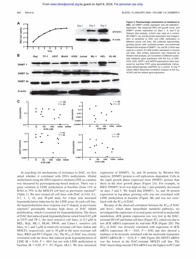
expression of DNMT1, 3a, and 3b proteins by Western blotanalysis. DNMT1 protein is cell replication–dependent. Cells inthe rapid growth phase expressed more DNMT1 protein thanthose in the slow growth phase (Figure 2A). For example, inRKO, DNMT1 level was high on day 1 and gradually decreasedon days 3 and 5. We found that DNMT1, 3a, and 3b proteinexpression in log-phase growing cells was not correlated withLINE methylation at baseline (Figure 2B) and was not corre-lated with the IC50 of DAC.
Because of the observed correlation between the IC50 of DACand Ara-C, which share intracellular metabolic pathways, weinvestigated the expression of several genes involved in nucleosidemetabolism. dCK protein expression was very low in the DAC-resistant DU145 and Jurkat cell lines (Figure 2C), which was due tolow dCK mRNA expression by real-time PCR (Figure 2D). TheIC50 of DAC was inversely correlated with expression of dCKmRNA expression (R � �0.63, P � .038) and also showed atendency to be inversely correlated with the nucleoside transporterhENT1 mRNA (R � �0.54, P � .068; Figure 2D). hENT1 mRNAwas the lowest in the DAC-resistant MOLT4 cell line. TheDAC-inactivating enzyme CDA mRNA was the highest in PC3 and
1 10 100 1000 10000 1000000.000001
0.00001
0.0001
0.001
0.01
0.1
1
IC50 (nM)
CDA
/GAP
DH
1 10 100 1000 10000 1000000.0001
0.001
0.01
0.1
IC50 (nM)
hEN
T2/G
APDH
r = -.62P = .03
r = .45P = .17
r = -54P = .06 r = -.48
P = .11
1 10 100 1000 10000 1000000.01
0.1
1
IC50 (nM)
dCK/
GAPD
H
1 10 100 1000 10000 1000000.0001
0.001
0.01
0.1
IC50 (nM)
hEN
T1/G
APD
H
HL-60HELML-1JurkatK562Molt4U937TF-1Raji
DNMT 1
DNMT 3b
DNMT 3a
ß-actin
B
C
0 1 2 3 4 5 6 7 8219
220
221
222
223
224
Day
RKO
Cell v
iabilit
y
ß-actin
DNMT1
ADay 1 Day 3 Day 5
Figure 2. Pharmacologic mechanisms of resistance toDAC. (A) DNMT1 protein expression was cell replication–dependent. We measured RKO cell growth curve, andDNMT1 protein expression on days 1, 3, and 5 byWestern blot analysis. �-Actin was used as a control.(B) DNMT1, 3a, and 3b protein expression was indepen-dent of sensitivity to DAC and LINE methylation indifferent cancer cell lines. We collected exponentiallygrowing cancer cells, extracted protein, and performedWestern blot analysis of DNMT1, 3a, and 3b. �-Actin wasused as a control. (C) dCK protein expression in severalcell lines. dCK protein expression was measured byWestern blot analysis. (D) Correlation of different nucleo-side metabolic gene expression with the IC50 of DAC.DCK, CDA, hENT1, and hENT2 expressions were mea-sured by real-time PCR using glyceraldehyde 3-phos-phate dehydrogenase (GAPDH) as a control. R and Pvalues reflect Spearman correlation analysis of the IC50
of DAC with the relative gene expression.
662 QIN et al BLOOD, 15 JANUARY 2009 � VOLUME 113, NUMBER 3
For personal use only.on April 8, 2016. by guest www.bloodjournal.orgFrom
DU145, respectively. Thus, 4 of the 5 cell lines most resistant toDAC have measurable alterations in one of these genes, while noneof the 9 most sensitive cell lines had any such alterations. Incontrast, the IC50 of AZA was not correlated with expression ofDCK, CDA, hENT1, and hENT2, respectively (data not shown).Interestingly, the Raji cell line was resistant to AZA, but sensitiveto DAC and Ara-C. DCK promoter is a CpG-rich region, which canbe silenced by DNA hypermethylation. We next measured DCKmethylation level in all these cell lines by pyrosequencing analysis,but could not detect any aberrant hypermethylation even in DU145and Jurkat with the lowest gene expression (data not shown). Inaddition, alternative splicing of DCK was not not observed inresistant clones (data not shown). It is likely that reduced DCKmRNA expression in several cell lines, such as DU145 or Jurkat,might be caused by other mechanisms such as histone deacetyla-tion or microRNAs targeting DCK.
The data described above relate to natural resistance to DAC(and AZA). To study this problem in a model of inducedresistance, we treated HL60 with DAC, selected resistantcolonies (HL60R) in methylcellulose medium, and found afrequency of resistance of 1.2 � 10�4. We replated 11 DAC-resistant colonies and found that resistance was stable and couldlast for more than 2 years even when cultured in the absence ofDAC. To investigate mechanisms of induced resistance to DACin these clones, we measured DAC hypomethylation inductionof LINE-repetitive elements. The hypomethylation induced bylow doses of DAC (0.2 and 2 �M) in HL60D cells was almostlost in the DAC-resistant cells (Figure 3A). To study whetherthis resistance affects gene expression induction by DAC, wemeasured mRNA for RIL, a tumor suppressor gene that issilenced in HL60 by DNA hypermethylation.5 RIL reactivation
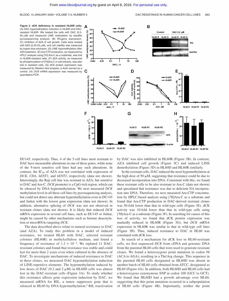
by DAC was also inhibited in HL60R (Figure 3B). In contrast,AZA inhibited cell growth (Figure 3C) and induced LINEdemethylation (Figure 3D) in HL60D and HL60R similarly.
In the resistant cells, DAC induced the most hypomethylation atthe high dose of 50 �M, suggesting that resistance could be due todecreased incorporation into DNA. Consistent with this, we foundthese resistant cells to be also resistant to Ara-C (data not shown)and speculated that resistance was due to deficient NA incorpora-tion into DNA. Therefore, we next measured Ara-CTP concentra-tion by HPLC-based analysis using [3H]Ara-C as a substrate andfound that Ara-CTP production in DAC-derived resistant cloneswas 50-fold lower than that in wild-type cells (Figure 3E). dCKactivity was 10-fold lower than that in wild-type cells using[3H]Ara-C as a substrate (Figure 3F). In searching for causes of thisloss of activity, we found that dCK protein expression wasmarkedly reduced in HL60R (Figure 3G), but DCK mRNAexpression in HL60R was similar to that in wild-type cell lines(Figure 3H). Thus, induced resistance to DAC in HL60 wascorrelated with dCK loss.
In search of a mechanism for dCK loss in HL60-resistantcells, we first sequenced DCK from cDNA and genomic DNAfrom the parental HL60 cells that were used to generate resistantclones. We found a heterozygous point mutation in codon 98(ACA to AGA), resulting in a Thr/Arg change. This sequence inthe parental HL60 cells designated as HL60D was absent inanother batch of HL60 cells obtained from ATCC, designated asHL60 (Figure 4A). In addition, both HL60D and HL60 cells hada heterozygous synonymous SNP in codon 100 (GCC to GCT).We found that HL60D had a growth advantage over HL60,suggesting that this point mutation occurred in a subpopulationof HL60 cells (Figure 4B). Importantly, neither the point
Figure 3. dCK deficiency in resistant HL60R cells.(A) DAC hypomethylation induction in HL60D and DAC-resistant HL60R. We treated the cells with DAC (0.2-50 �M) and measured LINE methylation by bisulfitepyrosequencing analysis. (B) RILgene expression.(C) Inhibition of AZA of cell growth. Cells were treatedwith AZA (0.25-25 �M), and cell viability was measuredby trypan blue exclusion. (D) LINE hypomethylation afterAZA treatment. (E) Ara-CTP production, as measured byHPLC analysis using [3H] Ara-C as a substrate, was lostin HL60R-resistant cells. (F) dCK activity, as measuredby phosphorylation of [3H]Ara-C in cell extracts, was alsolost in resistant cells. (G) dCK protein expression wasmeasured by Western blot analysis. �-Actin served as acontrol. (H) DCK mRNA expression was measured byquantitative PCR.
DAC RESISTANCE IN HUMAN CANCER CELL LINES 663BLOOD, 15 JANUARY 2009 � VOLUME 113, NUMBER 3
For personal use only.on April 8, 2016. by guest www.bloodjournal.orgFrom
mutation nor the adjacent SNP were seen in more than100 alleles in other investigated cell lines and normal cells. Wenext sequenced dCK cDNA and genomic DNA in the resistantcells HL60R (derived from HL60D), looking for additionalmutations. Among the 11 resistant clones, 3 sequenced HL60Rclones had exon 3 LOH of the wild-type allele, leaving behindexclusively the allele containing the AGA point mutation atcodon 98 and GCC at codon 100. Eight clones had a heterozy-gous deletion in exon 1 between nucleotides 154 and 197 ofDCK mRNA that abolished the ATG site (Table 2 and Figure 4).To determine whether this heterozygous deletion lead to bial-lelic mutation, we performed TA cloning (Invitrogen) andsequencing of DCK cDNA from exon 1-4. We found 2 mutantDCK alleles: the allele with a point mutation at codon 98 andanother allele with a 42-bp deletion in exon 1. To determinewhether the mutations were spontaneous and preexisting, wedesigned PCR primers spanning the deleted region of DCK exon 1
and successfully amplified that region from parental HL60D cells(Figure 4D), thus demonstrating that resistance was preexisting andspontaneous.
To eliminate preexisting DAC-resistant clones, we used limiteddilution to isolate individual cell-derived colonies of HL60D.After eachcolony expanded to approximately 106 cells, we selected for DAC-resistant clones in methylcellulose medium and obtained 9 resistantclones. The average frequency of resistance in these cells was 1.2 � 10�5
(Table 2). We measured allelic status of DCK by pyrosequencinganalysis using genomic DNA and found that of 9 single resistantcolonies, 7 had LOH of DCK, resulting in a single mutant allele withArg in codon 98 in exon 3. Two clones had a single base insertioncausing a frameshift (TA/TAA) in exon 6 of DCK (Table 2). cDNAcloning and sequencing confirmed that in each case, the frameshiftmutation was in the allele unmutated at exon 3. LOH of DCK istherefore the most common mechanism that gives rise to inducedresistance to DAC in HL60. To confirm that resistance is caused by
Figure 4. Spontaneous origin of resistance to DAC.(A) Two subclones of the HL60 cell line. HL60D devel-oped a heterozygous 454CG point mutation of DCK.This mutation was absent in another batch of HL60obtained from ATCC. (B) The HL60D cell line had agrowth advantage over the HL60 cell line. Cell numberwas counted from day 1 to 7. (C) Heterozygous deletionin exon 1 of DCK in HL60R cells. (D) Deletion waspreexisting in HL60D cells and absent in HL60 cells. Wedesigned a set of primers that spanned the deletedregion and amplified that region from parental HL60Dcells. (E) HL60R cells transfected with DCK restoredsensitivity to DAC. We transfected wild-type DCK intoHL60R and selected stably transfected cells by G418.HL60R and DCK-transfected (HL60T) cells were treatedwith DAC at 0.02, 0.2, 2, and 20 �M, respectively. Cellviability was counted. (F) Transfection of DCK cDNArestored dCK protein expression. dCK protein expres-sion was measured in HL60R and HL60T cells byWestern blot analysis.
Table 2. Summary of resistance to DAC in HL60D cells
GroupSelected resistant
clonesDAC resistance
frequency
Mutation
LOH Deletion Frameshift
HL6O 0 Undetected 0 0 0
HL6OD (pool) 11 1.2 � 10�4 3 8 0
HL6OD (clone) 9 1.2 � 10�5 7 0 2
664 QIN et al BLOOD, 15 JANUARY 2009 � VOLUME 113, NUMBER 3
For personal use only.on April 8, 2016. by guest www.bloodjournal.orgFrom
DCK loss, we cloned wild-type DCK into a pEGFP-N1 vectors andtransfected the vector into HL60R. We selected stably G418-resistantcells, designated as HL60T. Transfection of DCK restored dCK proteinexpression and sensitivity to DAC (Figure 4E,F), suggesting that DACresistance was due to functional loss of dCK, which was due to LOH ofthe DCK locus.
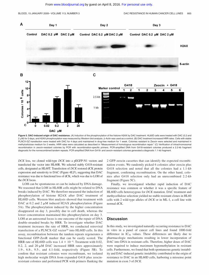
LOH can be spontaneous or can be induced by DNA damage.We reasoned that LOH in HL60R cells might be related to DNAbreaks induced by DAC. We therefore measured the induction ofphosphorylation of histone H2AX after DAC treatment ofHL60D cells. Western blot analysis showed that treatment withDAC at 0.2 and 2 �M induced H2AX phosphorylation (Figure5A). The phosphorylation induced by the higher concentrationdisappeared on day 3, possibly due to cell death, whereas thelower concentration maintained this phosphorylation on day 3.LOH at an autosomal locus is one outcome of the repair of DNAdouble-stranded breaks by HRR. To investigate whether DACtreatment increases the rate of HRR, we conducted retroviraltransfection of a PLNCX-GZ vector24 into HL60D cells. In thisassay, recombination between the tandem repeats regenerates afunctional GFP-ZeoR marker that can be easily scored. TheHRR rate of HL60D cells was 1.4 � 10�6. Treatment with 0.02,0.2, 2, and 20 �M DAC increased HRR rates approximately1.9-, 6.8-, 9.5-, and 4.1-fold, respectively (Figure 5B). Toconfirm that resistant colonies were recombinant, we isolatedhigh molecular weight DNA from expanded G418 plus zeocin-resistant colonies and performed PCR with primers flanking the
2 GFP-zeocin cassettes that can identify the expected recombi-nation events. We randomly picked 6 colonies after zeocin plusG418 selection and noted that all the colonies had a 1.1-kbfragment, confirming recombination. On the other hand, colo-nies after G418 selection only had an unrecombined 2.2-kbfragment (Figure 5C).
Finally, we investigated whether rapid induction of DACresistance was common or whether it was a specific feature ofHL60D cells heterozygous for DCK mutation. DAC treatment andmethycellulose selection yielded no stable resistant clones in HL60cells with 2 wild-type alleles of DCK or in ML-1, a cell line withnormal dCK.
Discussion
In this study, we investigated naturally occurring resistance to DACin vitro in a panel of cancer cell lines and found 1000-folddifference in IC50 values. These differences are likely due topharmacologic mechanisms resulting in lower incorporation ofDAC into DNA in resistant cells. Therefore, higher doses of DACwere required to induce maximum hypomethylation in resistantcells. Furthermore, we found that both spontaneous genetic instabil-ity and DAC-induced genetic instability contributed to the origin ofresistance to DAC in an HL60D cells, harboring a missense pointmutation in exon 3 of DCK.
Figure 5. DAC-induced origin of DAC resistance. (A) Induction of the phosphorylation of the histone H2AX by DAC treatment. HL60D cells were treated with DAC (0.2 and2 �M) for 3 days, and H2AX phosphorylation was measured by Western blot analysis. �-Actin was used as a control. (B) DAC treatment increased HRR rates. Cells with stablePLNCX-GZ transfection were treated with DAC for 4 days and maintained in drug-free medium for 1 week. Colonies resistant to Zeocin were selected and maintained inmethylcellulose medium for 2 weeks. HRR rates were calculated as described in “Measurement of homologous recombination repair.” (C) Verification of intrachromosomalrecombination in zeocin-resistant colonies by PCR with recombination-specific primers. PCR-amplified DNA from G418-resistant colonies produced a 2.2-kb fragmentdiagnostic for the nonrecombined tandem repeats. PCR-amplified DNA from G418- and zeocin-resistant colonies generated a diagnostic 1.1-kb fragment.
DAC RESISTANCE IN HUMAN CANCER CELL LINES 665BLOOD, 15 JANUARY 2009 � VOLUME 113, NUMBER 3
For personal use only.on April 8, 2016. by guest www.bloodjournal.orgFrom

Most in vitro resistance to NA results from lack of incorporationinto DNA.26 Consistent with this, we found that resistance to DACwas correlated with multiple pharmacologic mechanisms of aber-rant nucleoside metabolism, which result in less incorporation ofDAC into DNA, but this was not directly proven by measuringDAC incorporation. It remains experimentally very difficult toanswer this because DAC incorporation is at low levels. DAC has ashort half-life (minutes) and is quickly deaminated by CDA onceinside cells. It is also chemically unstable upon DNA extraction andHPLC analysis. The 5,6 bond in cytosine is very labile.27,28
Nevertheless, in HL60, we demonstrated that DAC resistance iscaused by loss of DCK, and DAC sensitivity is highly correlatedwith Ara-C sensitivity, which shares NA metabolism. The AraCTPexperiments are indirect, but they are a very good approximation ofdCK activity. Thus, the data in favor of incorporation as amechanism of resistance are strong. Clearly, however, multiplemechanisms may be active in different cells.
DAC exerts its effect through induction of hypomethylation atlow doses and cytotoxicity at high doses. DAC induced a U-shapedcurve of hypomethylation in a panel of cancer cell lines. This mightbe explained by the fact that low-dose DAC can covalently trapDNA methytransferases without cell-cycle arrest, whereas highdose DAC inhibits DNA synthesis and induces cell-cycle arrest,leading to less hypomethylation induction. Clinical usage of theagent is mostly at low doses, however.4 Interestingly, resistance toDAC was unrelated to DNMT levels and LINE methylation, amarker of global DNA methylation. Cancers have gene-specifichypermethylation and global hypomethylation.29 Our data suggestthat differences in the methylation levels of repetitive DNAelements (reflecting global methylation) are not simply explainedby DNMT levels and do not impact on sensitivity to DAC. Rather,DAC hypomethylation induction and gene reactivation are im-paired in resistant cells by insufficient incorporation of DAC intoDNA. A potential question is whether downstream pathways thatled to gene reactivation are related to resistance to DAC, providingadequate drug incorporation. The fact that DAC treatment activatesthousands of genes, not all of which are silenced by promoterhypermethylation,30 makes this kind of study difficult. However, inthis panel of cell lines, the cross-resistance between DAC andAra-C and the lack of cross-resistance between DAC and AZAsuggests that resistance to gene reactivation perhaps is lessimportant than DAC incorporation. Nevertheless, resistance toDAC is likely to be multifactorial in a specific cell line. Forexample, DU145 demonstrated low levels of DCK, hENT1, andhigh levels of CDA. The doses needed for the maximum hypomethy-lation are the best marker for determining the sensitivity orresistance to DAC based on our results.
In our studies, we were able to ascribe resistance to 2 potentialmechanisms: (1) preexisting genetic instability and selection forresistant clones and (2) DAC-induced genetic instability. Mostmalignant cell populations are characterized by “genetic instabil-ity” that can be shown to be directly involved in the generation ofphenotypic drug resistance. Using fluctuation analysis, researchershave demonstrated random and spontaneous origins of resistancephenotypes for some antineoplastic agents, such as topoisomeraseII inhibitor, folic acid antagonists, and antibiotic agents.10-16 In thisstudy, we amplified mutations preexisting in rare resistant cellsfrom a pool of parental cells by PCR, providing the most directevidence of a spontaneous origin of resistance to DAC and otherNAs. On the other hand, NAs affect the structural integrity ofDNA, leading to stalled replication forks and chain termination.The DNA damage sensors ATM, ATR, and DNA-PK recognize
these events and signal for DNA repair.31 We observed that DACtreatment induced histone H2AX phosphorylation, a marker ofdouble-strand DNA breaks, and increased the rate of HRR, whichmight give rise to LOH of DCK and resistance to DAC. Severalcytostatic drugs, including aphidicolin, Ara-C, hydroxyurea, andmethotrexate, induce HRR through inhibition of DNA synthesis.32
Sensitivity to DAC in our studies was closely related tosensitivity to Ara-C, but not to AZA except in the PC3 cell line,which had high CDA levels capable of deaminating all these drugsinto inactive forms. DAC and Ara-C need the same dCK enzymefor initial phosphorylation. Instead, AZA uses UCK for initialphosphorylation. Therefore, DAC resistance related to dCK lossdoes not lead to AZA resistance, and this lack of cross-resistancecould be exploited therapeutically. Indeed, a recent study hasshown that some patients can respond to DAC after showingclinical resistance to AZA.33
The relevance of our findings to in vivo resistance need to beexamined. Low levels of DCK gene expression or low dCK activitywere correlated with a poor response to Ara-C and cladribine(2-chlorodeoxyadenosine) in childhood acute lymphoblastic leuke-mias (ALLs) and lymphoproliferative disorders,34,35 and to gemcit-abine (a cytosine analog) in advanced pancreatic cancer.36 How-ever, not all studies were consistent in this regard.37,38 Ourcombined data suggest that the best approach to examining in vivoresistance to DAC would be to measure DAC incorporation intoDNA, an experiment that is not technically feasible at presentbecause of the low doses of DAC used clinically. An alternateapproach may be to measure all parameters of DAC metabolism(eg, dCK, hENT, CDA) in treated patients and correlate them withsensitivity to the drug.
In conclusion, this study provides important in vitro models forunderstanding the mechanisms and origin of resistance to DAC,which may lead to strategies to overcome DAC resistance in vivo.The lack of cross-resistance between DAC and AZA in some cellscould also have important therapeutic implications.
Acknowledgments
We thank Dr. William Plunkett and Billie Nowak for general adviceand help in the measurement of Ara-CTP and dCK activity, Drs LuXiongbin and Lawrence A. Donehower (Baylor College of Medi-cine, Houston, TX) for providing the PLNCX-GZ, pVSV-G, andpHit60 vectors for studying homologous recombination, and DrGergely Keszler at Semmelweis University (Budapest, Hungary)for providing a dCK antibody.
This work was supported by National Institutes of Health grantsCA100632 and CA108631 and by the Rosalie B. Hite GraduateFellowship at University of Texas Health Science Center atHouston.
Authorship
Contribution: T.Q. performed research; J.J. and J.-P.J.I. designedresearch; and J. Si and J. Shu analyzed data.
Conflict-of-interest disclosure: The authors declare no compet-ing financial interests.
Correspondence: Jean-Pierre J. Issa, Department of Leukemia,The University of Texas M. D. Anderson Cancer Center, Houston,TX; e-mail: [email protected].
666 QIN et al BLOOD, 15 JANUARY 2009 � VOLUME 113, NUMBER 3
For personal use only.on April 8, 2016. by guest www.bloodjournal.orgFrom

References
1. Sollars VE. Epigenetic modification as an en-abling mechanism for leukemic transformation.Front Biosci. 2005;10:1635-1646.
2. Villa R, De Santis F, Gutierrez A, Minucci S,Pelicci PG, Di Croce L. Epigenetic gene silencingin acute promyelocytic leukemia. Biochem Phar-macol. 2004;68:1247-1254.
3. Mizuno S, Chijiwa T, Okamura T, et al. Expressionof DNA methyltransferases DNMT1, 3A, and 3Bin normal hematopoiesis and in acute and chronicmyelogenous leukemia. Blood. 2001;97:1172-1179.
4. Issa JP. DNA methylation as a therapeutic targetin cancer. Clin Cancer Res. 2007;13:1634-1637.
5. Qin T, Youssef EM, Jelinek J, et al. Effect of cytar-abine and decitabine in combination in humanleukemic cell lines. Clin Cancer Res. 2007;13:4225-4232.
6. Lyko F, Brown R. DNA methyltransferase inhibi-tors and the development of epigenetic cancertherapies. J Natl Cancer Inst. 2005;97:1498-1506.
7. Kroep JR, Loves WJ, van der Wilt CL, et al. Pre-treatment deoxycytidine kinase levels predict invivo gemcitabine sensitivity. Mol Cancer Ther.2002;1:371-376.
8. Flasshove M, Strumberg D, Ayscue L, et al.Structural analysis of the deoxycytidine kinasegene in patients with acute myeloid leukemia andresistance to cytosine arabinoside. Leukemia.1994;8:780-785.
9. Stegmann AP, Honders MW, Hagemeijer A, Hoe-bee B, Willemze R, Landegent JE. In vitro-in-duced resistance to the deoxycytidine analoguescytarabine (AraC) and 5-aza-2�-deoxycytidine(DAC) in a rat model for acute myeloid leukemiais mediated by mutations in the deoxycytidinekinase (DCK) gene. Ann Hematol. 1995;71:41-47.
10. Wang G, Wilson TJ, Jiang Q, Taylor DE. Sponta-neous mutations that confer antibiotic resistancein Helicobacter pylori. Antimicrob Agents Che-mother. 2001;45:727-733.
11. Vogler AJ, Busch JD, Percy-Fine S, Tipton-Hunton C, Smith KL, Keim P. Molecular analysisof rifampin resistance in Bacillus anthracis andBacillus cereus. Antimicrob Agents Chemother.2002;46:511-513.
12. Jaffrezou JP, Chen G, Duran GE, Kuhl JS, SikicBI. Mutation rates and mechanisms of resistanceto etoposide determined from fluctuation analy-sis. J Natl Cancer Inst. 1994;86:1152-1158.
13. Poche H, Varshaver NB, Geissler E. Cyclohexi-
mide resistance in Chinese hamster cells. I.Spontaneous mutagenesis. Mutat Res. 1975;27:399-406.
14. Goldie JH, Coldman AJ. Genetic instability in thedevelopment of drug resistance. Semin Oncol.1985;12:222-230.
15. Monnat Jr, RJ. Molecular analysis of spontane-ous hypoxanthine phosphoribosyltransferase mu-tations in thioguanine-resistant HL-60 human leu-kemia cells. Cancer Res. 1989;49:81-87.
16. Siminovitch L. On the nature of hereditable varia-tion in cultured somatic cells. Cell. 1976;7:1-11.
17. Chen RZ, Pettersson U, Beard C, Jackson-Grusby L, Jaenisch R. DNA hypomethylationleads to elevated mutation rates. Nature. 1998;395:89-93.
18. Lengauer C, Kinzler KW, Vogelstein B. Geneticinstabilities in human cancers. Nature. 1998;396:643-649.
19. Raderschall E, Stout K, Freier S, Suckow V,Schweiger S, Haaf T. Elevated levels of Rad51recombination protein in tumor cells. Cancer Res.2002;62:219-225.
20. Ji W, Hernandez R, Zhang XY, et al. DNA de-methylation and pericentromeric rearrangementsof chromosome 1. Mutat Res. 1997;379:33-41.
21. Chou TC, Talalay P. Quantitative analysis ofdose-effect relationships: the combined effects ofmultiple drugs or enzyme inhibitors. Adv EnzymeRegul. 1984;22:27-55.
22. Rodriguez Jr CO, Plunkett W, Paff MT, et al. High-performance liquid chromatography method forthe determination and quantitation of arabinosyl-guanine triphosphate and fludarabine triphos-phate in human cells. J Chromatogr B Biomed SciAppl. 2000;745:421-430.
23. Saunders PP, Lai MM. Nucleoside kinase activi-ties of Chinese hamster ovary cells. Biochim Bio-phys Acta. 1983;761:135-141.
24. Lu X, Lozano G, Donehower LA. Activities of wild-type and mutant p53 in suppression of homolo-gous recombination as measured by a retroviralvector system. Mutat Res. 2003;522:69-83.
25. Estecio MR, Gharibyan V, Shen L, et al. LINE-1hypomethylation in cancer is highly variable andinversely correlated with microsatellite instability.PLoS ONE. 2007;2:e399.
26. Galmarini CM, Mackey JR, Dumontet C. Nucleo-side analogues: mechanisms of drug resistanceand reversal strategies. Leukemia. 2001;15:875-890.
27. Momparler RL. Pharmacology of 5-aza-2�-deoxy-
cytidine (decitabine). Semin Hematol. 2005;42:S9-S16.
28. Yoo CB, Jeong S, Egger G, et al. Delivery of5-aza-2�-deoxycytidine to cells using oligode-oxynucleotides. Cancer Res. 2007;67:6400-6408.
29. Robertson KD, Jones PA. DNA methylation: past,present and future directions. Carcinogenesis.2000;21:461-467.
30. Schmelz K, Sattler N, Wagner M, Lubbert M,Dorken B, Tamm I. Induction of gene expressionby 5-aza-2�-deoxycytidine in acute myeloid leuke-mia (AML) and myelodysplastic syndrome (MDS)but not epithelial cells by DNA-methylation-de-pendent and -independent mechanisms. Leuke-mia. 2005;19:103-111.
31. Sampath D, Rao VA, Plunkett W. Mechanisms ofapoptosis induction by nucleoside analogs. On-cogene. 2003;22:9063-9074.
32. Arnaudeau C, Tenorio ME, Jenssen D, HelledayT. Inhibition of DNA synthesis is a potent mecha-nism by which cytostatic drugs induce homolo-gous recombination in mammalian cells. MutatRes. 2000;461:221-228.
33. Borthakur G, Ahdab SE, Ravandi F, et al. Activityof decitabine in patients with myelodysplasticsyndrome previously treated with azacitidine.Leuk Lymphoma. 2008;49:690-695.
34. Kakihara T, Fukuda T, Tanaka A, et al. Expressionof deoxycytidine kinase (dCK) gene in leukemiccells in childhood: decreased expression of dCKgene in relapsed leukemia. Leuk Lymphoma.1998;31:405-409.
35. Stammler G, Zintl F, Sauerbrey A, Volm M. De-oxycytidine kinase mRNA expression in child-hood acute lymphoblastic leukemia. AnticancerDrugs. 1997;8:517-521.
36. Sebastiani V, Ricci F, Rubio-Viqueira B, et al. Im-munohistochemical and genetic evaluation of de-oxycytidine kinase in pancreatic cancer: relation-ship to molecular mechanisms of gemcitabineresistance and survival. Clin Cancer Res. 2006;12:2492-2497.
37. Veuger MJ, Honders MW, Willemze R, BargeRM. Deoxycytidine kinase expression and activityin patients with resistant versus sensitive acutemyeloid leukemia. Eur J Haematol. 2002;69:171-178.
38. Leiby JM, Snider KM, Kraut EH, Metz EN,Malspeis L, Grever MR. Phase II trial of9-beta-D-arabinofuranosyl-2-fluoroadenine 5�-monophosphate in non-Hodgkin lymphoma: pro-spective comparison of response with deoxycytidinekinase activity. Cancer Res. 1987;47:2719-2722.
DAC RESISTANCE IN HUMAN CANCER CELL LINES 667BLOOD, 15 JANUARY 2009 � VOLUME 113, NUMBER 3
For personal use only.on April 8, 2016. by guest www.bloodjournal.orgFrom

online October 17, 2008 originally publisheddoi:10.1182/blood-2008-02-140038
2009 113: 659-667
Taichun Qin, Jaroslav Jelinek, Jiali Si, Jingmin Shu and Jean-Pierre J. Issa cell lines
-deoxycytidine in human cancer′Mechanisms of resistance to 5-aza-2
http://www.bloodjournal.org/content/113/3/659.full.htmlUpdated information and services can be found at:
(1470 articles)Myeloid Neoplasia Articles on similar topics can be found in the following Blood collections
http://www.bloodjournal.org/site/misc/rights.xhtml#repub_requestsInformation about reproducing this article in parts or in its entirety may be found online at:
http://www.bloodjournal.org/site/misc/rights.xhtml#reprintsInformation about ordering reprints may be found online at:
http://www.bloodjournal.org/site/subscriptions/index.xhtmlInformation about subscriptions and ASH membership may be found online at:
Copyright 2011 by The American Society of Hematology; all rights reserved.of Hematology, 2021 L St, NW, Suite 900, Washington DC 20036.Blood (print ISSN 0006-4971, online ISSN 1528-0020), is published weekly by the American Society
For personal use only.on April 8, 2016. by guest www.bloodjournal.orgFrom
Related Documents