Rev. Cardiovasc. Med. 2022; 23(7): 227 https://doi.org/10.31083/j.rcm2307227 Copyright: © 2022 The Author(s). Published by IMR Press. This is an open access article under the CC BY 4.0 license. Publisher’s Note: IMR Press stays neutral with regard to jurisdictional claims in published maps and institutional affiliations. Review Mechanisms of Myocardial Ischemia in Cancer Patients: A State-of-the-Art Review of Obstructive Versus Non-Obstructive Causes Dinu V. Balanescu 1 , Richard Bloomingdale 2 , Teodora Donisan 1 , Eric H. Yang 3 , Purvi Parwani 4 , Cezar Iliescu 5 , Joerg Herrmann 6 , Ivan Hanson 2, * 1 Department of Internal Medicine, Beaumont Hospital, Royal Oak, MI 48073, USA 2 Department of Cardiology, Beaumont Hospital, Royal Oak, MI 48073, USA 3 UCLA Cardio-Oncology Program, Division of Cardiology, Department of Medicine, University of California at Los Angeles, Los Angeles, CA 90095, USA 4 Department of Cardiology, Loma Linda University International Heart Institute, Loma Linda, CA 92354, USA 5 Department of Cardiology, The University of Texas MD Anderson Cancer Center, Houston, TX 77030, USA 6 Department of Cardiovascular Medicine, Mayo Clinic, Rochester, MN 55903, USA *Correspondence: [email protected] (Ivan Hanson) Academic Editors: Yen-Wen Wu, Hung-Ju Lin, Yen-Wen Liu and Pei-Wei Shueng Submitted: 28 March 2022 Revised: 10 May 2022 Accepted: 16 May 2022 Published: 24 June 2022 Abstract In patients with cancer, myocardial infarction (MI) has distinct features and mechanisms compared to the non-oncology population. Triggers of myocardial ischemia specific to the oncology population have been increasingly identified. Coronary plaque disruption, coronary vasospasm, coronary microvascular dysfunction, spontaneous coronary artery dissection, and coronary oxygen supply-demand mismatch are all causes of MI that have been shown to have specific triggers related to either the treatments or complications of cancer. MI can occur in the presence or absence of atherosclerotic coronary artery disease (CAD). MI with nonobstructive CAD (MINOCA) is a heterogeneous syndrome that has distinct pathophysiology and different epidemiology from MI with significant CAD (MI-CAD). Recognition and differentiation of MI-CAD and MINOCA is essential in the oncology population, due to unique etiology and impact on diagnosis, management, and overall outcomes. There are currently no reports in the literature concerning MINOCA as a unified syndrome in oncology patients. The purpose of this review is to analyze the literature for studies related to known triggers of myocardial ischemia in cancer patients, with a focus on MINOCA. We propose that certain cancer treatments can induce MINOCA-like states, and further research is warranted to investigate mechanisms that may be unique to certain cancer states and types of treatment. Keywords: cardio-oncology; cancer; MINOCA; myocardial infarction in the absence of obstructive coronary artery disease Graphical Abstract. Mechanisms of ischemia in cancer patients include coronary plaque disruption, coronary va- sospasm, microvascular dysfunction, extrinsic tumor compression, spontaneous coronary artery dissection, and oxy- gen supply-demand mismatch. Abbreviations: A II, angiotensin II; ADP, adenosine-5’-diphosphate; Ca, calcium; EPC, endothelial progenitor cells; ET-1, endothelin 1; MI-CAD, myocardial infarction with obstructive coronary artery disease; MINOCA, myocardial infarction with non-obstructive coronary arteries; NET, neutrophil extracellular traps; NO, nitric ox- ide; PF4, platelet factor 4; PLT, platelet; ROS, reactive oxygen species; TGF-β, tumor growth factor-β; TxA, thromboxane A; VEGF, vascular endothelial growth factor.

Welcome message from author
This document is posted to help you gain knowledge. Please leave a comment to let me know what you think about it! Share it to your friends and learn new things together.
Transcript

Rev. Cardiovasc. Med. 2022; 23(7): 227https://doi.org/10.31083/j.rcm2307227
Copyright: © 2022 The Author(s). Published by IMR Press.This is an open access article under the CC BY 4.0 license.
Publisher’s Note: IMR Press stays neutral with regard to jurisdictional claims in published maps and institutional affiliations.
Review
Mechanisms of Myocardial Ischemia in Cancer Patients: AState-of-the-Art Review of Obstructive Versus Non-Obstructive CausesDinu V. Balanescu1, Richard Bloomingdale2, Teodora Donisan1, Eric H. Yang3,Purvi Parwani4, Cezar Iliescu5, Joerg Herrmann6, Ivan Hanson2,*1Department of Internal Medicine, Beaumont Hospital, Royal Oak, MI 48073, USA2Department of Cardiology, Beaumont Hospital, Royal Oak, MI 48073, USA3UCLA Cardio-Oncology Program, Division of Cardiology, Department of Medicine, University of California at Los Angeles, Los Angeles, CA 90095, USA4Department of Cardiology, Loma Linda University International Heart Institute, Loma Linda, CA 92354, USA5Department of Cardiology, The University of Texas MD Anderson Cancer Center, Houston, TX 77030, USA6Department of Cardiovascular Medicine, Mayo Clinic, Rochester, MN 55903, USA*Correspondence: [email protected] (Ivan Hanson)Academic Editors: Yen-Wen Wu, Hung-Ju Lin, Yen-Wen Liu and Pei-Wei ShuengSubmitted: 28 March 2022 Revised: 10 May 2022 Accepted: 16 May 2022 Published: 24 June 2022
AbstractIn patients with cancer, myocardial infarction (MI) has distinct features and mechanisms compared to the non-oncology population.Triggers of myocardial ischemia specific to the oncology population have been increasingly identified. Coronary plaque disruption,coronary vasospasm, coronary microvascular dysfunction, spontaneous coronary artery dissection, and coronary oxygen supply-demandmismatch are all causes of MI that have been shown to have specific triggers related to either the treatments or complications of cancer.MI can occur in the presence or absence of atherosclerotic coronary artery disease (CAD). MI with nonobstructive CAD (MINOCA)is a heterogeneous syndrome that has distinct pathophysiology and different epidemiology from MI with significant CAD (MI-CAD).Recognition and differentiation of MI-CAD and MINOCA is essential in the oncology population, due to unique etiology and impacton diagnosis, management, and overall outcomes. There are currently no reports in the literature concerning MINOCA as a unifiedsyndrome in oncology patients. The purpose of this review is to analyze the literature for studies related to known triggers of myocardialischemia in cancer patients, with a focus on MINOCA. We propose that certain cancer treatments can induce MINOCA-like states, andfurther research is warranted to investigate mechanisms that may be unique to certain cancer states and types of treatment.
Keywords: cardio-oncology; cancer; MINOCA; myocardial infarction in the absence of obstructive coronary artery disease
Graphical Abstract. Mechanisms of ischemia in cancer patients include coronary plaque disruption, coronary va-sospasm, microvascular dysfunction, extrinsic tumor compression, spontaneous coronary artery dissection, and oxy-gen supply-demand mismatch. Abbreviations: A II, angiotensin II; ADP, adenosine-5’-diphosphate; Ca, calcium; EPC,endothelial progenitor cells; ET-1, endothelin 1; MI-CAD, myocardial infarction with obstructive coronary artery disease;MINOCA, myocardial infarction with non-obstructive coronary arteries; NET, neutrophil extracellular traps; NO, nitric ox-ide; PF4, platelet factor 4; PLT, platelet; ROS, reactive oxygen species; TGF-β, tumor growth factor-β; TxA, thromboxaneA; VEGF, vascular endothelial growth factor.

1. IntroductionMyocardial infarction (MI) in cancer patients has dis-
tinct features and mechanisms compared to the general,non-oncology population. MI can broadly be categorizedinto MI due to coronary artery disease (MI-CAD) and MIin the absence of coronary obstructive disease (MINOCA).MI-CAD is the most common cause of MI in both cancerand non-cancer patients. Although traditional cardiovascu-lar risk factors apply to both patients with and without can-cer, the overall risk for MI in oncology patients is higherdue to both cancer-related processes and anti-cancer ther-apies [1]. Even in the absence of cardiotoxic anti-cancertreatments, cancer patients can be found with high levelsof cardiac biomarkers, suggesting subclinical myocardialdamage of unclear etiology and associated with worse out-comes [2,3]. MINOCA is a newly recognized heteroge-neous syndrome that has distinct pathophysiology and epi-demiology when compared to MI-CAD [4]. The preva-lence of MINOCA among patients presenting with suspi-cion of acute MI was reported as high as 14% [5]. Recentdata suggest that patients presenting with ST-elevation MI(STEMI) who have a history of cancer are more likely tohave MINOCA rather than MI-CAD compared to patientswith STEMI without an oncologic history (17% vs. 8%, re-spectively) [6]. Recognition of this condition and distinc-tion from MI-CAD are essential, as MINOCA may be mis-diagnosed as non-cardiac, with significant cardiovascularmanagement and outcome implications.
With the recent rapid rise of cardio-oncology, triggersof myocardial ischemia specific to the oncology populationhave been increasingly identified [7–9]. Coronary plaquedisruption, coronary vasospasm, coronary microvasculardysfunction, oxygen supply-demand mismatch, and spon-taneous coronary artery dissection (SCAD), are all causesof MI that have been shown to have specific triggers relatedto either the treatments or complications of cancer [10–12].The multiple etiologies of MINOCA each portend differentprognoses and require individualized management strate-gies [12]. Currently there is a paucity of data in the litera-ture concerning MINOCA as a unified syndrome in oncol-ogy patients. The purpose of this review is to analyze theliterature for studies related to known triggers of myocar-dial ischemia and infarction in cancer patients with a focuson MINOCA and propose that certain cancer states, and/ortheir treatments can induce MINOCA-like states.
2. DefinitionsThe Fourth Universal Definition of MI, issued by the
Joint European Society of Cardiology (ESC), AmericanCollege of Cardiology (ACC), American Heart Association(AHA), and World Heart Federation (WHF) Task Force, iswidely accepted and used in clinical practice [13]. Thismost recent iteration of the universal definition ofMI classi-
fies troponin elevation as being due to acute ischemia (lead-ing to myocardial infarction) and not acute ischemia driven(e.g., myocardial injury due to acute myocarditis).
MINOCA is a recently described entity that canbroadly be defined by these criteria: (1) acute MI accord-ing to the Fourth Universal Definition of MI; (2) exclu-sion of missed obstructive coronary disease (e.g., coronaryemboli/thrombi, coronary dissection); (3) no coronary le-sions ≥50% in a major epicardial vessel; (4) no coronarylesions with FFR <0.8; (5) no other identifiable cause forthe presentation [12,14]. MINOCA can be considered asa working diagnosis, when known causes of elevated tro-ponin are excluded and our current diagnostic capacity hasbeen reached. Initial studies of MINOCA considered thecomplete absence of coronary disease as a diagnostic ne-cessity [15]. It is currently generally accepted that the di-agnosis can be established even in the presence of mod-erate coronary disease (i.e., obstructing <50% of the lu-men) [4]. Echocardiography, ventriculography, and cardiacmagnetic resonance (CMR) imaging should be part of thework-up when assessing for MINOCA to comprehensivelysearch for etiologies that may mimic the syndrome (Fig. 1).CMR is an essential diagnostic tool in these patients since itcan evaluate the non-ischemic and ischemic etiology oncethe working diagnosis of MINOCA is established. Data onthe functional assessment of coronary lesions in MINOCAare sparse. It was noted that a significant number of le-sions in the 30%–50% range by angiography are function-ally hemodynamically significant on fractional flow reserve(FFR) or cardiac positron emission tomography (PET) andresponsible for MI-CAD [16,17]. There are no data on in-stantaneous free-wave ratio in MINOCA. A special cate-gory of cancer patients undergoing therapy with immunecheckpoint inhibitors (ICI) and possible myocarditis canhave indeterminate CMR findings [18] and in some casesrequire endomyocardial biopsy (EMB), which can be chal-lenging in the acute coronary setting where the addition ofantiplatelet and anticoagulation increases the bleeding riskand the subsequent risk of perforation and tamponade. In-tracoronary imaging and functional assessment should alsobe used to identify specific causes of MINOCA once an is-chemic etiology is established via CMR [12].
Despite MINOCA having an overall 1-year mortal-ity rate of 4.7%, data on the impact of distinct manage-ment strategies based on the specific diagnosis is lackingin the literature [19]. This may be due to MINOCA be-ing a relatively heterogeneous entity, with various etiolo-gies included in the disease spectrum, with an evolving def-inition. The diagnosis of MINOCA can be difficult, giventhat many chemotherapies and targeted therapies can causenon-specific cardiac biomarker elevations and/or EKG orechocardiographic abnormalities, which can also be com-pounded by other concomitates states associated with com-plications of cancer and its therapies (e.g., sepsis, hypov-
2
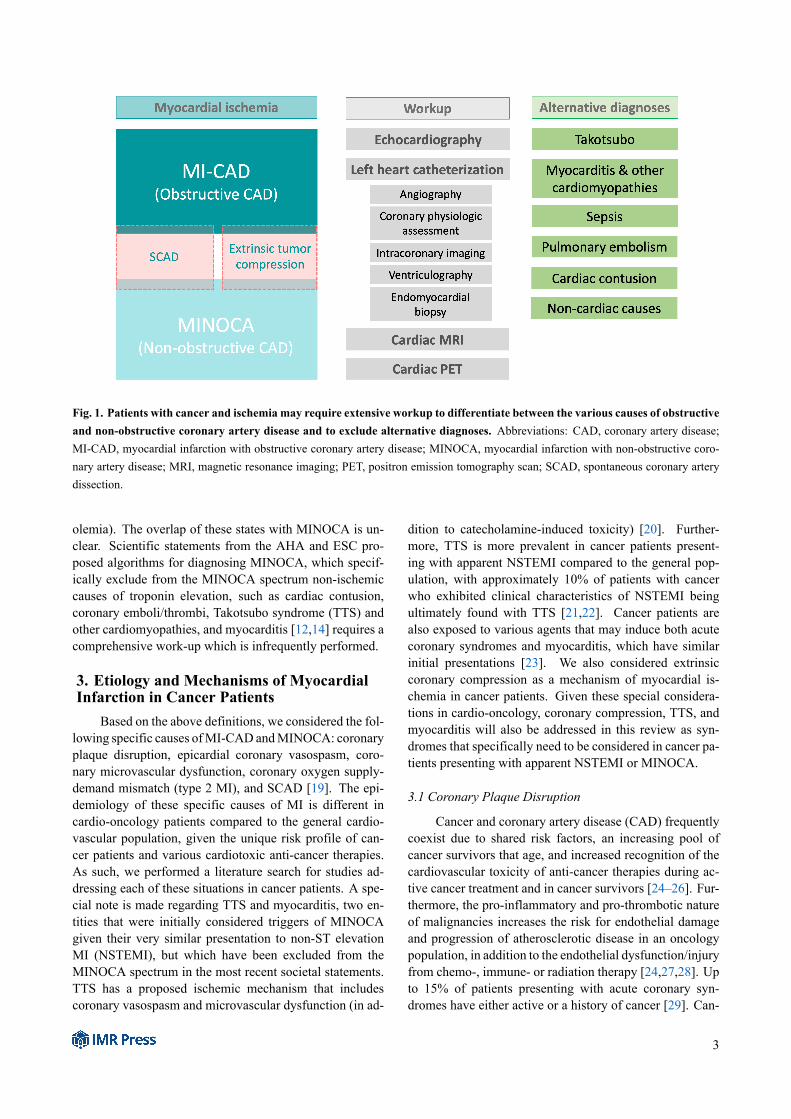
Fig. 1. Patients with cancer and ischemia may require extensive workup to differentiate between the various causes of obstructiveand non-obstructive coronary artery disease and to exclude alternative diagnoses. Abbreviations: CAD, coronary artery disease;MI-CAD, myocardial infarction with obstructive coronary artery disease; MINOCA, myocardial infarction with non-obstructive coro-nary artery disease; MRI, magnetic resonance imaging; PET, positron emission tomography scan; SCAD, spontaneous coronary arterydissection.
olemia). The overlap of these states with MINOCA is un-clear. Scientific statements from the AHA and ESC pro-posed algorithms for diagnosing MINOCA, which specif-ically exclude from the MINOCA spectrum non-ischemiccauses of troponin elevation, such as cardiac contusion,coronary emboli/thrombi, Takotsubo syndrome (TTS) andother cardiomyopathies, and myocarditis [12,14] requires acomprehensive work-up which is infrequently performed.
3. Etiology and Mechanisms of MyocardialInfarction in Cancer Patients
Based on the above definitions, we considered the fol-lowing specific causes ofMI-CAD andMINOCA: coronaryplaque disruption, epicardial coronary vasospasm, coro-nary microvascular dysfunction, coronary oxygen supply-demand mismatch (type 2 MI), and SCAD [19]. The epi-demiology of these specific causes of MI is different incardio-oncology patients compared to the general cardio-vascular population, given the unique risk profile of can-cer patients and various cardiotoxic anti-cancer therapies.As such, we performed a literature search for studies ad-dressing each of these situations in cancer patients. A spe-cial note is made regarding TTS and myocarditis, two en-tities that were initially considered triggers of MINOCAgiven their very similar presentation to non-ST elevationMI (NSTEMI), but which have been excluded from theMINOCA spectrum in the most recent societal statements.TTS has a proposed ischemic mechanism that includescoronary vasospasm and microvascular dysfunction (in ad-
dition to catecholamine-induced toxicity) [20]. Further-more, TTS is more prevalent in cancer patients present-ing with apparent NSTEMI compared to the general pop-ulation, with approximately 10% of patients with cancerwho exhibited clinical characteristics of NSTEMI beingultimately found with TTS [21,22]. Cancer patients arealso exposed to various agents that may induce both acutecoronary syndromes and myocarditis, which have similarinitial presentations [23]. We also considered extrinsiccoronary compression as a mechanism of myocardial is-chemia in cancer patients. Given these special considera-tions in cardio-oncology, coronary compression, TTS, andmyocarditis will also be addressed in this review as syn-dromes that specifically need to be considered in cancer pa-tients presenting with apparent NSTEMI or MINOCA.
3.1 Coronary Plaque Disruption
Cancer and coronary artery disease (CAD) frequentlycoexist due to shared risk factors, an increasing pool ofcancer survivors that age, and increased recognition of thecardiovascular toxicity of anti-cancer therapies during ac-tive cancer treatment and in cancer survivors [24–26]. Fur-thermore, the pro-inflammatory and pro-thrombotic natureof malignancies increases the risk for endothelial damageand progression of atherosclerotic disease in an oncologypopulation, in addition to the endothelial dysfunction/injuryfrom chemo-, immune- or radiation therapy [24,27,28]. Upto 15% of patients presenting with acute coronary syn-dromes have either active or a history of cancer [29]. Can-
3

Table 1. Mechanisms of cancer treatment-induced myocardial ischemia (MI-CAD + MINOCA).Plaque disruption andprothrombotic effects
Vasospasm Microvascular dysfunction
Alkylating agents (cisplatin, cyclophosphamide) + + +Antimetabolites (5-fluorouracil, capecitabine) - + +Anthracyclines (doxorubicin) - + +Microtubule-binding agents (paclitaxel) - + +Antitumor antibiotics (bleomycin) + +Plant alkaloids (vincristine, etoposide) + + +Proteasome inhibitors (bortezomib, carfilzomib) +Anti-VEGF (bevacizumab) + + +TKI inhibitors (e.g., ponatinib, sorafenib, sunitinib, axi-tinib, pazopanib)
+ + +
Immune checkpoint inhibitors (e.g., pembrolizumab,nivolumab, atezolizumab, ipilimumab)
+ - -
CAR T-cell therapy + - -Radiotherapy + + +Abbreviations: CAR, chimeric antigen receptor; TKI, tyrosine kinase inhibitor; VEGF, vascular endothelial growth factor.Adapted from Herrmann J, Yang EH, Iliescu CA, Cilingiroglu M, Charitakis K, Hakeem A, et al. Vascular Toxicities of Cancer Therapies:The Old and the New–An Evolving Avenue. Circulation. 2016;133(13):1272-89.
cer can be considered a risk factor for CAD, with increas-ing data suggesting the direct cause-and-effect relationshipbetween cancer and CAD [28]. Supply-demand mismatchin stable CAD is common in cancer patients due to a highrisk of anemia, sepsis, tachycardia, and hypovolemia, in thispopulation, although this represents a different mechanismand will be discussed separately.
Similar to MI-CAD, the fundamental atheroscleroticmechanism of MINOCA is coronary plaque disruption[30,31]. Plaques prone to disruption (“vulnerable”) aregenerally angiographically mild [32]. More commonlyin MINOCA than MI-CAD, plaque disruption occurs inpositively-remodeled lesions, i.e., lesions expanding out-ward from the coronary wall instead of obstructing the lu-men, thus not evident on regular coronary angiography [32].These lesions require definitive assessment with intracoro-nary imaging, either intravascular ultrasound (IVUS) or,preferred if available, optical coherence tomography (OCT)[31]. Cancer patients appear to have accelerated vascularaging as reflected by increased calcium scores when com-pared to non-cancer patients, which, in turn, also placesthem at a higher risk for acute MI [24], be it MINOCA orMI-CAD.
Plaque disruption includes the following 3 mecha-nisms: plaque rupture, plaque erosion, and calcified nod-ules [12] all with a common pathophysiologic endpoint ofacute MI via thrombosis. The risk of venous thrombo-sis in cancer patients has been extensively studied and iswell-established, however, recent evidence suggests thatthe risk of arterial thrombosis is currently underestimated[27,33,34]. Cancer patients have been shown to have ele-vated levels of platelet activation markers, such as plateletfactor 4, P-selectin, and soluble CD40 ligand [35]. Can-cerous cells have also been shown in vitro to directly in-
duce platelet activation and aggregation [36]. The mecha-nism, generally termed tumor cell-induced platelet aggrega-tion, involves several molecular pathways, including ADP,thrombin, tissue factor, metalloproteinases, thromboxaneA2, and VEGF [36,37]. There is increasing evidence sup-porting the involvement of neutrophil extracellular traps(NETs) in cancer-induced thrombosis, which may clinicallypresent with elevated serum troponins in ischemic strokepatients [38,39]. In a small 2016 case series on 31 patientsassessing causes of troponin elevation in patients with is-chemic stroke, the only significant difference between pa-tients with and without elevated serum troponin was thepresence of active cancer [39]. Three of the patients withtroponin elevation in this study were diagnosed with activecancer post-mortem. On autopsy, these patients were allfound with only mild coronary atherosclerosis, no throm-botic occlusions, but with widespread coronary microvas-cular thrombosis, disseminated focal areas of myocardialdamage, and presence of NETs. Although the main focusof the study was ischemic stroke, the authors reported NET-associated myocardial arterial microthrombosis in the coro-nary vasculature due to cancer as a cause of troponin eleva-tion in the context of no epicardial coronary disease [39].Interestingly, the risk of arterial thrombosis persists even inthe setting of chronic thrombocytopenia (a frequent comor-bidity in the course of malignancies) [40].
In addition to direct cancer-induced mechanisms ofthrombosis, there is also a strong association between anti-cancer therapies and a risk of arterial thrombosis [41] (Ta-ble 1). This includes numerous classes of chemotherapeuticagents, immunotherapies, and radiation therapy. For ex-ample, Animal and human studies showed that radiation-therapy leads to accelerated atherosclerosis and vulnerableplaque development [42].
4

Although there are no studies currently in the litera-ture specifically assessing MINOCA in cancer patients, ev-idence regarding this syndrome and arterial thrombosis incardio-oncology may be inferred from several works. Acase report of a patient who developed sudden cardiac deathwhile on cisplatin, bleomycin, and etoposide for testicu-lar cancer suggested demonstrated an acute fibrin throm-bus on autopsy overlying mild atherosclerotic disease [43].Cisplatin in particular may be related to this effect, as ithas been described on forearm venous occlusion plethys-mography to induce acute and transient endothelial toxicity[44]. A recent study from Israel analyzed consecutive pa-tients who underwent coronary angiography for clinicallydefined acute MI and who were found without obstructiveCAD [45]. The study included 174 such patients who werematched with a control group of 348 adults with MI-CAD.The authors identified MINOCA presenting as NSTEMI tobe a significant independent risk factor for occult malignan-cies (odds ratio 4.6) and attributed this effect mainly to arte-rial thromboembolism, although they couldn’t definitivelyrule out other of the known triggers of MINOCA [45]. An-other recent case-control analysis of coronary angiographyfindings in 240 cancer patients and 240 non-cancer patientsidentified a lower burden of angiographically-detectablecoronary disease in the cancer group [46]. A significantlimitation of that analysis was the inclusion of all patientsundergoing coronary angiography without adjusting for in-dications for cardiac catheterization. Although the authorsconcluded that this “cancer paradox” may be due to can-cer patients being referred for coronary angiography for in-dications other than CAD suspicion, and there’s no analy-sis of the prevalence of patients who had acute MI criteria.These findings also raise the hypothesis of a higher preva-lence of MINOCA in the cancer group. A landmark studyby Navi et al. [34] assessing 280,000 cancer patients inthe Surveillance, Epidemiology, and End Results (SEER)database matched each patient to patients from the Medi-care database. The authors identified MI via ICD codes,including numerous possible forms such as plaque rupture,embolism, vasospasm, and other forms of thrombosis. Adiagnosis of cancer carried a significant hazard ratio (HR)of 2.2 for arterial thrombotic events and of 2.9 for MI. Ofnote, the authors excluded patients with a diagnosis of CADone year prior to the cancer diagnosis. However, the SEERstudy did not include angiographic data, nor did it analyzespecific triggers of MI, so it is unknown how many of theincluded patients with myocardial ischemia had MINOCAversus other forms of MI.
Immune checkpoint inhibitors (ICI) are increasinglyand successfully used in the treatment of numerous can-cers. These agents are associated with a range of immune-related adverse events (iRAEs), of which cardiotoxicity isamong the most severe. Although the main focus in theliterature with regards to cardiotoxicity has been on ICI-induced myocarditis, recently, multiple reports have been
published suggesting a direct causal effect of ICI on coro-nary plaque disruption [47–49]. In a recent study of 1215patients with cancer who received ICI, approximately 1%of patients developed either myocardial infarction or an is-chemic stroke within 6 months of ICI treatment [50]. Thesame incidence of arterial thrombotic events after ICI ther-apy was described in a systematic review of 10,106 subjects[51]. The underlying mechanism appears to be a change inatherosclerotic inflammatory cell composition triggered byICI [52]. Given these observations and the ubiquitous use ofICI, these agents should be recognized as potentially linkedtoMI and/orMINOCA, although further study is warranted.
Clonal hematopoiesis of indeterminate potential(CHIP) has recently emerged as an independent risk factorfor CAD [53]. Mutations seen in CHIP are also seenin certain hematologic cancers, such as myelodysplasticsyndromes and acute myeloid leukemia [54]. Patientswith CHIP have a 10-time higher risk of developinga hematologic malignancy compared to those withoutCHIP [53]. The precise mechanism through which CHIPinduces atherosclerotic disease is unclear. Although thereare no reports currently of CHIP-associated acute MI,clinicians should be aware of CHIP as a causative agentfor atherosclerotic disease.
Data regarding MINOCA secondary to non-hemodynamically significant coronary atherosclerosisin cancer patients is extremely limited. What is clear is thatcancer patients are at increased risk for plaque disruptionand arterial thrombosis, which increases risk of bothMI-CAD and MINOCA. Further studies to advance theunderstanding of arterial thrombosis leading to MINOCAin cancer patients is essential to optimize managementand develop preventive strategies, particularly in patientsreceiving thrombogenic anti-cancer therapies.
3.2 Epicardial Coronary Vasospasm
Coronary artery spasm (CAS) is an important causeof MINOCA, described in up to 46% of MINOCA patients[55]. Intense CAS may be significant enough to impedeblood flow and cause myocardial ischemia. The diagnosismay be missed on coronary angiography, as the spasm mayresolve before the procedure. Vasospasm can occur both inthe absence or the presence of CAD, as atherosclerosis mayprecipitate vasospasm [56]. Definitive diagnosis requiresprovocative testing, the current standard being high-doseintracoronary acetylcholine boluses followed by coronaryangiography. Although CAS may occur without apparenttriggers, several anti-cancer therapies have been well docu-mented to induce infarction by vasospasm.
The classic chemotherapies extensively described asinducing CAS are the fluoropyrimidines 5-fluorouracil (5-FU) and its oral prodrug, capecitabine. The pathophysi-ology of 5-FU cardiotoxicity is multifactorial. Histologystudies found changes such as pan-cardiac inflammatorychanges, coronary arterial spasm, hemorrhagic infarction of
5

ventricular walls, myocardial interstitial fibrosis, dissem-inated myocardial necrosis, and coronary microthrombo-sis [57,58]. These changes were found to be dependenton treatment dose and schedule. Between 1–19% of pa-tients receiving 5-FU develop chest pain attributed to CAS[59,60], irrespective of pre-existing cardiovascular disease[61,62]. This effect is amplified in the setting of simul-taneous administration of other chemotherapeutic agents,especially leucovorin or cisplatin [62–64]. The significantrange may be attributed to differences between administra-tion methods, underlying CAD, or use of other anti-cancertreatments. A prospective study on 102 unselected patientstreated with 5-FU were followed with ECG, echocardiog-raphy, and radionuclide ventriculography at baseline and3 months from starting 5-FU. Nineteen of the 102 patientsdeveloped severe chest pain, with EKG changes suggestiveof myocardial infarction [65]. Six of them underwent coro-nary angiography. None of them were found with signif-icant CAD. The authors of these study report that cardiacenzymes were measured initially negative in these patients.However, multiple reports of troponin elevation with nor-mal coronary angiography in patients receiving 5-FU un-dergoing extensive cardiac assessment have been published[66,67]. A 2009 systematic review of fluoropyrimidine-associated cardiotoxicity describes a 12% prevalence ofincreased cardiac enzymes [62]. These findings are con-sistent with MINOCA. Further angiographic data in pa-tients with suspected 5-FU or capecitabine cardiotoxicityare limited to individual case reports. These reports con-sistently show the lack of significant coronary artery dis-ease [66,68–72]. The mechanism best supported by bothpreclinical and clinical data for these findings is CAS re-lated to endothelial dysfunction [9,73,74]. Although incon-sistently reported, CAS and brachial artery vasoconstric-tion have been directly demonstrated during angiography[75,76]. The risk of recurrence of such ischemic eventswith 5-FU rechallenge is as high as 90% [77]. This ef-fect is “cross-reactive” with cisplatin, although there havebeen reports of successful capecitabine use following 5-FUcardiotoxicity [78]. Therefore, special considerations areneeded when considering 5-FU rechallenge in patients with5-FU-induced MINOCA via CAS. If no other anti-cancerregimen is reasonable, several strategies may be attempted,such as bolus instead of 5-FU infusion [79,80], or givinglow-dose aspirin and a calcium-channel blocker and long-acting nitrate at least 72 hours prior to 5-FU administration(although this approach is mainly based on consensus ratherthan evidence-backed) [81].
A number of different chemotherapies are also associ-ated with CAS. Cisplatin has been associated with numer-ous vascular toxicities. There are few reports of MI withtroponin measurements and documented coronary angiog-raphy following cisplatin administration. These reports at-tributed the ischemic event to CAS [82–86]. Troponin el-evation was inconsistently present, but coronary angiogra-
phy recurrently showed no significant CAD, consistent withMINOCA. Notably, one report documented that acetyl-choline provocation induced severe coronary vasospasm as-sociated with chest pain and significant ST elevation [82].Cisplatin-induced MINOCA via CAS may occur early dur-ing the treatment regimen or delayed for years after com-pleting cisplatin treatment [82]. Since cisplatin has beenassociated with type 1 MI due to coronary thrombosis, an-giographic assessment (optimally invasive) is advised in pa-tients treated with this agent presenting with apparent ACS[11].
Vasospasm has been proposed as the underlyingmech-anism of taxane-induced ACS. Paclitaxel is an antimicro-tubule agent which has been linked with ACS, acute heartfailure, bradycardia, and cardiovascular mortality [87].Paclitaxel-induced MI is a rare adverse event, estimated tooccur in ~0.26% of cases [88]. There are several case re-ports of paclitaxel-induced MI, with inconsistent troponinelevation, transient ST-elevation, demonstrated coronaryvasospasm, and both obstructive and non-obstructive CAD[88–90]. The proposed mechanism of taxane-induced va-sospasm is reduced calcium release in the sarcoplasmicreticulum [91].
Angiogenesis inhibition is currently expanding as acancer treatment strategy. Vascular endothelial growth fac-tor (VEGF) inhibitors are increasingly being used as partof this strategy. Physiologically, VEGF is essential to nor-mal endothelial function and maintaining hemostasis andthrombosis [87]. Low levels of VEGF have been associatedwith increased cardiovascular mortality in patients withknown or suspected CAD [92,93]. VEGF inhibitors includethe monoclonal antibodies bevacizumab and regorafeniband the small molecule tyrosine kinase inhibitors (TKIs)such as ponatinib, sorafenib, sunitinib, axitinib, and pa-zopanib. Both bevacizumab and small molecule TKIs havebeen strongly associated with arterial thrombotic events[94], however, only small molecule TKIs have been foundto also induce vasospasm. Data related to TKI-inducedCAS is limited to case reports [95,96]. Troponin elevationsin cases of non-obstructive CAD were not consistently re-ported, making MINOCA an unclear entity related to TKIs[95–97]. In patients with known CAD, performing a stresstest and treatment with aspirin and a statin prior to and dur-ing TKI therapy is reasonable, as well as treating with cal-cium channel blockers should vasospasm be identified.
More recently, proteasome inhibitors have been linkedto CAS [98]. Bortezomib and carfilzomib are proteasomeinhibitors used in the treatment of multiple myeloma. Al-though these agents are strongly associated with acute heartfailure thus leading to MINOCA indirectly via type 2 MI,they have also been linked to CAS.Murine studies on carfil-zomib suggest that this agent impairs vasodilation throughan endothelium-dependent mechanism and increases thespasmogenic effect of other agents [98]. Bortezomib is an-other frequently used proteasome inhibitor which has been
6
shown to induce CAS in humans [99,100]. Vasospasmwas mainly described in the left coronary system, mostfrequently in the left anterior descending artery. Calciumchannel blockade inconsistently improved symptoms andrecovery of cardiac function. In vitro, nifedipine wasless effective than nitroglycerin at inhibiting proteasome-inhibitor-induced vasospasm, which suggest using nitratesin this setting as opposed to calcium channel blockers [98].
Symptomatic CAS has also been reported in pa-tients undergoing radiation therapy (RT) [101,102]. Sev-eral mechanisms have been hypothesized for RT-inducedCAS, such as radiation-induced pericarditis and radiation-induced vasculitis or arteritis [103]. More recently, a directeffect of RT on vascular reactivity has been described. RTimpairs endothelium-dependent vasorelaxation by decreas-ing nitric oxide availability, an effect which may persist foryears [104–106]. RT-induced CAS appears to be refrac-tory to vasodilators and may improve with glucocorticoids[102].
CAS is one of the most common mechanisms ofMINOCA in cancer patients. Although most frequentlyprecipitated by chemotherapy, chronic inflammation andoxidative stress intrinsic to the cancer status predispose pa-tients to this adverse event. Troponin elevation may or maynot occur in cases of chemotherapy-induced CAS and va-sospasm may not be directly identified on coronary angiog-raphy. However, MINOCA in this setting should be rec-ognized and prophylactic measures should be implementedwhen agents known to cause CAS are being considered aspart of cancer therapy. Calcium channel blockers are first-line therapies, although not always resolving symptoms orpreventing recurrences. Further studies into the mecha-nisms and effective prophylactic and therapeutic measuresof CAS-induced MINOCA in cancer patients are required.
3.3 Coronary Microvascular Dysfunction
The coronary microcirculation is not readily visual-ized on routine clinical imaging modalities, despite ac-counting for >70% of coronary resistance in the setting ofno obstructive CAD [107]. Coronary microvascular dys-function (CMD) has been described in 30–50% of casespresenting with chest discomfort and non-obstructive CADon coronary angiography [108]. The criteria for microvas-cular angina were standardized by the Coronary Vasomo-tion Disorders International Study Group and they include:(1) symptoms suggestive of myocardial ischemia; (2) ob-jective evidence of myocardial ischemia; (3) absence of ob-structive CAD by angiography or FFR; (4) confirmation ofreduced coronary blood flow reserve and/or inducible mi-crovascular spasm [108]. Although microvascular anginais not equivalent to MI, it may progress to MINOCA ifundiagnosed and untreated. Only a minority of patientswith CMD will have regional wall motion abnormalitieson echocardiography [108], highlighting the need for multi-modality imaging, with cardiac PET playing a role in iden-
tifying these patients. CMD is a predictive factor for otherCVD, in particular heart failure with preserved ejectionfraction. CMDhas also recently been associatedwith a two-fold increased risk of developing solid-tumor cancer, sug-gesting the need for increased awareness of CMD in cardio-oncology [109].
The mechanisms through which anti-cancer therapiescause CMD are similar to those responsible for epicar-dial coronary disease with endothelial dysfunction playinga central role, resulting from decreased nitric oxide pro-duction, oxidative stress with release of reactive oxygenspecies, and increased endothelin-1 and angiotensin II re-lease and production. Other mechanisms that also lead toCMD in cancer patients are atherosclerosis, thrombosis,microvascular CAS, hormonal effects, and autonomic dys-function. Anti-cancer treatments have been shown to in-duce CMD via the above mechanisms.
VEGF inhibitors have been associated with arterialthrombotic events and CAS, as mentioned above. Theabnormal vasoreactivity triggered by these agents may beevenmore significant on the coronarymicrocirculation thanthe epicardial coronaries [110]. Bevacizumab is a VEGFinhibitor used in multiple cancers. All patients with knownheart failure should undergo coronary angiography prior toinitiating bevacizumab to exclude CAD [111]. The mecha-nism of CMD induced by VEGF inhibitors is decreased ni-tric oxide production impairing endothelium-mediated va-sodilation [112] and increased endothelin-1 and angiotensinII production [113]. Although bevacizumab cardiotoxic-ity is well-recognized and arterial thrombotic events are amajor concern [114], data on bevacizumab-induced MI arescarce. Murine models showed a twofold increase in serumtroponin levels in mice following a 3-week treatment withbevacizumab, as well as evidence of myocardial necrosisas early as 2 weeks of treatment [115,116]. Human data onbevacizumab-induced MINOCA is limited to case reportswhich also include coronary angiography data [117,118].Although no mechanism has been clearly identified, giventhe toxicity profile of bevacizumab, coronary microthrom-bosis is a reasonable hypothesis as the underlying mecha-nism of these events, although further studies are needed.Nicorandil, a vasodilator agent, was successfully used totreat microvascular angina associated with bevacizumab[118]. Third-generation TKIs have been notoriously as-sociated with rapidly-progressive vasculopathy. Unique tosunitinib is the observation that in mice, it induced rarefi-cation of microvascular pericytes without changing capil-lary density, with subsequent development of microvascu-lar dysfunction and impaired coronary flow reserve [119].Ponatinib has been shown to cause microvascular coronaryangiopathy by inducing von Willebrand factor-mediatedplatelet-endothelial adhesion [120]. Myocardial contrastechocardiography was used as a rapid bedside diagnosisof coronary microvascular disease in cases of suspectedponatinib-induced acute MI with elevated troponin [121].
7
Radiation-induced CAD (RI-CAD) is an importantcause of morbidity in patients who undergo RT with me-diastinal involvement. The risk of CAD in such patients isincreased as much as 2.5 times compared to patients with-out radiation therapy exposure [122]. RT with incidentalcardiac exposure can disrupt the capillary endothelial struc-ture and cause direct myocyte injury, leading to episodesof ischemia, collagen deposition, and fibrosis [123,124].Biochemically, the result is an increase in transforminggrowth factor-beta, which leads to a pro-thrombotic andpro-inflammatory state which predisposes to acceleratedatherosclerosis. This effect may present even in the absenceof prior CAD or traditional cardiovascular risk factors, al-though the presence of these elements shortens the time tothe development of atherosclerosis [125]. The dose of radi-ation is linearly associated with the risk of RI-CAD [126].In vitro, this effect was not augmented by trastuzumab,which may translate to the use of trastuzumab without con-cern for microvascular dysfunction [127]. The evaluationof patients with RI-CAD is similar to ACS, however, inpatients found with non-obstructive CAD, further work-upwith functional testing or cardiac MRI is advised. If CMDis diagnosed, aggressive cardiovascular risk factor manage-ment should be immediately started with close follow-up.
In addition to the VEGF inhibitors and RT, recently,doxorubicin has been shown ex vivo to induce significantimpairment of coronary arteriolar function in vessel sam-ples collected from adults undergoing cardiopulmonary by-pass surgery [128]. Interestingly, this effect was insignifi-cant in pediatric coronary microcirculation.
Although there are few angiographic and serologicdata regarding CMD as a cause of MINOCA in cancer pa-tients, by definition CMD is part of the MINOCA spec-trum. Doxorubicin, VEGF inhibitors, and RT are all asso-ciated with significant morbidity related to CMD that mayprogress to overt MINOCA. Chest pain or anginal equiv-alents should not be dismissed as non-cardiac in patientsreceiving these therapies with unremarkable coronary an-giography and microvascular angina should be consideredas a leading diagnosis.
3.4 Coronary Oxygen Supply-Demand Mismatch
According to the Fourth Universal Definition of My-ocardial Infarction, type 2 MI (T2MI) is the result of my-ocardial oxygen supply-demand mismatch [13]. In patientsdiagnosed with MI, T2MI is up to 48% prevalent [129].In patients with T2MI, MINOCA can be diagnosed whena plausible trigger for MI exists in the absence of angio-graphic or imaging evidence that would suggest another di-agnosis [12]. One of the most common causes of T2MIis tachyarrhythmia-associated acute MI, with other poten-tial causes being anemia, hypotension, or thyrotoxicosis[130]. Given the nature of malignant disease, these con-ditions are prevalent in the cancer population, placing themat high risk for T2MI. However, there are few data on can-
cer patients with T2MI, as invasive assessment is generallydeferred in comorbid patients with sufficient clinical evi-dence of a T2MI with low suspicion of obstructive coro-nary disease. Cancer patients with T2MI have been shownto have worse overall survival than those with type 1 MI,potentially related to a higher burden of non-cardiac co-morbidities, although etiologic mortality data has not beenreported [131,132]. A retrospective cohort study from theMayo Clinic of patients with active hematologic malignan-cies diagnosed with ACS found that 67% of studied patientswho underwent coronary angiography had T2MI, consis-tent with MINOCA. Only 17.5% of patients with NSTEMIin this study underwent coronary angiography, with T2MIsuspected in the majority of cases with invasive work-updeferred [133]. Differentiating clinically between T2MI,other forms of cardiotoxicity, and the pure definition ofMINOCA in cancer patients, can be difficult and further re-search is needed regarding the optimalmanagement of thesecases.
3.5 Spontaneous Coronary Artery Dissection
Spontaneous coronary artery dissection (SCAD) is anontraumatic, nonatherosclerotic cause of ACS and sud-den cardiac death [134]. SCAD was thought to be veryrare, including in cancer patients, but recent efforts founda higher prevalence than previously believed and providea better understanding of this clinical entity [135]. SCADhas typically been described in middle aged women (87–95% of SCAD), but it can occur anytime from late teensto the ninth decade of life [135]. The mechanism for acuteMI in SCAD is the development of a hematoma within theintima or between the intima and media, compressing thecoronary true lumen. The hematoma is thought to arise intwoways: an endothelial-intimal disruption creates a “flap”through which blood can enter the sub-intimal space—the“inside-out” hypothesis; and possible de novo disruptionof vasa vasorum in the media, causing a hematoma with-out any communication with the true lumen—the “outside-in” hypothesis [136,137]. SCAD can lead to MINOCA incases where the false lumen is nonobstructive or in acuteintracoronary thrombosis in the absence of prior signifi-cant atherosclerotic disease. SCAD may require intracoro-nary imaging techniques for definitive diagnosis [138]. Themechanism of SCAD is unclear, but thought to be due to anintrinsic vascular vulnerability superimposed with an acutecatecholamine surge (i.e., emotional stress, physical activ-ity, medications) [139]. SCAD seems to occur indepen-dently of atherosclerosis, not being associated with conven-tional cardiac risk factors. SCAD has been reported to havevarious triggers [140], some of which are not usually asso-ciated with acute MI, such as emotional or physical stress[139,141].
There are few case reports of SCAD occurring in can-cer patients undergoing chemotherapy with 5-FU and/orcisplatin [142–144], bone marrow transplant for chronic
8

lymphocytic leukemia [145], and in patients without activecancer treatment [146]. None of these patients had signifi-cant atherosclerotic disease on coronary angiography. Be-cause of the overwhelming majority of SCAD cases pre-senting in women, sex hormones have been studied to as-sess any pathogenic mechanism. It is unclear what thismechanism is or if sex hormones are involved, as SCAD canoccur in pregnant, postpartum, nulliparous, multiparous,and post-menopausal women [147,148], and contraceptiveand postmenopausal hormone use are similar to generalpopulation [149,150]. There have been no studies regardingthe relationship between hormone-altering cancer therapies(e.g., in breast, endometrial, testicular, or prostate cancers)and SCAD. There are no reports of intracoronary imagingused in cancer patients with SCAD, so it is reasonable to hy-pothesize that there are a number of MINOCA cases causedby SCAD that remain undiagnosed. Thorough intracoro-nary imaging should be considered in suspected cases ofMINOCA.
3.6 Special Considerations: Extrinsic CoronaryCompression, Takotsubo Syndrome, Myocarditis
In addition to the above specific causes of myocardialischemia, in cancer patients, several special considerationsshould be noted.
Patients with intrathoracic masses are at risk for acuteMI from extrinsic coronary compression. Although thisetiology hasn’t been proposed as a cause of MINOCA insocietal guidelines, it may conform to the definition ofMINOCA. Both primary and metastatic tumors and bothcardiac and extracardiacmassesmay compress on any coro-nary artery. Small epicardial branches have been morefrequently involved, although there have also been re-ports of left main and proximal left anterior descendingartery involvement [151,152]. Patients may present withboth STEMI and non-STEMI and may be found with bothangiographically significant and non-significant stenoses[153,154]. Coronary angiography may show completelynormal coronaries in young patients, leading to suspicionof CAS as the etiology of MINOCA. However, furthertesting with intracoronary imaging, cardiac CT, or CMRmay be warranted, particularly in young patients with clearMINOCA and otherwise no cardiovascular history or riskfactors, which may reveal intramyocardial metastases ofmediastinal tumors [153]. Cardiac primary tumors or ex-tracardiac malignancies with secondary cardiac determina-tion with or without coronary compression have abnormalcardiac biomarkers, ECG, and should be differentiated fromother causes of ACS and MINOCA.
Special situations worth noting in cardio-oncology pa-tients are TTS and myocarditis. Although these syndromesare no longer considered MINOCA per the most recent so-cietal documents, TTS and myocarditis have a high preva-lence and unique triggers in cardio-oncology patients, mak-ing them worth noting as causes of ischemia-like presen-
tations with no obstructive coronary disease. In fact, asmuch as 20% of cancer patients presenting with suspicionof non-STEMI are ultimately diagnosed with TTS and up to30% of patients initially diagnosed withMINOCA based oncoronary angiography are ultimately found with myocardi-tis following advanced non-invasive imaging [22,155]. Theexact pathogenic mechanism of TTS is still unknown andmay in fact be related to other causes of MINOCA, suchas CAS and CMD [156]. Emotional stress related to thecancer diagnosis and treatment, the pro-inflammatory stateof malignancy, and chemoradiation may all precipitate TTS[157]. Numerous classes of chemotherapeutic agents havebeen recognized as triggering TTS and myocarditis, includ-ing novel immunotherapies such as lenalidomide and im-mune checkpoint inhibitors [23,158–160]. Patients treatedwith these agentsmay bemisdiagnosed as havingMINOCAwhile they have an unrecognized myocarditis. There areno prospective clinical trial data to guide management ofcancer-related TTS or myocarditis. TTS is generally treatedwith guideline-directed medical therapy for heart failurewith reduced ejection fraction regardless of apparent trig-ger, although outcome data does not show a clear benefit ofany regimen [156,161]. Distinguishing between MINOCAand cardiomyopathies or inflammatory syndromes such asmyocarditis can be difficult and more investigation is re-quired to determine optimal management.
4. ConclusionsIschemic assessment in cancer should involve tro-
ponin assessment in combination with other cardiacbiomarkers and novel imaging modalities given the com-plex and heterogeneous pathophysiology of cancer. MI-CAD and MINOCA in cardio-oncology patients haveunique triggers, each portending different managementstrategies and prognoses. Multiple chemotherapeutic reg-imens may trigger MI, with the main mechanisms be-ing coronary thrombosis, CAS, and CMD. Although fur-ther studies are needed in investigating the mechanisms ofMI for individual cancer treatments, integrating the termMINOCA in classifying forms of cardiotoxicity may beuseful throughout the diagnostic process. Historically theoriginal “type I/type II” cardiotoxicity paradigm was usedto define major types of cardiotoxicity [162] but was over-simplified and primarily focused on the mechanisms ofcardiotoxicity of anthracyclines and anti-HER2 treatments.However, as previously outlined in this review, there areboth historical and novel cancer treatments that may in-duce MINOCA-like states which raises the potential ap-plicability of this term as a form of cardiotoxicity. Fur-ther study of mechanisms and noninvasive and invasivediagnostic strategies are needed to further understand theunique mechanistic aspects of MINOCA syndromes in spe-cific cancer treatments and/or biology. The diagnosis ofMINOCA implies the presence of true myocardial ischemiaand causes of non-ischemicmyocardial injury should be ex-
9

cluded. The dynamics of cardiac biomarkers, intracoronaryimaging, and multimodality imaging should be consideredas part of comprehensive cardiovascular work-up in cancerpatients presenting with ACS and non-obstructive CAD oncoronary angiography.
Author ContributionsConceptualization, DVB and IH. Methodology, DVB,
RB, TD, EHY, PP, CI, JH, and IH. Resources, DVB, RB,TD, and IH. Writing-original draft preparation, DVB, RB,and TD. Writing-review and editing, DVB, RB, TD, EHY,PP, CI, JH, and IH. Figures, DVB and TD. All authors haveread and agreed to the published version of the manuscript.
Ethics Approval and Consent to ParticipateNot applicable.
AcknowledgmentWewould like to express our gratitude to all those who
helped us during the writing of this manuscript, as well asthe editorial board and peer reviewers.
FundingThis research received no external funding.
Conflict of InterestThe authors declare no conflict of interest.
References[1] Potts JE, Iliescu CA, Lopez Mattei JC, Martinez SC, Holmvang
L, Ludman P, et al. Percutaneous coronary intervention in cancerpatients: a report of the prevalence and outcomes in the UnitedStates. European Heart Journal. 2019; 40: 1790–1800.
[2] Pavo N, Raderer M, Hülsmann M, Neuhold S, Adlbrecht C,Strunk G, et al. Cardiovascular biomarkers in patients with can-cer and their association with all-cause mortality. Heart. 2015;101: 1874–1880.
[3] Finke D, Heckmann MB, Wilhelm S, Entenmann L, Hund H,Bougatf N, et al. Coronary artery disease, left ventricular func-tion and cardiac biomarkers determine all-cause mortality incancer patients-a large monocenter cohort study. Clinical Re-search in Cardiology. 2022. (in press)
[4] Safdar B, Spatz ES, Dreyer RP, Beltrame JF, Lichtman JH, Sper-tus JA, et al. Presentation, Clinical Profile, and Prognosis ofYoung Patients with Myocardial Infarction with NonobstructiveCoronary Arteries (MINOCA): Results from the VIRGO Study.Journal of the American Heart Association. 2018; 7: e009174.
[5] Ballesteros-Ortega D, Martínez-González O, Gómez-CaseroRB, Quintana-Díaz M, de Miguel-Balsa E, Martín-Parra C, etal. Characteristics of patients with myocardial infarction withnonobstructive coronary arteries (MINOCA) from the ARIAM-SEMICYUC registry: development of a score for predictingMINOCA. Vascular Health and Risk Management. 2019; 15:57–67.
[6] Iglesias-Garriz I, Delgado I, Prieto-Salvador I, Garrote C,García-Palomo A, Fernández-Vazquez F. Previously diagnosedcancer and mortality after ST-segment elevation acute myocar-dial infarction treated with primary angioplasty. Catheterizationand Cardiovascular Interventions. 2020; 95: 1269–1274.
[7] Donisan T, Balanescu DV, Palaskas N, Lopez-Mattei J,Karimzad K, Kim P, et al. Cardiac Interventional Procedures inCardio-Oncology Patients. Cardiology Clinics. 2019; 37: 469–486.
[8] Giza DE, Lopez-Mattei J, Vejpongsa P, Munoz E, Iliescu G,Kitkungvan D, et al. Stress-Induced Cardiomyopathy in Can-cer Patients. The American Journal of Cardiology. 2017; 120:2284–2288.
[9] GizaDE, Boccalandro F, Lopez-Mattei J, IliescuG,KarimzadK,Kim P, et al. Ischemic Heart Disease: Special Considerations inCardio-Oncology. Current Treatment Options in CardiovascularMedicine. 2017; 19: 37.
[10] Hayes SN, Kim ESH, Saw J, Adlam D, Arslanian-Engoren C,Economy KE, et al. Spontaneous Coronary Artery Dissection:Current State of the Science: a Scientific Statement from theAmerican Heart Association. Circulation. 2018; 137: e523–e557.
[11] Herrmann J, Yang EH, Iliescu CA, CilingirogluM, Charitakis K,Hakeem A, et al. Vascular Toxicities of Cancer Therapies: TheOld and the New–An Evolving Avenue. Circulation. 2016; 133:1272–1289.
[12] Tamis-Holland JE, Jneid H, Reynolds HR, Agewall S, BrilakisES, Brown TM, et al. Contemporary Diagnosis and Manage-ment of Patients with Myocardial Infarction in the Absence ofObstructive Coronary Artery Disease: a Scientific Statementfrom the American Heart Association. Circulation. 2019; 139:e891–e908.
[13] Thygesen K, Alpert JS, Jaffe AS, Chaitman BR, Bax JJ, MorrowDA, et al. Fourth Universal Definition of Myocardial Infarction(2018). Journal of the American College of Cardiology. 2018;72: 2231–2264.
[14] Collet JP, Thiele H, Barbato E, Barthélémy O, Bauersachs J,Bhatt DL, et al. 2020 ESC Guidelines for the management ofacute coronary syndromes in patients presenting without persis-tent ST-segment elevation: The Task Force for the managementof acute coronary syndromes in patients presenting without per-sistent ST-segment elevation of the European Society of Cardi-ology (ESC). European Heart Journal. 2021; 42: 1289–1367.
[15] Agewall S, Eurenius L, Hofman-Bang C,Malmqvist K, FrickM,Jernberg T, et al. Myocardial infarction with angiographicallynormal coronary arteries. Atherosclerosis. 2011; 219: 10–14.
[16] Curzen N, Rana O, Nicholas Z, Golledge P, Zaman A, OldroydK, et al. Does Routine Pressure Wire Assessment InfluenceManagement Strategy at Coronary Angiography for Diagnosisof Chest Pain?: the RIPCORD study. Circulation: Cardiovascu-lar Interventions. 2014; 7: 248–255.
[17] Nappi AG, BodenWE. Does Physiology Trump Anatomy as the“Best Course” to Guide PCI Decision Making and Outcomes?.Journal of the American College of Cardiology. 2016; 67: 1712–1714.
[18] Zhang L, Awadalla M, Mahmood SS, Nohria A, Hassan MZO,Thuny F, et al. Cardiovascular magnetic resonance in immunecheckpoint inhibitor-associated myocarditis. European HeartJournal. 2020; 41: 1733–1743.
[19] Pasupathy S, Air T, Dreyer RP, Tavella R, Beltrame JF. System-atic Review of Patients Presenting with Suspected MyocardialInfarction and Nonobstructive Coronary Arteries. Circulation.2015; 131: 861–870.
[20] Dawson DK. Acute stress-induced (takotsubo) cardiomyopathy.Heart. 2018; 104: 96–102.
[21] Liu VY, Agha AM, Lopez-Mattei J, Palaskas N, Kim P, Thomp-son K, et al. Interventional Cardio-Oncology: Adding a NewDimension to the Cardio-Oncology Field. Frontiers in Cardio-vascular Medicine. 2018; 5: 48.
[22] Munoz E, Iliescu G, Vejpongsa P, Charitakis K, Karimzad K,Lopez-Mattei J, et al. Takotsubo Stress Cardiomyopathy: ”Good
10
News” in Cancer Patients? Journal of the American College ofCardiology. 2016; 68: 1143–1144.
[23] BonacaMP, Olenchock BA, Salem J, Wiviott SD, Ederhy S, Co-hen A, et al. Myocarditis in the Setting of Cancer Therapeutics:Proposed Case Definitions for Emerging Clinical Syndromes inCardio-Oncology. Circulation. 2019; 140: 80–91.
[24] Whitlock MC, Yeboah J, Burke GL, Chen H, Klepin HD, Hund-ley WG. Cancer and Its Association With the Development ofCoronary Artery Calcification: An Assessment From the Multi-Ethnic Study of Atherosclerosis. Journal of the American HeartAssociation. 2015; 4: e002533.
[25] Barac A, Murtagh G, Carver JR, Chen MH, Freeman AM, Her-rmann J, et al. Cardiovascular Health of Patients with Cancerand Cancer Survivors: A Roadmap to the Next Level. Journalof the American College of Cardiology. 2015; 65: 2739–2746.
[26] Winther JF, Bhatia S, Cederkvist L, Gudmundsdottir T,Madanat-Harjuoja L, Tryggvadottir L, et al. Risk of cardiovas-cular disease among Nordic childhood cancer survivors with di-abetes mellitus: a report from adult life after childhood cancerin Scandinavia. Cancer. 2018; 124: 4393–4400.
[27] Oren O, Herrmann J. Arterial events in cancer patients—thecase of acute coronary thrombosis. Journal of Thoracic Disease.2018; 10: S4367–S4385.
[28] Giza DE, Iliescu G, Hassan S, Marmagkiolis K, Iliescu C. Can-cer as a Risk Factor for Cardiovascular Disease. Current Oncol-ogy Reports. 2017; 19: 39.
[29] Banasiak W, Zymliński R, Undas A. Optimal management ofcancer patients with acute coronary syndrome. Polish Archivesof Internal Medicine. 2018; 128: 244–253.
[30] Niccoli G, Scalone G, Crea F. Acute myocardial infarction withno obstructive coronary atherosclerosis: mechanisms and man-agement. European Heart Journal. 2015; 36: 475–481.
[31] Opolski MP, Spiewak M, Marczak M, Debski A, Knaapen P,Schumacher SP, et al. Mechanisms of Myocardial Infarction inPatients with Nonobstructive Coronary Artery Disease: ResultsFrom the Optical Coherence Tomography Study. JACC: Cardio-vascular Imaging. 2019; 12: 2210–2221.
[32] Libby P, Pasterkamp G, Crea F, Jang I. Reassessing the Mech-anisms of Acute Coronary Syndromes. Circulation Research.2019; 124: 150–160.
[33] Abdol Razak NB, Jones G, Bhandari M, Berndt MC, MetharomP. Cancer-Associated Thrombosis: An Overview of Mecha-nisms, Risk Factors, and Treatment. Cancers. 2018; 10: 380.
[34] Navi BB, Reiner AS, Kamel H, Iadecola C, Okin PM, ElkindMSV, et al. Risk of Arterial Thromboembolism in Patients withCancer. Journal of the American College of Cardiology. 2017;70: 926–938.
[35] Connolly GC, Phipps RP, Francis CW. Platelets and Cancer-Associated Thrombosis. Seminars in Oncology. 2014; 41: 302–310.
[36] van Es N, Sturk A, Middeldorp S, Nieuwland R. Effects of Can-cer on Platelets. Seminars in Oncology. 2014; 41: 311–318.
[37] Mezouar S, Frère C, Darbousset R, Mege D, Crescence L,Dignat-George F, et al. Role of platelets in cancer and cancer-associated thrombosis: Experimental and clinical evidences.Thrombosis Research. 2016; 139: 65–76.
[38] Demers M, Wagner DD. NETosis: a new factor in tumor pro-gression and cancer-associated thrombosis. Seminars in Throm-bosis and Hemostasis. 2014; 40: 277–283.
[39] Thålin C, Demers M, Blomgren B, Wong SL, von Arbin M, vonHeijne A, et al. NETosis promotes cancer-associated arterial mi-crothrombosis presenting as ischemic stroke with troponin ele-vation. Thrombosis Research. 2016; 139: 56–64.
[40] Iliescu C, Balanescu DV, Donisan T, Giza DE, Muñoz GonzalezED, Cilingiroglu M, et al. Safety of Diagnostic and TherapeuticCardiac Catheterization in Cancer PatientsWith Acute Coronary
Syndrome and Chronic Thrombocytopenia. The American Jour-nal of Cardiology. 2018; 122: 1465–1470.
[41] Aronson D, Brenner B. Arterial thrombosis and cancer. Throm-bosis Research. 2018; 164: S23–S28.
[42] Baselet B, Rombouts C, Benotmane AM, Baatout S, Aerts A.Cardiovascular diseases related to ionizing radiation: the risk oflow-dose exposure (Review). International Journal of MolecularMedicine. 2016; 38: 1623–1641.
[43] Panella M, Ross JE, Garvin K, Martin A. Cardiac SuddenDeath as a Result of Acute Coronary Artery Thrombosis dur-ing Chemotherapy for Testicular Carcinoma. Journal of ForensicSciences. 2010; 55: 1384–1388.
[44] Cameron AC, McMahon K, Hall M, Neves KB, Rios FJ, Mon-tezano AC, et al. Comprehensive Characterization of the Vas-cular Effects of Cisplatin-Based Chemotherapy in Patients withTesticular Cancer. JACC: CardioOncology. 2020; 2: 443–455.
[45] Kobo OM, Vainer Evgrafov E, Cohen Y, Lerner Y, Khatib A,Hoffman R, et al. Non-ST-Elevation Myocardial Infarction withNon-significant Coronary Artery Disease as a Symptom of Oc-cult or New Malignancy. The Israel Medical Association Jour-nal. 2019; 21: 381–385.
[46] Balanescu DV, Monlezun DJ, Donisan T, Boone D, Cervoni-Curet F, Palaskas N, et al. A Cancer Paradox: Machine-LearningBacked Propensity-Score Analysis of Coronary AngiographyFindings in Cardio-Oncology. The Journal of Invasive Cardiol-ogy. 2019; 31: 21–26.
[47] Cautela J, Rouby F, Salem J, Alexandre J, Scemama U, Dol-ladille C, et al. Acute Coronary Syndrome with Immune Check-point Inhibitors: a Proof-of-Concept Case and Pharmacovigi-lance Analysis of a Life-Threatening Adverse Event. CanadianJournal of Cardiology. 2020; 36: 476–481.
[48] Tomita Y, Sueta D, Kakiuchi Y, Saeki S, Saruwatari K, SakataS, et al. Acute coronary syndrome as a possible immune-relatedadverse event in a lung cancer patient achieving a complete re-sponse to anti-PD-1 immune checkpoint antibody. Annals ofOncology. 2017; 28: 2893–2895.
[49] Masson R, Manthripragada G, Liu R, Tavakoli J, Mok K. Pos-sible Precipitation of Acute Coronary Syndrome with ImmuneCheckpoint Blockade: A Case Report. The Permanente Journal.2020; 24: 1.
[50] Bar J, Markel G, Gottfried T, Percik R, Leibowitz-Amit R,Berger R, et al. Acute vascular events as a possibly related ad-verse event of immunotherapy: a single-institute retrospectivestudy. European Journal of Cancer. 2019; 120: 122–131.
[51] Solinas C, Saba L, Sganzerla P, Petrelli F. Venous and arterialthromboembolic events with immune checkpoint inhibitors: asystematic review. Thrombosis Research. 2020; 196: 444–453.
[52] Newman JL, Stone JR. Immune checkpoint inhibition altersthe inflammatory cell composition of human coronary arteryatherosclerosis. Cardiovascular Pathology. 2019; 43: 107148.
[53] Jaiswal S, Natarajan P, Silver AJ, Gibson CJ, Bick AG, ShvartzE, et al. Clonal Hematopoiesis and Risk of Atherosclerotic Car-diovascular Disease. New England Journal of Medicine. 2017;377: 111–121.
[54] Sperling AS, Gibson CJ, Ebert BL. The genetics of myelodys-plastic syndrome: from clonal haematopoiesis to secondaryleukaemia. Nature Reviews Cancer. 2017; 17: 5–19.
[55] Montone RA, Niccoli G, Fracassi F, Russo M, Gurgoglione F,Cammà G, et al. Patients with acute myocardial infarction andnon-obstructive coronary arteries: safety and prognostic rele-vance of invasive coronary provocative tests. European HeartJournal. 2018; 39: 91–98.
[56] Shepherd JT, Vanhoutte PM. Spasm of the Coronary Arteries:Causes and Consequences (the Scientist’s Viewpoint). MayoClinic Proceedings. 1985; 60: 33–46.
[57] Tsibiribi P, Bui-Xuan C, Bui-Xuan B, Lombard-Bohas C,
11
Duperret S, Belkhiria M, et al. Cardiac lesions induced by 5-fluorouracil in the rabbit. Human and Experimental Toxicology.2006; 25: 305–309.
[58] Kumar S, Gupta RK, Samal N. 5-fluorouracil induced cardiotox-icity in albino rats. Polish Journal of Medicine and Pharmacy.1995; 27: 63–66.
[59] Meyer CC, Calis KA, Burke LB, Walawander CA, GraselaTH. Symptomatic cardiotoxicity associated with 5-fluorouracil.Pharmacotherapy. 1997; 17: 729–736.
[60] Südhoff T, Enderle M-, Pahlke M, Petz C, Teschendorf C,Graeven U, et al. 5-Fluorouracil induces arterial vasocontrac-tions. Annals of Oncology. 2004; 15: 661–664.
[61] Jensen SA, Hasbak P, Mortensen J, Sørensen JB. FluorouracilInduces Myocardial Ischemia with Increases of Plasma BrainNatriuretic Peptide and Lactic Acid but without Dysfunction ofLeft Ventricle. Journal of Clinical Oncology. 2010; 28: 5280–5286.
[62] Saif MW, Shah MM, Shah AR. Fluoropyrimidine-associatedcardiotoxicity: revisited. Expert Opinion on Drug Safety. 2009;8: 191–202.
[63] MeydanN, Kundak I, Yavuzsen T, Oztop I, Barutca S, YilmazU,et al. Cardiotoxicity of de Gramont’s regimen: incidence, clini-cal characteristics and long-term follow-up. Japanese Journal ofClinical Oncology. 2005; 35: 265–270.
[64] Polk A, Vaage-NilsenM, Vistisen K, Nielsen DL. Cardiotoxicityin cancer patients treated with 5-fluorouracil or capecitabine: asystematic review of incidence, manifestations and predisposingfactors. Cancer Treatment Reviews. 2013; 39: 974–984.
[65] Wacker A, Lersch C, Scherpinski U, Reindl L, Seyfarth M.High Incidence of Angina pectoris in Patients Treated with 5-Fluorouracil. A planned surveillance study with 102 patients.Oncology. 2003; 65: 108–112.
[66] Shoemaker LK, Arora U, Rocha Lima CMS. 5-Fluorouracil-Induced Coronary Vasospasm. Cancer Control. 2004; 11: 46–49.
[67] Yuan C, Parekh H, Allegra C, George TJ, Starr JS. 5-FU inducedcardiotoxicity: case series and review of the literature. Cardio-Oncology. 2019; 5: 13.
[68] Kim S, Kwak C, Lee B, Kim SB, Sir J, Cho W, et al. ACase of Severe Coronary Spasm Associated with 5-FluorouracilChemotherapy. The Korean Journal of Internal Medicine. 2012;27: 342–345.
[69] Tsiamis E, Synetos A, Stefanadis C. Capecitabine may inducecoronary artery vasospasm. Hellenic Journal of Cardiology.2012; 53: 320–323.
[70] Tajik R, Saadat H, Taherkhani M, Movahed MR. Angina In-duced by 5-Fluorouracil Infusion in a Patient with Normal Coro-naries. The American Heart Hospital Journal. 2010; 8: E111–E112.
[71] Alter P, HerzumM, Soufi M, Schaefer J, Maisch B. Cardiotoxic-ity of 5-Fluorouracil. Cardiovascular and Hematological Agentsin Medicinal Chemistry. 2006; 4: 1–5.
[72] Camaro C, Danse PW, Bosker HA. Acute chest pain in a patienttreated with capecitabine. Netherlands Heart Journal. 2009; 17:288–291.
[73] Sara JD, Kaur J, Khodadadi R, RehmanM, Lobo R, ChakrabartiS, et al. 5-fluorouracil and cardiotoxicity: a review. TherapeuticAdvances in Medical Oncology. 2018; 10: 1758835918780140.
[74] Dalzell JR, Samuel LM. The spectrum of 5-fluorouracil car-diotoxicity. Anti-Cancer Drugs. 2009; 20: 79–80.
[75] Luwaert RJ, Descamps O, Majois F, Chaudron J, BeauduinM. Coronary artery spasm induced by 5-fluorouracil. EuropeanHeart Journal. 1991; 12: 468–470.
[76] Ghosh AK, Crake T, Manisty C, Westwood M. Pericardial Dis-ease in Cancer Patients. Current Treatment Options in Cardio-vascular Medicine. 2018; 20: 60.
[77] de Forni M, Malet-Martino MC, Jaillais P, Shubinski RE,Bachaud JM, Lemaire L, et al. Cardiotoxicity of high-dose con-tinuous infusion fluorouracil: a prospective clinical study. Jour-nal of Clinical Oncology. 1992; 10: 1795–1801.
[78] Saneeymehri SS, Markey KR, Mahipal A. Paradoxical effectof capecitabine in 5-fluorouracil-induced cardiotoxicity: a casevignette and literature review. Journal of Oncology PharmacyPractice. 2016; 22: 552–555.
[79] Clasen SC, Ky BX, O’Quinn R, Giantonio B, Teitelbaum U,Carver JR. Fluoropyrimidine-induced cardiac toxicity: chal-lenging the current paradigm. Journal of Gastrointestinal Oncol-ogy. 2017; 8: 970–979.
[80] Kosmas C, KallistratosMS, Kopterides P, Syrios J, Skopelitis H,Mylonakis N, et al. Cardiotoxicity of fluoropyrimidines in dif-ferent schedules of administration: a prospective study. Journalof Cancer Research and Clinical Oncology. 2008; 134: 75–82.
[81] Akpek G, Hartshorn KL. Failure of oral nitrate and calciumchannel blocker therapy to prevent 5-fluorouracil-related my-ocardial ischemia: a case report. Cancer Chemotherapy andPharmacology. 1999; 43: 157–161.
[82] Sasaki W, Wada H, Sakakura K, Matsuda J, Ibe T, Hayashi T,et al. Coronary vasospasm induced by cisplatin for seminoma.Clinical Case Reports. 2020; 8: 190–193.
[83] Hanchate LP, Sharma SR,Madyalkar S. Cisplatin Induced AcuteMyocardial Infarction and Dyslipidemia. Journal of Clinical andDiagnostic Research. 2017; 11: OD05–OD07.
[84] Rao A, Kumar R, Narayanan G. A rare case of cisplatin-inducedacute myocardial infarction in a patient receiving chemoradia-tion for lung cancer. Journal of Cancer Research and Therapeu-tics. 2015; 11: 983–985.
[85] Fukuda M, Oka M, Itoh N, Sakamoto T, Mori H, Hayakawa A,et al. Vasospastic Angina Likely Related to Cisplatin-containingChemotherapy and Thoracic Irradiation for Lung Cancer. Inter-nal Medicine. 1999; 38: 436–438.
[86] Stefenelli T, Kuzmits R, Ulrich W, Glogar D. Acute vasculartoxicity after combination chemotherapy with cisplatin, vinblas-tine, and bleomycin for testicular cancer. European Heart Jour-nal. 1988; 9: 552–556.
[87] Hassan SA, Palaskas N, Kim P, Iliescu C, Lopez-Mattei J,Mouhayar E, et al. Chemotherapeutic Agents and the Risk ofIschemia and Arterial Thrombosis. Current Atherosclerosis Re-ports. 2018; 20: 10.
[88] Gupta S, Ghosh J, Bajpai J, Maheshwari A, Shah K. Acute non-ST elevation myocardial infarction following paclitaxel admin-istration for ovarian carcinoma: a case report and review of lit-erature. Journal of Cancer Research and Therapeutics. 2012; 8:442–444.
[89] Gemici G, Çinçin A, DeǧertekinM,OktayA. Paclitaxel-inducedST-Segment Elevations. Clinical Cardiology. 2009; 32: E94–E96.
[90] Rawal G, Yadav S, Kumar R. Paclitaxel Induced Acute ST El-evation Myocardial Infarction: A Rare Case Report. Journal ofClinical and Diagnostic Research. 2016; 10: XD01–XD02.
[91] McGuire WP. Taxol: a Unique Antineoplastic Agent with Sig-nificant Activity in Advanced Ovarian Epithelial Neoplasms.Annals of Internal Medicine. 1989; 111: 273–279.
[92] Wada H, Suzuki M, MatsudaM, Ajiro Y, Shinozaki T, SakagamiS, et al. VEGF-C and Mortality in Patients With Suspected orKnown Coronary Artery Disease. Journal of the American HeartAssociation. 2018; 7: e010355.
[93] Huang A, Qi X, Cui Y, Wu Y, Zhou S, Zhang M. Serum VEGF:Diagnostic Value of Acute Coronary Syndrome from StableAngina Pectoris and Prognostic Value of Coronary Artery Dis-ease. Cardiology Research and Practice. 2020; 2020: 6786302.
[94] Sudasena D, Balanescu DV, Donisan T, Hassan S, Palaskas N,Kim P, et al. Fulminant Vascular and Cardiac Toxicity Associ-
12
ated with Tyrosine Kinase Inhibitor Sorafenib. CardiovascularToxicology. 2019; 19: 382–387.
[95] Fiets RB, Staal AHJ, Cramer GE, Blijlevens NMA. Coronaryartery spasms due to tyrosine kinase inhibitors used in chronicmyeloid leukemia. The Netherlands Journal of Medicine. 2018;76: 330–335.
[96] Moslehi JJ, Deininger M. Tyrosine Kinase Inhibitor-AssociatedCardiovascular Toxicity in Chronic Myeloid Leukemia. Journalof Clinical Oncology. 2015; 33: 4210–4218.
[97] Touyz RM, Herrmann J. Cardiotoxicity with vascular endothe-lial growth factor inhibitor therapy. NPJ Precision Oncology.2018; 2: 13.
[98] Chen-Scarabelli C, Corsetti G, Pasini E, Dioguardi FS, Sahni G,Narula J, et al. Spasmogenic Effects of the Proteasome InhibitorCarfilzomib on Coronary Resistance, Vascular Tone and Reac-tivity. EBioMedicine. 2017; 21: 206–212.
[99] Takamatsu H, Yamashita T, Kotani T, Sawazaki A, OkumuraH, Nakao S. Ischemic heart disease associated with bortezomibtreatment combined with dexamethasone in a patient with mul-tiple myeloma. International Journal of Hematology. 2010; 91:903–906.
[100] Yasui T, Shioyama W, Oboshi M, Nishikawa T, Kamada R,Oka T, et al. Coronary spastic angina in a multiple myelomapatient treated with bortezomib, lenalidomide, and dexametha-sone. Journal of Cardiology Cases. 2020; 21: 197–199.
[101] Miller DD, Waters DD, Dangoisse V, David P. SymptomaticCoronary Artery Spasm Following Radiotherapy for Hodgkin’sDisease. Chest. 1983; 83: 284–285.
[102] Yahalom J, Hasin Y, Fuks Z. Acute myocardial infarction withnormal coronary arteriogram after mantle field radiation therapyfor Hodgkin’s disease. Cancer. 1983; 52: 637–641.
[103] Herrmann J. Vascular toxic effects of cancer therapies. NatureReviews Cardiology. 2020; 17: 503–522.
[104] Soloviev AI, Tishkin SM, Parshikov AV, Ivanova IV, Gon-charov EV, Gurney AM.Mechanisms of endothelial dysfunctionafter ionized radiation: selective impairment of the nitric ox-ide component of endothelium-dependent vasodilation. BritishJournal of Pharmacology. 2003; 138: 837–844.
[105] Sugihara T, Hattori Y, Yamamoto Y, Qi F, Ichikawa R, SatoA, et al. Preferential Impairment of Nitric Oxide-MediatedEndothelium-Dependent Relaxation in Human Cervical Arter-ies after Irradiation. Circulation. 1999; 100: 635–641.
[106] Levesque L, Lam M, Allaire P, Mondat M, Houle S, BeaudoinG, et al. Effects of Radiation Therapy on Vascular Responsive-ness. Journal of Cardiovascular Pharmacology. 2001; 37: 381–393.
[107] Beltrame JF, Crea F, Camici P. Advances in Coronary Mi-crovascular Dysfunction. Heart, Lung and Circulation. 2009; 18:19–27.
[108] Ong P, Camici PG, Beltrame JF, Crea F, Shimokawa H,Sechtem U, et al. International standardization of diagnostic cri-teria for microvascular angina. International Journal of Cardiol-ogy. 2018; 250: 16–20.
[109] Toya T, Sara JD, Corban MT, Taher R, Godo S, Herrmann J,et al. Assessment of peripheral endothelial function predicts fu-ture risk of solid-tumor cancer. European Journal of PreventiveCardiology. 2020; 27: 608–618.
[110] Herrmann J, Kaski JC, LermanA. Coronarymicrovascular dys-function in the clinical setting: from mystery to reality. Euro-pean Heart Journal. 2012; 33: 2771–2782b.
[111] Economopoulou P, Kotsakis A, Kapiris I, Kentepozidis N. Can-cer therapy and cardiovascular risk: focus on bevacizumab. Can-cer Management and Research. 2015; 7: 133–143.
[112] Sane DC, Anton L, Brosnihan KB. Angiogenic growth factorsand hypertension. Angiogenesis. 2004; 7: 193–201.
[113] Kamba T, McDonald DM. Mechanisms of adverse effects of
anti-VEGF therapy for cancer. British Journal of Cancer. 2007;96: 1788–1795.
[114] Totzeck M, Mincu RI, Rassaf T. Cardiovascular AdverseEvents in Patients with Cancer Treated with Bevacizumab: aMeta-Analysis of more than 20 000 Patients. Journal of theAmerican Heart Association. 2017; 6: e006278.
[115] Chen C, Yamaguchi H, Lee H, Du Y, Lee H, Xia W, etal. Dual Targeting of Tumor Angiogenesis and Chemother-apy by Endostatin-Cytosine Deaminase-Uracil Phosphoribosyl-transferase. Molecular Cancer Therapeutics. 2011; 10: 1327–1336.
[116] Bordun K, Premecz S, daSilva M, Mandal S, Goyal V, Glavi-novic T, et al. The utility of cardiac biomarkers and echocardio-graphy for the early detection of bevacizumab- and sunitinib-mediated cardiotoxicity. American Journal of Physiology-Heartand Circulatory Physiology. 2015; 309: H692–H701.
[117] Oladiran O, Nazir S. Bevacizumab: a Rare Cause of Non-ischemic Cardiomyopathy. Case Reports in Cardiology. 2018;2018: 1361326.
[118] Katoh M, Takeda N, Arimoto T, Abe H, Oda K, Osuga Y, etal. Bevacizumab-Related Microvascular Angina and its Man-agement with Nicorandil. International Heart Journal. 2017; 58:803–805.
[119] Chintalgattu V, Rees ML, Culver JC, Goel A, Jiffar T, ZhangJ, et al. Coronary Microvascular Pericytes are the Cellular Tar-get of Sunitinib Malate–Induced Cardiotoxicity. Science Trans-lational Medicine. 2013; 5: 187ra69.
[120] Latifi Y, Moccetti F, Wu M, Xie A, Packwood W, Qi Y, et al.Thrombotic microangiopathy as a cause of cardiovascular tox-icity from the BCR-ABL1 tyrosine kinase inhibitor ponatinib.Blood. 2019; 133: 1597–1606.
[121] WuMD, Hodovan J, Kumar K, Moulton B, Olson S, Gilbert A,et al. Ponatinib coronary microangiopathy: novel bedside diag-nostic approach and management with N-acetylcysteine. BloodAdvances. 2020; 4: 4083–4085.
[122] Andersen R, Wethal T, Günther A, Fosså A, Edvardsen T,Fosså SD, et al. Relation of coronary artery calcium score topremature coronary artery disease in survivors >15 years ofHodgkin’s lymphoma. The American Journal of Cardiology.2010; 105: 149–152.
[123] Fajardo LF. The unique physiology of endothelial cells and itsimplications in radiobiology. Frontiers of Radiation Therapy andOncology. 1989; 23: 96–112.
[124] Walaszczyk A, Szołtysek K, Jelonek K, Polańska J, Dörr W,Haagen J, et al. Heart irradiation reduces microvascular den-sity and accumulation of HSPA1 in mice. Strahlentherapie UndOnkologie. 2018; 194: 235–242.
[125] Amromin GD, Gildenhorn HL, Solomon RD, Nadkarni BB,Jacobs ML. The synergism of x-irradiation and cholesterol-fatfeeding on the development of coronary artery lesions. Journalof Atherosclerosis Research. 1964; 4: 325–334.
[126] Darby SC, Ewertz M, McGale P, Bennet AM, Blom-GoldmanU, Brønnum D, et al. Risk of ischemic heart disease in womenafter radiotherapy for breast cancer. The New England Journalof Medicine. 2013; 368: 987–998.
[127] Seemann I, te Poele JAM, Song J, Hoving S, Russell NS, Stew-art FA. Radiation- and anthracycline-induced cardiac toxicityand the influence of ErbB2 blocking agents. Breast Cancer Re-search and Treatment. 2013; 141: 385–395.
[128] Hader SN, Zinkevich N, Norwood Toro LE, Kriegel AJ,Kong A, Freed JK, et al. Detrimental effects of chemotherapyon human coronary microvascular function. American Journalof Physiology-Heart and Circulatory Physiology. 2019; 317:H705–H710.
[129] Sandoval Y, Smith SW, Thordsen SE, Apple FS. Sup-ply/Demand Type 2Myocardial Infarction: should we be paying
13
more attention? Journal of the American College of Cardiology.2014; 63: 2079–2087.
[130] Nestelberger T, Boeddinghaus J, Badertscher P, Twerenbold R,Wildi K, Breitenbücher D, et al. Effect of Definition on Inci-dence and Prognosis of Type 2 Myocardial Infarction. Journalof the American College of Cardiology. 2017; 70: 1558–1568.
[131] Balanescu DV, Donisan T, Deswal A, Palaskas N, Song J,Lopez-Mattei J, et al. Acute myocardial infarction in a high-riskcancer population: Outcomes following conservative versus in-vasive management. International Journal of Cardiology. 2020;313: 1–8.
[132] Vora AN, Wang TY, Hellkamp AS, Thomas L, Henry TD,Goyal A, et al. Differences in Short- and Long-Term Out-comes among Older Patients with ST-Elevation Versus Non-ST-Elevation Myocardial Infarction with Angiographically ProvenCoronary Artery Disease. Circulation: Cardiovascular Qualityand Outcomes. 2016; 9: 513–522.
[133] Park JY, GuoW, Al-Hijji M, El Sabbagh A, Begna KH, Haber-mann TM, et al. Acute coronary syndromes in patients with ac-tive hematologic malignancies - Incidence, management, andoutcomes. International Journal of Cardiology. 2019; 275: 6–12.
[134] Saw J, Mancini GBJ, Humphries KH. Contemporary Reviewon Spontaneous Coronary Artery Dissection. Journal of theAmerican College of Cardiology. 2016; 68: 297–312.
[135] Hayes SN, Tweet MS, Adlam D, Kim ESH, Gulati R, Price JE,et al. Spontaneous Coronary Artery Dissection: JACC State-of-the-Art Review. Journal of the American College of Cardiology.2020; 76: 961–984.
[136] Waterbury TM, Tweet MS, Hayes SN, Eleid MF, Bell MR,Lerman A, et al. Early Natural History of Spontaneous Coro-nary Artery Dissection. Circulation: Cardiovascular Interven-tions. 2018; 11: e006772.
[137] Waterbury TM, Tarantini G, Vogel B,Mehran R, Gersh BJ, Gu-lati R. Non-atherosclerotic causes of acute coronary syndromes.Nature Reviews Cardiology. 2020; 17: 229–241.
[138] Choi S, Nam C, Bae H, Cho Y, Yoon H, Hur S, et al. Spon-taneous coronary artery dissection diagnosed by intravascularultrasound and followed up by cardiac computed tomography.The Korean Journal of Internal Medicine. 2013; 28: 370–373.
[139] Saw J, Aymong E, Sedlak T, Buller CE, Starovoytov A, RicciD, et al. Spontaneous Coronary Artery Dissection: associationwith predisposing arteriopathies and precipitating stressors andcardiovascular outcomes. Circulation: Cardiovascular Interven-tions. 2014; 7: 645–655.
[140] Krittanawong C, Kumar A, Johnson KW, Luo Y, Yue B, WangZ, et al. Conditions and Factors Associated with SpontaneousCoronary Artery Dissection (from a National Population-BasedCohort Study). The American Journal of Cardiology. 2019; 123:249–253.
[141] Tweet MS, Hayes SN, Pitta SR, Simari RD, Lerman A, LennonRJ, et al. Clinical Features, Management, and Prognosis ofSpontaneous Coronary Artery Dissection. Circulation. 2012;126: 579–588.
[142] Abbott JD, Curtis JP, Murad K, Kramer HM, Remetz MS,Setaro JF, et al. Spontaneous Coronary Artery Dissection ina Woman Receiving 5-Fluorouracil–a case report. Angiology.2003; 54: 721–724.
[143] Hart K, Patel S, Kovoor J. Spontaneous Coronary Artery Dis-section Associated with Anal Cancer Management with Fluo-rouracil and Radiotherapy. Cureus. 2019; 11: e4979.
[144] Ghosh N, Chow C, Korley V, Chisholm R. An unusual caseof chronic coronary artery dissection: did cisplatin play a role?Canadian Journal of Cardiology. 2008; 24: 795–797.
[145] Mir MA, Patnaik MM, Herrmann J. Spontaneous coronaryartery dissection during hematopoietic stem cell infusion. Blood.
2013; 122: 3388–3389.[146] Karabay CY, Biteker M, Zehir R, Bezgin T, Tanboga H, Can
MM, et al. Multiple spontaneous coronary artery dissection as-sociated with Trousseau’s syndrome. Cardiology Journal. 2010;17: 625–627.
[147] Kok SN, Hayes SN, Cutrer FM, Raphael CE, Gulati R, BestPJM, et al. Prevalence and Clinical Factors of Migraine in Pa-tients with Spontaneous Coronary Artery Dissection. Journal ofthe American Heart Association. 2018; 7: e010140.
[148] Saw J, Starovoytov A, Humphries K, Sheth T, So D, MinhasK, et al. Canadian spontaneous coronary artery dissection cohortstudy: in-hospital and 30-day outcomes. European Heart Jour-nal. 2019; 40: 1188–1197.
[149] Daniels K, Abma J. Current contraceptive status amongwomenaged 15–49: United States, 2015–2017. NCHSData Brief. 2018;1–8.
[150] Sprague BL, Trentham-Dietz A, Cronin KA. A Sustained De-cline in Postmenopausal Hormone Use: results from the Na-tional Health and Nutrition Examination Survey, 1999–2010.Obstetrics and Gynecology. 2012; 120: 595–603.
[151] Weinberg BA, Pinkerton CA,Waller BF. External compressionby metastatic squamous cell carcinoma: a rare cause of left maincoronary artery narrowing. Clinical Cardiology. 1990; 13: 360–366.
[152] Rehman KA, Betancor J, Xu B, Tan CD, Rodriguez ER, AsherCR, et al. An Unusual Cause of Acute Myocardial InfarctionCaused by a Large Pulmonary Artery Intimal Sarcoma. CASE.2017; 1: 107–113.
[153] Gue YX, Anwar M, Gorog DA. A rare cause of myocardialinfarction with non-obstructive coronary arteries-case report ofST-segment elevation myocardial infarction caused by a medi-astinal mass. European Heart Journal - Case Reports. 2018; 2:yty008.
[154] Lang NN, Newby DE. Cardiac compression causing fatal acuteST-segment elevation myocardial infarction. QJM. 2012; 105:883–885.
[155] Tornvall P, Gerbaud E, Behaghel A, Chopard R, Collste O, La-raudogoitia E, et al. Myocarditis or “true” infarction by cardiacmagnetic resonance in patients with a clinical diagnosis of my-ocardial infarction without obstructive coronary disease: a meta-analysis of individual patient data. Atherosclerosis. 2015; 241:87–91.
[156] Desai A, Noor A, Joshi S, Kim AS. Takotsubo cardiomyopathyin cancer patients. Cardio-Oncology. 2019; 5: 7.
[157] Madias JE. What is/are the trigger(s) of takotsubo syndrome incancer patients receiving chemotherapy? International Journalof Cardiology. 2016; 222: 253.
[158] Ederhy S, Dolladille C, Thuny F, Alexandre J, Cohen A. Takot-subo syndrome in patients with cancer treated with immunecheckpoint inhibitors: a new adverse cardiac complication. Eu-ropean Journal of Heart Failure. 2019; 21: 945–947.
[159] Tse YH, Chan WS, Chim CS, Tse HF. Lenalidomide-inducedfocal myocarditis mimicking acute ST segment elevation my-ocardial infarction. Postgraduate Medical Journal. 2021; 97:762–763.
[160] Jacob R, Strati P, Palaskas N, Lopez-Mattei JC, MarmagkiolisK, Buja LM, et al. Lenalidomide-InducedMyocarditis, Rare ButPossibly Fatal Toxicity of a Commonly Used Immunotherapy.JACC: Case Reports. 2020; 2: 2095–2100.
[161] Fazio G, Pizzuto C, Barbaro G, Sutera L, Incalcaterra E, EvolaG, et al. Chronic pharmacological treatment in takotsubo car-diomyopathy. International Journal of Cardiology. 2008; 127:121–123.
[162] Ewer MS, Lippman SM. Type II chemotherapy-related cardiacdysfunction: time to recognize a new entity. Journal of ClinicalOncology. 2005; 23: 2900–2902.
14
Related Documents