1 Mechanisms of hypothermic neuroprotection Paul P. Drury, BSc, 1 Eleanor R. Gunn, 1 Laura Bennet, 1 Alistair Jan Gunn, MBChB, PhD, 2 1. MBChB/PhD Student, Department of Physiology, University of Auckland, Auckland, New Zealand, [email protected] 2. MBChB Student, Department of Physiology, University of Auckland, Auckland, New Zealand, [email protected] 3. Professor of Physiology, Department of Physiology, University of Auckland, Auckland, New Zealand, [email protected] 4. Professor of Physiology and Paediatrics, University of Auckland, Auckland, New Zealand. [email protected] This work was supported by grants from the Health Research Council of New Zealand, the Auckland Medical Research Foundation and Lottery Health Grants Board New Zealand. PD was supported by the New Zealand Neurological Foundation W&B Miller Doctoral Scholarship. Disclosure/conflict of interest: All authors report no conflict of interest. Correspondence: Alistair Jan Gunn, Department of Physiology, Faculty of Medical and Health Sciences, University of Auckland, Private Bag 92019, Auckland, NZ Phone: +649 3737599 Fax: +649 3737499 Email: [email protected] Running title: Hypothermic neuroprotection Key words: Therapeutic hypothermia; neuroprotection; fetal sheep; mechanisms

Welcome message from author
This document is posted to help you gain knowledge. Please leave a comment to let me know what you think about it! Share it to your friends and learn new things together.
Transcript
1
Mechanisms of hypothermic neuroprotection
Paul P. Drury, BSc,1 Eleanor R. Gunn,
1 Laura Bennet,
1 Alistair Jan Gunn, MBChB, PhD,
2
1. MBChB/PhD Student, Department of Physiology, University of Auckland, Auckland, New
Zealand, [email protected]
2. MBChB Student, Department of Physiology, University of Auckland, Auckland, New
Zealand, [email protected]
3. Professor of Physiology, Department of Physiology, University of Auckland, Auckland, New
Zealand, [email protected]
4. Professor of Physiology and Paediatrics, University of Auckland, Auckland, New Zealand.
This work was supported by grants from the Health Research Council of New Zealand, the
Auckland Medical Research Foundation and Lottery Health Grants Board New Zealand. PD was
supported by the New Zealand Neurological Foundation W&B Miller Doctoral Scholarship.
Disclosure/conflict of interest: All authors report no conflict of interest.
Correspondence:
Alistair Jan Gunn,
Department of Physiology,
Faculty of Medical and Health Sciences,
University of Auckland, Private Bag 92019, Auckland, NZ
Phone: +649 3737599
Fax: +649 3737499
Email: [email protected]
Running title: Hypothermic neuroprotection
Key words: Therapeutic hypothermia; neuroprotection; fetal sheep; mechanisms
2
Synopsis
Prolonged, moderate cerebral hypothermia initiated within a few hours after severe hypoxia-
ischemia and continued until resolution of the acute phase of delayed cell death can reduce acute
brain injury, and improve long-term behavioral recovery in term infants and in adults after
cardiac arrest. Perhaps surprisingly, the specific mechanisms of hypothermic neuroprotection
remain unclear, at least in part because hypothermia suppresses a broad range of potential
injurious factors. In the present review we critically examine proposed mechanisms in relation to
the known window of opportunity for effective protection with hypothermia. Better knowledge
of the mechanisms of hypothermia is critical to help guide the rational development of future
combination treatments to augment neuroprotection with hypothermia, and to identify those most
likely to benefit.
3
Key words: Therapeutic hypothermia; fetal sheep; newborn infant; hypoxia-ischemia;
neuroprotection; neonatal encephalopathy
Key Points
Prolonged, mild hypothermia helps reduce anoxic depolarization, excitotoxicity, free
radical exposure and blood brain barrier dysfunction during hypoxia-
ischemia/reperfusion
The ‘latent’ phase of recovery, before delayed deterioration after hypoxia-ischemia,
represents the window of opportunity for hypothermic neuroprotection
Key targets of delayed hypothermia in the latent phase include programmed cell death,
microglial activation and abnormal excitatory receptor activity
Hypothermia is not generally protective after the onset of the secondary mitochondrial
failure, but may help reduce secondary, seizure-mediated, extension of injury
We hypothesize that overall, mild hypothermia suppresses secondary injury processes
without impairing recovery of normal brain homeostasis

4
Introduction
There is now compelling clinical evidence from meta-analyses of large randomized controlled
trials that in term infants with moderate to severe hypoxic-ischemic encephalopathy, prolonged,
moderate cerebral hypothermia initiated within a few hours after birth and continued until
resolution of the acute phase of delayed cell death reduces neural injury,1, 2
and improves
neurodevelopmental outcome in the medium to long-term.3-5
The specific mechanisms of this
protection remain surprisingly unclear, in part paradoxically because a very wide range of
potentially deleterious mechanisms are suppressed, making it difficult to distinguish between
changes during cooling that are critically beneficial, compared with those that are indifferent or
even deleterious. In the present review we will critically assess potential mechanisms of
hypothermic neuroprotection in relation to the window of opportunity for cooling after severe
hypoxia-ischemia (HI).
The evolution of hypoxic-ischemic injury
The central insight that underpinned development of therapeutic hypothermia was that hypoxic-
ischemic (HI) injury evolves over time. We now know that although neurons may die during the
actual ischemic or asphyxial event (the “primary” phase), many cells initially recover at least
partially from the primary insult in a “latent” phase during which oxidative metabolism is at least
partially restored despite continuing suppression of EEG activity.6-8
After moderate to severe
injury, this is typically followed by secondary deterioration, starting hours later (approximately 6
to 15 h), with delayed seizures,9 cytotoxic edema, accumulation of excitatory amino acids
(EAAs), failure of mitochondrial oxidative activity,8, 10
and ultimately, cell death.11
More severe
5
primary insults are typically associated with more severe primary damage,12
and more rapidly
developing cell death.12, 13
What can we learn from the window of opportunity for hypothermia?
It is not completely clear when in this process evolving cell death becomes irreversible.
Empirically, neuroprotection requires that hypothermia is started during the so-called ‘latent’ or
early recovery phase of transient restoration of cerebral oxidative metabolism, before secondary
failure of oxidative metabolism, and continued until after resolution of the secondary phase.9, 13-16
Thus, pragmatically, the window for treatment appears to close after the start of secondary
energy failure, corresponding with an ‘irreversible’ stage in the evolution of delayed cell death.17
Mechanisms of action of hypothermia during hypoxia-ischemia
At the most fundamental level, injury requires a period of insufficient delivery of oxygen and
substrates such as glucose (and lactate in the fetus) such that neurons and glia cannot maintain
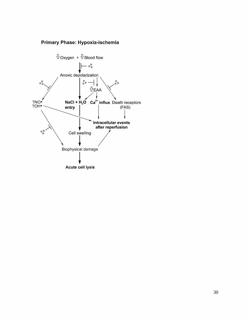
homeostasis. As outlined in Figure 1, the key mechanisms of primary injury and death include:
1. Anoxic depolarization. Once the neuron’s supply of high-energy metabolites such as ATP
can no longer be maintained during HI, the energy dependent mechanisms of intracellular
homeostasis including the Na+/K
+ ATP dependent pump begin to fail. Neuronal
depolarization opens sodium and calcium channels, leading to rapid entry of these cations
into cells (and potassium out). This creates an osmotic and electrochemical gradient that
in turn favors further chloride and water entry leading to cell swelling (cytotoxic edema).
If sufficiently severe, this may lead to acute cell lysis.18
6
Even after surprisingly prolonged and severe insults, however, many swollen neurons can still
recover, at least temporarily, if the hypoxic insult is reversed or the osmotic environment is
manipulated. Evidence suggests that several additional factors act to increase cell injury during
and following depolarization, including:
2. extracellular accumulation of EAAs, mediated by increased release after neuronal
depolarization coupled with impaired energy dependent re-uptake by astrocytes,19
which
in turn promote further receptor mediated cell swelling and intracellular calcium entry;18
3. generation of oxygen free radicals such as the highly toxic hydroxyl radical (•OH),
leading to lipid peroxidation and DNA/RNA fragmentation;20, 21
4. neuronal nitric oxide synthase (nNOS) mediated release of the reactive oxygen species
NO•,22
which can damage key lipoproteins in cell membrane, organelles and
mitochondria;
These damaging events are partly balanced by protective responses that help reduce cell injury,
including:
1. inhibitory amino acids such as γ-aminobutyric acid that accumulate to much greater
levels in the developing brain than in adult animals.19
2. adenosine, an inhibitory neuromodulator derived from breakdown of ATP that helps
delay onset and reduces the severity of energy failure during asphyxia.23
Hypothermia protects the brain during severe HI by:
7
1. a graded reduction in cerebral metabolism of about 5% for every degree of temperature
reduction,24
which delays the onset of anoxic cell depolarization. The protective effects of
intra-insult hypothermia are not simply due to reduced metabolism, since cooling
substantially reduces damage for a given absolute duration of depolarization compared to
normothermia.25
Additional factors include:
2. reduced accumulation of EAAs during intra-ischemic hypothermia in adult and newborn
animals.26, 27
This is primarily due to the delay in depolarization, although there is evidence
for a reduction in the rate of release even after depolarization has occurred.28
3. suppression of NO and superoxide formation, presumptively due to slowing of chemical
reactions, as shown in hippocampal slice cultures,29
during ischemia and reperfusion in
rodents,30
cardiac arrest in young adult dogs,31
and during and immediately after HI in the
piglet.27
Cooling during reperfusion
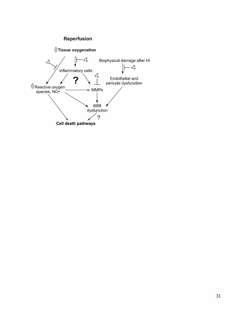
After cerebral circulation and oxygenation are restored at end of the insult, oxidative metabolism
rapidly recovers in surviving cells and cytotoxic edema resolves over approximately 30 to 60
minutes.7, 19, 32
The key events outlined in Figure 2 include:
1. EAA levels rapidly fall in parallel with resolution of the acute cell swelling;19
2. the rapid restoration of tissue oxygenation is associated with a further rapid burst of NO and
superoxide formation;27
8
3. breakdown of the blood brain barrier, allowing large proteins to leak out in the extracellular
space. This may increase brain swelling and is associated with degradation of key regulatory
proteins in the vascular basement membrane, at least in part mediated by induction of
enzymes called metaloproteases.33
Hypothermia started immediately after reperfusion in newborn piglets appeared to accelerate this
resolution as shown by reduced extracellular levels of EAAs, and reduced NO efflux in the
brain.27
Further, in adult rats, cooling after global ischemia was associated with reduced blood
brain barrier (BBB) leakiness and brain edema 24 h later, provided that it was induced within 1 h
after ischemia, apparently through inhibition of metalloproteinases.33
However,
metalloproteinase inhibition after HI in neonatal rats has had inconsistent effects.34
Taken with
the observation that hypothermia is neuroprotective even when delayed by more than an hour
after HI,9, 13, 15, 35
it seems unlikely that these mechanisms are critical to its beneficial effects.
Are excitotoxicity and free radicals relevant to post-insult cooling?
It is now known that:
1. both extracellular accumulation of EAAs and excess free radical production largely resolve
during reperfusion after the insult and appear to have returned to normal values during the
latent phase of recovery from HI;19, 21, 27, 36
2. in vitro, intra-insult hypothermia did not prevent intracellular accumulation of calcium during
cardiac arrest in vivo,37
or during EAA exposure in vitro;38
3. cooling initiated after wash-out of EAAs prevented neuronal degeneration in vitro.38
9
Thus, the ability of hypothermia to reduce release of excitotoxins does not appear to be central to
its neuroprotective effects even during HI, and cannot easily account for the protective effects of
delayed cooling. These data suggest that the critical effect of hypothermia is to block the
intracellular sequelae of depolarization and EAA exposure.
Cell death mechanisms in the latent phase
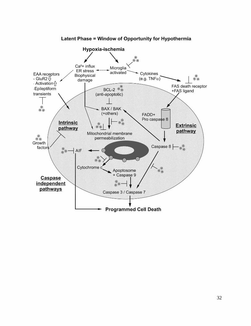
Although the mechanisms of delayed cell loss are clearly multifactorial, there is increasing
evidence that key pathways include activation of programmed cell death pathways, augmented
by the inflammatory reaction and abnormal receptor activity as shown in Figure 3. Programmed
cell death is activated by:
1. excessive calcium influx during and after HI,39
promotes depolarization of the mitochondria
(the ‘intrinsic’ pathway of apoptosis),40
leading to permeabilization of the outer membrane of
the mitochondria, with release of pro-apoptotic proteins, including cytochrome c.
2. abnormal excitatory receptor activity promoting further Ca2+
entry;
3. loss of trophic support from astrocytic growth factors,41
4. secondary inflammatory reaction to HI,42
with release of cytokines and activation of cell
surface death receptors (and thus the ‘extrinsic’ apoptosis pathway).43
Evidence that hypothermia can suppress programmed cell death
10
Post-insult hypothermia typically suppresses hypoxia-
associated protein synthesis,44
and multiple gene
responses to ischemia, particularly genes involved in
calcium homeostasis, cellular and synaptic integrity,
inflammation, cell death, and apoptosis.45
Thus it is
plausible that hypothermia would help prevent ‘active’
forms of cell death. Although studies using
morphological criteria for apoptotic cell death have had
inconsistent outcomes,44
in practice post-hypoxic cell
death represents a continuum between apoptosis and
necrosis, as recently reviewed.46
Activation of caspase-3, the final ‘executioner’ caspase, is a
reasonable, although nonspecific, marker of activation of apoptotic pathways.
In vitro, mild hypothermia directly suppressed neuronal apoptosis induced by serum deprivation,
with reduced activation of caspases -3, -8, and -9 after 24 h, and reduced cytochrome c
translocation, consistent with suppression of both the intrinsic and extrinsic pathways of
apoptosis.47
Further, hypothermia during focal ischemia in adult rats reduced expression of the
cell death receptor Fas and activation of caspase-8, supporting a direct effect on the extrinsic
pathway of apoptosis.48
These studies examined forms of intra-insult cooling. However, in vivo, in the near-term fetal
sheep, hypothermia delayed for 90 min after ischemia markedly suppressed caspase-3 activation
in white matter.14
Similarly, in postnatal day 7 (P7) rat, an age when brain development is
comparable to the late preterm human infant,49
immediate induction of hypothermia after HI
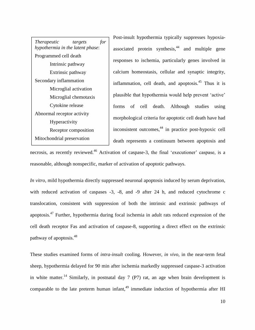
Therapeutic targets for
hypothermia in the latent phase:
Programmed cell death
Intrinsic pathway
Extrinsic pathway
Secondary inflammation
Microglial activation
Microglial chemotaxis
Cytokine release
Abnormal receptor activity
Hyperactivity
Receptor composition
Mitochondrial preservation
11
reduced caspase-3 expression in the cortical infarct,50
and in pre-oligodendrocytes.51
In adult
rats, after transient focal or global ischemia mild hypothermia suppressed activated caspase-3
immunoreactivity,52, 53
upregulated the anti-apoptotic protein bcl-2, reduced expression of the
proapoptotic protein p53,54
and attenuated release of cytochrome c.53, 55, 56
In adult minipigs,
cooling after cardiac arrest reduced opening of the mitochondrial permeability pores.57
Finally, combined treatment with the anti-apoptotic agent, insulin-like growth factor 1, and
hypothermia starting 4.5 h after cerebral ischemia in near-term fetal sheep did not show additive
neuroprotection,58
suggesting that these treatments were working in part though overlapping
mechanisms.
Inflammatory second messengers
Brain injury leads to induction of the inflammatory cascade with increased release of cytokines
and interleukins (IL).59
These compounds are believed to exacerbate delayed injury, whether by
direct neurotoxicity and induction of the extrinsic pathway of apoptosis or by promoting
leukocyte diapedesis into the ischemic brain. For example, TNF-α and interferon-γ mediated
iNOS expression were associated with mitochondrial DNA damage and apoptosis in cultured
oligodendrocytes.60
In vitro, hypothermia inhibits microglia proliferation, chemotaxsis, and induction of pro-
inflammatory cytokines, and the translocation and binding of a key inflammatory signal, nuclear
factor-kappaB, and attenuated microglia neurotoxicity, during and critically, after exposure to
both hypoxia and lipopolysaccharide.61-64
In some settings, cooling may also increase release of
anti-inflammatory cytokines.65
In adult animals, hypothermia after transient focal ischemia and
12
brief cardiac arrest attenuated subsequent increases in cytokines such as interleukin-1β (IL-1 β)
and tumor necrosis factor alpha (TNF-α).66
Consistent with this, post-insult hypothermia
suppressed activated microglia after transient ischemia or asphyxia in fetal sheep.14, 67-69
Intriguingly, despite potent suppression of microglia by hypothermia, it has little effect on
astrocytic proliferation in vitro.61
This raises the possibility that the hypothermic protection
against post-ischemic neuronal damage may be, in part, the result of differential effects on glia,
with suppression of microglial activation but relative sparing of restoration of the normal
homeostatic environment by astrocytes.
Excitotoxicity
In contrast to their role during the primary and reperfusion phases, given that extracellular levels
rapidly return to baseline values,19, 27
the importance of EAAs after reperfusion is surprisingly
unclear. In the temperature controlled environment of the fetal sheep, anti-excitotoxin therapy
limited to the secondary phase did not reduce neuronal injury in severely injured parasagittal
cortex and had only limited neuroprotective effects in other regions.70, 71
Nevertheless, even with normal levels of extracellular glutamate, excitotoxicity may still play an
indirect role. There is evidence of pathological hyperexcitability of glutamate receptors after HI
in P10 rats, with improved neuronal outcome after receptor blockade.72
Consistent with this, in
preterm fetal sheep, treatment with glutamate antagonist after asphyxia reduced neuronal loss,73
although protection was much less than with hypothermia started at a similar time.69
Further, in
adult animals, neuronal death after ischemia has been associated with a selective, delayed change
in the composition of the alpha-amino-3-hydroxy-5-methyl-4-isoxazolepropionic acid (AMPA)

13
receptor, with specific down-regulation of GluR2, a subunit that limits Ca2+
influx. Hypothermia
has been found to:
1. attenuate the post-ischemic reduction in the GluR2 subunit in adult gerbils;74
2. suppress excessive transient epileptiform activity in the first 6 h after asphyxia in preterm
fetal sheep,75
with a close correlation between suppression and neuroprotection.
Further studies are needed to confirm whether these mechanisms are important after hypoxic-
ischemic injury in the term-equivalent brain.
Protection of the mitochondria
Mitochondrial failure is a hallmark of delayed cell death.8 Clearly, maintaining mitochondrial
function is crucial in promoting survival after HI. Post-ischemic hypothermia maintains
mitochondrial respiratory activity after 2 h reperfusion in the adult gerbil,76
and minipig,57
and
intra-ischemic hypothermia has been shown to preserve activity after 4 days recovery in neonatal
rats.77
It is unclear though whether this reflects direct protection of the mitochondria, or whether
it is secondary to suppression of inflammation and programmed cell death.
Induction of growth factors
Perhaps surprisingly in view of the general tendency of hypothermia to suppress new protein
synthesis, there is evidence in the adult rat that mild hypothermia after cardiac arrest is
associated with augmentation of the increase in levels of growth factors such as brain-derived
neurotrophic factor (BDNF) and others,78, 79
which might help protect injured cells. Despite this,
14
BDNF infusion in normothermic animals was not neuroprotective.80
Thus, induction of these
growth factors does not seem to be a major mechanism of hypothermic neuroprotection.
Hypothermia in the Secondary Phase
There is compelling evidence that hypothermia started in the latent phase must be continued for
48 h or more to achieve optimal neuroprotection.11
The precise reasons are unknown. The most
likely explanation is that it is necessary to continue suppressing the programmed cell death and
inflammatory pathways until normal homeostasis returns. However, it could in part reflect
suppression of secondary events in this phase, including hyperperfusion, cytotoxic edema and
delayed seizures (Figure 4).
Cerebral metabolism
During the latent phase cerebral blood flow and metabolism are both suppressed. This
suppression is actively mediated by multiple neuroinhibitory pathways,81
and likely helps
mitigate the effects of abnormal excitatory activity. From 6 to 8 h, hyperperfusion develops
Effects of hypothermia during the secondary phase
1. Possibly contributing to neuroprotection
a. Reduced seizure burden may protect less severely
injured areas of the brain by reducing anaerobic stress
2. Not contributing to neuroprotection
a. Reduced cerebral hyperperfusion.
b. Reduced cytotoxic oedema
15
progressively, to a maximum after 36 to 48 h.9, 15
Hypothermia suppresses the secondary
hyperperfusion after ischemia in the fetal sheep,9, 15
but late hypothermia that was not protective
also effectively suppressed it.15
Clinically, hypothermia markedly attenuated the secondary fall
in the cerebral vascular resistance index, but reduced its predictive value.82
Thus, this effect
appears to be independent of neuroprotection.
Secondary cytotoxic edema
Similarly, neuroprotection with delayed cerebral cooling started 90 min after cerebral ischemia
potently suppresses secondary cytotoxic edema in near-term fetal sheep.9 However, strikingly,
late induction of hypothermia (8.5 h after ischemia) also completely prevented secondary
cytotoxic edema in the same paradigm, despite no significant neuroprotection.16
These findings
are highly consistent with the ability of hypothermia to reduce brain swelling after brain trauma
and in other clinical settings,83
and suggest that it is not a direct mechanism of neuroprotection.
Seizures
Intense, difficult to treat seizures are one of defining characteristics of neonatal
encephalopathy.84
Intense excitation during seizures leads to excessive local metabolic demand,
which can potentially cause local neuronal death.85
In near-term fetal sheep, treatment with MK-
801, a highly potent, selective glutamate antagonist, between 6 and 24 h after cerebral ischemia
prevented delayed post-ischemic seizures.70
Despite this, there was no improvement in
parasagittal neuronal loss, and only a modest improvement in less damaged regions such as the
temporal lobe. These data suggest that severe seizure activity in the secondary phase can
contribute to spreading of injury from the core area of damage to more mildly affected regions.
16
Clinically and experimentally, there is evidence of reduced seizure burden and reduced intensity
of seizures during cooling.75, 86, 87
Thus, the reduced metabolic demand associated with
hypothermia in this phase might help to protect less severely injured regions from further
injury.16
Final conclusions
The mechanisms underlying hypothermic neuroprotection are multifactorial, as summarized in
table 1. Suppression of excitotoxicity, oxidative stress, inflammation, intracellular signaling and
programmed cell death are all effects of hypothermia at different times. Critically, it is
suppression of ‘downstream’ events after anoxic depolarization and excitotoxity that appear to be
critical to hypothermic neuroprotection. We speculate that the differential effects of mild
hypothermia to suppress programmed cell death and microglial activation without suppressing
the recovery of normal homeostasis is central to long-term brain recovery. Further elucidation of
these downstream pathways, particularly in the latent phase and during long-term recovery, will
help us to design effective combination therapies.
17
Figure legends
Figure 1. Flow chart illustrating injurious events during hypoxia-ischemia and potential
therapeutic targets for hypothermia. EAAs: excitatory amino acids. NO•: nitric oxide. OH•:
hydroxyl free radical.
Figure 2. Flow chart illustrating potential therapeutic targets for hypothermia during reperfusion
from hypoxia-ischemia (HI). NO: nitric oxide. MMPs: matrix metalloproteinases. BBB: blood
brain barrier.
Figure 3. Flow chart illustrating key therapeutic targets for hypothermia during the latent phase
of recovery after hypoxia-ischemia. EAA: Excitatory amino acid. GluR2: calcium impermeable
subtype of alpha-amino-3-hydroxy-5-methyl-4-isoxazolepropionic acid receptor. ER:
Endoplasmic reticulum. TNF: Tumor necrosis factor alpha. FADD: Fas-Associated protein
with Death Domain. BCL-2: B-cell lymphoma 2 family of proteins. BAX: Bcl-2 associated X
protein. BAK: Bcl-2 antagonist/killer. AIF: Apoptosis inducing factor.
Figure 4. Flow chart illustrating potential therapeutic targets for hypothermia, during the phase
of secondary deterioration after hypoxia-ischemia.
18
References
1. Rutherford M, Ramenghi LA, Edwards AD, et al. Assessment of brain tissue injury after
moderate hypothermia in neonates with hypoxic-ischaemic encephalopathy: a nested
substudy of a randomised controlled trial. Lancet Neurol 2010;9:39-45.
2. Shankaran S, Barnes PD, Hintz SR, et al. Brain injury following trial of hypothermia for
neonatal hypoxic-ischaemic encephalopathy. Arch Dis Child Fetal Neonatal Ed
2012;97:F398-404.
3. Edwards AD, Brocklehurst P, Gunn AJ, et al. Neurological outcomes at 18 months of age
after moderate hypothermia for perinatal hypoxic ischaemic encephalopathy: synthesis
and meta-analysis of trial data. BMJ 2010;340:c363.
4. Guillet R, Edwards AD, Thoresen M, et al. Seven- to eight-year follow-up of the
CoolCap trial of head cooling for neonatal encephalopathy. Pediatr Res 2012;71:205-209.
5. Shankaran S, Pappas A, McDonald SA, et al. Childhood outcomes after hypothermia for
neonatal encephalopathy. N Engl J Med 2012;366:2085-2092.
6. Azzopardi D, Wyatt JS, Cady EB, et al. Prognosis of newborn infants with hypoxic-
ischemic brain injury assessed by phosphorus magnetic resonance spectroscopy. Pediatr
Res 1989;25:445-451.
7. Iwata O, Iwata S, Bainbridge A, et al. Supra- and sub-baseline phosphocreatine recovery
in developing brain after transient hypoxia-ischaemia: relation to baseline energetics,
insult severity and outcome. Brain 2008;131:2220-2226.
8. Bennet L, Roelfsema V, Pathipati P, et al. Relationship between evolving epileptiform
activity and delayed loss of mitochondrial activity after asphyxia measured by near-
infrared spectroscopy in preterm fetal sheep. J Physiol 2006;572:141-154.
19
9. Gunn AJ, Gunn TR, de Haan HH, et al. Dramatic neuronal rescue with prolonged
selective head cooling after ischemia in fetal lambs. J Clin Invest 1997;99:248-256.
10. Lorek A, Takei Y, Cady EB, et al. Delayed ("secondary") cerebral energy failure after
acute hypoxia-ischemia in the newborn piglet: continuous 48-hour studies by phosphorus
magnetic resonance spectroscopy. Pediatr Res 1994;36:699-706.
11. Gunn AJ, Thoresen M. Hypothermic neuroprotection. NeuroRx 2006;3:154-169.
12. Williams CE, Gunn AJ, Mallard C, et al. Outcome after ischemia in the developing sheep
brain: an electroencephalographic and histological study. Ann Neurol 1992;31:14-21.
13. Sabir H, Scull-Brown E, Liu X, et al. Immediate hypothermia is not neuroprotective after
severe hypoxia-ischemia and is deleterious when delayed by 12 hours in neonatal rats.
Stroke 2012;43:3364-3370.
14. Roelfsema V, Bennet L, George S, et al. The window of opportunity for cerebral
hypothermia and white matter injury after cerebral ischemia in near-term fetal sheep. J
Cereb Blood Flow Metab 2004;24:877-886.
15. Gunn AJ, Gunn TR, Gunning MI, et al. Neuroprotection with prolonged head cooling
started before postischemic seizures in fetal sheep. Pediatrics 1998;102:1098-1106.
16. Gunn AJ, Bennet L, Gunning MI, et al. Cerebral hypothermia is not neuroprotective
when started after postischemic seizures in fetal sheep. Pediatr Res 1999;46:274-280.
17. Gunn AJ, Gluckman PD. Head cooling for neonatal encephalopathy: the state of the art.
Clin Obstet Gynecol 2007;50:636-651.
18. Rothman SM, Olney JW. Excitotoxicity and the NMDA receptor--still lethal after eight
years. Trends Neurosci 1995;18:57-58.
20
19. Tan WK, Williams CE, During MJ, et al. Accumulation of cytotoxins during the
development of seizures and edema after hypoxic-ischemic injury in late gestation fetal
sheep. Pediatr Res 1996;39:791-797.
20. Bagenholm R, Nilsson UA, Gotborg CW, et al. Free radicals are formed in the brain of
fetal sheep during reperfusion after cerebral ischemia. Pediatr Res 1998;43:271-275.
21. Fraser M, Bennet L, van Zijl PL, et al. Extracellular amino acids and peroxidation
products in the periventricular white matter during and after cerebral ischemia in preterm
fetal sheep. J Neurochem 2008;105:2214–2223.
22. Wei G, Dawson VL, Zweier JL. Role of neuronal and endothelial nitric oxide synthase in
nitric oxide generation in the brain following cerebral ischemia. Biochim Biophys Acta
1999;1455:23-34.
23. Hunter CJ, Bennet L, Power GG, et al. Key neuroprotective role for endogenous
adenosine A1 receptor activation during asphyxia in the fetal sheep. Stroke
2003;34:2240-2245.
24. Laptook AR, Corbett RJ, Sterett R, et al. Quantitative relationship between brain
temperature and energy utilization rate measured in vivo using 31P and 1H magnetic
resonance spectroscopy. Pediatr Res 1995;38:919-925.
25. Bart RD, Takaoka S, Pearlstein RD, et al. Interactions between hypothermia and the
latency to ischemic depolarization: implications for neuroprotection. Anesthesiology
1998;88:1266-1273.
26. Nakashima K, Todd MM. Effects of hypothermia on the rate of excitatory amino acid
release after ischemic depolarization. Stroke 1996;27:913-918.
21
27. Thoresen M, Satas S, Puka-Sundvall M, et al. Post-hypoxic hypothermia reduces
cerebrocortical release of NO and excitotoxins. Neuroreport 1997;8:3359-3362.
28. Nakashima K, Todd MM. Effects of hypothermia, pentobarbital, and isoflurane on
postdepolarization amino acid release during complete global cerebral ischemia.
Anesthesiology 1996;85:161-168.
29. McManus T, Sadgrove M, Pringle AK, et al. Intraischaemic hypothermia reduces free
radical production and protects against ischaemic insults in cultured hippocampal slices. J
Neurochem 2004;91:327-336.
30. Lei B, Adachi N, Arai T. The effect of hypothermia on H2O2 production during ischemia
and reperfusion: a microdialysis study in the gerbil hippocampus. NeurosciLett
1997;222:91-94.
31. Lei B, Tan X, Cai H, et al. Effect of moderate hypothermia on lipid peroxidation in
canine brain tissue after cardiac arrest and resuscitation. Stroke 1994;25:147-152.
32. Bennet L, Roelfsema V, Dean J, et al. Regulation of cytochrome oxidase redox state
during umbilical cord occlusion in preterm fetal sheep. Am J Physiol Regul Integr Comp
Physiol 2007;292:R1569-R1576.
33. Nagel S, Su Y, Horstmann S, et al. Minocycline and hypothermia for reperfusion injury
after focal cerebral ischemia in the rat: effects on BBB breakdown and MMP expression
in the acute and subacute phase. Brain Res 2008;1188:198-206.
34. Ranasinghe HS, Scheepens A, Sirimanne E, et al. Inhibition of MMP-9 activity following
hypoxic ischemia in the developing brain using a highly specific inhibitor. Dev Neurosci
2012;34:417-427.
22
35. Colbourne F, Corbett D, Zhao Z, et al. Prolonged but delayed postischemic hypothermia:
a long-term outcome study in the rat middle cerebral artery occlusion model. J Cereb
Blood Flow Metab 2000;20:1702-1708.
36. Bagenholm R, Nilsson UA, Kjellmer I. Formation of free radicals in hypoxic ischemic
brain damage in the neonatal rat, assessed by an endogenous spin trap and lipid
peroxidation. Brain Res 1997;773:132-138.
37. Kristian T, Katsura K, Siesjo BK. The influence of moderate hypothermia on cellular
calcium uptake in complete ischaemia: implications for the excitotoxic hypothesis. Acta
Physiol Scand 1992;146:531-532.
38. Bruno VM, Goldberg MP, Dugan LL, et al. Neuroprotective effect of hypothermia in
cortical cultures exposed to oxygen-glucose deprivation or excitatory amino acids. J
Neurochem 1994;63:1398-1406.
39. Zipfel GJ, Babcock DJ, Lee JM, et al. Neuronal apoptosis after CNS injury: the roles of
glutamate and calcium. J Neurotrauma 2000;17:857-869.
40. Schild L, Keilhoff G, Augustin W, et al. Distinct Ca2+ thresholds determine cytochrome
c release or permeability transition pore opening in brain mitochondria. FASEB
2001;15:565-567.
41. Clawson TF, Vannucci SJ, Wang GM, et al. Hypoxia-ischemia-induced apoptotic cell
death correlates with IGF-I mRNA decrease in neonatal rat brain. Biol Signals Recept
1999;8:281-293.
42. Giulian D, Vaca K. Inflammatory glia mediate delayed neuronal damage after ischemia
in the central nervous system. Stroke 1993;24:I84-90.
23
43. Graham EM, Sheldon RA, Flock DL, et al. Neonatal mice lacking functional Fas death
receptors are resistant to hypoxic-ischemic brain injury. Neurobiol Dis 2004;17:89-98.
44. Bossenmeyer-Pourie C, Koziel V, Daval JL. Effects of hypothermia on hypoxia-induced
apoptosis in cultured neurons from developing rat forebrain: comparison with
preconditioning. Pediatr Res 2000;47:385-391.
45. Nagel S, Papadakis M, Pfleger K, et al. Microarray analysis of the global gene expression
profile following hypothermia and transient focal cerebral ischemia. Neuroscience
2012;208:109-122.
46. Northington FJ, Chavez-Valdez R, Martin LJ. Neuronal cell death in neonatal hypoxia-
ischemia. Ann Neurol 2011;69:743-758.
47. Xu L, Yenari MA, Steinberg GK, et al. Mild hypothermia reduces apoptosis of mouse
neurons in vitro early in the cascade. J Cereb Blood Flow Metab 2002;22:21-28.
48. Liu L, Kim JY, Koike MA, et al. FasL shedding is reduced by hypothermia in
experimental stroke. J Neurochem 2008;106:541-550.
49. Rice JE, 3rd, Vannucci RC, Brierley JB. The influence of immaturity on hypoxic-
ischemic brain damage in the rat. Ann Neurol 1981;9:131-141.
50. Askalan R, Wang C, Shi H, et al. The effect of postischemic hypothermia on apoptotic
cell death in the neonatal rat brain. Dev Neurosci 2011;33:320-329.
51. Xiong M, Chen LX, Ma SM, et al. Short-term effects of hypothermia on axonal injury,
preoligodendrocyte accumulation and oligodendrocyte myelination after hypoxia-
ischemia in the hippocampus of immature rat brain. Dev Neurosci 2013;35:17-27.
24
52. Zgavc T, De Geyter D, Ceulemans AG, et al. Mild hypothermia reduces activated
caspase-3 up to 1 week after a focal cerebral ischemia induced by endothelin-1 in rats.
Brain Res 2013;1501:81-88.
53. Zhao H, Yenari MA, Cheng D, et al. Biphasic cytochrome c release after transient global
ischemia and its inhibition by hypothermia. J Cereb Blood Flow Metab 2005;25:1119-
1129.
54. Zhang H, Xu G, Zhang J, et al. Mild hypothermia reduces ischemic neuron death via
altering the expression of p53 and bcl-2. Neurol Res 2010;32:384-389.
55. Yenari MA, Iwayama S, Cheng D, et al. Mild hypothermia attenuates cytochrome C
release but does not alter Bcl -2 expression or caspase activation after experimental
stroke. J Cereb Blood Flow Metab 2002;22:29-38.
56. Zhao H, Yenari MA, Sapolsky RM, et al. Mild postischemic hypothermia prolongs the
time window for gene therapy by inhibiting cytochrome C release. Stroke 2004;35:572-
577.
57. Gong P, Hua R, Zhang Y, et al. Hypothermia-induced neuroprotection is associated with
reduced mitochondrial membrane permeability in a swine model of cardiac arrest. J
Cereb Blood Flow Metab 2013;33:928-934.
58. George SA, Bennet L, Weaver-Mikaere L, et al. White matter protection with insulin
like-growth factor 1 (IGF-1) and hypothermia is not additive after severe reversible
cerebral ischemia in term fetal sheep. Dev Neurosci 2011;33:280-287.
59. Hagberg H, Mallard C, Jacobsson B. Role of cytokines in preterm labour and brain
injury. BJOG 2005;112 Suppl 1:16-18.
25
60. Druzhyna NM, Musiyenko SI, Wilson GL, et al. Cytokines induce nitric oxide-mediated
mtDNA damage and apoptosis in oligodendrocytes. Protective role of targeting 8-
oxoguanine glycosylase to mitochondria. J Biol Chem 2005;280:21673-21679.
61. Si QS, Nakamura Y, Kataoka K. Hypothermic suppression of microglial activation in
culture: inhibition of cell proliferation and production of nitric oxide and superoxide.
Neuroscience 1997;81:223-229.
62. Seo JW, Kim JH, Seo M, et al. Time-dependent effects of hypothermia on microglial
activation and migration. J Neuroinflammation 2012;9:164.
63. Schmitt KR, Diestel A, Lehnardt S, et al. Hypothermia suppresses inflammation via ERK
signaling pathway in stimulated microglial cells. J Neuroimmunol 2007;189:7-16.
64. Yenari MA, Han HS. Influence of hypothermia on post-ischemic inflammation: role of
nuclear factor kappa B (NFkappaB). Neurochem Int 2006;49:164-169.
65. Diestel A, Troeller S, Billecke N, et al. Mechanisms of hypothermia-induced cell
protection mediated by microglial cells in vitro. Eur J Neurosci 2010;31:779-787.
66. Meybohm P, Gruenewald M, Zacharowski KD, et al. Mild hypothermia alone or in
combination with anesthetic post-conditioning reduces expression of inflammatory
cytokines in the cerebral cortex of pigs after cardiopulmonary resuscitation. Critical care
2010;14:R21.
67. George SA, Barrett RD, Bennet L, et al. Nonadditive neuroprotection with early
glutamate receptor blockade and delayed hypothermia after asphyxia in preterm fetal
sheep. Stroke 2012;43:3114-3117.
26
68. Barrett RD, Bennet L, Naylor A, et al. Effect of cerebral hypothermia and asphyxia on
the subventricular zone and white matter tracts in preterm fetal sheep. Brain Res
2012;1469:35-42.
69. Bennet L, Roelfsema V, George S, et al. The effect of cerebral hypothermia on white and
grey matter injury induced by severe hypoxia in preterm fetal sheep. J Physiol
2007;578:491-506.
70. Tan WK, Williams CE, Gunn AJ, et al. Suppression of postischemic epileptiform activity
with MK-801 improves neural outcome in fetal sheep. Ann Neurol 1992;32:677-682.
71. Gressens P, Le Verche V, Fraser M, et al. Pitfalls in the quest of neuroprotectants for the
perinatal brain. Dev Neurosci 2011;33:189-198.
72. Jensen FE, Wang C, Stafstrom CE, et al. Acute and chronic increases in excitability in rat
hippocampal slices after perinatal hypoxia In vivo. J Neurophysiol 1998;79:73-81.
73. Dean JM, George SA, Wassink G, et al. Suppression of post hypoxic-ischemic EEG
transients with dizocilpine is associated with partial striatal protection in the preterm fetal
sheep. Neuropharmacology 2006;50:491-503.
74. Colbourne F, Grooms SY, Zukin RS, et al. Hypothermia rescues hippocampal CA1
neurons and attenuates down-regulation of the AMPA receptor GluR2 subunit after
forebrain ischemia. Proc Natl Acad Sci U S A 2003.
75. Bennet L, Dean JM, Wassink G, et al. Differential effects of hypothermia on early and
late epileptiform events after severe hypoxia in preterm fetal sheep. J Neurophysiol
2007;97:572-578.
27
76. Canevari L, Console A, Tendi EA, et al. Effect of postischaemic hypothermia on the
mitochondrial damage induced by ischaemia and reperfusion in the gerbil. Brain Res
1999;817:241-245.
77. Nakai A, Shibazaki Y, Taniuchi Y, et al. Influence of mild hypothermia on delayed
mitochondrial dysfunction after transient intrauterine ischemia in the immature rat brain.
Brain Res Dev Brain Res 2001;128:1-7.
78. D'Cruz BJ, Fertig KC, Filiano AJ, et al. Hypothermic reperfusion after cardiac arrest
augments brain-derived neurotrophic factor activation. J Cereb Blood Flow Metab
2002;22:843-851.
79. Schmidt KM, D'Cruz BJ, DeFranco DB, et al. Cardiac arrest and hypothermia increase
GDNF in brain. Acad Emerg Med 2003;10:480.
80. Callaway CW, Ramos R, Logue ES, et al. Brain-derived neurotrophic factor does not
improve recovery after cardiac arrest in rats. Neurosci Lett 2008;445:103-107.
81. Jensen EC, Bennet L, Hunter CJ, et al. Post-hypoxic hypoperfusion is associated with
suppression of cerebral metabolism and increased tissue oxygenation in near-term fetal
sheep. J Physiol 2006;572:131-139.
82. Elstad M, Whitelaw A, Thoresen M. Cerebral Resistance Index is less predictive in
hypothermic encephalopathic newborns. Acta Paediatr 2011;100:1344-1349.
83. Sadaka F, Veremakis C. Therapeutic hypothermia for the management of intracranial
hypertension in severe traumatic brain injury: a systematic review. Brain Inj
2012;26:899-908.
84. Williams CE, Gunn AJ, Synek B, et al. Delayed seizures occurring with hypoxic-
ischemic encephalopathy in the fetal sheep. Pediatr Res 1990;27:561-565.
28
85. Miller SP, Weiss J, Barnwell A, et al. Seizure-associated brain injury in term newborns
with perinatal asphyxia. Neurology 2002;58:542-548.
86. Srinivasakumar P, Zempel J, Wallendorf M, et al. Therapeutic hypothermia in neonatal
hypoxic ischemic encephalopathy: electrographic seizures and magnetic resonance
imaging evidence of injury. J Pediatr 2013;163:465-470.
87. Glass HC, Nash KB, Bonifacio SL, et al. Seizures and magnetic resonance imaging-
detected brain injury in newborns cooled for hypoxic-ischemic encephalopathy. J Pediatr
2011;159:731-735 e731.

29
Table 1. Potential mechanisms of hypothermic neuroprotection
Mechanism of
injury
Relevance to therapeutic hypothermia?
Anoxic
depolarization
Limited. Relevant to cooling during hypoxia-ischemia such as surgery
Accumulation of
EAAs / ROS
Limited. Reduced rate of release of EAAs / ROS by cooling during HI.
Little evidence that it is affected by delayed cooling
Prevention of BBB
breakdown
Limited. Early induction of hypothermia after ischemia can prevent BBB
breakdown, however, hypothermia is neuroprotective when delayed after
the apparent critical window for protecting the BBB
Programmed cell
death
Strong. Hypothermia is associated with suppression of caspase-3,
hypoxia-associated protein synthesis, the mitochondrial permeability
transition, and components of the intrinsic and extrinsic pathways
Secondary
inflammation
Strong. Mild hypothermia potently suppresses microglial activation,
production of inflammatory cytokines and other neurotoxins
Abnormal
glutamate receptor
activation
Moderate. Hypothermia reduces adverse changes in composition of the
AMPA receptor and suppresses epileptiform transients / abnormal
receptor activation in the latent phase. The effect correlates with
neuroprotection, but more studies needed to determine the role of these
effects at term
Cerebral
hyperperfusion
Unlikely. Hypothermia extends the phase of cerebral hypoperfusion and
reduces hyperperfusion independently of neuroprotection
Cytotoxic edema Unlikely. Hypothermia potently suppressed delayed cytotoxic edema, but
independently of neuroprotection
Induction of growth
factors
Limited. In some settings hypothermia can augment the increase in some
growth factors after HI, but not clear whether this is a significant
contributor to neuroprotection
Electrographic
seizures
Limited. Potentially, hypothermia may reduce injury in penumbral, more
mildly affected regions by reduced neural metabolism or anti-
excitotoxicity effects; however, the neuroprotective effects of delayed
hypothermia are much greater than anticonvulsants alone.
30
31
32

33
Related Documents