1 INVITED REVIEW ARTICLE Nagoya J. Med. Sci. 71. 1 ~ 10, 2009 MECHANISMS OF ASBESTOS-INDUCED CARCINOGENESIS SHINYA TOYOKUNI Department of Pathology and Biological Responses, Nagoya University Graduate School of Medicine, Nagoya 466-8550, Japan ABSTRACT Respiratory exposure to asbestos fibers has been associated with diffuse malignant mesothelioma (DMM) in humans. Despite advancements in the molecular analyses of human DMM and the development of animal models, the carcinogenic mechanisms of the disease remain unclear. There are basically three hypotheses regarding the pathogenesis of asbestos-induced DMM, which may be summarized as follows: (1) the “oxidative stress theory” is based on the fact that phagocytic cells that engulf asbestos fibers produce large amounts of free radicals due to their inability to digest the fibers, and epidemiological studies indicating that iron-containing asbestos fibers appear more carcinogenic; (2) the “chromosome tangling theory” postulates that asbestos fibers damage chromosomes when cells divide; and (3) the “theory of adsorption of many specific proteins as well as carcinogenic molecules” states that asbestos fibers in vivo concentrate proteins or chemicals including the components of cigarette smoke. Elucidation of the major mechanisms underlying DMM would be helpful for the development of novel strategies to prevent DMM induction in people who have already been exposed to asbestos. Key Words: Asbestos, Mesothelioma, Iron, Oxidative stress INTRODUCTION Asbestos fibers have been heavily used in industry since World War II to the present because of their durability, heat-resistance, and low cost. 1,2) However, in 1987, the IARC designated asbestos fibers as a Group I (definite) carcinogen for humans (http://monographs.iarc.fr/ENG/ Classification/crthgr01.php), and asbestos fibers were banned in many Western countries in the 1990’s. 1,2) In June of 2005, asbestos-associated deaths suddenly attracted widespread attention in Japan when it was reported that over the past 26 years, 79 factory workers using asbestos died from a rare asbestos-associated cancer called diffuse malignant mesothelioma (DMM). 3) Furthermore, people who lived near those factories also suffered from the same fatal disease. 4) The characteristics of DMM are as follows: (1) it is associated with repeated asbestos expo- sure, 5,6) (2) once diagnosed, prognosis is overwhelmingly poor, 7) and (3) it takes 30 to 40 years after the start of asbestos exposure for DMM to occur. 5,6) Currently, approximately 1,000 and 3,000 patients are diagnosed each year with DMM in Japan and the United States, respectively. It is expected that the incidence of DMM in Japan will peak in 2025 with a cumulative 100,000 Corresponding author: Shinya Toyokuni, M.D., Ph.D. Department of Pathology and Biological Responses, Nagoya University Graduate School of Medicine, 65 Tsurumai-cho, Showa-ku, Nagoya 466-8550, Japan Phone: +81-52-744-2086, Fax: +81-52-744-2091, E-mail: [email protected]

Welcome message from author
This document is posted to help you gain knowledge. Please leave a comment to let me know what you think about it! Share it to your friends and learn new things together.
Transcript
1
INVITED REVIEW ARTICLE
Nagoya J. Med. Sci. 71. 1 ~ 10, 2009
MECHANISMS OF ASBESTOS-INDUCED CARCINOGENESIS
SHINYA TOYOKUNI
Department of Pathology and Biological Responses, Nagoya University Graduate School of Medicine, Nagoya 466-8550, Japan
ABSTRACT
Respiratory exposure to asbestos fibers has been associated with diffuse malignant mesothelioma (DMM) in humans. Despite advancements in the molecular analyses of human DMM and the development of animal models, the carcinogenic mechanisms of the disease remain unclear. There are basically three hypotheses regarding the pathogenesis of asbestos-induced DMM, which may be summarized as follows: (1) the “oxidative stress theory” is based on the fact that phagocytic cells that engulf asbestos fibers produce large amounts of free radicals due to their inability to digest the fibers, and epidemiological studies indicating that iron-containing asbestos fibers appear more carcinogenic; (2) the “chromosome tangling theory” postulates that asbestos fibers damage chromosomes when cells divide; and (3) the “theory of adsorption of many specific proteins as well as carcinogenic molecules” states that asbestos fibers in vivo concentrate proteins or chemicals including the components of cigarette smoke. Elucidation of the major mechanisms underlying DMM would be helpful for the development of novel strategies to prevent DMM induction in people who have already been exposed to asbestos.
Key Words: Asbestos, Mesothelioma, Iron, Oxidative stress
INTRODUCTION
Asbestos fibers have been heavily used in industry since World War II to the present because of their durability, heat-resistance, and low cost.1,2) However, in 1987, the IARC designated asbestos fibers as a Group I (definite) carcinogen for humans (http://monographs.iarc.fr/ENG/ Classification/crthgr01.php), and asbestos fibers were banned in many Western countries in the 1990’s.1,2) In June of 2005, asbestos-associated deaths suddenly attracted widespread attention in Japan when it was reported that over the past 26 years, 79 factory workers using asbestos died from a rare asbestos-associated cancer called diffuse malignant mesothelioma (DMM).3) Furthermore, people who lived near those factories also suffered from the same fatal disease.4)
The characteristics of DMM are as follows: (1) it is associated with repeated asbestos expo-sure,5,6) (2) once diagnosed, prognosis is overwhelmingly poor,7) and (3) it takes 30 to 40 years after the start of asbestos exposure for DMM to occur.5,6) Currently, approximately 1,000 and 3,000 patients are diagnosed each year with DMM in Japan and the United States, respectively. It is expected that the incidence of DMM in Japan will peak in 2025 with a cumulative 100,000
Corresponding author: Shinya Toyokuni, M.D., Ph.D.
Department of Pathology and Biological Responses, Nagoya University Graduate School of Medicine,
65 Tsurumai-cho, Showa-ku, Nagoya 466-8550, Japan
Phone: +81-52-744-2086, Fax: +81-52-744-2091, E-mail: [email protected]
2
Shinya Toyokuni
deaths predicted from this neoplasm.8) This article briefly reviews the history, epidemiology, and genomic analysis of human DMM, and discusses the possible molecular mechanisms underlying asbestos-induced carcinogenesis.
Asbestos fibersAsbestos is a naturally occurring mineral conventionally divided into two mineralogic groups.
The amphiboles include crocidolite (blue asbestos), amosite (brown asbestos), tremolite, anthophyl-lite, and actinolite. Among the amphiboles, only crocidolite and amosite have widespread commercial utilization. The noncommercial amphiboles (the most commonly occurring and widely distributed amphibole asbestos mineral group) are primarily significant as contaminants of other minerals, such as chrysotile.9) The second group of asbestos minerals is the serpentine group, of which chrysotile (white asbestos) is the sole variety.
The amphibole and serpentine minerals occur both as asbestiform (fibrous) and nonasbestiform (massive) varieties of identical chemical composition. Amphibole crystallization is believed to occur initially as the massive form under conditions of moderate temperature and pressure, with transformation into the fibrous form occurring when the unstable massive form is submitted to rock stresses. Similarly, serpentine minerals first crystallize as the massive form, with chrysotile being subsequently formed by recrystallization.10)
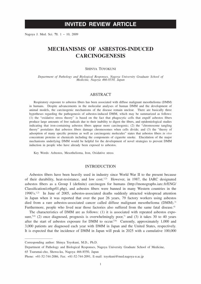
Chrysotile is a hydrated magnesium silicate with the chemical composition indicated in Table 1. Individual fibrils of chrysotile have diameters of 20–40 nm. Crushing of chrysotile ore produces fiber bundles consisting of variable numbers of aggregated individual fibrils. These
Table 1 Composition and characteristics of asbestos fibers
Name Composition Source Morphology
Chrysotile Mg6Si4O10(OH)8 U.S. and Canada Curly, pliable
Crocidolite Na2(Fe3+)2(Fe2+)3Si8O22(OH)2 South AfricaWestern Australia
Rodlike, durable
Amosite (Fe, Mg)7Si8O22(OH)2 South Africa Rodlike, durable
Anthophyllite (Mg, Fe)7Si8O22(OH)2 Finland Rodlike, durable
Tremolite Ca2Mg5Si8O22(OH)2 Exists in some deposits of Canadian chrysotile
Rodlike, durable
Actinolite Ca2(Mg, Fe)5Si8O22(OH)2 Not mined Rodlike, durable
Fig. 1 Scanning electron micrographs contrast curved fibers of chrysotile asbestos (left) with straight fibers of crocidolite (center) and amosite (right; bar=3 mm). Asbestos fibers are from UICC.

3
ASBESTOS-INDUCED CARCINOGENESIS
fibers have varying lengths that may exceed 100 mm. Typically, chrysotile fibers exhibit a curved, curly, or wavy morphology, which is most apparent in fiber bundles exceeding 10 mm in length (Fig. 1). In addition, the ends of chrysotile fiber bundles often exhibit a splayed appearance because of the separation of individual fibrillar units. This curly morphology influences the interceptive deposition of chrysotile fibers, which in turn, affects the depth of penetration into the lower respiratory tract. Inhalational studies in rats have shown that substantial numbers of chrysotile fibers 5 mm or greater in length can penetrate into the lung periphery.11,12)
The amphiboles are a group of hydrated silicates with a wide range of cation substitutions within the silicate backbone of the crystal structure. The predicted chemical formulas of the asbestiform varieties of amphibole minerals are summarized in Table 1. The diameters of individual fibers vary considerably with substantial overlap among members of the amphibole group. However, crocidolite generally has the finest fiber diameters (Fig. 1). Amosite fibers are on average somewhat thicker, and the noncommercial amphibole fibers tend to be the coarsest.
History and epidemiologyThe discovery of this rare tumor known as diffuse malignant mesothelioma (DMM), and the
subsequent controversies that arose about its causation by specific forms of commercial asbestos is a long and complex story. During the late 19th and early 20th centuries, there was a general consensus that some sarcomas arose from the pleura when there was no evidence of a primary tumor elsewhere. There are three major histological types of DMM in the present classification, namely, epithelioid, sarcomatoid, and biphasic.13) Miller and Wynn14) were the first to recognize that a peritoneal neoplasm was able to present both epithelial and fibroblastic characteristics because of the embryologic relationship of these cells to the mesoderm.
In 1924, Robertson’s article15) on endothelioma of the pleura was probably the most thorough review of the literature then available. At the time of the article’s publication, endotheliomas or primary pleural malignancies were certainly rare. In 1940, Ewing, who raised the question of the influence of chronic irritation and inflammation in causing connective tissue changes in the pleura, speculated that some cases of pleural malignancies were associated with tuberculosis.16) Many of the previously reported cases showed evidence of coexistent tuberculosis. The trauma and chronic inflammation as a cause of pleural transformation were reviewed by Ewing. During the 1940’s and 50’s, in the confusion about whether mesothelioma was truly a separate clinical entity, there were five different opinions as to the source of the tumor: (1) an aberrant nest of lung epithelium turned malignant within the lining of the pleura; (2) the endothelial lining of the subpleural lymphatics was the source of the tumor, hence the name endothelioma; (3) the tumor arose from the pleural capillary endothelium or endothelial lining of the subpleural lymphatics, or both; (4) the tumor originated from the mesothelial lining of the pleura itself, or was a mesothelial-derived tumor or a mesothelioma; (5) such tumors of epithelial origin always arise from a primary tumor elsewhere.
Amidst confusion over the concept and origin of the tumor, early reports began to filter in that some patients with asbestosis developed an unusual form of pleural malignancy. The first was by Wedler, who reported the results of 29 autopsies on asbestos workers in Germany.17) Four had bronchial cancers and two others had a malignant pleural growth. He commented about his own impression that the incidence of cancer, which was 20% for malignant tumors in this population, was much too high to have occurred by chance. The seminal year for establishing the association between asbestos exposure and mesothelioma was 1960, when Wagner et al. published a paper entitled “Diffuse malignant mesothelioma and asbestos exposure in North-western Cape Province.”5) The paper was very controversial at that time, since it described 33 cases of DMM with exposure to only one type of asbestos, the so-called Cape Blue asbestos

4
Shinya Toyokuni
mined in the asbestos hills west of Kimberly in the northwest Cape Province of South Africa. He also reported that the tumor was rarely seen elsewhere in South Africa. In 1962, Wagner was able to produce mesothelial tumors of the pleura by direct implantation of asbestos dust in laboratory animals.18) The association between asbestos exposure and diffuse abdominal tumors was established in the English literature by Enticknap and Smither in 1964.19)
Of particular interest epidemiologically was the case control study of Newhouse and Thomp-son.20) They diagnosed 83 patients with mesothelioma in association with a Cape Blue asbestos factory that had opened in London in 1913. There were 27 peritoneal tumors and 56 pleural tumors. The factory had used only Cape crocidolite exclusively until 1926, when small amounts of amosite and chrysotile were added. Particularly distressing was the discovery of 36 patients with no known employment or domestic exposure to asbestos. Eleven of these patients lived within a half mile of the asbestos factory, suggesting neighborhood exposure. A paradigm shift occurred. By 1970, it was generally accepted that low-level exposure to northwest Cape Blue crocidolite was capable of causing mesothelioma. By 1966, the import of crocidolite asbestos was voluntarily abandoned in England, and new asbestos regulations accepting the relationship between asbestos and mesothelioma were adopted in 1969. The history of the early years of the discovery of mesothelioma is an example of how slowly the medical community accepts new discoveries.21) Acceptance was partly delayed by the lack of specific mesothelial cell markers such as are available today to ensure proper diagnosis (calretinin, podoplanin, WT1, cytokeratin, etc.). Since then, extensive epidemiological data have accumulated.22) The general current consensus is that the risk of mesothelioma and lung cancer is greatest with crocidolite, less with amosite, and apparently less with chrysotile. With regard to amosite and chrysotile, there appears to be a higher risk in manufacturing than in mining and milling.
Genomic analysis of human diffuse malignant mesotheliomaCarcinogenesis is a multi-stage process, consisting of genetic and epigenetic alterations. It
is generally accepted that virtually all neoplasms undergo certain genetic changes, whether they are large deletions, amplifications or just a point mutation. Mesothelioma tumors accumulate a spectrum of acquired genetic lesions during the molecular pathogenesis leading to overt cancer. Perhaps reflecting the unique history of asbestos exposure routinely seen in mesothelioma patients, many of the well characterized mutations found in other cancers such as p53 and ras family alterations are not common features in DMM.23) Nonetheless, a variety of well-defined molecular abnormalities have been identified in the majority of DMM cases. The lack of a heritable model for mesothelioma, such as that observed in breast and colon cancers, has fueled the genetic studies of asbestos-induced mesothelioma. Chromosome banding techniques have revealed that most DMMs have complex karyotypes.24,25) The karyotypes of 39 DMMs repeatedly exhibited extensive aneuploidy and structural rearrangements of various chromosomes, in particular the short arms of chromosomes 1, 3, and 9, and the long arm of chromosome 6. The loss of one copy of chromosome 22 is the single most consistent numerical change observed in DMMs.26) Here, I mention three tumor suppressor genes that are mutated in the majority of human DMM cases.
Multiple lines of research have revealed that among the most commonly acquired genetic abnormalities in cancer is the loss of G1 to S checkpoint control. When mesothelioma cell lines and tumors were examined for the presence of p16INK4A gene product, a cyclin-dependent kinase inihibitor, it was found to be absent in all cases.27) Furthermore, a non-deletional loss of p16INK4A gene expression via epigenetic mechanisms of the genes at the 9p21 locus is less common in mesothelioma than in other cancers.28,29) Potentially, a loss of genetic material at the 9p21 locus leads to the loss of cell cycle regulation through both the pRb and p53 pathways.30)
Subsequent positional cloning isolated the NF2 gene as the targeted gene whose loss of

5
ASBESTOS-INDUCED CARCINOGENESIS
function accounts for this clinical syndrome. The 70-kd NF2 gene product is a moesin ezrin radixin-like protein that maps to 22q11-q13.1 and has also been called both Merlin and Schwannomin. NF2 is associated with a high frequency of the loss of chromosome 22, and inactivating mutations are frequently observed in this gene.31,32) Recently, it was reported that YAP1 is involved in mesothelial cell growth; the transcriptional co-activator activity of YAP1 is functionally inhibited by Merlin through phosphorylation and cytoplasmic retention of YAP1.33)
Mutations of the WT1 gene in mesothelioma were noted after the rather striking finding that the WT1 protein is routinely expressed in normal mesothelium as well as in urogenital tissues. Furthermore, DMMs were generally found to express elevated levels of WT1 protein, as detected by immunohistochemistry. The presence of nuclear staining for WT1 has been reported in 75% to 100% of mesothelioma tumors and cell lines examined to date.34)
Animal models of diffuse malignant mesotheliomaIt is well established that inhalational, intraperitoneal and intrapleural exposure to asbestos
fibers causes mesothelioma and/or lung cancer.35-38) This has been reviewed in detail else-where.1,21) A macroscopic appearance of rat peritoneal mesothelioma induced by crocidolite is shown in Fig. 2. DMM was successfully induced also by repeated intraperitoneal injections of ferric saccharate39) that is deposited in the peritoneum. In contrast, the repeated intraperitoneal administration of ferric nitrilotriacetate, an iron chelate soluble at neutral pH, has been found to induce oxidative stress in the renal proximal tubules, ultimately leading to a high incidence of renal cell carcinoma.40-42)
Fig. 2 Peritoneal diffuse malignant mesothelioma induced by intraperitoneal injection of crocidolite in a female rat. Arrows: various size of grape-like tumors are observed in mesentery.
6
Shinya Toyokuni
Mechanisms of carcinogenesisThe strong association between asbestos exposure and malignant mesothelioma has been widely
accepted since 1960. Although asbestos is the primary etiologic agent for this tumor, a certain number of patients who develop mesothelioma have no known asbestos exposure. Radiation,43)
nonasbestos mineral fibers, organic chemicals, chronic inflammation,44) and simian virus 40 (SV40) exposure have also been suggested as risk factors for mesothelioma in humans. Although the “SV40 oncogenic virus contamination theory” (polio vaccination) does not appear to be a major factor, at least in Japan,45,46) there is evidence that this oncogenic polyomavirus can play a variety of promotional roles in the carcinogenic processes of mesothelioma.47)
Currently, there are three hypotheses regarding the pathogenesis of asbestos-induced DMM, which may be summarized as follows: (1) the “oxidative stress theory”48) is based on the fact that asbestos fibers are foreign bodies, and that epidemiological studies show asbestos fibers containing iron (a transitional metal which catalyzes free radical generation) to be more carcinogenic;49) (2) the “chromosome tangling theory” postulates that asbestos fibers damage chromosomes when cells divide;50) and (3) the “theory of adsorption of many specific proteins as well as carcinogenic molecules” states that asbestos in vivo contains chemicals including components of cigarette smoke.48)
Here each hypothesis is described in greater detail, and include my own interpretations (Fig. 3). In the “oxidative stress theory,” there are two distinct players, namely, endogenous iron in asbestos fibers and phagocytes. Regarding the former, surface iron can catalyze the production of reactive oxygen species. Recently, my laboratory has shown, using several different methods, that amosite and crocidolite are good catalysts for free-radical generation.51) In the pathologic diagnosis of asbestos exposure, the presence of asbestos (ferruginous) bodies is an important hall-mark. Indeed, excess iron is carcinogenic not only to animals, but also to humans.41,42,52) In the latter case, “frustrated macrophages engulfing overly long asbestos fibers” have been frequently shown. It is well established that chronic inflammation is a risk factor for carcinogenesis.53) It is interesting to note here that one of the major target genes in ferric nitrilotriacetate-induced renal cell carcinoma is p16INK4A arising from homozygous deletion or methylation of its promoter region.54) I believe that this is not a coincidence, and that the genetic locus of p16INK4A is some-how susceptible to oxidative stress by iron. Additionally, the mesodermal origin of target cells in both carcinogeneses may be associated. My laboratory is interested in whether there are any genomic loci susceptible to oxidative stress. We are currently working on this hypothesis under the concept of oxygenomics.42,55) This idea is important for the elucidation of the carcinogenic mechanism of human DMM according to the “oxidative stress theory.”
It is somewhat more difficult to understand the “chromosome tangling hypothesis.” We recently found that asbestos fibers including crocidolite are actively taken up by several different kinds of cultured cells.51) Furthermore, those fibers enter both the cytoplasm and the nucleus. In this situation, asbestos fibers may tangle with chromosomes when cells divide. Whether there is a specificity of tangling for any chromosomal region is the next question to be addressed.
Finally, as for the “adsorption theory,” it is well known that the surface of asbestos fibers have a high affinity for certain proteins and molecules. This appears to be at least partly as-sociated with positive or negative charges on the asbestos surface. MacCorkle et al. reported an interesting finding that cytoskeletal and cell cycle proteins have high affinities for CCD-18 cells.56) However, it remains unclear how an asbestos (ferruginous) body is produced following exposure to asbestos. Recently, we found that, although chrysotile does not contain iron as a constituent, it concentrates iron in the surrounding tissue, probably via hemolysis.51) It is well known that it takes 30 to 40 years from the start of asbestos exposure for DMM to manifest itself. This probably includes the time needed for the asbestos fibers to reach the parietal pleura

7
ASBESTOS-INDUCED CARCINOGENESIS
Fig. 3 Possible mechanisms of asbestos-induced carcinogenesis.
8
Shinya Toyokuni
according to the negative pressure of the pleural cavity.
Concluding remarksThe molecular mechanism of asbestos-induced carcinogenesis is complex. We believe that
catalytic iron, whether endogenously or by other mechanisms, plays an important role in this carcinogenesis. These observations suggest that the modulation of iron status is one of the measures for the prevention of DMM in those already exposed to asbestos. The shape, size, and adsorbing nature of the fibers also appear to be critically important. Recently, doubts have arisen concerning the safety of commercially available carbon nanotubes,57,58) which may possess the same carcinogenicity as asbestos fibers because of their similar characteristics. Ample care has to be taken to prevent a tragedy similar to the one caused by asbestos exposure.
ACKNOWLDGMENTS
This work was supported in part by a MEXT grant (Special Coordination Funds for Promot-ing Science and Technology), a Grant-in-Aid for Cancer Research from the Ministry of Health, Labour and Welfare of Japan, and a Grant-in-Aid from the Ministry of Education, Culture, Sports, Science and Technology of Japan.
REFERENCES
1) Roggli VL, Oury TD, Sporn TA (editors). Pathology of Asbestos-associated Diseases. 2004, Springer Verlag, New York.
2) Dadson RF, Hammnar SP (editors). Asbestos: Risk Assessment, Epidemiology, and Health Effects. 2006, CRC Press, Taylor & Francis Group, Boca Raton.
3) 79 Kubota workers killed by asbestos over 26 years. 2005.06.30, The Daily Yomiuri. 4) Asbestos ‘silent time bomb’ feared / Revelations of workers’ deaths leave residents near plants wondering
who’s next. 2005.07.07, The Daily Yomiuri. 5) Wagner JC, Sleggs CA, Marchand P. Diffuse pleural mesothelioma and asbestos exposure in North Western
Cape Province. Br J Ind Med, 1960; 17: 260–271. 6) Spirtas R, Heineman E, Bernstein L, Beebe G, Keehn R, Stark A, Harlow B, Benichou J. Malignant
mesothelioma: attributable risk of asbestos exposure. Occup Environ Med, 1994; 51: 804–811. 7) Edwards J, Abrams K, Leverment J, Spyt T, Waller D, O’Byrne K. Prognostic factors for malignant
mesothelioma in 142 patients: validation of CALGB and EORTC prognostic scoring systems. Thorax, 2000; 55: 731–735.
8) Robinson B, Lake R. Advances in malignant mesothelioma. N Engl J Med, 2005; 353: 1591–1603. 9) Addison J, Davies L. Analysis of amphibole asbestos in chrysotile and other minerals. Ann Occup Hyg,
1990; 34: 159–175.10) Pooley FD. Asbestos mineralogy. In: Asbestos Mineralogy, Antman K, Aisner J (editors). pp. 3–27, 1987,
Grune & Stratton, Inc., Orlando.11) Brody AR, Hill LH, Adkins B, O’Connor RW. Chrysotile asbestos inhalation in rats: deposition pattern and
reaction of alveolar epithelium and pulmonary macrophages. Am Rev Respir Dis, 1981; 123: 670–679.12) Lee KP. Lung response to particulates with emphasis on asbestos and other fibrous dusts. CRC Crit Rev
Toxicol, 1985; 14: 33–86.13) Churg A, Cagle PT, Roggli VL. (editors). Tumors of the Serosal Membranes. 2006, ARP Press, Silver
Spring.14) Miller J, Wynn WH. A malignant tumor arising from the endothelium of the peritoneum and provoking a
mucoid acidic fluid. J Pathol Bact, 1908; 12: 127–280.15) Robertson HE. Endothelioma of the pleura. J Cancer Res, 1924; 8: 317–375.16) Ewing J. Neoplastic Diseases, 4th edition. Pp 355–359, 1940, WB Saunders, Philadelphia.17) Wedler HW. Lung cancer in asbestos patients. Dtsch Arch Klin Med, 1943; 191: 189–209.18) Wagner JC. Experimental production of mesothelial tumors of the pleura by implantation of dusts in

9
ASBESTOS-INDUCED CARCINOGENESIS
laboratory animals. Nature, 1962; 196: 180.19) Enticknap JB, Smither WJ. Peritoneal tumors in asbestosis. Br J Ind Med, 1964; 21: 20.20) Newhouse ML, Thompson H. Mesothelioma of pleura and peritoneum following exposure to asbestos in
the London area. Br J Ind Med, 1965; 22: 261–269.21) Pass HI, Vogelzang NJ, Carbone M (editors). Malignant mesothelioma: advances in pathogenesis, diagnosis,
and translational therapies. 2005, Springer Science+Business Media Inc., New York.22) McDonald JC. Asbestos. In: Epidemiology of Work-related Diseases, McDonald JC (editor). 2000, BMC
Books, London.23) Carbone M, Kratzke R, Testa J. The pathogenesis of mesothelioma. Semin Oncol, 2002; 29: 2–17.24) Murthy S, Testa J. Asbestos, chromosomal deletions, and tumor suppressor gene alterations in human
malignant mesothelioma. J Cell Physiol, 1999; 180: 150–157.25) Testa JR, Pass HI, Carbone M. Molecular biology of mesothelioma. In: Molecular Biology of Mesothelioma,
Devita VT, Jr., Hellman S, Rosenberg SA (editors). pp. 1937–1943, 2001, Lippincott Williams & Wilkins, Philadelphia.
26) Taguchi T, Jhanwar S, Siegfried J, Keller S, Testa J. Recurrent deletions of specific chromosomal sites in 1p, 3p, 6q, and 9p in human malignant mesothelioma. Cancer Res, 1993; 53: 4349–4355.
27) Kratzke R, Otterson G, Lincoln C, Ewing S, Oie H, Geradts J, Kaye F. Immunohistochemical analysis of the p16INK4 cyclin-dependent kinase inhibitor in malignant mesothelioma. J Natl Cancer Inst, 1995; 87: 1870–1875.
28) Toyooka S, Pass H, Shivapurkar N, Fukuyama Y, Maruyama R, Toyooka K, Gilcrease M, Farinas A, Minna J, Gazdar A. Aberrant methylation and simian virus 40 tag sequences in malignant mesothelioma. Cancer Res, 2001; 61: 5727–5730.
29) Hirao T, Bueno R, Chen C, Gordon G, Heilig E, Kelsey K. Alterations of the p16 (INK4) locus in human malignant mesothelial tumors. Carcinogenesis, 2002; 23: 1127–1130.
30) Kamijo T, Weber JD, Zambetti G, Zindy F, Roussel MF, Sherr CJ. Functional and physical interactions of the ARF tumor suppressor with p53 and Mdm2. Proc Natl Acad Sci USA, 1998; 95: 8292–8297.
31) Sekido Y, Pass H, Bader S, Mew D, Christman M, Gazdar A, Minna J. Neurofibromatosis type 2 (NF2) gene is somatically mutated in mesothelioma but not in lung cancer. Cancer Res, 1995; 55: 1227–1231.
32) Bianchi A, Mitsunaga S, Cheng J, Klein W, Jhanwar S, Seizinger B, Kley N, Klein-Szanto A, Testa J. High frequency of inactivating mutations in the neurofibromatosis type 2 gene (NF2) in primary malignant mesotheliomas. Proc Natl Acad Sci USA, 1995; 92: 10854–10858.
33) Yokoyama T, Osada H, Murakami H, Tatematsu Y, Taniguchi T, Kondo Y, Yatabe Y, Hasegawa Y, Shimokata K, Horio Y, Hida T, Sekido Y. YAP1 is involved in mesothelioma development and negatively regulated by Merlin through phosphorylation. Carcinogenesis, 2008; 29: 2139–2146.
34) Amin K, Litzky L, Smythe W, Mooney A, Morris J, Mews D, Pass H, Kari C, Rodeck U, Rauscher F. Wilms’ tumor 1 susceptibility (WT1) gene products are selectively expressed in malignant mesothelioma. Am J Pathol, 1995; 146: 344–356.
35) Wagner J, Berry G, Skidmore J, Timbrell V. The effects of the inhalation of asbestos in rats. Br J Cancer, 1974; 29: 252–269.
36) Bolton R, Davis J, Donaldson K, Wright A. Variations in the carcinogenicity of mineral fibres. Ann Occup Hyg, 1982; 26: 569–582.
37) Whitaker D, Shilkin K, Walters M. Cytologic and tissue culture characteristics of asbestos-induced meso-thelioma in rats. Acta Cytol, 1984; 28: 185–189.
38) Suzuki Y, Kohyama N. Malignant mesothelioma induced by asbestos and zeolite in the mouse peritoneal cavity. Environ Res, 1984; 35: 277–292.
39) Okada S, Hamazaki S, Toyokuni S, Midorikawa O. Induction of mesothelioma by intraperitoneal injections of ferric saccharate in male Wistar rats. Br J Cancer, 1989; 60: 708–711.
40) Ebina Y, Okada S, Hamazaki S, Ogino F, Li JL, Midorikawa O. Nephrotoxicity and renal cell carcinoma af-ter use of iron- and aluminum-nitrilotriacetate complexes in rats. J Natl Cancer Inst, 1986; 76: 107–113.
41) Toyokuni S. Molecular mechanisms of oxidative stress-induced carcinogenesis: from epidemiology to oxygenomics. IUBMB Life, 2008; 60: 441–447.
42) Toyokuni S. Role of iron in carcinogenesis: cancer as a ferrotoxic disease. Cancer Sci, 2009; 100: 9–16. 43) Sanders C. Pleural mesothelioma in the rat following exposure to 239PuO2. Health Phys, 1992; 63:
695–697.44) Peterson J, Greenberg S, Buffler P. Non-asbestos-related malignant mesothelioma: a review. Cancer, 1984;
54: 951–960.45) Jin M, Sawa H, Suzuki T, Shimizu K, Makino Y, Tanaka S, Nojima T, Fujioka Y, Asamoto M, Suko

10
Shinya Toyokuni
N, Fujita M, Nagashima K. Investigation of simian virus 40 large T antigen in 18 autopsied malignant mesothelioma patients in Japan. J Med Virol, 2004; 74: 668–676.
46) Aoe K, Hiraki A, Murakami T, Toyooka S, Shivapurkar N, Gazdar A, Sueoka N, Taguchi K, Kamei T, Takeyama H, Sugi K, Kishimoto T. Infrequent existence of simian virus 40 large T antigen DNA in malignant mesothelioma in Japan. Cancer Sci, 2006; 97: 292–295.
47) Gazdar A, Butel J, Carbone M. SV40 and human tumours: myth, association or causality? Nat Rev Cancer, 2002; 2: 957–964.
48) Kamp DW, Graceffa P, Pryor WA, Weitzman SA. The role of free radicals in asbestos-induced diseases. Free Radic Biol Med, 1992; 12: 293–315.
49) McDonald A, McDonald J, Pooley F. Mineral fibre content of lung in mesothelial tumours in North America. Ann Occup Hyg, 1982; 26: 417–422.
50) Wang N, Jaurand M, Magne L, Kheuang L, Pinchon M, Bignon J. The interactions between asbestos fibers and metaphase chromosomes of rat pleural mesothelial cells in culture: a scanning and transmission electron microscopic study. Am J Pathol, 1987; 126: 343–349.
51) Jiang L, Nagai H, Ohara H, Hara S, Tachibana M, Hirano S, Shinohara Y, Kohyama N, Akatsuka S, Toyokuni S. Characteristics and modifying factors of asbestos-induced oxidative DNA damage. Cancer Sci, 2008; 99: 2142–2151.
52) Toyokuni S. Iron-induced carcinogenesis: the role of redox regulation. Free Radic Biol Med, 1996; 20: 553–566.
53) Weitzman SA, Gordon LI. Inflammation and cancer: role of phagocyte-generated oxidants in carcinogenesis. Blood, 1990; 76: 655–663.
54) Tanaka T, Iwasa Y, Kondo S, Hiai H, Toyokuni S. High incidence of allelic loss on chromosome 5 and inactivation of p15INK4B and p16INK4A tumor suppressor genes in oxystress-induced renal cell carcinoma of rats. Oncogene, 1999; 18: 3793–3797.
55) Akatsuka S, Aung TT, Dutta KK, Jiang L, Lee WH, Liu YT, Onuki J, Shirase T, Yamasaki K, Ochi H, Naito Y, Yoshikawa T, Kasai H, Tominaga Y, Sakumi K, Nakabeppu Y, Kawai Y, Uchida K, Yamasaki A, Tsuruyama T, Yamada Y, Toyokuni S. Contrasting genome-wide distribution of 8-hydroxyguanine and acrolein-modified adenine during oxidative stress-induced renal carcinogenesis. Am J Pathol, 2006; 169: 1328–1342.
56) MacCorkle R, Slattery S, Nash D, Brinkley B. Intracellular protein binding to asbestos induces aneuploidy in human lung fibroblasts. Cell Motil Cytoskeleton, 2006; 63: 646–657.
57) Poland C, Duffin R, Kinloch I, Maynard A, Wallace W, Seaton A, Stone V, Brown S, Macnee W, Donaldson K. Carbon nanotubes introduced into the abdominal cavity of mice show asbestos-like pathogenicity in a pilot study. Nat Nanotechnol, 2008; 3: 423–428.
58) Takagi A, Hirose A, Nishimura T, Fukumori N, Ogata A, Ohashi N, Kitajima S, Kanno J. Induction of mesothelioma in p53+/– mouse by intraperitoneal application of multi-wall carbon nanotube. J Toxicol Sci, 2008; 33: 105–116.
Related Documents