Kidney International, Vol. 58 (2000), pp. 1481–1491 Mechanism of chronic obstructive uropathy: Increased expression of apoptosis-promoting molecules YEONG-JIN CHOI,ELZBIETA BARANOWSKA-DACA,VINH NGUYEN,TAKEHIKO KOJI, CHRISTIE M. BALLANTYNE,DAVID SHEIKH-HAMAD,WADI N. SUKI, and LUAN D. TRUONG Renal Pathology Laboratory, Department of Pathology, Department of Medicine, The Methodist Hospital and Baylor College of Medicine, Houston, Texas, USA, and Department of Histology and Cell Biology, Nagasaki School of Medicine, Nagasaki, Japan a progressive increase throughout the experiment. Apoptosis Mechanism of chronic obstructive uropathy: Increased expres- was minimal at all experimental time points for control and sion of apoptosis-promoting molecules. contralateral kidneys. Compared with control and contralateral Background. We have demonstrated that renal tubular and kidneys, the ligated kidneys displayed a dynamic expression interstitial cells undergo pronounced apoptosis during the of mRNAs for many apoptosis-related molecules, which in- course of chronic obstructive uropathy (COU). Apoptosis is a complex cellular process consisting of multiple steps, each of cluded an up to threefold increase for Fas, Fas ligand, TNF-R1, which is mediated by families of related molecules. These fami- TRAIL, TRADD, RIP, and caspase-8, and an up to twofold lies may include receptor/ligand molecules such as Fas, Fas li- increase for FADD and FAP, but there was little change for gand, tumor necrosis factor receptor-1 (TNFR-1), and TNF- FAF. These mRNAs increased between days 4 and 15, decreased related apoptosis inducing ligand (TRAIL); signal transduction until day 30, but then increased again until day 45. The rise adapter molecules such as Fas-associated death domain (FADD), and fall of mRNAs between days 4 and 30 paralleled a similar TNFR-1 associated death domain (TRADD), receptor-inter- fluctuation in tubular cell apoptosis in that period. The subse- acting protein (RIP), Fas-associated factor (FAF), and Fas- quent increase of mRNAs was correlated with a continuous associated phosphatase (FAP); or effector molecules such as rise of interstitial cell apoptosis. We demonstrated a positive caspases. However, the mechanism of tubular cell apoptosis, immunostaining for Fas and Fas ligand in the tubular cells at as well as the pathogenetic relevance of these apoptosis-related early time points as well as in interstitial inflammatory cells at molecules in COU, remains poorly understood. later time points. Although increased expression of TRAIL, Methods. Kidneys were harvested from sham-operated con- TRADD, RIP, and caspase-8 was noted in tubular cells, there trol mice and mice with COU created by left ureter ligation was no staining for these molecules in interstitial cells. sacrificed in groups of three at days 4, 15, 30, and 45. To detect Conclusion. The current study documents a dynamic expres- apoptotic tubular and interstitial cells, in situ end labeling of sion of several molecules that are known to mediate the most fragmented DNA was performed. To detect the expression of crucial steps of apoptosis. It implicates these molecules in apoptosis-related molecules, ribonuclease protection assay was COU-associated renal cell apoptosis and in the pathogenesis used with specific antisense RNA probes for Fas, Fas ligand, of this condition. It also lays the foundation for interventional TNFR-1, TRAIL, FADD, TRADD, RIP, FAF, FAP, and cas- studies, including genetic engineering, to evaluate the molecu- pase-8. Immunostaining for Fas, Fas ligand, TRAIL, TRADD, lar control of apoptosis associated with COU. RIP, and caspase-8 was also performed. To assess the role of these molecules in COU-associated renal cell apoptosis, the frequencies of apoptotic tubular and interstitial cells were sepa- rately quantitated for each experimental time point, and their Obstruction of urine outflow results in a constellation patterns of variation were correlated with those of apoptosis- of renal parenchymal changes collectively called chronic related molecules. obstructive uropathy (COU). These changes include tu- Results. The obstructed kidneys displayed increased apopto- bular atrophy, interstitial fibrosis, and interstitial inflam- sis of both tubular and interstitial cells. Tubular cell apoptosis mation, all of which are associated with progressive renal appeared at day 4 after ureter ligation, peaked (fivefold of control) at day 15, and decreased gradually until the end of tissue loss [1–4]. We have previously demonstrated in a the experiment. In contrast, interstitial cell apoptosis sustained rat model of COU created by unilateral ureteral ligation that a significant number of tubular cells undergo apo- ptosis. This process may be pathogenetically important Key words: tubular cell apoptosis, interstitial cell apoptosis, ribo- nuclease protection assay, obstructive uropathy, cell death. since it could be a major factor responsible for the pro- gressive renal tissue loss seen in kidneys with COU. In Received for publication April 26, 1999 addition, apoptosis of interstitial cells was also docu- and in revised form April 6, 2000 Accepted for publication April 18, 2000 mented and may be relevant to the interstitial changes in this model [5]. 2000 by the International Society of Nephrology 1481

Welcome message from author
This document is posted to help you gain knowledge. Please leave a comment to let me know what you think about it! Share it to your friends and learn new things together.
Transcript
Kidney International, Vol. 58 (2000), pp. 1481–1491
Mechanism of chronic obstructive uropathy:Increased expression of apoptosis-promoting molecules
YEONG-JIN CHOI, ELZBIETA BARANOWSKA-DACA, VINH NGUYEN, TAKEHIKO KOJI,CHRISTIE M. BALLANTYNE, DAVID SHEIKH-HAMAD, WADI N. SUKI, and LUAN D. TRUONG
Renal Pathology Laboratory, Department of Pathology, Department of Medicine, The Methodist Hospital and BaylorCollege of Medicine, Houston, Texas, USA, and Department of Histology and Cell Biology, Nagasaki School of Medicine,Nagasaki, Japan
a progressive increase throughout the experiment. ApoptosisMechanism of chronic obstructive uropathy: Increased expres-was minimal at all experimental time points for control andsion of apoptosis-promoting molecules.contralateral kidneys. Compared with control and contralateralBackground. We have demonstrated that renal tubular andkidneys, the ligated kidneys displayed a dynamic expressioninterstitial cells undergo pronounced apoptosis during theof mRNAs for many apoptosis-related molecules, which in-course of chronic obstructive uropathy (COU). Apoptosis is a
complex cellular process consisting of multiple steps, each of cluded an up to threefold increase for Fas, Fas ligand, TNF-R1,which is mediated by families of related molecules. These fami- TRAIL, TRADD, RIP, and caspase-8, and an up to twofoldlies may include receptor/ligand molecules such as Fas, Fas li- increase for FADD and FAP, but there was little change forgand, tumor necrosis factor receptor-1 (TNFR-1), and TNF- FAF. These mRNAs increased between days 4 and 15, decreasedrelated apoptosis inducing ligand (TRAIL); signal transduction until day 30, but then increased again until day 45. The riseadapter molecules such as Fas-associated death domain (FADD), and fall of mRNAs between days 4 and 30 paralleled a similarTNFR-1 associated death domain (TRADD), receptor-inter- fluctuation in tubular cell apoptosis in that period. The subse-acting protein (RIP), Fas-associated factor (FAF), and Fas- quent increase of mRNAs was correlated with a continuousassociated phosphatase (FAP); or effector molecules such as rise of interstitial cell apoptosis. We demonstrated a positivecaspases. However, the mechanism of tubular cell apoptosis, immunostaining for Fas and Fas ligand in the tubular cells atas well as the pathogenetic relevance of these apoptosis-related early time points as well as in interstitial inflammatory cells atmolecules in COU, remains poorly understood. later time points. Although increased expression of TRAIL,
Methods. Kidneys were harvested from sham-operated con- TRADD, RIP, and caspase-8 was noted in tubular cells, theretrol mice and mice with COU created by left ureter ligation was no staining for these molecules in interstitial cells.sacrificed in groups of three at days 4, 15, 30, and 45. To detect Conclusion. The current study documents a dynamic expres-apoptotic tubular and interstitial cells, in situ end labeling of sion of several molecules that are known to mediate the mostfragmented DNA was performed. To detect the expression of crucial steps of apoptosis. It implicates these molecules inapoptosis-related molecules, ribonuclease protection assay was COU-associated renal cell apoptosis and in the pathogenesisused with specific antisense RNA probes for Fas, Fas ligand, of this condition. It also lays the foundation for interventionalTNFR-1, TRAIL, FADD, TRADD, RIP, FAF, FAP, and cas- studies, including genetic engineering, to evaluate the molecu-pase-8. Immunostaining for Fas, Fas ligand, TRAIL, TRADD, lar control of apoptosis associated with COU.RIP, and caspase-8 was also performed. To assess the role ofthese molecules in COU-associated renal cell apoptosis, thefrequencies of apoptotic tubular and interstitial cells were sepa-rately quantitated for each experimental time point, and their Obstruction of urine outflow results in a constellationpatterns of variation were correlated with those of apoptosis- of renal parenchymal changes collectively called chronicrelated molecules. obstructive uropathy (COU). These changes include tu-Results. The obstructed kidneys displayed increased apopto-
bular atrophy, interstitial fibrosis, and interstitial inflam-sis of both tubular and interstitial cells. Tubular cell apoptosismation, all of which are associated with progressive renalappeared at day 4 after ureter ligation, peaked (fivefold of
control) at day 15, and decreased gradually until the end of tissue loss [1–4]. We have previously demonstrated in athe experiment. In contrast, interstitial cell apoptosis sustained rat model of COU created by unilateral ureteral ligation
that a significant number of tubular cells undergo apo-ptosis. This process may be pathogenetically importantKey words: tubular cell apoptosis, interstitial cell apoptosis, ribo-
nuclease protection assay, obstructive uropathy, cell death. since it could be a major factor responsible for the pro-gressive renal tissue loss seen in kidneys with COU. In
Received for publication April 26, 1999addition, apoptosis of interstitial cells was also docu-and in revised form April 6, 2000
Accepted for publication April 18, 2000 mented and may be relevant to the interstitial changesin this model [5]. 2000 by the International Society of Nephrology
1481
Choi et al: Apoptosis-promoting molecules in COU1482
Apoptosis is a genetically determined process of cell METHODSdeath involving multiple distinct steps, each of which Experimental designis under strict control by timely expressed molecules Under inhalation anesthesia with methoxyflurane,belonging to one of three distinct families, that is, recep- C57B16 male mice (Harlan Animal Farm, Houston, TX,tor/ligand, adapter, and effector families [6–11]. Al- USA) weighing 25 to 35 g were subjected to completethough the mechanism and the essential components of ligation of the left ureter at the ureteropelvic junctionapoptosis remain to be elucidated, it is generally ac- using double silk suture. Animals were subsequentlycepted that apoptosis is initiated by a direct physical placed on a regular diet and allowed free access to tapinteraction between a variety of death signals in the water and were then sacrificed at days 4, 15, 30, and 45.extracellular environment and their death receptors, in- These time points were chosen since, in a pilot study,
they were shown to span the entire quantitative spectrumcluding Fas, tumor necrosis factor receptor-1 (TNFR-1),of tubular and interstitial cell apoptosis. Three to sixand TNF-related apoptosis inducing ligand (TRAIL) re-mice were used for each time point. For control, a groupceptor, which are strategically situated on the cell mem-of six mice was sham-operated and sacrificed at day 0.brane. Apoptosis is initiated when these receptors are
engaged by their ligands, that is, Fas ligand, TNF, andTissue preparation
TRAIL, respectively, which are either free in the circula-Control, ligated, and contralateral kidneys were har-tion or bound to the membrane of cytotoxic effector
vested. Portions of the obtained kidney tissue were fixedcells [6, 9, 11–20]. The death receptor/ligand complexes,in 10% buffered formalin for routine histology and inin turn, bind to specialized domains of several intracellu-situ end labeling of fragmented DNA. Other portionslar molecules collectively called adapter molecules, whichwere frozen in embedding media (OCT compound;
include TNFR-1 associated death domain (TRADD),Sakura Finetek, Torrance, CA, USA) for frozen sec-
receptor-interacting protein (RIP), Fas-associated death tion immunohistochemistry. Both frozen and paraffin-domain (FADD), Fas-associated factor (FAF), and Fas- embedded kidney tissues were cut into 4 mm sectionsassociated phosphatase (FAP) [9, 21–29]. The activated for subsequent staining. The remaining kidney tissuesadapter molecules, then, trigger a cascade of intracellular from animals of the same experimental duration weresignal transduction, resulting in an orderly activation pooled together and snap frozen in liquid nitrogen atof a family of cytosolic or nuclear proteolytic enzymes 2708C for subsequent RNA extraction.collectively called caspases. Caspases [8, 10, 30] serve as
Detection of tubular cell apoptosis by in situ endthe final effector molecules of the death pathway andlabeling of fragmented DNAsare responsible for most of the structural changes that are
observed in apoptotic cells regardless of origin. Among One of the most sensitive and specific markers for apo-ptosis is fragmentation of nuclear DNA. For the detectionmembers of this family, caspase-8 is probably the mostof tubular cell apoptosis, in situ end labeling of fragmentedcritical molecule since it represents the last element inDNA was performed as previously detailed [5].the chain event connecting death signals to other cas-
pases [8, 10, 30, 31]. Among these molecules, FAP is theRibonuclease protection assayonly one that inhibits apoptosis, whereas the remainders
The ribonuclease (RNase) protection assay (RPA) wasare apoptosis promoters.used to detect and quantitate the expression of mRNAsAlthough apoptosis of tubular or interstitial cells rep-of apoptosis-related molecules in kidney tissue [32, 33].resents a pathogenetically fundamental change in COU,A cocktail of probes is used to detect simultaneouslyhow it happens has not been elucidated. To lay the foun-the mRNAs of several preselected functionally relateddation for investigation in this direction, we have at-molecules. The assay was performed as follows. For eachtempted to catalog and quantitate the renal expressiontime point, kidney tissue from three to six animals was
of mRNAs of the apoptosis-related molecules mentioned pooled together, and total cellular RNA was isolatedpreviously in this article in a model of COU created by from control, ligated, and contralateral kidney using theunilateral ureter ligation in mice. Against the background RNAzol-B method, according to the manufacturer’s in-of an ever-increasing number of apoptosis-related mole- structions (Tel-Test, Friendswood, TX, USA). Subse-cules that may or may not be pertinent to apoptosis of quently, RNase protection assay was performed usinga specific cell population in a single organ, this approach RiboQuant RNase protection assay kit (Pharmingen, Sanshould not only offer an insight into what molecules are Diego, CA, USA). This kit contained cDNA templates forrelevant to apoptosis in kidneys with COU, but also pro- Fas, Fas ligand, TNFR-1, TRAIL, TRADD, RIP, FADD,vide some guidelines for subsequent focus on the mole- FAF, FAP, and caspase-8. The kit also contained cDNA
templates for ribosomal protein L32 and glyceraldehydecules that are up-regulated in a faithful fashion in COU.
Choi et al: Apoptosis-promoting molecules in COU 1483
3-phosphate dehydrogenase (GAPDH) as internal con- apoptotic cells against the total number of tubular cells inthe same fields. Quantitation of interstitial cell apoptosistrols. Labeled antisense RNA probes were synthesized
from these cDNA templates using [a-32P] UTP in an in was also performed in the same manner. It should benoted that although both interstitial fibroblasts and inter-vitro transcription reaction performed according to the
manufacturer’s instruction and hybridized with 10 mg stitial inflammatory cells underwent apoptosis, it is notpossible to differentiate precisely these two types of cellsof total RNA extracted from the control, ligated, and
contralateral kidneys for 16 hours at 568C. The hybrid- when they become apoptotic. Separate quantitation forthem, therefore, was not carried out. Immunostainingized products were treated with a digestion mixture in-
cluding RNase and proteinase. During this procedure, for the previously mentioned antibodies was evaluatedon a 0 to 41 scale (0 5 no staining, 11 5 less than 10%the unhybridized RNAs and the free RNA probes were
digested, but the hybridized RNAs were protected from of cells stained, 21 5 10 to 30%, 31 5 30 to 70%, and41 5 70 to 100%). For this purpose, 5 to 10 randomdigestion (RNase protection assay). The hybridized RNAs
were heat denatured and electrophoresed on polyacryl- fields were evaluated under 320 magnification.amide gel for two hours at 50 W at 45 to 508C. The gelwas then dried and exposed to x-ray film at 2708C. The
RESULTSresultant bands were scanned and quantitated using Pho-
Pathologic findingstoShop and UTHSCSA software. Band intensity wasnormalized to that of GAPDH in the same reaction. The The ligated kidneys uniformly developed COU char-
acterized by progressive tubulointerstitial changes, whereasassay was performed three times on the pooled RNA.To confirm the original observations, RPA was also re- the glomeruli and blood vessels remained normal through-
out the experimental period. The tubular changes in-peated on pooled renal tissues from new groups of micesubjected to unilateral ureteral ligation, following the cluded atrophy, dilation, and simplification of the tubular
epithelium, and tubular cell apoptosis; the interstitialsame protocols for those included in the original study.The data were expressed as means 6 SD of the results changes included fibrosis, inflammatory cell infiltration,
increased number of fibroblasts, and apoptosis of inter-obtained from the original and the repeated studies.stitial cells. No significant changes were noted in the
Immunohistochemical staining control and contralateral kidneys (Fig. 1).Immunostaining was performed to assess the cellu-
Apoptosislar sources of the up-regulated apoptosis-related mole-cules. Immunostaining for Fas and Fas ligand was done Although rare apoptotic tubular or interstitial cells
were noted in routine tissue preparation, accurate identi-with affinity-purified and immunohistochemically suit-able rabbit polyclonal antibodies, which were success- fication and quantitation of apoptotic cells were greatly
facilitated by in situ end labeling for fragmented DNAfully developed in our laboratories. These antibodieshave been shown to be monospecific by Western blotting (Fig. 2A, B). Tubular cell apoptosis was first noted at
day 4 after ureteral ligation, increased and peaked (five-[34]. Recently, monospecific rabbit polyclonal antibodiesfor TRAIL, TRADD, RIP, and caspase-8 p20 have also fold of control) at day 15, subsequently decreased rapidly
until day 30, and then decreased gradually until the endbecome available (Santa Cruz, Santa Cruz, CA, USA).They were used for immunostaining in this study. Forma- of the experiment (Fig. 3A). Apoptotic interstitial cells
were less numerous than apoptotic tubular cells in thelin-fixed, paraffin-embedded tissue sections were usedfor antibodies against Fas (1:200 dilution) and Fas ligand initial phase of the study, but gradually increased through-
out the entire experimental period (Fig. 3B). Although(1:100 dilution). Frozen tissue sections were used forantibodies against TRAIL, TRADD, RIP, and caspase-8 it was not possible to differentiate the types of interstitial
cells that underwent apoptosis, inflammatory cells werep20 (1:50 dilution for each antibody). The antibodyagainst caspase 8 p20 recognizes both the proenzyme and probably the more frequent cell types, especially toward
the end of the experiment. Apoptotic cells were rarelyits p20 activation fragment. A standard avidin-biotin-peroxidase technique was performed for control, contra- seen in the glomeruli and were virtually not present in
control or contralateral kidneys throughout the experi-lateral, and ligated kidney tissue sections. Positive andnegative controls were also included. ment.
RNase protection assay forQuantitative studiesapoptosis-related moleculesTubular cells undergoing apoptosis were quantitated
by counting, under a 340 eyepiece of a Nikon micro- The results of the original and the repeated studieswere similar. The mRNAs of several apoptosis-relatedscope, all apoptotic tubular cells within 5 to 10 random
fields in either cortex or medulla. The frequency of apo- molecules were identified at a low level in control kid-neys. The contralateral kidneys displayed the same pat-ptotic tubular cells was expressed as the percentage of
Choi et al: Apoptosis-promoting molecules in COU1484
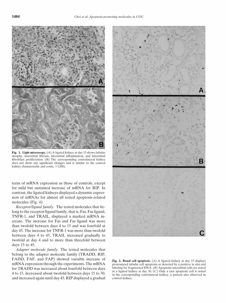
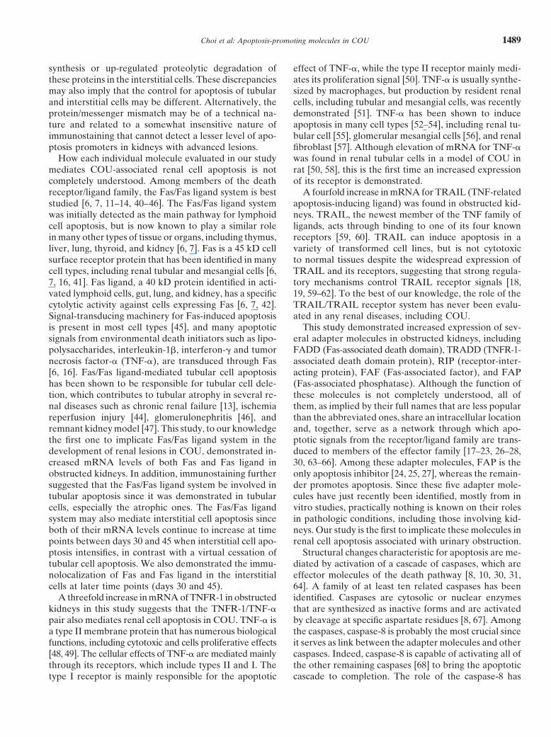
Fig. 1. Light microscopy. (A) A ligated kidney at day 15 shows tubularatrophy, interstitial fibrosis, interstitial inflammation, and interstitialfibroblast proliferation. (B) The corresponding contralateral kidneydoes not show any significant changes and is similar to the controlkidney (hematoxylin and eosin, 31200).
terns of mRNA expression as those of controls, exceptfor mild but sustained increase of mRNA for RIP. Incontrast, the ligated kidneys displayed a dynamic expres-sion of mRNAs for almost all tested apoptosis-relatedmolecules (Fig. 4).
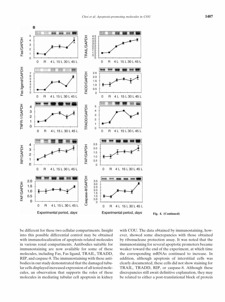
Receptor/ligand family. The tested molecules that be-long to the receptor/ligand family, that is, Fas, Fas ligand,TNFR-1, and TRAIL, displayed a marked mRNA in-crease. The increase for Fas and Fas ligand was morethan twofold between days 4 to 15 and was fourfold atday 45. The increase for TNFR-1 was more than twofoldbetween days 4 to 45. TRAIL increased gradually totwofold at day 4 and to more than threefold betweendays 15 to 45.
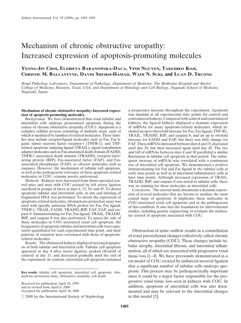
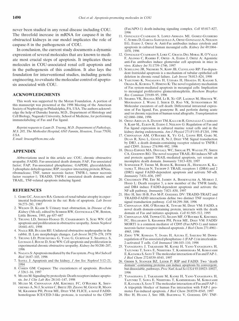
Adapter molecule family. The tested molecules thatbelong to the adapter molecule family (TRADD, RIP,FADD, FAF, and FAP) showed variable increase of Fig. 2. Renal cell apoptosis. (A) A ligated kidney at day 15 displaysmRNA expression through the experiments. The mRNA pronounced tubular cell apoptosis as detected by a positive in situ end
labeling for fragmented DNA. (B) Apoptotic interstitial cells are notedfor TRADD was increased about fourfold between daysin a ligated kidney at day 30. (C ) Only a rare apoptotic cell is noted
4 to 15, decreased about twofold between days 15 to 30, in the corresponding contralateral kidney, a pattern also observed incontrol kidney.and increased again until day 45. RIP displayed a gradual
Choi et al: Apoptosis-promoting molecules in COU 1485
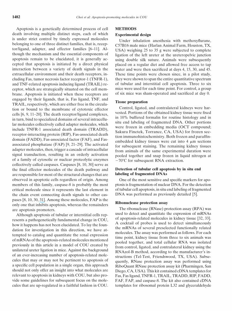
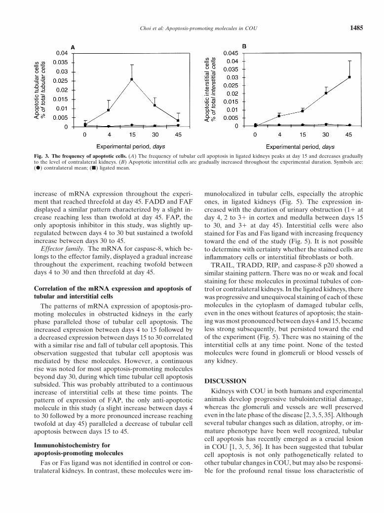
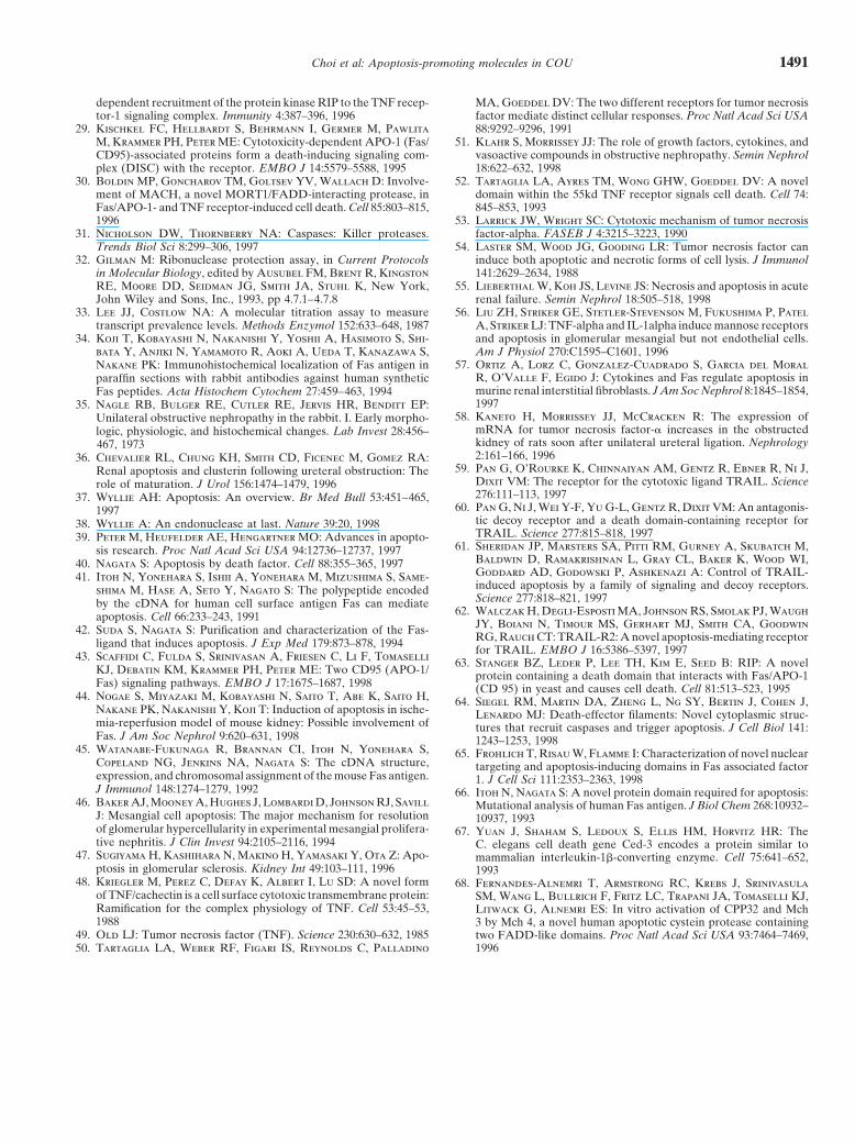
Fig. 3. The frequency of apoptotic cells. (A) The frequency of tubular cell apoptosis in ligated kidneys peaks at day 15 and decreases graduallyto the level of contralateral kidneys. (B) Apoptotic interstitial cells are gradually increased throughout the experimental duration. Symbols are:(d) contralateral mean; (j) ligated mean.
increase of mRNA expression throughout the experi- munolocalized in tubular cells, especially the atrophicment that reached threefold at day 45. FADD and FAF ones, in ligated kidneys (Fig. 5). The expression in-displayed a similar pattern characterized by a slight in- creased with the duration of urinary obstruction (11 atcrease reaching less than twofold at day 45. FAP, the day 4, 2 to 31 in cortex and medulla between days 15only apoptosis inhibitor in this study, was slightly up- to 30, and 31 at day 45). Interstitial cells were alsoregulated between days 4 to 30 but sustained a twofold stained for Fas and Fas ligand with increasing frequencyincrease between days 30 to 45. toward the end of the study (Fig. 5). It is not possible
Effector family. The mRNA for caspase-8, which be- to determine with certainty whether the stained cells arelongs to the effector family, displayed a gradual increase inflammatory cells or interstitial fibroblasts or both.throughout the experiment, reaching twofold between TRAIL, TRADD, RIP, and caspase-8 p20 showed adays 4 to 30 and then threefold at day 45. similar staining pattern. There was no or weak and focal
staining for these molecules in proximal tubules of con-Correlation of the mRNA expression and apoptosis of trol or contralateral kidneys. In the ligated kidneys, theretubular and interstitial cells was progressive and unequivocal staining of each of these
molecules in the cytoplasm of damaged tubular cells,The patterns of mRNA expression of apoptosis-pro-even in the ones without features of apoptosis; the stain-moting molecules in obstructed kidneys in the earlying was most pronounced between days 4 and 15, becamephase paralleled those of tubular cell apoptosis. Theless strong subsequently, but persisted toward the endincreased expression between days 4 to 15 followed byof the experiment (Fig. 5). There was no staining of thea decreased expression between days 15 to 30 correlatedinterstitial cells at any time point. None of the testedwith a similar rise and fall of tubular cell apoptosis. Thismolecules were found in glomeruli or blood vessels ofobservation suggested that tubular cell apoptosis wasany kidney.mediated by these molecules. However, a continuous
rise was noted for most apoptosis-promoting moleculesbeyond day 30, during which time tubular cell apoptosis
DISCUSSIONsubsided. This was probably attributed to a continuousKidneys with COU in both humans and experimentalincrease of interstitial cells at these time points. The
animals develop progressive tubulointerstitial damage,pattern of expression of FAP, the only anti-apoptoticwhereas the glomeruli and vessels are well preservedmolecule in this study (a slight increase between days 4even in the late phase of the disease [2, 3, 5, 35]. Althoughto 30 followed by a more pronounced increase reachingseveral tubular changes such as dilation, atrophy, or im-twofold at day 45) paralleled a decrease of tubular cellmature phenotype have been well recognized, tubularapoptosis between days 15 to 45.cell apoptosis has recently emerged as a crucial lesion
Immunohistochemistry for in COU [1, 3, 5, 36]. It has been suggested that tubularapoptosis-promoting molecules cell apoptosis is not only pathogenetically related to
other tubular changes in COU, but may also be responsi-Fas or Fas ligand was not identified in control or con-tralateral kidneys. In contrast, these molecules were im- ble for the profound renal tissue loss characteristic of
Choi et al: Apoptosis-promoting molecules in COU1486
this condition. Interstitial damage, including fibrosis, in-flammatory cell infiltrate, and vascular remodeling, isalso seen in COU, the severity of which was recentlyshown to correlate positively with the extent of intersti-tial cell apoptosis [5]. These observations strongly implya disturbance of tubular and interstitial cell cycles, in-cluding an increased apoptosis of these cells, in thepathogenesis of COU.
How the general mechanism of apoptosis, gleanedmostly from in vitro studies, is relevant to that of renalcell apoptosis in COU has not been evaluated. A largenumber of environmental factors known to cause apo-ptosis, such as hypoxia, ischemia, cytokines, growth fac-tors, angiotensin II, and reactive oxygen species [3], areall activated during the course of COU, but it remainsunclear whether they are, indeed, the initiators of apo-ptosis in this condition [37–39]. Another fundamentalquestion is which ones, among the large number ofknown apoptosis-related molecules, are functional inCOU. This question, indeed, is relevant since it has beenamply demonstrated that apoptosis can proceed throughdifferent distinct pathways, each of which may be tissueor cell specific and may be mediated by different sets ofmolecules [6–10, 17–31].
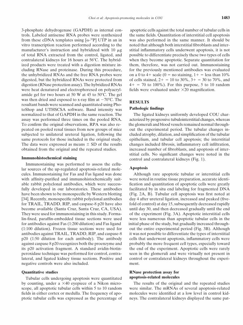
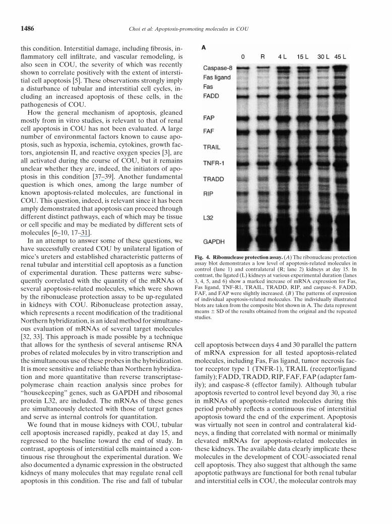
In an attempt to answer some of these questions, wehave successfully created COU by unilateral ligation ofmice’s ureters and established characteristic patterns of Fig. 4. Ribonuclease protection assay. (A) The ribonuclease protection
assay blot demonstrates a low level of apoptosis-related molecules inrenal tubular and interstitial cell apoptosis as a functioncontrol (lane 1) and contralateral (R; lane 2) kidneys at day 15. Inof experimental duration. These patterns were subse- contrast, the ligated (L) kidneys at various experimental duration (lanes
quently correlated with the quantity of the mRNAs of 3, 4, 5, and 6) show a marked increase of mRNA expression for Fas,Fas ligand, TNF-R1, TRAIL, TRADD, RIP, and caspase-8. FADD,several apoptosis-related molecules, which were shownFAF, and FAP were slightly increased. (B ) The patterns of expressionby the ribonuclease protection assay to be up-regulated of individual apoptosis-related molecules. The individually illustrated
in kidneys with COU. Ribonuclease protection assay, blots are taken from the composite blot shown in A. The data representmeans 6 SD of the results obtained from the original and the repeatedwhich represents a recent modification of the traditionalstudies.Northern hybridization, is an ideal method for simultane-
ous evaluation of mRNAs of several target molecules[32, 33]. This approach is made possible by a techniquethat allows for the synthesis of several antisense RNA cell apoptosis between days 4 and 30 parallel the pattern
of mRNA expression for all tested apoptosis-relatedprobes of related molecules by in vitro transcription andthe simultaneous use of these probes in the hybridization. molecules, including Fas, Fas ligand, tumor necrosis fac-
tor receptor type 1 (TNFR-1), TRAIL (receptor/ligandIt is more sensitive and reliable than Northern hybridiza-tion and more quantitative than reverse transcriptase- family); FADD, TRADD, RIP, FAF, FAP (adapter fam-
ily); and caspase-8 (effector family). Although tubularpolymerase chain reaction analysis since probes for“housekeeping” genes, such as GAPDH and ribosomal apoptosis reverted to control level beyond day 30, a rise
in mRNAs of apoptosis-related molecules during thisprotein L32, are included. The mRNAs of these genesare simultaneously detected with those of target genes period probably reflects a continuous rise of interstitial
apoptosis toward the end of the experiment. Apoptosisand serve as internal controls for quantitation.We found that in mouse kidneys with COU, tubular was virtually not seen in control and contralateral kid-
neys, a finding that correlated with normal or minimallycell apoptosis increased rapidly, peaked at day 15, andregressed to the baseline toward the end of study. In elevated mRNAs for apoptosis-related molecules in
these kidneys. The available data clearly implicate thesecontrast, apoptosis of interstitial cells maintained a con-tinuous rise throughout the experimental duration. We molecules in the development of COU-associated renal
cell apoptosis. They also suggest that although the samealso documented a dynamic expression in the obstructedkidneys of many molecules that may regulate renal cell apoptotic pathways are functional for both renal tubular
and interstitial cells in COU, the molecular controls mayapoptosis in this condition. The rise and fall of tubular
Choi et al: Apoptosis-promoting molecules in COU 1487
Fig. 4. (Continued)
be different for these two cellular compartments. Insight with COU. The data obtained by immunostaining, how-ever, showed some discrepancies with those obtainedinto this possible differential control may be obtained
with immunolocalization of apoptosis-related molecules by ribonuclease protection assay. It was noted that theimmunostaining for several apoptotic promoters becamein various renal compartments. Antibodies suitable for
immunostaining are now available for some of these weaker toward the end of the experiment, at which timethe corresponding mRNAs continued to increase. Inmolecules, including Fas, Fas ligand, TRAIL, TRADD,
RIP, and caspase-8. The immunostaining with these anti- addition, although apoptosis of interstitial cells wasclearly documented, these cells did not show staining forbodies in our study demonstrated that the damaged tubu-
lar cells displayed increased expression of all tested mole- TRAIL, TRADD, RIP, or caspase-8. Although thesediscrepancies still await definitive explanation, they maycules, an observation that supports the roles of these
molecules in mediating tubular cell apoptosis in kidney be related to either a post-translational block of protein

Fig. 5. Immunostaining for apoptosis-promoting molecules. Paraffin-embedded kidney tissue stained with Fas (A–C ) and Fas ligand (D–F )polyclonal antibodies. The expression is noted mostly in atrophic tubules at day 15; there is positive staining for both interstitial and tubular cellsat day 45. Acetone fixed-frozen sections of kidney tissues were stained by TRAIL (G–I ), TRADD (J–L), RIP (M–O), and caspase 8 (P–R)polyclonal antibodies. The expression is noted in the damaged tubular cells of the cortex at day 15; it is subsequently decreased and limited to afew tubules at day 45. There was no staining for interstitial cells (avidin-biotin peroxidase, 3600 for all panels).
Choi et al: Apoptosis-promoting molecules in COU 1489
synthesis or up-regulated proteolytic degradation of effect of TNF-a, while the type II receptor mainly medi-ates its proliferation signal [50]. TNF-a is usually synthe-these proteins in the interstitial cells. These discrepancies
may also imply that the control for apoptosis of tubular sized by macrophages, but production by resident renalcells, including tubular and mesangial cells, was recentlyand interstitial cells may be different. Alternatively, the
protein/messenger mismatch may be of a technical na- demonstrated [51]. TNF-a has been shown to induceapoptosis in many cell types [52–54], including renal tu-ture and related to a somewhat insensitive nature of
immunostaining that cannot detect a lesser level of apo- bular cell [55], glomerular mesangial cells [56], and renalfibroblast [57]. Although elevation of mRNA for TNF-aptosis promoters in kidneys with advanced lesions.
How each individual molecule evaluated in our study was found in renal tubular cells in a model of COU inrat [50, 58], this is the first time an increased expressionmediates COU-associated renal cell apoptosis is not
completely understood. Among members of the death of its receptor is demonstrated.A fourfold increase in mRNA for TRAIL (TNF-relatedreceptor/ligand family, the Fas/Fas ligand system is best
studied [6, 7, 11–14, 40–46]. The Fas/Fas ligand system apoptosis-inducing ligand) was found in obstructed kid-neys. TRAIL, the newest member of the TNF family ofwas initially detected as the main pathway for lymphoid
cell apoptosis, but is now known to play a similar role ligands, acts through binding to one of its four knownreceptors [59, 60]. TRAIL can induce apoptosis in ain many other types of tissue or organs, including thymus,
liver, lung, thyroid, and kidney [6, 7]. Fas is a 45 kD cell variety of transformed cell lines, but is not cytotoxicto normal tissues despite the widespread expression ofsurface receptor protein that has been identified in many
cell types, including renal tubular and mesangial cells [6, TRAIL and its receptors, suggesting that strong regula-tory mechanisms control TRAIL receptor signals [18,7, 16, 41]. Fas ligand, a 40 kD protein identified in acti-
vated lymphoid cells, gut, lung, and kidney, has a specific 19, 59–62]. To the best of our knowledge, the role of theTRAIL/TRAIL receptor system has never been evalu-cytolytic activity against cells expressing Fas [6, 7, 42].
Signal-transducing machinery for Fas-induced apoptosis ated in any renal diseases, including COU.This study demonstrated increased expression of sev-is present in most cell types [45], and many apoptotic
signals from environmental death initiators such as lipo- eral adapter molecules in obstructed kidneys, includingFADD (Fas-associated death domain), TRADD (TNFR-1-polysaccharides, interleukin-1b, interferon-g and tumor
necrosis factor-a (TNF-a), are transduced through Fas associated death domain protein), RIP (receptor-inter-acting protein), FAF (Fas-associated factor), and FAP[6, 16]. Fas/Fas ligand-mediated tubular cell apoptosis
has been shown to be responsible for tubular cell dele- (Fas-associated phosphatase). Although the function ofthese molecules is not completely understood, all oftion, which contributes to tubular atrophy in several re-
nal diseases such as chronic renal failure [13], ischemia them, as implied by their full names that are less popularthan the abbreviated ones, share an intracellular locationreperfusion injury [44], glomerulonephritis [46], and
remnant kidney model [47]. This study, to our knowledge and, together, serve as a network through which apo-ptotic signals from the receptor/ligand family are trans-the first one to implicate Fas/Fas ligand system in the
development of renal lesions in COU, demonstrated in- duced to members of the effector family [17–23, 26–28,30, 63–66]. Among these adapter molecules, FAP is thecreased mRNA levels of both Fas and Fas ligand in
obstructed kidneys. In addition, immunostaining further only apoptosis inhibitor [24, 25, 27], whereas the remain-der promotes apoptosis. Since these five adapter mole-suggested that the Fas/Fas ligand system be involved in
tubular apoptosis since it was demonstrated in tubular cules have just recently been identified, mostly from invitro studies, practically nothing is known on their rolescells, especially the atrophic ones. The Fas/Fas ligand
system may also mediate interstitial cell apoptosis since in pathologic conditions, including those involving kid-neys. Our study is the first to implicate these molecules inboth of their mRNA levels continue to increase at time
points between days 30 and 45 when interstitial cell apo- renal cell apoptosis associated with urinary obstruction.Structural changes characteristic for apoptosis are me-ptosis intensifies, in contrast with a virtual cessation of
tubular cell apoptosis. We also demonstrated the immu- diated by activation of a cascade of caspases, which areeffector molecules of the death pathway [8, 10, 30, 31,nolocalization of Fas and Fas ligand in the interstitial
cells at later time points (days 30 and 45). 64]. A family of at least ten related caspases has beenidentified. Caspases are cytosolic or nuclear enzymesA threefold increase in mRNA of TNFR-1 in obstructed
kidneys in this study suggests that the TNFR-1/TNF-a that are synthesized as inactive forms and are activatedby cleavage at specific aspartate residues [8, 67]. Amongpair also mediates renal cell apoptosis in COU. TNF-a is
a type II membrane protein that has numerous biological the caspases, caspase-8 is probably the most crucial sinceit serves as link between the adapter molecules and otherfunctions, including cytotoxic and cells proliferative effects
[48, 49]. The cellular effects of TNF-a are mediated mainly caspases. Indeed, caspase-8 is capable of activating all ofthe other remaining caspases [68] to bring the apoptoticthrough its receptors, which include types II and I. The
type I receptor is mainly responsible for the apoptotic cascade to completion. The role of the caspase-8 has
Choi et al: Apoptosis-promoting molecules in COU1490
(Fas/APO-1) death-inducing signaling complex. Cell 85:817–827,never been studied in any renal disease including COU.1996
The threefold increase in mRNA for caspase-8 in the 11. Gonzalez-Cuadrado S, Lopez-Armada MJ, Gomez-GuerreroC, Subira D, Garcia-Sahuquillo A, Ortiz-Gonzalez A, Neilsonobstructed kidneys in our model implicates the role ofEG, Egido J, Ortiz A: Anti-Fas antibodies induce cytolysis andcaspase-8 in the pathogenesis of COU.apoptosis in cultured human mesangial cells. Kidney Int 49:1064–
In conclusion, the current study documents a dynamic 1070, 199612. Gonzalez-Cuadrado S, Lorz C, Gracia Del Moral R, O’Valleexpression of several molecules that are known to medi-
F, Alonso C, Ramiro F, Ortiz- A, Egido J, Ortiz A: Agonisticate most crucial steps of apoptosis. It implicates theseanti-Fas antibodies induce glomerular cell apoptosis in mice in
molecules in COU-associated renal cell apoptosis and vivo. Kidney Int 51:1739–1746, 199713. Schelling JR, Nkemere N, Kopp JB, Cleveland RP: Fas-depen-in the pathogenesis of this condition. It also lays the
dent fratricidal apoptosis is a mechanism of tubular epithelial cellfoundation for interventional studies, including genetic deletion in chronic renal failure. Lab Invest 78:813–824, 1998engineering, to evaluate the molecular control of apopto- 14. Yasutomo K, Nagasawa H, Uehara H, Hisaeda H, Kagami S,
Okada K, Kuroda Y, Himeno K: The novel regulatory mechanismsis associated with COU.of Fas system-mediated apoptosis in mesangial cells: Implicationto mesangial proliferative glomerulonephritis. Biochem Biophys
ACKNOWLEDGMENTS Res Commun 219:89–95, 199615. Sharma VK, Bologa RM, Li B, Xu GP, Lagman M, Hiscock W,
This work was supported by the Moran Foundation. A portion of Mouradian J, Wang J, Serur D, Rao VK, Suthanthiran M:this manuscript was presented at the 1998 Meeting of the American Molecular executors of cell death: Differential intrarenal expres-Society of Nephrology in Philadelphia, PA, USA. The authors acknowl- sion of Fas ligand, Fas, granzyme B, and perforin during acuteedge the help of Daisuke Isuruta, M.D., Department of Histology and and /or chronic rejection of human renal allografts. TransplantationCell Biology, Nagasaki University, School of Medicine, for performing 62:1860–1866, 1996immunolabeling of Fas and Fas ligand. 16. Ortiz-Arduan A, Danoff TM, Kalluri R, Gonzalez-Cuadrado
S, Karp SL, Elkon K, Egido J, Neilson G: Regulation of Fas andReprint requests to Luan D. Truong, M.D. Department of Pathology, Fas ligand expression in cultured murine renal cells and in the
M.S. 205, The Methodist Hospital, 6565 Fannin, Houston, Texas 77030, kidney during endotoxemia. Am J Physiol 271:F1193–F1201, 1996USA. 17. Chinnaiyan AM, O’Rourke K, Yu G-L, Lyons RH, Garg M,E-mail: [email protected] Duan R, Xing L, Gentz R, Ni J, Dixit VM: Signal transduction
by DR3, a death domain-containing receptor related to TNFR-1and CD95. Science 274:990–992, 1996
18. Degli-Esposti MA, Dougall WC, Smolak PJ, Waugh JY, SmithAPPENDIXCA, Goodwin RG: The novel receptor TRAIL-R4 induces NF-kBand protects against TRAIL-mediated apoptosis, yet retains anAbbreviations used in this article are: COU, chronic obstructiveincomplete death domain. Immunity 7:813–820, 1997uropathy; FADD, Fas-associated death domain; FAF, Fas-associated
19. Schneider P, Thome M, Burns K, Bodmer JL, Hofmann K, Ka-factor; FAP, Fas-associated phosphatase; GAPDH, glyceraldehydetaoka T, Holler N, Tschopp J: TRAIL receptors 1 (DR4) and 23-phosphate dehydrogenase; RIP, receptor-interacting protein; RNase,(DR5) signal FADD-dependent apoptosis and activate NF-kB.ribonuclease; TNF, tumor necrosis factor; TNFR-1, tumor necrosisImmunity 7:831–836, 1997factor receptor-1; TRADD, TNFR-1 associated death domain; and
20. Chaudhaty PM, Eby M, Jasmin A, Bookwalter A, Murray J,TRAIL, TNF-related apoptosis inducing ligand.Hood L: Death receptor 5, a new member of the TNFR family,and DR4 induce FADD-dependent apoptosis and activate the
REFERENCES NF-kB pathway. Immunity 7:821–830, 199721. Hsu H, Shu H-B, Pan M-P, Goeddel DV: TRADD-TRAF2 and
1. Gobe GC, Axelsen RA: Genesis of renal tubular atrophy in exper- TRADD-FADD interactions define two distinct TNF receptor-1imental hydronephrosis in the rat: Role of apoptosis. Lab Invest signal transduction pathway. Cell 84:299–308, 199656:273–281, 1987 22. Chinnaiyan AM, O’Rourke K, Tewari M, Dixit VM: FADD, a
2. Wilson D, Klahr S: Urinary tract obstruction, in Disease of the novel death domain-containing protein, interacts with the deathKidney (5th ed), edited by Schrier RW, Gottschalk CW, Boston, domain of Fas and initiates apoptosis. Cell 81:505–512, 1995Little Brown, 1993, pp 657–687 23. Chinnaiyan AM, Tepper CG, Seldin MF, O’Rourke K, Kischkel
3. Truong LD, Sheikh-Hamad D, Chakraborty S, Suki WN: Cell FC, Hellbardt S, Krammer PH, Peter ME, Dixit VM: FADD/apoptosis and proliferation in obstructive uropathy. Semin Nephrol MORT1 is a common mediator of CD95 (Fas/APO-1) and tumor18:641–651, 1998 necrosis factor receptor-induced apoptosis. J Biol Chem 271:4961–
4. Nagle RB, Bulger RE: Unilateral obstructive nephropathy in the 4965, 1996rabbit. II. Late morphologic changes. Lab Invest 38:270–278, 1978 24. Zhou YW, Komada Y, Inaba H, Azuma E, Sakurai M: Down-
5. Truong LD, Petrusevska G, Yang G, Gurpinar T, Shappell S, regulation of Fas-associated phosphatase-1 (FAP-1) in interleukin-Lechago J, Rouse D, Suki WN: Cell apoptosis and proliferation in 2-activated T cells. Cell Immunol 186:103–110, 1998experimental chronic obstructive uropathy. Kidney Int 50:200–207, 25. Yanagisawa J, Takahashi M, Kanki H, Yano-Yanagisawa H,1996 Tazunoki T, Sawa E, Nishitoba T, Kamishohara M, Kobayashi
6. Nagata S: Apoptosis mediated by the Fas system. Prog Mol Subcell E, Kataoka S, Sato T: The molecular interaction of Fas and FAP-1.Biol 16:87–103, 1996 J Biol Chem 272:8539–8545, 1997
7. Savill J: Apoptosis and the kidney. J Am Soc Nephrol 5:12–21, 26. Grimm S, Stanger BZ, Leder P: RIP and FADD: Two “death1994 domain”-containing proteins can induce apoptosis by convergent,
8. Cohen GM: Caspases: The executioners of apoptosis. Biochem but dissociable, pathways. Proc Natl Acad Sci USA 93:10923–10927,J 326:1–16, 1997 1996
9. Muzio M: Signaling by proteolysis: Death receptors induce apopto- 27. Yanagisawa J, Takahashi M, Kanki H, Yano-Yanagisawa H,sis. Int J Clin Lab Res 28:141–147, 1998 Tazuniki T, Sawa E, Nishitoba T, Kamishohara M, Kobayashi
10. Muzio M, Chinnaiyan AM, Kischkel FC, O’Rourke K, Shev- E, Kataoka S, Sato T: The molecular interaction of Fas and FAP-1:chenko A, Ni J, Scaffidi C, Bretz JD, Zhang M, Gentz R, Mann A tripeptide blocker of human Fas interaction with FAP-1 pro-M, Krammer PH, Peter ME, Dixit VM: FLICE, a novel FADD- motes Fas-induced apoptosis. J Biol Chem 28:8539–8545, 1997
28. Hsu H, Huang J, Shu HB, Baichwal V, Goeddel DV: TNF-homologous ICE/CED-3-like protease, is recruited to the CD95
Choi et al: Apoptosis-promoting molecules in COU 1491
dependent recruitment of the protein kinase RIP to the TNF recep- MA, Goeddel DV: The two different receptors for tumor necrosisfactor mediate distinct cellular responses. Proc Natl Acad Sci USAtor-1 signaling complex. Immunity 4:387–396, 1996
29. Kischkel FC, Hellbardt S, Behrmann I, Germer M, Pawlita 88:9292–9296, 199151. Klahr S, Morrissey JJ: The role of growth factors, cytokines, andM, Krammer PH, Peter ME: Cytotoxicity-dependent APO-1 (Fas/
CD95)-associated proteins form a death-inducing signaling com- vasoactive compounds in obstructive nephropathy. Semin Nephrol18:622–632, 1998plex (DISC) with the receptor. EMBO J 14:5579–5588, 1995
30. Boldin MP, Goncharov TM, Goltsev YV, Wallach D: Involve- 52. Tartaglia LA, Ayres TM, Wong GHW, Goeddel DV: A noveldomain within the 55kd TNF receptor signals cell death. Cell 74:ment of MACH, a novel MORT1/FADD-interacting protease, in
Fas/APO-1- and TNF receptor-induced cell death. Cell 85:803–815, 845–853, 199353. Larrick JW, Wright SC: Cytotoxic mechanism of tumor necrosis1996
31. Nicholson DW, Thornberry NA: Caspases: Killer proteases. factor-alpha. FASEB J 4:3215–3223, 199054. Laster SM, Wood JG, Gooding LR: Tumor necrosis factor canTrends Biol Sci 8:299–306, 1997
32. Gilman M: Ribonuclease protection assay, in Current Protocols induce both apoptotic and necrotic forms of cell lysis. J Immunol141:2629–2634, 1988in Molecular Biology, edited by Ausubel FM, Brent R, Kingston
RE, Moore DD, Seidman JG, Smith JA, Stuhl K, New York, 55. Lieberthal W, Koh JS, Levine JS: Necrosis and apoptosis in acuterenal failure. Semin Nephrol 18:505–518, 1998John Wiley and Sons, Inc., 1993, pp 4.7.1–4.7.8
33. Lee JJ, Costlow NA: A molecular titration assay to measure 56. Liu ZH, Striker GE, Stetler-Stevenson M, Fukushima P, PatelA, Striker LJ: TNF-alpha and IL-1alpha induce mannose receptorstranscript prevalence levels. Methods Enzymol 152:633–648, 1987
34. Koji T, Kobayashi N, Nakanishi Y, Yoshii A, Hasimoto S, Shi- and apoptosis in glomerular mesangial but not endothelial cells.Am J Physiol 270:C1595–C1601, 1996bata Y, Anjiki N, Yamamoto R, Aoki A, Ueda T, Kanazawa S,
57. Ortiz A, Lorz C, Gonzalez-Cuadrado S, Garcia del MoralNakane PK: Immunohistochemical localization of Fas antigen inR, O’Valle F, Egido J: Cytokines and Fas regulate apoptosis inparaffin sections with rabbit antibodies against human syntheticmurine renal interstitial fibroblasts. J Am Soc Nephrol 8:1845–1854,Fas peptides. Acta Histochem Cytochem 27:459–463, 1994199735. Nagle RB, Bulger RE, Cutler RE, Jervis HR, Benditt EP:
58. Kaneto H, Morrissey JJ, McCracken R: The expression ofUnilateral obstructive nephropathy in the rabbit. I. Early morpho-mRNA for tumor necrosis factor-a increases in the obstructedlogic, physiologic, and histochemical changes. Lab Invest 28:456–kidney of rats soon after unilateral ureteral ligation. Nephrology467, 19732:161–166, 199636. Chevalier RL, Chung KH, Smith CD, Ficenec M, Gomez RA:
59. Pan G, O’Rourke K, Chinnaiyan AM, Gentz R, Ebner R, Ni J,Renal apoptosis and clusterin following ureteral obstruction: TheDixit VM: The receptor for the cytotoxic ligand TRAIL. Sciencerole of maturation. J Urol 156:1474–1479, 1996276:111–113, 199737. Wyllie AH: Apoptosis: An overview. Br Med Bull 53:451–465,
60. Pan G, Ni J, Wei Y-F, Yu G-L, Gentz R, Dixit VM: An antagonis-1997tic decoy receptor and a death domain-containing receptor for38. Wyllie A: An endonuclease at last. Nature 39:20, 1998TRAIL. Science 277:815–818, 199739. Peter M, Heufelder AE, Hengartner MO: Advances in apopto-
61. Sheridan JP, Marsters SA, Pitti RM, Gurney A, Skubatch M,sis research. Proc Natl Acad Sci USA 94:12736–12737, 1997Baldwin D, Ramakrishnan L, Gray CL, Baker K, Wood WI,40. Nagata S: Apoptosis by death factor. Cell 88:355–365, 1997Goddard AD, Godowski P, Ashkenazi A: Control of TRAIL-41. Itoh N, Yonehara S, Ishii A, Yonehara M, Mizushima S, Same-induced apoptosis by a family of signaling and decoy receptors.shima M, Hase A, Seto Y, Nagato S: The polypeptide encodedScience 277:818–821, 1997by the cDNA for human cell surface antigen Fas can mediate
62. Walczak H, Degli-Esposti MA, Johnson RS, Smolak PJ, Waughapoptosis. Cell 66:233–243, 1991JY, Boiani N, Timour MS, Gerhart MJ, Smith CA, Goodwin42. Suda S, Nagata S: Purification and characterization of the Fas-RG, Rauch CT: TRAIL-R2: A novel apoptosis-mediating receptorligand that induces apoptosis. J Exp Med 179:873–878, 1994 for TRAIL. EMBO J 16:5386–5397, 199743. Scaffidi C, Fulda S, Srinivasan A, Friesen C, Li F, Tomaselli 63. Stanger BZ, Leder P, Lee TH, Kim E, Seed B: RIP: A novelKJ, Debatin KM, Krammer PH, Peter ME: Two CD95 (APO-1/ protein containing a death domain that interacts with Fas/APO-1
Fas) signaling pathways. EMBO J 17:1675–1687, 1998 (CD 95) in yeast and causes cell death. Cell 81:513–523, 199544. Nogae S, Miyazaki M, Kobayashi N, Saito T, Abe K, Saito H, 64. Siegel RM, Martin DA, Zheng L, Ng SY, Bertin J, Cohen J,
Nakane PK, Nakanishi Y, Koji T: Induction of apoptosis in ische- Lenardo MJ: Death-effector filaments: Novel cytoplasmic struc-mia-reperfusion model of mouse kidney: Possible involvement of tures that recruit caspases and trigger apoptosis. J Cell Biol 141:Fas. J Am Soc Nephrol 9:620–631, 1998 1243–1253, 1998
45. Watanabe-Fukunaga R, Brannan CI, Itoh N, Yonehara S, 65. Frohlich T, Risau W, Flamme I: Characterization of novel nuclearCopeland NG, Jenkins NA, Nagata S: The cDNA structure, targeting and apoptosis-inducing domains in Fas associated factorexpression, and chromosomal assignment of the mouse Fas antigen. 1. J Cell Sci 111:2353–2363, 1998J Immunol 148:1274–1279, 1992 66. Itoh N, Nagata S: A novel protein domain required for apoptosis:
46. Baker AJ, Mooney A, Hughes J, Lombardi D, Johnson RJ, Savill Mutational analysis of human Fas antigen. J Biol Chem 268:10932–J: Mesangial cell apoptosis: The major mechanism for resolution 10937, 1993of glomerular hypercellularity in experimental mesangial prolifera- 67. Yuan J, Shaham S, Ledoux S, Ellis HM, Horvitz HR: Thetive nephritis. J Clin Invest 94:2105–2116, 1994 C. elegans cell death gene Ced-3 encodes a protein similar to
47. Sugiyama H, Kashihara N, Makino H, Yamasaki Y, Ota Z: Apo- mammalian interleukin-1b-converting enzyme. Cell 75:641–652,ptosis in glomerular sclerosis. Kidney Int 49:103–111, 1996 1993
48. Kriegler M, Perez C, Defay K, Albert I, Lu SD: A novel form 68. Fernandes-Alnemri T, Armstrong RC, Krebs J, Srinivasulaof TNF/cachectin is a cell surface cytotoxic transmembrane protein: SM, Wang L, Bullrich F, Fritz LC, Trapani JA, Tomaselli KJ,Ramification for the complex physiology of TNF. Cell 53:45–53, Litwack G, Alnemri ES: In vitro activation of CPP32 and Mch1988 3 by Mch 4, a novel human apoptotic cystein protease containing
49. Old LJ: Tumor necrosis factor (TNF). Science 230:630–632, 1985 two FADD-like domains. Proc Natl Acad Sci USA 93:7464–7469,199650. Tartaglia LA, Weber RF, Figari IS, Reynolds C, Palladino
Related Documents