Journal of Neurochemistry Lippincott—Raven Publishers, Philadelphia © 1997 International Society for Neurochemistry Mechanism of Cellular 3- (4,5-Dimethylthiazol-2-yl) -2,5- Diphenyltetrazolium Bromide (MTT) Reduction Yuanbin Liu, *Danjel A. Peterson, Hideo Kimura, and David Schubert Cellular Neurobiology Laboratory and * Laboratory of Genetics, Salk Institute for Biological Studies, San Diego, California, U.S.A. Abstract: 3- (4,5-Dimethylthiazol-2-yl) -2‚5-diphenyltet- razolium bromide (MIT) reduction is one of the most frequently used methods for measuring cell proliferation and neural cytotoxicity. lt is widely assumed that MIT is reduced by active mitochondria in living cells. By using isolated mitochondria from rat brain and B12 cells, we indeed found that malate, glutamate, and succinate sup- port MIT reduction by isolated mitochondria. However, the data presented in this study do not support the exclu- sive role of mitochondria in MIT reduction by intact cells. Using a variety of approaches, we found that MIT reduc- tion by B12 cells is confined to intracellular vesicles that later give rise to the needle-like MIT formazan at the cell surface. Some of these vesicles were identified as endosomes or lysosomes. In addition, MIT was found to be membrane impermeable. These and other results suggest that MIT is taken up by cells through endocyto- sis and that reduced MIT formazan accumulates in the endosomal/lysosomal compartment and is then trans- ported to the cell surface through exocytosis. Key Words: 3-(4,5-Dimethylthiazol-2-yl)-2,5-diphenyltet- razolium bromide— Endocytosis—Exocytosis—Oxido- reductase—Viability assay. J. Neurochem. 69, 581 —593 (1997). Tetrazolium salts are a large group of heterocyclic organic compounds that form highly colored and often insoluble formazans after reduction. First prepared in 1894, these compounds have been used widely as indi- cators of both biological redox systems and viability (Altman, 1976). 3-. (4,5-Dimethylthiazol-2-yl) -2,5-di- phenyltetrazolium bromide (MTT) is a monotetrazo- hum salt, the reduction of which is one of the most frequently used methods for measuring cell prolifera- tion and cytotoxicity (Mosmann, 1983). Despite the widespread use of the MTT assay as a measure of cell viability and proliferation, the mecha- nism of cellular MTT reduction is poorly understood. An earlier study by Slater et al. (1963), using respira- tory chain inhibitors, showed that succinate-dependent MTT reduction by rat liver homogenates occurred at two sites along the mitochondrial electron transport chain (coenzyme Q and cytochrome c; Fig. 1). Al- though this finding has often been taken as evidence that MTT is reduced by active mitochondria in viable cells, definitive proof for the association of mitochon- dna with MTT reduction in intact cells has been lack- ing because many nonmitochondrial dehydrogenases or fiavin oxidases can also reduce MTT (Altman, 1976; Burdon et al., 1993). Without a clear understanding of the site and the enzymatic system involved in cellu- lar MTT reduction, it has been difficult to explain the discrepancies between the MTT assay and other mea- sures of cell growth and viability (Jabber et al., 1989; Berridge and Tan, 1993), and the effect of extracellular D-glucose concentration and cellular pyridine nucleo- tide concentration on cellular MTT reduction (Vistica et al., 1991). Nikkhah et al. (1992) and Shearman et al. (1995) found that MTT formazan was deposited intracellularly in a granular form with a predominantly perinuclear localization, and prolonged incubation with MTT also resulted in the formation of needle-like formazan crystals at the cell surface. Beri-idge and Tan (1993) found that NADH and NADPH were much better substrates than succinate in supporting MTT re- duction by subcellular components after fractionation, and that the mitochondrial electron transport chain in- hibitor rotenone failed to affect MTT reduction by in- tact cells. The exclusive role of mitochondria in cellu- lar MTT reduction has been questioned (Berridge and Tan, 1993; Shearman et al., 1995), yet the exact cellu- lar site and the enzyme involved in cellular MTT re- duction have not been identified. The data presented below show that cellular MTT Received January 17, 1997; revised manuscript received April 1, 1997; accepted April 2, 1997. Address correspondence and reprint requests to Dr. Y. Liu at Cellular Neurobiology Laboratory, Salk Institute for Biological Stud- ies, P.O. Box 85800, San Diego, CA 92186-5800, U.S.A. Abbreviations used: BODIPY-C 5-DMB-ceramide, N- [5-(5,7-di- methyl-BODIPY) - I -pentanoyl l-D- etythro-sphingosine; DIC, differ- ential interference contrast; DiOC6 (3), 3,3 ‘-dihexyloxacarbocya- nine iodide; DPI, diphenylene iodonium; ER, endoplasmic reticu- lum; FCCP, carbonyl cyanide p -trifluorornethoxyphenylhydrazone; MDCK, Madin—Darby canine kidney; MTT, 3- (4,5-dimethylthia- zol-2-yl )-2,5-diphenyltetrazolium bromide; NEM, N-ethylmalei- mide; PBS, phosphate-buffered saline. 581

Welcome message from author
This document is posted to help you gain knowledge. Please leave a comment to let me know what you think about it! Share it to your friends and learn new things together.
Transcript

Journal ofNeurochemistryLippincott—Raven Publishers, Philadelphia© 1997 International Society for Neurochemistry
Mechanism of Cellular 3- (4,5-Dimethylthiazol-2-yl) -2,5-Diphenyltetrazolium Bromide (MTT) Reduction
Yuanbin Liu, *Danjel A. Peterson, Hideo Kimura, and David Schubert
Cellular Neurobiology Laboratory and * Laboratory of Genetics, Salk Institutefor Biological Studies, San Diego, California, U.S.A.
Abstract: 3- (4,5-Dimethylthiazol-2-yl) -2‚5-diphenyltet-razolium bromide (MIT) reduction is one of the mostfrequently used methods for measuring cell proliferationand neural cytotoxicity. lt is widely assumed that MIT isreduced by active mitochondria in living cells. By usingisolated mitochondria from rat brain and B12 cells, weindeed found that malate, glutamate, and succinate sup-port MIT reduction by isolated mitochondria. However,the data presented in this studydo notsupport the exclu-sive role of mitochondria in MIT reduction by intact cells.Using a variety of approaches, we found that MIT reduc-tion by B12 cells is confined to intracellular vesicles thatlater give rise to the needle-like MIT formazan at thecell surface. Some of these vesicles were identified asendosomes or lysosomes. In addition, MIT was foundto be membrane impermeable. These and other resultssuggest that MIT is taken up by cells through endocyto-sis and that reduced MIT formazan accumulates in theendosomal/lysosomal compartment and is then trans-ported to the cell surface through exocytosis. KeyWords: 3-(4,5-Dimethylthiazol-2-yl)-2,5-diphenyltet-razolium bromide— Endocytosis—Exocytosis—Oxido-reductase—Viability assay.J. Neurochem. 69, 581 —593 (1997).
Tetrazolium salts are a large group of heterocyclicorganic compounds that form highly colored and ofteninsoluble formazans after reduction. First prepared in1894, these compounds have been used widely as indi-cators of both biological redox systems and viability(Altman, 1976). 3-. (4,5-Dimethylthiazol-2-yl)-2,5-di-phenyltetrazolium bromide (MTT) is a monotetrazo-hum salt, the reduction of which is one of the mostfrequently used methods for measuring cell prolifera-tion and cytotoxicity (Mosmann, 1983).
Despite the widespread use of the MTT assay as ameasure of cell viability and proliferation, the mecha-nism of cellular MTT reduction is poorly understood.An earlier study by Slater et al. (1963), using respira-tory chain inhibitors, showed that succinate-dependentMTT reduction by rat liver homogenates occurred attwo sites along the mitochondrial electron transportchain (coenzyme Q and cytochrome c; Fig. 1). Al-
though this finding has often been taken as evidencethat MTT is reduced by active mitochondria in viablecells, definitive proof for the association of mitochon-dna with MTT reduction in intact cells has been lack-ing because many nonmitochondrial dehydrogenasesor fiavin oxidases can also reduce MTT (Altman, 1976;Burdon et al., 1993). Without a clear understandingof the site and the enzymatic system involved in cellu-lar MTT reduction, it has been difficult to explain thediscrepancies between the MTT assay and other mea-sures of cell growth and viability (Jabber et al., 1989;Berridge and Tan, 1993), and the effect of extracellularD-glucose concentration and cellular pyridine nucleo-tide concentration on cellular MTT reduction (Visticaet al., 1991). Nikkhah et al. (1992) and Shearman etal. (1995) found that MTT formazan was depositedintracellularly in a granular form with a predominantlyperinuclear localization, and prolonged incubation withMTT also resulted in the formation of needle-likeformazan crystals at the cell surface. Beri-idge and Tan(1993) found that NADH and NADPH were muchbetter substrates than succinate in supporting MTT re-duction by subcellular components after fractionation,and that the mitochondrial electron transport chain in-hibitor rotenone failed to affect MTT reduction by in-tact cells. The exclusive role of mitochondria in cellu-lar MTT reduction has been questioned (Berridge andTan, 1993; Shearman et al., 1995), yet the exact cellu-lar site and the enzyme involved in cellular MTT re-duction have not been identified.
The data presented below show that cellular MTT
Received January 17, 1997; revised manuscript received April 1,1997; accepted April 2, 1997.
Address correspondence and reprint requests to Dr. Y. Liu atCellular Neurobiology Laboratory, Salk Institute for Biological Stud-ies, P.O. Box 85800, San Diego, CA 92186-5800, U.S.A.
Abbreviations used: BODIPY-C5-DMB-ceramide, N- [5-(5,7-di-
methyl-BODIPY) - I -pentanoyl l-D- etythro-sphingosine; DIC, differ-ential interference contrast; DiOC6 (3), 3,3 ‘-dihexyloxacarbocya-nine iodide; DPI, diphenylene iodonium; ER, endoplasmic reticu-lum; FCCP, carbonyl cyanide p -trifluorornethoxyphenylhydrazone;MDCK, Madin—Darby canine kidney; MTT, 3-(4,5-dimethylthia-zol-2-yl )-2,5-diphenyltetrazolium bromide; NEM, N-ethylmalei-mide; PBS, phosphate-buffered saline.
581

582 Y LIU ET AL.
reduction can occur outside of the mitochondria in B 12cells. Cellular MTT reduction is confined to perinu-clear vesicles, some of which are identified as endo-somes/lysosomes. In addition, MTT is not permeableto lipid membrane. These and other data suggest thatMTT is taken up by cells through endocytosis andis reduced by an N-ethylmaleimide (NEM)-sensitiveflavin oxidase. MTT formazan is then transported tothe cell surface through exocytosis.
MATERIALS AND METHODS
Cell lines and materialsB 12 cells are from a collection of cell lines made from
nitrosoethylurea-induced rat brain tumors and are phenotypi-cally similar to neural precursorcells (Schubert et al., 1974).A suspension-growing variant of B 12 was selected bygrowth in Petri dishes over a 6-month periodand subsequentcloning. Primary culture of rat cortical neurons was preparedas described (Behl et al., 1994). p°cells, which lack mito-chondrial DNA and the wild-type 143B cells (King andAttardi, 1989), were kindly providedby G. Attardi (Califor-nia Institute of Technology). The epithelial Madin—Darbycanine kidney (MDCK) cells were from I. S. Trowbridge(Salk Institute). A HeLa cell line that is defective in recep-tor-mediated endocytosis due to the overexpression of a tem-perature-sensitive dynamin mutant (Damke et al., 1995) wasa generous gift of S. Schmid (Scripps Research Institute).The fluorescent dyes were from Molecular Probes. All otherreagents were from Sigma or Calbiochem.
Preparation of mitochondriaRat brain mitochondria were isolated according to a
method that yields a mixture of “free“ plus synaptosomalmitochondria of high functional integrity (Rosenthal et al.,1987). The rat was killed by decapitation and the entireforebrain was placed in ice-cold buffer containing 225 mMmannitol, 75 mM sucrose, 1 mM EGTA, 5 mM HEPES, pH7.4, and 1 mg/ml bovine serum albumin (MSE buffer). Thetissue was minced, washed with MSE, and homogenized in10 ml of MSE containing5mg Nagase (a type ofcollagenasefrom Sigma). The volume of the homogenate was broughtto 30 ml with MSE and centrifuged at 2,000 g for 3 min.The supernatant was centrifuged at 12,000 g for 8 min. Theresultant pellet, containing free mitochondria and synapto-somes, was resuspended in MSE containing 0.02% digitoninto disrupt the synaptosomal plasma membrane andto releaseany trapped mitochondria, and then centrifuged at 12,000 gfor 10 min. The pellet was resuspended in MSE mediumminus EGTA andcentrifuged at 12,000 g for 10 min. Finally,the pellet was resuspended in MSE minus EGTA for subse-quent studies.
Mitochondria from cultured B12 cells were prepared ac-cording to Moreadith andFiskum (1984). In brief, —~2X l0~cells were permeabilized by digitonin titration. The volumeof the cell suspension was doubled and centrifuged at 3,000g for 3 min to yield the permeabilized cell pellet. The cellpellet was resuspended and homogenized with a Potter—Elvehjem homogenizer. Mitochondria were then preparedfrom the homogenate through differential centrifugation ac-cording to Moreadith and Fiskum (1984). The mitochondriaprepared by this method had respiratory control ratios of7—10.
Subcehlular fractionationSubcellular fractions from Bl2 cells were prepared ac-
cording to Berridge and Tan (1993). About 3 X l0~cellswere washed twice with ice-coldbuffer A (0.25 mM sucrose,20 mM HEPES, pH 7.4, and 1 mM EDTA) and homoge-nized with a glass Dounce homogenizer (80 strokes using atight-fitting pestle). The homogenate was centrifuged at 700g for 10 min. Thepellet was washed twice at 700 g in bufferA and designated as the nuclear fraction. The supernatantwas then centrifuged at 12,000 g for 10 min. The pellet wasagain washed twice at 12,000 g in buffer A and designatedas the mitochondrial fraction. The 12,000-g supematant wascentrifuged at 100,000 g for 1 h. The 100,000-g pellet wastaken as the microsomal fraction without further washing,and the 100,000-g supernatant was taken as cytosol.
MTT reductionMTT reduction by intact cells, subcellular fractions, or
small reductive substances was performed in 96-well micro-titer plates containing 100 ~l medium or buffer. The reactionwas started by adding 10 btl/well of a 2.5 mg/ml MTTstock in Dulbecco‘s phosphate-buffered saline (PBS) andterminated by adding 100 ~il of a solubilization solutioncontaining 50% N,N‘-dimethylformamide and 20% sodiumdodecyl sulfate (pH 4.8). Absorbance values at 570 nm weredetermined on the next day with an automatic microtiterplate reader, using 630 nm as the reference wavelength. ForMTT reduction by B12 cells, l0~cells per well were platedin Dulbecco‘s modified Eagle‘s medium supplemented with5% dialyzed fetal calf serum. Afterovernight incubation, thecells were treated with test reagents for the indicatedperiodsof time followed by 3 h of MTT reduction at 37°C.MTTreduction by isolated mitochondria was performed in KC1buffer (125 mM KC1, 2 mM K2HPO4, 1 mM MgC12, and20mM HEPES, adjusted to pH 7.4 with KOH) with 0.1 mgof protein/mi of mitochondria supported with succinate ormalate plus glutamate, or all of these reagents. MTT reduc-tion by B 12 cell subcellular fractions wasperformed at 37°Cwith 0.5 mg of protein/ml of subcellular fraction in the pres-ence of 0.2 mM of NADH or NADPH. MTT reduction bysmall reductive substances was performed in PBS (pH 7.4)at 37°Cfor 1 h in the presence of 1 mMreductive substances.
Purification of MTT formazan—containingorganelles and marker enzyme assays
B12 cells (2 X l0~cells) were allowed to reduce MTT(0.25 mg/ml) in culture media for 15 min. The cells werepelleted and homogenized in ice-cold buffer A (0.25 mMsucrose, 20 mM HEPES, pH 7.4, and 1 mM EDTA) with aglass Dounce homogenizer (100 strokes using a tight-fittingpestle). The homogenate was centrifuged at 700 g for 10min to sediment nuclei and unbroken cells. The supernatantwas centrifugedat 3,000 g for 10 min. The pelletwaswashedtwice. Themarker enzymeactivities of the 700- g supernatantand the 3,000-g pellet were assayed for various organelle-specific marker enzymes according to themethods describedby Beaufay et al. (1974), as follows: succinate—cytochromec reductasefor mitochondria, /1- N-acetylglucosaminidase forlysosomes, NADPH—cytochrome c reductase for the endo-plasmic reticulum (ER), catahase for peroxisomes (Aebi,1984), and galactosyltransferase for the Golgi apparatus.Protein concentration was determined according to themethod of Lowry et al. (1951).
J. Neurochem., Vol. 69, No. 2, 1997

MECHANISM OF CELLULAR MTT REDUCTION 583
MTT reduction by purified rat erythrocyte lysateRat erythrocytes were purified according to Beutler and
West (1976). In brief, ‘-.~2ml of blood was collected inheparin (10 U/ml) from the heart of an anesthetized rat.Erythrocytes were freed of white blood cells and most plate-lets by passing through a column of microcrystalline cellu-lose/ct-cellulose (1:1, wt/wt; bed volume, 2 ml) that hadbeen previously washed with PBS (10 mM sodium phos-phate, pH 7.4, 0.154 M NaC1, and 10 mM glucose). Theerythrocytes were eluted with 15 ml of PBS and washedthree times with PBS to remove plasma. They were thenresuspended in PBS and lysed with a sonicator probe (fivepulses, 15 s/s). The ability of the lysate (25 mg of protein/ml) to reduce MTT (0.25 mg/mi) was then tested at 37°Cfor 3 h. Hemoglobin in the lysate was found to interferewith MTT formazan reading. Most of the hemoglobin wasremoved by first centrifuging the lysate at 12,000 g for 10min to sediment the water-insoluble MTT formazan. Thepellet was then dissolved in the solubilization solution forMTT formazan determination using a 96-well plate.
Photomicroscopy and scanning laser confocalmicroscopy
Cells growing on 35-mm dishes were examined or photo-graphed with a Nikon light microscope equipped witha water- and oil-immersible objective lens using Kodak EPT64T film. Cells growing on glass slide chambers were stainedwith MTT- and subcellular organelle—specific fluorescentdyes, washed, and viewed on a Bio-Rad MRC 1024 confocalimaging system coupled to aZeiss Axiovertmicroscope (Pe-terson et al., 1996). Subcellularorganelles were stained withthefollowing fluorescentdyes: mitochondriawith rhodamine123 (Johnson et al., 1980), ER with 3,3‘-dihexyloxacar-bocyanine iodide [DiOC6(3)] (Terasaki et al., 1984),Golgi apparatus with { N- [5- (5,7-dimethyl-BODIPY) -1-pentanoyl I-D- erythro - sphingosine } (BODIPY-C5-DMB -
ceramide) (Pagano et al., 1991), endosome/lysosome withlucifer yellow CH (Swanson et al., 1985), and acidic vesi-des (which include both endosomes and lysosomes) withacridineorange (Yoshimori et al., 1991). Because theconfo-cal microscope is equipped with a krypton/argon laser, exci-tation or emission wavelengths that are closest to the avail-able spectrum lines of the laser source were used. Thesewavelengths are not necessarily the maximum excitation oremission wavelengths ofthe fluorescentdyes. The excitationwavelength and emission wavelength used for rhodamine123, DIOC6(3), BODIPY-C5-DMB-ceramide, and luciferyellow CH are the same, which was 488 nm for excitationand 522 nm (bandwidth, 35 nm) for emission. The excitationwavelength and emission wavelength for acridine orangewere 568 and 605 nm (bandwidth, 32 nm), respectively.The optical slice thickness used to capture the fluorescentimages is 0.45 ~im and the optical slice thickness used tocapture the bright-field images is 1.5 ~tm using differentialinterference contrast (DIC) optics. The superimposition ofimages was done on a 20-in, computer screen; the computerprogram aids the examination of colocahization by allowingrepeated take away—add back of one image (dynamic dis-play of color), so that one can be sure whether a particularMTT formazan vesicle is superimposed with nearby fluo-rescent organelles.
Encapsulation of MTT in liposomesMTT and trypan blue were encapsulated in large unilamel-
lar liposomes by reverse-phase evaporation (Szoka and Pa-
pahadjopoulos, 1978). In brief, 66 ~zmolof total lipids (mo-lar ratio: egg phosphatidylcholine/egg phosphatidylgly-cerol/cholesterol = 1:4:5) dissolved in chloroform wasdriedunder nitrogen by a rotary evaporator. The lipids were redis-solved with 3 ml of diethyl ether and mixed with 1 ml of1:10 dilution of PBS containing 5 mg/mI MTT or 5 mg/mitrypan blue. The mixture was sonicated under nitrogen for5 min at 20°Cin a bath-type sonicator. The resulting suspen-sion was then placed on the rotary evaporator and the etherwas removed under vacuum (water aspirator) at 20°C.Thegel formed during evaporation was broken up by vortexmixing and 1 ml of PBS was added. The residual ether wasremoved by using the water aspirator until an opalescentaqueous suspension wasobtained. Unencapsulated MTT wasremoved by overnight dialysis at 4°Cin 500 ml of PBS withfour buffer changes.
The membrane permeability of MTT and trypan blue wasdetermined by dialyzing the liposomes (0.4 ml) against 100ml of PBS for 1 h at 37°C, and the MTT or trypan blueremaining in the liposomes was then determined by spectro-photometry in the presence of 0.5% Triton X-100 to disruptthe liposomes. Spectra of the samples were obtained witha Shimadzu UV 1 60U UV-Visible spectrophotometer. Thewavelengths of maximum absorption for trypan blue andMTT are 616 and 302 nm, respectively. Absorbance valuesat these wavelengths were then used to calculate therecoveryof MTT or trypan blue after dialysis.
RESULTS
Characterization of MTT reduction by isolatedrat brain mitochondria
Mitochondrial MTT reduction has been studied inrat liver homogenates in the presence of succinate(Slater et al., 1963) and with isolated mitochondriain the presence of succinate or NADH or NADPH(Berridge and Tan, 1993). These studies did not, how-ever, examine mitochondrial MTT reduction supportedby the physiologically important NADH-iinked sub-strates such as pyruvate, glutamate, and malate. Usingrat brain mitochondria that have a high degree of mem-brane integrity (Rosenthal et al., 1987), we found thatsuccinate as well as NADH-linked substrates (malateplus glutamate) supported mitochondrial MTT reduc-tion (Table 1). As expected, MTT reduction was en-hanced by >60% in state 3 respiration (in the presenceof ADP) compared with state 4 respiration (no ADP)with either succinate or malate plus glutamate as sub-strate. With the amount of mitochondria used in thisexperiment, the concentrations of the respiratory inhib-itors are saturating as judged from oxygen consump-tion experiments (Moreadith and Fiskum, 1984; Ro-senthal et al., 1987). It can be inferred from the respira-tory inhibitor results that 50% of the malate plusglutamate—supported MTT reduction occurred in com-plex I before the blocking point of rotenone and an-other 50% occurred after the rotenone-acting site incomplex I (Fig. 1). The inhibitor results with succinateas substrate are similar to those of Slater et al. (1963)and Berridge and Tan (1993), with .= 10% occurringbetween complex II and the inhibitory point of anti-
J. Neurochem., Vol. 69, No. 2, 1997

584 Y LIU ET AL.
TABLE 1. MTT reduction by isolated rat brain mitochondria
Substrate Control
Treatment (% of control)
Rotenone Anti A NaN5 Olig FCCP
Suc 3.3 ±0.4°(100%) 101±3 92±3 131±6 131±5 61±3
Suc + ADP 5.5 ±0.7(100%) 97±4 88±3 93±7 105±6 70±6
Mal + Glu 4.7 ±0.2(100%) 53±2 94±2 84±4 91±6 67±4
Mal + Glu + ADP 7.6 ±0.7(100%) 49±2 93±6 80±4 79±7 65±5
Mal + Glu + Suc 7.7 ±0.6(100%) 94±4 91±6 90±9 92±2 66±3
Mal + Glu + Suc + ADP 10.5 ±0.9(100%) 82±2 85±3 78±5 90±2 66±3
MTT reduction by isolated rat brain mitochondria (0.1 mg/mI) was supported by 6 mM succinate (Suc),or 6 mM malate (Mal) pIus 6 mM glutamate (Glu), or all three substrates in the absence or presence of0.1 mM ADP. The reaction was allowed to proceed for 30 min in KC1 buffer (pH 7.4) at 37°Cin thepresence of 0.25 mg/ml MTT before solubilization. Respiratory inhibitors were added at the same time asMTT. Concentrations for rotenone, antimycin A (Anti A), and FCCP were 2 1jM, 20 mM for NaN5, and5 ~sg/mlfor oligomycin (Olig). Data are mean ±SD values of three experiments.
Specific activity (A570_530~‚~/mgof protein/h).
mycin A in complex III and 90% occurring betweenthe inhibitory point of antimycin A in complex III andthe inhibitory site of azide (or cyanide) in complex IV.Carbonyl cyanide p -trifluoromethoxyphenylhydrazone(FCCP), a protonophore that uncouples mitochondrialoxidative phosphorylation, inhibited mitochondrialMTT reduction supported by either succinate or malateplus glutamate.
The characteristics of mitochondrial MTTreduction are different from those of intact cells
To compare the characteristics of MTT reduction byisolated mitochondria and intact cells, the B 12 cellline, a rat CNS cell line that is sensitive to amyloid f
3protein toxicity (Schubert et al., 1974; Behl et al.,1994), was used. Because mitochondria in intact cellshave access to both succinate and NADH-linked sub-strates and the respiratory state of intact cells lies be-tween state 4 and state 3 (Myers et al., 1995), incuba-tion of isolated mitochondria with both succinate andNADH-linked substrates in the presence of ADP wouldbest mimic intact cells. As shown in Table 2, MTTreduction by both intact B 12 cells and mitochondriaisolated from B12 cells was not affected by rotenone
when isolated mitochondria were supported by bothsuccinate and malate plus glutamate in the presence ofADP. In contrast, the effect of FCCP on MTT reduc-tion by intact B12 cells and by mitochondria isolatedfrom B12 cells is clearly different. FCCP enhancedMTT reduction by intact B 12 cells but severely inhib-ited MTT reduction by mitochondria isolated from B 12cells (Table 2). Antimycin A and sodium azide alsoinhibited MTT reduction by isolated mitochondria butdid not affect the MTT reduction by intact cells (Table2). Respiratory inhibitors such as FCCP, sodium azide,rotenone, and antimycin A are all able to reach mito-chondria in intact living cells as judged from oxygenconsumption experiments (Myers et al., 1995). Themechanism by which FCCP increases MTT reductionin intact cells is not clear.
All subcellular fractions can reduce MTT whensupplied with NADH or NADPH
To look for other cellular MTT reduction activities,subcellular fractions of B 12 cells were prepared bydifferential centrifugation and their ability to reduceMTT in the presence of NADH or NADPH was stud-ied. As shown in Table 3A, all major subcellular frac-
FIG. 1. Mitochondrial electron transport chain and the acting sites of respiratory inhibitors. I, II, Ill, IV, and V denote mitochondrialelectron transport chain complexes I, Il, Ill, IV, and V, respectively.
J. Neurochem., Vol. 69, No. 2, 1997
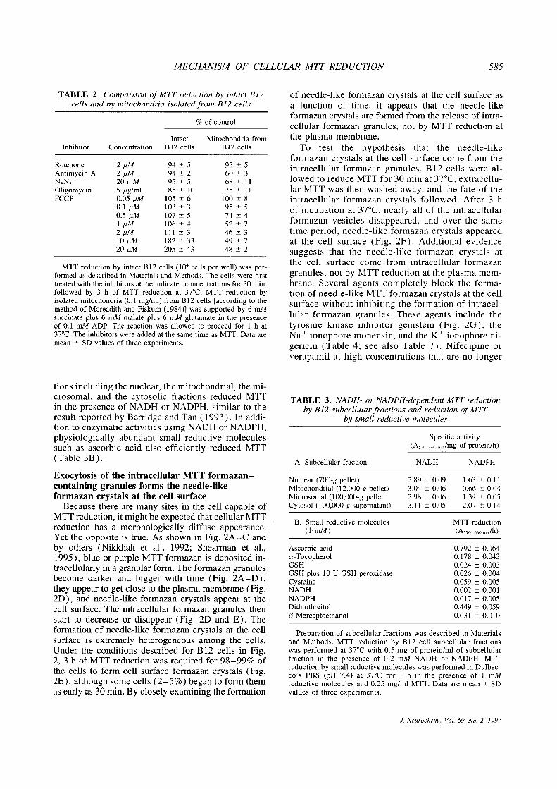
MECHANISM OF CELLULAR MiT REDUCTION 585
TABLE 2. Comparison of MIT reduction by intact B12cells and by mitochondria isolated from B12 cells
% of control
Intact Mitochondria fromInhibitor Concentration B 12 cells B12 cells
Rotenone 2 jiM 94 ±5 95 ±5Antimycin A 2 jiM 94 ±2 60 ±3NaN~ 20mM 95 ±5 68 ±11Oligomycin 5 jig/ml 85 ±10 75 ±11FCCP 0.05 jiM
0.1/iM
0.54M
ljiM2jiMlOjiM2OjiM
105 ±6103±3107±5106±4111±3182±33205±43
100 ±895±5
74±452±246±349±248±2
MTT reduction by intact B 12 cells (l0~cells per well) was per-formed as described in Materials and Methods. The cells were firsttreated with the inhibitors at the indicated concentrations for 30 mm,followed by 3 h of MTT reduction at 37°C. MTT reduction byisolated mitochondria (0.1 mg/ml) from B 12 cells [according to themethod of Moreadith and Fiskum (1984)1 was supported by 6 mMsuccinate plus 6 mM malate plus 6 mM glutamate in the presenceof 0.1 mill ADP. The reaction was allowed to proceed for 1 h at37°C.The inhibitors were added at the same time as MTT. Data aremean ±SD values of three experiments.
tions including the nuclear, the mitochondrial, the mi-crosomal, and the cytosolic fractions reduced MTTin the presence of NADH or NADPH, similar to theresult reported by Berridge and Tan (1993). In addi-tion to enzymatic activities using NADH or NADPH,physiologically abundant small reductive moleculessuch as ascorbic acid also efficiently reduced MTT(Table 3B).
Exocytosis of the intracellular MTT formazan—containing granules forms the needle-likeformazan crystals at the cell surface
Because there are many sites in the cell capable ofMTT reduction, it might be expected that cellularMTTreduction has a morphologically diffuse appearance.Yet the opposite is true. As shown in Fig. 2A—C andby others (Nikkhah et al., 1992; Shearman et al.,1995), blue or purple MTT formazan is deposited in-tracellularly in a granular form. The formazan granulesbecome darker and bigger with time (Fig. 2A—D),they appear to get close to the plasma membrane (Fig.2D), and needle-like formazan crystals appear at thecell surface. The intracellular formazan granules thenstart to decrease or disappear (Fig. 2D and E). Theformation of needle-like formazan crystals at the cellsurface is extremely heterogeneous among the cells.Under the conditions described for B 12 cells in Fig.2, 3 h of MTT reduction was required for 98—99% ofthe cells to form cell surface formazan crystals (Fig.2E), although some cells (2—5%) began to form themas early as 30 min. By closely examining the formation
of needle-like formazan crystals at the cell surface asa function of time, it appears that the needle-likeformazan crystals are formed from the release of intra-cellular formazan granules, not by MTT reduction atthe plasma membrane.
To test the hypothesis that the needle-likeformazan crystals at the cell surface come from theintracellular formazan granules, B12 cells were al-lowed to reduce MTT for 30 min at 37°C,extracellu-lar MTT was then washed away, and the fate of theintracellular formazan crystals followed. After 3 hof incubation at 37°C,nearly all of the intracellularformazan vesicles disappeared, and over the sametime period, needle-like formazan crystals appearedat the cell surface (Fig. 2F). Additional evidencesuggests that the needle-like formazan crystals atthe cell surface come from intracellular formazangranules, not by MTT reduction at the plasma mem-brane. Several agents completely block the forma-tion of needle-like MTT formazan crystals at the cellsurface without inhibiting the formation of intracel-lular formazan granules. These agents include thetyrosine kinase inhibitor genistein (Fig. 2G), theNa + ionophore monensin, and the K + ionophore ni-gericin (Table 4; see also Table 7). Nifedipine orverapamil at high concentrations that are no longer
TABLE 3. NADH- or NADPH-dependent MIT reductionby B12 subcellular fractions and reduction of MIT
by small reductive molecules
A. Subcellular fraction
Specific activity(A570_630 rn. /mg of protein/h)
NADH NADPH
Nuclear (700-g pellet) 2.89 ±0.09 1.63 ±0.11Mitochondrial (12,000-g pellet) 3.04 ±0.06 0.66 ±0.04Microsomal (100,000-g pellet 2.98 ±0.06 1.34 ±0.05Cytosol (100,000-g supernatant) 3.11 ±0.05 2.07 ±0.14
B. Small reductive molecules MTT reduction(1 mAl) (A570_630 ‚m/lnl)
Ascorbic acid 0.792 ±0.064a-Tocopherol 0.178 ± 0.043GSH 0.024 ±0.003GSH plus 10 U GSH peroxidase 0.026 ±0.004Cysteine 0.059 ±0.005NADH 0.002 ±0.001NADPH 0.017 ±0.005Dithiothreitol 0.449 ±0.059/3-Mercaptoethanol 0.031 ±0.010
Preparation of subcellular fractions was described in Materialsand Methods. MTT reduction by B12 cell subcellular fractionswas performed at 37°Cwith 0.5 mg of protein/ml of subcellularfraction in the presence of 0.2 mM NADH or NADPH. MTTreduction by small reductive molecules was performed in Dulbec-co‘s PBS (pH 7.4) at 37°C for 1 h in the presence of 1 mMreductive molecules and 0.25 mg/ml MTT. Data are mean ±SDvalues of three experiments.
J. Neurochem., Vol. 69, No. 2, 1997

586 Y LIU ET AL.
FIG. 2. Microscopic examination ofMTl reduction by intact living cells;the time course and the exocytosis ofMIT formazan. Bi 2 cells (A—G) orpri-mary culture of rat cortical neurons(H) on 35-mm dishes were incubatedwith 0.25 mg/mI MIT at 37°Cand ex-amined after various periods of time.A—E: MIT reduction by Bi2 cells for5(A), 10(B), 30(C), 90(0), and 180mm(E). F: MIT reduction by B12 cellsfor 30 min followed by the washout ofunreduced MIT and 180 min of incu-bation at 37°C.G: MIT reduction (180mm) by Bi2 cells in the presence of80 jiM genistein. H: MUreduction (1h) by primary culture of rat corticalneurons. Scale bar = 25 jim for allpanels.
specific for their calcium channel—blocking effectalso blocked needle formation.
How do the intracellular MTT formazan granulesgive rise to the needle-like formazan crystals at thecell surface? It is possible that cytoplasmic formazancrystals can grow into the extracellular space after pen-etrating the plasma membrane. However, extensive mi-croscopic monitoring of intracellular MTT formazan—containing vesicles as a function of time does not sup-port this scenario, because needle-like MTT formazancrystals do not form intracellularly and the cells withformazan needles on their cell surface (Fig. 2F) stillhave intact plasma membranes as judged by propidium
iodide staining (data not shown). To our knowledge,the only known process that delivers intracellular vesi-de or granule content to the extracellular surface isexocytosis. The confinement of MTT formazan to in-tracellular granules and the exocytosis of intracellularMTT formazan—containing granules to form needle-like formazan crystals at the cell surfacewere observedwith all of the mammalian cells examined, which in-clude B12 and PC12 cells,primary cultures of rat corti-cal neurons, MDCK epithelial cells, and L929 fibro-sarcoma cells. Figure 2H shows MTT reduction andMTT formazan exocytosis by primary cultures of ratcortical neurons.
J. Neurochem., Vol. 69, No. 2, 1997

MECHANISM OF CELLULAR MiT REDUCTION 587
TABLE 4. Genistein, Na~or K~ionophores, andnifedipine block the formation of needle-like
MTTformazan at the cell surface
InhibitorConcentration
(j.tM)% of cells exocytosing
MTT formazan
Control 0 97 ±2Genistein 10
204080
90 ±565±735±6
0Nigericin 2
51020
87 ±558±68±30
Monensin 102550
100
73 ±652±77±3
0Nifedipine 100
250500
1,000
51 ±610±33±10
B12 cells growing on 35-mm dishes were incubated with MTT(0.25 mg/mi) and the inhibitors at indicated concentrations at thesame time. MTT reduction was allowed to proceed for 3 h. Thepercentage of cells exocytosing MTT formazan was then countedunder microscope. Data are mean ±SD values ofthree experiments.
Intracellular MTT formazan granules do notcolocalize with mitochondria, ER, or Golgiapparatus, but partially colocalize withendosomes/lysosomes
When optically sectioned by the scanning laser con-focal microscope, the intracellular MTT formazangranules clearly have a vesicle-like structure (Fig. 3,left panels). The identity of the intracellular MTTformazan granules or vesicles was studied by doublestaining with MTT and subcellular organelle—specificfluorescent dyes. Through dynamic display of color(see Materials and Methods), it was found that MTTformazan—containing vesicles are adjacent to, but notsuperimposed with, rhodamine 123— stained mitochon-dna (Fig. 3, row 1, see enlarged inset). In contrast,there is frequent superimposable staining of MTTformazan and acridine orange, which stains the acidicendosomes and lysosomes (Fig. 3, row 4). It is possi-ble that superimposed structures are separate but ontop of each other if the depth of the image exceeds thedepth of one of the organelles. However, the superim-posed MTT and acridine orange staining in the whitebox of Fig. 3 (row 4) must be the result of colocaliza-lion because it is unlikely for such geometrically regis-tered staining superimposition to be the result of ran-dom physical stacking. The shape and morphology ofthe ER and Golgi staining (Fig. 3, rows 2 and 3)are so different from those of the intracellular MTTformazan—containing granules that one can readilyconclude they are not the same structure. These resultssuggest that intracellular MTT formazan vesicl~sdonot correspond to mitochondria, ER, or Golgi appara-
tus. MTT formazan vesicles do, however, partiallycolocalize with acidic vesicles stained with acridineorange, indicating at least some of the formazan-con-taming vesicles are endosomes/lysosomes. It is sur-prising that intracellular MTT formazan granules didnot colocalize with the staining of lucifer yellow CH,which also stains endosomes and lysosomes (data notshown, see Discussion). It is possible that MTTformazan may absorb the fluorescence of rhodamine123 so that colocalization is not detectable. However,this possibility was ruled out by experiments usingisolated mitochondria and another mitochondria dye(MitoTracker red CMXRos) whose emitted fluores-cence falls at almost the maximum absorption wave-lengths of dissolved MIT formazan. Rhodamine 123cannot be used to do the experiment with isolated mito-chondria because it is not washable. MIT formazancrystals (undissolved state) deposited in or around theisolated mitochondria was found to absorb very littlefluorescence when the same mitochondria were alsostained with MitoTracker red CMXRos (data notshown). In addition, the rhodamine 123—stained mito-chondria that are adjacent to MIT formazan vesicles(Fig. 3, first row) also argue against the idea that mito-chondria in intact cells reduce MIT and the reducedMIT formazan in turn makes them undetectable withrhodamine 123 staining. Ihe simplest explanation isthat the MIT formazan vesicles are not mitochondria.
b provide additional information about the identityof the intracellular MTl formazan—containing vesi-des, they were biochemically characterized. Theformazan-containing vesicles might have a differentdensity from other organelles because of their forma-zan content. Indeed, apparently pure formazan vesicles(judged by light-microscopic examination) were ob-tained by differential centrifugation at 3,000 g, a forcethat normally does not sediment any intracellular or-ganelles except the nucleus (see Materials and Meth-ods for details). Ihe isolated MTl formazan vesicleswere tested for marker enzyme activities specific tovarious intracellular organelles. As shown in Table 5,both the MTl formazan content and /3-N-acetylgluco-saminidase activity (a marker enzyme for lysosomes)of the purified MIT formazan vesicles were enrichedby more than fourfold compared with the 700- g super-natant, a fraction obtained after centrifugation of thecell homogenate at 700 g to remove nuclei and unbro-ken cells. The marker enzyme activities for other intra-cellular organelles (including mitochondria, peroxi-somes, ER, and Golgi apparatus) were not enriched.The total MTT formazan content of the purified MIIformazan—containing vesicles (the 3,000-g pellet)comprised 15% of the total MTl formazan that waspresent in the 700-g supernatant (calculated from theA570_630 nm/mg of protein value and the total proteincontent of the 700-g supernatant or-the 3,000-g pellet).The rest of the 85% MTT formazan cannot be easilyseparated from intracellular organelles and was notstudied. These results are consistent with the sugges-
J. Neurochem., Vol. 69, No. 2, 1997

588 Y. LIU ET AL.
FIG. 3. Laser scanning confocal microscopy of living Bi2 cells double stained with MU and subcellular organelle—specific fluorescentdyes. Top to bottom: row 1, 0.25 mg/mI MU plus 10 jig/mI rhodamine 123 for 10 mm; row 2, 0.25 mg/mI MIT plus 0.5 jig/mIOiOC6(3) for 10 mm; row 3, 0.25 mg/mI MU plus 5 jiM BODIPY-C5-DMB-ceramide for 20 mm; row 4, 0.25 mg/mI MU plus 10 jig/ml acridine orange for 10 min. Left panels are DIC images showing MU formazan—containing granules or vesicles; middle panelsare fluorescent images showing subcellular organelles; and right panels are mergers of DIC and fluorescent images. Scale bar = 10jim for all panels.
tion that at least some of the MII formazan—con-taming vesicles are lysosomes.
MTT is not permeable to lipid membrane andmay be taken up into cells via endocytosis
The observations that cellular MIT formazan depo-sition is confined to intracellular granules, that at leastsome of the formazan granules are endosomes/lyso-somes, and that the needle-like formazan crystals atthe cell surface are formed through the exocytosis ofthe intracellular formazan granules suggest that all of
the intracellular formazan may end up in the endoso-mal/lysosomal compartment before being exocytosed.Because mitochondria, ER, and Golgi apparatus werenot found to reduce MIT in intact cells (as indicatedby the confocal microscope experiment and the markerenzyme assay of isolated intracellular MIT formazangranules), although they are able to reduce MII invitro, it is possible that MTl is not permeable to theplasma membrane, but that it is taken into the cellthrough endocytosis and reduced in the endosomal/lysosomal compartment. Reduced MTT formazan is
J. Neurochem., Vol. 69, No. 2, 1997

MECHANISM OF CELLULAR MiT REDUCTION 589
TABLE 5. Marker enzyme activities of the partially purifiedMTLformazan — containing vesicles
Enzymatic activity 700-g supernatant 3,000-g pellet
MIT formazan (A575~630nm/mg of protein) 3.6 16.3 (450%“)
ß-N-Acetylglucosaminidase (nmollmg of proteinlmin) 60.0 245.4 (409%)Succinate—cytochrome c reductase (nmoi/mg of proteinlmin) 7.57 5.41 (71%)NADPH—cytochrome c reductase (nmol/mg of protein/mm) 20.5 18.9 (92%)Catalase (k/mg of protein) 0.07 0.04 (57%)Galactosyltransferase (nmol/mg of protein/b) 0.13 0.09 (69%)
B12 cells were allowed to reduce MTT (0.25 mg/ml) for 15 min and homogenized as described inMaterials and Methods. The homogenate was centrifuged at 700 g for 10 min to sediment nuclei andunbroken cells. The 700-g supernatant was centrifuged at 3,000 g for 10 min. The 3,000-g pellet waswashed twice. The marker enzyme activities of the 700-g supernatant and 3,000-g pellet were assayed asin cited publications.
Percentage of the activity in the 700-g supernatant.
then transported to the extracellular surface throughexocytosis to form the needle-like formazan crystals.
To examine the permeability of MTl to artificiallipid membranes, an aqueous solution of MIT wasencapsulated in large unilamellar liposomes (lipidcomposition: egg phosphatidylcholine/egg phosphati-dyiglycerol/cholesterol = 1:4:5 in molar ratio)through reverse-phase evaporation (Szoka and Papa-hadjopoulos, 1978). Ihe membrane permeability ofMTl was determmned by dialyzing the liposomesagainst 100 ml of buffer for 1 h at 37°C,and the MITremaining in the liposomes was then determined byspectrophotometry in the presence of 0.5% Iriton X-100 to disrupt the liposomes. As the lipid compositionof liposomes affects their membrane permeability, themembrane-impermeable dye trypan blue was also en-capsulated into liposomes to serve as a positive control.MTl is readily encapsulated in the liposomes, and only5.67 ± 1.56% (mean ± SD values of three experi-ments) of the trapped MIT in the liposomes was re-leased during a 1-h incubation period at 37°C. Forcomparison, 3.60 ± 1.69% (mean ± SD values ofthree experiments) of the encapsulated trypan blue wasreleased under the same conditions. These results arenot statistically different (p > 0.05) and indicate thatMII is essentially impermeable to lipid membranesbut do not exclude the possibility that the plasma mem-brane of intact cells can be permeable to MTl througha protein-facilitated mechanism.
To examine the permeability of plasma membraneto MIT and the role of endocytosis in cellular MITreduction, we studied the effect of low temperature,a well-documented inhibitor of endocytosis, on MITreduction by intact cells. MIT reduction by intact B 12cells was completely blocked at 4°C(Table 6), a tem-perature that also completely blocks endocytosis(Steinman et al., 1983) as well as many enzymaticprocesses. In contrast, B12 cell lysates and ascorbicacid, an abundant intracellular reducing molecule,were still able to reduce MIT at 4°Calthough at amuch slower rate (Table 6). These results indicate thatcells were capable of MTT reduction at 4°Cby either
enzymatic or nonenzymatic processes and that MTlwas unable to get inside of the cells when endocytosiswas blocked. However, this result cannot exclude thepossibility that a protein-facilitated MTl transportmechanism is also inhibited at 4°C. Because there aremultiple forms of fluid-phase endocytosis (Robinsonet al., 1996) and we are not aware of an inhibitor thatcanblock all forms of fluid-phase endocytosis at 37°C,a type of cell that is completely devoid of endocytosisat 37°Cis required to examine the membrane perme-ability of MTT at 37°C. Erythrocytes are cells thathave no intracellular organelles and do not undergoendocytosis (they are not stained with fluorescent en-docytosis marker such as lucifer yellow CH, data notshown), but do contain dehydrogenases (such as glu-cose-6-phosphate dehydrogenase) and small reductivemolecules (such as ascorbic acid) that are capable ofMTl reduction (Altman, 1976). Indeed, we found thatthe cell lysate of purified rat erythrocytes was able toreduce MIT, although the activity was relatively low
TABLE 6. Effect of temperature on MTL reduction byascorbic acid and intact B12 cells or their lysates
Reductive substance 4°C 37°C
Ascorbic acid (mM) A57~_650‚rn/3 h A570_650 nm/l h1.0 0.275 ±0.047 0.725 ±0.0420.5 0.152 ±0.016 0.331 ±0.0230.2 0.015 ±0.003 0.081 ±0.0050.1 0.004 ±0.001 0.038 ±0.003
B12 cell lysate (mg) A57o_63~nm/3h A57~_ 3onm/3h0.740 0.127 ±0.002 1.226 ±0.0280.370 0.064 ±0.001 0.935 ±0.0520.148 0.025 ±0.002 0.850 ±0.0340.074 0.010 ±0.001 0.509 ±0.052
Intact B 12 cells(cells per well) A570650 nm/12 h A570_630 nmI3 h
l0~ 0.000 0.373 ±0.047
MU reduction was performed under the conditions specified inTables 2 and 3 and measured as described in Materials and Methods.Data are mean ±SD values of three triplicate determinations. B 12cell lysates were prepared on ice with a sonication probe using fivepulses of 10-s duration.
J. Neurochem., Vol. 69, No. 2, 1997

590 Y LIU ET AL
FIG. 4. Rat erythrocytes do not reduce MU. Rat blood cells were incubated with 0.25 mg/mI MIT pIus 10 jig/mI acridine orange for1 h at 37°C.A: Phase-contrast image. B: Fluorescent image of the same field. The arrows indicate that the cells stained with MU arealso stained with acridine orange, showing that cells with intracellular organelles, therefore capable of endocytosis, are able to reduceMU. Scale bar = 10 jim.
(0.082 ±0.004 A570_630 nm/mg of protein/3 h at 37°C,mean ± SD values of three determinations). In con-trast, intact rat erythrocytes did not appear to reduceMTl at 37°C, although other blood cells that haveintracellular organelles (nucleus and acidic vesiclesstained with acridine orange) reduced MIT (seearrows in Fig. 4). These results indicate that the plasmamembrane of rat erythrocytes is not permeable to MITat 37°Cbut do not exclude the possibility that theplasma membrane of other types of cells is permeableto MIT through an endocytosis-independent mecha-nism. For example, the MTI-staining pattern in rathepatocytes appears diffuse (data not shown), and itis possible that the chemical-metabolizing hepatocytehas a protein-facilitated mechanism to transport MITinto the cytosol.
If MIT gets into cytosol through a protein-facili-tated mechanism, it will be reduced to formazan bythe many possible MIT-reducing activities in the cell.One possible mechanism for the accumulation offormazan in the endosome/lysosome compartment isintracellular autophagy. Autophagy is the process bywhich a cell sequesters part of its cytosol or intracellu-lar organelles and then delivers it to the lysosome fordegradation (Dunn, 1990; Gordon et al., 1992). Toexamine the possible role of autophagy in cellularMII reduction, we used 3-methyladenine, a knowninhibitor of intracellular autophagy in hepatocytes(Gordon et al., 1992). With B 12 cells, this reagent didnot affect MII reduction (lable 7), the distributionpattern of the intracellular formazan granules, or theirexocytosis. The percentage of cells exocytosing MIIformazan for control cells and 3-methyladenine—treated cells (10 mM 3-methyladenine, 2-h incubationat 37°C)are 4 ± 2 vs. 5 ± 3% (mean ± SD valuesof three experiments) after 30 min of MIT reductionand 85 ±8 vs. 83 ±6% after 2 h of MII reduction.We were also unable to demonstrate significant activeautophagy in Bl2 cells using particulate fraction—associated lactate dehydrogenase activity (Kopitz et
al., 1990) as a measure of autophagy. These resultsdo not support the notion that autophagy is involvedin cellular MIT reduction in B12 cells and suggestthat the plasma membrane of B12 cells is not perme-able to MIT. However, we do not exclude the possi-bility that 3-methyladenine does not work in Bl2cells.
TABLE 7. Agents that affect MTL reduction by B12 cells
Inhibitor Concentration % of control
Sulthydryl agentsSodium iodoacetate 1 mM 53 ±2NEM 100 jiM
SOjiM2OjiM
O14±283±7
p-Hydroxymercuribenzoate 200 jiMlOOjiM5OjiM201iM
O3±2
12±798±10
Inhibitor of tyrosine kinaseGenistein 80 jiM 161 ±13
Inhibitors of endosome/lysosomeacidification
Nigericin 10 jiM 81 ±7Monensin 50 jiM 96 ±5Bafilomycin Al 0.2 jiM 84 ±8
Inhibitor of intracellular autophagy3-Methyladenine 10 mM 91 ±5
Calcium channel blockerNifedipine 0.5 mM 144 ±9
Inhibitor of fiavin proteinDPI 50gM
20gM10gM5gM1gM
014±535±745±566±3
MTT reduction by B 12 cells (l0~cells per well) was performedas described in Materials and Methods. The cells were first treatedwith the inhibitors at indicated concentrations for 30 mm, and therewas no loss of cell viability under these conditions, as judged bypropidium iodide staining. MTT reduction by the cells was thenallowed to proceed for 3 h at 37°C.
J. Neurochem., Vol. 69, No. 2, 1997

MECHANISM OF CELLULAR MTL REDUCTION 59‘
FIG. 5. Effects of NEM and DPI on MIT reduction and the endocytosis of lucifer yellow CH by B12 cells. Left panels: MU(0.25 mg/ml) reduction for 30 min at 37°C.Right panels: Lucifer yellow CH (LY-CH, 1 mg/mI) uptake for 1 h at 37°C.A and B: Control. C andD: Cells treated with 0.5 mM NEM for 30 min. E and F: Cells treated with 50 jiM DPI for 30 min. Scale bar = 25 jim.
The cellular MTl reductase is an NEM-sensitiveflavin oxidase
Cellular MIT reduction can be severely inhibitedor abolished by the sulfhydryl agent NEM and theflavin protein inhibitor diphenylene iodonium (DPI)(Table 7), neither of which has significant inhibitoryeffects on the endocytosis of lucifer yellow CH, al-though NEM appears to enhance lucifer yellow CHuptake through an unknown mechanism (Fig. 5).NEM inhibits the acidification of the endocytic andexocytic pathways (Mellman et al., 1986). Becausethe inhibition of intracellular acidification by bafilo-mycin A, (Bowman et al., 1988) and nigericin (Mell-man et al., 1986) did not significantly affect cellularMIT reduction (lable 7), it is likely that NEM inhibitsthe cellular MIT reductase directly. p-Hydroxymer-curibenzoate, another sulfhydryl agent that inhibits theplasma membrane ferricyanide reductase (Crane et al.,1985), is also a strong inhibitor of cellular MIT reduc-tion (Table 7). Together, these results suggest that
the cellular MIT reductase is a thiol-containing flavinoxidase. NEM, DPI, and p -hydroxymercuribenzoatealso inhibited MII reduction by isolated mitochondriaand nuclear and microsomal fractions (data notshown), suggesting that the MIT-reducing enzymesin these subcelluiar fractions also contain the thiol andthe flavin groups.
DISCUSSION
The exclusive role of mitochondria in MTlreduction by intact cells is questionable
Because isolated mitochondria are able to reduceMII in the presence of succinate (Slater et al., 1963),it has been widely assumed that cellular MII reduc-tion is performed by active mitochondria in living cells.In the present study, we find that NADH-linked mito-chondrial substrates (such as maläte, glutamate, or pyr-uvate) are equally good at supporting MII reductionby isolated rat brain mitochondria, identifying an addi-
J. Neurochem., Vol. 69, No. 2, 1997

592 Y LIU ET AL.
tional MII reducing site along the mitochondrial elec-tron transport chain. However, the following results donot support an exclusive role of mitochondria in cellu-lar MII reduction. (1) The characteristics of MITreduction by intact cells are clearly different from thoseof isolated mitochondria, because FCCP, a protono-phore that uncouples mitochondrial oxidative phos-phorylation, enhanced MII reduction by intact B 12cells but severely inhibited MIT reduction by mito-chondria isolated from B 12 cells. (2) The intracellularMII formazan vesicles do not correspond to mito-chondria (Fig. 3). (3) Mitochondrial marker enzymeactivities are not increased in isolated MIT formazan—containing vesicles (Table 5). (4) p°cells, a humancell line that lacks mitochondrial DNA and a functionalelectron transport chain (King and Attardi, 1989), arestill able to reduce MIT although at a much slowerrate than the wild-type 143B cells (MIT reduction:0.321 ± 0.067 A570 nm/mg of protein/3D min for p°cells and 0.956 ±0.134 A570 nm/mg of protein/3D minfor l43B cells, mean ± SD values of three experi-ments). (5) In an electron microscopic study of cellu-lar MII reduction, Nikkhah et al. (1992) found thatMII formazan was accumulated in electron-lucid yes-ides that are apparently different from the electron-dense mitochondria. It follows that the use of cellularMII reduction as an indicator of cellular mitochon-drial succinate dehydrogenase or mitochondrial dehy-drogenase is questionable unless definitive evidence ispresented that mitochondria are indeed responsible.
Cellular MIT reduction: reduction ofendocytosed MII and the exocytosis of MITformazan
This study demonstrates that cellular MII reductionis confined to perinuclear vesicles, some of which wereidentified as endosomes/lysosomes. The exocytosis ofthe intracellular MII formazan — containing vesiclesgives rise to the needle-like formazan crystals at thecell surface, which further suggests that the MIIformazan ends up in the endosomal/lysosomal com-partment before exocytosis. In addition, MIT is notpermeable to lipid membranes. Ihese and other datasuggest that MII is reduced by an NEM-sensitiveflavin oxidase. Ihe MIT formazan is then accumulatedin the endosome/lysosome compartment and trans-ported to cell surface through exocytosis. The datasuggest that MIT is taken into cells through endocyto-sis and reduced primarily in the endosome/lysosomecompartment. The possibility that MII is transportedto cytosol by a protein-facilitated mechanism, reducedat multiple intracellular sites, and then accumulated inthe endosomal/ lysosomal compartment cannot, how-ever, be excluded. The membrane permeability ofMII could also vary with different types of cells.
MIT may be taken into cells through fluid-phaseendocytosis or pinocytosis because MII does not bindto tissue or to the cell surface (Altman, 1976), andMIT reduction is not inhibited in a HeLa cell line
(Damke et al., 1995) that is defective in receptor-mediated endocytosis (data not shown). However, in-tracellular MIT formazan—containing vesicles do notcolocalize with vesicles containing the pinocytosismarker lucifer yellow CH as analyzed by laserconfocalmicroscopy. This result does not rule out the possibilitythat the MII formazan—containing vesicles are endo-somes/lysosomes, for endosomes are diverse and un-dergoing dynamic sorting. In addition, there are alsomultiple forms of pinocytosis that are poorly character-ized (Robinson et al., 1996). In any case, the sheernumber of the MII formazan—containing vesicles inthe cell suggests that they must have an importantphysiological function.
The implications for using MTl reduction as acytotoxicity assay
With the understanding that MIT is endocytosedand that reduced MII formazan is transported to cellsurface through exocytosis, many of the published dis-crepancies between the MIT assay and other measuresof cell growth and viability can be explained. Endocy-tosis and reduction of MIT apparently require the AlPand NADH generated through glycolysis, which ex-plains why D-glucose and pyridine nucleotides affectcellular MIT reduction ( Vistica et al., 1991). Ihe rateof MII formazan exocytosis also has a major effecton cellular MIT reduction. The exocytosed MITformazan forms needle-like crystals at the cell surface,which appear to inhibit further MIT uptake, for cellswhose surface are covered with MII formazan crys-tals have very fewMIT formazan granules in the cyto-sol despite the continued presence of unreduced MITin the culture medium (Fig. 2). In addition, B12 cells,whose MIT formazan exocytosis was blocked by gen-istein, reduced 60% more MIT than the control cellsduring a 3-h incubation period at 37°C.Different typesof cells have tremendous differences in their rate ofMII formazan exocytosis. Even the same types ofcells at different stages of their growth curve havequite different rates of MIT formazan exocytosis. Forexample, it takes >6 h for nearly 100% of exponen-tially dividing B 12 cells to exocytose MII formazan,whereas it takes only 3 h for stationary-phase B 12 cellstodo the same. This would explain the effect of culturetime on MIT reduction (Jabber et al., 1989). TheMIT assay as a measure of cell viability is still validbecause it measures endocytosis, a fundamental featureof most living cells. However, it should be consideredthat factors affecting the endocytosis of MII, the exo-cytosis of MII formazan, and the cellular MII redue-tase activity will all be able to affect cellular MIIreduction. Finally, inhibition of cellular MII reduc-tion is a specific early indicator of amyloid 13 proteinneurotoxicity in Alzheimer‘s disease (Behl et al., 1992,1994; Shearman et al., 1995). The findings in thisstudy will help clarify the mechanism by which amy-bid /3 protein inhibits cellular MII reduction, thus
J. Neurochem., Vol. 69, No. 2, 1997

MECHANISM OF CELLULAR MTL REDUCTION 593
providing insight into the early events of amyloid /3protein neurotoxicity.
Acknowledgment: We thank Drs. Pamela Maher, YutakaSagara, and Yonghong Li for reading the manuscript. Thehelp from Richard Dargusch, Shirley Tan, Beverly Toth,Martin Latterish, Tammy Goedken, and Soon Lee, duringthis study, is gratefully acknowledged. This study was sup-ported by grants from the National Institutes of Health(R01NS09658-25 andR01AG11383-03) to David Schubert.Daniel A. Peterson was supported by NINDS P01 NS23121.Hideo Kimura was supported by grants from the NationalInstitutes of Health (R29N531202) and the Alzheimer‘s As-sociation.
REFERENCESAebi H. (1984) Catalase in vitro. Methods Enzymol. 105, 121 126.Altman F. P. (1976) Tetrazohum salts and formazans. Prog. Histo-
chem. Cytochem. 9, 1—56.Beaufay H., Amar-Costesec A., Feytmans E., Thines-Sempoux D.,
Wibo M., Robbi M., and Berthet J. (1974) Analytical study ofmicrosomes and isolated subcellular membranes from rat liver:I. Biochemical methods. J. Cell Biol. 61, 88—200.
BehI C., Davis J. B., Cole G. M., and Schubert D. (1992) VitaminE protects nerve cells from amyloid ß protein toxicity. Biochem.Biophys. Res. Commun. 186, 944—950.
Behl C., Davis J. B., Lesley R., and Schubert D. (1994) Hydrogenperoxide mediates amyloid ß protein toxicity. Cell 77, 817—827.
Berridge M. V. and Tan A. N. (1993) Characterization ofthe cellularreduction of 3- (4,5-dimethylthiazol-yl )-2,5-diphenyitetrazol-ium bromide (MTT): subcellular localization, substrate depen-dence and involvement of mitochondrial electron transport inMU reduction. Arch. Biochem. Biophys. 303, 474—482.
Beutler E. and West C. (1976) The removal of leukocytes andplatelets from whole blood. J. Clin. Lab. Res. 88, 328—333.
Bowman E. J., Siebers A., and Altendorf K. (1988) Bafilomycins: aclass of inhibitors of membrane ATPases from microorganism,animai cells, and plant cells. Proc. Natl. Acad. Sci. USA 85,7972—7976.
Burdon R. H., Gill V., and Rice-Evens C. (1993) Reduction of atetrazolium salt and superoxide generation in human tumor cells(HeLa). Free Radic. Res. Commun. 18, 369—380.
Crane F. L., Sun I. L., Clark M. G., Grebing C., and Löw H. (1985)Transplasma-membrane redox systems in growth and develop-ment. Biochim. Biophys. Acta 811, 233—264.
Damke H., Baba T., van der Bliek A. M., and Schmid S. L. (1995)Clathrin-independent pinocytosis is induced in cells overex-pressing a temperature-sensitive mutant of dynammn. J. CellBiol. 131, 69—80.
Dunn W. A. Jr. (1990) Studies on the mechanisms of autophagy. J.Cell Biol. 110, 1923—1945.
Gordon P. B., Høyvik H., and Seglen P. 0. (1992) Prelysosomaland lysosomal connections between autophagy and endocytosis.Biochem. J. 283, 361—369.
Jabber S. A. B., Twentyman P. R., and Watson J. V. (1989) TheMTT assay underestimates the growth inhibitory effects of in-terferon. Br. J. Cancer 60, 523—528.
Johnson L. V., Walsh M. L., and Chen L. B. (1980) Localization ofmitochondria in living cells with rhodamine 123. Proc. NatI.Acad. Sei. USA 77, 990—994.
King M. P. and Attardi G. (1989) Human cells lacking mtDNA:repopulation with exogenous mitochondria by complementa-tion. Science 246, 500—503.
Kopitz J., Kisen G. 0., Gorden P. B., Bohley P., and Seglen P. 0.(1990) Nonselective autophagy of cytosolic enzymes by iso-lated rat hepatocytes. J. Cell Biol. 111, 941—953.
Lowry 0. H., Rosebrough N. J., Fan A. L., and Randall R. J. (1951)Protein measurement with the Folin phenol reagent. J. Biol.Chem. 193, 265—275.
Mellman I., Fuchs R., and Helenius A. (1986) Acidification of theendocytic and exocytic pathways. Anna. Rev. Biochem. 55,663—700.
Moreadith R. W. and Fiskum G. (1984) Isolation of mitochondriafrom ascites tumor cells permeabilized with digitonin. Anal.Biochem. 137, 360—367.
Mosmann T. (1983) Rapid colorimetric assay for cellular growthand survival: application to proliferation and cytotoxicity. J.Immunol. Methods 65, 55—63.
Myers M. K., Fiskum G., Liu Y., Simmens S. J., Bredesen D. E., andMurphy A. N. (1995) Bel-2 protects neural cells from cyanide!aglycemia-induced lipid oxidation, mitochondrial injury, andloss of viability. J. Neurochem. 65, 2432—2440.
Nikkhah G., Tonn J. C., Hoffmann 0., Kraemer H-P., Darling J. L.,Schönmayr R., and Schachenmayr W. (1992) The MTT assayfor chemosensitivity testing of human tumors of the centralnervous system. J. Neurooncol. 13, 1—11.
Orci L., Tagaya M., Amherdt M., Perrelet A., Donaldson J. G.,Lippincott-Schwartz J., Kiausner R. D., and Rothman J. E.(1991) Brefeldin A, a drug that blocks secretion, prevents theassembly of non-clathrin-coated buds on Golgi cisternae. Cell64, 1183—1195.
Pagano R. E., Martin 0. C., Kang H. C., and Haugland R. P. (1991)A novel fluorescent ceramide analogue for studying membranetraffic in animai cells: accumulation at the Golgi apparatus re-sults in altered spectral properties of the sphingohpid precursor.J. Cell Biol. 113, 1267—1279.
Peterson D. A., Lucidi-Phillipi C. A., Murphy D. P., Ray J., andGage F. H. (1996) Fibroblast growth factor-2 protects entorhi-nal layer II glutamatergic neurons from axotomy-induced celldeath. J. Neurosci. 16, 886—898.
Robinson M. S. and Kreis T. E. (1992) Recruitment of coat proteinsonto Golgi membranes in intact and permeabilized cells: effectsof brefeldin A and G protein activators. Cell 69, 129—138.
Robinson M. S., Watts C., and Zerial M. (1996) Membrane dynam-ics in endocytosis. Cell 84, 13—21.
Rosenthal R. E., Hamud F., Fiskum G., Varghese P. J., and SharpeS. (1987) Cerebral ischemia and reperfusion: prevention ofbrain mitochondrial injury by lidoflazize. J. Cereb. Blood FlowMetab. 7, 752—758.
Schubert D., Heinemann S., Carlisle W., Tarika H., Kimes B., SteinbachJ. H., Culp W., and Brandt B. L. (1974) Cional cell lines fromthe rat central nervous system. Nature 249, 224—227.
Shearman M. S., Hawtin S. R., and Tailor V. J. (1995) The intracel-lular component of 3- (4,5-dimethylthiazol-2-yl)-2,5-diphenyl-tetrazolium bromide (MTT) reduction is specifically inhibitedby /3-amyloid peptides. J. Neurochem. 65, 218—227.
Slater T. F., Sawyer B., and Sträuh U. (1963) Studies on succinate-tetrazolium reductase system. Ill. Points of coupling of four differ-ent tetrazolium salts. Biochim. Biophys. Acta 77, 383 —393.
Steinman R. M., Mellman I., Muller W. A., and Cohn Z. A. (1983)Endocytosis and the recycling of plasma membrane. J. CellBiol. 96, 1—27.
Swanson J. A., Yirinec B. D., and Silverstein S. C. (1985) Phorbolesters and horseradish peroxidase stimulate pinocytosis and re-direct the flow of pinocytosed fluid in macrophages. J. CellBiol. 100, 85 1—859.
Szoka F. and Papahadjopoulos D. (1978) Procedure for preparationof liposomes with large internal aqueous space and high captureby reverse-phase evaporation. Proc. NatI. Acad. Sei. USA 75,4194—4198.
Terasalci M., Song J., Wong J. R., Weiss M. J., and Chen L. B. (1984)Localization of endoplasmic reticulum in living and glutaralde-hyde-fixed cells with fluorescent dyes. Cell 38, 101—108.
Vistica D. T., Skehan P., Scudiero D., Monks A., and Boyd M. R.(1991) Tetrazolium-basedassays for cellular viability: a criticalexamination of selected parameters affecting formazan produc-tion. Cancer Res. 51, 25 15—2520.
Yoshimori T., Yamamoto A., Moriyama.Y., Futai M., and TashiroY. (1991) Bafilomycin AI, a specific inhibitor of vacuolar-typeH + -ATPase, inhibits acidification and protein degradation inlysosomes of cultured cells. J. Biol. Chem. 266, 17707—17712.
J. Neurochem., Vol. 69, No. 2, 1997
Related Documents