MQP-BIO-DSA-3095 MECHANISM BY WHICH HEART REGULATES PERIPHERAL METABOLISM IN OBESITY A Major Qualifying Project Report Submitted to the Faculty of the WORCESTER POLYTECHNIC INSTITUTE in partial fulfillment of the requirements for the Degree of Bachelor of Science in Biology and Biotechnology by _________________________ Monolina Binny May 1, 2014 APPROVED: _________________________ _________________________ Jason Kim, PhD David Adams, PhD Department of Molecular Medicine Biology and Biotechnology UMass Medical School WPI Project Advisor MAJOR ADVISOR

Welcome message from author
This document is posted to help you gain knowledge. Please leave a comment to let me know what you think about it! Share it to your friends and learn new things together.
Transcript

MQP-BIO-DSA-3095
MECHANISM BY WHICH HEART REGULATES PERIPHERAL METABOLISM IN OBESITY
A Major Qualifying Project Report
Submitted to the Faculty of the
WORCESTER POLYTECHNIC INSTITUTE
in partial fulfillment of the requirements for the
Degree of Bachelor of Science
in
Biology and Biotechnology
by
_________________________ Monolina Binny
May 1, 2014
APPROVED:
_________________________ _________________________ Jason Kim, PhD David Adams, PhD Department of Molecular Medicine Biology and Biotechnology UMass Medical School WPI Project Advisor MAJOR ADVISOR

2
ABSTRACT
Although cardiac natriuretic peptides (NPs) have well characterized roles as vasodilators,
recent evidence indicates some NPs may also act to help regulate carbohydrate metabolism in brown
fat. The purpose of this project was to examine whether ventricular (or brain-type) natriuretic
peptide (B-type, BNP) affects glucose metabolism in tissues other than brown fat, especially in
mouse skeletal muscle. Mice were fed a high-fat diet for 8 weeks, then glucose uptake in muscle was
assayed using 2-[14C]-deoxyglucose-6-phosphate, and glycogen synthesis was monitored using 3H-
glucose labeling into muscle glycogen. The data indicate that BNP treatment did not alter glucose
metabolism in skeletal muscle, so perhaps BNP has no glucose metabolic effect on this particular
peripheral tissue.

3
TABLE OF CONTENTS Signature Page ………………………………….…………….………………………. 1 Abstract ……………………………………………………………………………… 2 Table of Contents ……………………………………………………………….…… 3 Acknowledgements ………………………………………………………………….. 4 Background ………………………………………………………………………….. 5 Project Purpose ……………………………………………………………………… 15 Methods ……………………………………………………………………………... 16 Results ………………………………………………………………………………. 22 Discussion …………………………………………………………………………... 25 Bibliography ……………………………………….………………………………... 28

4
ACKNOWLEDGEMENTS
I would like to especially thank Dr. Jason Kim, my Major Advisor, for allowing me to work
in his lab, and for all his help throughout the entire project. Next, I would like to thank Dr. Dae
Young Jung and Xiaodi Hu for showing me how to perform the glucose uptake and glycogen
synthesis assays, and for providing me with the BNP-treated and the wild-type mouse quadriceps
tissues and solutions associated with the assays. I would also like to thank Kevin Hsu for helping
me with the assays. Lastly, I thank Dr. Dave Adams, my WPI Project Advisor for helping me
initiate the project, and for helping me edit the MQP report.

5
BACKGROUND Obesity and Heart Failure
Obesity, as indexed by an elevated body mass index (BMI), is an increasingly widespread
condition worldwide. Obesity is also a well-known independent cardiovascular risk factor (Calle et
al., 1999). The use of the body mass index (BMI) to classify patients as underweight (BMI 18.5),
normal (BMI 18.5–24.9), overweight (BMI 25.0–29.9), or obese (BMI 30) as per World Health
Organization (WHO) guidelines reveals numbers that are staggering: nearly 70% of adults in the
United States are classified as overweight or obese (Bayes-Genis et al., 2007). The obesity problem
will cause higher rates of cardiovascular disease in the future (Redfield, 2002). A high body mass
index (BMI) correlates with hypertension, insulin resistance, and dyslipidemia, and contributes to
harmful hemodynamic and morphologic cardiovascular modifications (deDivitis et al., 1981; Wang
et al., 2004). In the general population, an elevated body mass index (BMI) contributes to an
increased risk for various cardiovascular diseases and heart failure (HF). According to The Acute
Decompensated Heart Failure National Registry (ADHERE), a national observational registry of
hospital data on patients admitted with HF in the USA, of the patients who have undergone HF,
about 30% were overweight or obese (Fonarow et al., 2007). The Renfrew-Paisley study (Bayes-
Genis et al., 2007), a 20-year follow-up population-based study of >15,000 individuals, demonstrated
that obesity is associated with an increase in a broad range of fatal and nonfatal cardiovascular
events. This study found a clear association between obesity and an increased risk for heart failure
(HF), including hospital admissions for HF. Based on the growing pandemic of obesity in the
United States and elsewhere, the HF problem will likely continue to increase.
Heart failure (HF) is a multisystem disorder which occurs when the heart is not able to
sufficiently maintain blood flow to meet the needs of the body. The symptoms of heart failure are

6
cardiac abnormalities, skeletal muscle problems, and renal dysfunction, sympathetic nervous system
acting up and various neurohormonal changes (Weber et al., 2001; Smooke et al., 2005; Horwich et
al., 2006; Kimmenade et al., 2009). Heart failure is a serious problem that results in high costs
associated with disability and health care costs. HF is the main instigator of various cardiovascular
diseases and mortality, and is one of the major reasons for hospitalization in industrial nations
(Fonarow et al., 2007). Heart failure affects more than 5 million people in the United States and
about 0.4 to 2.0 percent of the European population (Miyamoto et al., 2014). Most patients greater
than 65 years of age staying in a hospital suffer from chronic heart failure (CHF) (Joseph et al.,
2009). CHF is also associated with high mortality in the U.S.
The association between obesity and heart failure strongly suggests that there is a
cardiomyopathy of obesity. Many complications may arise due to obesity-mediated changes. These
complications include aiding atherosclerosis, problematic shifts in cardiac load and in functional
efficiency, rise of adverse inflammatory conditions, predisposition to proteinuria and renal
dysfunction, and disordered sleep breathing or dyspnea (Fonarow et al., 2007; Jung et al., 2013).
Obesity could contribute to heart failure in multiple ways. Obese people have been reported
to contain a huge proportion of lipid in or around myocytes and to eventually suffer from
lipotoxicity. Heart failure can arise from restrictive lung disease and disordered sleep breathing,
both of which are caused by obesity. Obesity influences conditions such as hypertension, diabetes,
valvular heart disease and these conditions secondarily cause ventricular dysfunction (Smooke et al.,
2005; Litwin, 2006; Daniels et al., 2006). “Obesity is a chronic inflammatory state,” (Litwin, 2006)
and therefore fibrosis can occur, or cardiac function hampered, when inflammatory cytokines are
secreted (Jung et al., 2013). Increased weight is proportional to high cardiac output or increased
load on the heart. This exposes latent ventricular dysfunction, or gradually the person suffers a heart
failure due to chronic overload (Litwin, 2006). Other diseases such as hypertension, hyperlipidemia,

7
and diabetes can rise from obesity, and the secondary diseases can harm cardiac operation by giving
rise to ischemic heart disease (Litwin, 2006; Daniels and Maisel, 2007; Wang et al., 2004). Metabolic
shifts are associated with obesity which impairs the production of energy or causes a rise in oxidative
free radical formation in the heart (Smooke et al., 2005).
Current studies indicate that obesity also impacts ventricular natriuretic peptide (BNP) levels
(discussed in the next section). Individuals who have higher BMI usually have lower circulating
levels of BNP (Chainani-Wu et al., 2010). BNP is a protein comprised of 32 amino acids, and is
secreted by cardiac ventricle cells (Daniels and Maisel, 2007). The inverse relationship between BNP
and BMI has been demonstrated in multiple population studies, including those of healthy people,
patients suffering from dyspnea, and patients with chronic heart failure.
The American College of Cardiology (ACC) and the American Heart Association (AHA) in
collaboration with the International Society for Heart and Lung Transplantation recommended
angiotensin converting enzyme inhibitors (ACEIs), beta-blockers, spironolactone, and diuretics as
optimal treatments of heart failure (Hunt et al., 2001). However, optimal treatment is usually
achieved through a balance of physician experience and patient drug tolerance (Jourdain et al., 2007).
Several studies support the diagnostic and prognostic value of BNP plasma levels in Chronic HF
(Remme and Swedberg, 2001). Most drugs used to treat heart failure cause a major decline in the
BNP levels. In the Valsartan Heart Failure Trial, valsartan was administered to patients along with
conventional therapy (Daniels and Maisel, 2007; Jourdain et al., 2007). This treatment caused a
drastic reduction in plasma BNP levels compared with the placebo group. The Randomized
Aldactone Evaluation Study also demonstrated that spironolactone noticeably decreased BNP levels
in patients with severe heart condition (Jourdain et al., 2007). However, the effect of beta-blockers
on BNP is still debatable.

8
The Obesity Paradox in Heart Failure
It has been presumed that obesity aids in increased mortality risk in people with established
heart failure, but unexpectedly, obese patients with heart failure have a better prognosis than patients
with normal weight (Fonarow et al., 2007; Iwanaga et al., 2007). A number of studies have
confirmed that BMI is actually inversely associated with long-term mortality in chronic HF; the
presence of obesity is associated with better survival in chronic HF patients. This phenomenon is
known as the obesity paradox because an increase of cardiovascular disease is also due to the effects
of obesity. It is possible that individuals with high BMI may acquire HF symptoms at an earlier and
less severe stage, and this may result in a better survival of obese patients due to a diminished
activation of natriuretic peptides, enhanced protection against endotoxin or inflammatory cytokines,
or an increased nutritional and metabolic reserve. Alternatively, individuals who undergo cachexia
and wasting, i.e. with a low BMI, may suffer through advanced HF which may result in high
mortality (Bayes-Genis et al., 2007). The obesity paradox also shows in cases with individuals with
acute dyspnea regardless of having acute destabilized heart failure. A study analyzing patients with
acute dyspnea taken to an emergency room showed that a higher BMI was associated with a lower
death rate in individuals with and without acute HF. BMI was significantly and inversely associated
with survival in a univariate analysis (Bayes-Genis et al., 2007).
Obesity influences systolic and diastolic ventricular function, and has also been recognized as
a major risk issue for the development of coronary artery disease and heart failure. Physiologically,
“natriuretic peptides and lipolysis have been closely linked, and adipose tissues are intimately related
to the natriuretic clearance receptor” (Mehra et al., 2004). Therefore, pathophysiologic mechanisms
underlying the relationship between obesity and cardiovascular disease consequences could partially
be linked to the effect of natriuretic peptides (Mehra et al., 2004).

9
Cardiac Natriuretic Peptides
Cardiac natriuretic peptides are vasodilator hormones that play a vital role in fluid and
hemodynamic homeostasis. The natriuretic peptides (NPs) consist of atrial NP (ANP) and its
ventricular companion (B-type, brain-type or BNP). NPs are considered cardiac or vascular
hormones that decrease vascular tone and increase circulating blood (Bordicchia et al., 2012). Two
types of receptors bind NPs: guanylyl cyclase-A (GCA) and guanylyl cyclase-B (GCB). Receptor
stimulation increases the synthesis of intracellular second messenger cyclic GMP (cGMP) through
the activation of cGMP-dependent protein kinase (cGK)-I (Lucas et al., 2000). By increasing
cGMP, NPs exercise their biological effects and create a signal transduction pathway that increases
nitric oxide (NO) (Bordicchia et al., 2012).
Another member of the NP receptor family, type-C receptor (NPR-C), performs the role of
a clearance receptor, binding ANP and BNP, integrating them into the cytoplasm, thus removing
them from the circulation. NP receptors are also expressed in adipose tissue of both rats and
humans. When rats undergo fasting, their NPR-C levels in adipose tissue decreases (Bordicchia et
al., 2012). Thus, cardiac NP receptors play a role in adipocyte metabolism as well as a role in
clearing natriuretic peptides from the circulation. However, it is unclear whether NPs and their
receptors affect metabolism in non-adipocyte tissues.
ANP and BNP can help cure congestive heart failure. Studies have found that the
intravenous infusion of ANP or BNP into patients with heart failure reduces cardiac pre- and post-
load, and results in beneficial hemodynamic function (Maki et al., 2001).
Lipolysis in human adipocytes is significantly increased by ANP. Activtion of β-adrenergic
receptors (β-ARs) initiates lipolysis (Bordicchia et al., 2012). The catabolism of triacylglycerol (TAG)
stored in cellular lipid droplets is managed through the biochemical pathway called lipolysis. TAG
undergoes hydrolytic cleavage into non-esterified fatty acids, essential precursors for lipid and
10
membrane synthesis and mediators in cell signaling processes, which are then used as energy
substrates (Bordicchia et al., 2012). Thus, lipolysis is important in lipid and energy homeostasis.
Lipolysis occurs in essentially all tissues and cell types, but is mostly found in white and brown
adipose tissue. However, the ability of NPs to cause lipolysis “is primate-specific and is apparently
absent from rodent adipose tissue” (Bordicchia et al, 2012). β-ARs are classic stimulators of lipolysis,
and they are responsible for an increase of cAMP levels that triggers cAMP-dependent protein
kinase (PKA). PKA phosphorylates crucial targets in the fat cell, such as hormone-sensitive lipase
(HSL) and the abundant lipid droplet–associated perilipins. NPs also activate the guanylyl cyclase
containing NPRA that produces the second messenger cGMP to stimulate cGMP-dependent
protein kinase (PKG) (Bordicchia et al., 2012; Lucas et al., 2000). PKA and PKG have similar
motifs for substrate phosphorylation. NPs and β-agonists appear to share a common mechanism for
the growth of lipolysis, as they phosphorylate the same targets in adipocytes: NPs phosphorylate
through PKG, and β-agonists through PKA (Miyashita et al., 2009). “A physiological role for NPs in
exercise-induced lipolysis in humans has been suggested” (Bordicchia et al., 2012).
Heart has been found to play a central role in regulating the supply of fatty acids for both
cardiac and skeletal muscle under aerobic conditions. The release of NPs coupled with exercise
causes an increased cardiac output (Jung et al., 2013). Postprandial fat oxidation in humans and
weight loss positively correlates with rises in circulating NPs (Bordicchia et al., 2012). The relation
between NPs and adipose tissue is significant because “obese human subjects with metabolic
syndrome often show reduced circulating NPs and biological efficacy (e.g., blood pressure control)”
(Bordicchia et al, 2012). In various population studies, plasma NPs and BMI have shown an inverse
relationship.

11
BNP
Ventricular natriuretic peptide, also termed brain or B-type natriuretic peptide (BNP), was
originally isolated from porcine brain in 1988 (Sudoh et al., 1988). BNP is a 32-amino acid peptide
containing a 17-amino acid ring (Figure-1) (Gorenjak, 2003). The highest levels of BNP are found
in the ventricular myocardium. Human pro-BNP contains 108 amino acids, and includes the mature
32-amino acid long BNP and a 76 aa long amino-terminal fragment NT-pro-BNP.
Figure-1: Structure of the B-Type Natriuretic Peptide (BNP). BNP is synthesized as a high molecular weight precursor pro-BNP. The N-terminal pro-BNP includes residues 1-76, while the 32 aa biologically active form of BNP includes residues 77-108. The lower diagram shows the 32 amino acid sequence of BNP including the 17 amino acid ring structure common to all natriuretic peptides. (Gorenjak, 2003)
Particulate Guanylate Cyclases (pGC) and Natriuretic Peptides
Particulate guanylate cyclases (pGC) are membrane-bound cGMP-forming enzymes with
extracellular domains that bind ANP and BNP (Lucas et al., 2000; Hobbs, 2014). Volume-induced
stretch in the cardiac atria signals the cells to release natriuretic peptides, which (as discussed above)

12
relax vascular smooth muscle and stimulate natriuresis to lower systemic blood pressure (Gorenjak,
2003; Hobbs, 2014). Some evidence exists for a reciprocal regulation of cGMP-mediated responses
in the vasculature using a balance of soluble and particulate guanylyl cyclases (Miyashita et al., 2009).
Low Levels of Natriuretic Peptides in Obesity
Besides having improved prognosis for obese individuals who suffer from cardiovascular
disease, the levels of BNP and NT-proBNP are considerably lower in obese individuals compared to
lean individuals with heart failure (Kimmenade et al., 2009; Chainani-Wu et al., 2010). Both BNP
and NT-proBNP are suppressed with increasing BMI (Taylor et al., 2006; Bayes-Genis et al., 2007).
A decrease in vasodilator NT-proBNP levels in obesity occurs despite the heavier patients having
higher blood pressures. Several potential mechanisms have been hypothesized for the inverse
association between NPs and BMI (Table 1).
In the Antoni Bayes-Genis study (2007), the NP clearance receptor NPR-C was isolated in
adipose tissue in humans, and “both elevated NPR-C expression and increased secretion of neutral
endopeptidases were demonstrated in patients with obesity.” The results of the study show that the
clearance of NPs could be increased in obese patients with HF, and this may partly explain the
impact of BMI on BNP plasma levels (Sarzani et al., 1996).
Lower levels of circulating BNP in obese individuals may also be a consequence of impaired
synthesis and secretion of NT-proBNP from myocytes. NT-pro-A-type NP activity has an inversely
proportional relationship to BMI (the Framingham Heart Study); lean mass initiates higher BMI
values and lower NT-proBNP levels (Bayes-Genis et al., 2007). Sex steroid hormones, most
probably androgens, may cause the synthesis or release of NPs from cardiomyocytes to be reduced.
Therefore, these studies suggest that the negative correlation between NPs and BMI is probably not
due to increased clearance of the peptide, but more likely a decrease in synthesis or release.

13
Table-1: Summary of the Potential Role of Natriuretic Peptides in Obesity (Bayes-Genis et al., 2007)
Diagnostic and Prognostic Use of ANP and BNP
Determining the serum levels of natriuretic peptides (NPs) might be useful when monitoring
patients with ventricular dysfunction (Gorenjak, 2003). Much research has demonstrated that the
cellular levels of NPs can serve as excellent biochemical markers for helping determine risk and the
stratification of HF patients; when compared to other neurohormones NPs appeared to be superior.
BNP measurement is usually an accurate marker when diagnosing HF in patients with acute
dyspnea, but high NP plasma concentrations are also found in asymptomatic patients with left
ventricular dysfunction. Therefore, these peptides may have a role in HF markers.
BNP is high in patients with impaired left ventricular function, and is found to be more
useful than ANP when detecting left ventricular systolic dysfunction. The elevated “negative
predictive value of BNP” in the identification of these dysfunctions shows how BNP can be utilized
in a clinical environment (Iwanaga et al., 2007). Thus, NP may be used as a both prognostic marker
or as a diagnostic marker. Rise in BNP plasma levels may also act as an independent predictor of the

14
condition of “congestive HF and of mortality in patients with chronic HF” (Iwanaga et al., 2007).
European clinics have already included BNP as a diagnostic tool for chronic HF.
Figure-2: The Decrease of BNP in Obese Individuals. The diagram on the left shows the distribution of patients with heart failure versus which category of BMI the patients fall under. The diagram on the right shows the levels of plasma BNP in association with BMI category, showing a decrease with increasing BMI. (Iwanaga et al., 2007)
Figure-3: Chart of the Levels of BNP Versus BMI Levels. The diagram shows The BNP cut-point values where sensitivity and specificity are equal differ across different BMI groups. “To achieve 90% sensitivity, the BNP cut-point in the lean patients is 170 pg/mL, whereas in the severely/morbidly obese patients the equivalent cut-point is only 54 pg/mL. Specificity at the 90% sensitivity level shown was at least 70% for all 3 groups.” (Daniels and Maisel, 2007)
15
PROJECT PURPOSE
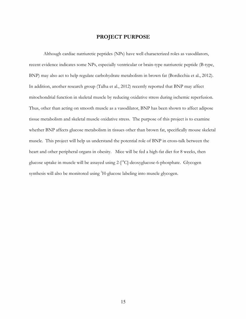
Although cardiac natriuretic peptides (NPs) have well characterized roles as vasodilators,
recent evidence indicates some NPs, especially ventricular or brain-type natriuretic peptide (B-type,
BNP) may also act to help regulate carbohydrate metabolism in brown fat (Bordicchia et al., 2012).
In addition, another research group (Talha et al., 2012) recently reported that BNP may affect
mitochondrial function in skeletal muscle by reducing oxidative stress during ischemic reperfusion.
Thus, other than acting on smooth muscle as a vasodilator, BNP has been shown to affect adipose
tissue metabolism and skeletal muscle oxidative stress. The purpose of this project is to examine
whether BNP affects glucose metabolism in tissues other than brown fat, specifically mouse skeletal
muscle. This project will help us understand the potential role of BNP in cross-talk between the
heart and other peripheral organs in obesity. Mice will be fed a high-fat diet for 8 weeks, then
glucose uptake in muscle will be assayed using 2-[14C]-deoxyglucose-6-phosphate. Glycogen
synthesis will also be monitored using 3H-glucose labeling into muscle glycogen.

16
METHODS
Mouse Strains and Feeding
For this study, male mice were used because hormonal and menstrual cycles could affect the
metabolic phenotypes of female mice. This study used one genetic strain C57BL6/J for BNP
treatments and an equal number of generations of back-crosses. The WT control group was
C57BL6/J mice with the same genetic background as the experimental mice. Mice were gender-
matched and age-matched; mouse age can have a profound effect on glucose metabolism. Young
mice at 3-4 months of age were chosen since they have the optimal size and maturity. The mice were
housed under controlled temperature (23°C) and lighting, with free access to water, and were given
high-fat diet food. Stress considerably affects glucose metabolism, so all efforts were made to
minimize stress. Transportation was kept to a minimum. The mice were fed high-fat diet for only 8
weeks as this time period was relatively easy to minimize stress. All mice were individually housed
to avoid stress from fighting.
BNP Infusion
BNP (Anaspec Inc., cat no. 61152) was dissolved in sterile water, and 50 µl of that stock was
added to sterile saline (0.9% NaCl) to prepare a final BNP concentration of 100 ng/ml. The control
group of mice received pumps containing saline only. After perfusion for various times, quadriceps
skeletal muscle was rapidly isolated, frozen in liquid nitrogen, and stored at −80°C. Mouse total
body weight and the weights of individual dissected organs were measured.
Hyperinsulinemic-Euglycemic Clamp in Conscious Mice
A euglycemic clamp was used to maintain normal blood glucose levels, while increasing
insulin levels. This assay was performed following a fed state; food was removed in the morning

17
and the clamp was started at least 5 hours after food removal. This approach ensures null or minimal
effects of gastrointestinal absorption of nutrients at the start of the clamp. On the day of the
euglycemic clamp experiment, each mouse was placed in a rat-size restrainer with its tail tape-
tethered at one end. This approach minimizes stress during the experiments. The intravenous
catheter was connected to the infusion pump, and the procedure was applied for 2 hours with saline
before the start of the euglycemic clamp to allow mice to acclimatize to this partially-restrained state
and to recover from the initial stressful handling. Less than 0.5 cm of tail end was cut to obtain
blood samples from the tail vessels during the experiment, and then the tail was taped to prevent
bleeding. All blood samples were taken from the tail vessels after carefully removing the tape and
using tail massage. After blood sampling, the tail end was taped again to prevent bleeding, and the
procedure was repeated during the clamp.
During the 2-hour acclimation period, D-[3H] glucose (0.05 Ci/min) was infused using a
microdialysis pump to assess the basal rate of whole body glucose turnover. A blood sample was
collected at the end for measuring plasma glucose, insulin, and [3H] glucose concentrations (basal
parameters). Following the basal period, a 2-hour hyperinsulinemic-euglycemic clamp with a primed
and continuous infusion of human insulin was performed to raise the plasma insulin levels to
approximately 300 pM (within the high end of a physiological range). Blood samples were collected
at 10-20 minute intervals for the measurement of plasma glucose concentrations using a clinical
glucose analyzer. A 20% glucose solution was infused at variable rates to maintain plasma glucose at
basal concentrations during the euglycemic clamp. Insulin-stimulated whole body glucose turnover
was estimated using [3-3H] glucose at various times throughout the clamp. A bolus of a solution of
2-deoxy-D-[1-14C] glucose was administered 75 minutes after the start of the clamp to estimate
insulin-stimulated glucose uptake in individual organs. Blood samples were taken at 80, 85, 90, 100,
110, and 120 minutes for the measurement of plasma [3H] glucose, 3H2O, and 2-[14C] DG

18
concentrations. A final blood sample (20 l) was taken at the end of the clamp to measure plasma
concentrations of glucose and insulin. At the end of the euglycemic clamps, the mice were
anesthetized, and then skeletal muscles (quadriceps) were dissected. The isolated tissues were rapidly
frozen in liquid nitrogen (N2), and stored in a -80oC freezer until biochemical/molecular analysis. A
summary of the clamp tests is shown in Figure-4.
Figure-4: Schematic Set-up of the Hyperinsulinemic-Euglycemic Clamp. The diagram notes the use of a glucose solution used to maintain normoglycemia throughout the procedure, and an insulin solution used to stimulate glucose uptake. Also shown are the times at which blood samples were taken (Kim, 2009).
Glucose Uptake
The rate of insulin-stimulated glucose uptake in individual organs was determined using 2-
[14C] deoxy-glucose (Figure-5). 2-[14C] DG was taken up by cells, phosphorylated by hexokinase to
become -6-P, and was not further metabolized. Thus, the organ-specific accumulations and levels of
2-[14C] DG-6-P found during the clamp reflect insulin-stimulated glucose uptake in individual
organs. Poly-prep columns prefilled with AG 1-X8 resin (Bio-Rad) were used in anion-exchange
chromatography to separate 2-[14C] DG-6-phosphate from non-phosphorylated 2-[14C] DG to

19
measure glucose uptake in individual organs. A solution containing 0.2 M formic acid and 0.5 M
ammonium acetate was used for anion-exchange chromatography.
Figure-5: Summary of the Glucose Uptake Assay. The diagram notes the use 14C- labeled deoxy-glucose which is incorporated into cells and phosphorylated to the 6-phosphatge state with no further metabolism (Kim, 2009).
The intracellular concentrations of 2-[14C] DG-6-P were determined by homogenizing 50 -
100 mg of frozen tissue samples in 10-volumes of dH2O (usually 50 mg of tissue in 500 ml of dH2O)
in glass tubes using a tissue homogenizer. Following homogenization, the glass tubes containing
homogenate were heated at 100°C for 10 min, and then vortexed for 2 s, and cooled to room
temperature. The homogenized samples were centrifuge at 16,000 × g for 5 minutes to pellet cell
debris. 33 ml of homogenate (supernatant) were added to 467 ml of dH2O in a scintillation vial
labeled “total” sample. 5 ml of scintillation cocktail were added and then vortexed. The samples
were counted for 14C using a liquid scintillation counter (total 14C samples). 333 ml of homogenate
(supernatant) was transferred to the anion-exchange columns for the separation of 2-[14C] DG-6-P
from non-phosphorylated 2-[14C] DG. The columns were washed with 2 ml of dH2O three times the
samples were collected into a scintillation vial labeled “wash” sample. The “wash” samples were

20
vortexed, and 500 ml of “wash” samples were transferred to another set of scintillation vials to be
counted for 14C using a liquid scintillation counter (wash samples containing 2-[14C] DG). The
columns were eluted 3X with 2 ml of 0.2 M formic acid/0.5 ammonium acetate, and the samples
were collected into a scintillation vial labeled “eluate” sample. The “eluate” samples were vortexed,
and 500 ml of “eluate” samples were transferred to another set of scintillation vials to be counted
for 14C using a liquid scintillation counter (eluate samples containing 2-[14C] DG-6-P) (Hong et al.,
2013).
Assay of Glycogen Synthesis in Individual Organs
The rate of insulin-stimulated glycogen synthesis was determined using [3- 3H] glucose,
which is taken up by cells and metabolized via glycolysis and glycogen synthesis. Approximately 50
mg of tissue samples were added to 500 ml of 30% KOH. After homogenization, the samples were
heated to about 100°C for 15 min, vortexed for 2 s, and cooled to room temperature. 3 ml of ice-
cold 95% ethanol was added to the homogenate samples, covered with parafilm, and incubated in a
freezer for 1 hour. Following incubation, the samples were centrifuged to obtain glycogen pellets,
and the ethanol supernatants were discarded. The ethanol wash process was repeated two more
times. Following the third wash, the glycogen pellets were dried. 0.6 ml of dH2O was added to the
glycogen pellet, and the pellet was completely dissolved by vortexing. 500 ml of the samples were
mixed with 3 ml of scintillation cocktail to count for 3H-glycogen using a liquid scintillation counter.
The assay is summarized in Figure-6.

21
Figure-6: Summary of the Glycogen Synthesis Assay. The diagram notes the use 3H-glucose which is metabolically converted inside the cell to glycogen. Glycolysis is calculated as the difference between glucose uptake and glycogen synthesis (Kim, 2009).
Calculations and Statistics All data are expressed as means ± SE. The significance of the difference in mean values
between BNP-treated mice versus WT mice was evaluated using the Student’s t test. The statistical
significance was at the P < 0.05 level.

22
RESULTS Although natriuretic peptides (NPs) have well characterized roles in vasodilation, recent data
indicates that some NPs may act on adipose tissue to alter lipid or carbohydrate metabolism
(Bordicchia et al., 2012). Mice treated with B-type NP (BNP) exhibit greater oxygen consumption
and energy expenditure through the activation of brown adipose tissue (BAT) and the browning of
white adipose tissue (WAT), thus BNP might have a function in controlling adipocyte metabolism.
In addition, another research group (Talha et al., 2012) recently reported that BNP may affect
mitochondrial function in skeletal muscle by reducing oxidative stress during ischemic reperfusion.
Thus, other than acting on smooth muscle as a vasodilator, BNP has been shown to affect adipose
tissue metabolism and skeletal muscle oxidative stress. We hypothesized that BNP may alter
carbohydrate metabolism in non-adipocyte tissues, such as skeletal muscle. Our lab has shown that
BNP is released by the heart under stress, but the metabolic role of NPs on muscle remains
unknown. This project investigated whether BNP can affect carbohydrate metabolism in wild-type
mice. Biochemical assays were performed to determine the rate of glucose uptake, the rate of
glycogen synthesis, and the effect on glycolysis in BNP-treated and saline treated mice.
Glucose is an important fuel for muscle, and normal glucose metabolism is vital for health.
Glucose enters the muscle cell via facilitated diffusion through the GLUT4 glucose transporter
which translocates from intracellular storage depots to the plasma membrane in response to insulin
or muscle contraction (Dolinsky and Dyck, 2006). Glucose uptake differs depending on the
metabolic needs of the tissue and the availability of glucose. We hypothesized that BNP-treated
mice will exhibit improved glucose uptake, i.e. be able to utilize glucose more efficiently and exhibit
improved insulin action, glucose disposal, and enhanced muscle glycogen storage than vehicle-
treated mice. Muscle glucose uptake was determined by measuring the muscle content of 2-[14C]-

23
deoxyglucose-6-phosphate. An anion-exchange column was used to quantitate the phosphorylated
version (column eluate) from the non-phosphorylated 2-[14C]-deoxy-glucose (column wash). The
data indicate that glucose uptake was slightly reduced in BNP-treated mice compared to saline-
treated mice (Figure-7) but the decrease was not statistically significant (p = 0.318).
Figure-7: Skeletal Muscle Glucose Uptake Rate. The rate of glucose uptake was monitored using the 2-[14C]-deoxyglucose-6-phosphate assay as described in Methods, for saline treated mice (green) and BNP-treated mice (red).
The rate of glycolysis was measured using [3-3H]-glucose metabolism (Figure-8), and shows
a similar small decrease for the BNP-treated WT mice, but again the decrease was not significant
(p=0.325).

24
Figure-8: Skeletal Muscle Glycolysis Rate. The rate of glycolysis was measured using a 3H-glucose metabolism assay as described in Methods, for saline treated mice (blue) and BNP-treated mice (orange).
The rate of glycogen synthesis was measured by subtracting the rate of [3-3H]-glucose
glycolysis from total glucose uptake (Figure-9), and shows a similar slight decrease for the BNP-
treated WT mice compared to saline-treated mice, but the decrease was not significant (p=0.644).
Figure-9: Skeletal Muscle Glycogen Synthesis Rate. The rate of glycogen synthesis was measured by subtracting the rate of glycolysis from total 3H-glucose uptake as described in Methods, for saline treated mice (orange) and BNP-treated mice (blue).

25
DISCUSSION
Many studies have shown that obesity influences the secretion level of ventricular or B-type
natriuretic peptide (BNP). Obese people with elevated BMI’s frequently have lower levels of BNP
compared to lean or normal weight individuals (Bayes-Genis et al., 2007). This inverse relationship
between BNP and BMI has been demonstrated in multiple population studies, including those of
healthy people of various weights, patients suffering from dyspnea, and patients with chronic heart
failure. BNP levels may be used as a diagnostic marker for heart failure (HF) (Bayes-Genis et al.,
2007). In what is known as the obesity paradox, individuals with a high BMI may acquire HF
symptoms at an early less severe stage, which surprisingly may result in a better survival due to a
diminished activation of natriuretic peptides, enhanced protection against endotoxin or
inflammatory cytokines, or an increased nutritional and metabolic reserve (Fonarow et al., 2007;
Iwanaga et al., 2007).
High levels of BNP can act as a poor prognostic marker for HF. An elevated BNP plasma
level, secreted predominantly by ventricular myocytes, has been suggested as an independent
predictor of the severity of congestive HF and of mortality in patients with chronic HF (Zellner et
al., 1999). BNP may be a useful adjunct to the standard multimarker panel currently used for
patients presenting with chest pain. High levels of BNP may help diagnose patients with non-ST
segment elevation myocardial infarction (NSTEMI), however there are other diagnostic markers
such as non-diagnostic levels of serum creatine kinase-MB (CK-MB) (a marker for acute myocardial
infarction), troponin I (another marker for acute myocardial infarction) (Green and Hill, 2012).
BNP levels provide valuable prognostic information pertaining to mortality, independent of other
risk indicators such as old age, renal insufficiency, or heart left ventricular dysfunction (Hong, 2008).

26
As with other natriuretic peptides, BNP may act as a vasodilator to influence coronary
vascular tone. But recent evidence also indicates it may act on adipose tissue to alter lipid or
carbohydrate metabolism (Bordicchia et al., 2012). Mice treated with BNP exhibited greater oxygen
consumption and energy expenditure through the activation of brown adipose tissue and the
browning of white adipose tissue, thus BNP might have a function in controlling brown fat
(Bordicchia et al., 2012).
Due to BNP’s role in affecting adipocyte metabolism, we hypothesized that BNP might
affect the metabolism of other peripheral tissues, including skeletal muscle. Mice were fed a high-fat
diet for 8 weeks, then glucose uptake in muscle was assayed using 2-[14C]-deoxyglucose-6-phosphate.
Glycogen synthesis was also monitored using 3H-glucose labeling into muscle glycogen.
However, the data (Figures 7, 8 and 9) indicated that BNP treatment did not appear to
significantly alter glucose metabolism in skeletal muscle. This is an important negative data since
another research group has reported that BNP may affect mitochondria in skeletal muscle. Pre-
treatment of mice with BNP reduced skeletal muscle mitochondrial dysfunction and oxidative stress
after ischemia-reperfusion (IR) (Talha et al., 2012). Their study hypothesized that since BNP can
reduce the extent of myocardial infarction, BNP may reduce skeletal muscle mitochondrial
dysfunction and oxidative stress through mitochondrial K(ATP) channel opening after ischemia-
reperfusion. The BNP pre-treatment in normal mice reduced the Bax-to-Bcl2 ratio, and reduced
oxidative stress. The BNP protection against deleterious IR effects on skeletal muscles was
abolished by treating with 5-hydroxydecanoic acid, and Caspase-3 activities did not change
significantly (Talha et al., 2012). Conversely, BNP injected during mouse ischemia failed to protect
against muscle injury. In addition to maintaining the activity of mitochondrial respiratory chain
complexes and possibly decreasing apoptosis, pre-treatment with BNP appeared to protect skeletal
muscle against IR-induced lesions, most likely by decreasing excessive production of radical oxygen

27
in species and opening mK(ATP) channels (Talha et al., 2012). These findings from other labs
indicate that BNP may affect adipose tissues and skeletal muscles. Based on our data, BNP may not
have any influence on skeletal muscle glucose metabolism (no significant effect on glucose uptake,
glycogen synthesis, or glycolysis). So, instead of affecting carbohydrate metabolism, BNP may
reduce oxidative stress and protect muscles against IR-induced lesions. But in adipose tissues, BNP
might have a direct metabolic role in converting white adipose tissue into brown fat, and activating
brown adipose tissue (Bordicchia et al., 2012). Therefore, BNP could be useful in studying adipose
tissue metabolism, and studying skeletal muscle mitochondrial dysfunction and oxidative stress.
And if BNP were infused into patients, perhaps it would not alter skeletal muscle glucose
metabolism.
One problem encountered in this project was that due to the limited supply of both saline-
treated and BNP-treated quadriceps tissues of C57BL6/J obese mice, I was unable to repeat the
experimental procedures for verification, and had to conduct the experiments in various parts with
extreme precaution over several days. So, a repeat of the experiment is necessary.
In the future, I plan to investigate whether BNP affects the metabolism of peripheral tissues
in diabetic mice. In one study, plasma BNP levels were higher in diabetic patients with chronic renal
failure (CRF) compared to non-diabetic patients, which suggests that plasma BNP might have a
potential in monitoring cardiac function in diabetic individuals with CRF (Ogawa et al., 2003). Other
studies suggest that increased circulating BNP levels in the presence of micro-albuminuria might be
a useful marker for early diastolic dysfunction in diabetic patients (Chan, 2001). Thus, BNP may
have several important roles in determining the cardiac state of diabetic individuals.

28
BIBLIOGRAPHY Bayes-Genis A, Lloyd-Jones DM, van Kimmenade RR, Lainchbury JG, Richards AM, Ordonez- Llanos J, Santalo M, Pinto YM, Januzzi JL Jr. (2007) Effect of body mass index on
diagnostic and prognostic usefulness of amino-terminal pro-brain natriuretic peptide in patients with acute dyspnea. Archives of Internal Medicine, 2007; 167: 400–407. Bordicchia M, Liu D, Amri EZ, Ailhaud G, Dessì-Fulgheri P, Zhang C, Takahashi N, Sarzani R,
Collins S (2012) Cardiac natriuretic peptides act via p38 MAPK to induce the brown fat thermogenic program in mouse and human adipocytes. The Journal of Clinical Investigation, 122(3): 1022-1036.
Calle EE, Thun MJ, Petrelli JM, Rodriguez C, Heath CW Jr. (1999) Body-mass index and mortality
in a prospective cohort of U.S. adults. New England Journal of Medicine, 341(15): 1097-1105. http://www.ncbi.nlm.nih.gov/pubmed/10511607
Chainani-Wu N, Weidner G, Purnell DM, Frenda S, Merritt-Worden T, Kemp C, Kersh E, Ornish
D (2010) Relation of B-type natriuretic peptide levels to body mass index after comprehensive lifestyle changes. The American Journal of Cardiology, 105(11): 1570-1576.
Chan NN (2001) Brain natriuretic peptide as a potential marker of diastolic dysfunction in type 2
diabetes. American Diabetes Association: Diabetes Care, 24(11): 2019-2020. Daniels LB, Clopton P, Bhalla V, Krishnaswamy P, Nowak RM, McCord J, Hollander JE, Duc P,
Omland T, Storrow AB, Abraham WT, Wu AH, Steg PG, Westheim A, Knudsen CW, Perez A, Kazanegra R, Herrmann HC, McCullough PA, Maisel AS (2006) How obesity affects the cut-points for B-type natriuretic peptide in the diagnosis of acute heart failure. Results from the Breathing Not Properly Multinational Study. American Heart Journal, 151(5): 999-1005. http://www.sciencedirect.com/science/article/pii/S0002870305009488#
Daniels LB, Maisel AS (2007) Natriuretic Peptides. Journal of the American College of Cardiology, 50(25): 2357-2368. deDivitiis O, Fazio S, Petitto M, Maddalena G, Contaldo F, Mancini M (1981) Obesity and cardiac
function. Circulation, 64(3): 477-482. Dolinsky VW, Dyck JB (2006) Role of AMP-activated protein kinase in healthy and diseased hearts.
American Journal of Physiology - Heart and Circulatory Physiology, 291: H2557-H2569. http://ajpheart.physiology.org/content/291/6/H2557
Fonarow GC, Srikanthan P, Costanzo MR, Cintron GB, Lopatin M, & ADHERE Scientific
Advisory Committee and Investigators (2007) An obesity paradox in acute heart failure: Analysis of body mass index and in hospital mortality for 108,927 patients in the acute decompensated heart failure national registry. American Heart Journal, 153(1): 74-81.
Gorenjak M (2003) Natriuretic peptides in assessment of ventricular dysfunction. International Federation of Clinical Chemistry and Laboratory Medicine.

29
http://www.ifcc.org/ifcc-communications-publications-division-(cpd)/ifcc-publications/ejifcc-(journal)/e-journal-volumes/ejifcc-2003-vol-14/vol-14-n%C2%B0-2/natriuretic-peptides-in-assessment-of-ventricular-dysfunction/
Green GB, Hill PM (2012) Chest Pain: Cardiac or Not. In Tintinalli's Emergency Medicine Manual: American College of Emergency Physicians: 7(1)-7(7). http://www.mhprofessional.com/content/media/Tintin52_0.pdf
Hobbs A (2014) Guanylate cyclase research. University College London.
http://www.ucl.ac.uk/npp/research/ah Hong MK (2008) B-type natriuretic peptide. Acute coronary syndrome: Multidisciplinary and pathway-based
approach (pp. 43) Springer. Hong EG, Kim BW, Jung DY, Kim JH, Yu T, Seixas Da Silva W, Friedline RH, Bianco SD, Seslar
SP, Wakimoto H, Berul CI, Russell KS, Lee KW, Larsen PR, Bianco AC, Kim JK (2013) Cardiac expression of human type 2 iodothyronine deiodinase increases glucose metabolism and protects against doxorubicin-induced cardiac dysfunction in male mice Endocrinology, 154: 3937-3946.
Horwich TB, Hamilton MA, Fonarow GC (2006) B-Type Natriuretic Peptide Levels in Obese
Patients With Advanced Heart Failure. Journal of the American College of Cardiology, 47(1): 85–90. http://www.sciencedirect.com/science/article/pii/S073510970502351X
Hunt SA, Baker DW, Chin MH, Cinquegrani MP, Feldmanmd AM, Francis GS, Ganiats TG,
Goldstein S, Gregoratos G, Jessup M, Noble R, Packer M, Silver MA, Stevenson L (2001) ACC/AHA guidelines for the evaluation and management of chronic heart failure in the adult: Executive summary A report of the American College of Cardiology/American heart association task force on practice guidelines (committee to revise the 1995 guidelines for the evaluation and management of heart failure). Journal of the American Heart Association, 104: 2996-3007.
Hunt SA, Baker DW, Chin MH, Cinquegrani MP, Feldman AM, Francis GS, Ganiats TG, Goldstein
S, Gregoratos G, Jessup ML, Noble RJ, Packer M, Silver MA, Stevenson LW, Gibbons RJ, Antman EM, Alpert JS, Faxon DP, Fuster V, Gregoratos G, Jacobs AK, Hiratzka LF, Russell RO, Smith SC Jr (2001) ACC/AHA Guidelines for the Evaluation and Management of Chronic Heart Failure in the Adult: Executive Summary A Report of the American College of Cardiology/American Heart Association. Circulation, 104(24): 2996-3007.
Iwanaga Y, Kihara Y, Niizuma S, Noguchi T, Nonogi H, Kita T, Goto Y (2007) BNP in overweight
and obese patients with heart failure: An analysis based on the BNP-LV diastolic wall stress relationship. Journal of Cardiac Failure, 13(8): 663-667.
Joseph SM, Cedars AM, Ewald GA, Geltman EM, Mann DL (2009) Acute decompensated heart failure: contemporary medical management. Texas Heart Institute Journal, 36(6): 510- 520.

30
Jourdain P, Jondeau G, Funck F, Gueffet P, Helloco AL, Donal E, Aupetit JF, Aumont MC, Galinier M, Eicher JC, Cohen-Solal A, Juillière Y (2007) Plasma brain natriuretic peptide-guided therapy to improve outcome in heart failure: The STARS-BNP multicenter study. Journal of the American College of Cardiology, 49(16): 1733-1739.
Jung DY, Ko HJ, Lichtman EI, Lee E, Lawton E, Ong H, Yu K, Azuma Y, Friedline RH, Lee KW,
Kim JK (2013) Short-term weight loss attenuates local tissue inflammation and improves insulin sensitivity without affecting adipose inflammation in obese mice. American Journal of Physiology - Endocrinology and Metabolism, 304: E964–E976.
Kim JK (2009) Hyperinsulinemic–Euglycemic clamp to assess insulin sensitivity in vivo. C. Stocker
(Ed.), Type 2 diabetes: Methods in molecular biology (pp. 221-238). Kimmenade RRJ, Januzzi JL, Bakker JA, Houben AJ, Rennenberg R, Kroon AA, Crijns HM,
Dieijen-Visser MP, Leeuw PW, MD, Pinto YJ (2009) Renal clearance of B-type natriuretic peptide and amino terminal pro-B-type natriuretic peptide a mechanistic study in hypertensive subjects. Journal of the American College of Cardiology, 53(10): 884-890.
Litwin S (2006) The growing problem of obesity and the heart. Journal of the American College of
Cardiology, 47: 617-619. Lucas KA, Pitari GM, Kazerounian S, Ruiz-Stewart I, Park J, Schulz S, Chepenik KP, Waldman SA
(2000) Guanylyl cyclases and signaling by cyclic GMP. Pharmacological Reviews, 52(3): 375-414. http://pharmrev.aspetjournals.org/content/52/3/375.full.pdf
Maki T, Yamaguchi YF, Yoshida H, Mori M, Takada T, Horikawa E, Okano K, Takeo S (2001)
Long-term treatment with neutral endopeptidase inhibitor improves cardiac function and reduces natriuretic peptides in rats with chronic heart failure. Cardiovascular Research, 51(3): 608–617. http://cardiovascres.oxfordjournals.org/content/51/3/608.full.pdf
Mehra MR, Uber PA, Park MH, Scott RL, Ventura HO, Harris BC, Frohlich ED (2004)
Obesity and suppressed B-type natriuretic peptide levels in heart failure. Journal of the American College of Cardiology, 43(9): 1590-1595.
Miyamoto SD, Stauffer BL, Nakano S, Sobus R, Nunley K, Nelson P, Sucharov CC (2014) Beta- adrenergic adaptation in paediatric idiopathic dilated cardiomyopathy. European Heart Journal, 35(1): 33-41. doi: 10.1093/eurheartj/ehs229. Epub 2012 Jul 26. Miyashita K, Itoh H, Tsujimoto H, Tamura N, Fukunaga Y, Sone M, Yamahara K, Taura D,
Inuzuka M, Sonoyama T, Nakao K (2009) Natriuretic peptides/cGMP/cGMP-dependent protein kinase cascades promote muscle mitochondrial biogenesis and prevent obesity. Diabetes, 58(12): 2880-2892. http://www.ncbi.nlm.nih.gov/pmc/articles/PMC2780866/
Ogawa S, Takeuchi K, Ito S (2003) Plasma BNP levels in the treatment of type 2 diabetes with
pioglitazone. The Journal of Clinical Endocrinology & Metabolism, 88(8): 3993-3996. Redfield MM (2002) Heart failure: an epidemic of uncertain proportions. New England Journal of Medicine, 347(18): 1442-1444.

31
Remme WJ, Swedberg K (2001) Guidelines for the diagnosis and treatment of chronic heart failure. European Heart Journal, 22(17): 1527-1560. Sarzani R, Dessi-Fulgheri P, Paci VM, Espinosa E, Rappelli A (1996) Expression of natriuretic
peptide receptors in human adipose and other tissues. Journal of Endocrinological Investigation, 1996; 19: 581–585.
Smooke S, Horwich TB, Fonarow GC (2005) Insulin-treated diabetes is associated with a marked
increase in mortality in patients with advanced heart failure. American Heart Journal, 149(1): 168-174.
Sudoh T, Kangawa K, Minamino N, Matsuo H (1988) A new natriuretic peptide in porcine brain. Nature, 332(6159): 78-81. Talha S, Bouitbir J, Charles AL, Zoll J, Goette-Di Marco P, Meziani F, Piquard F, Geny B (2012)
Pretreatment with brain natriuretic peptide reduces skeletal muscle mitochondrial dysfunction and oxidative stress after ischemia-reperfusion. Journal of Applied Physiology, 114(2): 172-179.
Taylor AT, Christenson RH, Rao K, Jorge M, Gottlieb SS (2006) B-type natriuretic peptide and N-
terminal pro B-type natriuretic peptide are depressed in obesity despite higher left ventricular end diastolic pressures. American Heart Journal, 152(6): 1071-1076.
Wang TJ, Larson MG, Levy D, Benjamin EJ, Leip EP, Wilson PW, Vasan RS (2004) Impact of
obesity on plasma natriuretic peptide levels. Circulation, 109(5): 594-600. https://circ.ahajournals.org/content/109/5/594.full
Weber MA, Neutel JM, Smith DG (2001) Contrasting clinical properties and exercise responses in
obese and lean hypertensive patients. Journal of the American College of Cardiology, 37(1): 169-174. Zellner C, Protter AA, Ko E, Pothireddy MR, DeMarco T, Hutchison SJ, Chou TM, Chatterjee K,
Sudhir K (1999). Coronary vasodilator effects of BNP: Mechanisms of action in coronary conductance and resistance arteries. American Journal of Physiology, 276: H1049-H1057.
Related Documents


