Welcome message from author
This document is posted to help you gain knowledge. Please leave a comment to let me know what you think about it! Share it to your friends and learn new things together.
Transcript
MECHANICS OF SPASTIC MUSCLE AND EFFECTS OF
TREATMENT TECHNIQUES: ASSESSMENTS WITH
INTRA-OPERATIVE AND ANIMAL EXPERIMENTS
by
Filiz Ate³
B.Sc., Electronics Engineering, �stanbul University, 2003
M.Sc., Biomedical Engineering, Bo§aziçi University, 2005
Submitted to the Institute of Biomedical Engineering
in partial ful�llment of the requirements
for the degree of
Doctor
of
Philosophy
Bo§aziçi University
2013
ii
MECHANICS OF SPASTIC MUSCLE AND EFFECTS OF
TREATMENT TECHNIQUES: ASSESSMENTS WITH
INTRA-OPERATIVE AND ANIMAL EXPERIMENTS
APPROVED BY:
Assoc. Prof. Dr. Can Ali Yücesoy . . . . . . . . . . . . . . . . . . .
(Thesis Advisor)
Prof. Dr. Cengizhan Öztürk . . . . . . . . . . . . . . . . . . .
Prof. Dr. Mehmed Özkan . . . . . . . . . . . . . . . . . . .
Prof. Dr. Muharrem �nan . . . . . . . . . . . . . . . . . . .
Assoc. Prof. Dr. Umut Akgün . . . . . . . . . . . . . . . . . . .
DATE OF APPROVAL: 23 January 2013

iii
ACKNOWLEDGMENTS
It is with immense gratitude that I acknowledge the support and help of my
supervisor Assoc. Prof. Dr. Can A. Yücesoy. This thesis would not have been possible
without his great e�orts.
I wish to thank Prof. Dr. Peter Huijing for his guidance and valuable suggestions
and also Guus Baan for his continual and precious help during animal surgery.
I owe my sincere thanks to Prof. Dr. Cengizhan Öztürk and Prof. Dr. Mehmed
Özkan for being in the thesis progress committee and for the helpful suggestions during
this period.
It gives me great pleasure in acknowledging the support and help of Prof. Dr.
Mustafa Karahan and his team in Marmara University, Medical School, Department
of Orthopedics and Traumatology, and also Assoc. Prof. Dr. Umut Akgün, who made
incredible contributions.
I am indebted to our collaborators Prof. Dr. Yener Temelli, and his team in
Istanbul University, Istanbul Medical School, Department of Orthopedics and Trauma-
tology and also Assoc. Prof. Ekin Akalan for their helps during the collaboration.
I would like to thank Adnan Kurt for designing novel instruments for our ex-
periments and solving the problems in setup with his incredible talent. I also wish to
thank Renan Mert Özel for the helps during data processing.
I wish to thank the members of the crew of Biomechanics Laboratory; Ahu
Nur Türko§lu, Uluç Pamuk, Sevgi Umur, Alper Yaman, Önder Emre Ar�kan, Selen
Ersoy, Rana N. Özde³lik, Gülay Hocao§lu, Arda Arpak, F. Oya Aytürk, Zeynep Susam,
Begüm Anlar, Y. Turgay Ertugay, Gizem Sar�ba³, Ferah �lhan, Zeynep �eref Ferlengez,

iv
Bora Yaman. �Once a biomechanics lab member, always a biomechanics lab member �
I owe special thanks to my friends Nermin Topalo§lu, Ahu Nur Türko§lu, Murat
Tümer, Ay³e Sena Kaba³ Sarp, Didar Talat, Burcu Tunç, Muhammed Av³ar, Sevinç
Mutlu, Mehmet Kocatürk, Eda Çapa, Bora Büyüksaraç, Özlem Özmen Okur, Esin
Karahan, Meltem Sevgi, Özgür Tabako§lu, Mehmet Yumak, Mustafa Kemal Ruhi,
Engin Baysoy with whom we crossed paths at the Institute of Biomedical Engineering.
I warmly thank to my friends Selcan Ç�nar, �ule Süzük, Özüm Seda Duran,
Özge Özy�lmaz, Sergül Aydore, Merve Arkan, Burçin Duan, Gülay Tezgel for making
life more bearable.
This thesis would not have been possible unless the support and love of Ate³
family; my parents Nimet and Adem, my siblings Ali Murat, Fatih, Mehmet Yavuz,
my sister in law Özlem, and my nephews Atahan, Alper Kaan.
v
Abstract
MECHANICS OF SPASTIC MUSCLE AND EFFECTS OFTREATMENT TECHNIQUES: ASSESSMENTS WITHINTRA-OPERATIVE AND ANIMAL EXPERIMENTS
Present thesis is focused on mechanics of spastic human muscles and the e�ects
of widely used treatment methods in the context of the determinant role of epimuscu-
lar myofascial force transmission (EMFT). A novel intra-operative method was devel-
oped to measure human Gracilis (GRA) muscle isometric forces with respect to knee
angle. In healthy subjects, GRA was shown to have very large operational length
range. For spastic cerebral palsy patients on the other hand, GRA muscle did not
show �abnormal� mechanical characteristics: (i) Length range was not narrowed and
(ii) high �exion forces were not available. Such abnormality occurred if its antago-
nist vastus medialis is activated simultaneously. Therefore, EMFT mechanism through
inter-antagonistic interaction was suggested to determine human muscle characteristics
in spasticity. E�ects of treatment methods were investigated in animal experiments:
(1) Muscle lengthening surgery was shown to a�ect (i) proximal and distal sides di�er-
entially and (ii) non-operated neighboring muscle as well. (2) Botulinum Toxin Type-A
(BTX-A) administration was shown to change the mechanics of not only the injected
but also non-injected muscles in conditions close to in vivo. Additional to active force
reductions (i) the narrowed length range of force exertion and (ii) pronounced passive
force increase contradictory to the aim were shown. EMFT mechanism was concluded
to be determinant for the treatment methods as well.
Keywords: Epimuscular myofascial force transmission, spastic cerebral palsy,
intra-operative human experiments, rat anterior crural compartment, muscle length-
ening surgery, aponeurotomy, botulinum toxin type-A.
vi
ÖZET
SPAST�K KAS MEKAN���N�N VE TEDAV�YÖNTEMLER�N�N ETK�LER�N�N �NTRA-OPERAT�F
DENEYLER VE HAYVAN DENEYLER� �LE �NCELENMES�
Bu tez, epimüsküler miyoba§dokusal kuvvet iletimi (EMK�) çerçevesinde spastik
kas mekani§ine ve yayg�n kullan�lan tedavi yöntemlerine odaklanm�³t�r. �nsan gracilis
(GRA) kas� izometrik kuvvetini diz aç�s�n�n fonksiyonu olarak ölçmek üzere yeni bir
yöntem geli³tirilmi³tir. Sa§l�kl� deneklerin GRA kas�n�n geni³ bir operasyonel boy ar-
al�§� oldu§u gösterilmi³tir. Spastik serebral palsili hastalar�n GRA kas�n�n ise anomali
göstermedi§i bulunmu³tur: (i) kas�n boy aral�§� daralmam�³t�r ve (ii) yüksek �eksiyon
kuvvetleri gözlenmemi³tir. Bu tür bir anomali antagonisti vastus medialis ile e³ zamanl�
uyar�ld�§�nda ortaya ç�km�³t�r. Bu nedenle, EMK� mekanizmas�n�n inter-antagonist
etkile³im üzerinden spastisite durumunda insan kas� karakteristi§ini belirleyici oldu§u
önerilmi³tir. Tedavi yöntemlerinin etkileri ise hayvan deneyleri ile incelenmi³tir: (1)
Kas uzatma ameliyat�n�n (i) proksimal ve distalde de§i³ken etkileri oldu§u ve (ii) opere
edilmeyen kom³u kaslar� da etkiledi§i gösterilmi³tir. (2) Botulinum Toksin Tip-A
(BTX-A) uygulamas�n�n in vivo ya yak�n ko³ullarda hem enjekte edilen hem edilmeyen
kaslar� etkiledi§i gösterilmi³tir. Aktif kuvvet dü³ü³lerinin yan� s�ra uygulaman�n hede-
�yle çeli³en (i) aktif kuvvet etkime boy aral�§�nda daralma ve (ii) belirgin pasif kuvvet
art�³� görülmü³tür. EMK� mekanizmas�n�n tedavi yöntemlerinde de belirleyici odu§u
sonucuna var�lm�³t�r.
Anahtar Sözcükler:Epimüsküler miyoba§dokusal kuvvet iletimi, spastik beyin felci,
insanda intra-operatif deneyler, s�çan anterior krural kompartman�, kas uzatma op-
erasyonu, aponevroz gev³etme, botulinum toksin tip-A.
vii
Contents
ACKNOWLEDGMENTS . . . . . . . . . . . . . . . . . . . . . . . . . . . . . . iii
Abstract . . . . . . . . . . . . . . . . . . . . . . . . . . . . . . . . . . . . . . . . v
ÖZET . . . . . . . . . . . . . . . . . . . . . . . . . . . . . . . . . . . . . . . . . vi
List of Figures . . . . . . . . . . . . . . . . . . . . . . . . . . . . . . . . . . . . xi
List of Tables . . . . . . . . . . . . . . . . . . . . . . . . . . . . . . . . . . . . . xiii
LIST OF SYMBOLS . . . . . . . . . . . . . . . . . . . . . . . . . . . . . . . . . xiv
LIST OF ABBREVIATIONS . . . . . . . . . . . . . . . . . . . . . . . . . . . . xv
1. GENERAL INTRODUCTION . . . . . . . . . . . . . . . . . . . . . . . . . 1
1.1 Muscle Force-Length Characteristics and Transmission of Forces . . . . 1
1.2 Myofascial Force Transmission . . . . . . . . . . . . . . . . . . . . . . 2
1.3 Mechanics of Spastic Muscle . . . . . . . . . . . . . . . . . . . . . . . 3
1.4 Clinical and Surgical Interventions to Correct Impaired Joint Function 3
1.5 Goals and Overview of Dissertation . . . . . . . . . . . . . . . . . . . . 5
2. INTRAOPERATIVE MEASUREMENT OF HUMAN GRACILIS MUSCLE
ISOMETRIC FORCES AS A FUNCTION OF KNEE ANGLE . . . . . . . 7
2.1 Introduction . . . . . . . . . . . . . . . . . . . . . . . . . . . . . . . . 7
2.2 Methods . . . . . . . . . . . . . . . . . . . . . . . . . . . . . . . . . . . 8
2.2.1 Subjects . . . . . . . . . . . . . . . . . . . . . . . . . . . . . . . 8
2.2.2 Methods . . . . . . . . . . . . . . . . . . . . . . . . . . . . . . . 8
2.2.3 Processing of Data . . . . . . . . . . . . . . . . . . . . . . . . . 11
2.3 Results . . . . . . . . . . . . . . . . . . . . . . . . . . . . . . . . . . . . 12
2.3.1 Peak GRA forces and inter-subject variability . . . . . . . . . . 12
2.3.2 Knee joint angle-GRA force characteristics . . . . . . . . . . . 12
2.3.3 Length history e�ects . . . . . . . . . . . . . . . . . . . . . . . 13
2.4 Discussion . . . . . . . . . . . . . . . . . . . . . . . . . . . . . . . . . . 13
2.4.1 Intraoperative experiments and our present approach . . . . . . 13
2.4.2 Functional joint range of motion . . . . . . . . . . . . . . . . . . 16
2.4.3 Length history e�ects do occur in human muscle . . . . . . . . . 17
2.4.4 Limitations and implications . . . . . . . . . . . . . . . . . . . . 18
viii
2.4.4.1 Lack of passive force data . . . . . . . . . . . . . . . . 18
2.4.4.2 Implications of EMFT . . . . . . . . . . . . . . . . . . 18
3. HUMAN SPASTIC GRACILIS MUSCLE ISOMETRIC FORCES AS A FUNC-
TION OF KNEE ANGLE SHOW NO ABNORMAL MUSCULAR MECHAN-
ICS . . . . . . . . . . . . . . . . . . . . . . . . . . . . . . . . . . . . . . . . . 21
3.1 Introduction . . . . . . . . . . . . . . . . . . . . . . . . . . . . . . . . 21
3.2 Methods . . . . . . . . . . . . . . . . . . . . . . . . . . . . . . . . . . . 22
3.2.1 Patients . . . . . . . . . . . . . . . . . . . . . . . . . . . . . . . 22
3.2.2 Methods . . . . . . . . . . . . . . . . . . . . . . . . . . . . . . 24
3.2.3 Processing of data . . . . . . . . . . . . . . . . . . . . . . . . . 26
3.2.3.1 Clinical Measures . . . . . . . . . . . . . . . . . . . . . 26
3.2.3.2 Experimental measures . . . . . . . . . . . . . . . . . 26
3.2.3.3 Clinical and experimental measures compared . . . . . 28
3.3 Results . . . . . . . . . . . . . . . . . . . . . . . . . . . . . . . . . . . . 29
3.3.1 Clinical Measures . . . . . . . . . . . . . . . . . . . . . . . . . 29
3.3.2 Experimental measures . . . . . . . . . . . . . . . . . . . . . . 29
3.3.3 Clinical vs. experimental measures . . . . . . . . . . . . . . . . 31
3.4 Discussion . . . . . . . . . . . . . . . . . . . . . . . . . . . . . . . . . . 32
3.4.1 The intraoperative measurement method . . . . . . . . . . . . . 32
3.4.2 Experimental data show no abnormal mechanical characteristics
for spastic GRA muscle . . . . . . . . . . . . . . . . . . . . . . . 33
3.4.3 Mechanisms which may be responsible with the present �ndings 35
4. SIMULTANEOUS AGONISTIC-ANTAGONISTIC STIMULATION CAUSES
PARALLELISM BETWEEN MECHANICS OF SPASTIC GRACILIS MUS-
CLE AND THE PATIENTS' MOVEMENT LIMITATION . . . . . . . . . . 40
4.1 Introduction . . . . . . . . . . . . . . . . . . . . . . . . . . . . . . . . 40
4.2 Methods . . . . . . . . . . . . . . . . . . . . . . . . . . . . . . . . . . . 41
4.2.1 Patients . . . . . . . . . . . . . . . . . . . . . . . . . . . . . . . 41
4.2.2 Methods . . . . . . . . . . . . . . . . . . . . . . . . . . . . . . 42
4.2.3 Processing of Data . . . . . . . . . . . . . . . . . . . . . . . . . 44
4.3 Results . . . . . . . . . . . . . . . . . . . . . . . . . . . . . . . . . . . . 46
4.3.1 Clinical Data . . . . . . . . . . . . . . . . . . . . . . . . . . . . 46
ix
4.3.2 Experimental Data . . . . . . . . . . . . . . . . . . . . . . . . . 47
4.4 Discussion . . . . . . . . . . . . . . . . . . . . . . . . . . . . . . . . . . 50
4.4.1 Joint range of motion . . . . . . . . . . . . . . . . . . . . . . . . 51
4.4.2 Availability of high muscle force . . . . . . . . . . . . . . . . . . 52
5. MUSCLE LENGTHENING CAUSES DIFFERENTIAL ACUTE MECHANI-
CAL EFFECTS IN BOTH TARGETED AND NON-TARGETED SYNERGIS-
TIC MUSCLES . . . . . . . . . . . . . . . . . . . . . . . . . . . . . . . . . 54
5.1 Introduction . . . . . . . . . . . . . . . . . . . . . . . . . . . . . . . . 54
5.2 Methods . . . . . . . . . . . . . . . . . . . . . . . . . . . . . . . . . . . 55
5.2.1 Surgical procedures and preparation for experiments . . . . . . 55
5.2.2 Experimental conditions and procedure . . . . . . . . . . . . . . 56
5.2.3 Experimental protocol . . . . . . . . . . . . . . . . . . . . . . . 57
5.2.4 Processing of experimental data and statistics . . . . . . . . . . 57
5.3 Results . . . . . . . . . . . . . . . . . . . . . . . . . . . . . . . . . . . . 59
5.4 Discussion . . . . . . . . . . . . . . . . . . . . . . . . . . . . . . . . . . 62
6. BTX-A ADMINISTRATION TO THE TARGETMUSCLE AFFECTS FORCES
OF ALL MUSCLES WITHIN AN INTACT COMPARTMENT . . . . . . . 70
6.1 Introduction . . . . . . . . . . . . . . . . . . . . . . . . . . . . . . . . 70
6.2 Methods . . . . . . . . . . . . . . . . . . . . . . . . . . . . . . . . . . . 72
6.2.1 Surgical procedures . . . . . . . . . . . . . . . . . . . . . . . . 73
6.2.2 Experimental set-up . . . . . . . . . . . . . . . . . . . . . . . . 74
6.2.3 Experimental conditions and procedures . . . . . . . . . . . . . 74
6.2.4 Processing of experimental data and statistics . . . . . . . . . . 76
6.3 Results . . . . . . . . . . . . . . . . . . . . . . . . . . . . . . . . . . . . 78
6.4 Discussion . . . . . . . . . . . . . . . . . . . . . . . . . . . . . . . . . . 83
6.5 Conclusions . . . . . . . . . . . . . . . . . . . . . . . . . . . . . . . . . 90
7. EFFECTS OF BTX-A ON NON-INJECTED BI-ARTICULAR MUSCLE IN-
CLUDE A NARROWER LENGTH RANGE OF FORCE EXERTION AND
INCREASED PASSIVE FORCE . . . . . . . . . . . . . . . . . . . . . . . . 91
7.1 Introduction . . . . . . . . . . . . . . . . . . . . . . . . . . . . . . . . . 91
7.2 Methods . . . . . . . . . . . . . . . . . . . . . . . . . . . . . . . . . . . 93
7.2.1 Assessment of the e�ects of BTX on muscular mechanics . . . . 93
x
7.2.2 Surgical Procedures . . . . . . . . . . . . . . . . . . . . . . . . . 93
7.2.3 Experimental set-up . . . . . . . . . . . . . . . . . . . . . . . . 96
7.2.4 Experimental conditions and procedures . . . . . . . . . . . . . 96
7.2.5 Assessments of the e�ects of BTX-A on intramuscular connective
tissue content . . . . . . . . . . . . . . . . . . . . . . . . . . . . 97
7.2.6 Data processing and statistics . . . . . . . . . . . . . . . . . . . 98
7.3 Results . . . . . . . . . . . . . . . . . . . . . . . . . . . . . . . . . . . . 100
7.3.1 E�ects of BTX-A on the TA and EHL muscles . . . . . . . . . 100
7.3.2 E�ects of BTX-A after proximal lengthening of the EDL . . . . 101
7.3.3 E�ects of BTX-A after distal lengthening of the EDL . . . . . 105
7.3.4 Distal vs. proximal lengthening condition . . . . . . . . . . . . 105
7.3.5 E�ects of BTX-A on intramuscular connective tissue content . 107
7.4 Discussion . . . . . . . . . . . . . . . . . . . . . . . . . . . . . . . . . . 107
8. GENERAL DISCUSSION AND CONCLUSIONS . . . . . . . . . . . . . . . 116
8.1 Mechanics of Human Spastic Muscles, Limitations, and Future Directions116
8.2 Treating Spastic Cerebral Palsy, Limitations, and Future Directions . . 119
8.2.1 Muscle lengthening surgery . . . . . . . . . . . . . . . . . . . . 119
8.2.2 BTX-A Application . . . . . . . . . . . . . . . . . . . . . . . . 120
Appendix A. PRECONDITIONING REMOVES LENGTHHISTORY EFFECTS AND
ENSURES SUCCESSIVE FORCE-LENGTH MEASUREMENTS . . . . . . . 122
A.1 Introduction . . . . . . . . . . . . . . . . . . . . . . . . . . . . . . . . . 122
A.2 Methods . . . . . . . . . . . . . . . . . . . . . . . . . . . . . . . . . . . 123
A.2.1 Surgical procedures . . . . . . . . . . . . . . . . . . . . . . . . . 123
A.2.2 Experimental conditions and procedure . . . . . . . . . . . . . . 123
A.2.3 Processing of data and statistics . . . . . . . . . . . . . . . . . . 124
A.3 Results . . . . . . . . . . . . . . . . . . . . . . . . . . . . . . . . . . . . 126
A.3.1 EDL . . . . . . . . . . . . . . . . . . . . . . . . . . . . . . . . . 126
A.3.2 TA+EHL . . . . . . . . . . . . . . . . . . . . . . . . . . . . . . 128
A.4 Discussion . . . . . . . . . . . . . . . . . . . . . . . . . . . . . . . . . . 128
REFERENCES . . . . . . . . . . . . . . . . . . . . . . . . . . . . . . . . . . . . 129
xi
List of Figures
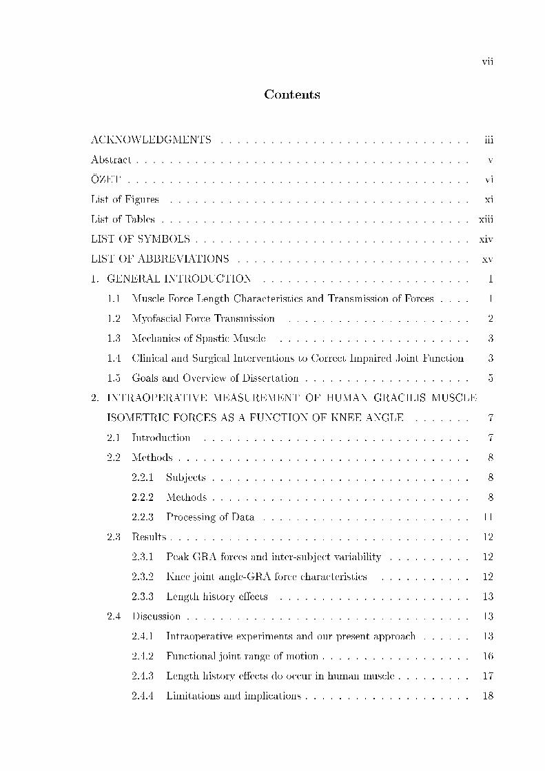
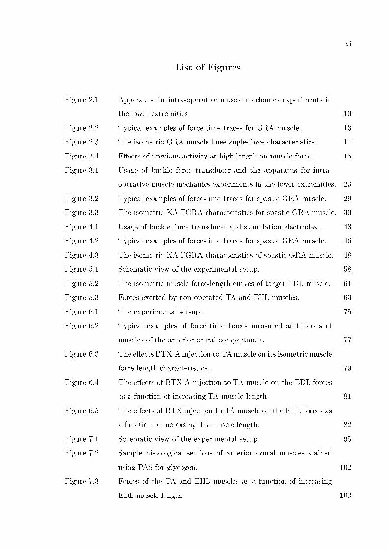
Figure 2.1 Apparatus for intra-operative muscle mechanics experiments in
the lower extremities. 10
Figure 2.2 Typical examples of force-time traces for GRA muscle. 13
Figure 2.3 The isometric GRA muscle knee angle-force characteristics. 14
Figure 2.4 E�ects of previous activity at high length on muscle force. 15
Figure 3.1 Usage of buckle force transducer and the apparatus for intra-
operative muscle mechanics experiments in the lower extremities. 23
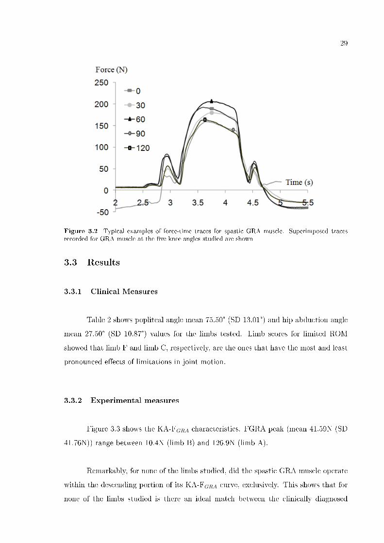
Figure 3.2 Typical examples of force-time traces for spastic GRA muscle. 29
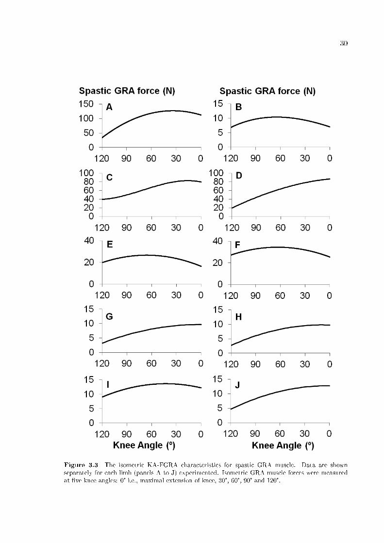
Figure 3.3 The isometric KA-FGRA characteristics for spastic GRA muscle. 30
Figure 4.1 Usage of buckle force transducer and stimulation electrodes. 43
Figure 4.2 Typical examples of force-time traces for spastic GRA muscle. 46
Figure 4.3 The isometric KA-FGRA characteristics of spastic GRA muscle. 48
Figure 5.1 Schematic view of the experimental setup. 58
Figure 5.2 The isometric muscle force-length curves of target EDL muscle. 61
Figure 5.3 Forces exerted by non-operated TA and EHL muscles. 63
Figure 6.1 The experimental set-up. 75
Figure 6.2 Typical examples of force time traces measured at tendons of
muscles of the anterior crural compartment. 77
Figure 6.3 The e�ects BTX-A injection to TA muscle on its isometric muscle
force-length characteristics. 79
Figure 6.4 The e�ects of BTX-A injection to TA muscle on the EDL forces
as a function of increasing TA muscle length. 81
Figure 6.5 The e�ects of BTX injection to TA muscle on the EHL forces as
a function of increasing TA muscle length. 82
Figure 7.1 Schematic view of the experimental setup. 95
Figure 7.2 Sample histological sections of anterior crural muscles stained
using PAS for glycogen. 102
Figure 7.3 Forces of the TA and EHL muscles as a function of increasing
EDL muscle length. 103
xii
Figure 7.4 EDL force-length characteristics obtained after proximal length-
ening. 104
Figure 7.5 EDL force-length characteristics obtained after distal lengthen-
ing. 106
Figure 7.6 Sample histological sections of anterior crural muscles stained
using Trichrome Gomori for collagen. 107
Figure A.1 The experimental set-up. 125
Figure A.2 Force-length characteristics of (a) EDL distal, (b) EDL proximal,
and (c) TA+EHL muscles obtained after distal lengthening. 127
xiii
List of Tables
Table 2.1 Anthropometric data, peak GRA forces and peak GRA tendon
stresses 9
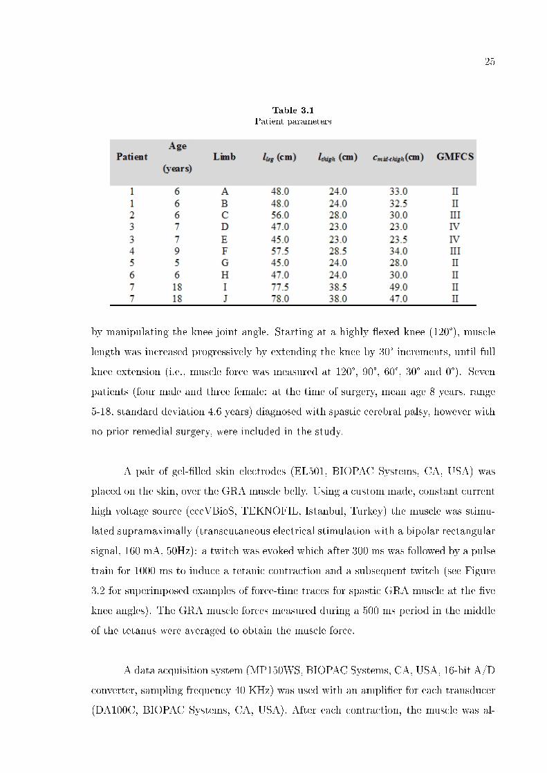
Table 3.1 Patient parameters 25
Table 3.2 Clinical measures characterizing motion limitation and experi-
mental measures 27
Table 4.1 Patient Parameters 44
Table 4.2 Clinical measures characterizing motion limitation and experi-
mental measures 49

xiv
LIST OF SYMBOLS
° Degree
C Celcius
cm Centimeter
Hz Hertz
kg Kilogram
mA Milli Amper
ml Milliliter
mm2 Square Millimeter
MPA Mega Pascal
ms Milli second
N Newton
p Probability Value
xv
LIST OF ABBREVIATIONS
Δlmt Muscle-tendon Length
ANOVA Analysis of Variance
ACL Anterior Cruciate Ligament
AFO Ankle Foot Orthosis
AT Aponeurotomy
BTX-A Botulinum Toxin Type - A
CC Control Contractions
cmid−thigh Mid-thigh Circumference
CP Cerebral Palsy
ECM Extracellular Matrix
EDL Extensor Digitorum Longus
EHL Extensor Hallicus Longus
EMFT Epimuscular Myofascial Force Transmission
Fa Active Force
FEM Finite Element Modeling
FGRA Gracilis Muscle Force
FL Force Length
Fp Passive Force
Ft Total Force
FT Force Transducer
GMFCS Gross Motor Functional Classi�cation System
GRA Gracilis
HAA Hip Abduction Angle
KA Knee Angle
Lambda Tonic Stretch Re�ex Threshold
lleg Leg Length
lopt Optimum Length
lrange Leg Range
xvi
lref Reference Length
lthigh Thigh Length
MFT Myofascial Force Transmission
MRI Magnetic Resonance Imaging
PA Popliteal Angle
PAS Periodic acid-Schi�
PD Preparatory Dissection
rho Spearman's Rank Correlation Coe�cient
ROM Range of Motion
SD Standard Deviation
SE Standard Error
TA Tibialis Anterior
US Ultrasound
VM Vastus Medialis
1
1. GENERAL INTRODUCTION
1.1 Muscle Force-Length Characteristics and Transmission of
Forces
Forces generated by skeletal muscle �bers and transmitted to the bones cause
joint motion. Force-length characteristics representing the maximal force with respect
to the length shows functional potential of muscle independent from velocity and ac-
tivation parameters such as stimulation frequency or amplitute [e.g. 1]. One of the
determinants of such potential is muscle architecture de�ned with muscle length, �ber
length, pennation angle, �ber type distribution, and number of sarcomeres in series and
in parallel [2]. Muscle length denoting muscle excursion determines the joint range of
motion [3-4] and it is typically associated with �ber length [5] i.e., number of sarcom-
eres in series mostly related with velocity of movement with pennation angle and �ber
type distribution [6-8]. Number of sarcomeres in parallel on the other hand translates
to the muscle cross sectional area and it is directly related to force production capacity
with also pennation angle [4, 9-11].
Additional to such architectural properties, as part of structural and morpholog-
ical features of muscles, transmission mechanisms of the forces are other determinants
of muscle function. Myotendinous junctions having specialized morphological features
are one of the paths of force transmission [12]: forces generated by sarcomeres in a
muscle �ber are transmitted to the bone through aponeurosis and tendon attached to
the bone. Myotendinous connections are very important for joint motion; however,
they are not exclusive pathways. Force transmission shown to have more complicated
mechanism includes also lateral pathways: transmission of forces from sarcomeres to
the extracellular matrix via special proteins [13-14]. Considering the continuity of
extracellular matrix with epimysium and collagen rich fascial structures, such lateral
pathways transmit forces to the neighboring �bers, muscles, and non-muscular struc-
tures.
2
1.2 Myofascial Force Transmission
Transmission of forces from myo�ber to its extracellular matrix composed of
collagen �bers is called intramuscular myofascial force transmission (MFT) [15-17].
Such transmission may occur along the endomysial perimeter of muscle �ber having
tightly arranged collagen layers [18]. In addition to intramuscular MFT, forces are also
transmitted from extracellular matrix of a muscle to the adjacent muscle's extracellu-
lar matrix. It is referred to as intermuscular MFT [19-20] if it is through the direct
connections between the muscles. Transmission of force from the extracellular matrix
of a muscle to surrounding non-muscular structures and bone is referred to as extra-
muscular MFT [15, 20-22]. Since inter- and extramuscular connections constitute an
integral system and cannot be distinguished they are de�ned as epimuscular myofascial
force transmission (EMFT) [23].
Myofascial loads representing the amount of EMFT were quanti�ed as the dif-
ference between muscle forces measured from proximal and distal tendons at speci�c
muscle lengths. Such myofascial loads shown to be prominent [15, 22, 24] indicates
the important role of inter- and extramuscular connections. If a muscle is not isolated,
shape of the force-length curve representing its characteristics changes drastically due
to EMFT [23, 25]. Another indicator of myofascial loads are force alterations measured
due to the relative positional di�erences between muscles even their lengths are �xed
[26-27]. The muscle model developed with �nite element modeling (FEM) method
[28] also revealed the e�ects of intra-, inter-, and extramuscular connections: Sarcom-
ere length heterogeneity with stress distribution along �ber bundles was shown as an
evidence of EMFT determining muscle force production capacity [25].
Consequently, muscles do not act as independent actuators. These previous
�ndings showing how EMFT modi�es muscle force-length characteristics did suggest
the functional role of this transmission mechanism in health and also in pathology.
3
1.3 Mechanics of Spastic Muscle
Cerebral palsy (CP) is a neuromuscular disorder caused by damage of the devel-
oping brain. Most of the cerebral palsy patients su�er from spasticity which is a form
of hypertonia [29-31] characterized by velocity dependent exaggerated re�exes [31-34].
Continuously activated muscles remain at low lengths and adapt to the immobiliza-
tion at shortened position [35-37]. Thus, in long term, contracture formation [38-42]
with muscle and soft tissue shortening [39] accompanies to spasticity even anti-spastic
treatments are applied [43-44]. Contracture as a reason or a consequence is associated
with decreased joint range of motion thus impaired function such as �exed hips, knees
and equines deformity at the ankles [40, 45-48].
Many architectural changes due to spasticity were reported: muscle shortening
[49-51], decrease in muscle volume [51-54] and cross-sectional area [52, 55-56], increased
sti�ness [57-59], �ber type alterations and increased amount of extracellular collagen
[59]. Even many structural changes for spastic muscles were found and the impaired
function is known to be related with these changes, no previous study revealed the
relationship between joint range and speci�c muscle function.
On the other hand, EMFT occurs also for CP patients [60-61] and known to
have important role for spasticity [62-63] in consistent with the increased sti�ness,
implications of this knowledge needs further examinations.
1.4 Clinical and Surgical Interventions to Correct Impaired
Joint Function
There are various methods for the treatment of spastic cerebral palsy from
physical therapy to neuromuscular surgical interventions applied depending on the
severity of the symptoms. To reduce spasticity injection of botulinum toxin type A
(BTX-A) causing muscle paralysis by inhibiting acetylcholine release [64-65] is used.
4
The e�ects of BTX-A have been widely studied by quantifying the area of paralysis [66],
compound muscle action potential [67] and electromyography [68]. However, reports
on mechanical parameters, e.g., twitch and tetanic force have been limited to selected
muscle lengths or joint positions [e.g. 67, 69]. Even it is well known that BTX-A
injected to the muscles spread through the fascia [70] and a�ects neighboring muscles
[71-72], the e�ects of BTX-A on the mechanical characteristics of targeted and non-
targeted neighboring muscles are not known.
Surgical interventions are performed on the patients having severe impairments
due to contracture formation. Remedial surgery known as muscle recession [73-74],
muscle release [75], muscle lengthening [76-80], and aponeurotomy (AT) [81] involves
cutting of an intramuscular aponeurosis transversely to its longitudinal direction. Lim-
ited e�ects of aponeurotomy per se were shown previously even a discontinuity oc-
curring. However, subsequent rupturing of intramuscular connections denoting the
removal of intramuscular MFT had major e�ects on force-length characteristics [82].
Following studies pronounced the altering role of extramuscular MFT on the e�ects of
AT [83-84]. Such results evoke a question if most of the epimuscular connections are
intact similar to in vivo condition how aponeurotomized muscle and its neighbors are
a�ected.
5
1.5 Goals and Overview of Dissertation
Present thesis is focused on spastic muscle mechanics and the treatment methods
used in the context of the determinant role of epimuscular myofascial force transmission
(EMFT). The goals of the study and the publications addressed in sequence in the
following chapters are
Chapter 2 aimed at developing an intra-operative method to measure human
muscle isometric forces directly with respect to joint angle and measure human gracilis
(GRA) muscle characteristics in health. It is published as
Yucesoy, C.A., Ate³, F., Akgün U., Karahan M., "Measurement of human gra-
cilis muscle isometric forces as a function of knee angle, intraoperatively," J Biome-
chanics, Vol. 43, p. 2665-71, 2010.
Chapter 3 aimed at measuring the forces of activated spastic GRA muscle as
a function of knee joint angle and to test the following hypotheses: (i) The muscle's
joint range of force exertion is narrow and (ii) High muscle forces are available at �exed
joint positions. It is published as
Ates, F., Temelli, Y., and Yucesoy, C.A., "Human spastic Gracilis muscle iso-
metric forces measured intraoperatively as a function of knee angle show no abnormal
muscular mechanics," Clin Biomech (Bristol, Avon), 2012
Chapter 4 aimed at measuring spastic GRA muscle characteristics during si-
multaneous stimulation with its antagonist and to test (i) if the joint range of force
exertion is narrow and (ii) if GRA muscle has higher force exertion capacity at low
lengths.
Chapter 5 aimed at revealing the e�ects of muscle lengthening surgery on rat
muscles and test the following hypotheses: (i) E�ects of AT on the target muscle are
di�erent at distal and proximal tendons, (ii) forces of non-targeted synergistic muscles
6
are a�ected as well, and (iii) preparatory dissection performed to reach the target
aponeurosis is responsible from some of these e�ects. It is submitted as
Ate³, F., Huijing P.A., Yucesoy, C.A., 2013. Muscle lengthening surgery causes
di�erential acute mechanical e�ects in both targeted and non-targeted synergistic mus-
cles, Journal of Electromyography and Kinesiology, in revision.
Chapter 6 aimed at testing the e�ects of BTX-A on (i) not only the injected
but also the non-injected rat muscles and also (ii) EMFT mechanism. It is published
as
Yucesoy, C.A., Ar�kan Ö.E., Ate³ F., 2012. BTX-A Administration to the Target
Muscle A�ects Forces of All Muscles Within an Intact Compartment and Epimuscular
Myofascial Force Transmission, Journal of Biomechanical Engineering, 134:111002-1-9.
Chapter 7 aimed at testing the e�ects of BTX-A on (i) the active contribution
of bi-articular neighboring muscle as well as (ii) the passive forces for both proximal
and distal joints it spans. It is submitted as
Ate³, F., Yucesoy, C.A., 2013. E�ects of BTX-A on non-injected bi-articular
muscle include a narrower length range of force exertion and increased passive force,
Muscle & Nerve, in revision.
7
2. INTRAOPERATIVE MEASUREMENT OF HUMAN
GRACILIS MUSCLE ISOMETRIC FORCES AS A
FUNCTION OF KNEE ANGLE
2.1 Introduction
Muscle force-length characteristics comprise one of the most important elements
of muscular mechanics. Such characteristics have been widely studied using highly
standardized experimental procedures in numerous animals [e.g., 85, 86]. In contrast,
although to an appreciable extent, the main goal of the research conducted is to under-
stand the function of human muscles, data available are mostly obtained from indirect
approaches including cadaver studies [87], joint torque measurements [88-89], modeling
[89] and use of dynamometry and ultrasound [90]. In only a limited number of studies,
direct measurement of human muscle forces was performed during activities in vivo
[91]. Direct measurements of isometric force-length characteristics in human muscles
were also rare and limited to the upper extremities [60, 92-93]. Improved understand-
ing of human muscle functioning in health and disease necessitates collection of data
that relates isometric muscle force to joint angle, directly.
Recent experiments in the rat have shown that previous activity at high (over
optimum) muscle lengths causes considerable active force changes (predominantly a
decrease) at lower lengths [15, 62]. Such length history e�ects present importance:
they comprise a distinct muscle mechanics phenomenon with unclear mechanisms and
implications. However, their occurrence has not been shown in human muscle.
The goals of this study were (1) to measure intraoperatively the previously
unstudied isometric forces of activated human Gracilis (GRA) muscle as a function of
knee joint angle and (2) to test our hypothesis that length history e�ects substantiate
also in human muscle. Experiments were conducted during anterior cruciate ligament
(ACL) reconstruction surgery.
8
2.2 Methods
Surgical and experimental procedures, in strict agreement with the guidelines of
Helsinki declaration were approved by a Committee on Ethics of Human Experimen-
tation at Marmara University, Istanbul.University, Istanbul.
2.2.1 Subjects
Seven male and one female patients (mean age 25 years, range 19-32, standard
deviation 4.7 years) undergoing ACL reconstruction surgery; however, with no former
musculoskeletal pathology were included in the study. Prior to surgery, (1) after a full
explanation of the purpose and methodology of the experiments, the subjects provided
their informed consent and (2) subject anthropometric data were collected.
2.2.2 Methods
Subjects were under general anesthesia but no muscle relaxants were used. All
intraoperative experiments were performed after routine incisions to reach the distal
GRA tendon and before any other surgical procedures of ACL reconstruction.
Using a scalpel blade (number 10), 2 cm oblique skin incision was made parallel
to the GRA tendon palpated 1 cm below the tuberosity of the tibia and 3 cm below
the medial joint gap. Sartorial fascia covering hamstring tendons was cut to expose
the GRA tendon with two incisions: (i) parallel to GRA tendon and (ii) parallel to
longitudinal axis of the tibia. Subsequently, a buckle force transducer (NK Biotechnical
Engineering Co., Minneapolis, Minnesota, USA, for further details [94] was mounted
over the tendon. Note that prior to each experiment, the force transducer was (i)
calibrated using bovine tendon strips (with rectangular cross section, dimensions ap-
proximating 7 x 2 mm, similar to human GRA distal tendon) and (ii) sterilized (using
dry gas at maximally 500 C).
9
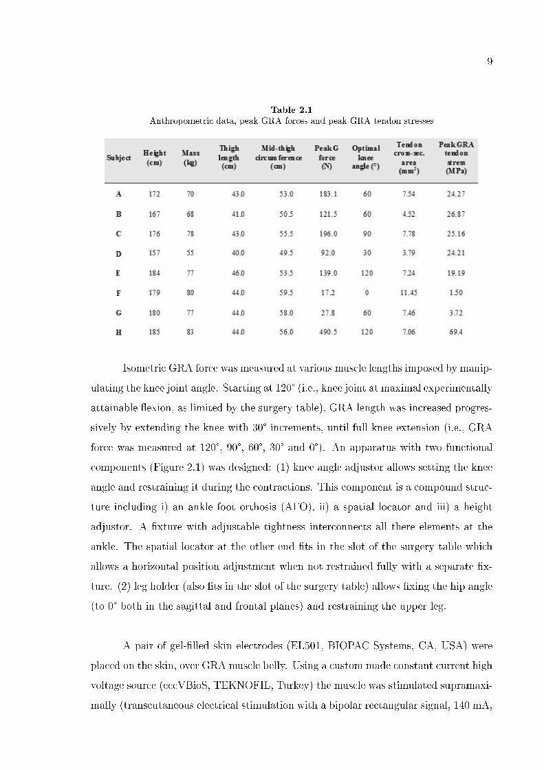
Table 2.1
Anthropometric data, peak GRA forces and peak GRA tendon stresses
Isometric GRA force was measured at various muscle lengths imposed by manip-
ulating the knee joint angle. Starting at 120° (i.e., knee joint at maximal experimentally
attainable �exion, as limited by the surgery table), GRA length was increased progres-
sively by extending the knee with 30° increments, until full knee extension (i.e., GRA
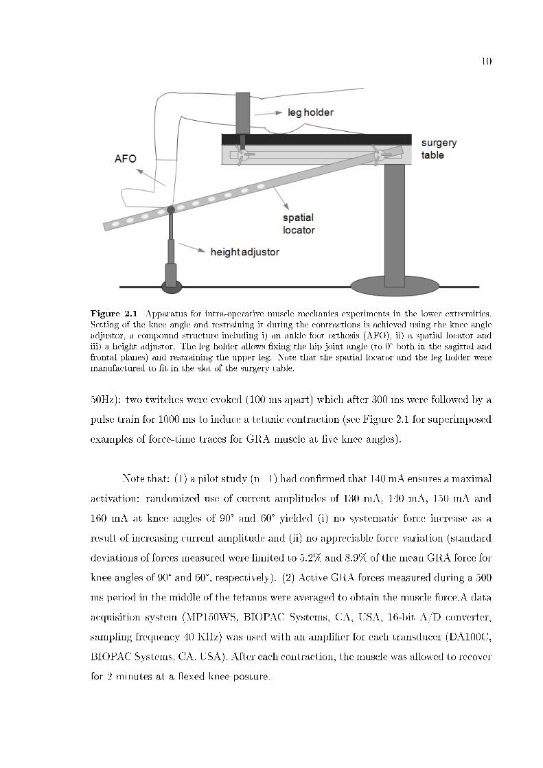
force was measured at 120°, 90°, 60°, 30° and 0°). An apparatus with two functional
components (Figure 2.1) was designed: (1) knee angle adjustor allows setting the knee
angle and restraining it during the contractions. This component is a compound struc-
ture including i) an ankle foot orthosis (AFO), ii) a spatial locator and iii) a height
adjustor. A �xture with adjustable tightness interconnects all there elements at the
ankle. The spatial locator at the other end �ts in the slot of the surgery table which
allows a horizontal position adjustment when not restrained fully with a separate �x-
ture. (2) leg holder (also �ts in the slot of the surgery table) allows �xing the hip angle
(to 0° both in the sagittal and frontal planes) and restraining the upper leg.
A pair of gel-�lled skin electrodes (EL501, BIOPAC Systems, CA, USA) were
placed on the skin, over GRA muscle belly. Using a custom made constant current high
voltage source (cccVBioS, TEKNOFIL, Turkey) the muscle was stimulated supramaxi-
mally (transcutaneous electrical stimulation with a bipolar rectangular signal, 140 mA,
10
Figure 2.1 Apparatus for intra-operative muscle mechanics experiments in the lower extremities.Setting of the knee angle and restraining it during the contractions is achieved using the knee angleadjustor, a compound structure including i) an ankle foot orthosis (AFO), ii) a spatial locator andiii) a height adjustor. The leg holder allows �xing the hip joint angle (to 0° both in the sagittal andfrontal planes) and restraining the upper leg. Note that the spatial locator and the leg holder weremanufactured to �t in the slot of the surgery table.
50Hz): two twitches were evoked (100 ms apart) which after 300 ms were followed by a
pulse train for 1000 ms to induce a tetanic contraction (see Figure 2.1 for superimposed
examples of force-time traces for GRA muscle at �ve knee angles).
Note that: (1) a pilot study (n=1) had con�rmed that 140 mA ensures a maximal
activation: randomized use of current amplitudes of 130 mA, 140 mA, 150 mA and
160 mA at knee angles of 90° and 60° yielded (i) no systematic force increase as a
result of increasing current amplitude and (ii) no appreciable force variation (standard
deviations of forces measured were limited to 5.2% and 8.9% of the mean GRA force for
knee angles of 90° and 60°, respectively). (2) Active GRA forces measured during a 500
ms period in the middle of the tetanus were averaged to obtain the muscle force.A data
acquisition system (MP150WS, BIOPAC Systems, CA, USA, 16-bit A/D converter,
sampling frequency 40 KHz) was used with an ampli�er for each transducer (DA100C,
BIOPAC Systems, CA, USA). After each contraction, the muscle was allowed to recover
for 2 minutes at a �exed knee posture.
11
Subsequent to collection of a complete set of knee joint angle-force data, control
measurements were performed at lower GRA length (corresponding to 90° knee angle)
in order to test if previous activity at high length (imposed by full knee extension) had
any e�ect on the force exerted. All experimental preparations and data collection were
completed within 20 min, the maximal study duration allowed by the ethical commit-
tee. Diagnostic upper leg MRI images of the subjects were used to determine GRA
muscle tendon cross-sectional areas: using the built in software of the Picture Archiv-
ing and Communication System, area of tendon cross-section marked in an image slice
located distally, approximately where force transducers were mounted were determined
as number of pixels in the marked area x pixel area.
2.2.3 Processing of Data
(1) Peak GRA forces as well as GRA tendon stresses (Peak GRA force/tendon
cross-sectional area) and the corresponding optimal knee angles were studied. Pearson's
correlation coe�cient was calculated to quantify inter-subject variability between the
peak force and stress values and subject (i) weigh ii) mid-upper leg perimeter and (iii)
upper leg length. Correlations were considered signi�cant at p<0.05.
(2) Knee joint angle-GRA muscle force characteristics were studied separately
for each subject: operational portion of the force-length characteristics were character-
ized.
(3) E�ects of previous activity at high length on muscle force exerted at lower
lengths were assessed: control force for each subject measured at 90º knee angle was
compared to the force measured at identical knee angle during collection of knee angle-
GRA muscle force data. Existence of a correlation between optimal knee angle and
history e�ect was assessed: Pearson's correlation coe�cient was calculated using abso-
lute values of % force changes. Correlation was considered signi�cant at p<0.05.
12
2.3 Results
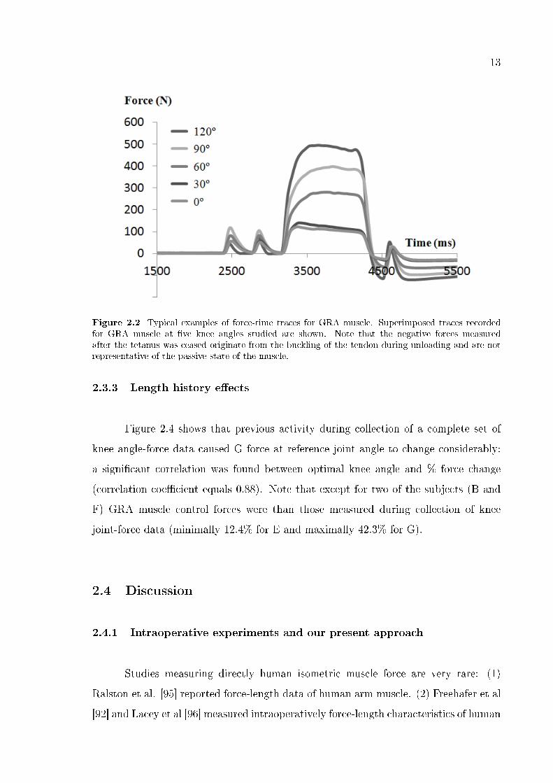
2.3.1 Peak GRA forces and inter-subject variability
Table 1 shows the key anthropometric parameters of the subjects as well as the
magnitude and the corresponding knee joint angle for the peak GRA forces measured.
Peak GRA forces show a sizable inter-subject variability: peak force (mean = 178.5 ±
270.3 N) ranges between 17.2 N and 490.5 N. Mean peak GRA tendon stress equals 24.4
± 20.6 MPa. Optimal knee angles (mean = 67.5 ± 41.7°) include all angles studied.
Only a limited correlation was found between peak GRA force and subject mass
as well as mid-thigh perimeter (correlation coe�cient equals 0.34 and -0.17, respec-
tively) whereas, almost no correlation was found between peak GRA force and thigh
length (correlation coe�cient equals 0.02). None of these correlations were statistically
signi�cant. Similarly, correlations between peak GRA tendon stress and subject mass,
mid-thigh perimeter and thigh length (correlation coe�cient equals 0.14, -0.24, and
-0.06, respectively) were insigni�cant.
2.3.2 Knee joint angle-GRA force characteristics
Knee joint angle-GRA force characteristics (Figure 2.3) for only two subjects
(E and H) indicate that GRA muscle may operate in the descending limb of its force-
length characteristics. In contrast, for a majority of the subjects, such operational
range includes parts of both ascending and descending limbs. Nevertheless, even for
those subjects Figure 2.3 shows sizable inter-subject variability presenting itself as
shape di�erences in knee joint angle-GRA force characteristics.
An important �nding is that for none of the knee angles studied, GRA muscle
was at active slack length indicating that this length corresponds to a knee �exion over
120°.
13
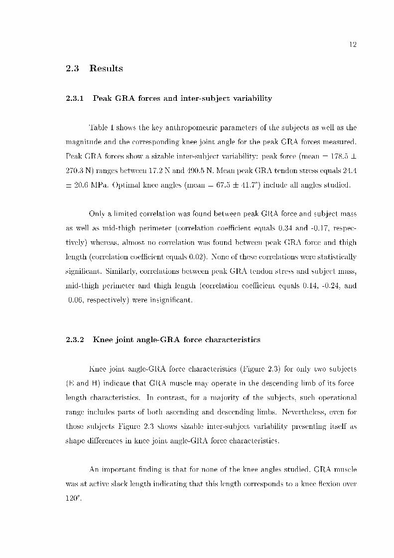
Figure 2.2 Typical examples of force-time traces for GRA muscle. Superimposed traces recordedfor GRA muscle at �ve knee angles studied are shown. Note that the negative forces measuredafter the tetanus was ceased originate from the buckling of the tendon during unloading and are notrepresentative of the passive state of the muscle.
2.3.3 Length history e�ects
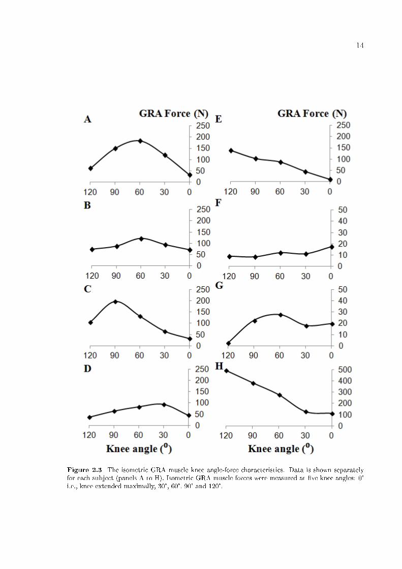
Figure 2.4 shows that previous activity during collection of a complete set of
knee angle-force data caused G force at reference joint angle to change considerably:
a signi�cant correlation was found between optimal knee angle and % force change
(correlation coe�cient equals 0.88). Note that except for two of the subjects (B and
F) GRA muscle control forces were than those measured during collection of knee
joint-force data (minimally 12.4% for E and maximally 42.3% for G).
2.4 Discussion
2.4.1 Intraoperative experiments and our present approach
Studies measuring directly human isometric muscle force are very rare: (1)
Ralston et al. [95] reported force-length data of human arm muscle. (2) Freehafer et al
[92] and Lacey et al [96] measured intraoperatively force-length characteristics of human
14
Figure 2.3 The isometric GRA muscle knee angle-force characteristics. Data is shown separatelyfor each subject (panels A to H). Isometric GRA muscle forces were measured at �ve knee angles: 0°i.e., knee extended maximally, 30°, 60°, 90° and 120°.
15
Figure 2.4 E�ects of previous activity at high length on muscle force. Data shown separately foreach subject compares the control forces measured at knee angle = 90º with the force measuredat identical knee angle during collection of knee angle-GRA muscle force data. Note that percentdi�erences shown for each subject in gray characters indicate a force increase (for subjects B and F)whereas; the remainder indicate a force decrease.
lower arm muscles. Despite such pioneering contributions, the experimental setup of
those authors was not capable of avoiding all movement artifacts of the arm. (3) A
systematic set of studies reporting intraoperatively measured human �exor carpi ulnaris
muscle force-length data were published recently by Smeulders, Kreulen and colleagues
[e.g. 61, 93, 97]. Using well designed experimental procedures and setup, these authors
were able to remove limb movement artifacts and standardize their approach. They
therefore, made a major contribution not only to intraoperative experimentation but
also to our understanding of human muscle mechanics.
Our present intraoperative approach features to the same capacity, the ability
to ensure the isometric nature of experiments. Moreover, data, more representative of
the situation in vivo can be obtained: the buckle force transducers allow measuring (1)
after minimal tissue interventions and (2) muscle force directly as a function of joint
angle. The latter yields simultaneous length changes of all muscular and non-muscular
structure as it occurs in vivo.
16
2.4.2 Functional joint range of motion
Our results show that human GRA muscle is capable of producing non-zero
active forces even for the shortest length studied hence, at least for a joint range between
0° to 120°. This �nding is in concert with those of Ward et al. [98] who showed that G
is among the lower extremity muscles that feature the greatest excursion. Note that,
for fundamental motions like walking and sit-to-stand, maximal knee �exion was shown
to equal 110° [99] and 103° [100], respectively. Our results show that GRA muscle is
capable of contributing to knee �exor moment also for such joint positions.
In isometric experiments performed in animal [e.g. 23] and human muscles [e.g.
61], in situ data produced include typically both ascending and descending limbs of
muscle force-length characteristics. However, studies inferring to in vivo condition
have reported that muscle functional joint range does not correspond fully to such in
situ length range of active force exertion. For example, Lieber and coworkers [101-103]
showed that synergistic extensor carpi radialis brevis and longus muscles function in the
descending limb of their force-length characteristics. In contrast, human gastrocnemius
muscle was reported to operate within the ascending limb [89, 104]. Our present
results suggest in agreement with these studies that human GRA muscle does not
operate within the entire length range of force exertion. However, for a majority of
the subjects (except subjects E and H) it appears to function in both of the ascending
and descending limbs. Nevertheless, even for those subjects inter-subject variability
is considerable: the knee angle-muscle force curves show shape di�erences as well as
di�erent optimal knee angles. Moreover, data of subjects E and H indicate that their
GRA muscles operate within the descending limb, exclusively. Therefore, we conclude
that the functional joint range of motion for human GRA muscle is at least as wide as
full knee extension to 120° of knee �exion, however; the portion of the knee angle-muscle
force relationship operationalized is not unique but individual speci�c.
17
2.4.3 Length history e�ects do occur in human muscle
Our results con�rmed our hypothesis that length history e�ects [e.g. 15, 105]
do occur also in human muscle. The exact mechanism is not immediately apparent;
however, due to the cyclic testing nature (increasing muscle lengths and shortening
before control measurements), one may consider hysteresis as a determinant. Our
recent �ndings do indicate such viscoelastic intra- and epimuscular tissue role [106]:
(i) a second control contraction after a longer recovery period (15 min) showed even
greater history e�ects however, (ii) interfering surgically with the fascial connections
of muscle caused history e�ects to decrease.
Nevertheless, such energy dissipation mechanism is expected to cause a force
reduction. Although, for a majority of the present and previously reported data this
is the case, there are remarkable exceptions with possibly important messages: (1)
Previous activity of a muscle at high length was shown to cause a certain force increase
in its restrained synergistic [106] or even antagonistic [107] muscles. (2) For two of
the present subjects, our results show such force increase also for the target muscle.
Activity speci�cally over muscle optimum length has been argued to cause force reduc-
ing length history e�ects [62]. Accordingly, knee angle-force characteristics of subject
F (Figure 2.2 F) suggest that GRA muscle was not activated over optimum length
even for maximal knee extension. However, the considerably increased control force for
subject B is clearly exceptional: knee angle-force characteristics indicate strongly that
for maximal knee extension, GRA muscle was at a higher length than optimum length.
Note that unlike the previous animal experiments reporting such history e�ects, mus-
cle lengths were manipulated presently by altering the joint angle. Moment arms that
could vary among subjects and di�erences in position of the target muscle relative to
its neighboring muscular and nonmuscular structures (nontargetted muscles were re-
strained in animal experiments) may a�ect the lengths of the tissues involved. Muscle
relative position reported as a key determinant of epimuscular myofascial force trans-
mission [26-27, 108-109] was shown to alter muscular mechanics substantially [e.g. 27,
108]. Nevertheless, the exceptional results discussed here suggest that time dependent
material properties may not be the exclusive cause for the history e�ects indicating
18
more complex mechanisms involving also the contractile elements. Possible history
dependent role played by the active and passive components of muscle tissue [e.g., 110]
should be considered and addressed in speci�cally planned new studies.
2.4.4 Limitations and implications
2.4.4.1 Lack of passive force data. Both prior and subsequent to the tetanus,
measurement of passive force was not possible: (1) the amplitude of the twitches evoked
before the tetanus appears to be not high enough to remove the GRA tendon slack. (2)
Since the tendon buckles during unloading, the force transducers working on a principle
of torque measurement [94] measure negative forces, not representative of the passive
state after the tetanus was ceased. In new studies, successful passive data collection
could be possible by either increasing the twitch current amplitude or by measuring
passive force-joint angle relationship separately.
2.4.4.2 Implications of EMFT . Our research groups have shown that due to
myofascial force transmission [111] occurring epimuscularly [23] forces produced in
neighboring synergistic rat muscles can be integrated with the force of the agonist and
exerted onto the bone from its tendon [22]. Moreover, experimentally manipulating
such force transmission pathways (e.g., dissection of connective tissues at muscle bellies
or removal of synergistic muscles) has been shown to change the magnitude of muscle
force exerted at the same lengths, explained by sarcomere length changes [23, 25].
Therefore, due to epimuscular myofascial force transmission, the shape of the muscle
length�force characteristics was shown to change as a function of di�erent mechanical
conditions in which the muscle functions.
We expect important implications of this mechanism:
(1) Harvesting the distal tendon of GRA muscle often together with semitendi-
nosus (ST) muscle is a common technique in ACL reconstruction. Although, such direct

19
impairment of the myotendinous force transmission path implies post-operative knee
�exion de�ciency, several studies reported only a small reduction in peak knee �exion
moment after recovery, if any [e.g., 112, 113-115]. Accordingly, such post-operatively
unchanged peak knee �exion moment may be ascribable, at least in part, to epimus-
cular myofascial force transmission from GRA and ST muscles to the knee joint via
neighboring hamstrings muscles.
(2) Epimuscular myofascial force transmission may be an important factor re-
sponsible at least in part with the inter-subject variability shown presently: (i) plausible
di�erences in the mechanical properties of the muscle's epimuscular connections among
di�erent subjects are conceivable to cause shape di�erences in their knee angle-muscle
force relationships and hence, lead to functional portions of this relationship to be indi-
vidual speci�c. Smeulders et al. [61] reported data indicating e�ectiveness of myofascial
force transmission in human muscles and ascribed apparent inter-subject variability to
such mechanism additional to spasticity related di�erences in muscle properties. (ii)
Note that although care was taken presently to stimulate the targeted GRA muscle
exclusively, unintentional marginal stimulation of also the neighboring hamstrings was
not completely impossible due to the surface electrodes used. In such a situation, addi-
tional force transmitted via myofascial pathways from a neighboring muscle onto GRA
muscle may also be responsible with some of the inter-subject variability.
For a broader consideration of muscle-tendon biomechanical function and inter-
subject variability, tendon stress is a valuable parameter. Addressing bone, muscle
and tendon stresses across species, importance of biomechanical consequences of scal-
ing was reviewed extensively by Biewener [116]. In speci�c studies e.g., on kangaroo
rat locomotion [117-118] determining how stress scales with force yielded important
insights allowing relating animal size and motion strategies. It should be noted that
presently much less inter-subject variability was shown for peak GRA tendon stresses.
Nevertheless, a surprising �nding was that tendon cross-sectional area of subject F was
the largest and tendon of subject H was not oversized. Therefore, there was no fully
consistent scaling of tendon size and force. This suggests that certain inter-subject
variability should originate from di�erential force production capacity of the muscle
20
which implies much higher contribution of the GRA muscle to knee �exion moment for
some individuals which is likely to have consequences post-operatively. New studies
are indicated to study simultaneously the knee angle-force characteristics of also other
hamstrings in order to (1) assess the e�ects of epimuscular myofascial force transmission
and (2) determine the relative contribution of GRA, ST and e.g. semimembranosus
muscles to knee moment. The lack of correlation shown between typical subject anthro-
pometrics and peak muscle force and even stress indicates the di�culty of estimation
of the contribution of human muscles to joint moments for di�erent individuals using
such indirect measurements.
In conclusion, mean peak GRA force, mean peak GRA tendon stress and optimal
knee angle equaled 178.5 ± 270.3 N, 24.4 ± 20.6 MPa and 67.5 ± 41.7°, respectively.
A substantial inter-subject variability was found and our results indicate that typical
subject anthropometrics cannot be used as predictors. The functional joint range of
motion for human GRA muscle was at least as wide as full knee extension to 120° of
knee �exion. However; the portion of the knee angle-muscle force relationship oper-
ationalized by GRA muscle is not unique but individual speci�c. Previous isometric
activity of a human muscle at high length was shown for the �rst time to a�ect muscle
forces measured at lower lengths causing for most of the subjects a decrease.
21
3. HUMAN SPASTIC GRACILIS MUSCLE ISOMETRIC
FORCES AS A FUNCTION OF KNEE ANGLE SHOW NO
ABNORMAL MUSCULAR MECHANICS
3.1 Introduction
Cerebral palsy is a movement disorder caused by damage of the developing
brain. Skeletal muscle spasticity is the central aspect of such disorders associated with
exaggerated stretch re�exes caused by diminished inhibition [32, 34]. Due to that, if
the spastic muscle is stretched rapidly, it will resist lengthening actively and, therefore,
remain at low length. Such increased resistance to stretch leads to a long lasting,
shortened condition of the muscle often followed by structural changes [e.g., 39, 119].
Contractures [38-39, 42] and muscle hypertonicity [29-31] that commonly occur in the
lower extremities cause children su�ering from spastic cerebral palsy to walk with the
hips and knees �exed and with equines deformity at the ankles.
We should be able to show that the mechanics of the spastic muscle are repre-
sentative of the functional de�ciencies clearly apparent in the joints. The limited joint
range of motion of the patients suggests that spastic muscle may not be capable of
exerting force for the entire range of joint angles attainable from a �exed joint position
to full extension. The fact that the joint is forcefully kept in a �exed position sug-
gests that the spastic muscle should be capable of exerting high forces at lower muscle
lengths. In order to demonstrate such characteristics, it is necessary to collect data
that relate isometric muscle force to joint angle, directly. Measurements of spastic
muscle forces of this nature are rarely done in the upper extremities [61, 93] and, to
our knowledge, never in the lower extremities.
In the present study, using our recently developed methods [120], we measured
the forces of activated spastic Gracilis (GRA) muscle as a function of knee joint angle
during remedial surgery. Our goal was to test the following hypotheses: (1) the muscle's
22
joint range of force exertion is narrow. (2) High muscle forces are available at �exed
joint positions corresponding to low muscle lengths.
3.2 Methods
Surgical and experimental procedures, in strict agreement with the guidelines
of the Helsinki declaration, were approved by a Committee on Ethics of Human Ex-
perimentation at Istanbul University, Istanbul.
3.2.1 Patients
Seven patients (four male and three female: at the time of surgery, mean age 8
years, range 5-18, standard deviation 4.6 years) diagnosed with spastic cerebral palsy,
however with no prior remedial surgery, were included in the study.
The Gross Motor Functional Classi�cation System (GMFCS) [121-122] was used
to classify the mobility of the patients. Those who were included in the study attained
scores of level II or higher, indicating the severity of their limited mobility (Table 1).
In short, all patients tested were in need of physical assistance for walking (in order to
avoid a fall and rapid buildup of fatigue) and also a support was necessary for sitting
and standing (level II). For some patients, additional aids including a wheelchair or a
body support walker were needed for mobility (level III and IV). Additional patient
classi�cation was done based on popliteal angle [the angle between hip and knee at hip
in 90° �exion, see 123] and hip abduction angle measured when the hip is in extended
position [124] (Table 2). Clinical tests led to a decision that all patients required
remedial surgery including release of hamstrings and hip adductors.
All patients were operated on bilaterally. For three of the patients, separate
experiments were performed on both legs, whereas for the remainder, only one leg was
experimented on due to time limitations imposed by subsequent multilevel surgery.
23
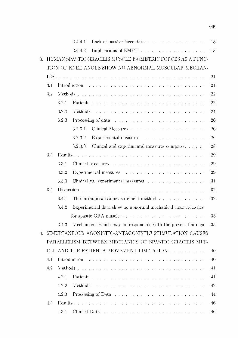
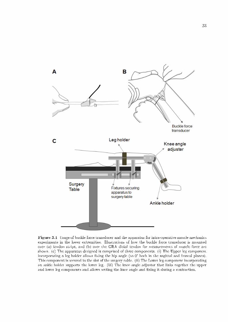
Figure 3.1 Usage of buckle force transducer and the apparatus for intra-operative muscle mechanicsexperiments in the lower extremities. Illustrations of how the buckle force transducer is mountedover (a) tendon strips, and (b) over the GRA distal tendon for measurements of muscle force areshown. (c) The apparatus designed is comprised of three components. (i) The Upper leg componentincorporating a leg holder allows �xing the hip angle (to 0° both in the sagittal and frontal planes).This component is secured to the slot of the surgery table. (ii) The Lower leg component incorporatingan ankle holder supports the lower leg. (iii) The knee angle adjustor that links together the upperand lower leg components and allows setting the knee angle and �xing it during a contraction.
24
Therefore, a total of ten knee angle-GRA muscle force data sets were collected. Prior
to surgery, (1) after a full explanation of the purpose and methodology of the experi-
ments, the patients or their parents provided their informed consent, and (2) patient
anthropometric data were collected.
3.2.2 Methods
The patients received general anesthesia and no muscle relaxants or tourni-
quet was used. All intraoperative experiments were performed after routine incisions
to reach the distal GRA tendon and before any other surgical procedures of muscle
lengthening surgery.
Using a scalpel blade (number 18), a longitudinal skin incision of 2.5cm was
made immediately above the popliteal fossa. After cutting the adipose tissue and the
fascia lata, the distal GRA tendon was exposed. Subsequently, a buckle force trans-
ducer (S-shape, dimensions: width=12mm length=20mm and height=9mm, TEKNOFIL,
Turkey) was mounted and �xed over the tendon. Note that prior to each experiment,
the force transducer was (i) calibrated using bovine tendon strips (with rectangular
cross section, dimensions 7x2mm2 representative of that of the GRA distal tendon)
and (ii) sterilized (using dry gas at maximally 500 C). See Figure 3.1a and b, respec-
tively, for illustrations of how a buckle force transducer is mounted over tendon strips
and over the GRA distal tendon for measurements of muscle force.
An apparatus comprised of three components (Figure 3.1c) was designed: (1)
the upper leg component was secured with two �xtures to the slot of the surgery table
and the leg holder, which had an adjustable position on it that allowed supporting the
upper leg and �xing the hip angle (to 0° both in the sagittal and frontal planes); (2)
the knee angle adjustor combining the upper and lower leg components allowed setting
the knee angle and �xing it during the contractions; (3) the lower leg component with
an ankle holder that had an adjustable position on it allowing support of the lower leg.
Isometric spastic GRA muscle force was measured at various muscle lengths imposed
25
Table 3.1
Patient parameters
by manipulating the knee joint angle. Starting at a highly �exed knee (120°), muscle
length was increased progressively by extending the knee by 30° increments, until full
knee extension (i.e., muscle force was measured at 120°, 90°, 60°, 30° and 0°). Seven
patients (four male and three female: at the time of surgery, mean age 8 years, range
5-18, standard deviation 4.6 years) diagnosed with spastic cerebral palsy, however with
no prior remedial surgery, were included in the study.
A pair of gel-�lled skin electrodes (EL501, BIOPAC Systems, CA, USA) was
placed on the skin, over the GRA muscle belly. Using a custom made, constant current
high voltage source (cccVBioS, TEKNOFIL, Istanbul, Turkey) the muscle was stimu-
lated supramaximally (transcutaneous electrical stimulation with a bipolar rectangular
signal, 160 mA, 50Hz): a twitch was evoked which after 300 ms was followed by a pulse
train for 1000 ms to induce a tetanic contraction and a subsequent twitch (see Figure
3.2 for superimposed examples of force-time traces for spastic GRA muscle at the �ve
knee angles). The GRA muscle forces measured during a 500 ms period in the middle
of the tetanus were averaged to obtain the muscle force.
A data acquisition system (MP150WS, BIOPAC Systems, CA, USA, 16-bit A/D
converter, sampling frequency 40 KHz) was used with an ampli�er for each transducer
(DA100C, BIOPAC Systems, CA, USA). After each contraction, the muscle was al-
26
lowed to recover for 2 minutes in a �exed knee posture.
All experimental preparations and data collection were completed within 30
minutes, the maximal study duration allowed by the ethics committee.
3.2.3 Processing of data
3.2.3.1 Clinical Measures. The popliteal and hip abduction angles for each limb
were normalized for the values of these angles representing the most severe limitations
to joint motion among our patients using equations (1) and (2):
PAnormalized = PAPAmax
(1)
where, PA and PAnormalized are the actual and normalized popliteal angle for
a particular limb, respectively. PAmax is the maximal popliteal angle value measured
among the limbs studied.
HAAnormalized = HAAHAAmax
(2)
where, HAA and HAAnormalized are the actual and normalized hip abduction
angle for a particular limb, respectively. HAAmax is the maximal hip abduction angle
value measured among the limbs studied. The GRAmuscle is a �exor of the knee as well
as an adductor of the hip. Therefore, summed PAnormalized and HAAnormalized
represented a limb score for limited range of motion (ROM) (Table 2).
3.2.3.2 Experimental measures. Using a least squares criterion, data for total
GRA muscle force (FGRA) in relation to knee joint angle (KA) were �tted with a
polynomial function
FGRA= a0 + a1KA+a2KA2 + ... + anKAn
27
Table 3.2
Clinical measures characterizing motion limitation and experimental measures
a0, a1. . . an are coe�cients determined in the �tting process. The lowest order of
the polynomials that still added a signi�cant improvement to the description of changes
of KA and FGRA data were selected using a one-way analysis of variance (ANOVA)
[125]. The �tted KA-FGRA characteristics of the patients were studied separately for
each limb for characterization of the mechanics of the spastic GRA muscle.
Experimentally obtained measures were used for an objective assessment of our
hypotheses based on (i) joint range of muscle force exertion and (ii) availability of high
muscle force at low muscle length. We considered that a narrow joint range together
with the availability of the peak muscle force at the lowest muscle length studied would
provide an ideal match between the clinically diagnosed impaired joint motion and the
experimentally determined KA-FGRA characteristics. Therefore, we �rst studied our
data to see if the spastic GRA muscle operates within the descending portion of its
KA-FGRA curve, exclusively. For KA-FGRA curves other than that, the following
procedures were followed.
Measures based on joint range of muscle force exertion
If the peak GRA muscle force (FGRApeak) was attained within the knee joint
28
range studied such that the increase trend of the force ceased before full knee extension,
the spastic GRA muscle was concluded to operate within both the ascending and
descending portions of its KA-FGRA curve. Therefore, the joint range of muscle force
exertion is not as narrow as for the ideal match. The proximity of the KA at which FGRA
peak was exerted (KAFGRApeak) to the maximal knee �exion angle studied indicates
how narrow the joint range of force exertion of each limb is. If, on the other hand,
FGRApeak was attained only at KA=0°, such that the increase trend of the force was
not ceased, the spastic GRA muscle was concluded to operate exclusively within the
ascending portion of its KA-FGRA curve. Therefore, such curves were considered not
to represent a narrow joint range, and hence not representative of the compromised
joint motion regarding this measure. In order to distinguish those limbs, the slope
of the KA-FGRA curve for KA=5° to 0° range was calculated. The steepest slope was
considered to represent the widest joint range of force exertion among the limbs studied.
Additionally, KA-FGRA curves were extrapolated to determine the KA corresponding
to muscle active slack length (i.e., the shortest length at which the muscle can still
exert non-zero force).
Measures based on the availability of muscle force
Even for the limbs that do not show a narrow knee joint range of GRA muscle
force exertion, we considered that the availability of high muscle force at knee �exion
represents characteristics of spastic muscle. In order to quantify that, the percentage
of FGRApeak exerted at KA=120° (%FGRApeak/120º) was calculated. Additionally, the
availability of high muscle force at full knee extension was assessed. In order to quantify
that, the percentage of FGRApeak exerted at KA=0° (%FGRApeak/0º was calculated.
3.2.3.3 Clinical and experimental measures compared. Spearman's Rank cor-
relation coe�cient was calculated to test if limb scores for limited ROM are corre-
lated with the key determinants of KA-FGRA characteristics, i.e., (i) KAFGRApeak, (ii)
%FGRApeak/120º and, (iii) %FGRApeak/0º. Correlations were considered signi�cant at
P<0.05.
29
Figure 3.2 Typical examples of force-time traces for spastic GRA muscle. Superimposed tracesrecorded for GRA muscle at the �ve knee angles studied are shown
3.3 Results
3.3.1 Clinical Measures
Table 2 shows popliteal angle mean 75.50° (SD 13.01°) and hip abduction angle
mean 27.50° (SD 10.87°) values for the limbs tested. Limb scores for limited ROM
showed that limb F and limb C, respectively, are the ones that have the most and least
pronounced e�ects of limitations in joint motion.
3.3.2 Experimental measures
Figure 3.3 shows the KA-FGRA characteristics. FGRA peak (mean 41.59N (SD
41.76N)) range between 10.4N (limb B) and 126.9N (limb A).
Remarkably, for none of the limbs studied, did the spastic GRA muscle operate
within the descending portion of its KA-FGRA curve, exclusively. This shows that for
none of the limbs studied is there an ideal match between the clinically diagnosed
30
Figure 3.3 The isometric KA-FGRA characteristics for spastic GRA muscle. Data are shownseparately for each limb (panels A to J) experimented. Isometric GRA muscle forces were measuredat �ve knee angles: 0° i.e., maximal extension of knee, 30°, 60°, 90° and 120°.
31
decreased mobility and the experimentally determined KA-FGRA characteristics.
Measures based on joint range of muscle force exertion
For all limbs and for all the knee angles studied, non-zero muscle forces were
measured. This indicates that the GRA muscle's operational joint range of muscle
force exertion includes the entire joint range studied. Therefore, the muscle's active
slack length corresponds to a knee �exion of over 120° (minimally 134° for limb A, as
determined using extrapolated data). For only 6 limbs (A, B, C, E, F and I), was it
possible to determine the KAFGRA peak within the knee joint range studied (Table
2). The operational joint range of force exertion for limb E (KAFGRApeak = 66°) was
found to be the narrowest. For the remaining 4 limbs (D, G, H and J), the operational
joint range of force exertion was shown to include exclusively the ascending portion of
their KA-FGRA curves. The steepest slope at KA=0°of these curves was calculated for
limb D, indicating the widest operational joint range of force exertion.
Measures based on availability of muscle force
Table 2 shows that as much as 79.1% and 74.6% of FGRApeak are available at
the lowest muscle length studied respectively for limbs F and E. For the remainder of
the limbs, maximally 66.2% (limb I) and minimally 22.4% (limb D) of FGRApeak were
available at such low length. The GRA muscle was capable of exerting high forces even
at the highest length studied: minimally for limb E %FGRApeak/0º =61.6 (Table 2).
3.3.3 Clinical vs. experimental measures
No signi�cant correlations were found between limb scores for limited ROM
and the key determinants of KA-FGRA characteristics: Spearman's rank correlation
coe�cients were only 0.03 (p = 0.93), 0.24 (p = 0.48), and 0.02 (p = 0.96), respectively,
for a comparison with respect to KAFGRApeak, %FGRApeak/120ºand, %FGRApeak/0º.
32
3.4 Discussion
3.4.1 The intraoperative measurement method
Clinical measures of the GMFCS as well as popliteal and hip abduction angles
indicated decreased mobility of all of our patients requiring multilevel surgery involving
lengthening of the GRA muscle. The purpose of this study was to test our hypotheses
to demonstrate that the mechanics of the spastic GRA muscle are representative of
such impaired joint motion.
Prior to any routine surgical procedures, except for the incisions required for
exposing the distal GRA tendon, the muscle forces were measured using buckle force
transducers. This avoids any dissection of the muscle belly and therefore any potential
damage to innervation and blood supply to the muscle. An important advantage of our
methods is that the di�culty of matching muscle forces and corresponding joint angles
is eliminated by measuring muscle forces directly as a function of knee joint angle.
Therefore, the experimental conditions were the closest possible to those attainable in
vivo. To our knowledge, such data for spastic GRA muscle are presented for the �rst
time.
Before discussing the implications of the results obtained, it is important to
consider some of the limitations of this study: (I) Measurement of passive force was
not possible: (1) prior to the tetanus, the twitches did not always remove the GRA
tendon slack entirely; (2) After the tetanus was ceased, since the tendon buckles dur-
ing unloading, the force transducers working on a principle of torque measurement
[94] measure negative forces, not representative of the passive state. Due to that, our
results are not capable of re�ecting on high passive tissue sti�ness considered to char-
acterize spastic muscle in general [57-58] and spastic GRA muscle in particular [59].
(II) Although the availability of muscle force data per joint angle allows for making
clinically relevant interpretations, because the moment arm lengths of muscles vary
with varying joint angles [e.g. 126, 127], relating the changes in knee joint angle to ac-
tual muscle length change is not possible. To our knowledge no study is available that
33
reports such relationship for spastic GRA muscle. Therefore, presenty the arguments
on spastic GRA forces measured are based on our assumptions that �exion of the knee
joint causes GRA muscle length to decrease. Intuitively, this is a tenable assumption.
However, the measurement of muscle force-length characteristics can allow for a more
direct addressing of muscle length related issues, e.g., the availability of high muscle
force at low muscle length. After being modi�ed, our methods can be used for such
measurements, e.g., by attaching a regular force transducer to the tendon and measur-
ing isometric muscle forces subsequent to altered position of the transducer as done in
anmimal experiments [e.g. 23]. However, in order for this to be possible, the distal
GRA tendon has to be transected, which is not included in the allowable procedures
of muscle lengthening surgery. Medical imaging modalities may be used in relating the
changes in knee joint angle to actual muscle length change. New studies are indicated
to address this issue.
3.4.2 Experimental data show no abnormal mechanical characteristics for
spastic GRA muscle
Spasticity is a motor disorder characterized by a velocity-dependent resistance
to stretch [31]. The feature central to spastic muscle is hypertonia [39, 128]. Such
increased muscle tone originates in part from increased stretch re�ex activity [34].
However, methods (e.g., injection of botulinum-toxin) to suppress the problem are
not fully e�ective [44]. Therefore, this is not likely to be the sole cause of increased
muscle tone due to which spastic muscle tissue is considered typically as sti�ened. A
suggestion is that there is a passive component to the sti�ening of spastic muscle tissue
[30, 58]. Chronic activation of the a�ected muscle causes acute muscle shortening [39],
and an adaptation to such state is referred to as contracture. Contractured muscle in
the clinics is considered to limit the ROM around a joint even in the absence of any
active force exertion [129].
Presently, we evaluated for each limb the outcome of clinical examinations per-
formed in the passive state. A popliteal angle greater than 50° in age groups of 5
34
years and older was shown to indicate abnormal knee �exor tightness [123]. For all
limbs studied presently, this limit was exceeded. Also hip abduction of less than 40°
was shown to indicate abnormality [75, 130]. Limb C was at the limit of such abnor-
mality, which was exceeded for all other limbs. Therefore, the pre-operative clinical
examinations performed presently did indicate a severely limited range of knee and hip
joint motion suggesting strongly an occurrence of muscle contracture. However, our
experimental results show for all limbs tested that the activated spastic GRA muscle
is capable of exerting non-zero forces at all of the joint angles studied. This shows
that the functional joint ROM for spastic GRA muscle is at least as wide as a full
knee extension to 120° of knee �exion. Using the same methods, we tested recently the
GRA muscle of healthy adults undergoing anterior cruciate ligament reconstruction
surgery [120]. Obviously, at least because those patients were of a di�erent age group,
sizable di�erences in muscle force data are to be expected. Yet, there is full qualita-
tive agreement between our present and previous results that functional joint ROM
is as wide for spastic GRA muscle as for healthy GRA muscle. Our present �nding
suggesting that muscle optimum length corresponds to a high muscle length does not
show any particular dissimilarity to the mechanical characteristics of healthy muscle.
Therefore, we conclude that the KA-FGRA characteristics of spastic GRA muscle are
not representative of the limited ROM the patients are su�ering and reject our �rst
hypothesis. Moreover, our results showed that the limb scores for limited ROM and the
key determinants of KA-FGRA characteristics are not correlated. Therefore, although
muscle contracture alone is known to limit joint ROM, our present �ndings indicate
that it may not a�ect joint angle-muscle force characteristics for activated muscle.
During daily life, joint ROM may be limited by the muscle's active resistance
capacity, rather than by its passive resistance. A parameter that could objectively char-
acterize this was the availability of high force at low length: even if the spastic muscle
was capable of exerting muscle force for a ROM that appears wide enough for daily
activities, high muscle forces exerted at low length may prevent the operationalization
of the remainder of the ROM and cause movement disorder.
Our results show that for none of the limbs tested was the highest muscle force
35
available at the lowest muscle length studied. For limbs E and F, a sizable portion
of the peak muscle force (as high as 79%) was measured at low GRA muscle length.
This suggests for these limbs that the shape of the KA-FGRA curve may be in rea-
sonable agreement with an expected shape for spastic muscle. However, for limbs B
and I approximately 66%, and for the remainder of the limbs less than only 50% of the
peak muscle force was available at low length. Moreover, muscle forces for some limbs,
including E and F, appear to be low. Our methods were con�rmed to stimulate the
muscle maximally [120]. However, a decrease in neuronal drive in cerebral palsy pa-
tients [131-132] is possible. A decreased muscle size is also likely [51, 53]. Such factors
may have caused the muscle to be �weakened�. In sum, no supreme active resistance
capacity of spastic GRA muscle to stretch was shown presently. This contradicts an ex-
pectation that spastic muscle is a source of high forces that cause movement limitation
at the joint. Therefore, also our second hypothesis can be rejected.
3.4.3 Mechanisms which may be responsible with the present �ndings
In a recent study on the muscles important for propulsion in hemiplegic subjects,
Riad et al. [133] reported that muscle volumes assessed using magnetic resonance
imaging (MRI) analyses were decreased compared to the noninvolved side, with an
exception for the GRA muscle. This may suggest that the GRA muscle is relatively
spared in hemiplegic cerebral palsy. However, in another recent study, Smith et al. [59]
reported increased collagen content and increased sti�ness for the extracellular matrix
of GRA muscle of subjects with cerebral palsy. The existence of such adaptation of
intramuscular connective tissues indicates that this muscle also is a�ected in children
with spastic cerebral palsy. Yet, two issues should be considered: GRA muscle (1) in
healthy subjects was shown to have long �bers [134] and large excursion [98], and (2) is
not a primary knee �exor. The former suggests that this muscle may operate over very
large joint �exions even if it su�ers from contracture secondary to cerebral palsy. This
could be responsible for the lack of the narrow operational joint range of force exertion
shown presently. The latter suggests that the dominant source of high forces that cause
movement limitation at the joint may be the hamstrings. It was shown that the �ber
36
length of the semimembranosus and biceps femoris muscles are three to four times
shorter than that of the GRA muscle [134]. Therefore, it may be more likely to �nd a
relationship between contracture magnitude and active force-joint angle relationships
in such muscles with shorter �bers. Note that Makihara et al. [134] showed that also
the semitendinosus muscle has long muscle �bers comparable to those of the GRA
muscle. However, in children with cerebral palsy, the results of Oberhofer et al. [51]
obtained by using MRI analyses and anatomically-based modeling techniques are not
in full agreement with the expectations related to muscle architecture in relation to
tissue adaptation: these authors showed that the semimembranosus was shortened
together with the much longer �bered semitendinosus, whereas the biceps femoris was
not shortened signi�cantly. Additionally, after analyzing crouch gait with a graphic-
based model accompanied by 3D kinematic data, Delp et al. [135] showed that only
for a minority of their subjects were the lengths of hamstring muscles shorter than
normal. These �ndings do not seem to indicate univocally that the present �ndings are
due to the particular muscle architecture of the GRA muscle. However, new studies
are indicated in which hamstrings are tested.
Adaptation to a prolonged shortened state of muscle is considered as a charac-
teristic spasticity-related e�ect yielding a reduction in the number of sarcomeres within
muscle �bers and shortened muscle �bers [136]. Studies using ultrasound imaging [50,
137] show no evidence for fascicle length change in spastic gastrocnemius muscle. Nev-
ertheless, it is believed that sarcomere numbers within fascicles may change in the
spastic muscle even though fascicle lengths may not change [59]. Therefore, one pa-
rameter which has been used to assess the e�ects of a contracture is increased sarcomere
length. Dr. Lieber's group developed elegant measurement techniques and reported
that sarcomere lengths are indeed higher in spastic muscle [59, 138-139]. Smith et al.
[59] concluded that the increased sarcomere length shown for spastic GRA muscle is an
indicator of spastic muscle being under higher passive stress, and hence plays an impor-
tant role in contracture formation. However, an additional consequence of increased
sarcomere length is that this would favor the production of active force at shorter
muscle lengths. The present �ndings are not capable of showing whether spastic GRA
muscle forces measured at the �exed knee position are higher than those of typically
37
developing subjects. Nevertheless, the data showing that no particularly high muscle
force is measured at knee �exion do not strongly support the expectation of favored
production of active force at shorter muscle lengths. On the other hand, increased
sarcomere length of spastic muscle also suggests that at high muscle lengths, sarcom-
eres should be overstretched. Presently, even at full knee extension, the spastic GRA
muscle was capable of exerting high forces (on average 87.7% of FGRA peak). This
implies that sarcomeres are not at very high lengths, unfavorable for force production.
However, due to the unavailability of passive muscle forces, studying active muscle
forces and hence inferring sarcomere lengths at high muscle length is not possible.
Nevertheless, Smeulders et al. [97] did show a contrasting �nding to the expectation
of existence of overstretched sarcomeres indirectly: a majority of the maximum active
force of the spastic �exor carpi ulnaris muscle was available at maximal extension of
the wrist, indicating abundant overlap of the sarcomeres at high muscle length and
hence no sarcomere overstretching. Moreover, these authors also reported that at high
�exor carpi ulnaris lengths, the passive force measured was not exceptionally high.
Therefore, considering increased sarcomere length as a determinant for the e�ects of a
contracture does not seem to explain the present �ndings consistently.
The present experiments were performed at hip angle �xed to 0°. As the GRA
muscle is also a hip �exor, any added �exion of the hip to the test position plausibly
would have caused the muscle to be shortened proximally. This suggests that the spas-
tic GRA muscle may have been tested at even lower lengths than presently studied for
the same maximal knee �exion tested. As one of the key goals of the present study was
to assess the muscle's force at lower muscle lengths, this may be seen as a limitation.
However, an added proximal shortening imposed with a �exed hip position is expected
to lead to even lower %FGRA peak | 120° values to be measured. In contrast, introduc-
ing certain added hip extension to the test position may allow measurement of higher
%FGRA peak | 120° values. It is important to note that both modi�cations were not
feasible due to limitations by the surgery table as well as the surgical procedures. On
the other hand, the testing of hamstrings in the present experimental conditions may
have caused these hip extensors to be tested at lower lengths for the same maximal
knee �exion tested. If possibilities can be created, these alternatives should be tested

38
in new studies.
Based on our �ndings and the mechanisms considered above, considering the
question �what could be the origin of high forces within the spastic paretic limb?�
posed recently [62] may contribute to this discussion. This question was posed because
in the characteristic joint positions (e.g., �exed knee) of cerebral palsy patients, the
spastic muscle is at low length. For healthy muscle, this means a low capacity for active
force exertion. Con�rming Huijing's implicit assumption, our results showed that this
is not particularly di�erent for spastic muscle. Dr. Huijing proposed that the source of
forces high enough to generate moments that keep the joint in the particular position is
the antagonistic muscles which are at higher length, favorable for higher force exertion.
He hypothesized that forces generated within sarcomeres of an antagonistic muscle by
epimuscular myofascial force transmission (EMFT) [for a review of key concepts see
63, 140] can be exerted at the distal tendon of the spastic muscle.
Note that unlike the animal experiments on healthy muscles [e.g. 107], there is
no direct evidence for antagonistic EMFT to occur in cerebral palsy. However, Kreulen
et al. [60] did show that epimuscular connections (i.e., EMFT pathways) comprised of
e.g., neurovascular tracts and compartmental boundaries [for pictures, see 20, 63] play
a mechanical role in spastic muscle: subsequent to the tenotomy of �exor carpi ulnaris
muscle, they tested whether dissecting the muscle's epimuscular connections a�ects
the muscle length: i) in the neutral wrist position and ii) on passively moving the
wrist. In the neutral wrist position, tenotomy alone (no dissection of the epimuscular
connections done) caused a minor shortening of the passive muscle and only a limited
further shortening was found after the muscle was tetanized. In contrast, after partial
dissection of the muscles' epimuscular connections its shortening increased substantially
in both passive and active conditions. On passively moving the wrist, the authors
showed that muscle excursions measured in intact condition and after tenotomy alone
were very similar. However, dissection caused a dramatic decrease in the muscles'
excursion. These results suggest that the EMFT mechanism plays a role in muscle
spasticity.
39
In the present experimental conditions, epimuscular connections were intact.
However, among others, an important di�erence of the test conditions to those of
daily activities in which several muscles are activated simultaneously is that solely the
target GRA muscle was activated. Therefore, although the mechanical interaction of
the spastic GRA muscle with its surrounding structures was probable, no EMFT of
active antagonistic force was possible. This may be responsible at least in part for
the �normal� function of the spastic muscle shown presently, and supports Huijing's
hypothesis implicitly.
On the other hand, myofascial loads acting on the muscle �bers are expected to
make the force balance determining the length of a sarcomere much more involved than
that determined solely by the interaction of the sarcomeres arranged in series within
the same �ber [141]. This can cause distribution of lengths of sarcomeres [22, 25, 109],
a major e�ect of EMFT. Using MRI, such loads that may originate from stretching
epimuscular connections were shown to cause substantial deformations within length-
ened as well as restrained human calf muscles, in vivo [142]. In the case of the si-
multaneous activation of muscles within a limb, much enhanced myofascial loads are
conceivable to cause sizable sarcomere length heterogeneity. This may have two rel-
evant implications: (1) overstretched sarcomeres may be found locally, but this may
not necessarily be a general e�ect within the entire spastic muscle, (2) major shape
changes shown to occur in muscle force-length characteristics [23, 25] due to altered
sarcomere length heterogeneity may yield joint angle-muscle force characteristics that
are more representative of the movement disorder.
In conclusion, the knee angle-muscle force characteristics of the spastic GRA
muscle are not representative of the pathological condition occurring at the joint, in-
dicating no abnormal muscular mechanics. An explanation for this may be activation
of the muscle alone.
40
4. SIMULTANEOUS AGONISTIC-ANTAGONISTIC
STIMULATION CAUSES PARALLELISM BETWEEN
MECHANICS OF SPASTIC GRACILIS MUSCLE AND THE
PATIENTS' MOVEMENT LIMITATION
4.1 Introduction
Many cerebral palsy, multiple sclerosis, and stroke, patients su�er from spasticity
which is a form of hypertonia characterized by velocity dependent exaggerated re�exes
[31, 33]. In long term, even anti-spastic treatments are applied [43-44] contracture
formation with muscle and soft tissue shortening [39] accompanies spasticity [40-41].
Decreased joint range of motion and impaired function [40, 45-48] is associated with
contracture.
Due to spastic CP, previous studies showed muscle shortening for gastrocnemius
[49-50], for semitendinosus, semimembranosus [51], and a decrease in muscle volume for
gastrocnemius [54], hamstrings [51], adductors [53], and anterior muscles [52]. However,
such changes in muscles and muscle groups shown with ultrasound (US) or magnetic
resonance imaging (MRI) modalities were not linked with the joint function. On the
other hand, Smith et al. [59] reported increased collagen content and sti�ness for the
extracellular matrix for the spastic gracilis (GRA) muscle whereas an MRI study [133]
showed that in contrast to other muscles, the decrease in GRA muscle volume was not
signi�cant.
Our previous study [143] reporting spastic GRA muscle's isometric forces as a
function of knee joint angle for the �rst time in the literature showed no abnormality
for GRA muscle characteristics: in contrast to the clinically pathological function of
the knee joint, spastic GRA muscle (i) showed no narrow operational joint range of
force exertion and (ii) active resistance capacity of this muscle was not supreme at low
41
lengths. Such results showing qualitative similarity with previous ones we reported
for healthy GRA [120] suggest that stimulation electrically of solely the target GRA
muscle in the experiments and in contrast to the typical in vivo conditions which
involve simultaneous contraction of several muscles may be the reason of such normal
mechanics. This condition limits epimuscular myofascial force transmission (EMFT)
[62, 107], which has been shown to change the magnitude of muscle force and how it is
related to muscle length [16, 111, 140-141]. Therefore, we hypothesized that the knee
joint angle-muscle force curves of spastic GRA muscle activated simultaneously with
its antagonist vastus medialis (VM) show abnormal mechanics representative of joint
movement disorder.
4.2 Methods
Surgical and experimental procedures, in strict agreement with the guidelines
of the Helsinki declaration, were approved by a Committee on Ethics of Human Ex-
perimentation at Istanbul University, Istanbul.
4.2.1 Patients
Six patients (four male and two female: at the time of surgery, mean age 10.7
years, range 6-16, standard deviation 3.6 years) diagnosed with spastic cerebral palsy,
however with no prior remedial surgery, were included in the study. The Gross Motor
Functional Classi�cation System (GMFCS) [121-122] was used to classify the mobility
of the patients. Those who were included in the study attained scores of level II to
IV, indicating the severity of their limited mobility (Table 4.1). Additional patient
classi�cation was done based on popliteal angle [the angle between hip and knee at
hip in 90° �exion, see 123] and abduction angle measured when the hip is in extended
position [124] (Table 4.2). Clinical tests led to a decision that all patients required
remedial surgery including release of hamstrings and hip adductors.
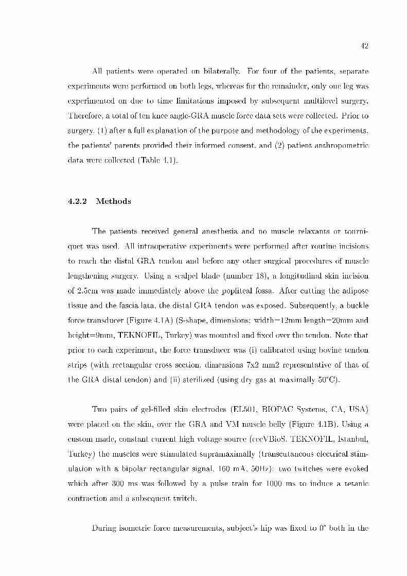
42
All patients were operated on bilaterally. For four of the patients, separate
experiments were performed on both legs, whereas for the remainder, only one leg was
experimented on due to time limitations imposed by subsequent multilevel surgery.
Therefore, a total of ten knee angle-GRA muscle force data sets were collected. Prior to
surgery, (1) after a full explanation of the purpose and methodology of the experiments,
the patients' parents provided their informed consent, and (2) patient anthropometric
data were collected (Table 4.1).
4.2.2 Methods
The patients received general anesthesia and no muscle relaxants or tourni-
quet was used. All intraoperative experiments were performed after routine incisions
to reach the distal GRA tendon and before any other surgical procedures of muscle
lengthening surgery. Using a scalpel blade (number 18), a longitudinal skin incision
of 2.5cm was made immediately above the popliteal fossa. After cutting the adipose
tissue and the fascia lata, the distal GRA tendon was exposed. Subsequently, a buckle
force transducer (Figure 4.1A) (S-shape, dimensions: width=12mm length=20mm and
height=9mm, TEKNOFIL, Turkey) was mounted and �xed over the tendon. Note that
prior to each experiment, the force transducer was (i) calibrated using bovine tendon
strips (with rectangular cross section, dimensions 7x2 mm2 representative of that of
the GRA distal tendon) and (ii) sterilized (using dry gas at maximally 50°C).
Two pairs of gel-�lled skin electrodes (EL501, BIOPAC Systems, CA, USA)
were placed on the skin, over the GRA and VM muscle belly (Figure 4.1B). Using a
custom made, constant current high voltage source (cccVBioS, TEKNOFIL, Istanbul,
Turkey) the muscles were stimulated supramaximally (transcutaneous electrical stim-
ulation with a bipolar rectangular signal, 160 mA, 50Hz): two twitches were evoked
which after 300 ms was followed by a pulse train for 1000 ms to induce a tetanic
contraction and a subsequent twitch.
During isometric force measurements, subject's hip was �xed to 0° both in the
43
Figure 4.1 Usage of buckle force transducer and stimulation electrodes. Illustrations of how (A) thebuckle force transducer is mounted over tendon strips, and (B) the skin electrodes are mounted overthe GRA and VM muscles are shown.
sagittal and frontal planes and the ankle was immobilized. GRA muscle isometric forces
were measured (i) at 120° and 90° of knee angle by stimulating GRA muscle exclusively,
afterwards (ii) from 120°, a highly �exed knee, to full extension with 30° increments (at
120°, 90°, 60°, 30° and 0°) by stimulating GRA and VM muscles simultaneously. Note
that, some patients were not capable of extending their knee fully (0°). Therefore,
for the limbs that full KA range was not feasible, the last force measurement was
performed at the most possible extended position of knee. A data acquisition system
(MP150WS, BIOPAC Systems, CA, USA, 16-bit A/D converter, sampling frequency
40 KHz) was used with an ampli�er for each transducer (DA100C, BIOPAC Systems,
CA, USA). After each contraction, the muscle was allowed to recover for 2 minutes in a
�exed knee posture. Data collection was completed within 30 min, the maximal study
duration allowed by the ethics committee.
A data acquisition system (MP150WS, BIOPAC Systems, CA, USA, 16-bit A/D
converter, sampling frequency 40 KHz) was used with an ampli�er for each transducer
(DA100C, BIOPAC Systems, CA, USA). After each contraction, the muscle was al-
lowed to recover for 2 minutes in a �exed knee posture. Data collection was completed
within 30 min, the maximal study duration allowed by the ethics committee.
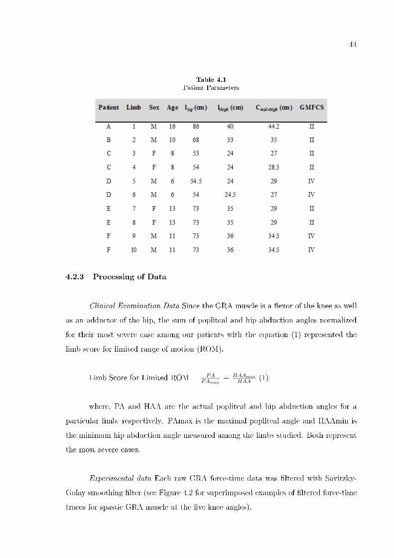
44
Table 4.1
Patient Parameters
4.2.3 Processing of Data
Clinical Examination Data Since the GRA muscle is a �exor of the knee as well
as an adductor of the hip, the sum of popliteal and hip abduction angles normalized
for their most severe case among our patients with the equation (1) represented the
limb score for limited range of motion (ROM).
Limb Score for Limited ROM = PAPAmax
+ HAAmin
HAA(1)
where, PA and HAA are the actual popliteal and hip abduction angles for a
particular limb, respectively. PAmax is the maximal popliteal angle and HAAmin is
the minimum hip abduction angle measured among the limbs studied. Both represent
the most severe cases.
Experimental data Each raw GRA force-time data was �ltered with Savitzky-
Golay smoothing �lter (see Figure 4.2 for superimposed examples of �ltered force-time
traces for spastic GRA muscle at the �ve knee angles).
45
The GRA muscle forces measured during a 100 ms period in the middle of the
tetanus were averaged to obtain the muscle total force. Using a least squares criterion,
data for total GRA muscle force (FGRA) in relation to knee joint angle (KA) were
�tted with a polynomial function
FGRA= a0 + a1KA+a2KA2 + ... + anKAn
a0, a1. . . an are coe�cients determined in the �tting process. The lowest order
of the polynomials that still added a signi�cant improvement to the description of
changes of KA and The lowest order of the polynomials that still added a signi�cant
improvement to the description of changes of KA and FGRA data were selected using a
one-way analysis of variance (ANOVA) [125]. The �tted KA-FGRA characteristics of the
patients were studied separately for each limb for characterization of the mechanics of
the spastic GRA muscle. Experimentally obtained measures were used for an objective
assessment of our hypotheses based on (i) joint range of muscle force exertion and (ii)
availability of high muscle force at low muscle length.
Joint range of muscle force exertion If the peak GRA muscle force (FGRApeak)
was attained within the knee joint range studied such that the increase trend of the force
ceased before full knee extension, the spastic GRA muscle was concluded to operate
within both the ascending and descending limbs of its KA-FGRA characteristics. If a
local minimum de�ned as the minimum force measured inside the range of KAs scanned
appeared, maximum force measured before that point was considered as Fpeak.
The proximity of the KA at which FGRA peak was exerted (KAFGRApeak) to
the maximal knee �exion angle studied indicates how narrow the joint range of force
exertion of each limb is. Therefore, the curves including local minimum or KAFGRApeak
closed to �exion were considered to represent a narrow joint range, and hence repre-
sentative of the compromised joint motion.
The availability of muscle force The availability of high muscle force at knee
�exion was considered to represent a typical characteristic of spastic muscle. In order to
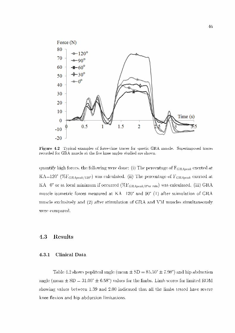
46
Figure 4.2 Typical examples of force-time traces for spastic GRA muscle. Superimposed tracesrecorded for GRA muscle at the �ve knee angles studied are shown.
quantify high forces, the following were done: (i) The percentage of FGRApeak exerted at
KA=120° (%FGRApeak/120º) was calculated. (ii) The percentage of FGRApeak exerted at
KA=0° or at local minimum if occurred (%FGRApeak/0ºor min) was calculated. (iii) GRA
muscle isometric forces measured at KA=120° and 90° (1) after stimulation of GRA
muscle exclusively and (2) after stimulation of GRA and VM muscles simultaneously
were compared.
4.3 Results
4.3.1 Clinical Data
Table 4.2 shows popliteal angle (mean ± SD = 85.50° ± 7.98°) and hip abduction
angle (mean ± SD = 31.00° ± 6.58°) values for the limbs. Limb scores for limited ROM
showing values between 1.39 and 2.00 indicated that all the limbs tested have severe
knee �exion and hip abduction limitations.
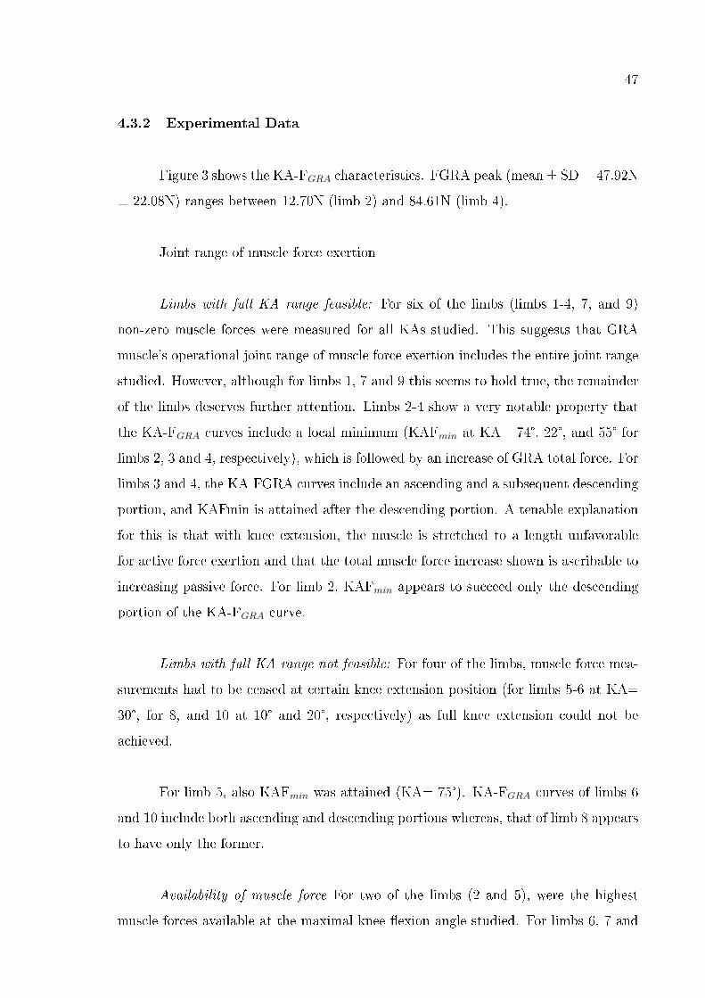
47
4.3.2 Experimental Data
Figure 3 shows the KA-FGRA characteristics. FGRA peak (mean ± SD = 47.92N
± 22.08N) ranges between 12.70N (limb 2) and 84.61N (limb 4).
Joint range of muscle force exertion
Limbs with full KA range feasible: For six of the limbs (limbs 1-4, 7, and 9)
non-zero muscle forces were measured for all KAs studied. This suggests that GRA
muscle's operational joint range of muscle force exertion includes the entire joint range
studied. However, although for limbs 1, 7 and 9 this seems to hold true, the remainder
of the limbs deserves further attention. Limbs 2-4 show a very notable property that
the KA-FGRA curves include a local minimum (KAFmin at KA= 74°, 22°, and 55° for
limbs 2, 3 and 4, respectively), which is followed by an increase of GRA total force. For
limbs 3 and 4, the KA-FGRA curves include an ascending and a subsequent descending
portion, and KAFmin is attained after the descending portion. A tenable explanation
for this is that with knee extension, the muscle is stretched to a length unfavorable
for active force exertion and that the total muscle force increase shown is ascribable to
increasing passive force. For limb 2, KAFmin appears to succeed only the descending
portion of the KA-FGRA curve.
Limbs with full KA range not feasible: For four of the limbs, muscle force mea-
surements had to be ceased at certain knee extension position (for limbs 5-6 at KA=
30°, for 8, and 10 at 10° and 20°, respectively) as full knee extension could not be
achieved.
For limb 5, also KAFmin was attained (KA= 75°). KA-FGRA curves of limbs 6
and 10 include both ascending and descending portions whereas, that of limb 8 appears
to have only the former.
Availability of muscle force For two of the limbs (2 and 5), were the highest
muscle forces available at the maximal knee �exion angle studied. For limbs 6, 7 and
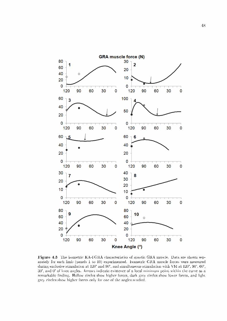
48
Figure 4.3 The isometric KA-FGRA characteristics of spastic GRA muscle. Data are shown sep-arately for each limb (panels 1 to 10) experimented. Isometric GRA muscle forces were measuredduring exclusive stimulation at 120° and 90°, and simultaneous stimulation with VM at 120°, 90°, 60°,30°, and 0° of knee angles. Arrows indicate existence of a local minimum point within the curve as aremarkable �nding. Hollow circles show higher forces, dark grey circles show lower forces, and lightgrey circles show higher forces only for one of the angles studied.
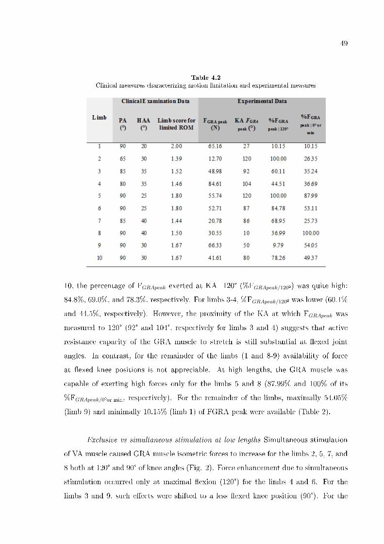
49
Table 4.2
Clinical measures characterizing motion limitation and experimental measures
10, the percentage of FGRApeak exerted at KA=120° (%FGRApeak/120º) was quite high:
84.8%, 69.0%, and 78.3%, respectively. For limbs 3-4, %FGRApeak/120º was lower (60.1%
and 44.5%, respectively). However, the proximity of the KA at which FGRApeak was
measured to 120° (92° and 104°, respectively for limbs 3 and 4) suggests that active
resistance capacity of the GRA muscle to stretch is still substantial at �exed joint
angles. In contrast, for the remainder of the limbs (1 and 8-9) availability of force
at �exed knee positions is not appreciable. At high lengths, the GRA muscle was
capable of exerting high forces only for the limbs 5 and 8 (87.99% and 100% of its
%FGRApeak/0ºor min, respectively). For the remainder of the limbs, maximally 54.05%
(limb 9) and minimally 10.15% (limb 1) of FGRA peak were available (Table 2).
Exclusive vs simultaneous stimulation at low lengths Simultaneous stimulation
of VA muscle caused GRA muscle isometric forces to increase for the limbs 2, 5, 7, and
8 both at 120° and 90° of knee angles (Fig. 2). Force enhancement due to simultaneous
stimulation occurred only at maximal �exion (120°) for the limbs 4 and 6. For the
limbs 3 and 9, such e�ects were shifted to a less �exed knee position (90°). For the
50
limbs 1 and 10 on the other hand, simultaneous stimulation did cause force reduction
at both positions.
In conclusion, (i) local minima for the limbs 2, 3, 4, and 5, (ii) the proximity
of the KA at which FGRA peak was measured to 120° for all of the limbs except 1,
8, and 10, (iii) low forces at extended knee positions, and (iv) force enhancement at
�exion for most of the limbs showed that simultaneous stimulation caused length range
of GRA muscle to be narrowed and active resistance of muscle to increase at �exed
knee positions. Therefore, our hypothesis is con�rmed for the majority of the limbs
tested.
4.4 Discussion
EMFT de�nes the transmission of muscle forces to the neighboring muscular
and non-muscular structures through the epimysium. This is because the muscles'
epimysium is continuous with myofascial structures [23, 140]. Such transmission of
force was shown with earlier animal experiments to occur between antagonist muscles
[107, 144]. Moreover, its functional role in vivo was also proved for human lower leg
muscles [142]. Such mechanism is important also for surgical treatments [83]. On the
other hand, there is no direct evidence for occurrence of inter-antagonistic EMFT for
spastic muscles. However, Kreulen et al. [60] did show that the epimuscular connections
for �exor carpi ulnaris muscle of cerebral palsy patients play a substantial mechanical
role. Since an increase in sti�ness of the spastic muscles [30, 57-58] and their connective
tissues [59] have been reported, we may expect that the role of EMFT mechanism for
the spastic limbs may even be more pronounced than in health. Our previous results
showing no abnormal mechanics for spastic GRA muscle which was stimulated alone
may support that idea implicitly because even though the muscle compartment with
almost all of its connective tissues was intact, no other muscle was activated. In this
study, we tested whether the spastic GRA muscle characteristics change to represent
better a characteristic that can be expected from a spastic muscle if it is stimulated
simultaneously with its antagonist VM. Our hypotheses on both narrowing length range
51
and higher force exertion at �exion are con�rmed for the majority of the limbs tested.
Before discussing the probable mechanisms causing such results, it should be
noted, in the present study, the pre-operative clinical examinations showing (i) CP
patients had GMFCS values indicating decreased mobility and causing a decision for
lengthening surgery on GRA muscle as well and (ii) popliteal angles greater than 50°
[123] and hip abduction angles lower than or equal to 40° [75, 130] indicated severe
knee joint problem. Since the popliteal angle measures under anesthesia were shown
not to di�er from clinic [145], we may accept that these limbs have permanent and
prominent contractures in daily life. Additionally, (iii) four of the limbs tested (5, 6, 8,
and 10) showed lacking full knee extension. Therefore, all the limbs experimented had
GRA muscle contracture and severe joint impairment passively.
4.4.1 Joint range of motion
Present results showed a narrowing for length range of spastic GRA muscle.
Healthy GRA has long �bers [134] and large excursion [98], thus its length range of
force production is expected to be wide comparably. Our previous results showed that
the operational joint range was at least as wide as 120° from the most �exed position of
the knee tested to the full extension for both healthy [120] and spastic GRAmuscle [143]
if they are stimulated alone. Presently, mean KAFGRA peak value was 78° and 7 of the
limbs tested produced their peak forces at more �exed position than 80°. Compared
to the previous results [143] showing peak forces produced between 66° and 0° of knee
angles (mean 29°) for exclusive stimulation of spastic GRA, the proximity of the KA at
which FGRA peak was measured to 120° and thus, a shift of KAFGRA peak to more
�exed positions occurred. Therefore, such present results indicated that if stimulated
simultaneously with VM, spastic GRA muscle operational range is narrowed.
Two of the limbs (2 and 5) operational only at the descending portion and three
of the remaining limbs (6, 7, 10) mostly at the descending portion of their KA-FGRA
characteristics indicated that at least for half of the limbs, simultaneous stimulation
52
of antagonist VM changed the characteristics of spastic GRA majorly and supported
our hypothesis on narrowing range as well. Considering that the maximum ankle
torque previously shown to be at more plantar �exion for CP patients than normal
children [146] our results indicate that narrowed range of GRA muscle represents the
abnormality at the joint.
Additionally, %48 shown for mean %FGRA peak | 0°or min indicated a major
force reduction for most of the limbs at knee extension. This ratio relating peak
forces to the forces produced at high muscle lengths was about half of the previous
ones shown for the exclusively stimulated GRA muscle [143]. More interestingly, the
increasing trend of the KA-FGRA curve shown after decreasing for four of the limbs
(2, 3, 4, and 5) resembles to the ideal force-length characteristics of a muscle having
all sarcomeres at high lengths and no extra capacity for active force production. Thus,
the passive force increase being responsible from such high forces is compatible with
the narrowing of length range with simultaneous stimulation.
No previous study reported the force production capacity of VM with respect
to its length or knee joint angle. However, for stable standing and walking, it is
probable that VM with other quadriceps produces quite amount of forces at extended
knee positions. Compromised force production with general weakness due to cerebral
palsy is also probable [147-148]. Nevertheless, considering that the spastic GRA having
increased sti�ness with its extramuscular structures due to CP may serve a probable
path for inter-antagonistic force transmission.
4.4.2 Availability of high muscle force
Our results showing (i) active resistance capacity of the GRA muscle at �exion
with the exception of limbs 1, 8-9 and (ii) force enhancement during simultaneous
activity of VM with the exception of limbs 1 and 10 indicated availability of high �exion
forces. It should be noted that antagonistic muscle activity occurs at many multi-joint
movements in daily life [149-150]. Such co-contraction is reported to be even higher in
53
spasticity [151-153]. Thus, the e�ects of antagonistic muscle activation not only for joint
function but also for spastic �exors are conceivable. On the contrary, joint weakness
[147-148, 154] as well as changes causing muscle weakness [50, 52, 54] were reported
for spastic CP. Therefore, lower forces produced and transmitted by either �exor or
its antagonist corresponding to such low joint torques may be expected. Nevertheless,
our results reported considerable e�ects due to inter-antagonistic interaction with high
�exion forces for most of the limbs.
In conclusion, simultaneous stimulation of antagonistic VM muscle causes (i)
spastic GRA muscle peak forces to shift to the more �exed positions, (ii) operational
range of GRA muscle to be narrowed and (iii) operational portion of knee angle-muscle
force curve to shift to the �exion. Thus, our hypothesis on narrowed joint range of
motion is veri�ed. High �exion forces measured due to simultaneous activity con�rmed
our second hypothesis as well. It is suggested that inter-antagonistic interaction im-
poses myofascial loads on muscles through epimuscular connections sti�ened due to
spasticity. Such loads cause intramuscular alterations on spastic GRA muscle: Sar-
comeres are lengthened at �exion and thus, excessive lengthening occurs at knee ex-
tension. Therefore, �exion forces increase and length range decreases. Such probable
mechanism should be tested with spastic muscle model and also by adding histological
examinations to our experimental method improved to measure passive forces.
54
5. MUSCLE LENGTHENING CAUSES DIFFERENTIAL
ACUTE MECHANICAL EFFECTS IN BOTH TARGETED
AND NON-TARGETED SYNERGISTIC MUSCLES
5.1 Introduction
In remedial surgery, known under various names (muscle recession [73-74], mus-
cle release [155], muscle lengthening [76-80], aponeurotomy (AT) [81] involves cutting
of an intramuscular aponeurosis transversely. Preparatory dissection (PD) is performed
�rst to reach the target muscle [156], AT is used for correction of problems of range
of movement and joint position in spastic paresis. The most important acute e�ect
allowing lengthening of muscle is creation of a gap within the muscle. Enhancing the
compromised joint range of motion is a primary goal [157], but reduction of muscle
force to correct imbalances of force between antagonistic muscles [158] are additional
clinical aims.
The myotendinous junction is widely considered as the main [159] or implicitly
even the sole site [many studies in biomechanics e.g. 160] for transmission of forces
generated within sarcomeres onto the tendon and from there to bone. However, force
transmission is possible also via trans-sarcolemmal proteins connecting muscle �bers
along their full periphery of their length to the collagen reinforced extracellular matrix
(ECM) [for a review see 161]. As a consequence, muscle �bers have been shown to in-
teract mechanically with the ECM and with each other with a bundle [162-163]. Later,
for whole muscle, such transmission has been named, myofascial force transmission
(MFT) [111].
Moreover, muscle functioning within its normal context of connective tissues is
connected to surrounding muscles and non-muscular structures and epimuscular my-
ofascial force transmission (EMFT) occurs via such connections [e.g. 63, 140]. EMFT
has been shown to cause asymmetric e�ects at muscle's origin and insertion and depen-
55
dency of muscle characteristics on mechanical conditions within which it functions [140,
164]. Evidence for EMFT is accumulating for human muscles in vivo [142, 165-167].
It is quite conceivable that PD (e.g. opening of the compartment) may a�ect
MFT within a compartment and because of mechanical interaction between muscles,
also acute mechanical e�ects of AT could be present in muscles other than those on
which the main surgical intervention was performed. Post operative e�ects of AT were
investigated at the joint level by using e.g., gait analysis [157, 168]. However, e�ects
on the target and non-targeted muscles were not studied explicitly.
Therefore, we designed the present study to test for such e�ects of MFT and
test the following hypotheses for muscles within the anterior crural compartment of
the rat: (1) e�ects of muscle lengthening surgery on the target muscle are di�erent at
distal and proximal tendons, (2) forces of non-targeted synergistic muscles are a�ected
as well, and (3) PD is responsible from some of these e�ects.
5.2 Methods
5.2.1 Surgical procedures and preparation for experiments
Surgical and experimental procedures were approved by the Committee on
Ethics of Animal Experimentation at Bo§aziçi University. Young adult male Wis-
tar rats (n = 8, mean body mass 327.0 g (SD 18.4 g)) anaesthetized using intraperi-
toneal injection of urethane (1.2 ml/100 g body mass). Additional doses were given,
if necessary (maximally 0.5 ml). The animals were placed on a heated pad (Harvard
Apparatus) during surgery and data collection. Left hind limb skin and biceps femoris
muscle were removed and the anterior crural compartment, containing extensor digito-
rum longus (EDL), tibialis anterior (TA) and extensor hallucis longus (EHL) muscles
was exposed. Only limited distal fasciotomy was performed to remove the retinaculae,
leaving fully intact connective tissues at the muscle. At a reference position selected
(knee and ankle joint angles of 90° and 100° respectively), the four distal tendons of
56
EDL muscle were tied together using silk thread. Matching markers were placed on
distal tendons of EDL, TA and EHL muscles, as well as on the lower leg. Subsequently,
the distal tendons of EDL, as well as TA and EHL were cut as distally as possible.
The proximal EDL tendon was cut from the femur, with a small piece of the lateral
femur condyle still attached. To allow connection to force transducers, Kevlar threads
were sutured to all cut tendons. Within the femoral compartment, the sciatic nerve
was dissected and cut as proximal as possible. All its branches to muscles of that
compartment were cut.
5.2.2 Experimental conditions and procedure
The rat was mounted in the experimental set-up (Fig. 1). The femur and foot
were clamped, such that the knee was kept at 90° and the ankle in maximal plantar
�exion (180°) to allow for free passage of Kevlar threads. Each Kevlar thread was
connected to a force transducer (BLH Electronics Inc., Canton MA). The distal end
of the sciatic nerve was placed on a bipolar silver electrode. Room temperature was
kept at 26° C. Muscle and tendon tissues were irrigated regularly by isotonic saline to
prevent dehydration during the experiment.
Markers on EDL proximal tendon and distal tendons of TA and EHL were kept
in their reference positions at all times. EDL length was changed by moving its distal
force transducer (in steps of 1 mm), until 2 mm over distal optimum length, and EDL
length-force data were collected at proximal and distal EDL tendons. Distal forces of
TA and EHL were measured.
Muscles were activated maximally by supramaximal stimulation of the sciatic
nerve at a constant current of 2 mA (Biopac Systems stimulator, STMISOC: square
pulse width 0.1 ms, pulse train 400 ms, stimulation frequency 100 Hz). After setting
EDL to a new length, two twitches were evoked and the muscles were tetanized 300 ms
after the second twitch. At 200 ms after cessation of stimulation, another twitch was
evoked. After these contractions, muscles were allowed to recover for 2 min. (EDL at
57
low length, others at lref ).
5.2.3 Experimental protocol
Before acquiring data, muscle-tendon complexes and their epimuscular connec-
tions were preconditioned by isometric contractions, alternatingly at high and low EDL
lengths, until forces at low EDL length were reproducible (i.e. e�ects of previous ac-
tivity at high length [169] are removed).
Three conditions were studied: (1) Control with an intact anterior crural com-
partment. (2) After preparatory dissection (Post PD), performed to gain access to
the target muscle. Laterally, a probe was inserted from proximally into the anterior
crural compartment between EDL and TA until the middle of the compartment was
reached by the probe tip (i.e., approximately for 12 mm). Using a micro scissor, half of
the anterior crural compartmental fascia was cut (partial fasciotomy). Intermuscular
connections between EDL and TA along half of EDL length were removed by blunt
dissection (using a cotton swab). (3) Post-AT EDL proximal aponeurosis was tran-
sected at its middle, perpendicular to its longitudinal direction using a scalpel blade
(Surgeon, number 11).
Subsequently, muscles were activated with EDL at optimum length (of control
condition). This causes tearing of the ECM along muscle �bers located below the site
of AT. E�ects of PD, AT and subsequent tearing of the ECM together are referred to
as cumulative e�ects of muscle lengthening surgery.
5.2.4 Processing of experimental data and statistics
Muscle passive isometric forces were measured at 100 ms after the second twitch
before the tetanus. The mean total force during the tetanic plateau was calculated for
an interval of 200 ms starting after 150 ms of tetanic stimulation.
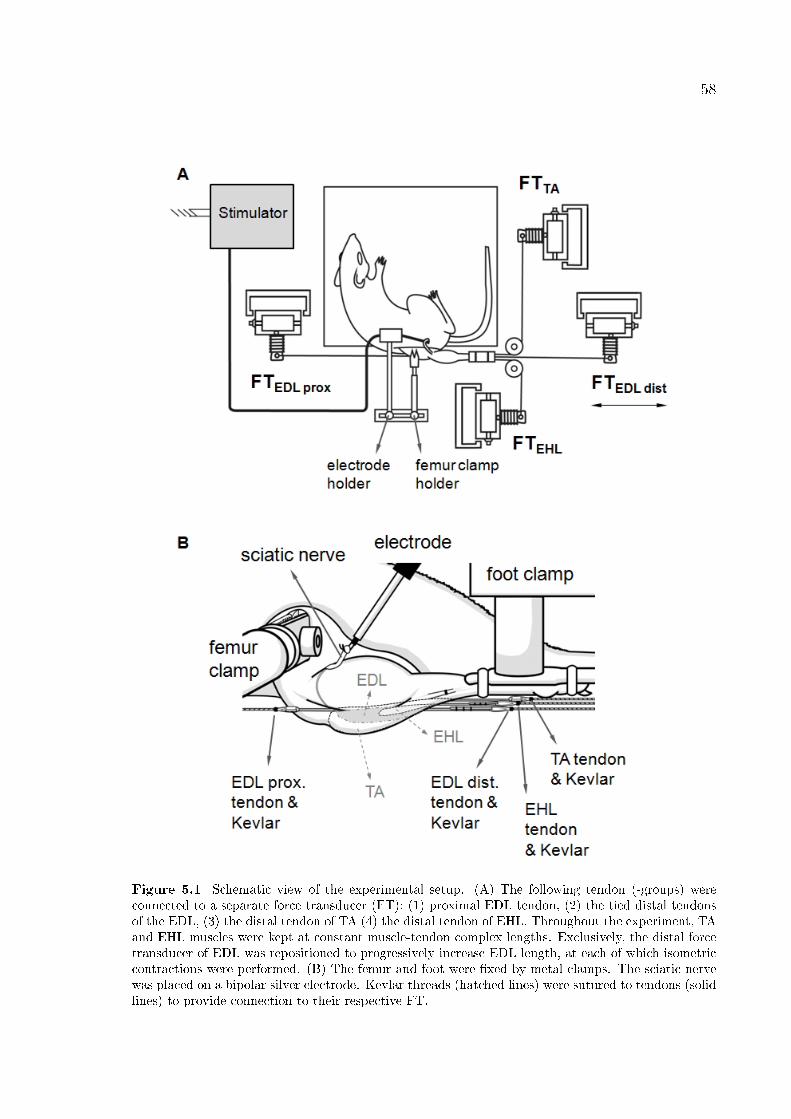
58
Figure 5.1 Schematic view of the experimental setup. (A) The following tendon (-groups) wereconnected to a separate force transducer (FT): (1) proximal EDL tendon, (2) the tied distal tendonsof the EDL, (3) the distal tendon of TA (4) the distal tendon of EHL. Throughout the experiment, TAand EHL muscles were kept at constant muscle-tendon complex lengths. Exclusively, the distal forcetransducer of EDL was repositioned to progressively increase EDL length, at each of which isometriccontractions were performed. (B) The femur and foot were �xed by metal clamps. The sciatic nervewas placed on a bipolar silver electrode. Kevlar threads (hatched lines) were sutured to tendons (solidlines) to provide connection to their respective FT.
59
Mathematical functions were least square �tted to the experimental data for
further treatment and averaging [e.g. 169]. Passive length-force data were �tted using
an exponential function and active muscle force was calculated by subtracting the
measured passive force from total force during muscle activity. Active length-force
data were �tted (polynomial function). The degree of the polynomial function that
most adequately described a particular set of length-force data was selected using an
analysis of variance (ANOVA) [125]. Optimal EDL force is de�ned as the maximum
of the polynomial, and its corresponding length as optimum length. One-way ANOVA
was also used to test for cumulative e�ects of muscle lengthening surgery on EDL's
length range of active force exertion i.e., the range between muscle active slack length
and optimum length. Two-way ANOVA's for repeated measures (factors: EDL length
and surgical condition) were used to analyze e�ects of the interventions on EDL length-
force characteristics and on forces of non-targeted TA and EHL. If signi�cant e�ects
were found, post hoc tests were performed using the Bonferroni procedure for multiple
pair wise comparisons to locate di�erences. p values < 0.05 were considered signi�cant.
5.3 Results
Target muscle-distal force ANOVA (factors: EDL length and condition) showed
signi�cant main e�ects on EDL distal active forces, and signi�cant interaction. Com-
pared to control condition (Fig. 2A), post hoc tests showed signi�cant cumulative
e�ects of AT causing EDL distal active forces to decrease (e.g., by 26.3% at optimum
length), in contrast to increases at the lowest muscle lengths. Note that no signi�-
cant e�ects of PD are found for distal EDL active force. Cumulative e�ects of muscle
lengthening surgery cause EDL distal length range of active force exertion to increase
signi�cantly (from mean 9.1 mm (SE 1.1 mm) to mean 10.7 (SE 0.9 mm)). For EDL
distal passive force, ANOVA (factors: EDL length and condition) showed signi�cant
main e�ects, and signi�cant interaction. Compared to the control condition, post hoc
tests showed (i) signi�cant cumulative e�ects of muscle lengthening surgery causing
EDL distal passive forces to increase (e.g., by 40.7% at optimum length). This increase
is explained by higher stretch of EDL epimuscular connections in proximal direction.
60
(ii) No signi�cant e�ects of PD were found for EDL distal passive force.
Target muscle-proximal force ANOVA (factors: EDL length and condition)
showed signi�cant main e�ects on EDL proximal active force and signi�cant inter-
action. Compared to control condition (Fig. 2B), post hoc tests showed signi�cant
cumulative e�ects of muscle lengthening surgery causing EDL proximal active forces to
decrease (e.g., by 44.5% at optimum length, note that this is more than the decrease
of its distal counterpart Fig. 2A). However, no signi�cant e�ects of PD were found
for EDL proximal active force (post hoc test). In contrast to e�ects found distally,
cumulative e�ects of muscle lengthening surgery caused no signi�cant change in length
range of active force exertion at the EDL proximal tendon. ANOVA (factors: EDL
length and condition) showed signi�cant e�ects of length on EDL proximal passive
forces; but neither signi�cant e�ects of condition nor of interaction.
Non-operated TA ANOVA (factors: EDL length and condition) showed signif-
icant main e�ects on TA active forces, but no signi�cant interaction. Compared to
control condition (Fig. 3A), post hoc tests showed signi�cant cumulative e�ects of
muscle lengthening surgery (mean decrease for EDL lengths studied equaled 11.9%).
Note that post hoc tests also indicate that PD causes TA active forces to decrease
(mean decrease for EDL lengths studied equaled 4.9%). ANOVA (factors: EDL length
and condition) showed signi�cant e�ects of condition on TA passive forces; but no sig-
ni�cant e�ects of length or a signi�cant interaction. Compared to control condition,
post hoc tests showed signi�cant cumulative e�ects of muscle lengthening surgery caus-
ing TA passive forces to decrease (the mean force decrease for the EDL lengths studied
equaled 11.6%). However, no signi�cant e�ects of PD were shown (post hoc test).
Non-operated EHL ANOVA (factors: EDL length and condition) showed sig-
ni�cant main e�ects on EHL active forces, but no signi�cant interaction. Compared
to control condition (Fig. 3B), post hoc tests showed signi�cant cumulative e�ects of
muscle lengthening surgery causing EHL distal active forces to decrease, despite its
constant muscle tendon complex length (for EDL lengths studied, mean force decrease
equals 8.4%). However, no signi�cant e�ects of PD on EHL active force were found
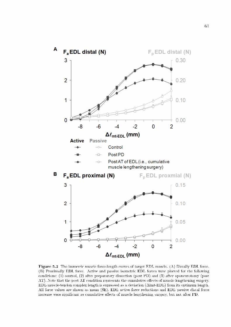
61
Figure 5.2 The isometric muscle force-length curves of target EDL muscle. (A) Distally EDL force.(B) Proximally EDL force. Active and passive isometric EDL forces were plotted for the followingconditions: (1) control, (2) after preparatory dissection (post PD) and (3) after aponeurotomy (postAT). Note that the post AT condition represents the cumulative e�ects of muscle lengthening surgery.EDL muscle-tendon complex length is expressed as a deviation (∆lmt-EDL) from its optimum length.All force values are shown as mean (SE). EDL active force reductions and EDL passive distal forceincrease were signi�cant as cumulative e�ects of muscle lengthening surgery, but not after PD.
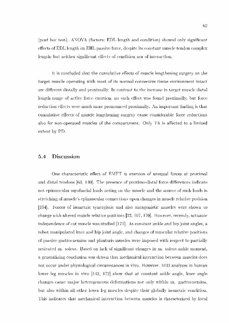
62
(post hoc test). ANOVA (factors: EDL length and condition) showed only signi�cant
e�ects of EDL length on EHL passive force, despite its constant muscle tendon complex
length; but neither signi�cant e�ects of condition nor of interaction.
It is concluded that the cumulative e�ects of muscle lengthening surgery on the
target muscle operating with most of its normal connective tissue environment intact
are di�erent distally and proximally. In contrast to the increase in target muscle distal
length range of active force exertion, no such e�ect was found proximally, but force
reduction e�ects were much more pronounced proximally. An important �nding is that
cumulative e�ects of muscle lengthening surgery cause considerable force reductions
also for non-operated muscles of the compartment. Only TA is a�ected to a limited
extent by PD.
5.4 Discussion
One characteristic e�ect of EMFT is exertion of unequal forces at proximal
and distal tendons [63, 140]. The presence of proximo-distal force di�erences indicate
net epimuscular myofascial loads acting on the muscle and the source of such loads is
stretching of muscle's epimuscular connections upon changes in muscle relative position
[164]. Forces of isometric synergistic and also antagonistic muscles were shown to
change with altered muscle relative positions [23, 107, 170]. However, recently, actuator
independence of cat muscle was studied [171]. At constant ankle and hip joint angles, a
robot manipulated knee and hip joint angle, and changes of muscular relative positions
of passive gastrocnemius and plantaris muscles were imposed with respect to partially
activated m. soleus. Based on lack of signi�cant changes in m. soleus ankle moment,
a generalizing conclusion was drawn that mechanical interaction between muscles does
not occur under physiological circumstances in vivo. However, MRI analyses in human
lower leg muscles in vivo [142, 172] show that at constant ankle angle, knee angle
changes cause major heterogeneous deformations not only within m. gastrocnemius,
but also within all other lower leg muscles despite their globally isometric condition.
This indicates that mechanical interaction between muscles is characterized by local
63
Figure 5.3 Forces exerted by non-operated TA and EHL muscles. (A) Distal TA force. (B) DistalEHL force. Active and passive isometric TA and EHL forces were plotted for the following conditions:(1) control, (2) after preparatory dissection (post PD) and (3) after aponeurotomy (post AT). Notethat the post AT condition represents the cumulative e�ects of muscle lengthening surgery. EDLmuscle-tendon complex length is expressed as a deviation (∆lmt-EDL) from its optimum length. Allforce values are shown as Non-operated TA ANOVA (factors: EDL length and condition) showedsigni�cant main e�ects on TA active forces, but no signi�cant interaction. Compared to controlcondition (Fig. 3A), post hoc tests showed signi�cant cumulative e�ects of muscle lengthening surgery(mean decrease for EDL lengths studied equaled 11.9%). Note that post hoc tests also indicate that PDcauses TA active forces to decrease (mean decrease for EDL lengths studied equaled 4.9%). ANOVA(factors: EDL length and condition) showed signi�cant e�ects of condition on TA passive forces; butno signi�cant e�ects of length or a signi�cant interaction. Compared to control condition, post hoctests showed signi�cant cumulative e�ects of muscle lengthening surgery causing TA passive forces todecrease (the mean force decrease for the EDL lengths studied equaled 11.6%). However, no signi�cante�ects of PD were shown (post hoc test).
64
epimuscular myofascial loads on those muscles with variable magnitude and direction.
If the net e�ect of EMFT on forces exerted at the tendon would be small, a mechanical
balance of counteracting epimuscular myofascial loads rather than their absence can
explain this. Therefore, in contrast to the conclusion of Maas and Sandercock [171], a
mechanical interaction of muscles is tenable in most conditions. We expect that this
plays a role in surgery as well.
In the control condition, the anterior crural compartment was not interfered
with, hence epimuscular connections were intact. PD performed in our experiment
to gain access to the AT site, as in clinical practice [156], was minimized to limit
interference with the compartment. We showed that even for a deeper target mus-
cle and its synergistic muscle, it does not contribute to cumulative e�ects of muscle
lengthening surgery. Present dissection was performed exclusively within the proximal
compartment half, leaving intact distal parts of fasciae. Meijer et al. [144] concluded
that these tissues are particularly sti� if TA and EHL are shortened which imposes a
stretch on them causing increased distally directed net epimuscular myofascial loads
acting on EDL. High sti�ness of connections is likely in our conditions as TA and EHL
are kept at lower lengths. Therefore, e�ects of PD on epimuscular myofascial loads
acting on deeper located muscles are likely to remain small. However, for TA, PD
alone, independent from EDL length, reduced its contribution to the moment exerted
at the distal joint. Therefore, e�ects of PD are con�ned to super�cial muscles, conceiv-
ably due to a reduced sti�ness of its epimuscular connections (e.g., the TA branch of
the neurovascular tract) creating conditions unfavorable for force exertion. In general,
more compartmental disruption is expected to cause reduction in muscle force [173],
additional to that caused by AT. If PD remains limited, remaining integrity of EMFT
pathways is likely to cause e�ects similar to those shown presently.
Previous studies on isolated muscle [82-83] allowed improvement of our under-
standing of mechanisms of AT a�ecting intramuscular mechanics. Although a discon-
tinuity within the aponeurosis directly prevents myotendinous force transmission for a
part of the muscle �bers, this intervention per se was shown to have only minor e�ects,
but subsequent rupturing of intramuscular connective tissues below the AT location
65
yields major e�ects on muscle length-force characteristics [82]. Therefore, a central
issue is interference with MFT, which e�ectively causes manipulation of myofascial
loads (i.e., forces exerted onto muscle �bers by the ECM and via that by sarcom-
eres located in the neighboring muscle �bers). This causes characteristic sarcomere
length distributions within the aponeurotomized muscle yielding changes in muscle
length-force characteristics. Enhanced sarcomere length heterogeneity within the tar-
get muscle causes an acute increase in muscle length range of force exertion. Therefore,
in addition to causing acutely a lengthening of muscle, AT facilitates exertion of active
force for an enhanced range of joint angles also. In addition to contributing to that
e�ect, major shortening of sarcomeres occurs within muscle �bers without myotendi-
nous connections to bone. This is responsible for most of the force reductions due to
AT.
An important present �nding is that e�ects on forces exerted at proximal and
distal tendons of the target muscle are not equal. This in accordance with the presence
of proximo-distal force di�erences indicating net epimuscular myofascial loads acting
on the muscle. Such loads were shown to change in vivo muscular mechanics substan-
tially [142] and a�ect local sarcomere lengths, forces exerted, as well as length range
of active force exertion [25]. Epimuscular myofascial loads plausibly cause sarcomeres
in the proximal ends of �bers of the target muscle located proximally to the AT site to
attain lengths less favorable for force exertion which explains the e�ect of higher force
reductions proximally. Lack of an increased proximal length range of active force exer-
tion indicates that such loads also diminish sarcomere length heterogeneity within the
target muscle. This may not only explain the asymmetry in e�ects of muscle length-
ening surgery for joints spanned by the target muscle, but also suggest that integrity
of the EMFT network limits acute e�ects of AT, also in clinical practice. Previous
modeling study [84] con�rmed that epimuscular myofascial loads present in conditions
representing removal of muscles, other than the target muscle from the compartment,
will cause diminished sarcomere shortening within the population of muscle �bers dis-
tal to the location of AT, and less pronounced sarcomere length heterogeneity within
the muscle. We conclude that acute cumulative e�ects of muscle lengthening surgery
on mechanics of the target muscle are a�ected to a large extent by EMFT.
66
For clinicians it is important to realize that surgically correcting functional prob-
lems at a distal joint may alter muscle mechanics at the proximal joint as well. This
suggests that care is indicated when considering the overall surgical outcome, partic-
ularly if the target muscle is bi- or poly-articular. For example, in combined equinus
and crouch gait [157], AT of m. gastrocnemius may improve gait because contribu-
tions to both plantar �exion and knee �exion moments may decrease. However, for
combined crouch and hip �exor tightness, AT of the semimembranosus [168] may dete-
riorate conditions at the proximal joint due to reduced contributions to hip extension
moment. The asymmetry shown for decreased contribution of the target muscle to mo-
ments exerted at the joints it spans is due to EMFT, since for truly isolated muscle this
would not occur. Note that, AT on the distal aponeurosis would prevent myotendinous
force transmission for a di�erent population of muscle �bers and may change acute
e�ects of the intervention. However, these muscle �bers are exposed also to EMFT.
This will also a�ect asymmetry e�ects shown presently only for a proximal AT. Also
note that imposed distal muscle length changes of the present study mimic movements
exclusively of the distal joint. Therefore, our results are not direct indicators of how
proximal length range of force exertion is a�ected. However, identical lengthening
imposed distally or proximally of intact EDL yielded asymmetric proximal and distal
length ranges of active force exertion [174]. This implies that, lengthening surgery of
a target muscle with most of its connective tissues intact should not be expected to
yield symmetrical lengthening e�ects for joints spanned.
A key �nding is that cumulative e�ects of muscle lengthening surgery involve
considerable reductions also of distal force exerted by non-operated synergistic mus-
cles. These results indicate that the surgeon should be aware of possible other and
unintended acute e�ects, in addition to those for the target muscle.
It should be noted that synergistic muscles will change lengths simultaneously
during joint movement and in similar direction as the agonist muscle. This may limit
changes of relative position of synergistic muscles. However, even after lengthening of
the whole anterior crural group, increased e�ects of EMFT were reported compared
to single EDL lengthening [144]. Note that such equal and simultaneously imposed
67
muscle length changes may di�er from in vivo conditions, as di�erences in moment
arms of synergistic muscles are present [175] contributing to relative movement of
muscles. Moreover, di�erences in number of joints spanned by synergistic muscles will
contribute also to relative movement of muscles and movement of muscle with respect
to bones. Joint angle changes in vivo have been shown to indicate e�ects of EMFT
such as interactions between �nger forces [176-177], as well as local displacements [166]
or deformations [142] within muscles remaining globally isometric. Therefore, acute
e�ects of muscle lengthening surgery on non-operated synergistic muscles, similar to
those shown presently, are plausible also for in vivo function.
Although motor control plays no role in our experiments, one should be aware
that surgical interventions may have important e�ects on the a�erent sensory machin-
ery. Firstly, PD alone may cause some of fascial a�erents [e.g. 178] to be disrupted
or may also a�ect sensory machinery via modi�ed sti�ness of compartmental connec-
tive tissues. Therefore, PD, even though we �nd it to have relatively small e�ects on
muscle length-force characteristics, in vivo may still cause neuromotor changes. In ad-
dition, within the aponeurotomized muscle, populations of muscle �bers, which loose
their myotendinous connections, shorten considerably subsequent to rupturing of in-
tramuscular connective tissues [82, 179]. Mechanical unloading of the intrafusal �bers
and reduced a�erent response was reported in conditions causing shortened extrafusal
�bers [180-181]. Therefore, for this shortened part of the aponeurotomized muscle, the
tonic stretch re�ex threshold may shift to higher values. Skeletal muscle spasticity is
characterized by exaggerated stretch re�exes [e.g. 34]. Therefore, this is regarded as a
positive e�ect of treatment. However, muscle �bers between the location of interven-
tion and the tendon attain in general higher lengths, which may cause opposite e�ects.
Modeling studies showed that an aponeurotomy closest to the tendon dominates the
e�ect and enhances intended mechanical e�ects [83]. This e�ect is so important that
additional aponeurotomies performed on the same model muscle do not further im-
prove the outcome considerably [182]. These arguments suggest that modi�cation of
the proprioceptive input may also be optimized by a choice of intervention location.
On the other hand, e�ects at a higher level of organization than the target muscle
are quite conceivable. Force reductions shown presently for synergistic muscles suggest
68
that their muscle �bers attain lower lengths as an e�ect of the surgical interventions.
Therefore, PD and AT may cause threshold of the tonic stretch re�ex of also these
muscles to shift to higher values. Similar e�ects of mechanical unloading of spindles of
co-contracting intact muscles were reported previously [183]. However, after tenotomy
of gastrocnemius and plantaris muscles, opposite �ndings were reported for a�erents
of soleus muscle [184]. This implies that, similar as for AT, tenotomy is not interfering
solely with myotendinous force transmission and that manipulation of mechanical in-
teractions between muscles may change a�erent sensory machinery in a complex way,
even if no damage were done to the sensory system.
Note that threshold of the tonic stretch re�ex (represented by lambda) is re-
garded as a key control variable in motor control studies based on Equilibrium-Point
Hypothesis [185-186]. According to this viewpoint, for a single muscle, surgical mod-
i�cation of muscle length may shift the equilibrium point of the system leading to a
change in lambda. This theory was elaborated and extended to be linked to the idea
that motor synergies and bodily movement is controlled at much higher levels of or-
ganization than the level of individual muscles [187-188]. Our results suggest that the
whole synergistic interaction is a�ected by modifying mechanics of one muscle compo-
nent within the synergy. The issues argued in the preceding paragraph may be relevant
for agreement of the present �ndings with motor control theories. In our view, intra-
and epimuscular MFT provides coupling between neuromuscular mechanisms suggest-
ing that it is an important determinant for the changes to occur in lambda. Muscle
sti�ness is a component of this and is important also for intended remedial e�ects of
muscle lengthening surgery. As AT causes a part of the muscle to shorten and PD
disrupts integrity of the connective tissues, an expected e�ect for most of the com-
partment is reduced sti�ness of muscular and non-muscular tissues. Compared to its
untreated properties, after healing, the aponeurosis was shown to be more compliant
and longer [189]. Therefore, e�ects of muscle lengthening surgery on mechanical, as
well as a�erent sensory machinery of the target muscle are likely also for the long-term.
Long-term adaptations in neuromotor properties due to potential changes in epimus-
cular connections need to be considered as well. Only partial or no neural adjustments
were found to occur after tendon transfer surgery [190-191]. Rectus femoris muscle,
69
transferred to a �exor insertion was reported not to move as a knee �exor [192]. This
in agreement with its epimuscular mechanical interaction with the knee extensors and
may explain absence of such adjustments. However, after AT, substantial neuromotor
adaptations were reported [193-194]. Those studies focused either on the target muscle
or a selected antagonist muscle, but not on synergistic muscles. As multilevel surgical
treatment is common [78, 195], systematic testing of neural control of relevant muscles
including non-targeted synergistic muscles is indicated.
In summary, e�ects of muscle lengthening surgery performed to improve im-
peded joint mechanical function are dominated by e�ects of EMFT causing (i) di�er-
ential e�ects at the proximal and distal tendons of a poly-articular target muscle, and
(ii) sizable e�ects also at unintended sites via non-operated muscles. These di�eren-
tial and unintended e�ects on muscle forces may yield additional favorable e�ects for
the target joint, but also contrasting e�ects particularly for the non-targeted joint. It
is therefore important to consider the role of EMFT in order to enhance control of
the surgical outcome of the operation. New speci�c studies are indicated to assess
neuromotor changes acutely as well as in the long-term.
70
6. BTX-A ADMINISTRATION TO THE TARGET MUSCLE
AFFECTS FORCES OF ALL MUSCLES WITHIN AN
INTACT COMPARTMENT
6.1 Introduction
Botulinum toxin type A (BTX-A) is a chemical denervant that acts at motor
nerve endings to block acetylcholine release [65], which causes paralysis of muscle �bers
[64], hence, muscle weakness (i.e., decreased ability for force production). Due to its
e�ectiveness in avoiding the development of contractures [196], BTX-A is used widely
in patients with cerebral palsy as an alternative treatment to surgery [197-199].
The e�ects of BTX-A have been widely studied by quantifying the area of paral-
ysis [66], compound muscle action potential [67] and electromyography [68]. However,
reports on mechanical parameters, e.g., twitch and tetanic force have been limited to
selected muscle lengths or joint positions [e.g. 67, 69]. Herzog et al. made a major
contribution to �lling this gap in the literature by measuring joint torques in a range of
joint angles [200-202]. Experiments on the rabbit quadriceps musculature showed that
BTX-A causes more pronounced reductions in knee extension torque at more �exed
knee positions [202]. This suggests that the e�ects of BTX-A on muscle forces and
increasing muscle length may be negatively correlated.
Recently, Yaraskavitch et al. showed that the force-length characteristics of
both injected soleus and non-injected plantaris muscles of the cat are a�ected by the
poison [71]. BTX-A has been shown to spread through muscle fascia [70], and its
e�ects beyond the injection site are plausible [72]. As these e�ects may not be con�ned
only to a neighboring muscle, an experimental model that involves measurement of
forces of mono- and biarticular muscles of an entire muscle compartment can allow for
a comprehensive assessment of the e�ects of BTX-A both at and beyond the injection
site.
71
Collagenous connections between adjacent muscles and extramuscular connec-
tive tissues such as collagen-reinforced neurovascular tracts and compartmental bound-
aries provide connections between the muscular and non-muscular structures of an
intact compartment [20, 63, 86]. These epimuscular connections feature complex me-
chanical properties. Similar to other connective tissue structures, these connections
have nonlinear force-deformation characteristics [203-205]. In addition, they have been
shown to be pre-strained [23] and to have inhomogeneous mechanical properties (e.g.,
the proximal parts of the neurovascular tracts of the anterior crural compartment of
the rat are sti�er than the distal parts [22]. Previous studies have shown the occur-
rence of epimuscular myofascial force transmission (EMFT) via these structures [23,
140-141]. Such EMFT is characterized by the interplay of sti�ness of muscular tissues
and epimuscular connections and it is determined by changes in the position of mus-
cle relative to its neighboring structures [e.g. 27, 109]. Recently it has been shown
that due to such force transmission, the global length changes of human gastrocnemius
muscle and local strains within the muscle can be very di�erent and despite its global
isometric condition, local and heterogeneous deformations were found also within the
soleus muscle [142].
BTX-A exposure changes the sti�ness of muscular tissues because it causes
paralysis of muscle �bers within parts of the muscle belly [66]. Therefore, compared to
the no BTX-A injected condition, identical changes in relative position globally of the
muscle-tendon complex is expected to lead locally to di�erent interactions between the
muscular tissues and their epimuscular connections. Consequently, the epimuscular
connections can operate at di�erent segments of their complex mechanical proper-
ties and cause the EMFT mechanism to change. Note that previously, the e�ects of
BTX-A on the non-injected adjacent muscle were explored after the intactness of the
compartment and the connections of the muscles with surrounding structures had been
disrupted and the two muscles' forces were not measured simultaneously [71]. There-
fore, the e�ects of BTX-A on the forces of muscles operating in an intact compartment
as well as on EMFT mechanism remain unknown. We hypothesized that BTX-A af-
fects (1) the forces of not only the injected but also the noninjected muscles of an
entire intact compartment, and (2) EMFT. The goal of this study was to test these hy-
72
potheses. Additionally, it was to assess the existence of a correlation between injected
muscle's length and the e�ects of BTX-A. These goals were addressed by measuring
the force-length properties of the injected muscle as well as the isometric forces of the
restrained non-injected muscles of the intact anterior crural compartment of the rat
simultaneously, and in conditions close to those in vivo.
6.2 Methods
Surgical and experimental procedures were approved by the Committee on the
Ethics of Animal Experimentation at Bogaziçi University. Male Wistar rats were di-
vided into two groups: (1) Control (n = 8, mean ± SD body mass = 318.5 ± 12.5 g).
(2) BTX (n = 8, mean ± SD body mass = 312.5 ± 14.6 g).
After imposing a mild sedation with an intraperitonal dose of 1mg/kg ketamine,
a circular region of approximately 15 mm radius from the center of the knee cap was
shaved. The tibialis anterior (TA) muscle was located by palpation when the ankle was
in maximal plantar �exion and the knee angle approximated 90°. After marking the
center of the knee cap, a second marker was placed at a point 10 mm distal to that,
along the tibia. The injection location was 5 mm lateral (along the direction normal to
the line segment drawn between the two markers) to the second marker and over the
TA muscle. All injections were made exclusively into this muscle, to a depth of 3 mm.
Note that at the site of injection, the diameter of the TA approximates 5 to 5.5 mm,
whereas the thickness of the skin approximates 0.7 to 1mm. Therefore, the injections
were made into the super�cial half of the TA muscle.
For the BTX group, each 100 unit vial of vacuum dried, botulinum type A
neurotoxin complex (BOTOX, Allergan Pharmaceuticals, Ireland) was reconstituted
with 0.9% sodium chloride. The animals received a one-time intramuscular BTX-A
injection at a total dose of 0.1 units. The injected volume equaled 20μl. The control
group was injected with the same volume of 0.9% saline solution exclusively. All
injections were performed �ve days prior to testing. The animals were kept separately
73
until the day of the experiment in standard cages and in a thermally regulated animal
care room with a 12h dark-light cycle.
6.2.1 Surgical procedures
The animals were anesthetized using an intraperitoneally injected urethane so-
lution (1.2 ml of 12.5% urethane solution/100 g body mass). Additional doses were
given if necessary (maximally 0.5 ml). Immediately following the experiments, the
animals were sacri�ced by the administration of an overdose of urethane solution.
During the surgery and data collection, the animals were kept on a heated pad
(Harvard Apparatus, Homoeothermic Blanket Control Unit) to prevent hypothermia.
A feedback system utilizing an integrated rectal thermometer allowed for the control
of body temperature at 37 °C by adjusting the temperature of the heated pad.
The skin and the biceps femoris muscle of the left hind limb were removed and
the anterior crural compartment including the extensor digitorum longus (EDL), the
TA, and the extensor hallucis longus (EHL) muscles were exposed. Only a limited
distal fasciotomy was performed to remove the retinaculae (i.e., the transverse crural
ligament and the crural cruciate ligament). The connective tissues at the muscle bellies
within the anterior crural compartment were left intact.
The speci�c combination of knee joint and ankle angles (120º and 100º, respec-
tively) was selected as the reference position. In the reference position, the four distal
tendons of the EDL muscle were tied together using silk thread. Matching markers
were placed on the distal tendons of the EDL, the TA and the EHL muscles, as well
as on a �xed location on the lower leg. Subsequently, the distal EDL tendon complex
as well as the TA and the EHL tendons were cut as distally as possible. The proximal
EDL tendon was cut from the femur, with a small piece of the lateral femoral condyle
still attached. In order to provide connection to force transducers, Kevlar threads were
sutured to: (1) the proximal tendon of the EDL muscle (2) the tied distal tendons of
74
the EDL muscle, (3) the distal tendon of the TA muscle, and (4) the distal tendon of
the EHL muscle.
Within the femoral compartment, the sciatic nerve was dissected free of other
tissues, during which process all nerve branches to the muscles of that compartment
were cut. Subsequently, the sciatic nerve was cut as proximally as possible.
6.2.2 Experimental set-up
The animal was mounted in the experimental set-up (Figure 6.1). The femur
and foot were �xed with metal clamps such that the ankle was in maximal plantar
�exion (180°) to allow for the free passage of the Kevlar threads to the distal force
transducers. The knee angle was set at 120º. Each Kevlar thread was connected to
a separate force transducer (BLH Electronics Inc., Canton MA). Care was taken to
ensure the alignment of the Kevlar threads were in the muscle line of pull. The distal
end of the sciatic nerve was placed on a bipolar silver electrode.
6.2.3 Experimental conditions and procedures
Room temperature was kept at 26ºC. For the duration of the experiment, muscle
and tendon tissues were irrigated regularly by isotonic saline to prevent dehydration.
The distal and proximal tendons of the EDL and the distal tendon of the EHL
muscles were kept in their reference positions at all times during the experiment. The
isometric TA force was measured at various muscle=tendon complex lengths. Starting
at muscle active slack length, the TA length was increased by moving its force trans-
ducer (in increments of 1 mm), until it was 2 mm over the length at which the highest
TA force was measured. TA muscle tendon complex length is expressed as deviation
(Δlmt TA) from its active slack length. Simultaneously, the proximal and distal EDL
forces and the distal EHL force were measured.

75
Figure 6.1 The experimental set-up. (a) Distal tendons of the tibialis anterior muscle (TA) and theextensor hallucis longus muscle (EHL) as well as the proximal and the tied distal tendons of the EDLmuscle (EDL proximal and EDL distal respectively) were each connected to a separate force transducer(FT). Throughout the experiment, the EDL and EHL muscles were kept at constant muscle-tendoncomplex lengths (Δlmt = 0). Exclusively, the TA muscle was lengthened (Δlmt TA) to progressivelyincreasing lengths, at which isometric contractions were performed. Lengthening (indicated by doublearrow) started from muscle active slack length at 1 mm increments by changing the position of theTA force transducer. (b) Experimental condition for joint angles: knee angle = 120º and the ankle isat maximal plantar �exion. The femur and the foot were �xed by metal clamps and the distal end ofthe sciatic nerve was placed on a bipolar silver electrode.
76
All muscles studied were activated maximally by supramaximal stimulation of
the sciatic nerve (Biopac Systems stimulator, STMISOC) using a constant current of
2mA (square pulse width 0.1ms). After setting the TA muscle to a target length, two
twitches were evoked and 300 ms after the second twitch, the muscles were tetanized
(pulse train 400 ms, frequency 100 Hz). At 200 ms after the tetanic contraction, another
twitch was evoked. After each application of this stimulation protocol, the muscles were
allowed to recover for 2 minutes. For the TA muscle, recovery was allowed to occur
near the active slack length, whereas the lengths of the other muscles were not altered.
6.2.4 Processing of experimental data and statistics
Muscle passive isometric forces were determined 100 ms after the second twitch,
and muscle total isometric forces were determined during the tetanic plateau (the
mean force for a 200 ms interval, 150 ms after evoking tetanic stimulation). Data for
total muscle force (Ft) in relation to muscle-tendon complex length were �tted with a
polynomial function using a least squares criterion
y=b0+b1x+b2x2 + ... + bnxn (1)
where x represents muscle-tendon complex length. b0, b1. . . bn are coe�cients
determined in the �tting process. Data for passive muscle force (Fp) in relation to
muscle-tendon complex length were �tted with an exponential function using a least
squares criterion
y=ea1+a2x (2)
where x represents passive muscle-tendon complex length and a1 and a2 are
coe�cients determined in the �tting process.
Polynomials that best described the experimental data were selected by using
one-way analysis of variance (ANOVA) [30]: the lowest order of the polynomials that
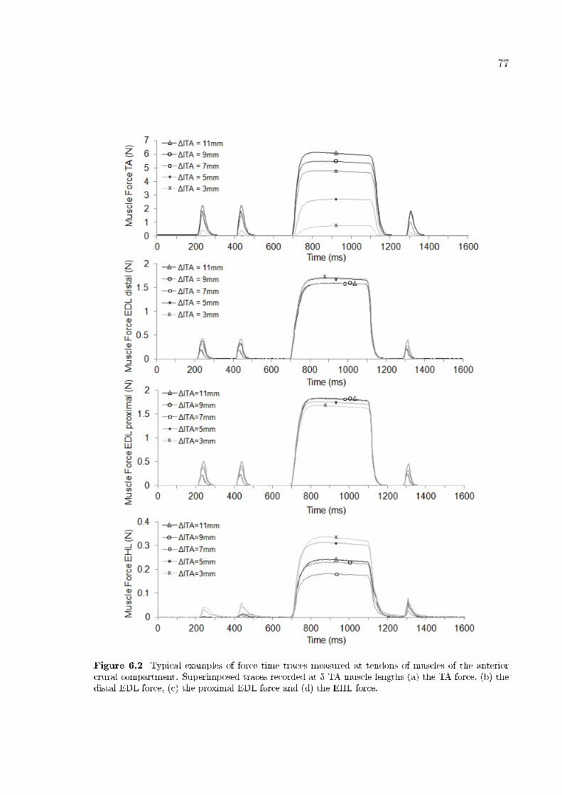
77
Figure 6.2 Typical examples of force time traces measured at tendons of muscles of the anteriorcrural compartment. Superimposed traces recorded at 5 TA muscle lengths (a) the TA force, (b) thedistal EDL force, (c) the proximal EDL force and (d) the EHL force.
78
still added a signi�cant improvement to the description of changes of muscle-tendon
complex length and muscle force data were selected. These polynomials were used
for two purposes: (1) Averaging of data and calculation of standard deviations. Per
each muscle studied, muscle forces at di�erent TA muscle-tendon complex lengths were
obtained by using these functions. Per each TA muscle-tendon complex length, forces
were averaged and standard deviations (SD) were calculated to determine the muscle's
force (mean ± SD). (2) Determining the maximal TA force and the corresponding
muscle length. For each individual TA muscle, the maximal TA force is de�ned as the
maximum value of the �tted polynomial for total muscle force and the corresponding
TA length is determined. One-way ANOVA was also used to test for the e�ects of BTX-
A injection on the TA muscle's length range of force production, i.e., the range between
muscle active slack length and the length at which maximal force is measured. Two-
way ANOVA for repeated measures (factors: TA muscle-tendon complex length and
animal group) was performed separately for the forces of each muscle. Di�erences were
considered signi�cant at p<0.05. If signi�cant main e�ects were found, Bonferroni post-
hoc tests were performed to further locate signi�cant force di�erences within the factors
[30]. Spearman's Rank correlation coe�cient was calculated to test if reductions in TA
total forces due to BTX-A injection are correlated with TA muscle-tendon complex
length. Reduction in force is calculated as the di�erence in mean force between the
control and the BTX animal groups at each TA muscle-tendon complex length and
expressed as a percentage of the mean force of the control group. Correlations were
considered signi�cant at p<0.05.
6.3 Results
Figure 6.2 shows superimposed examples of force�time traces for muscles of the
anterior crural compartment at 5 sample TA lengths selected from the range of 13 TA
lengths tested between muscle active slack length and the length 2 mm over the length
at which the highest TA force was measured.
TA force-length characteristics ANOVA (factors: TA length and animal group)
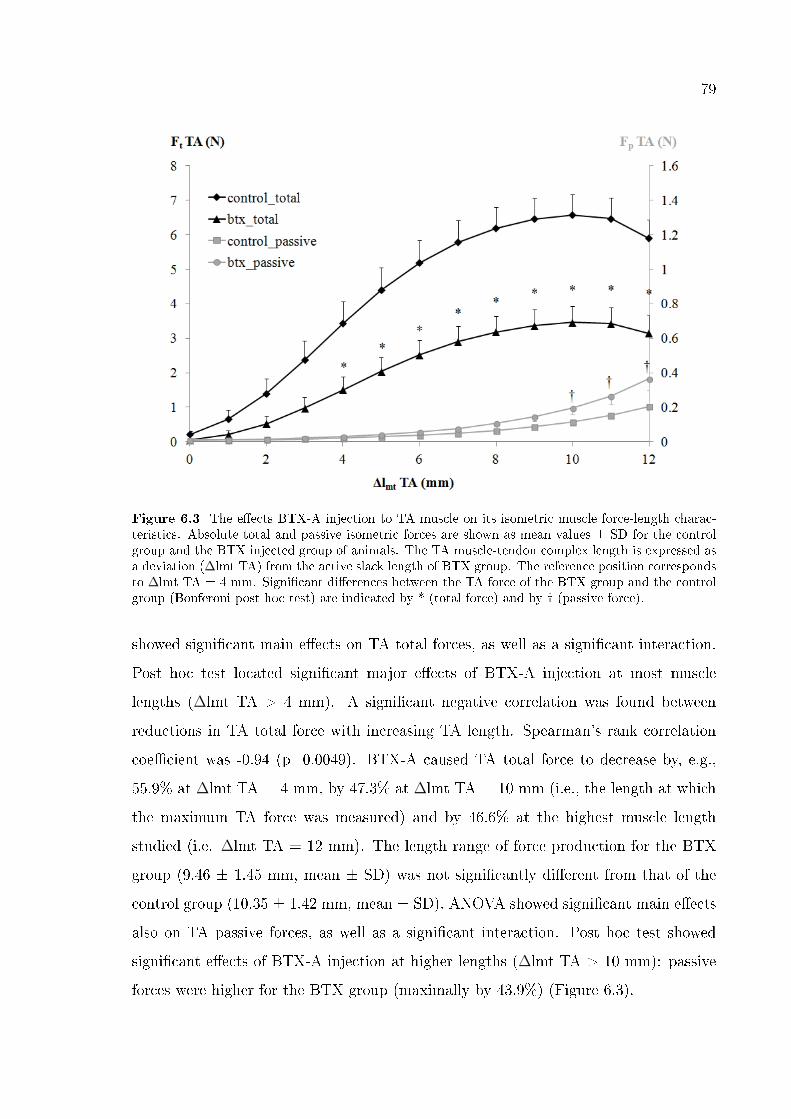
79
Figure 6.3 The e�ects BTX-A injection to TA muscle on its isometric muscle force-length charac-teristics. Absolute total and passive isometric forces are shown as mean values ± SD for the controlgroup and the BTX injected group of animals. The TA muscle-tendon complex length is expressed asa deviation (Δlmt TA) from the active slack length of BTX group. The reference position correspondsto Δlmt TA = 4 mm. Signi�cant di�erences between the TA force of the BTX group and the controlgroup (Bonferoni post hoc test) are indicated by * (total force) and by � (passive force).
showed signi�cant main e�ects on TA total forces, as well as a signi�cant interaction.
Post hoc test located signi�cant major e�ects of BTX-A injection at most muscle
lengths (Δlmt TA > 4 mm). A signi�cant negative correlation was found between
reductions in TA total force with increasing TA length. Spearman's rank correlation
coe�cient was -0.94 (p=0.0049). BTX-A caused TA total force to decrease by, e.g.,
55.9% at Δlmt TA = 4 mm, by 47.3% at Δlmt TA = 10 mm (i.e., the length at which
the maximum TA force was measured) and by 46.6% at the highest muscle length
studied (i.e. Δlmt TA = 12 mm). The length range of force production for the BTX
group (9.46 ± 1.45 mm, mean ± SD) was not signi�cantly di�erent from that of the
control group (10.35 ± 1.42 mm, mean ± SD). ANOVA showed signi�cant main e�ects
also on TA passive forces, as well as a signi�cant interaction. Post hoc test showed
signi�cant e�ects of BTX-A injection at higher lengths (Δlmt TA > 10 mm): passive
forces were higher for the BTX group (maximally by 43.9%) (Figure 6.3).
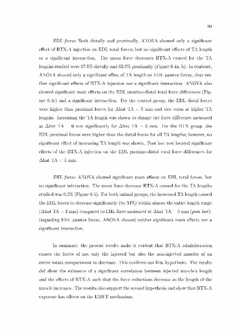
80
EDL forces Both distally and proximally, ANOVA showed only a signi�cant
e�ect of BTX-A injection on EDL total forces; but no signi�cant e�ects of TA length
or a signi�cant interaction. The mean force decreases BTX-A caused for the TA
lengths studied were 67.8% distally and 62.9% proximally (Figure 6.4a, b). In contrast,
ANOVA showed only a signi�cant e�ect of TA length on EDL passive forces, thus nei-
ther signi�cant e�ects of BTX-A injection nor a signi�cant interaction. ANOVA also
showed signi�cant main e�ects on the EDL proximo-distal total force di�erences (Fig-
ure 6.4c) and a signi�cant interaction. For the control group, the EDL distal forces
were higher than proximal forces for Δlmt TA < 5 mm and vice versa at higher TA
lengths. Increasing the TA length was shown to change the force di�erence measured
at Δlmt TA = 0 mm signi�cantly for Δlmt TA > 6 mm. For the BTX group, the
EDL proximal forces were higher than the distal forces for all TA lengths; however, no
signi�cant e�ect of increasing TA length was shown. Post hoc test located signi�cant
e�ects of the BTX-A injection on the EDL proximo-distal total force di�erences for
Δlmt TA < 5 mm.
EHL forces ANOVA showed signi�cant main e�ects on EHL total forces, but
no signi�cant interaction. The mean force decrease BTX-A caused for the TA lengths
studied was 9.2% (Figure 6.5). For both animal groups, the increased TA length caused
the EHL forces to decrease signi�cantly (by 34%) within almost the entire length range
(Δlmt TA > 2 mm) compared to EHL force measured at Δlmt TA = 0 mm (post hoc).
Regarding EHL passive forces, ANOVA showed neither signi�cant main e�ects nor a
signi�cant interaction.
In summary, the present results make it evident that BTX-A administration
causes the forces of not only the injected but also the non-injected muscles of an
entire intact compartment to decrease. This con�rms our �rst hypothesis. The results
did show the existence of a signi�cant correlation between injected muscle's length
and the e�ects of BTX-A such that the force reductions decrease as the length of the
muscle increases. The results also support the second hypothesis and show that BTX-A
exposure has e�ects on the EMFT mechanism.
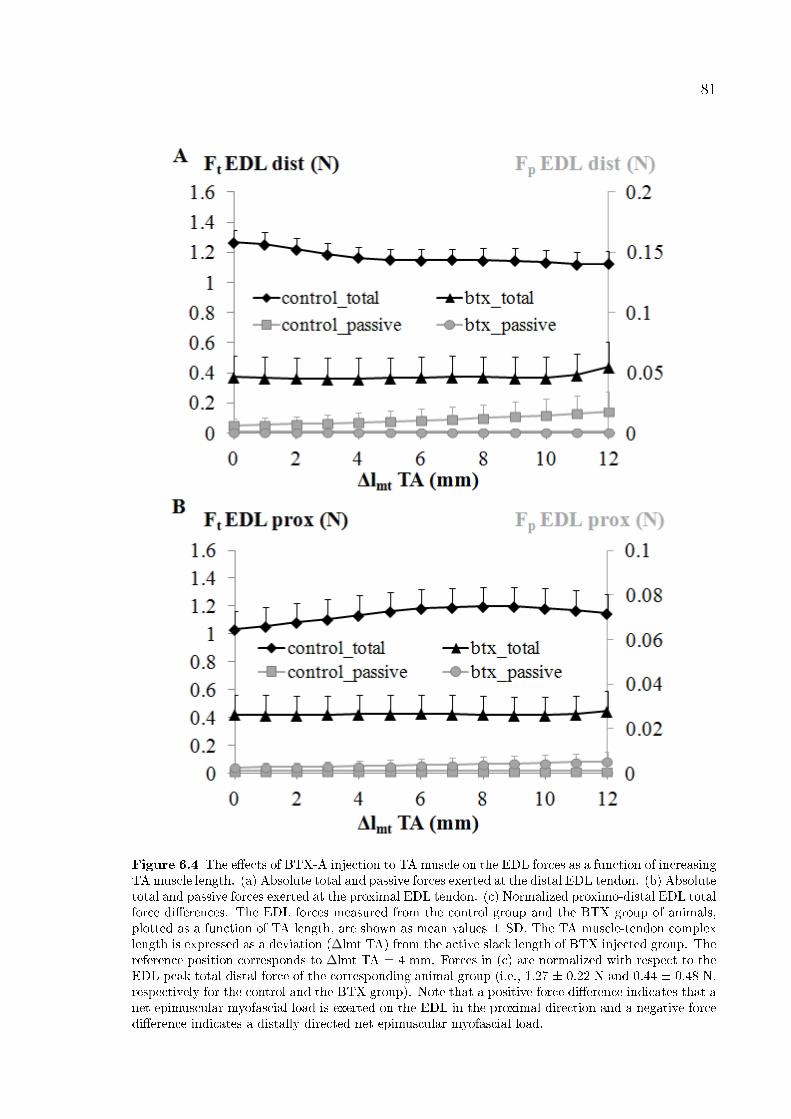
81
Figure 6.4 The e�ects of BTX-A injection to TA muscle on the EDL forces as a function of increasingTA muscle length. (a) Absolute total and passive forces exerted at the distal EDL tendon. (b) Absolutetotal and passive forces exerted at the proximal EDL tendon. (c) Normalized proximo-distal EDL totalforce di�erences. The EDL forces measured from the control group and the BTX group of animals,plotted as a function of TA length, are shown as mean values ± SD. The TA muscle-tendon complexlength is expressed as a deviation (Δlmt TA) from the active slack length of BTX injected group. Thereference position corresponds to Δlmt TA = 4 mm. Forces in (c) are normalized with respect to theEDL peak total distal force of the corresponding animal group (i.e., 1.27 ± 0.22 N and 0.44 ± 0.48 N,respectively for the control and the BTX group). Note that a positive force di�erence indicates that anet epimuscular myofascial load is exerted on the EDL in the proximal direction and a negative forcedi�erence indicates a distally directed net epimuscular myofascial load.
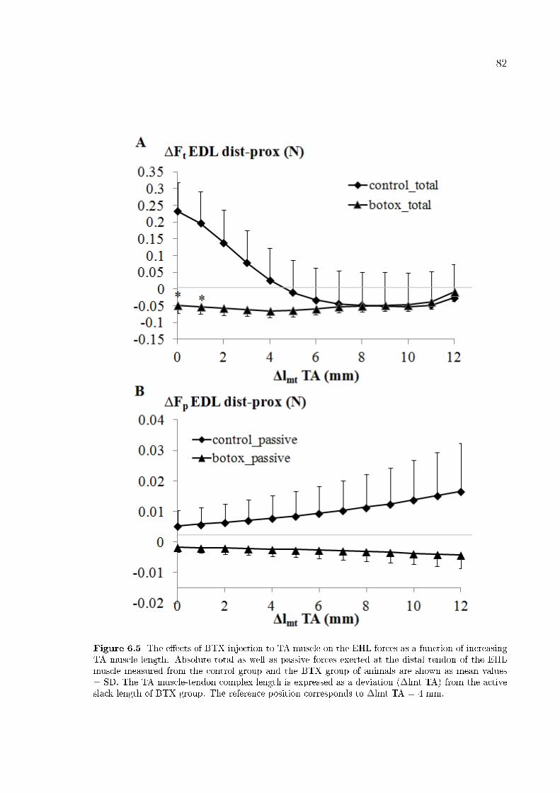
82
Figure 6.5 The e�ects of BTX injection to TA muscle on the EHL forces as a function of increasingTA muscle length. Absolute total as well as passive forces exerted at the distal tendon of the EHLmuscle measured from the control group and the BTX group of animals are shown as mean values± SD. The TA muscle-tendon complex length is expressed as a deviation (Δlmt TA) from the activeslack length of BTX group. The reference position corresponds to Δlmt TA = 4 mm.
83
6.4 Discussion
E�ects of BTX-A on the forces of the injected and the non-injected muscles of
the compartment An important �nding is that force decreases caused by BTX-A are
muscle length dependent such that the increased length of the injected muscle and the
reduction in force are negatively correlated. Similar results were shown in the study of
Yaraskavitch et al. [71] for the cat soleus muscle and were inferred from their torque-
joint angle data by Longino et al. [13] after testing the quadriceps musculature of
the rabbit. These �ndings obtained from di�erent muscles suggest that muscle length
dependency of the e�ects of BTX-A are important to consider. This may have clinical
implications some of which are discussed in a successive paragraph.
Yaraskavitch et al. [71] based their explanation for the observed change in the
force-length curve, i.e., a less steep ascending limb, on adaptation in the number of
in-series sarcomeres, which could occur 4 weeks post-injection. However, in the present
study, only 5 days postinjection, the occurrence of such adaptation is less likely. Dener-
vation of adult mice muscle was shown to have no e�ect on the sarcomere number [206].
Therefore, the present results suggest that length dependent e�ects of BTX-A may not
be due exclusively to changes in the number of in-series sarcomeres within the muscle.
Instead, a mechanism that involves changes in lengths of in series sarcomeres may be
responsible. For such mechanism, the fact that BTX-A causes paralysis of muscle �bers
within parts of the muscle belly [66] is central. Due to that, BTX-A injection may lead
to di�erences in two types of mechanical interactions occurring among the structures
comprising the muscle-tendon complex: (1) Muscle-tendon interactions Tendon tissue
has nonlinear force-deformation characteristics [205], and under lower magnitudes of
forces, it has been shown to be more compliant [207-208]. For the BTX-A injected
muscle, a general expected e�ect of the resulting reduction of muscle force is less ex-
tension of the tendon for the same muscle-tendon complex length. Therefore, a muscle
tendon complex length dependent shifting of the sarcomere lengths to higher lengths
is plausible. (2) Muscle �ber-extracellular matrix interactions Muscle �bers and the
extracellular matrix (ECM) are mechanically connected not only at the ends of the
muscle �bers, but also along their full peripheral length [161, 209-210]. Consequently,
84
muscle �bers can interact with the ECM and hence with each other mechanically [111,
162-163, 211]. In tetanized muscle, this mechanism has been shown to limit shorten-
ing of muscle �bers after tenotomy [111] and aponeurotomy [82, 179]: although some
muscle �bers lost their myotendinous connection to the insertion of the muscle, the
ECM connected to the muscle �bers via transsarcolemmal molecules [161] prevents the
sarcomeres within these muscle �bers from shortening to their active slack length. In
BTX-A injected muscle due to a lack of stimulation, the paralyzed muscle �bers do not
shorten as activated muscle �bers of a non-paralyzed muscle would do. Therefore, the
interaction mechanism described is expected to cause a resistance to the shortening of
the sarcomeres within the activated muscle �bers. A common indicated e�ect there-
fore appears to be the shifted lengths of the sarcomeres within the activated muscle
�bers. Recently, Turkoglu et al. [212] studied the principles of e�ects of BTX-A on
muscular mechanics by extending a �nite element model of rat muscle [28, 213]. They
activated only selected parts of the muscle, and the remainder parts were considered
to represent paralyzed muscle �bers. The model results show in agreement with [71]
the arguments posed above that sarcomeres do attain higher lengths. Consequently,
if the active sarcomeres of the partially paralyzed muscle are at the ascending limb of
their force length curve, they can produce more force compared to their counterparts
in the nonparalyzed muscle. This suggest that a net muscle force reduction e�ect of
BTX-A originates exclusively from the presence of paralyzed muscle �bers, as the ac-
tive ones may have even an enhanced potential of active force production. Note that
the model results [212] indicate that compared to lower muscle lengths, this potential
compromising the e�ectiveness of BTX-A is greater at intermediate muscle lengths be-
cause there is relatively more resistance to sarcomere shortening. On the other hand,
if the active sarcomeres are at the descending limb of their force-length curve, the op-
posite is valid, i.e., as they attain higher lengths, they produce less force. Note that in
conditions similar to the present ones i.e., for muscle with epimuscular connections to
its surrounding structures, previous model studies suggest heterogeneity of sarcomere
lengths within muscle �bers [23, 84, 109]. Particularly at higher muscle lengths, sar-
comere length heterogeneity may include sarcomeres at both ascending and descending
limbs of their force-length curves within the same muscle �bers [22, 25]. Therefore, for
such lengths, a more complex mechanism may determine the e�ectiveness of BTX-A
85
in force reduction. Recall that the present results show a decrease in the force reduc-
tion e�ect of BTX-A at higher muscle lengths. According to the mechanism proposed,
this suggests that most active sarcomeres may have been shifted to lengths favorable
for force production. It is important to note that the mechanisms considered here to
explain the length dependency of the e�ects of BTX-A are theoretical ones and they
should be elaborated and veri�ed in new studies. Medical imaging may be a feasible
method to test these e�ects. Recently in no BTX-A injected condition has magnetic
resonance imaging analyses shown the occurrence of local and heterogeneous muscle
tissue deformations as caused by joint angle changes in human muscles in vivo [142].
Such deformations involve variable magnitudes of local lengthening occuring simulta-
neously with local shortening at other locations and indicate that distribution locally
of lengths of muscle tissue is possible. Coupled with di�usion tensor imaging, such
analyses may allow for quanti�cation of length changes speci�cally along the muscle
�ber direction [214].
BTX-A is used in treating patients with cerebral palsy, e.g., in the management
of spastic equines gait [198-199] as well as in upper limb spasticity [215]. In these
patients, spasticity is responsible for movement disorders and functional disability that
can be characterized by a limited joint range of movement [38]. An improved un-
derstanding of how BTX-A a�ects the force production of muscle for di�erent muscle
lengths may be clinically relevant. However, the moment arm lengths of muscles vary
with varying joint angles [126-127], which make it di�cult to relate such understanding
directly to joint movement. Therefore, based on the present results, it cannot be con-
cluded that the e�ects of the treatment are variable for di�erent joint angles. Muscle
hypertonicity in spasticity [29-31] causes the joint to be forcefully kept in typically a
�exed position in which the muscle is expected to be short. It may be important that
a more pronounced muscle weakening e�ect is found for shorter muscle lengths. How-
ever, due to the indicated di�culty in relating muscle force-length properties to joint
movement, it cannot be concluded that more pronounced e�ects are available for the
joint positions that may correspond to short muscle lengths. Nevertheless, the �ndings
of Longino et al. [201] may support this expectation because these authors showed that
muscle weakness e�ects of BTX-A cumulatively on the rabbit knee extensors are knee
86
joint angle dependent, and are more pronounced for more extended knee positions.
More importantly, the results of the present study show that potentially all muscles
within a compartment can determine how BTX-A administration a�ects the mechanics
at the joint, even though only one of them is injected. A noteworthy implication of this
�nding is that the non-targeted muscles may have unintended e�ects also at the other
joints that they span. These �ndings are expected to have clinical relevance and the
experimental approaches developed are suitable for addressing them in new studies.
These studies should also impose muscle length changes for the non-targeted muscles.
Injection protocol employed in relation to the e�ects on muscle forces
[67] showed that the compound muscle action potential amplitude and the force
exerted by lower hind limb �exors of the rat decreased predominantly in the �rst four
days, with the decreases leveling o� by the �fth day. Presently, the aim was to assess
the short-term e�ects of BTX-A after stabilization. Therefore, all injections were
performed �ve days prior to testing. Note that the present BTX-A injection protocol
di�ers from common clinical practice: (1) the injected dose (0.1 U i.e., approximately
0.32 U/kg) was less than that used in patients for lower limb muscles (3-6 U/kg),
including children with cerebral palsy [216-218]. (2) A single injection was made to
the mid-belly of the target muscle instead of using multiple injection points [65, 70,
216, 219]. However, although they are not capable of showing whether an e�ective
distribution of toxin is achieved, the present results strongly suggest that a considerable
paralysis did occur within the target muscle. Shaari and Sanders showed that for the
rat TA muscle, even a dose of 0.02U (a �fth of the dose used presently) injected to the
mid-belly causes approximately a �fth of the total cross-sectional area to be paralyzed
only 24 hours following the injection [66]. Shaari and Sanders also reported that an
e�ective distribution of toxin is possible. Therefore, the quantity of BTX-A injected
does not represent a low dose for the rat TA muscle as the di�erence with respect to
the clinically used quantities would suggest and the present injection protocol was a
suitable one for studying the e�ects of BTX-A on this muscle. On the other hand, BTX-
A is reported to be highly di�usive [70]. Although it binds with high a�nity to local
targets within the muscle, a larger volume, single injection may cause that site to be
87
saturated and thus allow the spread of toxin to neighboring structures [216]. Therefore,
the present injection protocol may have promoted toxin leakage to the adjacent EDL
and EHL muscles. Yaraskavitch et al. explained their results with leakage of BTX-A
into the noninjected muscle [71]; additionally, it is possible that partial paralyzation
of these muscles is responsible for the presently measured force decreases. Note that
the spread of BTX-A beyond the injection site is considered to be a side e�ect [16, 56]
and has been argued to occur not only after localized injections [14-15], but may be
determined by the dose and concentration of injection [216]. New studies are indicated
to test for the role of di�erent injection protocols beyond the injection site within an
intact compartment.
BTX-A has e�ects on EMFT
After imposing length changes to a muscle, EMFT previously has been shown to
cause changes in forces of restrained muscles [e.g. 23, 141]. The present results showed
similar e�ects for the control group: (1) The increased TA length caused signi�cant
changes in EDL proximo-distal force di�erences. Note that such force di�erences [15,
22, 25] are characteristic e�ects of EMFT [26] and represent the resultant of epimuscu-
lar myofascial loads acting on the EDL muscle. These forces originate from stretching
epimuscular connections, which include direct collageneous connections between adja-
cent muscles as well as structures such as collagen-reinforced neurovascular tracts and
compartmental boundaries.
They also include forces generated within the sarcomeres of neighboring muscles
that are transmitted onto the EDL muscle via these structures [for a detailed discussion
see 107]. Initially, the present EDL proximo-distal force di�erences were in favor of the
distal force indicating that a resultant epimuscular myofascial load was acting on the
muscle in the proximal direction. At the initial TA length, the length of the EDL muscle
restrained at the reference position was conceivably higher causing interconnecting
epimuscular connections between these muscles to be stretched as a source of such
proximally directed loads.
88
However, the e�ect was reversed at higher TA lengths. (2) The increased TA
length also caused EHL force to change signi�cantly. No such EMFT e�ect on EHL
muscle has been reported since in the previous studies, forces of this muscle were mea-
sured together with the forces of TA muscle via their tied tendons [25, 170]. However,
EMFT between EHL and EDL muscles was shown previously to occur after impos-
ing EHL length changes [23]. In those experiments, prior to testing, anterior crural
compartment was opened and the TA muscle was removed. Therefore, only certain
epimuscular connections of EHL muscle were left intact. In contrast, presently signif-
icant EMFT e�ects of TA length changes in a fully intact compartment were shown,
which caused EHL forces to decrease approximately by a third. The results showed
that similarly to the control group, increased TA length caused signi�cant changes in
the EHL force such that the EMFT e�ects remained as profound in the BTX group.
This �nding can be interpreted as BTX-A did not a�ect EMFT between EHL and TA
muscles. However, like all muscles within the compartment, a muscle weakening e�ect
was found for the EHL muscle, indicating that its sti�ness in the active state decreased.
Yet, the length changes of the TA muscle yielded the same relative decrease in EHL
force for both groups. Therefore, the results indicate two �ndings: (1) the epimuscu-
lar connections between EHL and TA muscles remained su�ciently sti� to allow the
occurrence of EMFT. (2) BTX-A causes manipulation locally of their sti�ness, opera-
tionalized for the changes of muscle relative positions imposed. This is in agreement
with our expectation that BTX-A administration changes the interplay of sti�ness of
muscular tissues and their epimuscular connections, and con�rms that BTX-A a�ected
EMFT between the EHL and the TA muscles.
On the other hand, BTX-A exposure did a�ect EDL proximo-distal force dif-
ferences such that the e�ect of force di�erences initially in favor of the distal EDL
force disappeared. This is an indicator that the epimuscular myofascial loads acting on
the EDL muscle were manipulated by BTX-A. Also this result indicates that BTX-A
administration changes the interplay of sti�ness of muscular tissues and their epimus-
cular connections. Although it is not immediately apparent which component plays a
dominant role, it is plausible that the epimuscular connections between the EDL and
the TA muscles were less e�ective in EMFT after BTX-A administration.
89
A noteworthy e�ect presently shown is that at higher muscle lengths of the
BTX-A injected TA muscle, the passive forces were signi�cantly higher than those of
the control group. A tenable explanation for this e�ect is an increased sti�ness of the
intramuscular connective tissues of the TA muscle and its epimuscular connections in
combination. In agreement with this, the slope of the passive force-length curve of
the BTX-A group was at least 86% higher than that of the control group for ∆lmt
TA = 10 mm. Note that, in the passive state, any di�erence between the control
and BTX groups in terms of existence of paralyzed muscle parts vanishes. Therefore,
the increased passive TA forces cannot be ascribable to manipulated myofascial tissue
sti�ness, solely due to muscle relative position changes. Instead, it should also be
considered that structural changes possibly occurred in these tissues. Within the �rst
week following the denervation, atrophy of rat muscles was reported [220]. Also BTX-
A was shown to cause atrophy [221-222]. Billante et al. showed that BTX-A causes
decreased muscle �ber diameters as well as density [68]. Only for very high doses of
BTX-A, even existence of �brosis was observed by these authors within the muscle.
However, no evidence is available whether passive muscle force increases accompanied
such e�ects.
Moreover, these e�ects are limited to the intramuscular tissues exclusively. The
lack of direct data to show if tissue structural changes occurred presently is a limita-
tion of this study. However, increased passive force of BTX-A injected muscle with
intact epimuscular connections is an interesting �nding, implications of which on tissue
adaptation should be addressed in new speci�c studies.
The results show that BTX-A has e�ects on EMFT. However, these e�ects are
not uniform within the anterior crural compartment for the conditions studied and
they imply that EMFT is a�ected not only by muscle weakening but also by manipu-
lated mechanical properties of the connective tissues comprising the EMFT pathways.
EMFT has been regarded to play an important role in the mechanics of spastic paretic
muscle [62] and surgical treatment techniques of the related functional de�ciencies [63].
Such concepts are likely to have clinical implications also for the treatment of these
conditions using BTX-A. New studies are indicated to explore further the relationship
90
between the e�ects of BTX-A and those of EMFT.
6.5 Conclusions
The results show that exposure to BTX-A does a�ect the forces of all muscles
operating in an intact compartment: (1) length dependent force decreases were found
for the targeted TA muscle such that increased muscle length and the reduction in
muscle force are negatively correlated. However, no change in the muscles' length range
of active force production was found. (2) The simultaneously measured forces of the
non-injected synergistic EDL and EHL muscles also decreased signi�cantly, suggesting
the presence of unintended additional e�ects both for the targeted distal joint and
for the non-targeted proximal joint. The results also show that BTX-A exposure has
e�ects on the EMFT mechanism. However, these e�ects are not uniform within the
anterior crural compartment.
91
7. EFFECTS OF BTX-A ON NON-INJECTED
BI-ARTICULAR MUSCLE INCLUDE A NARROWER
LENGTH RANGE OF FORCE EXERTION AND
INCREASED PASSIVE FORCE
7.1 Introduction
Botulinum toxin type A (BTX-A) causes muscle paralysis by inhibiting acetyl-
choline release into the presynaptic cleft in the neuromuscular junction [e.g., 65]. This
chemodenervant is applied to spastic muscles of cerebral palsy (CP) patients to de-
crease muscle tonus [223-224]. A consequence of decreased hyperactivity is increased
joint range of motion [225-227]. Therefore, it is aimed at improving joint function
[228] and gait [218, 229-230]. BTX-A is also used to treat spasticity from several other
origins such as stroke [231-232] and multiple sclerosis [233].
The e�ects of BTX-A have been widely studied by quantifying such parameters
as the area of paralysis [66], electromyography [234] and compound muscle action
potential [235]. Reports on twitch and tetanic force have been limited to selected
muscle lengths or joint positions [e.g., 235, 236]. The di�culty of relating muscle
force-length characteristics directly to joint movement due to moment arm lengths
varying with joint angles [237-238] is apparent. Yet, such data have more potential
for an improved understanding of e�ects of BTX-A on joint mechanics. For example,
reduction of active force was shown to be variable as a function of muscle length instead
of being constant [239].
BTX-A has been shown to spread through muscle fascia [70], and its e�ects
beyond the injection site have been reported [240-242]. Therefore, it is also necessary
to measure the e�ects not only on the target muscle but also on others that are af-
fected. Recently, Yaraskavitch et al. [243] showed that the force-length characteristics
92
of both injected soleus and non-injected plantaris muscles of the cat are a�ected by the
poison. Exposure exclusively of the tibialis anterior (TA) muscle of the anterior crural
compartment of the rat a�ects the forces of also all other muscles i.e., m. extensor
digitorum longus (EDL) and m. extensor hallucis longus (EHL) within the compart-
ment [239]. On the other hand, it was reported to our knowledge for the �rst time
that passive forces of muscle exposed to BTX-A increase [239]. As by using BTX-A
in CP patients, an important goal is also to reduce passive resistance of the muscle at
the joint [244-247], this �nding is interesting.
For a bi-articular muscle, proximal and distal moment arms may di�er, causing
its contribution to mechanics of the joints it spans to be not symmetric. However,
this may not be the exclussive source of such asymmetry. In BTX-A free conditions
it was shown that the e�ects on muscle force-length characteristics of equal proximal
and distal lengthening of bi-articular EDL muscle of the rat are not symmetric: e.g.
distal lengthening yielded a lower distal optimal force than the proximal optimal force
measured after proximal lengthening [174]. This e�ect is acribable to mechanical in-
teraction of muscle with its surrounding muscular and non-muscular tissues via its
myofascial connections to those structures [63, 140-141]. Such mechanical interaction
is feasible in an intact muscle compartment, as it is in vivo [142, 165]. Based on
this, BTX-A may be expected to a�ect mechanics of a bi-articular muscle di�erently
proximally and distally. However, this has not been tested.
We aimed at testing the hypothesis that BTX-A administration to the TA mus-
cle of the rat a�ects the synergistic EDL muscle and causes changes to this muscle's
active as well as passive contribution to mechanics of the joints it spans. This goal
was addressed by measuring the force-length characteristics of the EDL muscle after
proximal as well as distal lengthening. Key parameters studied were active force reduc-
tion, length range of active force exertion and passive muscle forces. For completeness,
isometric forces of the TA and EHL muscles of the intact compartment were measured
simultaneously.
93
7.2 Methods
7.2.1 Assessment of the e�ects of BTX on muscular mechanics
Surgical and experimental procedures were approved by the Committee on the
Ethics of Animal Experimentation at Bo§aziçi University. Male Wistar rats were di-
vided into two groups: Control (n = 8, mean ± SD body mass = 300.0 ± 6.9 g) and
BTX (n = 8, mean ± SD body mass = 315.0 ± 6.3 g).
After imposing a mild sedation with an intraperitonal dose of 1mg/kg ketamine,
a circular region of approximately 15 mm radius from the center of the knee cap was
shaved. The tibialis anterior (TA) muscle was located by palpation when the ankle was
in maximal plantar �exion and the knee angle approximated 90°. After marking the
center of the knee cap, a second marker was placed at a point 10 mm distal to that,
along the tibia. The injection location was 5 mm lateral (along the direction normal to
the line segment drawn between the two markers) to the second marker and over the
TA muscle. All injections were made exclusively into this muscle, to a depth of 3 mm.
For the BTX group, each 100 unit vial of vacuum dried, botulinum type A neu-
rotoxin complex (BTX-A) (BOTOX, Allergan Pharmaceuticals, Ireland) was recon-
stituted with 0.9% sodium chloride. The animals received a one-time intramuscular
BTX-A injection at a total dose of 0.1 units. The injected volume equaled 20μl. The
control group was injected with the same volume of 0.9% saline solution exclusively.
All injections were performed �ve days prior to testing. The animals were kept sepa-
rately until the day of the experiment in standard cages and in a thermally regulated
animal care room with a 12h dark-light cycle.
7.2.2 Surgical Procedures
The animals were anesthetized using an intraperitoneally injected urethane so-
lution (1.2ml of 12.5% urethane solution/100g body mass). Additional doses were given
94
if necessary (maximally 0.5ml). Immediately following the experiments, the animals
were euthanized by the administration of an overdose of urethane solution.
During the surgery and data collection, the animals were kept on a heated pad
(Harvard Apparatus, Homoeothermic Blanket Control Unit) to prevent hypothermia.
A feedback system utilizing an integrated rectal thermometer allowed for the control
of body temperature at 37 °C by adjusting the temperature of the heated pad. The
skin and the biceps femoris muscle of the left hind limb were removed and the anterior
crural compartment including the EDL, the TA, and the EHL muscles were exposed.
Only a limited distal fasciotomy was performed to remove the retinaculae (i.e., the
transverse crural ligament and the crural cruciate ligament). The connective tissues at
the muscle bellies within the anterior crural compartment were left intact.
The combination of knee joint and ankle angles (120° and 100°, respectively)
was selected as the reference position. In the reference position, the four distal tendons
of the EDL muscle were tied together using silk thread. Matching markers were placed
on the distal tendons of the EDL, the TA and the EHL muscles, as well as on a �xed
location on the lower leg. Subsequently, the distal EDL tendon complex as well as the
TA and the EHL tendons were cut as distally as possible.
The femoral compartment was opened for two purposes: (1) to reach the prox-
imal tendon of the EDL. After reaching it, this tendon was cut from the femur, with
a small piece of the lateral femoral condyle still attached. (2) To expose the sciatic
nerve. After this was done, the sciatic nerve was dissected free of other tissues, during
which process all nerve branches to the muscles of the femoral compartment were cut.
Subsequently, the sciatic nerve was cut as proximally as possible.
In order to provide connection to force transducers, Kevlar threads were sutured
to: (1) the proximal tendon of the EDL muscle (2) the tied distal tendons of the EDL
muscle, (3) the distal tendon of the TA muscle, and (4) the distal tendon of the EHL
muscle.
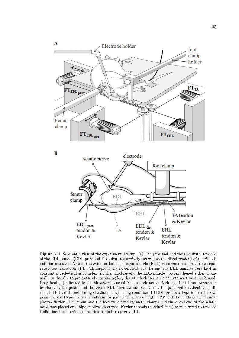
95
Figure 7.1 Schematic view of the experimental setup. (a) The proximal and the tied distal tendonsof the EDL muscle (EDL prox and EDL dist, respectively) as well as the distal tendons of the tibialisanterior muscle (TA) and the extensor hallucis longus muscle (EHL) were each connected to a sepa-rate force transducer (FT). Throughout the experiment, the TA and the EHL muscles were kept atconstant muscle-tendon complex lengths. Exclusively, the EDL muscle was lengthened either proxi-mally or distally to progressively increasing lengths, at which isometric contractions were performed.Lengthening (indicated by double arrow) started from muscle active slack length at 1mm incrementsby changing the position of the target EDL force transducer. During the proximal lengthening condi-tion, FTEDL dist, and during the distal lengthening condition, FTEDL prox was kept in its referenceposition. (b) Experimental condition for joint angles: knee angle=120° and the ankle is at maximalplantar �exion. The femur and the foot were �xed by metal clamps and the distal end of the sciaticnerve was placed on a bipolar silver electrode. Kevlar threads (hatched lines) were sutured to tendons(solid lines) to provide connection to their respective FT.

96
7.2.3 Experimental set-up
The animal was mounted in the experimental set-up (Fig. 1A). The femur and
foot were �xed with metal clamps such that the ankle was in maximal plantar �exion
(180°) to allow for the free passage of the Kevlar threads to the distal force transducers.
The knee angle was set at 120°. Each Kevlar thread was connected to a separate force
transducer (BLH Electronics Inc., Canton MA). Care was taken to ensure the alignment
of the Kevlar threads were in the muscle line of pull. The distal end of the sciatic nerve
was placed on a bipolar silver electrode (Fig. 1B).
7.2.4 Experimental conditions and procedures
Room temperature was kept at 26°C. For the duration of the experiment, muscle
and tendon tissues were irrigated regularly by isotonic saline to prevent dehydration.
The distal tendons of TA and EHL muscles were kept in their reference positions at
all times during the experiment. The isometric forces of all muscles were measured
simultaneously at various muscle-tendon complex lengths of the EDL either by mov-
ing its proximal force transducer in the proximal direction (i.e., proximal lengthening
condition) or by moving its distal force transducer in the distal direction (i.e., distal
lengthening condition): starting from its active slack length, the EDL length was in-
creased in increments of 1 mm, until it was 2 mm over the optimum length. EDL distal
tendon was kept in its reference position.
EDL muscle-tendon complex lengths are expressed as deviation from the related
optimum length (i.e., ∆lma EDL proximal or ∆lma EDL distal).
All muscles studied were activated maximally by supramaximal stimulation of
the sciatic nerve (Biopac Systems stimulator, STMISOC) using a constant current of
2mA (square pulse width 0.1ms). After setting the EDL muscle to a target length, two
twitches were evoked and 300ms after the second twitch, the muscles were tetanized
(pulse train 400ms, frequency 100Hz). At 200ms after the tetanic contraction, another
97
twitch was evoked. After each application of this stimulation protocol, the muscles
were allowed to recover for 2 minutes. For the EDL muscle, recovery was allowed to
occur near the active slack length, whereas the lengths of the other muscles were not
altered.
Subsequent to isometric force measurements, a glycogen depletion process was
carried out in the BTX group: the peroneal branch of sciatic nerve was stimulated
(20 Hz) continuously for 15 min [66, 70]. The anterior crural muscles were removed
immediately after the animal was euthanized and their mid portions were prepared
for histological assessment: �xation was followed by processing (Leica TP1020) and
para�n embedding (Leica EG1150H). 8µm sections were cut (Leica RM2255) for every
100µm. Glycogen staining was performed using periodic acid-Schi� (PAS) (Sigma-
Aldrich, USA). Photographs of stained sections were taken under microscope (Leica
DM2500, 10X magni�cations). E�ectiveness of PAS staining technique was assessed
(Fig. 2A) in the control group (n=1, with glycogen depletion and n=1, with no glycogen
depletion).
7.2.5 Assessments of the e�ects of BTX-A on intramuscular connective
tissue content
Changes in intramuscular connective tissue content were assessed histologically
in a separate set of male Wistar rats, again divided into two groups: Control (n =
6, mean ± SD body mass = 321.7 ± 29.3 g) and BTX (n = 6, mean ± SD body
mass = 319.3 ± 8.1 g). After the injection protocol described above was employed,
the animals were kept for �ve days in identical conditions with the previous set. TA,
EDL, and EHL muscles were removed. Total wet muscle mass was measured using
a scale (Precisa XB 320M) with high resolution (0.001 g). Subsequently, the muscles
were prepared for histological assessment, cut into sections and photographed using the
equipment described above. However, for this set, a detailed and quantitative analysis
was carried out: 5μm cross-sections were cut for every 20μm. The sections were stained
using Gomori's Trichrome (Bio-Optica-30-30110, Italy), which stain has been used in
98
distinguishing intramuscular collagen e.g., in neuromuscular diseases [248-249].
7.2.6 Data processing and statistics
Muscle isometric forces were determined 100ms after the second twitch. Force
in such inactivated state represents the mechanical resistance potential of the muscular
tissues at a particular muscle length tested and is referred to as passive muscle force
(Fp). Muscle total isometric forces were determined during the tetanic plateau (the
mean force for a 200ms interval, 150 ms after evoking tetanic stimulation). Data for
active muscle force (Fa) calculated by subtracting the measured passive force from
total force in relation to muscle-tendon complex length were �tted with a polynomial
function using a least squares criterion
y=b0+b1x+b2x2 + ... + bnxn (1)
where y represents Fa, x represents muscle-tendon complex length. b0, b1. . . bn
are coe�cients determined in the �tting process. Data for passive muscle force in
relation to muscle-tendon complex length were �tted with an exponential function
using a least squares criterion
y=ea1+a2x (2)
where y represents Fp and x represents passive muscle-tendon complex length
and a1 and a2 are coe�cients determined in the �tting process.
Polynomials that best described the experimental data were selected by using
one-way analysis of variance (ANOVA) [125]: the lowest order of the polynomials that
still added a signi�cant improvement to the description of changes of muscle-tendon
complex length and muscle force data were selected. These polynomials were used for
averaging of data and calculation of standard deviations. Per each muscle studied,
muscle forces at di�erent EDL muscle-tendon complex lengths were obtained by using
99
these functions. Per each EDL muscle-tendon complex length, forces were averaged
and standard deviations (SD) were calculated to determine the muscle's force (mean
± SD).
The major determinants of muscle length-force characteristics are muscle opti-
mal force (the maximum isometric force exerted by an active muscle), the corresponding
muscle length, and muscle active slack length (the shortest length at which the muscle
can still exert nonzero force). Muscle optimal force often is taken as an indication of a
muscle's capacity for force production, and the range from active slack length to length
at which optimal force is exerted is taken as an indicator of movement capability with
active force exertion within the potential range of motion of a certain joint.
In order to account for these issues, the polynomials obtained were used also for
the following purposes: determining (i) the optimal EDL force (i.e., for each individual
EDL muscle, the maximum value of the �tted polynomial for active muscle force) as
well as the corresponding muscle length and (ii) EDL active slack length. EDL muscle's
length range of active force exertion (lrange) was determined as the range between the
muscle's active slack length and the length at which optimal force is measured. Assessed
were the proximal lrange in proximal lengthening condition and distal lrange in distal
lengthening condition.
One-way ANOVA was also used to test for the e�ects of BTX administration
on EDL muscle's lrange and to compare changes to lrange in proximal lengthening vs.
distal lengthening conditions. Two-way ANOVA for repeated measures (factors: EDL
muscle-tendon complex length and animal group) was performed separately for the
forces of each muscle. Di�erences were considered signi�cant at p<0.05. If signi�cant
main e�ects were found, Bonferroni post-hoc tests were performed to further locate
signi�cant force di�erences within the factors [125].
Forces of both groups were aligned for their optimum length. Reduction in
active force is calculated as the di�erence in mean force between the control and the
BTX animal groups at each EDL muscle-tendon complex length and expressed as
100
a percentage of the mean force of the control group. Spearman's Rank correlation
coe�cient (ρ) was calculated to test if reductions due to BTX-A administration in (i)
EDL proximal active forces are correlated with EDL proximal muscle-tendon complex
length and (ii) EDL distal active forces are correlated with EDL distal muscle-tendon
complex length. Correlations were considered signi�cant at p<0.05. Glycogen staining
was assessed qualitatively to show that BTX-A causes partial paralysis for the muscles
of the anterior crural compartment.
Intramuscular connective tissue staining on the other hand was assessed quanti-
tatively to test whether BTX-A causes changes in the collagen content for these muscles.
Using a self programmed code (MATLAB, R2012a) section images were analyzed to
distinguish pixels stained in light green color (showing intramuscular connective tissue)
from pixels stained in dark blue (showing contractile material). For both control and
BTX groups (i) percentage of intramuscular connective tissue content in the sections
studied was quanti�ed and (ii) summed anterior crural muscle mass was normalized
for the body mass. One-way ANOVA was performed to test for the e�ects of BTX
administration. Di�erences were considered signi�cant at p<0.05.
7.3 Results
Glycogen staining shows that BTX-A administration causes partial paralysis for
all muscles of the anterior crural compartment (Fig. 2).
7.3.1 E�ects of BTX-A on the TA and EHL muscles
Signi�cant main e�ects of only BTX-A injection on forces of restrained TA and
EHL muscles were found. The mean active force decreases BTX-A caused for the EDL
lengths studied were 82.8% for proximal lengthening and 74.8% for distal lengthening
conditions, for the TA muscle (Fig. 3A, C) and 43.7% for proximal lengthening and
53.5% for distal lengthening conditions, for the EHL muscle (Fig. 3B, D). Passive EHL
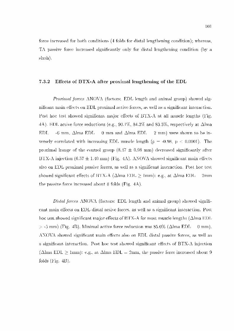
101
force increased for both conditions (4 folds for distal lengthening condition); whereas,
TA passive force increased signi�cantly only for distal lengthening condition (by a
sixth).
7.3.2 E�ects of BTX-A after proximal lengthening of the EDL
Proximal forces ANOVA (factors: EDL length and animal group) showed sig-
ni�cant main e�ects on EDL proximal active forces, as well as a signi�cant interaction.
Post hoc test showed signi�cant major e�ects of BTX-A at all muscle lengths (Fig.
4A). EDL active force reductions (e.g., 90.1%, 84.3% and 83.3%, respectively at ∆lma
EDL = -6 mm, ∆lma EDL = 0 mm and ∆lma EDL = 2 mm) were shown to be in-
versely correlated with increasing EDL muscle length (ρ = -0.98, p < 0.0001). The
proximal lrange of the control group (8.47 ± 0.98 mm) decreased signi�cantly after
BTX-A injection (6.57 ± 1.40 mm) (Fig. 4A). ANOVA showed signi�cant main e�ects
also on EDL proximal passive forces, as well as a signi�cant interaction. Post hoc test
showed signi�cant e�ects of BTX-A (∆lma EDL ≥ 1mm): e.g., at ∆lma EDL = 2mm
the passive force increased about 8 folds (Fig. 4A).
Distal forces ANOVA (factors: EDL length and animal group) showed signi�-
cant main e�ects on EDL distal active forces, as well as a signi�cant interaction. Post
hoc test showed signi�cant major e�ects of BTX-A for most muscle lengths (∆lma EDL
> -5 mm) (Fig. 4B). Minimal active force reduction was 85.0% (∆lma EDL = 0 mm).
ANOVA showed signi�cant main e�ects also on EDL distal passive forces, as well as
a signi�cant interaction. Post hoc test showed signi�cant e�ects of BTX-A injection
(∆lma EDL ≥ 1mm): e.g., at ∆lma EDL = 2mm, the passive force increased about 9
folds (Fig. 4B).
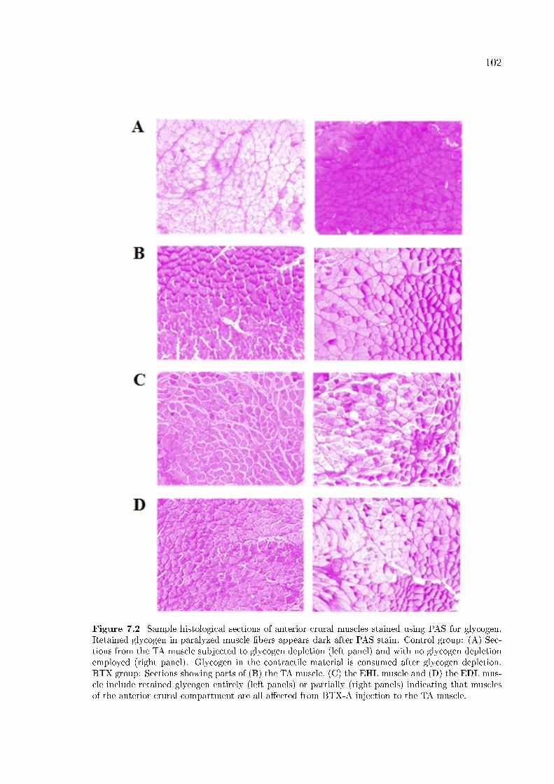
102
Figure 7.2 Sample histological sections of anterior crural muscles stained using PAS for glycogen.Retained glycogen in paralyzed muscle �bers appears dark after PAS stain. Control group: (A) Sec-tions from the TA muscle subjected to glycogen depletion (left panel) and with no glycogen depletionemployed (right panel). Glycogen in the contractile material is consumed after glycogen depletion.BTX group: Sections showing parts of (B) the TA muscle, (C) the EHL muscle and (D) the EDL mus-cle include retained glycogen entirely (left panels) or partially (right panels) indicating that musclesof the anterior crural compartment are all a�ected from BTX-A injection to the TA muscle.
103
Figure 7.3 Forces of the TA and EHL muscles as a function of increasing EDL muscle length. Activeas well as passive isometric muscle forces are shown as mean values ± SD for the control and BTXgroups. (A) TA and (B) EHL forces obtained after EDL proximal lengthening. (C) TA and (D) EHLforces obtained after EDL distal lengthening. EDL muscle-tendon complex length is expressed as adeviation from its optimum length.
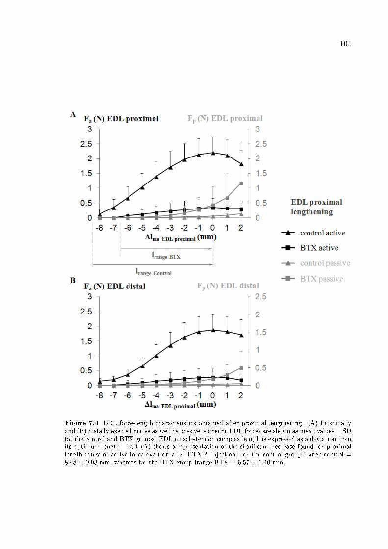
104
Figure 7.4 EDL force-length characteristics obtained after proximal lengthening. (A) Proximallyand (B) distally exerted active as well as passive isometric EDL forces are shown as mean values ± SDfor the control and BTX groups. EDL muscle-tendon complex length is expressed as a deviation fromits optimum length. Part (A) shows a representation of the signi�cant decrease found for proximallength range of active force exertion after BTX-A injection: for the control group lrange control =8.48 ± 0.98 mm, whereas for the BTX group lrange BTX = 6.57 ± 1.40 mm.
105
7.3.3 E�ects of BTX-A after distal lengthening of the EDL
Distal forces ANOVA (factors: EDL length and animal group) showed signi�-
cant main e�ects on EDL distal active forces, as well as a signi�cant interaction. Post
hoc test showed signi�cant major e�ects of BTX-A injection for most muscle lengths
(∆lma EDL > -5 mm) (Fig. 5A). However, EDL active force reduction (e.g., 82.8%,
85.6% and 83.7%, respectively at ∆lma EDL = -5 mm, ∆lma EDL = 0 mm and ∆lma
EDL = 2 mm) was not signi�cantly correlated with increasing EDL muscle length (ρ
= 0.19, p = 0.65). The distal lrange for the control group (8.24 ± 0.55 mm) decreased
signi�cantly after BTX-A injection (6.09 ± 1.36 mm) (Fig. 7.5A).
ANOVA showed signi�cant main e�ects on EDL distal passive forces, but no
signi�cant interaction. The mean passive force increase was about 3 folds (Fig. 7.5A).
Proximal forces ANOVA (factors: EDL length and animal group) showed sig-
ni�cant main e�ects on EDL proximal active forces, as well as a signi�cant interaction.
Post hoc test showed signi�cant major e�ects of BTX-A injection at all muscle lengths
(Fig. 7.5B). Minimal active force reduction was 83.7% (∆lma EDL = -6 mm).
ANOVA showed signi�cant main e�ects also on EDL proximal passive forces,
as well as a signi�cant interaction. Post hoc test showed signi�cant e�ects of BTX-A
injection (∆lma EDL ≥ 1mm): e.g., at ∆lma EDL = 2mm the passive force increased
about 12 folds (Fig. 7.5B).
7.3.4 Distal vs. proximal lengthening condition
An indicator of asymmetry of e�ects of BTX-A was that EDL active force re-
duction was inversely correlated with increasing EDL muscle length in the proximal
lengthening condition, whereas no such correlation was found in the distal lengthening
condition. However, decrease in distal lrange due to BTX-A injection was not signi�-
cantly di�erent than that in proximal lrange.
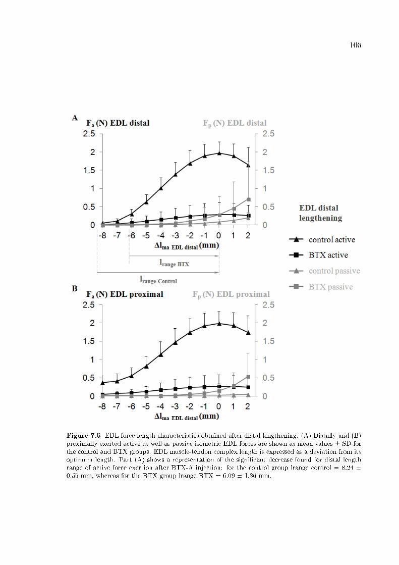
106
Figure 7.5 EDL force-length characteristics obtained after distal lengthening. (A) Distally and (B)proximally exerted active as well as passive isometric EDL forces are shown as mean values ± SD forthe control and BTX groups. EDL muscle-tendon complex length is expressed as a deviation from itsoptimum length. Part (A) shows a representation of the signi�cant decrease found for distal lengthrange of active force exertion after BTX-A injection: for the control group lrange control = 8.24 ±0.55 mm, whereas for the BTX group lrange BTX = 6.09 ± 1.36 mm.
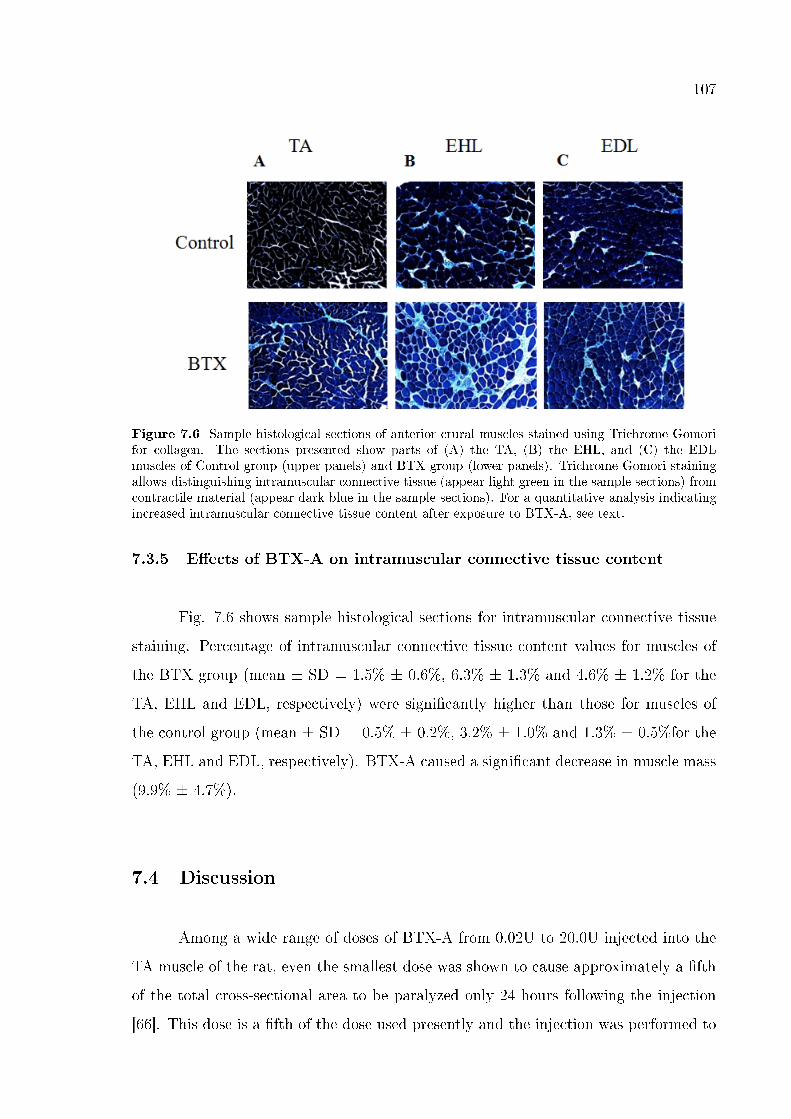
107
Figure 7.6 Sample histological sections of anterior crural muscles stained using Trichrome Gomorifor collagen. The sections presented show parts of (A) the TA, (B) the EHL, and (C) the EDLmuscles of Control group (upper panels) and BTX group (lower panels). Trichrome Gomori stainingallows distinguishing intramuscular connective tissue (appear light green in the sample sections) fromcontractile material (appear dark blue in the sample sections). For a quantitative analysis indicatingincreased intramuscular connective tissue content after exposure to BTX-A, see text.
7.3.5 E�ects of BTX-A on intramuscular connective tissue content
Fig. 7.6 shows sample histological sections for intramuscular connective tissue
staining. Percentage of intramuscular connective tissue content values for muscles of
the BTX group (mean ± SD = 1.5% ± 0.6%, 6.3% ± 1.3% and 4.6% ± 1.2% for the
TA, EHL and EDL, respectively) were signi�cantly higher than those for muscles of
the control group (mean ± SD = 0.5% ± 0.2%, 3.2% ± 1.0% and 1.3% ± 0.5%for the
TA, EHL and EDL, respectively). BTX-A caused a signi�cant decrease in muscle mass
(9.9% ± 4.7%).
7.4 Discussion
Among a wide range of doses of BTX-A from 0.02U to 20.0U injected into the
TA muscle of the rat, even the smallest dose was shown to cause approximately a �fth
of the total cross-sectional area to be paralyzed only 24 hours following the injection
[66]. This dose is a �fth of the dose used presently and the injection was performed to
108
the mid-belly of the TA as it was done in the present study. Misiaszek and Pearson
[250] demonstrated that the peak e�ectiveness of the BTX usually occurred around
day �ve and Cichon et al. [67] reported occurrence of most force decreases of the
lower hind limb �exors of the rat in the �rst four days after the injection and level
o� by the �fth day. On the other hand, remodeling of the neuromuscular junctions
may occur [250]. Nerve sprouting was observed after the �rst week [251]. Based on
these studies we determined the present injection protocol and aimed at assessing the
short-term e�ects of BTX-A. Our choice of conducting the experiments �ve days after
the injection conceivably allowed for testing after force decreases reach a steady state
and before remodeling of the neuromuscular junctions may become profound.
However, di�erences between the present protocol and common clinical practice
should be discussed for which, the following three issues appear to be important: (1)
Doses used in lower limb muscles of children with cerebral palsy range between 3 to
6U/kg [218, 240], whereas, the quantity of BTX-A injected presently equal approxi-
mately 0.32U/kg. (2) A single injection was made to the mid-belly of the target muscle
instead of using multiple injection points [216, 252]. (3) Electrical stimulation guidance
is often used in injection of BTX-A in the clinical practices [253-254], whereas such
guidance was not used presently.
Although the �rst issue suggests that in a direct comparison, the present dose
is much smaller than typical clinically used doses, substantial muscle force reductions
were shown indicating that it was quite e�ective in the rat muscles studied. We used
histological assessment to show existence of paralyzed muscle parts, which sustains
the above judgment. Shaari and Sanders [66] showed with more detailed histological
assessments that for the rat TA muscle, an increase of 10-25 fold of the dose (0.2U to
2.0U for 10µl and 0.2U to 5.0U for 25µl) was needed to double the area of paralysis.
However, at constant dose, they showed that it takes much more (100 fold) increase in
volume to achieve similar increase in the area of paralysis. These �ndings suggest that
the volume injected is also a key determinant however it is relatively less important in
determining the e�ects of BTX-A compared to the dose. On the other hand, although
it is in the same order of magnitude with what was used by Shaari and Sanders [66],
109
also the present volume injected (approximately 64µl/kg) appears very low in a direct
comparison with volumes used in lower limb muscles (2.5-8ml/kg) of children with
cerebral palsy [227, 240]. Other examples of dose and dilution volume values per kg
of the di�erent animals tested include 8.3U-0.83ml in the mouse [255], 3.5U-0.23ml
in the cat [71], 1 to10U-0.04ml in the rabbit [219]. Therefore, there is considerable
variability among the injection protocols used in di�erent species as well. The points
discussed here imply that an explicit relationship between animal studies and clinical
practice cannot be easily built. This is a general limitation and hence a limitation of
the present study as well. We suggest that new speci�c studies are necessary to bridge
the doses used in clinical and fundamental studies and a consideration of muscle size
and body mass is necessary in order to achieve a useful scaling of BTX-A doses and
volumes injected.
With regard to the second issue, a single injection as done presently has a higher
potential of saturating the local binding sites within the injected muscle and facilitat-
ing spread of BTX-A to neighboring structures [216]. Therefore, the present injection
protocol may have promoted toxin leakage to the adjacent EDL and EHL muscles.
Note however that the spread of BTX-A beyond the injection site was shown to oc-
cur also after multiple injections [70]. Such spread is considered as a side e�ect [216,
256-257] and hence it presents a challenge to control of the e�ects of the treatment.
On the other hand, studies with conclusions not in concert with these points were
also reported. Frasson et al. [240] showed recently that in children with hemiplegic
CP, BTX-A spreads from foot �exors to even antagonistic extensors. These authors
argue that such spread may be responsible in part for improving gait of the patients.
Although in animal studies e�ects of possible BTX-A spread were shown among syner-
gistic muscles [71, 239], Misiaszek and Pearson [250] showed in the cat that injection of
BTX to lateral gastrocnemius, soleus and plantaris muscles causes their EMG activity
to decrease whereas, that of the non-injected medial gastrocnemius increases. These
authors suggested that the non-injected muscle remains quite active and functional and
it compensates for the paralysis in the other muscles. Remarkably, the dose of BTX-A
injected was as high as 20 to 80U per muscle. This challenges the common clinical
expectation that BTX-A may di�use to adjacent muscles. Despite the fact that asso-
110
ciation of increased EMG signals with increased activity in the medial gastrocnemius
is a tenable one, no data directly showing absence of reduced force of this muscle is
available. These points indicate that spread of BTX-A to neighboring structures is a
complex and important issue. Therefore, its e�ects deserve to be understood well. Our
study helps improving such understanding in terms of muscular mechanics. However,
the mechanism of how such spread occurs remains unknown. Evidence was shown for
local di�usion [70] to cause such spread. E�ects of BTX-A injection were detected
even in the contralateral limb [258] suggesting that vascular di�usion is also likely.
Presently, both mechanisms may have played a role. In order to distinguish those,
new speci�c studies should be performed. Another limitation is that e�ects of possible
spread to antagonistic muscles are not assessed. This could be studied by extending
our experimental procedures.
Regarding the third issue, electrical stimulation guidance for BTX-A injection
was shown to improve the localization for deep-seated muscles [254]. Also, if the
target muscles are very small or adjacent to muscle groups that have similar functions
[253, 259], it is suggested for enhancing accuracy and speci�city. On the other hand,
such guidance was shown not to be superior to the manual placement method using
anatomical landmarks and palpation for a super�cial muscle such as gastrocnemius
muscle [260]. We studied presently e�ects of an injection to the TA muscle on force-
length characteristics of its bi-articular synergistic EDL muscle. It was not di�cult
to locate the super�cial TA using palpation and care was taken to standardize the
injection location and depth. However, if a deeper lying muscle is to be tested after
being injected, electrical stimulation guidance may improve speci�city considerably.
In the light of the discussion above, new studies are indicated to test for the role
of di�erent injection protocols at and beyond the injection site. Animal models are good
approaches to improve our understanding of fundamental phenomena regarding the
e�ects of BTX-A on muscular mechanics because of their controlled nature of testing.
Studying the e�ects directly on muscle forces and for a wide range of muscle lengths may
facilitate an improved understanding. The present �ndings obtained employing such
an approach con�rm our hypothesis and show that data measured from the bi-articular
111
EDL's tendon relocated to impose muscle length changes include decreased active forces
(muscle length dependent for the proximal lengthening condition) accompanied by a
narrower lrange both proximally and distally. Moreover, EDL passive forces were higher
in the BTX group. These �ndings include new viewpoints for a muscle a�ected by BTX-
A, with potentially important implications addressed in the subsequent paragraphs.
However, as discussed above the animal experiments do not directly represent a typical
scenario for human treatment.
An important �nding, which to our knowledge has not been reported before is
that BTX-A administration causes a decrease in the lrange of a muscle a�ected. Recent
experimental [239] and model data [212] imply that BTX-A causes sarcomeres of the
a�ected muscle to shift to longer lengths. The muscle is paralyzed partially and on
excitation, the paralyzed muscle �bers do not shorten as they would do in a BTX-A
free muscle. Due to that, the less sti� muscle can take up a greater portion the muscle-
tendon complex length. Moreover, interaction of muscle �bers with the extracellular
matrix (ECM) [28, 111, 162] can limit sarcomere shortening in the activated muscle
�bers. Modeling shows that this e�ect becomes more pronounced with increasing sti�-
ness of the ECM and hence it becomes increasingly important with increasing muscle
length [212]. In contrast, at very low muscle lengths it is not e�ectual. Occurrence of
such �longer sarcomere e�ect� can cause the muscle's optimum length to shift to a lower
length, which can be responsible for narrowing of lrange shown presently. Another ten-
able explanation is shifting of the muscle's active slack length to a higher length. Note
that since the BTX and control data were obtained from separate groups of animals,
a comparison of only the lrange can be done and it is not possible to distinguish if the
shifting of one of the characteristic muscle lengths dominates the other. Therefore, we
will consider the potential e�ects of both shifts in addressing the clinical implications
of narrowing of the lrange in the successive paragraph. On the other hand, the data
also show that length dependency of the force reductions is a possibility. This �nding
sustains the previous results that more pronounced active force reductions are likely at
lower muscle lengths [71, 239] or at joint angles that may correspond to short muscle
lengths [202].
112
Muscle hypertonicity in spasticity [29-30, 40] causes the joint to be forcefully
kept in typically a �exed position in which the muscle is expected to be short. Shifting
of the muscle's active slack length to a higher length and the possible occurrence
of more pronounced active force reductions at lower muscle lengths may mean that
a more pronounced muscle weakening is available for shorter muscle lengths. This
implies that after exposure to BTX-A, contribution of an a�ected muscle to the joint
moment in �exed joint positions is compromised more, which may be a preferable
e�ect. On the other hand, spasticity typically causes the joint range of motion to
be compromised [39-40, 261-262]. lrange is considered as an indicator of movement
capability with active force exertion of a muscle within the potential range of motion
of a certain joint. Compromised lrange indicates that the muscle may exert active
force for a smaller portion of such joint range i.e., the joint movement may be limited
before being limited by ligaments or bony constraints. Our present results show that
after exposure to BTX-A, occurrence of such compromised contribution of an a�ected
muscle to joint movement is also likely. It should be noted that we don't know whether
the lrange determined experimentally is fully functional in vivo. Although this may
be unlikely, narrowing of the lrange does not seem to agree well with an intention of
improving joint range of motion. Therefore, it may be regarded as an inferior e�ect.
It has been argued that in spasticity, muscle weakness itself is a primary source
of the functional problem [32, 52, 154, 244, 263] and that BTX-A treatment involves
a paradox of weakening the muscle to achieve better functionality [244]. Excessive
muscle weakening has been reported as an adverse e�ect [216, 231, 257, 264-266].
Awareness of potential e�ects of BTX-A on muscle lrange in addition to a variable force
reduction for di�erent muscle lengths may be important for a good clinical judgment
which was indicated as necessary [e.g., 244] to control the produced weakness. A
situation occurring frequently is that spasticity a�ects both distal and proximal joints
[224]. The present �ndings indicate that that the e�ects shown are conceivable for
both joints a bi-articular a�ected muscle spans. This may have clinically relevant
implications, which need to be tested in new studies. For example, in combined equinus
and crouch gait, contributions of m. gastrocnemius to both plantar �exion and knee
�exion moments may decrease. This appears to agree with the intended improvement
113
in gait. However, for combined crouch and hip �exor tightness, reduced contributions
of e.g., semimembranosus to hip extension moment may deteriorate conditions at the
proximal joint. A decreased lrange on the other hand, does not appear to be a favorable
e�ect for neither of such conditions. It should be noted that whether direct injection
of BTX-A into a bi-articular muscle or its di�usion from an injected muscle to such
muscle leads to the same e�ects remains unknown. Therefore, our results may be
representative of conditions in which such a muscle may be a�ected through leakage
of BTX-A only. It should also be noted that in the present conditions, asymmetry of
e�ects of proximal or distal length changes on muscle force-length characteristics remain
limited. Additionally, e�ects of BTX-A on the TA and EHL muscles were not a�ected
from changing EDL length. This shows in contrast to our previous animal experiments
performed in BTX-A free conditions [e.g., 25, 107] that mechanical interaction of muscle
with its surrounding muscular and non-muscular tissues plays a smaller role. Certain
e�ects of such epimuscular myofascial force transmission mechanism were reported in
our previous experimental study also after exposure to BTX-A but they were less
pronounced compared to those shown for the control group of animals [239]. This
may be due to the presence of paralyzed parts within the muscles of the compartment
i.e., due to reduced active force production as our results show that passive forces can
increase. The exact mechanical mechanism is not immediately apparent and needs to
be studied e.g., using muscle modeling [e.g., 25]. However, if partial muscle paralysis
is responsible for this e�ect, epimuscular myofascial force transmission is plausible to
a�ect muscular mechanics in long term after the discontuniation of BTX-A treatment.
The present results show that BTX-A changes the passive properties of the
muscular tissues as well. The slope of EDL passive force-length curves of the BTX-A
group was higher than that of the control group (e.g., for ∆lma EDL proximal ≥ 1
mm by approximately 9 folds). This is a positive indicator of increased sti�ness of
muscular connective tissues after exposure to BTX-A. Increased passive forces suggest
more speci�cally that sti�ness of the ECM may have increased. Such elevated ECM
sti�ness was reported recently for isolated rat muscle �ber bundles after BTX-A in-
jection [267]. Increased intramuscular connective tissue content shown for the BTX
group may explain this. Note that our histological assessment con�rms an increased

114
proportion of the ECM rather than a net increase of the ECM. Denervation leads to
atrophy of rat muscles within the �rst week [220]. Also BTX-A was reported to cause
atrophy [221-222]. Similarly our results indicate occurrence of atrophy as muscle mass
normalized to body mass was shown to decrease. Biliante et al. [68] showed that such
atrophy involves decreased muscle �ber diameters as well as density. Therefore, a net
increase of the ECM is tenable.
However, ruling out the possibility that the muscle's epimuscular connections
may have also become sti�er is not possible. One reason for this is that increased passive
forces were shown not only at higher muscle lengths at which the ECM is stretched, but
also at lower. Passive distal EDL forces measured after distal lengthening were higher
after BTX-A administration for all muscle lengths including short ones. Moreover, the
increased passive forces of the TA and EHL muscles were measured at lower lengths at
which they were restrained. These results indicate occurrence of an increased sti�ness
of also the epimuscular connections, which should be tested speci�cally.
The possibility that BTX-A causes an increase in the sti�ness of muscular con-
nective tissues does not seem to be in concert with the intended reduction of passive
resistance of spastic muscle at the joint [232, 246, 256, 268-269]. It should be noted
that the clinical �nding that BTX-A causing decreased passive resistance to stretch
[232, 270-271] is mainly ascribable to decreased hypertonicity of the spastic muscle i.e.,
a reduced spasticity [272]. Even in non-spastic mouse muscle, large doses of BTX-A
were shown to cause reduced tone and disrupted stretch re�ex, leading to decreased
passive resistance during stretching [273]. The present experiments were conducted
also on normal muscles however, the measurements do not include a passive stretching
with movement as used in the evaluation of spasticity, and no re�ex loop was active.
Therefore, a modulation of muscle tone is not a part of our �ndings, and the present
increase in muscle passive force re�ects enhanced muscular tissue sti�ness. The term
�sti�ness� is used interchangeably with �passive resistance to stretch� [272, 274]. We
consider that the latter involves the former, which characterizes mechanical resistance
of non-contractile muscular structures to increased length. Based on the present and
previous �ndings, we suggest the following. Decreased passive resistance to stretch af-
115
ter BTX-A injection is a net e�ect of decreased hypertonicity and may be compromised
by increased tissue sti�ness. On the other hand, the present �ndings were obtained
only �ve days after BTX-A injection. However, similar e�ects on passive mechanical
properties shown by Thacker et al. [267] were obtained 1 month after, suggesting their
continuation in time. These authors argued that the e�ects of BTX-A on normal muscle
and on spastic muscle may be di�erent. Recently, we have shown that human spastic
Gracilis muscle tested intra-operatively shows no abnormal mechanics [143]. However,
passive muscle force was not available together with data from healthy subjects. Given
the lack of data allowing for a direct comparison, also Thacker et al.'s suggestion may
explain the di�erence between �ndings obtained from conditions in health and spas-
ticity. Nevertheless, we believe whether BTX-A causes increased muscular connective
tissue sti�ness also in spasticity may be an important question that needs to be ad-
dressed in new speci�c studies. BTX-A injections are usually combined with additional
techniques such as strengthening exercises [275-276], exercises performed to improve
joint range of motion [277] as well as stretching and splinting [278-279]. The functional
bene�t expected by using BTX-A may depend largely on these techniques and our
�ndings may be relevant for them.
In concussion, our results indicate that the e�ects of BTX-A on muscular me-
chanics are more complex than just weakening of the a�ected muscle characterized
by a reduction in its active force. They also include a potential length dependency
of reduction of the active force and narrowing of the muscle's length range of active
force exertion. Moreover, not only the active properties but also the passive properties
may change featuring increased passive muscle forces. Therefore, BTX-A may lead to
compromised joint movement due to both a narrower active movement range and an
increased passive resistance. Spread of BTX-A to a bi-articular muscle may cause both
joints the muscle spans to be a�ected. Such e�ects may be clinically relevant because,
as some are additional to the intended ones, others may even be unintended.
116
8. GENERAL DISCUSSION AND CONCLUSIONS
In this thesis, the e�ects of spastic cerebral palsy on muscle mechanics were
investigated with human experiments and the e�ects of botulinum toxin administra-
tion as a conservative therapy applied to spastic muscles, and aponeurotomy (AT) as
a surgical method to correct impaired joint function were investigated with animal
experiments. Myofascial force transmission was addressed as a key concept both to
understand the etiology of the disease and treatment methods widely used.
8.1 Mechanics of Human Spastic Muscles, Limitations, and Fu-
ture Directions
The knowledge on the mechanics of human muscles is very important for under-
standing locomotion in health and also in disease. Most of the information gained from
the studies on muscle architecture by investigating cadavers [87] or by using modeling
[89] or imaging methods such as ultrasound [49-50] and magnetic resonance imaging
[51-53] did not give direct data on force production capacity of muscles. Other indirect
approaches used dynamometry [90] or torque measurements [88-89] were targeted the
joints. However, there has been a lack of information on relating the force production
capacity of speci�c muscles and the joint angle which is associated with functional
joint problems. One of our studies presented in chapter 2 [120] showed a capability to
close such gap for human GRA muscle with a novel intra-operative force measurement
method developed.
Healthy GRA muscle isometric forces with respect to knee angle measured
showed substantial inter-subject variability: the values of muscle forces, stress over
tendons, and the knee angle where the maximum force generated were di�erential for
the subjects. This study suggested that typical subject anthropometrics cannot be
used as predictors due to these experimentally obtained data not being correlated with
subject anthropometrics. It was also shown for the �rst time that GRA muscle in
117
health has very large operational length range at least it spans 120° of knee �exion to
full knee extension. This is consistent with its architectural features with long �bers
[134] and large excursion [98].
Additionally, this study [120] showed the occurrence of history e�ects at �rst
time for healthy human muscles. History e�ects de�ned as force changes at lower mus-
cle lengths due to muscle activity at high lengths should be taken into consideration
if consecutive measurements are performed since they interfere with the results. In
the Appendix-A, to ensure such measurements, a certain preconditioning method was
suggested. In chapter 5, this method was used in the animal experiments since con-
secutive measurements were performed: We tested the e�ects of aponeurotomy (AT)
following force-length data collections in the intact condition and after preparatory
dissections (PD)). On the other hand, for the human experiments presented at chapter
2-4, and also for the animal experiments using di�erent groups of animals to compare
the applied conditions such as BTX-A (presented at chapter 6 and 7) no isometric force
measurements were performed subsequent to activity at high length. Therefore, pre-
conditioning was not needed. It should be noted that if similar experiments including
consecutive data collection are planned in human, muscles should also be precondi-
tioned by activating at high and low lengths until the same amount of isometric force
was measured at low lengths.
In spastic cerebral palsy, treatments are mostly applied to particular muscles or
muscle groups such as administration of BTX-A or lengthening surgery performed on
knee �exors to correct crouch gait or on gastrocnemius or Achilles tendon to correct
equines deformity. However, the contributions of the speci�c muscles on the joints
they span are not well known. One of our present intra-operative studies on CP pa-
tients [143] revealed the contribution of spastic GRA muscle on knee joint. The main
outcome of this study was that the spastic GRA muscle did show no abnormal charac-
teristics. Even muscles were diagnosed with spasticity, (i) GRA did not have a narrow
joint range of force exertion and (ii) at low length, higher forces presumably causing
knee joint to stay in �exed position were not available. In the experiments, although
most of the neighboring structures were intact, the synergistic or antagonistic mus-
118
cles were not activated. Considering the experimental conditions including exclusive
stimulation of GRA, the possibility of Dr. Huijing's hypothesis [62] to be valid was
suggested: The source of high forces observed in the �exed joint positions should be
due to activation of antagonistic muscles at high lengths. Such approach should include
epimuscular myofascial force transmission mechanism to be held between antagonists
[107, 170]. We improved that approach and test if spastic GRA characteristics change
with simultaneous stimulation of its antagonist vastus medialis (VM) in the context
of (i) narrowed joint range and (ii) availability of high force at knee �exion. In this
study presented at Chapter 4, operational range of spastic GRA muscle was found to
be narrowed and peak GRA forces shifted to more �exed position due to simultane-
ous activity of VM. Also, comparably higher forces were measured at �exion. This
indicates the determinant role of inter-antagonistic force transmission due to sti�ened
epimuscular pathways.
Our results on human spastic muscles indicated that impeded joint motion is
not due to abnormal function of each muscle but simultaneous activation of agonist-
antagonists. It should be noted that our data are limited with a single muscle and the
e�ects of activating its antagonist. To understand the knee joint function in spastic
CP other muscles crossing the knee should be investigated. It may be critical to dis-
criminate the characteristics of long �bered GRA from that of the short �bered biceps
femoris since they are known to di�erentiate in function, they may also di�erentially
adapt to spasticity. Functional map for all knee �exors and extensors seems to be cru-
cial for also understanding the myofascial pathways which are presently shown to have
a major and determinant role. Another important limitation of our intra-operative
method is the lack of capability of measuring passive forces since the force transducers
work on a principle of torque measurement [94]. Therefore, our results do not re�ect
passive tissue sti�ness presumably high for spastic muscles [57-59].
In conclusion, our results suggesting the key role of inter-antagonistic force trans-
mission should be further investigated with spastic muscle model to reveal such mecha-
nism in detail and also by adding histological examinations to our experimental method
improved to measure passive forces.
119
8.2 Treating Spastic Cerebral Palsy, Limitations, and Future
Directions
Animal experimentation facilitates collecting speci�c information in a more con-
trolled manner even the limitations such as di�culty to re�ect the results to the clinics
should be considered. Experimental protocols have been highly standardized for nu-
merous animals to study muscle force-length characteristics. However, many protocols
included measuring forces at one muscle length or from only one tendon of a bi-articular
muscle and assumed that such measurements represent the isometric muscle function.
Moreover, treatment methods for neuromuscular disorders are determined according
to such approaches neglecting length e�ects and also not considering myofascial force
transmission. Our experimental protocols considering the intactness of most of the
epimuscular connections did show that the e�ects of some treatment methods are not
limited with the target muscle. Present studies on animals revealed that EMFT deter-
mines not only the etiology but also the treatment methods of spasticity.
8.2.1 Muscle lengthening surgery
Aponeurotomy (AT) i.e. cutting of the aponeurosis transversely aimed at length-
ening the contractured muscles. In surgery, it is applied after preparatory dissection
(PD) to reach the targeted area. Our experimental procedure mimicking PD and
subsequent AT on anterior crural muscles of rat (presented at chapter 5) showed (i)
di�erential e�ects at proximal and distal tendons of target muscle and (ii) substantial
force decrease for the non-operated neighboring muscle. Therefore, EMFT mechanism
was suggested to dominate the e�ects of muscle lengthening surgery. Since these dif-
ferential and unintended e�ects on muscle forces may yield additional favorable e�ects
for the targeted joint, but also contrasting e�ects particularly for the non-targeted
joint, we conclude that EMFT should be taken into account to improve the functional
outcome of the surgery planned to correct the impeded joint.
The importance of such e�ects was also shown for other treatment methods
120
such as tenotomy and tendon transfer surgery. For instance, Kreulen et al. [60] showed
for �exor carpi ulnaris muscle that distal tenotomy caused only a minor e�ect how-
ever, subsequent dissection of epimuscular connections of the muscle caused substantial
changes in both muscle shortening and activation. Also, previous studies [192, 280-281]
showed that e.g. �exor muscle whose tendon was transferred to support extension still
produces �exor moment. It was suggested that muscle connected to its origin with
epimuscular connections caused such unexpected outcomes. To further investigate the
implications of this approach, e.g. tendon splitting surgery or other conservative treat-
ments for musicians arm or repetitive strain injury should be tested with controlled
experimental methods like the ones presented in this thesis.
8.2.2 BTX-A Application
It is aimed at reducing muscle spasticity with the injection of BTX-A that
inhibits acetylcholine release into the presynaptic cleft in the neuromuscular junction
[e.g., 65]. Such treatment on the other hand creates a dilemma with its weakening e�ect
contrary to the need of CP patients to improve joint function. To prevent joint from
excessive weakening while reducing spasticity concurrently, it is essential to understand
the e�ects of BTX-A on the whole compartment. BTX-A injection has known to a�ect
the muscles beyond the injection site [70, 72]. However, such e�ects on the neighboring
muscles have not been studied systematically. A novel animal experimental model
[239] presented at chapter 6 was developed to investigate acutely the e�ects of BTX-
A on force-length characteristics not only of the injected but also of the non-injected
muscles of an entire intact compartment in conditions close to those in vivo. Additional
to the major active force reductions due to BTX-A, length dependency of the e�ects
(i.e. correlation between injected muscle's length and the e�ects of BTX-A) and in
contrast, passive force increases were shown. The following study focusing on bi-
articular muscle characteristics presented here at Chapter 7 showed (i) the narrowed
length range of force exertion that BTX-A caused even acutely and (ii) pronounced
passive force increase for all the muscles in the compartment which is in contrast to
the general aim of the treatment. Furthermore, the length dependency of the e�ects
121
was shown to be not valid for every experimental condition.
These studies focused on the acute e�ects of BTX-A suggested that to improve
the overall function unintended force reductions shown (i) for non-targeted joint due
to the di�erential e�ects on bi-articular target muscle and also (ii) for neighboring
muscle should be taken into account. One of the most important outcomes of this
study was to �nd a passive force increase due to BTX-A injection. Such passive force
enhancement was explained with increased and possibly sti�ened collagen content in
contrast to the atrophy occurs even at �fth day of BTX-A administration. In any
condition, passive force increase due to BTX-A shown in our present studies at �rst
time should be considered when determining additional treatments such as physical
therapy or strengthening exercises.
On the other hand, our studies showed that BTX-A caused a prominent reduc-
tion in proximo-distal force di�erences which are direct measures of myofascial loads.
Such disappearing of EMFT may be an important part of the BTX-A treatment mech-
anism. If the therapy with this toxin is e�ective since it diminishes the e�ects of
connections even the amount of ECM increases. Then, such mechanism should further
be revealed with new studies investigating how (i) the toxin cause collagen content
increase and (ii) epimuscular loads changes in the presence of other muscles than the
targeted. Experimental and modeling methods should both be used for that. More-
over, the changes in passive force, interaction of muscles, and accordingly the amount
of collagen are needed to be examined in long term.
122
Appendix A. PRECONDITIONING REMOVES LENGTH
HISTORY EFFECTS AND ENSURES SUCCESSIVE
FORCE-LENGTH MEASUREMENTS
A.1 Introduction
Previous activity at high (over optimum) muscle lengths a�ects muscular me-
chanics at lower lengths [15] causing typically a reduction in the force measured at
the same low length again. Such length history e�ects were shown to occur for tib-
ialis anterior [170], deep �exors [107], and extensor digitorum longus (EDL) [15, 282]
muscles of the rat, a commonly used species for an animal muscle mechanics model.
However, sizable length history e�ects were also reported for human gracilis muscle
[120] suggesting that they are not restricted to the rat.
Testing the e�ects of di�erent conditions with successive measurements of muscle
force-length (FL) characteristics is an important and common practice [24, 27, 92,
283-284] for muscle mechanics experimentation. In order to isolate the role of the
condition tested, length history e�ects need to be removed. A method proposed for
that purpose was preconditioning of the muscle to be lengthened [20]: subsequent
isometric contractions at high and low lengths to be performed prior to the experiment
until the force reduction at low length vanishes. These alternating contractions were
reported to eliminate length history e�ects for a set of FL data [20, 62, 174, 285-286].
However; the outcome of such preconditioning for successive FL measurements is not
known. The goal of this study was to test if such preconditioning is a safe method for
three consecutive FL measurements for both lengthened and restrained muscles.
123
A.2 Methods
A.2.1 Surgical procedures
Surgical and experimental procedures were approved by the Committee on
Ethics of Animal Experimentation at the Bo§aziçi University.
Male Wistar rats (n=8, mean body mass 325.5±13.7g) were anaesthetized using
intraperitoneally injected urethane solution (1.2 ml of 12.5% urethane solution/100 g
body mass). Additional doses were given if necessary (maximally 0.5 ml). Immediately
following experiments, animals were sacri�ced by using an overdose of urethane solu-
tion. After surgery needed to expose EDL, tibialis anterior (TA) and extensor hallicus
longus (EHL) muscles (Figure A1 a) the rat was mounted in the experimental setup
(Figure A1 b).
A.2.2 Experimental conditions and procedure
All muscles studied were activated maximally by stimulation of the sciatic nerve
(Biopac Systems stimulator, STMISOC) with a constant current of 2mA (0.1ms pulse
width, 100 Hz). After setting EDL to a target length, two twitches were evoked, 500 ms
after the second twitch, the muscles were tetanized and 400 ms subsequently, another
twitch was evoked. During the tetanic plateau, muscle total forces were measured and
averaged at an interval of 150 ms subsequent to 25 ms after evoking tetanic stimulation.
After each application of this stimulation protocol, the muscles were allowed to recover
at low muscle length, for 2 minutes.
Isometric forces were measured simultaneously from EDL proximal and TA+EHL
distal tendons (kept at their reference position at all times during the experiment) and
from the distal tendon of EDL which was repositioned (in 1 mm steps until 2 mm over
distal optimum length, starting at active slack length) to quantify FL characteristics.
124
Subsequent to a full measurement of each FL data, control contractions (CC)
were performed at EDL reference low length (lref, i.e., 3 mm over distal active slack
length) in order to determine the e�ects of previous activity at high length (over opti-
mum length) on forces exerted by the experimental muscles at lower lengths.
Preconditioning Isometric forces at low (lref) and high (lopt+2) lengths of EDL
muscle distally were measured sequentially until the force di�erence between succes-
sively measured forces at identical lengths were less than 3%. Each experiment involved
collection of a complete set of FL data and subsequent control measurements. The �rst
experiment (FL-1 and CC-1) was performed without any preconditioning. Therefore,
the data was prone to length history e�ects. Prior to the subsequent three experiments
(FL -2 to 4 and CC-2 to 4) however, a preconditioning was performed. Therefore, it
was tested (i) if such preconditioning does remove length history e�ects acutely and
(ii) if its e�ects are reliable for subsequent two experiments.
A.2.3 Processing of data and statistics
Data for muscle total forces (F) in relation to muscle-tendon complex length
(lmt) were �tted with a polynomial function using a least squares criterion
y=b0+b1x+b2x2 + ... + bnxn (1)
where y represents F and x represents lmt. b0, b1. . . bn are coe�cients deter-
mined in the �tting process. Polynomials that best described the experimental data
were selected by using one-way analysis of variance (ANOVA) [125]: the lowest or-
der of the polynomials that still added a signi�cant improvement to the description of
changes of muscle-tendon complex length and muscle force data were selected. These
functions were used for determining mean forces, standard errors (SE), and EDL opti-
mum length distally. One-way ANOVA was used to test for the history e�ects within
each experiment: di�erences between EDL forces exerted at lref (i) during collection
of FL data and (ii) after CC were considered signi�cant at p<0.05.
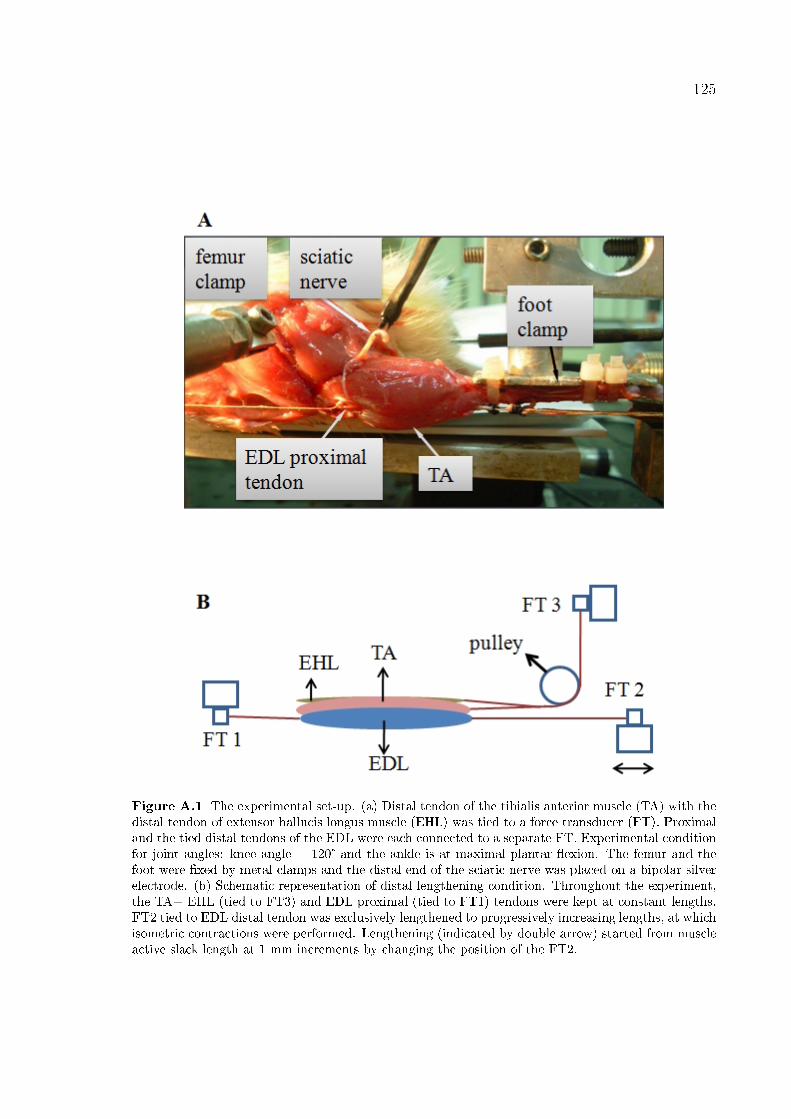
125
Figure A.1 The experimental set-up. (a) Distal tendon of the tibialis anterior muscle (TA) with thedistal tendon of extensor hallucis longus muscle (EHL) was tied to a force transducer (FT). Proximaland the tied distal tendons of the EDL were each connected to a separate FT. Experimental conditionfor joint angles: knee angle = 120° and the ankle is at maximal plantar �exion. The femur and thefoot were �xed by metal clamps and the distal end of the sciatic nerve was placed on a bipolar silverelectrode. (b) Schematic representation of distal lengthening condition. Throughout the experiment,the TA+ EHL (tied to FT3) and EDL proximal (tied to FT1) tendons were kept at constant lengths.FT2 tied to EDL distal tendon was exclusively lengthened to progressively increasing lengths, at whichisometric contractions were performed. Lengthening (indicated by double arrow) started from muscleactive slack length at 1 mm increments by changing the position of the FT2.
126
Two-way ANOVA for repeated measures (factors: EDL muscle-tendon complex
length (lmt) and experiment) was performed to test for the e�ects of preconditioning
across experiments on EDL muscle distal and proximal FL characteristics and TA+EHL
distal forces. Bonferroni post-hoc tests were performed to further locate signi�cant
di�erences.
A.3 Results
A.3.1 EDL
For experiment 1 and for both proximal and distal EDL forces, ANOVA showed
signi�cant di�erences (drops of 48.0% and 33.4%, respectively) between forces measured
at lref during collection of FL data and after CC. In contrast, after preconditioning
no such signi�cant force di�erences were shown indicating that preconditioning can
remove length history e�ects occurring at lower muscle lengths within three successive
experiments.
Both distally and proximally, ANOVA (factors EDL lmt and experiment) showed
signi�cant main e�ects, but no signi�cant interaction. Signi�cant e�ects were located
only between experiment 1 and 2 (post-hoc test) indicating that preconditioning re-
moves length history e�ects also across experiments. Lack of a signi�cant interaction
indicates that e�ects are similar for FL data at four experiments for all length range
spanned. On the other hand, if EDL forces collected during experiment 1 to 4 are
tested for each EDL length (one-way ANOVA), signi�cant e�ects of the experiment
were shown only for a few lengths around lref distally (Figure A.2a) and proximally
(Figure A.2b). The same test showed no signi�cant e�ects of experiments at any length
if FL -1 was excluded. Therefore, these analyses indicate that length history e�ects
are limited to lower muscle lengths and disappear after preconditioning.
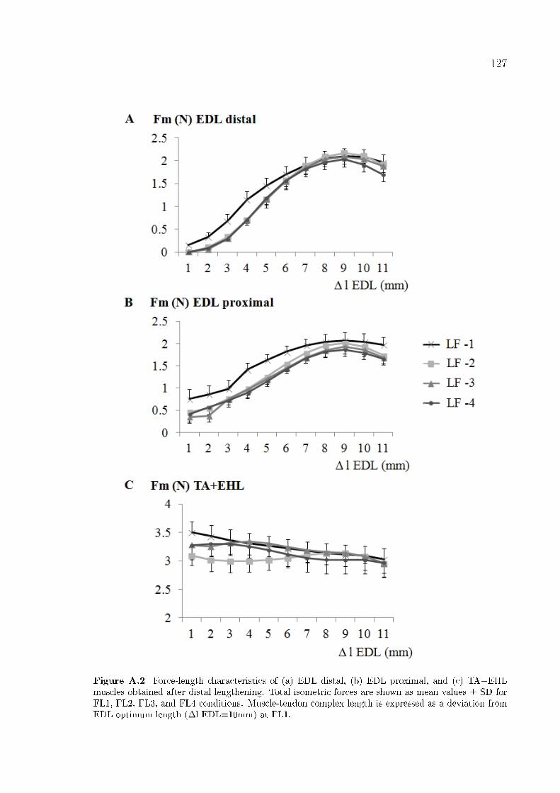
127
Figure A.2 Force-length characteristics of (a) EDL distal, (b) EDL proximal, and (c) TA+EHLmuscles obtained after distal lengthening. Total isometric forces are shown as mean values ± SD forFL1, FL2, FL3, and FL4 conditions. Muscle-tendon complex length is expressed as a deviation fromEDL optimum length (Δl EDL=10mm) at FL1.
128
A.3.2 TA+EHL
ANOVA showed no signi�cant di�erences between forces measured at lref during
collection of FL data and after CC within each experiment. Therefore, TA+EHL forces
are not a�ected by previous activity of its synergistic muscle at high length.
On the other hand, ANOVA (factors EDL lmt and experiment) showed only
a signi�cant e�ect of experiment thus neither signi�cant e�ects of EDL length nor a
signi�cant interaction. Signi�cant e�ects were located only between experiment 1 and
2 (post-hoc test). These �ndings of analyses performed across experiments indicate
that preconditioning of EDL muscle does a�ect the forces of TA+EHL similarly for all
EDL lengths and it also stabilizes forces of TA+EHL for three successive experiments
performed subsequently.
A.4 Discussion
The size of length history e�ects characterized by force depression particularly
at low muscle lengths following the activity at high lengths [15] was suggested to
be determined by CC at low reference length [62]. In previous studies [107, 170,
282], di�erences between the force measured during FL data collection and CC at
identical low length were used to quantify such e�ects. Our present results showing
length history e�ects for EDL before preconditioning con�rm previous �ndings. Also,
signi�cant e�ects across experiments shown for low lengths around lref indicate that
control measurements (in our case 3 mm over active slack length) performed after
collecting a set of FL data do re�ect the e�ects of previous activity and con�rm that
quantifying method used.
Mechanism of length history e�ects is not exactly apparent; however, viscoelas-
tic properties of muscle and fascial tissues presumably having major role suggest that
considering the experimental protocols particularly include lengthening, there is poten-
tial for systematic changes that may interfere with the results. With the intention of
129
collecting reproducible data, preconditioning of viscoelastic materials known to mini-
mize variability is performed [287-289]. Loading and unloading is a widely used method
for various biological tissues such as tendon [290], ligament [291], spinal cord [292], or
skin [293]. With same principle, by applying stretch and release on passive muscle
�bers of rat soleus with constant velocity were shown to stabilize sarcomere lengths
and forces after third cycle [294]. Basic warm-up and stretching exercises performed
mostly to prevent damage [295-296] also aimed to precondition muscles isometrically.
In our case, EDL was preconditioned by isometric contractions at high and low muscle
lengths to principally mimic the following FL data collection. Such preconditioning
tested systematically at our present study for the �rst time is shown to serve the pur-
pose of generating the same amount of force with non-signi�cant deviation at the same
length for identical conditions.
Additionally, our present study showed history e�ects for the restrained muscles
before preconditioning. Such e�ects may be ascribed to previously stretched inter- and
extramuscular connections with muscle lengthening. Thus, it suggests the substantial
role of connective tissue on history e�ects. Our previous results indicating the impor-
tance of both intermuscular connections and compartmental fascia are consistent with
present results: compartmental fasciotomy and subsequently dissecting the intermuscu-
lar connections of anterior crural muscles of rat caused history e�ects to decrease [231].
Therefore, passive tissues having a partial role on history e�ects should be considered
for preconditioning.
In conclusion, our present study showed that preconditioning removes history ef-
fects and ensures at least three consecutive force-length measurements for both length-
ened and restrained muscles. History e�ects limited with lower muscle lengths are
dependent on the activity of muscle at high lengths since major force reductions before
preconditioning were shown for the lengthened muscle. On the other hand, minor but
signi�cant e�ects shown for the restrained muscles due to being adjacent to the length-
ened muscle with stretched fascial connections during lengthening preconditioning was
also shown to stabilize the neighboring restrained muscles.
130
REFERENCES
1. Lieber, R.L., "Skeletal muscle architecture: implications for muscle function and surgical
tendon transfer," J Hand Ther, Vol. 6, p. 105-13, 1993.
2. Huijing, P.A., "Muscle, the motor of movement: properties in function, experiment and
modelling," J Electromyogr Kinesiol, Vol. 8, p. 61-77, 1998.
3. Maganaris, C.N., V. Baltzopoulos, and A.J. Sargeant, "In vivo measurements of the triceps
surae complex architecture in man: implications for muscle function," J Physiol, Vol. 512 (Pt 2),
p. 603-14, 1998.
4. Narici, M.V., L. Landoni, and A.E. Minetti, "Assessment of human knee extensor muscles stress
from in vivo physiological cross-sectional area and strength measurements," Eur J Appl Physiol
Occup Physiol, Vol. 65, p. 438-44, 1992.
5. Willems, M.E. and P.A. Huijing, "Heterogeneity of mean sarcomere length in different fibres:
effects on length range of active force production in rat muscle," Eur J Appl Physiol Occup
Physiol, Vol. 68, p. 489-96, 1994.
6. Fukunaga, T., et al., "Muscle architecture and function in humans," J Biomech, Vol. 30, p. 457-
63, 1997.
7. Gans, C. and A.S. Gaunt, "Muscle architecture in relation to function," J Biomech, Vol. 24
Suppl 1, p. 53-65, 1991.
8. Gans, C. and W.J. Bock, "The functional significance of muscle architecture--a theoretical
analysis," Ergeb Anat Entwicklungsgesch, Vol. 38, p. 115-42, 1965.
9. Narici, M.V., et al., "In vivo human gastrocnemius architecture with changing joint angle at rest
and during graded isometric contraction," J Physiol, Vol. 496 ( Pt 1), p. 287-97, 1996.
10 Kawakami, Y., et al., "Specific tension of elbow flexor and extensor muscles based on magnetic
resonance imaging," Eur J Appl Physiol Occup Physiol, Vol. 68, p. 139-47, 1994.
11. Rutherford, O.M. and D.A. Jones, "Measurement of fibre pennation using ultrasound in the
human quadriceps in vivo," Eur J Appl Physiol Occup Physiol, Vol. 65, p. 433-7, 1992.
12. Tidball, J.G., "Force transmission across muscle cell membranes," J Biomech, Vol. 24 Suppl 1,
p. 43-52, 1991.
13. Patel, T.J. and R.L. Lieber, "Force transmission in skeletal muscle: from actomyosin to external
131
tendons," Exerc Sport Sci Rev, Vol. 25, p. 321-63, 1997.
14. Pardo, J.V., J.D. Siliciano, and S.W. Craig, "A vinculin-containing cortical lattice in skeletal
muscle: transverse lattice elements ("costameres") mark sites of attachment between myofibrils
and sarcolemma," Proc Natl Acad Sci U S A, Vol. 80, p. 1008-12, 1983.
15. Huijing, P.A. and G.C. Baan, "Extramuscular myofascial force transmission within the rat
anterior tibial compartment: proximo-distal differences in muscle force," Acta Physiol Scand,
Vol. 173, p. 297-311, 2001.
16. Huijing, P.A., "Muscular force transmission necessitates a multilevel integrative approach to the
analysis of function of skeletal muscle," Exerc Sport Sci Rev, Vol. 31, p. 167-75, 2003.
17. Huijing, P., "Muscular force transmission: a unified, dual or multiple system? A review and
some explorative experimental results," Arch Physiol Biochem, Vol. 107, p. 292-311, 1999.
18. Nishimura, T., A. Hattori, and K. Takahashi, "Ultrastructure of the intramuscular connective
tissue in bovine skeletal muscle. A demonstration using the cell-maceration/scanning electron
microscope method," Acta Anat (Basel), Vol. 151, p. 250-7, 1994.
19. Huijing, P.A. and G.C. Baan, "Myofascial force transmission causes interaction between
adjacent muscles and connective tissue: effects of blunt dissection and compartmental
fasciotomy on length force characteristics of rat extensor digitorum longus muscle," Arch
Physiol Biochem, Vol. 109, p. 97-109, 2001.
20. Huijing, P.A., H. Maas, and G.C. Baan, "Compartmental fasciotomy and isolating a muscle
from neighboring muscles interfere with myofascial force transmission within the rat anterior
crural compartment," J Morphol, Vol. 256, p. 306-21, 2003.
21. Huijing, P.A., et al., "Extramuscular myofascial force transmission also occurs between
synergistic muscles and antagonistic muscles," J Electromyogr Kinesiol, Vol. 17, p. 680-9,
2007.
22. Yucesoy, C.A., et al., "Extramuscular myofascial force transmission: experiments and finite
element modeling," Arch Physiol Biochem, Vol. 111, p. 377-88, 2003.
23. Yucesoy, C.A., et al., "Pre-strained epimuscular connections cause muscular myofascial force
transmission to affect properties of synergistic EHL and EDL muscles of the rat," J Biomech
Eng, Vol. 127, p. 819-28, 2005.
24. Maas, H., H.J. Meijer, and P.A. Huijing, "Intermuscular interaction between synergists in rat
132
originates from both intermuscular and extramuscular myofascial force transmission," Cells
Tissues Organs, Vol. 181, p. 38-50, 2005.
25. Yucesoy, C.A., et al., "Effects of inter- and extramuscular myofascial force transmission on
adjacent synergistic muscles: assessment by experiments and finite-element modeling," J
Biomech, Vol. 36, p. 1797-811, 2003.
26. Maas, H., G.C. Baan, and P.A. Huijing, "Muscle force is determined also by muscle relative
position: isolated effects," J Biomech, Vol. 37, p. 99-110, 2004.
27. Maas, H., et al., "The relative position of EDL muscle affects the length of sarcomeres within
muscle fibers: experimental results and finite-element modeling," J Biomech Eng, Vol. 125, p.
745-53, 2003.
28. Yucesoy, C.A., et al., "Three-dimensional finite element modeling of skeletal muscle using a
two-domain approach: linked fiber-matrix mesh model," J Biomech, Vol. 35, p. 1253-62, 2002.
29. Brown, J.K., et al., "Neurophysiology of lower-limb function in hemiplegic children," Dev Med
Child Neurol, Vol. 33, p. 1037-47, 1991.
30. Mirbagheri, M.M., et al., "Intrinsic and reflex stiffness in normal and spastic, spinal cord injured
subjects," Exp Brain Res, Vol. 141, p. 446-59, 2001.
31. O'Dwyer, N.J. and L. Ada, "Reflex hyperexcitability and muscle contracture in relation to
spastic hypertonia," Curr Opin Neurol, Vol. 9, p. 451-5, 1996.
32. Gracies, J.M., "Pathophysiology of spastic paresis. I: Paresis and soft tissue changes," Muscle
Nerve, Vol. 31, p. 535-51, 2005.
33. Katz, R.T. and W.Z. Rymer, "Spastic hypertonia: mechanisms and measurement," Arch Phys
Med Rehabil, Vol. 70, p. 144-55, 1989.
34. Gracies, J.M., "Pathophysiology of spastic paresis. II: Emergence of muscle overactivity,"
Muscle Nerve, Vol. 31, p. 552-71, 2005.
35. Huijing, P.A. and R.T. Jaspers, "Adaptation of muscle size and myofascial force transmission: a
review and some new experimental results," Scand J Med Sci Sports, Vol. 15, p. 349-80, 2005.
36. Williams, P.E. and G. Goldspink, "Changes in sarcomere length and physiological properties in
immobilized muscle," J Anat, Vol. 127, p. 459-68, 1978.
37. Williams, P.E. and G. Goldspink, "Connective tissue changes in immobilised muscle," J Anat,
Vol. 138 ( Pt 2), p. 343-50, 1984.
133
38. Tardieu, C., et al., "Muscle hypoextensibility in children with cerebral palsy: I. Clinical and
experimental observations," Arch Phys Med Rehabil, Vol. 63, p. 97-102, 1982.
39. Botte, M.J., V.L. Nickel, and W.H. Akeson, "Spasticity and contracture. Physiologic aspects of
formation," Clin Orthop Relat Res, p. 7-18, 1988.
40. O'Dwyer, N.J., L. Ada, and P.D. Neilson, "Spasticity and muscle contracture following stroke,"
Brain, Vol. 119 ( Pt 5), p. 1737-49, 1996.
41. Sheean, G., "The pathophysiology of spasticity," Eur J Neurol, Vol. 9 Suppl 1, p. 3-9; dicussion
53-61, 2002.
42. Farmer, S.E. and M. James, "Contractures in orthopaedic and neurological conditions: a review
of causes and treatment," Disabil Rehabil, Vol. 23, p. 549-58, 2001.
43. Tedroff, K., et al., "Does loss of spasticity matter? A 10-year follow-up after selective dorsal
rhizotomy in cerebral palsy," Dev Med Child Neurol, Vol. 53, p. 724-9, 2011.
44. Olsson, M.C., et al., "Fibre type-specific increase in passive muscle tension in spinal cord-
injured subjects with spasticity," J Physiol, Vol. 577, p. 339-52, 2006.
45. McDowell, B.C., et al., "Passive Range of Motion in a Population-Based Sample of Children
with Spastic Cerebral Palsy Who Walk," Physical & Occupational Therapy in Pediatrics, Vol.
32, p. 139-150, 2012.
46. Lin, F.M. and M. Sabbahi, "Correlation of spasticity with hyperactive stretch reflexes and motor
dysfunction in hemiplegia," Arch Phys Med Rehabil, Vol. 80, p. 526-30, 1999.
46. Sheean, G. and J.R. McGuire, "Spastic hypertonia and movement disorders: pathophysiology,
clinical presentation, and quantification," PM R, Vol. 1, p. 827-33, 2009.
48. Nordmark, E., et al., "Development of lower limb range of motion from early childhood to
adolescence in cerebral palsy: a population-based study," BMC Med, Vol. 7, p. 65, 2009.
48. Fry, N.R., M. Gough, and A.P. Shortland, "Three-dimensional realisation of muscle morphology
and architecture using ultrasound," Gait Posture, Vol. 20, p. 177-82, 2004.
50. Malaiya, R., et al., "The morphology of the medial gastrocnemius in typically developing
children and children with spastic hemiplegic cerebral palsy," J Electromyogr Kinesiol, Vol. 17,
p. 657-63, 2007.
51. Oberhofer, K., et al., "Subject-specific modelling of lower limb muscles in children with
cerebral palsy," Clin Biomech (Bristol, Avon), Vol. 25, p. 88-94, 2010.
134
52. Elder, G.C., et al., "Contributing factors to muscle weakness in children with cerebral palsy,"
Dev Med Child Neurol, Vol. 45, p. 542-50, 2003.
53. Lampe, R., et al., "MRT-measurements of muscle volumes of the lower extremities of youths
with spastic hemiplegia caused by cerebral palsy," Brain Dev, Vol. 28, p. 500-6, 2006.
54. Barber, L., et al., "Medial gastrocnemius muscle volume and fascicle length in children aged 2
to 5 years with cerebral palsy," Dev Med Child Neurol, Vol. 53, p. 543-8, 2011.
55. Bandholm, T., et al., "Dorsiflexor muscle-group thickness in children with cerebral palsy:
relation to cross-sectional area," NeuroRehabilitation, Vol. 24, p. 299-306, 2009.
56. Moreau, N.G., S.A. Teefey, and D.L. Damiano, "In vivo muscle architecture and size of the
rectus femoris and vastus lateralis in children and adolescents with cerebral palsy," Dev Med
Child Neurol, Vol. 51, p. 800-6, 2009.
57. Harlaar, J., et al., "Passive stiffness characteristics of ankle plantar flexors in hemiplegia," Clin
Biomech (Bristol, Avon), Vol. 15, p. 261-70, 2000.
58. Sinkjaer, T. and I. Magnussen, "Passive, intrinsic and reflex-mediated stiffness in the ankle
extensors of hemiparetic patients," Brain, Vol. 117 ( Pt 2), p. 355-63, 1994.
59. Smith, L.R., et al., "Hamstring contractures in children with spastic cerebral palsy result from a
stiffer extracellular matrix and increased in vivo sarcomere length," J Physiol, Vol. 589, p.
2625-39, 2011.
60. Kreulen, M., et al., "Biomechanical effects of dissecting flexor carpi ulnaris," J Bone Joint Surg
Br, Vol. 85, p. 856-9, 2003.
61. Smeulders, M.J., et al., "Spastic muscle properties are affected by length changes of adjacent
structures," Muscle Nerve, Vol. 32, p. 208-15, 2005.
62. Huijing, P.A., "Epimuscular myofascial force transmission between antagonistic and synergistic
muscles can explain movement limitation in spastic paresis," J Electromyogr Kinesiol, Vol. 17,
p. 708-24, 2007.
63. Yucesoy, C.A. and P.A. Huijing, "Substantial effects of epimuscular myofascial force
transmission on muscular mechanics have major implications on spastic muscle and remedial
surgery," J Electromyogr Kinesiol, Vol. 17, p. 664-79, 2007.
64. Blasi, J., et al., "Botulinum neurotoxin A selectively cleaves the synaptic protein SNAP-25,"
Nature, Vol. 365, p. 160-3, 1993.
135
65. Brin, M.F., "Botulinum toxin: chemistry, pharmacology, toxicity, and immunology," Muscle
Nerve Suppl, Vol. 6, p. S146-68, 1997.
66. Shaari, C.M. and I. Sanders, "Quantifying how location and dose of botulinum toxin injections
affect muscle paralysis," Muscle Nerve, Vol. 16, p. 964-9, 1993.
67. Cichon, J.V., Jr., et al., "The effect of botulinum toxin type A injection on compound muscle
action potential in an in vivo rat model," Laryngoscope, Vol. 105, p. 144-8, 1995.
68. Billante, C.R., et al., "Comparison of neuromuscular blockade and recovery with botulinum
toxins A and F," Muscle Nerve, Vol. 26, p. 395-403, 2002.
69. Dimitrova, D.M., M.S. Shall, and S.J. Goldberg, "Short-term effects of botulinum toxin on the
lateral rectus muscle of the cat," Exp Brain Res, Vol. 147, p. 449-55, 2002.
70. Shaari, C.M., et al., "Quantifying the spread of botulinum toxin through muscle fascia,"
Laryngoscope, Vol. 101, p. 960-4, 1991.
71. Yaraskavitch, M., T. Leonard, and W. Herzog, "Botox produces functional weakness in non-
injected muscles adjacent to the target muscle," J Biomech, Vol. 41, p. 897-902, 2008.
72. Kuehn, B.M., "Studies, reports say botulinum toxins may have effects beyond injection site,"
JAMA, Vol. 299, p. 2261-3, 2008.
73. Rush, S.M., L.A. Ford, and G.A. Hamilton, "Morbidity associated with high gastrocnemius
recession: retrospective review of 126 cases," J Foot Ankle Surg, Vol. 45, p. 156-60, 2006.
74. Strayer, L.M., "Recession of the gastrocnemius; an operation to relieve spastic contracture of
the calf muscles.," J Bone Joint Surg, Vol. 32-A, p. 671–676, 1950.
75. Khot, A., et al., "Adductor release and chemodenervation in children with cerebral palsy: a pilot
study in 16 children," J Child Orthop, Vol. 2, p. 293-9, 2008.
76. Ma, F.Y., et al., "Lengthening and transfer of hamstrings for a flexion deformity of the knee in
children with bilateral cerebral palsy: technique and preliminary results," J Bone Joint Surg Br,
Vol. 88, p. 248-54, 2006.
77. Borton, D.C., et al., "Isolated calf lengthening in cerebral palsy. Outcome analysis of risk
factors.," J Bone Joint Surg Br., Vol. 83, p. 364-370, 2001.
78. Nene, A.V., G.A. Evans, and J.H. Patrick, "Simultaneous multiple operations for spastic
diplegia. Outcome and functional assessment of walking in 18 patients," J Bone Joint Surg Br,
Vol. 75, p. 488-94, 1993.
136
79. Sutherland, D.H., et al., "Psoas release at the pelvic brim in ambulatory patients with cerebral
palsy: operative technique and functional outcome," J Pediatr Orthop, Vol. 17, p. 563-70, 1997.
80. Park, M.S., et al., "Effects of distal hamstring lengthening on sagittal motion in patients with
diplegia: hamstring length and its clinical use," Gait Posture, Vol. 30, p. 487-91, 2009.
81. Baumann, J.U. and H.G. Koch, "Lengthening of the anterior aponeurosis of the gastrocnemius
muscle.," Operat Orthop Traumatol, Vol. 1, p. 254, 1989.
82. Yucesoy, C.A., et al., "Finite element modeling of aponeurotomy: altered intramuscular
myofascial force transmission yields complex sarcomere length distributions determining acute
effects," Biomech Model Mechanobiol, Vol. 6, p. 227-43, 2007.
83. Yucesoy, C.A. and P.A. Huijing, "Assessment by finite element modeling indicates that surgical
intramuscular aponeurotomy performed closer to the tendon enhances intended acute effects in
extramuscularly connected muscle," J Biomech Eng, Vol. 131, p. 021012, 2009.
84. Yucesoy, C.A., et al., "Extramuscular myofascial force transmission alters substantially the
acute effects of surgical aponeurotomy: assessment by finite element modeling," Biomech
Model Mechanobiol, Vol. 7, p. 175-89, 2008.
85. Gordon, A.M., A.F. Huxley, and F.J. Julian, "The variation in isometric tension with sarcomere
length in vertebrate muscle fibres.," J Physiol., Vol. 184, p. 170-192, 1966.
86. Maas, H., G.C. Baan, and P.A. Huijing, "Intermuscular interaction via myofascial force
transmission: effects of tibialis anterior and extensor hallucis longus length on force
transmission from rat extensor digitorum longus muscle," J Biomech, Vol. 34, p. 927-40, 2001.
87. Huijing, P.A., "Architecture of the human gastrocnemius muscle and some functional
consequences," Acta Anat (Basel), Vol. 123, p. 101-7, 1985.
88. Herzog, W. and H.E. ter Keurs, "Force-length relation of in-vivo human rectus femoris
muscles," Pflugers Arch, Vol. 411, p. 642-7, 1988.
89. Winter, S.L. and J.H. Challis, "Reconstruction of the human gastrocnemius force-length curve
in vivo: part 1-model-based validation of method," J Appl Biomech, Vol. 24, p. 197-206, 2008.
90. Maganaris, C.N., "Force-length characteristics of the in vivo human gastrocnemius muscle,"
Clin Anat, Vol. 16, p. 215-23, 2003.
91. Komi, P.V., S. Fukashiro, and M. Järvinen, "Biomechanical loading of Achilles tendon during
normal locomotion.," Clin Sports Med., Vol. 11, p. 521-531, 1992.
137
92. Freehafer, A.A., P.H. Peckham, and M.W. Keith, "Determination of muscle-tendon unit
properties during tendon transfer," J Hand Surg Am, Vol. 4, p. 331-9, 1979.
93. Smeulders, M.J., et al., "Intraoperative measurement of force-length relationship of human
forearm muscle," Clin Orthop Relat Res, p. 237-41, 2004.
94. An, K.N., et al., "Direct in vivo tendon force measurement system," J Biomech, Vol. 23, p.
1269-71, 1990.
95. Ralston, H.J., V.T. Inman, and A. Strait, "Mechanics of human isolated voluntary muscle.," Am
J Physiol., Vol. 151, p. 612-620, 1947.
96. Lacey, S.H., et al., "The posterior deltoid to triceps transfer: A clinical and biomechanical
assessment," J Hand Surg., Vol. 13A, p. 542-547, 1986.
97. Smeulders, M.J., et al., "Overstretching of sarcomeres may not cause cerebral palsy muscle
contracture," J Orthop Res, Vol. 22, p. 1331-5, 2004.
98. Ward, S.R., et al., "Are current measurements of lower extremity muscle architecture
accurate?," Clin Orthop Relat Res, Vol. 467, p. 1074-82, 2009.
99. Delval, A., et al., "Kinematic angular parameters in PD: reliability of joint angle curves and
comparison with healthy subjects.," Gait and Posture, Vol. 28, p. 495-501, 2008.
100. Seven, Y.B., N.E. Akalan, and C.A. Yucesoy, "Effects of back loading on the biomechanics of
sit-to-stand motion in healthy children.," Human Movement Science, Vol. 27, p. 65-79, 2008.
101. Lieber, R.L. and J. Friden, "Intraoperative measurement and biomechanical modeling of the
flexor carpi ulnaris-to-extensor carpi radialis longus tendon transfer," Journal of Biomechanical
Engineering, Vol. 119, p. 386-391, 1997.
102. Lieber, R.L. and J. Friden, "Musculoskeletal balance of the human wrist elucidated using
intraoperative laser diffraction.," Journal of Electromyography and Kinesiology, Vol. 8, p. 93-
100, 1998.
103. Lieber, R.L., et al., "Biomechanical properties of the brachioradialis muscle: Implications for
surgical tendon transfer.," J Hand Surg Am., Vol. 30, p. 273-282, 2005.
104. Maganaris, C.N., "Force-length characteristics of the in vivo human gastrocnemius muscle,"
Clin Anat, Vol. 16, p. 215-223, 2003.
105. Yucesoy, C.A., G.C. Baan, and P.A. Huijing, "Epimuscular myofascial force transmission
occurs in the rat between the deep flexor muscles and their antagonistic muscles," Journal of
138
Electromyography and Kinesiology, p. in press, 2009.
106. Ates, F., P.A. Huijing, and C.A. Yucesoy. "The role of muscle related fascial tissues in length-
history effects causing decreased active muscle force particularly at low lengths.". in Second
International Fascia Research Congress. Amsterdam, the Netherlands. 2009.
107. Yucesoy, C.A., G. Baan, and P.A. Huijing, "Epimuscular myofascial force transmission occurs
in the rat between the deep flexor muscles and their antagonistic muscles," J Electromyogr
Kinesiol, Vol. 20, p. 118-26, 2010.
108. Maas, H., et al., "Myofascial force transmission between a single muscle head and adjacent
tissues: length effects of head III of rat EDL," J Appl Physiol, Vol. 95, p. 2004-13, 2003.
109. Yucesoy, C.A., et al., "Mechanisms causing effects of muscle position on proximo-distal muscle
force differences in extra-muscular myofascial force transmission," Med Eng Phys, Vol. 28, p.
214-26, 2006.
110. Herzog, W., et al., "Mysteries of muscle contraction.," Journal of Applied Biomechanics, Vol.
24, p. 1-13, 2008.
111. Huijing, P.A., "Muscle as a collagen fiber reinforced composite: a review of force transmission
in muscle and whole limb," J Biomech, Vol. 32, p. 329-45, 1999.
112. Lipscomb, A.B., et al., "Evaluation of hamstring strength following use of semitendinosus and
gracilis tendons to reconstruct the anterior cruciate ligament.," Am J Sports Med., Vol. 10, p.
340-342, 1982.
113. Yasuda, K., et al., "Graft site morbidity with autogenous semitendinosus and gracilis tendons.,"
Am J Sports Med., Vol. 23, p. 706-714, 1995.
114. Maeda, A., et al., "Anterior cruciate ligament reconstruction with multistranded autogenous
semitendinosus tendon.," Vol. 24, p. 504-509, 1996.
115. Ohkoshi, Y., et al., "Changes in muscle strength properties caused by harvesting of autogenous
semitendinosus tendon for reconstruction of contralateral anterior cruciate ligament.,"
Arthroscopy., Vol. 14, p. 580-584, 1998.
116. Biewener, A.A., "Biomechanical consequences of scaling," J Exp Biol, Vol. 208, p. 1665-76,
2005.
117. Biewener, A.A. and R. Blickhan, "Kangaroo rat locomotion: design for elastic energy storage or
acceleration?," J Exp Biol, Vol. 140, p. 243-55, 1988.
139
118. Biewener, A.A., et al., "Muscle forces during locomotion in kangaroo rats: force platform and
tendon buckle measurements compared," J Exp Biol, Vol. 137, p. 191-205, 1988.
119. O'Dwyer, N., P. Neilson, and J. Nash, "Reduction of spasticity in cerebral palsy using feedback
of the tonic stretch reflex: a controlled study," Dev Med Child Neurol, Vol. 36, p. 770-86, 1994.
120. Yucesoy, C.A., et al., "Measurement of human gracilis muscle isometric forces as a function of
knee angle, intraoperatively," J Biomech, Vol. 43, p. 2665-71, 2010.
121. Rosenbaum, P.L., et al., "Prognosis for gross motor function in cerebral palsy: creation of motor
development curves," JAMA, Vol. 288, p. 1357-63, 2002.
122. Palisano, R., et al., "Development and reliability of a system to classify gross motor function in
children with cerebral palsy," Dev Med Child Neurol, Vol. 39, p. 214-23, 1997.
123. Katz, K., A. Rosenthal, and Z. Yosipovitch, "Normal ranges of popliteal angle in children," J
Pediatr Orthop, Vol. 12, p. 229-31, 1992.
124. Miller, F., et al., "Soft-tissue release for spastic hip subluxation in cerebral palsy," J Pediatr
Orthop, Vol. 17, p. 571-84, 1997.
125. Neter, J., et al., Applied Linear Statistical Models. Irwin, Homewood, IL. 1996.
126. Ettema, G.J.C., G. Styles, and V. and Kippers, "The moment arms of 23 muscle segments of the
upper limb with varying elbow and forearm positions: Implications for motor control. ," Human
Movement Science, Vol. 17, p. 201-220. 1998.
127. Kuechle, D.K., et al., "The relevance of the moment arm of shoulder muscles with respect to
axial rotation of the glenohumeral joint in four positions," Clin Biomech (Bristol, Avon), Vol.
15, p. 322-9, 2000.
128. Lance, J.W., Spasticity: disordered motor control, R.G. Feldman, R.R. Young, and W.P. Koella,
Editors. 1980, Year Book Medical Publishers: Chicago. p. 485-95.
129. Fergusson, D., B. Hutton, and A. Drodge, "The epidemiology of major joint contractures: a
systematic review of the literature," Clin Orthop Relat Res, Vol. 456, p. 22-9, 2007.
130. Marty, G.R., L.S. Dias, and D. Gaebler-Spira, "Selective posterior rhizotomy and soft-tissue
procedures for the treatment of cerebral diplegia," J Bone Joint Surg Am, Vol. 77, p. 713-8,
1995.
131. Rose, J. and K.C. McGill, "Neuromuscular activation and motor-unit firing characteristics in
cerebral palsy," Dev Med Child Neurol, Vol. 47, p. 329-36, 2005.
140
132. Stackhouse, S.K., S.A. Binder-Macleod, and S.C.K. Lee, "Voluntary muscle activation,
contractile properties, and fatigability in children with and without cerebral palsy," Muscle &
Nerve, Vol. 31, p. 594-601, 2005.
133. Riad, J., et al., "Are muscle volume differences related to concentric muscle work during
walking in spastic hemiplegic cerebral palsy?," Clin Orthop Relat Res, Vol. 470, p. 1278-85,
2012.
134. Makihara, Y., et al., "Decrease of knee flexion torque in patients with ACL reconstruction:
combined analysis of the architecture and function of the knee flexor muscles," Knee Surg
Sports Traumatol Arthrosc, Vol. 14, p. 310-7, 2006.
135. Delp, S.L., et al., "Hamstrings and psoas lengths during normal and crouch gait: implications for
muscle-tendon surgery," J Orthop Res, Vol. 14, p. 144-51, 1996.
136. Mohagheghi, A.A., et al., "In vivo gastrocnemius muscle fascicle length in children with and
without diplegic cerebral palsy," Dev Med Child Neurol, Vol. 50, p. 44-50, 2008.
137. Shortland, A.P., et al., "Architecture of the medial gastrocnemius in children with spastic
diplegia," Dev Med Child Neurol, Vol. 44, p. 158-63, 2002.
138. Lieber, R.L. and J. Friden, "Spasticity causes a fundamental rearrangement of muscle-joint
interaction," Muscle Nerve, Vol. 25, p. 265-70, 2002.
139. Ponten, E., S. Gantelius, and R.L. Lieber, "Intraoperative muscle measurements reveal a
relationship between contracture formation and muscle remodeling," Muscle Nerve, Vol. 36, p.
47-54, 2007.
140. Huijing, P.A., "Epimuscular myofascial force transmission: a historical review and implications
for new research. International Society of Biomechanics Muybridge Award Lecture, Taipei,
2007," J Biomech, Vol. 42, p. 9-21, 2009.
141. Yucesoy, C.A., "Epimuscular myofascial force transmission implies novel principles for
muscular mechanics," Exerc Sport Sci Rev, Vol. 38, p. 128-34, 2010.
142. Huijing, P.A., et al., "Effects of knee joint angle on global and local strains within human triceps
surae muscle: MRI analysis indicating in vivo myofascial force transmission between
synergistic muscles," Surg Radiol Anat, Vol. 33, p. 869-79, 2011.
143. Ates, F., Y. Temelli, and C.A. Yucesoy, "Human spastic Gracilis muscle isometric forces
measured intraoperatively as a function of knee angle show no abnormal muscular mechanics,"
141
Clin Biomech (Bristol, Avon), 2012.
144. Meijer, H.J., J.M. Rijkelijkhuizen, and P.A. Huijing, "Myofascial force transmission between
antagonistic rat lower limb muscles: effects of single muscle or muscle group lengthening," J
Electromyogr Kinesiol, Vol. 17, p. 698-707, 2007.
145. McMulkin, M.L., et al., "Range of motion measures under anesthesia compared with clinical
measures for children with cerebral palsy," J Pediatr Orthop B, Vol. 17, p. 277-80, 2008.
146. Brouwer, B., et al., "Reflex excitability and isometric force production in cerebral palsy: the
effect of serial casting," Dev Med Child Neurol, Vol. 40, p. 168-75, 1998.
147. Damiano, D.L., et al., "Spasticity versus strength in cerebral palsy: relationships among
involuntary resistance, voluntary torque, and motor function," Eur J Neurol, Vol. 8 Suppl 5, p.
40-9, 2001.
148. Wiley, M.E. and D.L. Damiano, "Lower-extremity strength profiles in spastic cerebral palsy,"
Dev Med Child Neurol, Vol. 40, p. 100-7, 1998.
149. Gribble, P.L., et al., "Role of cocontraction in arm movement accuracy," J Neurophysiol, Vol.
89, p. 2396-405, 2003.
150. Hansen, S., et al., "Coupling of antagonistic ankle muscles during co-contraction in humans,"
Exp Brain Res, Vol. 146, p. 282-92, 2002.
151. Damiano, D.L., et al., "Muscle force production and functional performance in spastic cerebral
palsy: relationship of cocontraction," Arch Phys Med Rehabil, Vol. 81, p. 895-900, 2000.
152. Ikeda, A.J., et al., "Quantification of cocontraction in spastic cerebral palsy," Electromyogr Clin
Neurophysiol, Vol. 38, p. 497-504, 1998.
153. O'Sullivan, M.C., et al., "Abnormal development of biceps brachii phasic stretch reflex and
persistence of short latency heteronymous reflexes from biceps to triceps brachii in spastic
cerebral palsy," Brain, Vol. 121 ( Pt 12), p. 2381-95, 1998.
154. Poon, D.M. and C.W. Hui-Chan, "Hyperactive stretch reflexes, co-contraction, and muscle
weakness in children with cerebral palsy," Dev Med Child Neurol, Vol. 51, p. 128-35, 2009.
155. Khot, A., et al., "Adductor release and chemodenervation in children with cerebral palsy: a pilot
study in 16 children.," J Child Orthop, Vol. 2, p. 293-299, 2008.
156. Saraph, V., et al., "The Baumann procedure for fixed contracture of the gastrosoleus in cerebral
palsy. Evaluation of function of the ankle after multilevel surgery.," J Bone Joint Surg Br., Vol.
142
82, p. 535-540, 2000.
157. Baddar, A., et al., "Ankle and knee coupling in patients with spastic diplegia: effects of
gastrocnemius-soleus lengthening," J Bone Joint Surg Am, Vol. 84-A, p. 736-44, 2002.
158. Nather, A., G.E. Fulford, and K. Stewart, "Treatment of valgus hindfoot in cerebral palsy by
peroneus brevis lengthening," Dev Med Child Neurol, Vol. 26, p. 335-40, 1984.
159. Tidball, J.G., "Myotendinous junction injury in relation to junction structure and molecular
composition," Exerc Sport Sci Rev, Vol. 19, p. 419-45, 1991.
160. Hawkins, D. and M. Bey, "Muscle and tendon force-length properties and their interactions in
vivo," J Biomech, Vol. 30, p. 63-70, 1997.
161. Berthier, C. and S. Blaineau, "Supramolecular organization of the subsarcolemmal cytoskeleton
of adult skeletal muscle fibers. A review," Biol Cell, Vol. 89, p. 413-34, 1997.
162. Street, S.F., "Lateral transmission of tension in frog myofibers: a myofibrillar network and
transverse cytoskeletal connections are possible transmitters," J Cell Physiol, Vol. 114, p. 346-
64, 1983.
163. Street, S.F. and R.W. Ramsey, "Sarcolemma: transmitter of active tension in frog skeletal
muscle," Science, Vol. 149, p. 1379-80, 1965.
164. Yucesoy, C.A., "Epimuscular myofascial force transmission implies novel principles for
muscular mechanics.," Exercise and Sport Science Reviews, Vol. 38, p. 128-134, 2010.
165. Carvalhais, V.O., et al., "Myofascial force transmission between the latissimus dorsi and gluteus
maximus muscles: An in vivo experiment," J Biomech, Vol. 46, p. 1003-7, 2013.
166. Bojsen-Moller, J., et al., "Intermuscular force transmission between human plantarflexor
muscles in vivo," J Appl Physiol, Vol. 109, p. 1608-18, 2010.
167. Oda, T., et al., "In vivo behavior of muscle fascicles and tendinous tissues of human
gastrocnemius and soleus muscles during twitch contraction," J Electromyogr Kinesiol, Vol. 17,
p. 587-95, 2007.
168. Zwick, E.B., et al., "Medial hamstring lengthening in the presence of hip flexor tightness in
spastic diplegia," Gait Posture, Vol. 16, p. 288-96, 2002.
169. Huijing, P.A. and G.C. Baan, "Extramuscular myofascial force transmission within the rat
anterior tibial compartment: Proximo-distal differences in muscle force," Acta Physiologica
Scandinavica, Vol. 173, p. 1-15, 2001.
143
170. Rijkelijkhuizen, J.M., et al., "Myofascial force transmission also occurs between antagonistic
muscles located within opposite compartments of the rat lower hind limb," J Electromyogr
Kinesiol, Vol. 17, p. 690-7, 2007.
171. Maas, H. and T.G. Sandercock, "Are skeletal muscles independent actuators? Force
transmission from soleus muscle in the cat," J Appl Physiol, Vol. 104, p. 1557-67, 2008.
172. Yucesoy, C.A., et al. "MRI analyses show in vivo occurrence of myofascial force transmission".
in XXIII International Society of Biomechanics Congress. Brussels, Belgium,. 2011.
173. Smeulders, M.J., et al., "Progressive surgical dissection for tendon transposition affects length-
force characteristics of rat flexor carpi ulnaris muscle," J Orthop Res, Vol. 20, p. 863-8, 2002.
174. Huijing, P.A. and G.C. Baan, "Myofascial force transmission: muscle relative position and
length determine agonist and synergist muscle force," J Appl Physiol, Vol. 94, p. 1092-107,
2003.
175. Lieber, R.L., "Muscle fiber length and moment arm coordination during dorsi- and
plantarflexion in the mouse hindlimb," Acta Anatomica, Vol. 159, p. 84-89, 1997.
176. Yu, W.S., et al., "Thumb and finger forces produced by motor units in the long flexor of the
human thumb," J Physiol, Vol. 583, p. 1145-54, 2007.
177. Zatsiorsky, V.M., Z.M. Li, and M.L. Latash, "Enslaving effects in multi-finger force
production," Exp Brain Res, Vol. 131, p. 187-95, 2000.
178. Benjamin, M., "The fascia of the limbs and back - a review," J Anat, Vol. 214, p. 1-18, 2009.
179. Jaspers, R.T., et al., "Acute effects of intramuscular aponeurotomy on rat gastrocnemius
medialis: force transmission, muscle force and sarcomere length," J Biomech, Vol. 32, p. 71-9,
1999.
180. Hagbarth, K.E., G. Wallen, and L. Lofstedt, "Muscle spindle activity in man during voluntary
fast alternating movements," J Neurol Neurosurg Psychiatry, Vol. 38, p. 625-35, 1975.
181. Wilson, L.R., S.C. Gandevia, and D. Burke, "Discharge of human muscle spindle afferents
innervating ankle dorsiflexors during target isometric contractions," J Physiol, Vol. 504 ( Pt 1),
p. 221-32, 1997.
182. Yucesoy, C.A., Z. Seref-Ferlengez, and P.A. Huijing, "In muscle lengthening surgery multiple
aponeurotomy does not improve intended acute effects and may counter-indicate: an assessment
by finite element modelling," Comput Methods Biomech Biomed Engin, Vol. 16, p. 12-25,
144
2013.
183. Burke, D., et al., "The responses of human muscle spindle endings to vibration during isometric
contraction," J Physiol, Vol. 261, p. 695-711, 1976.
184. Kawano, F., et al., "Tension- and afferent input-associated responses of neuromuscular system
of rats to hindlimb unloading and/or tenotomy," Am J Physiol Regul Integr Comp Physiol, Vol.
287, p. R76-86, 2004.
185. Feldman, A.G., "Once more on the equilibrium-point hypothesis (lambda model) for motor
control," J Mot Behav, Vol. 18, p. 17-54, 1986.
186. Latash, M.L., J.P. Scholz, and G. Schoner, "Motor control strategies revealed in the structure of
motor variability," Exerc Sport Sci Rev, Vol. 30, p. 26-31, 2002.
187. Latash, M.L., J.P. Scholz, and G. Schoner, "Toward a new theory of motor synergies," Motor
Control, Vol. 11, p. 276-308, 2007.
188. Latash, M.L., "Motor synergies and the equilibrium-point hypothesis," Motor Control, Vol. 14,
p. 294-322, 2010.
189. Jaspers, R.T., et al., "Healing of the aponeurosis during recovery from aponeurotomy:
morphological and histological adaptation and related changes in mechanical properties," J
Orthop Res, Vol. 23, p. 266-73, 2005.
190. Sperry, R.W., "The problem of central nervous reorganization after nerve regeneration and
muscle transposition," Q Rev Biol, Vol. 20, p. 311-69, 1945.
191. Slawinska, U. and S. Kasicki, "Altered electromyographic activity pattern of rat soleus muscle
transposed into the bed of antagonist muscle," J Neurosci, Vol. 22, p. 5808-12, 2002.
192. Asakawa, D.S., et al., "In vivo motion of the rectus femoris muscle after tendon transfer
surgery," J Biomech, Vol. 35, p. 1029-37, 2002.
193. Granata, K.P., M.F. Abel, and D.L. Damiano, "Joint angular velocity in spastic gait and the
influence of muscle-tendon lengthening," J Bone Joint Surg Am, Vol. 82, p. 174-86, 2000.
194. Buurke, J.H., et al., "Influence of hamstring lengthening on muscle activation timing," Gait
Posture, Vol. 20, p. 48-53, 2004.
195. Saraph, V., et al., "Multilevel surgery in spastic diplegia: evaluation by physical examination
and gait analysis in 25 children," J Pediatr Orthop, Vol. 22, p. 150-7, 2002.
196. Cosgrove, A.P. and H.K. Graham, "Botulinum toxin A prevents the development of contractures
145
in the hereditary spastic mouse," Dev Med Child Neurol, Vol. 36, p. 379-85, 1994.
197. Fock, J., et al., "Functional outcome following Botulinum toxin A injection to reduce spastic
equinus in adults with traumatic brain injury," Brain Inj, Vol. 18, p. 57-63, 2004.
198. Cardoso, E.S., et al., "Botulinum toxin type A for the treatment of the spastic equinus foot in
cerebral palsy," Pediatr Neurol, Vol. 34, p. 106-9, 2006.
199. Criswell, S.R., B.E. Crowner, and B.A. Racette, "The use of botulinum toxin therapy for lower-
extremity spasticity in children with cerebral palsy," Neurosurg Focus, Vol. 21, p. e1, 2006.
200. Herzog, W. and D. Longino, "The role of muscles in joint degeneration and osteoarthritis," J
Biomech, Vol. 40 Suppl 1, p. S54-63, 2007.
201. Longino, D., C. Frank, and W. Herzog, "Acute botulinum toxin-induced muscle weakness in the
anterior cruciate ligament-deficient rabbit," J Orthop Res, Vol. 23, p. 1404-10, 2005.
202. Longino, D., T.A. Butterfield, and W. Herzog, "Frequency and length-dependent effects of
Botulinum toxin-induced muscle weakness," J Biomech, Vol. 38, p. 609-13, 2005.
203. Ettema, G.J. and P.A. Huijing, "Properties of the tendinous structures and series elastic
component of EDL muscle-tendon complex of the rat," J Biomech, Vol. 22, p. 1209-15, 1989.
204. Strumpf, R.K., J.D. Humphrey, and F.C. Yin, "Biaxial mechanical properties of passive and
tetanized canine diaphragm," Am J Physiol, Vol. 265, p. H469-75, 1993.
205. Scott, S.H. and G.E. Loeb, "Mechanical properties of aponeurosis and tendon of the cat soleus
muscle during whole-muscle isometric contractions," J Morphol, Vol. 224, p. 73-86, 1995.
206. Williams, P.E. and G. Goldspink, "The effect of denervation and dystrophy on the adaptation of
sarcomere number to the functional length of the muscle in young and adult mice," J Anat, Vol.
122, p. 455-465, 1976.
207. Monti, R.J., et al., "Mechanical properties of rat soleus aponeurosis and tendon during variable
recruitment in situ," J Exp Biol, Vol. 206, p. 3437-45, 2003.
208. Maganaris, C.N. and J.P. Paul, "In vivo human tendon mechanical properties," J Physiol, Vol.
521 Pt 1, p. 307-13, 1999.
209. Passerieux, E., et al., "Physical continuity of the perimysium from myofibers to tendons:
involvement in lateral force transmission in skeletal muscle," J Struct Biol, Vol. 159, p. 19-28,
2007.
210. Nishimura, T., A. Hattori, and K. Takahashi, "Arrangement and identification of proteoglycans
146
in basement membrane and intramuscular connective tissue of bovine semitendinosus muscle,"
Acta Anat (Basel), Vol. 155, p. 257-65, 1996.
211. Huijing, P.A., G.C. Baan, and G.T. Rebel, "Non-myotendinous force transmission in rat
extensor digitorum longus muscle," J Exp Biol, Vol. 201, p. 683-91, 1998.
212. Turkoglu, A.N., P.A. Huijing, and C.A. Yucesoy. "Assessment of Principles of Effects of
Botulinum Toxin on Mechanics of Isolated Muscle Using Finite Element Modeling". in
Proceedings of the International Society of Biomechanics XXIIIrd Congress. Brussels, Belgium.
2011.
213. Yucesoy, C.A. and P.A. Huijing, "Specifically Tailored Use of the Finite Element Method to
Study Muscular Mechanics within the Context of Fascial Integrity: The Linked Fiber-Matrix
Mesh Model," International Journal for Multiscale Computational Engineering, Vol. 10, 2012.
214. Englund, E.K., et al., "Combined diffusion and strain tensor MRI reveals a heterogeneous,
planar pattern of strain development during isometric muscle contraction," Am J Physiol Regul
Integr Comp Physiol, Vol. 300, p. R1079-90, 2011.
215. Lowe, K., I. Novak, and A. Cusick, "Low-dose/high-concentration localized botulinum toxin A
improves upper limb movement and function in children with hemiplegic cerebral palsy," Dev
Med Child Neurol, Vol. 48, p. 170-5, 2006.
216. Graham, H.K., et al., "Recommendations for the use of botulinum toxin type A in the
management of cerebral palsy," Gait Posture, Vol. 11, p. 67-79, 2000.
217. Russman, B.S., A. Tilton, and M.E. Gormley, Jr., "Cerebral palsy: a rational approach to a
treatment protocol, and the role of botulinum toxin in treatment," Muscle Nerve Suppl, Vol. 6,
p. S181-93, 1997.
218. Koman, L.A., et al., "Management of cerebral palsy with botulinum-A toxin: preliminary
investigation," J Pediatr Orthop, Vol. 13, p. 489-95, 1993.
219. Borodic, G.E., et al., "Histologic assessment of dose-related diffusion and muscle fiber response
after therapeutic botulinum A toxin injections," Mov Disord, Vol. 9, p. 31-9, 1994.
220. Finol, H.J., D.M. Lewis, and R. Owens, "The effects of denervation on contractile properties or
rat skeletal muscle," J Physiol, Vol. 319, p. 81-92, 1981.
221. Dodd, S.L., et al., "Botulinum neurotoxin type A causes shifts in myosin heavy chain
composition in muscle," Toxicon, Vol. 46, p. 196-203, 2005.
147
222. Fortuna, R., et al., "Changes in contractile properties of muscles receiving repeat injections of
botulinum toxin (Botox)," J Biomech, Vol. 44, p. 39-44, 2011.
223. Lukban, M.B., R.L. Rosales, and D. Dressler, "Effectiveness of botulinum toxin A for upper and
lower limb spasticity in children with cerebral palsy: a summary of evidence," J Neural Transm,
Vol. 116, p. 319-31, 2009.
224. Molenaers, G., et al., "Long-term use of botulinum toxin type A in children with cerebral palsy:
treatment consistency," Eur J Paediatr Neurol, Vol. 13, p. 421-9, 2009.
225. Wong, V., "Use of botulinum toxin injection in 17 children with spastic cerebral palsy," Pediatr
Neurol, Vol. 18, p. 124-31, 1998.
226. Sutherland, D.H., et al., "Double-blind study of botulinum A toxin injections into the
gastrocnemius muscle in patients with cerebral palsy," Gait Posture, Vol. 10, p. 1-9, 1999.
227. Koman, L.A., et al., "Botulinum toxin type A neuromuscular blockade in the treatment of lower
extremity spasticity in cerebral palsy: a randomized, double-blind, placebo-controlled trial.
BOTOX Study Group," J Pediatr Orthop, Vol. 20, p. 108-15, 2000.
228. Love, S.C., et al., "The effect of botulinum toxin type A on the functional ability of the child
with spastic hemiplegia a randomized controlled trial," Eur J Neurol, Vol. 8 Suppl 5, p. 50-8,
2001.
229. Cosgrove, A.P., I.S. Corry, and H.K. Graham, "Botulinum toxin in the management of the lower
limb in cerebral palsy," Dev Med Child Neurol, Vol. 36, p. 386-96, 1994.
230. Ubhi, T., et al., "Randomised double blind placebo controlled trial of the effect of botulinum
toxin on walking in cerebral palsy," Arch Dis Child, Vol. 83, p. 481-7, 2000.
231. Bakheit, A.M., et al., "A randomized, double-blind, placebo-controlled, dose-ranging study to
compare the efficacy and safety of three doses of botulinum toxin type A (Dysport) with
placebo in upper limb spasticity after stroke," Stroke, Vol. 31, p. 2402-6, 2000.
232. Bhakta, B.B., et al., "Impact of botulinum toxin type A on disability and carer burden due to
arm spasticity after stroke: a randomised double blind placebo controlled trial," J Neurol
Neurosurg Psychiatry, Vol. 69, p. 217-21, 2000.
233. Van Der Walt, A., et al., "A double-blind, randomized, controlled study of botulinum toxin type
A in MS-related tremor," Neurology, Vol. 79, p. 92-9, 2012.
234. Billante, C.R., et al., "Comparison of neuromuscular blockade and recovery with botulinum
148
toxins A and F.," Muscle Nerve, Vol. 26, p. 395-403, 2002.
235. Cichon, J.V.J., et al., "The effect of botulinum toxin type A injection on compound muscle
action potential in an in vivo rat model.," Laryngoscope, Vol. 105, p. 144-148, 1995.
236. Dimitrova, D.M., M.S. Shall, and S.J. Goldberg, "Short-term effects of botulinum toxin on the
lateral rectus muscle of the cat.," Experimental Brain Research, Vol. 147, p. 449-455, 2002.
239. Ettema, G.J.C., G. Styles, and V. Kippers, "The moment arms of 23 muscle segments of the
upper limb with varying elbow and forearm positions: Implications for motor control," Human
Movement Science, Vol. 17, p. 201-220, 1998.
238. Kuechle, D.K., et al., "The relevance of the moment arm of shoulder muscles with respect to
axial rotation of the glenohumeral joint in four positions.," Clinical Biomechanics, Vol. 15, p.
322-329, 2000.
239. Yucesoy, C.A., Ö.E. Arıkan, and F. Ateş, "BTX-A Administration to the Target Muscle Affects
Forces of All Muscles Within an Intact Compartment and Epimuscular Myofascial Force
Transmission," Journal of Biomechanical Engineering, Vol. 134, p. 111002-1-9, 2012.
240. Frasson, E., et al., "Spread of botulinum neurotoxin type a at standard doses is inherent to the
successful treatment of spastic equinus foot in cerebral palsy: short-term neurophysiological and
clinical study," J Child Neurol, Vol. 27, p. 587-93, 2012.
241. Kuehn, B.M., "Studies, reports say botulinum toxins may have effects beyond injection site,"
JAMA-Journal of the American Medical Association, Vol. 299, p. 2261-2263, 2008.
242. Borodic, G.E., et al., "Botulinum A toxin for the treatment of spasmodic torticollis: dysphagia
and regional toxin spread," Head Neck, Vol. 12, p. 392-9, 1990.
243. Yaraskavitch, M., T. Leonard, and W. Herzog, "Botox produces functional weakness in non-
injected muscles adjacent to the target muscle.," Journal of Biomecanics, Vol. 41, p. 897-902,
2008.
244. Sheean, G.L., "Botulinum treatment of spasticity: why is it so difficult to show a functional
benefit?," Curr Opin Neurol, Vol. 14, p. 771-6, 2001.
245. Dietz, V., J. Quintern, and W. Berger, "Electrophysiological studies of gait in spasticity and
rigidity. Evidence that altered mechanical properties of muscle contribute to hypertonia," Brain,
Vol. 104, p. 431-49, 1981.
246. Tardieu, C., et al., "Toe-walking in children with cerebral palsy: contributions of contracture

149
and excessive contraction of triceps surae muscle," Phys Ther, Vol. 69, p. 656-62, 1989.
247. Kwon, D.R., et al., "Spastic cerebral palsy in children: dynamic sonoelastographic findings of
medial gastrocnemius," Radiology, Vol. 263, p. 794-801, 2012.
248. Sheehan, D.C. and B.B. Hrapchak, Theory and Practice of Histotechnology. Columbus, OH:
Battelle Memorial Institute. 1987.
249. Bancroft, J.D. and M. Gamble, Theory and practice of histological techniques. 6th ed.
Philadelphia:: Churchill Livingstone/Elsevier. 2008.
250. Misiaszek, J.E. and K.G. Pearson, "Adaptive changes in locomotor activity following botulinum
toxin injection in ankle extensor muscles of cats," J Neurophysiol, Vol. 87, p. 229-39, 2002.
251. Duchen, L.W., "Changes in motor innervation and cholinesterase localization induced by
botulinum toxin in skeletal muscle of the mouse: differences between fast and slow muscles," J
Neurol Neurosurg Psychiatry, Vol. 33, p. 40-54, 1970.
252. Borodic, G.E., et al., "Botulinum a toxin for spasmodic torticollis: multiple vs single injection
points per muscle," Head Neck, Vol. 14, p. 33-7, 1992.
253. Chin, T.Y., et al., "Accuracy of intramuscular injection of botulinum toxin A in juvenile
cerebral palsy: a comparison between manual needle placement and placement guided by
electrical stimulation," J Pediatr Orthop, Vol. 25, p. 286-91, 2005.
254. Wissel, J., et al., "European consensus table on the use of botulinum toxin type A in adult
spasticity," J Rehabil Med, Vol. 41, p. 13-25, 2009.
255. Carli, L., C. Montecucco, and O. Rossetto, "Assay of diffusion of different botulinum
neurotoxin type a formulations injected in the mouse leg," Muscle Nerve, Vol. 40, p. 374-80,
2009.
256. Hyman, N., et al., "Botulinum toxin (Dysport) treatment of hip adductor spasticity in multiple
sclerosis: a prospective, randomised, double blind, placebo controlled, dose ranging study," J
Neurol Neurosurg Psychiatry, Vol. 68, p. 707-12, 2000.
257. Hamdy, R.C., et al., "Safety and efficacy of botox injection in alleviating post-operative pain
and improving quality of life in lower extremity limb lengthening and deformity correction,"
Trials, Vol. 8, p. 27, 2007.
258. Fortuna, R., et al., "The effects of electrical stimulation exercise on muscles injected with
botulinum toxin type-A (botox)," J Biomech, Vol. 46, p. 36-42, 2013.
150
259. Lim, E.C., A.M. Quek, and R.C. Seet, "Botulinum toxin-A injections via electrical motor point
stimulation to treat writer's cramp: pilot study," Neurol Neurophysiol Neurosci, p. 4, 2006.
260. Picelli, A., et al., "Botulinum toxin type A injection into the gastrocnemius muscle for spastic
equinus in adults with stroke: a randomized controlled trial comparing manual needle
placement, electrical stimulation and ultrasonography-guided injection techniques," Am J Phys
Med Rehabil, Vol. 91, p. 957-64, 2012.
261. Sutherland, D.H. and J.R. Davids, "Common gait abnormalities of the knee in cerebral palsy,"
Clin Orthop Relat Res, p. 139-47, 1993.
262. Hagglund, G. and P. Wagner, "Spasticity of the gastrosoleus muscle is related to the
development of reduced passive dorsiflexion of the ankle in children with cerebral palsy: a
registry analysis of 2,796 examinations in 355 children," Acta Orthop, Vol. 82, p. 744-8, 2011.
263. Bourbonnais, D. and S. Vanden Noven, "Weakness in patients with hemiparesis," Am J Occup
Ther, Vol. 43, p. 313-9, 1989.
264. Koman, L.A., B.P. Smith, and J.S. Shilt, "Cerebral palsy," Lancet, Vol. 363, p. 1619-31, 2004.
265. Cote, T.R., et al., "Botulinum toxin type A injections: adverse events reported to the US Food
and Drug Administration in therapeutic and cosmetic cases," J Am Acad Dermatol, Vol. 53, p.
407-15, 2005.
266. Mahant, N., P.D. Clouston, and I.T. Lorentz, "The current use of botulinum toxin," J Clin
Neurosci, Vol. 7, p. 389-94, 2000.
267. Thacker, B.E., et al., "Passive mechanical properties and related proteins change with botulinum
neurotoxin A injection of normal skeletal muscle," J Orthop Res, Vol. 30, p. 497-502, 2012.
268. Dengler, R., et al., "Local botulinum toxin in the treatment of spastic drop foot," J Neurol, Vol.
239, p. 375-8, 1992.
269. Marsden, J., et al., "Muscle paresis and passive stiffness: key determinants in limiting function
in Hereditary and Sporadic Spastic Paraparesis," Gait Posture, Vol. 35, p. 266-71, 2012.
270. Park, G.Y. and D.R. Kwon, "Sonoelastographic evaluation of medial gastrocnemius muscles
intrinsic stiffness after rehabilitation therapy with botulinum toxin a injection in spastic cerebral
palsy," Arch Phys Med Rehabil, Vol. 93, p. 2085-9, 2012.
271. Colovic, H., et al., "Estimation of botulinum toxin type A efficacy on spasticity and functional
outcome in children with spastic cerebral palsy," Biomed Pap Med Fac Univ Palacky Olomouc
151
Czech Repub, Vol. 156, p. 41-7, 2012.
272. Miscio, G., et al., "Botulinum toxin in post-stroke patients: stiffness modifications and clinical
implications," J Neurol, Vol. 251, p. 189-96, 2004.
273. Haubruck, P., et al., "Botulinum neurotoxin a injections influence stretching of the
gastrocnemius muscle-tendon unit in an animal model," Toxins (Basel), Vol. 4, p. 605-19, 2012.
274. Mannava, S., et al., "Contributions of neural tone to in vivo passive muscle-tendon unit
biomechanical properties in a rat rotator cuff animal model," Ann Biomed Eng, Vol. 39, p.
1914-24, 2011.
275. Desloovere, K., et al., "The effect of different physiotherapy interventions in post-BTX-A
treatment of children with cerebral palsy," Eur J Paediatr Neurol, Vol. 16, p. 20-8, 2012.
276. Williams, S.A., et al., "Combining strength training and botulinum neurotoxin intervention in
children with cerebral palsy: the impact on muscle morphology and strength," Disabil Rehabil,
2012.
277. Bandholm, T., et al., "Neurorehabilitation with versus without resistance training after
botulinum toxin treatment in children with cerebral palsy: a randomized pilot study,"
NeuroRehabilitation, Vol. 30, p. 277-86, 2012.
278. Yasar, E., et al., "The efficacy of serial casting after botulinum toxin type A injection in
improving equinovarus deformity in patients with chronic stroke," Brain Inj, Vol. 24, p. 736-9,
2010.
279. Kanellopoulos, A.D., et al., "Long lasting benefits following the combination of static night
upper extremity splinting with botulinum toxin A injections in cerebral palsy children," Eur J
Phys Rehabil Med, Vol. 45, p. 501-6, 2009.
280. Maas, H. and P.A. Huijing, "Mechanical effect of rat flexor carpi ulnaris muscle after tendon
transfer: does it generate a wrist extension moment?," J Appl Physiol, Vol. 112, p. 607-14,
2012.
281. Delp, S.L., D.A. Ringwelski, and N.C. Carroll, "Transfer of the rectus femoris: effects of
transfer site on moment arms about the knee and hip," J Biomech, Vol. 27, p. 1201-11, 1994.
282. Ateş, F., P.A. Huijing, and C.A. Yucesoy, "Previous muscular activity at high length yields
sizable history effects causing decreased muscle force particularly at low lengths," Second
International Fascia Research Congress, Vol. Amsterdam, the Netherlands, 2009.
152
283. Abbott, B.C. and X.M. Aubert, "The force exerted by active striated muscle during and after
change of length," J Physiol, Vol. 117, p. 77-86, 1952.
284. Lieber, R.L. and S.C. Bodine-Fowler, "Skeletal muscle mechanics: implications for
rehabilitation," Phys Ther, Vol. 73, p. 844-56, 1993.
285. Kreulen, M. and M.J. Smeulders, "Assessment of Flexor carpi ulnaris function for tendon
transfer surgery," J Biomech, Vol. 41, p. 2130-5, 2008.
286. Meijer, H.J., G.C. Baan, and P.A. Huijing, "Myofascial force transmission is increasingly
important at lower forces: firing frequency-related length-force characteristics of rat extensor
digitorum longus," Acta Physiol (Oxf), Vol. 186, p. 185-95, 2006.
287. Fung, Y.C., Biomechanics: Mechanical Properties of Living Tissues. Second Edition ed.:
Springer. 1993.
288. Best, T.M., et al., "Characterization of the passive responses of live skeletal muscle using the
quasi-linear theory of viscoelasticity," J Biomech, Vol. 27, p. 413-9, 1994.
289. Giles, J.M., A.E. Black, and J.E. Bischoff, "Anomalous rate dependence of the preconditioned
response of soft tissue during load controlled deformation," J Biomech, Vol. 40, p. 777-85,
2007.
290. Einat, R. and L. Yoram, "Recruitment viscoelasticity of the tendon," J Biomech Eng, Vol. 131,
p. 111008, 2009.
291. Schatzmann, L., P. Brunner, and H.U. Staubli, "Effect of cyclic preconditioning on the tensile
properties of human quadriceps tendons and patellar ligaments," Knee Surg Sports Traumatol
Arthrosc, Vol. 6 Suppl 1, p. S56-61, 1998.
292. Cheng, S., E.C. Clarke, and L.E. Bilston, "The effects of preconditioning strain on measured
tissue properties," J Biomech, Vol. 42, p. 1360-2, 2009.
293. Lokshin, O. and Y. Lanir, "Viscoelasticity and preconditioning of rat skin under uniaxial
stretch: microstructural constitutive characterization," J Biomech Eng, Vol. 131, p. 031009,
2009.
294. Palmer, M.L., et al., "Non-uniform distribution of strain during stretch of relaxed skeletal
muscle fibers from rat soleus muscle," J Muscle Res Cell Motil, Vol. 32, p. 39-48, 2011.
295. Safran, M.R., et al., "The role of warmup in muscular injury prevention," Am J Sports Med,
Vol. 16, p. 123-9, 1988.

153
296. McArdle, F., et al., "Preconditioning of skeletal muscle against contraction-induced damage: the
role of adaptations to oxidants in mice," J Physiol, Vol. 561, p. 233-44, 2004.
Related Documents