J. exp. Biol. Ill, 155-170 (1984) 155 Great Britain © The Company of Biologists Limited 1984 MECHANICAL PROPERTIES AND CONTROL OF NON- MUSCULAR CATCH IN SPINE LIGAMENTS OF THE SEA URCHIN, STRONGYLOCENTROTUS FRANCISCANUS BY MOHAMMAD DIAB AND WM. F. GILLY Hopkins Marine Station of Stanford University, Department of Biological Sciences, Pacific Grove, CA 93950, U.SA. Accepted 16 January 1984 SUMMARY 1. Catch was induced in the spine ligaments of Strongylocentrotus fran- ciscanus (Agassiz) (surrounding muscle and nervous tissue removed) by mechanical agitation. 2. When the catch mechanism is set, the tensile stiffness of urchin liga- ment can change by nearly an order of magnitude and approaches that of vertebrate tendon. 3. Deflection of a spine in catch through an angle of more than 10° damages the catch mechanism. 4. Setting of catch depends on extracellular calcium. 5. Transglutaminase inhibitors (cadaverine and putrescine) prevent the setting of catch. 6. A model involving Ca-dependent activation of a transglutaminase-like enzyme is discussed as a possible basis for the setting of catch by cross- linking collagen fibrils. INTRODUCTION The spines of the sea urchin are connected to the animal's test by means of a ball and socket joint. Three concentric cylinders constitute this joint: an outer neuro- epithelial layer, a middle (non-striated) muscular layer, and an inner ligamental layer composed almost entirely of collagen fibrils. In the normal, relaxed state, the ligament is quite extensive, as bundles of collagen fibrils can slide past one another when the ligament is stretched (Hidaka & Takahashi, 1983). Thus, the urchin can move its spines freely in all directions by contracting that portion of the muscular layer facing the direction of deflection. This is accompanied by stretching of the relaxed ligament diametrically opposite and by 'active' (i.e. no kinking) shortening of the ligament facing the direction of deflection. The basis for these length changes is not yet under- stood, but the ligamental layer also contains a few, very small diameter muscle fibres and nerve endings (Smith, Wainwright, Baker & Cayer, 1981) which probably are involved in the mechanism. ey words: Sea urchin, catch, transglutaminase.

Welcome message from author
This document is posted to help you gain knowledge. Please leave a comment to let me know what you think about it! Share it to your friends and learn new things together.
Transcript

J. exp. Biol. Ill, 155-170 (1984) 155Great Britain © The Company of Biologists Limited 1984
MECHANICAL PROPERTIES AND CONTROL OF NON-MUSCULAR CATCH IN SPINE LIGAMENTS OF THE
SEA URCHIN, STRONGYLOCENTROTUSFRANCISCANUS
BY MOHAMMAD DIAB AND WM. F. GILLY
Hopkins Marine Station of Stanford University, Department of BiologicalSciences, Pacific Grove, CA 93950, U.SA.
Accepted 16 January 1984
SUMMARY
1. Catch was induced in the spine ligaments of Strongylocentrotus fran-ciscanus (Agassiz) (surrounding muscle and nervous tissue removed) bymechanical agitation.
2. When the catch mechanism is set, the tensile stiffness of urchin liga-ment can change by nearly an order of magnitude and approaches that ofvertebrate tendon.
3. Deflection of a spine in catch through an angle of more than 10°damages the catch mechanism.
4. Setting of catch depends on extracellular calcium.5. Transglutaminase inhibitors (cadaverine and putrescine) prevent the
setting of catch.6. A model involving Ca-dependent activation of a transglutaminase-like
enzyme is discussed as a possible basis for the setting of catch by cross-linking collagen fibrils.
INTRODUCTION
The spines of the sea urchin are connected to the animal's test by means of a balland socket joint. Three concentric cylinders constitute this joint: an outer neuro-epithelial layer, a middle (non-striated) muscular layer, and an inner ligamental layercomposed almost entirely of collagen fibrils. In the normal, relaxed state, the ligamentis quite extensive, as bundles of collagen fibrils can slide past one another when theligament is stretched (Hidaka & Takahashi, 1983). Thus, the urchin can move itsspines freely in all directions by contracting that portion of the muscular layer facingthe direction of deflection. This is accompanied by stretching of the relaxed ligamentdiametrically opposite and by 'active' (i.e. no kinking) shortening of the ligamentfacing the direction of deflection. The basis for these length changes is not yet under-stood, but the ligamental layer also contains a few, very small diameter muscle fibresand nerve endings (Smith, Wainwright, Baker & Cayer, 1981) which probably areinvolved in the mechanism.
ey words: Sea urchin, catch, transglutaminase.

156 M. DlAB AND W. F . GlLLY
Extensibility of the ligament and highly movable spines are not static featur^however. Agitation of the urchin can cause its spines to 'catch' and their movementdue to either active muscular activity or to an externally applied force to be stronglyinhibited. The basis for this catch is a rapid and dramatic increase in stiffness of theligament. Although the mechanisms for setting and maintaining catch are not under-stood, cross-linking of collagen fibrils which make up the inner ligamental layer seemsto be involved (Smith et al. 1981). Another factor may be the ionic composition ofthe inter-fibrillar matrix of the collagenous tissue (Wilkie, 1978; Smith et al. 1981).
This ligament 'catch' should not be confused with the superficially similarphenomenon which occurs in the non-striated muscle of many molluscs, as firstdescribed in Pecten maximus by von Uexkull (1912) and more recently studied byTwarog (1954, 1967, 1972). This catch is due to tonic muscular activity which isultimately controlled by the nervous system, whereas in the sea urchin, catch of thespine ligaments does not involve the outer muscular layer at all (Kawaguti &Kamishima, 1965).
In this paper, we examine the non-muscular type of catch. In contrast to mostprevious studies (Smith et al. 1981; Eylers, 1982; Hidaka & Takahashi, 1983;Hidaka, 1983) we have studied spine ligaments in situ, that is, attached to the spineand test, in the hope of minimizing mechanical trauma of this complex tissue.Biomechanical properties of the spine ligament in catch are presented, along withconsiderations of several possible control mechanisms for setting and maintainingcatch. Results suggest that catch is triggered neuronally, but that the physical processby which it is set and maintained may be enzymatic in nature.
METHODS
Strongylocentrotus franciscanus were obtained subtidally in Monterey Bay, CA.Specimens were kept in the laboratory in running, oxygenated sea water (15 °C).
The experimental preparation consisted of an urchin test with the Aristotle's lan-tern removed. The inside of the test was scraped to avoid participation of the internalnervous system (e.g. radial nerves). The ligaments to be studied were mechanicallystripped of surrounding muscular and neuro-epithelial layers. The test was heldsteady by pushing a rubber stopper through that portion previously occupied by theAristotle's lantern and firmly mounting the assembly in a glass dish filled with seawater. In all cases, the ligament being tested was just above the surface of the water.Room temperature was approximately 16— 20 °C.
Test spines were connected to a Pixie element force transducer (Endevco Corp.)by means of a rigid cylindrical cap fitted over their tips. The connection between thespine cap and force transducer was of rigid steel wire which could swivel at the spinecap to maintain a horizontal pull on the force transducer. The transducer was attachedto a micromanipulator, and lateral movement of the transducer produced spine deflec-tions through a desired angle. Angle was measured with a protractor attached toanother micromanipulator and held immediately behind the spine. Resistance todeflection by a spine would bend the semiconductor transducer element, therebychanging its resistance. This resistance change was transformed into a voltage outputby an operational amplifier circuit and recorded on a Brush 220 pen oscillograph
Ligament catch in sea urchin spines 157
• r c e measurement was essentially isometric; that is, the transducer bending was avery small fraction of spine deflection (<2%).
Stress-strain data were obtained using a tensometer (Tansey, 1983). Thepreparation, in this case a ligament in catch with spine and single ossicle of testattached, was held between one clamp mounted on an anchored double cantileverbeam and another clamp mounted on a plate moved by a lead screw. Forcedeveloped upon stretching was sensed by the deflection of the double beam andwas monitored by a linearly variable differential transformer (LVDT) on theanchored plate. Change in ligament length was monitored by another LVDTmounted on the moving plate.
The most effective way to induce catch in the spine ligament was found to bephysical agitation by bubbling in sea water for lOmin prior to experimentation.Attempts to trigger catch by electrical stimulation of a stripped ligament were notsuccessful.
All chemicals were applied in drops by means of a Pasteur pipette directly onto thestripped ligament in 2 ml volumes per trial. Solutions containing these chemicals werefreshly made up on the day of the experiment and stored in opaque vessels at 4 °C. Ca-free sea water contained (mmoll"1): 445NaCl, 60MgCl2, 10KC1, 1SEGTA andlOTris buffer (pH7-8). The entire test here was immersed in the solution.
RESULTS
Active muscular force vs ligament strength in catch
Since the catch ligament of the sea urchin spine is surrounded by muscular andneuro-epithelial layers, it is important to consider the strength of the activatedmuscles in comparison with that of the ligament in catch. A test spine with themuscular and neuro-epithelial layers intact was connected to the force transduceras described above. Muscular contraction was elicited by mechanical stimulationof the test at the base of the spine diametrically opposite the transducer. This causedthe muscle to contract and attempt deflection of the spine in the direction of thestimulus, thus pulling on the force transducer. Records of such contractions areshown in Fig. 1A. Following this experiment, the test spine was stripped of itsmuscle and set into catch by physical agitation. Force offered upon a 10° deflectionof the spine (F-10) is shown in Fig. IB. Comparison of the magnitude of this forcewith that in Fig. 1A shows that muscular and catch force are similar in magnitude.Hence, all experiments were run on ligaments stripped of surrounding muscle.This also avoided endogenous control of catch by the neuro-epithelium coveringthe test.
Characterization of catch and non-catch states in stripped ligaments
Fig. 2A shows a record of three F-10 responses from a spine out of catch. Fig. 2Bshows responses from the same spine after catch had been induced. Differences inamplitude and time course of the F-10 responses are clear. When out of catch, thespine offers a small, step-like F-10, but when in catch, F-10 shows an initial sharp peakAllowed by a decay to a steady state value greater than the out-of-catch F-10. This
M. DlAB AND W. F . GlLLY
1008
Fig. 1. Comparison of muscular force and ligament strength. (A) Contractile force developed by themuscular layer surrounding a spine ligament measured upon mechanical stimulation (arrows). (B)Force offered by the same ligament in catch upon a 10° spine deflection (F-10) at time indicated byarrow. Return of force to baseline corresponds to moving spine back to its original position.
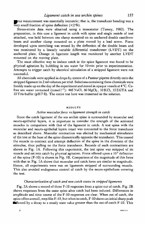
decay in catch records was consistently observed, but its extent was variable asillustrated in Fig. 2C. This spine, from the same test, shows a more marked decay.The basis of this decay is not completely understood, but it seems to be characteristicof the catch state. Presumably, the deflection of the spine and the concomitant stretch-ing of the ligament occur quickly relative to the relaxation time of the viscoelasticmaterial comprising the ligament. In all further experiments, F-10 is defined as peakamplitude of the force transient accompanying a rapid 10 ° deflection. This representsan apparent 'instantaneous' stiffness at the moment of stretch.
Differences in peak F-10 amplitude for catch and non-catch records are furtherillustrated in the histogram of Fig. 3, where F-10 values from 80 spines, some agitatedand some not, are plotted. From this, a quantitative indication of catch vs non-catchis evident. All non-agitated spines offered F-10 of less than 0*1 g. Spines which wq^

Ligament catch in sea urchin spines 159^itated fell mainly into the F-10 range of 0-34-0-64g. The former group (F-10 =0062 ± 0-027 g, mean ± S.D.) was deemed to be out-of-catch, and the remainder ofthe population in catch (F-10 = 0-464 ± 0-085g, mean± S.D.). Cases of F-10 valuessubstantially greater than the mean, e.g. 0-7 g in Fig. 3 and up to 1-5 g (notillustrated), were also occasionally observed in agitated spines. Whether this is due toan unusually strong catch state or reflects small errors in angular deflection measure-ments, differences in spine length, or some other variable is not clear.
T0-5g
100s
Fig. 2. Characteristic F-10 responses for ligaments in and out of catch. (A) Spine was deflected 10°(three trials at arrows) with ligament out of catch. (B) Same spine was deflected twice through 10°after setting catch. (C) Examples of F-10 responses for a spine from the same test with its ligamentin catch. Extent of sag in force during response is more pronounced than in (B). In each case thesudden decrease in force to the baseline marks the return of the spine to its initial position.

160 M. DlAB AND W. F . GlLLY
5 - •
I
0-5
Force (g)
10
Fig. 3. Histogram of F-10 values from a population of spines. Some spines were physically agitated(all those with F-10 values greater than 0' 1 g), and some were not (all those with values less than orequal to O'l g). The former category is denned as being in catch, the latter out-of-catch. All spineswere of approximately the same length.
Critical deflection angle for ligament damage
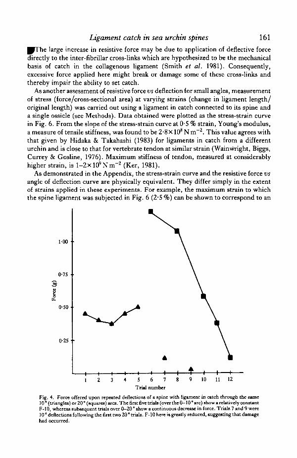
A spine that is set in catch cannot be deflected through a large angle withoutsuffering a drastic change in the properties of its ligament. Repeated spine deflectionsof 10 ° or less over the same arc yield resistive forces of similar magnitudes (connectedtriangles in Fig. 4), whereas repeated deflections of greater extent (20°, squares)result in a large decrease in resistive force. These results indicate that a ligament incatch begins to exhibit plastic flow for deflections of between 10 and 20°.
This phenomenon appears to reflect true damage of the ligament, in that it isirreversible on a time scale of days. Furthermore, the damaged portion of the ligamentis restricted to the span covered by the original deflection angle. Thus, if damageoccurs between 0 and 20° as in Fig. 3, then a deflection from 20 to 30° would stillexhibit an F-10 characteristic of the normal catch state.
Resistive force vs deflection for small angles
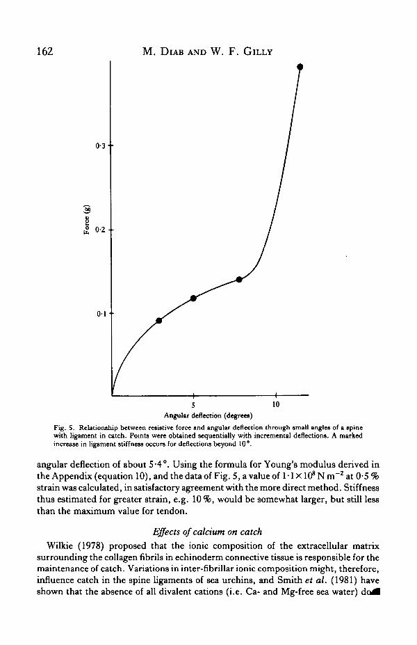
Spines in catch were deflected incrementally to different angles and the resistiveforce offered at each angle was recorded. Data from a typical experiment are plottedin Fig. 5. Most noteworthy is that for deflections of approximately 10°, there is adramatic increase in the slope of the curve relating force and angle. This angle coin-cides with the region of deflection in which the critical angle for damage was foundto lie.
Ligament catch in sea urchin spines 161
The large increase in resistive force may be due to application of deflective forcedirectly to the inter-fibrillar cross-links which are hypothesized to be the mechanicalbasis of catch in the collagenous ligament (Smith et al. 1981). Consequently,excessive force applied here might break or damage some of these cross-links andthereby impair the ability to set catch.
As another assessment of resistive force vs deflection for small angles, measurementof stress (force/cross-sectional area) at varying strains (change in ligament length/original length) was carried out using a ligament in catch connected to its spine anda single ossicle (see Methods). Data obtained were plotted as the stress-strain curvein Fig. 6. From the slope of the stress-strain curve at 0-5 % strain, Young's modulus,a measure of tensile stiffness, was found to be 2-8 X10⁸ N m~2. This value agrees withthat given by Hidaka & Takahashi (1983) for ligaments in catch from a differenturchin and is close to that for vertebrate tendon at similar strain (Wainwright, Biggs,Currey & Gosline, 1976). Maximum stiffness of tendon, measured at considerablyhigher strain, is l -2xlO9NnT2 (Ker, 1981).
As demonstrated in the Appendix, the stress-strain curve and the resistive force vsangle of deflection curve are physically equivalent. They differ simply in the extentof strains applied in these experiments. For example, the maximum strain to whichthe spine ligament was subjected in Fig. 6 (2-5 %) can be shown to correspond to an
1-00 •
0-75 •
0-50
0-25 •
5 6 7
Trul number
10 11 12
Fig. 4. Force offered upon repeated deflections of a spine with ligament in catch through the same10 ° (triangles) or 20 ° (squares) arcs. The first five trials (OVCT the 0-10 ° arc) show a relatively constantF-10, whereas subsequent trials over 0—20° show a continuous decrease in force. Trials 7 and 9 were10° deflections following the first two 20° trials. F-10 here is greatly reduced, suggesting that damagehad occurred.
162 M. DlAB AND W. F . GlLLY
0-3-
0 - 2 -
0 1
5Angular deflection (degrees)
10
Fig. 5. Relationship between resistive force and angular deflection through small angles of a spinewith ligament in catch. Points were obtained sequentially with incremental deflections. A markedincrease in ligament stiffness occurs for deflections beyond 10°.
angular deflection of about 5-4°. Using the formula for Young's modulus derived inthe Appendix (equation 10), and the data of Fig. 5, a value of 1-1 XlffNin"2 at 0-5%strain was calculated, in satisfactory agreement with the more direct method. Stiffnessthus estimated for greater strain, e.g. 10%, would be somewhat larger, but still lessthan the maximum value for tendon.
Effects of calcium on catchWilkie (1978) proposed that the ionic composition of the extracellular matrix
surrounding the collagen fibrils in echinoderm connective tissue is responsible for themaintenance of catch. Variations in inter-fibrillar ionic composition might, therefore,influence catch in the spine ligaments of sea urchins, and Smith et al. (1981) haveshown that the absence of all divalent cations (i.e. Ca- and Mg-free sea water)

Ligament catch in sea urchin spines 163
300
az•bX
150 - •
0-5 10 1-5 2-0% Strain
2-5
Fig. 6. Stress-strain curve for a spine ligament in catch. Points were obtained sequentially withincremental stretches (longitudinally applied - see text for details). Largest stretch is equivalent toan angular deflection of 5-4°.
prevent the setting of catch. Hidaka (1983) further showed that Mg could not sub-stitute for Ca in sustaining catch. This has been confirmed in the present study. Aligament out of catch was bathed in Ca-free sea water for 5 min and then agitated inthat medium for another 10 min. Pre- and post-agitation F-10 responses in two spinesthus studied were identical and of the non-catch sort, indicating that the setting ofcatch is Ca-dependent.
Smith et al. (1981) also reported that the prevention of catch by divalent cation-freesea water was reversible, but they gave no indication of the time course for reversal.Following the above experiments with Ca-free sea water, the preparations werebathed in running natural sea water (10-15 mmol I"1 Ca) at 15 °C and periodicallyagitated to determine the time course of catch recovery. Results from the most com-plete experiment are shown in Fig. 7. F-10 responses in Ca-free sea water (time zero)and in natural sea water for up to 3 h are characteristic of spine ligaments out of catch.After 12 h, recovery F-10 shows both the amplitude and time course (not illustrated)typical of ligaments in catch. Thus, recovery of catch-setting ability following Ca-freetreatment is very slow.
Are disulphide bonds involved in catch?, Collagen from the nematodeAscaris contains a high density of cysteine residues and

164 M. DlAB AND W. F . GlLLY
is known to form cross-links by means of disulphide bonds (Wainwright et ^1976). The possibility of disulphide cross-linking in sea urchin spine ligaments wastested by applying sulphydryl reagents to spine ligaments. Dithiothreitol (DTT,5 mmol I"1), which breaks disulphide bonds, was applied to two ligaments in catch.No abolition of catch occurred, as shown by similar F-10 responses before and afterDTT treatment. As another test, iV-ethylmaleimide (NEM, 5 mmolT1), whichprevents formation of disulphide linkages, was applied to two ligaments out of catchwhich were then agitated to induce catch. F-10 responses before and after agitationin the presence of NEM were typical of those for normal spines (e.g. Fig. 2).Results with both of these reagents suggest that disulphide linkages do not playa significant role in the setting or maintenance of catch, but this conclusion shouldbe regarded as tentative.
Recovery time (h)
Fig. 7. Recovery of the ability to set catch following treatment of a ligament with Ca-free sea water.Point at zero time was obtained in the Ca-free solution. The preparation was then placed in naturalsea water for recovery. Each point thereafter was obtained following 10 min agitation. Recovery timeis very slow; F-10 characteristic of catch is developed only after many hours.

Ligament catch in sea urchin spines 165
Transglutaminase inhibitors prevent the setting of catchIn vertebrates the enzyme transglutaminase catalyses the in vitro formation of
cross-links between fibronectin and collagen in cultured fibroblasts (Folk, 1980).Cadaverine (1,5-diaminopentane) and putrescine (1,4-diaminobutane) act as sub-strates for this enzyme and thereby inhibit its action when added to a system in excess(Curtis & Lorand, 1976). As a test of transglutaminase-like cross-linking in seaurchin ligaments, cadaverine and putrescine were applied to ligaments out of catchin an attempt to prevent catch setting. Two experiments with each substance at0-01 mmol I"1 concentration gave F-10 responses of the non-catch type before andafter agitation (see also Table 1). Thus, catch setting was prevented by these trans-glutaminase inhibitors. Application of cadaverine and putrescine to spines already incatch (two each) did not abolish or significantly weaken catch. These results stronglysuggest that cross-link formation in the spine ligament may involve a chemicalmechanism similar to that catalysed by transglutaminase, but that once formed, cross-links do not require enzyme activity.
Classical neurotransmitters do not appear to affect catch-settingThe results described so far have dealt with the mechanical and chemical properties
of the catch mechanism. In the living urchin, catch appears to be rapidly turned onthrough sensory input to the nervous system (Maeda, 1978). Neural processes havealso been found in the ligament itself and appear to be secretory in nature (Smith etal. 1981; Hidaka & Takahashi, 1983). Furthermore, in single-spine ossicle prepara-tions, both ligament 'tonus', measured as length change under a constant load(Takahashi, 1966), and apparent stiffness (Hidaka & Takahashi, 1983) are reported tobe increased by 0-1 mmol I"1 acetylcholine (ACh) and decreased by 0-01-0-1 mmol I"1
epinephrine. The extent of these changes relative to non-catch control spines isunclear however.
Table 1. Action of various substances on spine ligaments in and out of catch
Applied Test spineTime in/out Release Induce Prevent
Agent applied (N) (mmol I"1) (min) of catch catch catch catch
AcetylcholineAcetylcholine</-tubocurarineEpinephrineDopamineSerotoninD T TNEMCadaverineCadaverinePutrescinePutrescine
(4)(4)(2)(4)(2)(2)(2)(2)(2)(2)(2)(2)
0 10 110010-10 1550010010-01001
10103
103333
10101010
OutInInInInInIn
OutOutIn
OutIn
D T T and NEM represent dithiothreitol and N-ethylmaleimide, respectively.Only cadaverine and putrescine showed any effect on thejigament catch mechanism. Both substances
prevented the setting of catch.symbol represents a single trial: ( - ) indicates no effect, (+) indicates an effect.

166 M. DlAB AND W. F . GlIXY
Attempts were made in the present study to obtain more detailed informationthe controlling role of the nervous system using in situ stripped ligaments and F-responses as indicators of the state of catch. No effects were obtained for severalsuspected neurotransmitters, and results are summarized in Table 1.
ACh (0-1 mmol I"1) was applied to ligaments out of catch, to see if catch could beinduced, and to ligaments already in catch, to see if some extreme state of catch,normally not attainable by physical agitation, could be achieved (cf. Fig. 3). In everycase, ACh had no effect on F-10. As another test of cholinergic control, 1 mmol I"1
tf-tubocurarine was applied to a ligament in catch. Again, there was no effect on F-10.Hidaka (1983) suggested that the ACh-induced increase in stiffness reported by
Hidaka & Takahashi (1983) is transient in nature and begins to disappear after1-2 min of exposure. Thus, a 'desensitization'-like process may have masked any ACheffect in the present experiments where 10-min exposures were used.
Several catecholamines were also tested on ligaments already in catch. Epinephrine(0-01 mmoir1) had no inhibitory effect on catch as judged by the unchanged F-10response. This is in agreement with Hidaka & Takahashi (1983) who also found noinhibitory effects when quick stretches were used. Similarly, 0-1 mmol I"1 serotoninand 0-1 mmoir1 dopamine had no effect. These latter substances cause release ofcatch in the non-striated muscle of various molluscs (Florey, 1966).
As a check on the efficacy of the method for application of these substances,methylene blue sea water was applied in exactly the same manner as above. Oneminute following application, the ligament was cut open to expose its innermostsurface; this was found to be well stained. Therefore, there does not appear to be asignificant diffusional barrier to substances applied to the outer surface of a strippedligament.
DISCUSSION
This paper presents data on the mechanical properties of the spine ligaments in thesea urchin and considers some possible physiological control mechanisms for thesetting and releasing of catch. It confirms and extends on another genus of urchinsome basic findings of Smith et al. (1981) and Hidaka (1983) and the stiffnessmeasurements for ligaments in catch by Hidaka & Takahashi (1983). It also offerssuggestive evidence for a Ca-dependent enzymatic mechanism of collagen cross-linking as the chemical basis for setting catch.
One important result described here is the occurrence of structural damage orweakening when a spine in catch is deflected through an angle of greater than 10°. Asimilar alteration in ligament constitution may have occurred at approximately thesame amount of stretch in the 'movable' spines described by Smith et al. (1981).These ligaments showed a dramatic decrease in extensibility at a stretch of0-3-0-4mm (longitudinally applied), and this amount of stretch can be converted toan equivalent angle of deflection by a method employing the same principles as thatdescribed in the Appendix. Using a ligament radius of 1 -0 mm (estimated from theirmicrographs) and other parameter values cited in their report, the 'critical'0-3—0-4 mm stretch is equivalent to 16-9—21-8° of deflection, which corresponds tothat region where the critical angle for damage lies. Hidaka & Takahashi (1983)

Ligament catch in sea urchin spines 167vpuited that the apparent stiffness of a ligament in catch began to decrease atstretches of >10 %, and that tensile strength was exceeded by stretches of ~15 %. Infair agreement with these data, we observed ligament damage to begin at approximately8% stretch (-15°, cf. Figs 5, 6).
Exactly what happens at this angle requires a more detailed mechanical model forthe ligament system and the mechanism of catch maintenance by collagen fibrils.According to the ideas presented by Smith et al. (1981), this critical angle mayrepresent the limiting amount of stretch which the cross-links between collagen fibrilscan withstand before breaking. Since this breakage appears not to be readily reversible(cf. Fig. 4), it follows that the cross-links are not capable of rapid reformation oncebroken by excessive stretch.
This type of structural damage due to excessive stretch does not appear to be relatedto the force decay phenomenon in F-10 responses described in conjunction with Fig. 2.Decay in force following a quick stretch to a maintained angle of deflection correspondsto a slow stress relaxation due to the viscoelastic properties of the ligament. This does notreflect damage to the catch mechanism, as F-10 responses of nearly identical magnitudeand time course can be repeatedly obtained over the same arc of spine deflection. Fastand slow components of F-10 decay were seen in every ligament in catch studied. Thisbehaviour may involve the sliding of bundles of collagen fibrils past one another thatHidaka & Takahashi (1983) observed in relaxed ligaments under stretch. The extent ofsliding during catch is not known. A similar phenomenon may occur in holothurian der-mis, where stretch under a load produces fast and slow creep rates (Motokawa, 1982).
Another new finding in this work is that the transglutaminase inhibitors cadaverineand putrescine at very low concentration can prevent the setting of catch. This sug-gests a mechanism involving transglutaminase or a transglutaminase-like enzyme(TGL) for the formation of collagen cross-links. In support of this hypothesis is theobservation that the setting of catch is Ca-dependent, since Ca is required to activatevertebrate transglutaminase (Curtis & Lorand, 1976). Thus, if a Ca-dependent trans-glutaminase or TGL controls catch, then the absence of Ca should block its setting,as was observed.
What triggers the actual setting of catch remains unclear. All attempts to activatecatch in stripped ligaments with electrical stimulation or suspected neurotransmitterswere unsuccessful. The anatomical basis exists, nevertheless, for activation viasecretion of some substance from axonal processes inside the ligament itself (Smithet al. 1981), and this remains the most likely possibility.
These postulated mechanisms for triggering and setting catch are both criticallydependent on Ca ions. Thus, Ca might support both the neurosecretory release ofTGL from its intracellular storage site in the intra-ligamental nerve endings and thesubsequent activation of TGL which results in catch. This simple model is thusconsistent with both the pharmacological and anatomical data.
One problem with this, however, is the very long recovery time for the ligament toregain catch-setting ability when its Ca supply is replenished following treatment withCa-free sea water (many hours). Several explanations can be offered. One is that Ca-free sea water in some unknown way causes loss of inactive TGL from the intracellularstorage sites. Catch regaining would then require regeneration of the lost enzyme,
^•esumably a slow process.

168 M. DlAB AND W. F . GlLLY
A second explanation requires modification of the simple model. In this case, tltaTGL storage site may not be intracellular, and TGL must then be bound extra-cellularly in some Ca-dependent manner and activated by an additional cofactor. Itwould then be this cofactor which is stored in the axonal processes and released bynervous'activity to activate TGL and trigger catch. Depletion of Ca would result inthe inhibition of neurosecretory release of cofactor and the loss of inactive TGL fromits binding sites to the medium surrounding the spine. Again, the limiting step indetermining recovery time following treatment with Ca-free sea water would beregeneration of lost enzyme.
Summarizing, results described here suggest that nervous activity and the actionof neurotransmitters are not the sole elements involved in the mechanism of settingcatch. Nor does it seem likely that catch is directly controlled in vivo by changes inextracellular ionic strength, pH (Hidaka, 1983) or tonicity (Eylers, 1982). Thesefactors modify stiffness by two- to three-fold, considerably less than that observed inthe present study, and proposed mechanisms for in vivo changes are lacking.
Rather, we propose that catch in the spine ligaments of the sea urchin is mediatedby neurosecretory activation of a Ca-dependent enzymatic process involving trans-glutaminase or a similar enzyme. The sea urchin spine ligament is a complex com-posite material of apparently discontinuous collagen fibres in an unusual and veryinteresting matrix. Our model of TGL-activated cross-linking of collagen with itself
SPINE
L / LIGAMENT
Fig. 8. Schematic diagram of spine-ligament system (see Appendix). (A) Representation of spinedeflection through angle 6 which results in ligament stretching through a distance x. (B) Cross-sectionthrough the ligament. Spine deflection is about the dashed line, with the lower half of the ligamentas drawn being compressed (thus not contributing to the F-10), and the upper half being stretched(thus resisting stretch).
Ligament catch in sea urchin spines 169
n d possibly with other matrix components not only provides a basis for setting andmaintaining catch that is consistent with available morphological and physiologicaldata, but also suggests how interactions between collagen and matrix may change inthe TGL-activated catch process.
APPENDIX
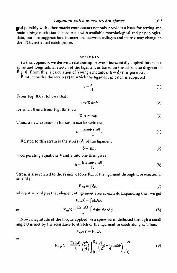
In this appendix we derive a relationship between horizontally applied force on aspine and longitudinal stretch of the ligament as based on the schematic diagram inFig. 8. From this, a calculation of Young's modulus, E = 6/e, is possible.
First, consider the strain (e) to which the ligament in catch is subjected:
From Fig. 8A it follows that:
x = Xsin8 (2)
for small 8 and from Fig. 8B that:
X = rsin<£. (3)
Thus, a new expression for strain can be written:
Related to this strain is the stress (<5) of the ligament:
<5 = eE. (5)
Incorporating equations 4 and 5 into one then gives:
c _ Ersin<ft sin8 .,.
Stress is also related to the resistive force F r a of the ligament through cross-sectionalarea (A):
(7)
where A = rdrd0 is that element of ligament area at each (f>. Expanding this, we get
Fro.X = JeEAX
„ v E s i n © f j . 2 . 1 i , , „ ,
or FrcX = — : — J rsirr<pdrd(p. (8)L
Now, magnitude of the torque applied on a spine when deflected through a smallangle 8 is met by the resistance to stretch of the ligament in catch along x. Thus,
F iY = F X
orP v_Esin8 /r4\1R2 ,, , . , ,F . p p iY— r -HW I ^ - T 8 1 1 1 2 ^ ! (9)
170 M. DlAB AND W. F . GlLLY
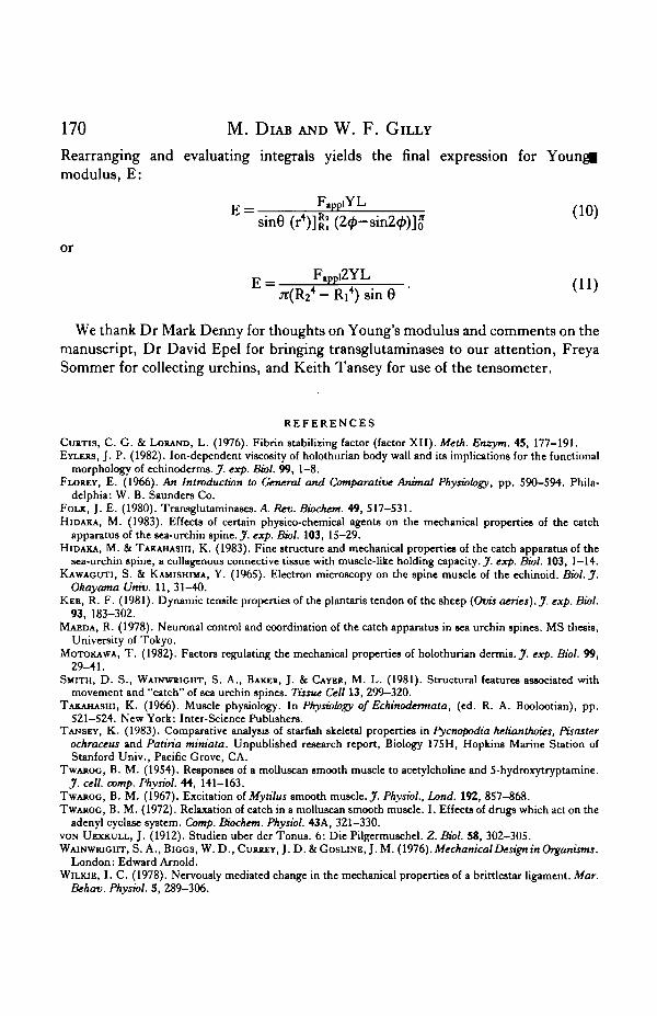
Rearranging and evaluating integrals yields the final expression for Youngimodulus, E:
F i Y Lv ;
K =sin6 (r4)]gj (24>-
or
p _ F.ppi2YLp
jr(R24-Ri4)sine •
We thank Dr Mark Denny for thoughts on Young's modulus and comments on themanuscript, Dr David Epel for bringing transglutaminases to our attention, FreyaSommer for collecting urchins, and Keith Tansey for use of the tensometer.
R E F E R E N C E S
CURTIS, C. G. & LORAND, L. (1976). Fibrin stabilising factor (factor XII). Meth. Enzym. 45, 177-191.EYLERS, J. P. (1982). Ion-dependent viscosity of holothurian body wall and its implications for the functional
morphology of echinoderms. J. exp. Biol. 99, 1-8.FLOREY, E. (1966). An Introduction to General and Comparative Animal Physiobgy, pp. 590-594. Phila-
delphia: W. B. Saunders Co.FOLK, J. E. (1980). Transglutaminases. A, Rev. Biochem. 49, 517-531.HIDAKA, M. (1983). Effects of certain physico-chemical agents on the mechanical properties of the catch
apparatus of the sea-urchin spine. J. exp. Biol. 103, 15—29.HIDAKA, M. & TAKAHASHI, K. (1983). Fine structure and mechanical properties of the catch apparatus of the
sea-urchin spine, a collagenous connective tissue with muscle-lite holding capacity. J. exp. Biol. 103, 1—14.KAWAGUTI, S. & KAMISHMA, Y. (1965). Electron microscopy on the spine muscle of the echinoid. Biol. J.
Okayama Univ. 11, 31-40.KER, R. F. (1981). Dynamic tensile properties of the plantaris tendon of the sheep (Ovis aeries). J. exp. Biol.
93, 183-302.MAEDA, R. (1978). Neuronal control and coordination of the catch apparatus in sea urchin spines. MS thesis,
University of Tokyo.MOTOKAWA, T. (1982). Factors regulating the mechanical properties of holothurian dermis. J. exp. Biol. 99,
29-41.SMITH, D. S., WAINWRIGHT, S. A., BAKER, J. & CAYEJ>, M. L. (1981). Structural features associated with
movement and "catch" of sea urchin spines. Tissue Cell 13, 299—320.TAKAHASHI, K. (1966). Muscle physiology. In Physiology of Echinodermata, (ed. R. A. Boolootian), pp.
521-524. New York: Inter-Science Publishers.TANSEY, K. (1983). Comparative analysis of starfish skeletal properties in Pycnopodia helianthoies, Pisaster
ochraceus and Patiria mimata. Unpublished research report, Biology 175H, Hopkins Marine Station ofStanford Univ., Pacific Grove, CA.
TWAROC, B. M. (1954). Responses of a molluscan smooth muscle to acetylcholine and 5-hydroxytryptamine.J. cell. comp. Physiol. 44, 141-163.
TWAROG, B. M. (1967). Excitation of Mytilus smooth muscle. J. Physiol., Land. 192, 857-868.TWAROG, B. M. (1972). Relaxation of catch in a molluscan smooth muscle. I. Effects of drugs which act on the
adenyl cyclase system. Comp. Biochem. Physiol. 43A, 321—330.VON UEXKULL, J. (1912). Studien uber der Tonus. 6: Die Pilgermuschel. Z. Biol. 58, 302-305.WAINWRIGHT, S. A., BIGGS, W. D., CURREY, J . D . & G O S U N E , J .M. (1976). Mechanical Design in Organisms.
London: Edward Arnold.WILKIE, I. C. (1978). Nervously mediated change in the mechanical properties of a brittlestar ligament. Mar.
Behav. Physiol. 5, 289-306.
Related Documents