HAL Id: tel-03696948 https://tel.archives-ouvertes.fr/tel-03696948 Submitted on 16 Jun 2022 HAL is a multi-disciplinary open access archive for the deposit and dissemination of sci- entific research documents, whether they are pub- lished or not. The documents may come from teaching and research institutions in France or abroad, or from public or private research centers. L’archive ouverte pluridisciplinaire HAL, est destinée au dépôt et à la diffusion de documents scientifiques de niveau recherche, publiés ou non, émanant des établissements d’enseignement et de recherche français ou étrangers, des laboratoires publics ou privés. Mécanismes moléculaires de la sécrétion hormonale et traitement anti-sécrétoire du phéochromocytome humain Laura Streit To cite this version: Laura Streit. Mécanismes moléculaires de la sécrétion hormonale et traitement anti-sécrétoire du phéochromocytome humain. Médecine humaine et pathologie. Université de Strasbourg, 2021. Français. NNT : 2021STRAJ018. tel-03696948

Welcome message from author
This document is posted to help you gain knowledge. Please leave a comment to let me know what you think about it! Share it to your friends and learn new things together.
Transcript
HAL Id: tel-03696948https://tel.archives-ouvertes.fr/tel-03696948
Submitted on 16 Jun 2022
HAL is a multi-disciplinary open accessarchive for the deposit and dissemination of sci-entific research documents, whether they are pub-lished or not. The documents may come fromteaching and research institutions in France orabroad, or from public or private research centers.
L’archive ouverte pluridisciplinaire HAL, estdestinée au dépôt et à la diffusion de documentsscientifiques de niveau recherche, publiés ou non,émanant des établissements d’enseignement et derecherche français ou étrangers, des laboratoirespublics ou privés.
Mécanismes moléculaires de la sécrétion hormonale ettraitement anti-sécrétoire du phéochromocytome humain
Laura Streit
To cite this version:Laura Streit. Mécanismes moléculaires de la sécrétion hormonale et traitement anti-sécrétoire duphéochromocytome humain. Médecine humaine et pathologie. Université de Strasbourg, 2021.Français. �NNT : 2021STRAJ018�. �tel-03696948�
UNIVERSITÉ DE STRASBOURG
ÉCOLE DOCTORALE des Sciences de la Vie et de la Santé
Institut des Neurosciences Cellulaires et Intégratives – CNRS UPR3212
THÈSE présentée par :
Laura STREIT
soutenue le : 1er juillet 2021
pour obtenir le grade de : Docteur de l’Université de Strasbourg
Discipline : Sciences du vivant
Spécialité : Aspects moléculaires et cellulaires de la biologie
Mécanismes moléculaires de la sécrétion hormonale
et traitement anti-sécrétoire du
phéochromocytome humain
THÈSE dirigée par : M. GASMAN Stéphane . . Directeur de recherche Inserm, Université de Strasbourg
RAPPORTEURS : M. DUBESSY Christophe . .Maître de conférences hors classe, Université de Rouen-Normandie
Mme GUERINEAU Nathalie - .Directrice de recherche CNRS, Université de Montpellier
EXAMINATRICE ET PRESIDENTE : Mme SIMONNEAUX Valérie Directrice de recherche CNRS, Université de Strasbourg
INVITÉS : M. ORY Stéphane Chercheur CNRS, Université de Strasbourg


REMERCIEMENTS
J’ai réalisé la première partie de ma thèse au sein de l’entreprise de
biotechnologies Firalis située à Huningue durant 1 an et 3 mois. J’ai ensuite rejoint
mon équipe « Traffic membranaire intracellulaire dans les systèmes nerveux et
neuroendocrines » dirigée par les Drs Stéphane Gasman et Nicolas Vitale, au sein
de l’Institut des Neurosciences Cellulaires et Intégratives à Strasbourg.
Je souhaiterais tout d’abord remercier les membres de mon jury de thèse,
Nathalie Guerineau, Christophe Dubessy, et Valérie Simonneaux d’avoir accepté
d’évaluer ce travail.
Je remercie Michel Barrot de m’avoir accueilli dans son laboratoire.
J’adresse des remerciements particuliers à mon directeur de thèse Stéphane
Gasman, qui m’a encadré depuis le stage de Master, et encouragé lors du difficile
passage du concours de l’école doctorale. Merci pour tes conseils et tes nombreuses
relectures de dossiers et d’articles (on voit le bout !!). Merci de m’avoir accordé ta
confiance pour ce projet, même si les phéos étaient parfois capricieux en manip !
Enfin, merci de m’avoir donné cette opportunité de réaliser une partie de ma thèse en
entreprise.
Un très grand merci à Stéphane Ory qui a été mon encadrant de manips.
Merci d’avoir été disponible pour toutes mes questions ! J’ai également apprécié nos
discussions moins scientifiques lors de nos « expéditions phéo » à Nancy !
Mes pensées vont ensuite à Marion R, qui m’a également encadré, et initié à
la culture de phéo et à l’ampérométrie (parfois compliqué). Merci surtout pour tous
nos moments passés ensemble et nos repas partagés le midi ! Merci à Sophie,
d’avoir été mon binôme de manips lors du lancement du projet, les sécrétions n’ont
plus de secret pour nous !! Je suis ravie que notre collaboration se poursuive pour ce
projet phéo. Je te souhaite le meilleur dans quelques mois lorsque tu soutiendras
aussi ta thèse.

Merci à Audrey pour tes qualités humaines et pour m’avoir aidé (très tôt le
matin) pour mes nombreuses sécrétions ! Je te souhaite le meilleur pour la suite !
Merci au duo inséparable Marion M et Margherita pour tous nos moments passés
ensemble, j’en garde d’excellents souvenirs.
Enfin, je tiens à remercier tous les membres de la Team Gasman-Vitale avec
lesquels j’ai eu du plaisir à travailler durant mes 4 ans de thèse. Sylvette et Nicolas
merci pour tous nos échanges notamment lors de nos « expéditions abattoir » pour
récupérer nos fameuses cellules chromaffines. Alex, merci pour ton optimisme
perpétuel, c’est toujours agréable de travailler et de discuter dans la bonne humeur !
Je te souhaite le meilleur, surtout maintenant que la culture de cellules chromaffines
n’a plus de secret pour toi ! Claudine, merci pour ta disponibilité et ton aide, tu es
toujours prête à rendre service aux autres ! Enfin, Michael, Sebahat, Petra, Qili,
Anne-Marie, merci pour les échanges que nous avons pu avoir durant ces années.
Merci à Sylvain Hugel d’avoir assuré les expériences d’électrophysiologie
toujours avec bonne humeur et le sourire, ça a été un plaisir de collaborer avec toi !
Mes remerciements vont également aux personnels hospitaliers du CHRU de
Nancy et de l’Hôpital Civil de Strasbourg, et plus particulièrement aux Pr Laurent
Brunaud, Dr Michel Vix, Pr Didier Mutter qui ont assuré mon approvisionnement
plus ou moins constant en phéochromocytomes.
Je tiens à remercier Hüseyin Firat pour m’avoir donné la chance de réaliser
une partie de ma thèse au sein de son entreprise.
Merci à mes coloc de bureau, Marion C pour les nombreuses qualifications
que tu as dû me faire passer et pour mon initiation en purif AKTA. Ton sens de
l’humour, nos conversations, et surtout nos débats sur les lapins vont me manquer !
Guillaume, le chef prod pour ses conseils toujours avisés et ses qualités humaines.
Un grand merci à Marine pour son soutien autour de nos nombreuses tartes
flambées et mojitos. Melek, pour nos échanges, l’initiation à l’arabe et notre séjour
fantastique en Tunisie. Et Yasemin, pour sa gentillesse, son aide en manip et sa
capacité extraordinaire à prendre des photos sous le bon angle.

Mes remerciements vont également à la Team spectrométrie-orbitrap : Maїté
et Maëva pour tous leurs coups de folie et leurs nombreuses blagues hilarantes qui
ont animé le labo !
Un grand merci à Samira pour sa bonne humeur contagieuse et nos
discussions beauté & Miloud pour sa gentillesse et la découverte des pâtisseries
marocaines !
Merci à Pascale pour ses conseils en transfection et culture d’hybridomes.
Enfin, je tiens à remercier l’ensemble du personnel de Firalis qui ont permis le
déroulement de cette partie de mon projet.
Un immense merci à la Robertsau Team !! Emeline et Mathieu, merci pour
votre soutien et de m’avoir permis de décompresser lors de nos apéros en
provenance de la cave à terroirs, nos soirées raclettes, restaurants et j’en passe !
Merci pour tous ces échanges, rires et bons moments passés ensemble (Mathieu le
canoë attend encore ta vengeance) !
À mes amis de longue date : Aurélie, Marion H, Aline merci pour votre
soutien depuis toutes ces années.
Un merci tout particulier à Nicolas, tu t’es toujours intéressé à mon projet, et
tu as fait preuve d’un soutien sans faille à toutes les étapes de cette thèse. Merci
pour ton énergie et tes encouragements quotidiens, et de n’avoir jamais douté de ma
réussite. Merci d’apporter tellement de bons moments et de positif à ma vie.
Pour finir, un merci infini à mes parents, vous m’avez donné l’opportunité de
faire toutes ces années d’études. Merci d’essayer de comprendre de temps en temps
les mystères du phéochromocytome !! Merci à mamie et papy, mes vacances à
« Wittring plage » sont toujours une bulle d’oxygène et de positivité dans ma vie. Une
pensée à mes deux autres grands parents qui auraient été fiers de ce que j’ai
accompli…. À mes deux sœurs, Sophie, merci pour ton soutien, tes relectures
pointilleuses et nos bons moments passés avec Schnouki à la maison. Anaïs, merci
d’avoir été mon binôme, mon repère depuis notre naissance. Depuis toutes ces
années nous avons partagé nos joies, nos peines et surtout nos réussites, merci
d’être toujours présente.


LISTE DES ABRÉVIATIONS
AC Adénylate Cyclase
ACTH Adreno CorticoTropic Hormone
ADN Acide DésoxyriboNucléique
AMPc Adénosine MonoPhosphate Cyclique
APUD Amine content and amine Precursor Uptake and Decarboxylation
Arf6 ADP-Ribosylation Factor 6
ARNm Acide RiboNucléique Messager
Arp2/3 Actin Related Protein 2/3 complex
ATP Adénosine TriPhosphate
Bax Bcl-2-Associated X
BIM23014 Lanréotide
Cdc42 Cell Division Control protein 42 homolog
CEA CarcinoEmbryonic Antigen
CGA ChromoGranine A
CGB ChromoGranine B
CI50 Concentration Inhibitrice Médiane
DAG DiAcylGlycérol
DD Dopamine Décarboxylase
DβH Dopamine-β-Hydroxylase
ERK Extracellular signal-Regulated Kinase
FDA Food and Drug Administration
GH Growth Hormone
GHIH Growth Hormone-Inhibiting Hormone
HIF Hypoxia-Inducible Factor
IGF-II Insulin-like Growth Factor-II
IRM Imagerie par Résonnance Magnétique
MAPK Mitogen-Activated Protein Kinase
MEN2 Multiple Endocrine Neoplasia type 2
Munc Mammalian UNCoordinated protein
NCAM Neural Cell Adhesion Molecule
NPY NeuroPeptide Y
N-WASP Neural Wiskott-Aldrich Syndrome Protein
NSE Neuron Specific Enolase
NSF N-ethylmaleimide-Sensitive Fusion protein
OCT Octréotide
PA Acide Phosphatidique
PACAP Pituitary Adenylate Cyclase-Activating Polypeptide
PASS Pheochromocytoma of the Adrenal gland Scaled Score
PGL ParaGangLiome
PET/CT Positron Emission Tomography/Computed Tomography
PHD Prolyl-HyDroxylase
PHEO Phéochromocytome
PI 4-kinase Phosphatidylinositol 4-kinase
PIP2 Phosphatidylinositol 4,5-bisphosphate
PKA Protéine Kinase A
PLD PhosphoLipase D
PNMT Phényléthanolamine N-Méthyl-Transférase
PPGL Phéochromocytome et ParaGangLiome
PRL PRoLactine
PRRT Peptide Receptor Radionuclide Therapy
PSA Prostate-Specific Antigen
PSAP Prostatic-Specific Acid Phosphatase
Rac1 Ras-related C3 botulinum toxin substrate 1
RalA Ras-related protein Ral-A
RE Réticulum Endoplasmique
REST RE-1 Silencing Transcription factor
Rho Ras homologous protein
RhoA Ras Homolog family member A
RT-PCR Reverse Transcription Polymerase Chain Reaction
SDHB Succinate DésHydrogénase sous-unité B
SMS 201–995 Octréotide
SNAP Soluble NSF Attachment Protein
SNAP-25 SyNaptosomal-Associated Protein, 25 kDa
SNARE Soluble N-ethylmaleimide-sensitive-factor Attachment protein REceptor
SOM230 Pasiréotide
SRIF Somatotropin Release-Inhibiting Factor
SST Somatostatine
SSTR Récepteur de la somatostatine
TGN Trans-Golgi Network
TH Tyrosine Hydroxylase
TNE Tumeur NeuroEndocrine
TNE-GEP Tumeur NeuroEndocrine Gastro-Entéro-Pancréatique
t-SNARE target SNARE
TSH Thyroid-Stimulating Hormone
VAMP-2 Vesicle-Associated Membrane Protein 2
VEGF Vascular Endothelium Growth Factor
VMA VanillylMandelic Acid
VMAT Vesicular MonoAmine Transporter
v-SNARE vesicular SNARE
5-HIAA Acide 5-hydroxyindolacétique

SOMMAIRE
AVANT-PROPOS .................................................................................................................................... 1
INTRODUCTION ..................................................................................................................................... 5
1. Le système neuroendocrinien : physiologie et tumeurs ............................................................... 5
1.1. Généralités ................................................................................................................................... 5
1.2. Les cellules neuroendocrines ....................................................................................................... 6
1.3. La régulation de fonctions vitales ................................................................................................. 7
1.4 Les tumeurs neuroendocrines et l’hypersécrétion ........................................................................ 8
1.4.1. Généralités ............................................................................................................................ 8
1.4.2. La problématique de l’hypersécrétion ................................................................................... 9
2. La glande médullosurrénale et le phéochromocytome ............................................................... 11
2.1. Anatomie et histologie de la glande surrénale ........................................................................... 11
2.2. Les cellules chromaffines ........................................................................................................... 12
2.3. Processus de sécrétion par les cellules chromaffines ............................................................... 12
2.3.1 Les granules de sécrétion à cœur dense ............................................................................. 12
2.3.1.1. Le contenu granulaire ................................................................................................... 12
2.3.1.2. Biogenèse ..................................................................................................................... 13
2.3.1.3. Maturation ..................................................................................................................... 15
2.3.2. L’exocytose régulée ............................................................................................................. 17
2.3.3. La régulation de l’exocytose ................................................................................................ 19
2.4. Effets physiologiques des molécules libérées par les cellules chromaffines ............................. 21
2.5. Le phéochromocytome ............................................................................................................... 25
2.5.1. Généralités .......................................................................................................................... 25
2.5.2. Historique............................................................................................................................. 26
2.5.3. Epidémiologie ...................................................................................................................... 27
2.5.4. Symptômes cliniques ........................................................................................................... 27
2.5.5 Les phéochromocytomes malins .......................................................................................... 28
2.5.6. La génétique ........................................................................................................................ 29
2.5.6.1. Prédisposition ............................................................................................................... 29
2.5.6.2. Cluster 1 : la voie pseudohypoxique ............................................................................ 32
2.5.6.3. Clusters 2 et 3 : les voies des récepteurs tyrosine kinases et Wnt .............................. 33
2.5.6.4. Les multiples conséquences des mutations ................................................................. 35
2.5.6.5. De la génétique au phénotype ..................................................................................... 35
2.5.7. Diagnostic biochimique et imagerie ..................................................................................... 36
2.5.8. Prise en charge clinique du phéochromocytome ................................................................ 37
2.5.8.1. Gestion préopératoire de la tumeur .............................................................................. 37
2.5.8.2. Approche chirurgicale ................................................................................................... 38
2.5.8.3. Management postopératoire du patient ........................................................................ 39
2.5.8.4. Les phéochromocytomes malins et inopérables : des cas complexes pour la prise
charge ........................................................................................................................................ 40
3. Les analogues somatostatinergiques : un espoir de traitement anti-sécrétoire ...................... 41
3.1. La somatostatine ........................................................................................................................ 41
3.1.1. Découverte et historique ...................................................................................................... 41
3.1.2. Biosynthèse ......................................................................................................................... 41
3.1.3. Les récepteurs somatostatinergiques et leurs signalisations .............................................. 43
3.1.3.1. Action anti-sécrétoire .................................................................................................... 43
3.1.3.2. Autres actions ............................................................................................................... 44
3.1.4. Fonctions physiologiques de la somatostatine .................................................................... 45
3.2. Les analogues de la somatostatine : les nouvelles molécules employées en cancérologie ..... 46
3.2.1. Du développement à aujourd’hui ......................................................................................... 46
3.2.2. Les récepteurs de la somatostatine et les tumeurs neuroendocrines ................................. 48
3.2.3. Les effets secondaires ......................................................................................................... 51
3.2.4. Utilisation en médecine nucléaire ........................................................................................ 51
RÉSULTATS ......................................................................................................................................... 53
1. Publication n°1 : Étude des mécanismes de l'exocytose régulée par le calcium dans le
phéochromocytome humain .............................................................................................................. 53
1.1. Objectifs et déroulement de l’étude ............................................................................................ 53
1.2. Résultats ..................................................................................................................................... 53
2. Publication n°2 : Effet des analogues de la somatostatine sur la sécrétion de catécholamines
par les cellules de phéochromocytomes humains .......................................................................... 55
2.1. Objectifs et déroulement de l’étude ............................................................................................ 55
2.2. Résultats ..................................................................................................................................... 56
DISCUSSION GÉNÉRALE ET RÉSULTATS COMPLÉMENTAIRES ................................................. 57
1. Critique méthodologique ................................................................................................................ 57
1.1. Les modèles utilisés ................................................................................................................... 57
1.2. La sécrétion mesurée sur des cellules en culture primaire est-elle le reflet de la sécrétion
tumorale ? .......................................................................................................................................... 59
2. L’hypersécrétion tumorale : quels mécanismes possibles ? ..................................................... 61
3. Effet fonctionnel des analogues de la somatostatine ................................................................. 64
3.1. Octréotide et SOM230, un impact différent sur la sécrétion catécholaminergique .................... 64
3.2. Mode d’action du SOM230 ......................................................................................................... 69
4. Le SOM230 : vers les prémices d’un traitement de l’hypersécrétion ? ..................................... 72
4.1. Dose utilisée ............................................................................................................................... 72
4.2. Effet anti-sécrétoire du SOM230 en clinique .............................................................................. 74
4.3. Effet pro-tumoral des catécholamines et molécules sécrétées par les phéochromocytomes ... 76
4.4. Le SOM230 une molécule aux multiples actions anti-tumorale ................................................. 78
4.5. Quelles sont les limites de l’utilisation du SOM230 ? ................................................................. 80
4.5.1. Le récepteur nicotinique ...................................................................................................... 80
4.5.2. Les effets secondaires du SOM230 .................................................................................... 81
4.6. Les drogues anti-sécrétoire pour le traitement du phéochromocytome ..................................... 82
5. Vers une amélioration du diagnostic du phéochromocytome humain ? .................................. 85
5.1. Problématique ............................................................................................................................ 85
5.2. Découverte de nouveaux biomarqueurs .................................................................................... 86
CONCLUSION....................................................................................................................................... 89
MATÉRIELS ET MÉTHODES ............................................................................................................... 91
1. Réactifs ............................................................................................................................................. 91
2. Échantillons de phéochromocytomes humains........................................................................... 91
3. Culture cellulaire ............................................................................................................................. 91
3.1. Culture primaire de cellules chromaffines bovines ..................................................................... 91
3.2. Culture primaire de cellules chromaffines humaines issues de phéochromocytome ................ 92
4. Techniques biochimiques .............................................................................................................. 93
4.1. Détection des récepteurs à la somatostatine par Western-blot ................................................. 93
4.2. Dosage des catécholamines par méthode fluorimétrique .......................................................... 94
5. Mesure de la sécrétion des catécholamines par ampérométrie à fibre de carbone................. 96
RÉFÉRENCES BIBLIOGRAPHIQUES ................................................................................................ 97
ANNEXES ........................................................................................................................................... 129

1
AVANT-PROPOS
Le système neuroendocrinien assure de nombreuses fonctions de commande
de l’organisme en sécrétant des hormones ainsi que des neuropeptides dans la
circulation sanguine. Dans les cellules neuroendocrines, ces molécules sont
stockées dans des granules de sécrétion puis libérées par un processus d’exocytose
régulé par le calcium. Ce mécanisme permet l’acheminement des granules vers la
membrane plasmique et la libération du contenu granulaire dans l’espace
extracellulaire. Ce type d’exocytose apparaît comme un processus crucial, qui doit
être étroitement contrôlé car la dérégulation de la sécrétion neuroendocrine est
associée à diverses pathologies dont les tumeurs neuroendocrines.
Malheureusement, toutes les cellules neuroendocrines de l'organisme peuvent
se transformer en cellules tumorales et potentiellement engendrer des cancers. Bien
que constituant un groupe très hétérogène, les tumeurs neuroendocrines possèdent
une caractéristique commune intéressante qui est un dysfonctionnement de leur
activité sécrétrice entraînant, dans la majorité des cas, une hypersécrétion des
hormones et peptides qu'elles stockent. Cette sécrétion anarchique pose problème
car elle peut engendrer des conséquences cliniques graves chez les patients. À ce
jour, les mécanismes moléculaires qui induisent et maintiennent l’hypersécrétion de
ces tumeurs ne sont pas connus et il n’existe aucune thérapie ciblée permettant de
l’empêcher.
L’équipe des Drs Gasman et Vitale s’attache à décrypter les mécanismes
moléculaires qui contrôlent les processus d’exocytose et d’endocytose au sein des
systèmes nerveux et neuroendocrines, et à en révéler les altérations potentielles
dans des pathologies importantes. Dans ce contexte, l’équipe a développé, il y a
quelques années, un projet de recherche qui s’intéresse à l’hypersécrétion des
cellules neuroendocrines tumorales. Forte de son expertise dans les mécanismes de
la sécrétion neuroendocrine et de sa connaissance de la cellule chromaffine, l’équipe
a choisi d’utiliser comme modèle expérimental le phéochromocytome, une tumeur
neuroendocrine de la glande médullosurrénale.
Avant-propos
2
J’ai toujours été attirée par ces deux grands domaines de la biologie que sont
la cancérologie et la neurobiologie avec un intérêt particulier pour l’aspect cellulaire.
Ainsi, ce projet sur les tumeurs neuroendocrines répondait à toutes mes attentes et
m’a tout de suite beaucoup plu. J’ai démarré mon stage de Master 2 de
Neurosciences en me familiarisant avec le modèle des cellules chromaffines et en
commençant à défricher le projet sur l’action potentielle d’analogues
somatostatinergiques sur la sécrétion des catécholamines. Au cours de mon Master,
Stéphane Gasman démarrait un projet en collaboration avec deux entreprises,
Caprion à Montréal au Canada et Firalis à Huningue en France. Ce projet collaboratif
a commencé par une analyse en spectrométrie de masse du profil protéique de
plasmas d’une cohorte de 40 patients atteints de phéochromocytome, en les
comparant au profil protéique de plasmas d’une cohorte de volontaires sains, ils ont
pu mettre en évidence deux combinaisons de biomarqueurs potentiels permettant de
distinguer les patients des sujets contrôles avec une sensibilité et une spécificité
supérieures au test actuellement utilisé. À la fin de mon Master, j’hésitais longuement
à me lancer dans un doctorat. Cependant, me destinant à une carrière dans
l’industrie, le projet collaboratif avec les deux entreprises sur la mise au point de
nouveaux outils de diagnostic m’attirait fortement. C’est ainsi que j’ai tenté l’aventure
d’une thèse à l’interface entre la recherche académique et la recherche industrielle.
J’ai réalisé la première partie de ma thèse au sein de l’entreprise Firalis (1 an
et 3 mois) sur le projet de développement de nouveaux outils permettant, à terme, la
mise au point d’un test diagnostic plus fiable du phéochromocytome. Cette
immersion dans le monde de l’entreprise a été très enrichissante et m’a beaucoup
appris sur le fonctionnement de la recherche privée. D’un point de vue technique, j’ai
pu y acquérir de nouvelles compétences comme la production de protéines
recombinantes et la fabrication d’anticorps monoclonaux nécessaires au
développement de tests ELISA. Le projet est toujours en cours et, essentiellement
pour des raisons de confidentialité, cette partie ne sera pas approfondie dans le
manuscrit mais simplement abordée dans la discussion générale.
Avant-propos
3
Après ces 15 mois en entreprise, j’ai poursuivi ma thèse à l’Institut des
Neurosciences Cellulaires et Intégratives. Mes résultats prometteurs de Master 2
montrant une action inhibitrice d’un analogue de la somatostatine sur la sécrétion
des catécholamines dans les cellules chromaffines bovines devaient être poursuivis,
notamment en testant cet analogue sur des cellules tumorales humaines de
phéochromocytomes. C’est à partir de ce moment que j’ai passé de longues heures
au poste d’ampérométrie à fibre de carbone. J’ai utilisé cette technique pour mesurer
la sécrétion des catécholamines à partir de cellules chromaffines tumorales
humaines cultivées directement à partir de phéochromocytomes fraîchement opérés.
Mon objectif ici était double : caractériser le profil sécrétoire des cellules chromaffines
tumorales afin d’en aborder les mécanismes sous-jacents, et tenter d’inhiber cette
sécrétion tumorale anarchique par l’action des analogues somatostatinergiques.
Entamé depuis plusieurs années déjà au laboratoire, le premier axe de recherche
visait à caractériser la sécrétion catécholaminergique des phéochromocytomes. Ma
participation a permis d’analyser plus de tumeurs et d’obtenir assez d’analyses pour
dresser les premières hypothèses. Nous avons ainsi montré que l’hypersécrétion est
bien la conséquence d’une dérégulation du processus d’exocytose et non pas
simplement un effet de masse dû à la prolifération tumorale. Ce constat fût appuyé
par un crible protéomique, auquel je n’ai pas participé, qui a montré que de
nombreuses protéines de l’exocytose voient leur expression modifiée dans le
phéochromocytome en comparaison au tissu non tumoral. Le second axe fut la
continuité de mon travail de Master 2, et m’a permis de montrer que le pasiréotide a
une action inhibitrice efficace sur la sécrétion des phéochromocytomes humains,
ouvrant ainsi des perspectives très intéressantes en clinique. Ainsi, au cours de ma
thèse, j’ai participé à trois axes de recherche ayant pour but d’améliorer la
compréhension, le diagnostic et le traitement de l’hypersécrétion du
phéochromocytome.
La première partie de ce manuscrit consiste en une introduction générale dans
laquelle je présente le système neuroendocrinien ainsi que les tumeurs
neuroendocrines qui en dérivent, tout en insistant sur la problématique de
l’hypersécrétion. Je me focalise ensuite sur la glande surrénale et les cellules
chromaffines, en m’attardant sur le processus d’exocytose ainsi que le rôle
physiologique des molécules libérées. J’enchaîne sur une description détaillée du
Avant-propos
4
phéochromocytome, allant de sa découverte jusqu’aux connaissances actuelles en
termes de clinique, génétique et diagnostic. Je termine en introduisant la
somatostatine et le développement de ses analogues en pointant leurs intérêts et
leurs utilisations cliniques. La deuxième partie de mon manuscrit présente mes
résultats, intégrés avec d’autres, sous la forme d’un article scientifique soumis et d’un
article en préparation. La troisième partie est constituée d’une discussion générale
qui inclut quelques résultats complémentaires. Je clos ce manuscrit avec une
conclusion générale de mes travaux de thèse. Enfin, je décris l’ensemble du matériel
et des méthodes que j’ai utilisés lors de ma thèse. En complément, vous trouverez la
liste détaillée des interventions scientifiques que j’ai effectuées au cours de mon
doctorat, ainsi que la revue sur la sécrétion des tumeurs neuroendocrines et les
GTPases de type Rho, que j’ai écrite afin de compenser l’absence de mes précieux
phéochromocytomes lors du premier confinement de la pandémie de la COVID-19.
Je vous souhaite une agréable lecture.

INTRODUCTION

5
INTRODUCTION
1. Le système neuroendocrinien : physiologie et tumeurs
1.1. Généralités
Le système neuroendocrinien est formé par l’ensemble des glandes et cellules
neuroendocrines de l’organisme qui permettent le maintien de l’équilibre fonctionnel
indispensable à la vie. En effet, les cellules qui le constituent ont une capacité à
produire des hormones et des neuropeptides qui sont déversés dans la circulation
sanguine. Une fois libérées, ces molécules vont agir sur leurs récepteurs cibles,
permettant le contrôle des fonctions de l’organisme.
En 1870, Heidenhain décrit pour la première fois une population de cellules
neuroendocrines dans l'intestin grêle, puis en 1938, Feyrter pose l'hypothèse d'un
système neuroendocrinien diffus (de Herder et al., 2016). En effet, les cellules
neuroendocrines sont ubiquitaires dans l’organisme, une partie est dispersée à
l’intérieur d’un organe ou d’un tissu, formant ce qu’on appelle le système endocrinien
diffus, où elles sont disposées en groupe à proximité des capillaires. Des cellules
neuroendocrines sont ainsi dispersées dans le tractus gastro-entéro-pancréatique et
pulmonaire. Elles sont également présentes dans une moindre mesure dans les
voies urogénitales et la peau. Tandis qu’une autre partie des cellules forme des
organes endocriniens tels que l’hypothalamus, l’hypophyse ou la glande
médullosurrénale. Le pancréas endocrine n’a pas de classification clairement définie
et peut appartenir à l’un des groupes (Figure 1) (Montuenga et al., 2003).
Introduction
6
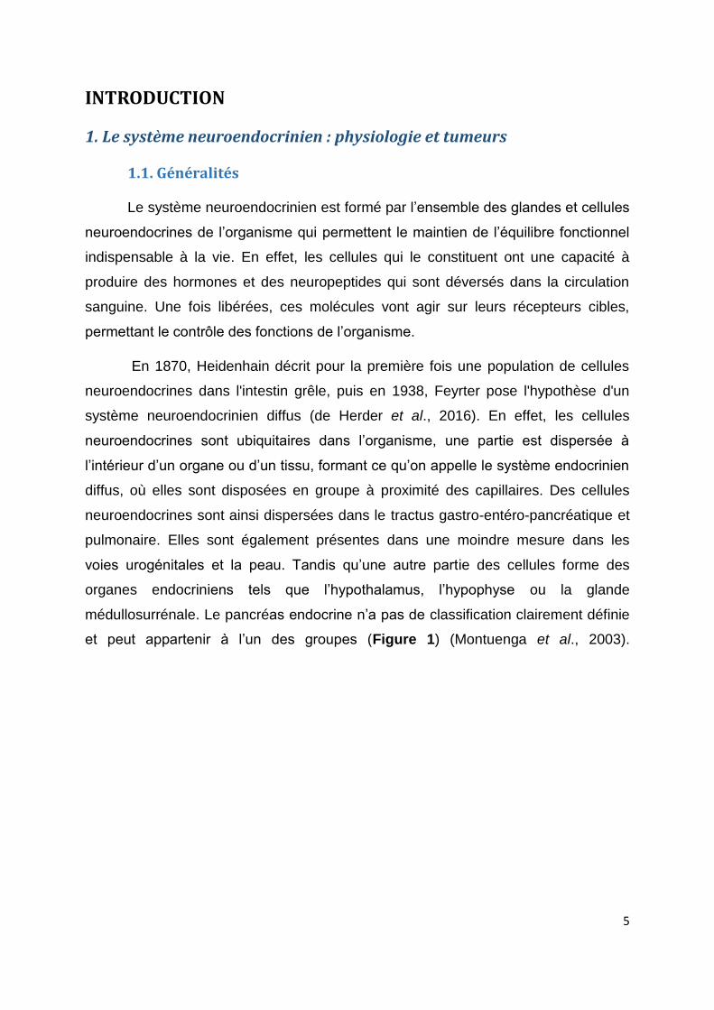
Figure 1 : Représentation du système neuroendocrinien et localisation des cellules
neuroendocrines dans le corps humain. Une partie des cellules neuroendocrines est dispersée
dans l’organisme formant le système neuroendocrinien diffus, tandis qu’une partie forme des organes
à part entière.
1.2. Les cellules neuroendocrines
Les cellules neuroendocrines ont diverses origines embryologiques. Ainsi, la
crête neurale donne naissance aux cellules chromaffines de la glande
médullosurrénale, aux cellules principales des paraganglions extra-surrénaux et aux
cellules C thyroïdiennes (Adams and Bronner-Fraser, 2009), tandis que les cellules
neuroendocrines intestinales, pancréatiques et pulmonaires dérivent de l’endoderme
(Andrew et al., 1998).
Bien que d’origines diverses et dispersées dans tout l’organisme, les cellules
neuroendocrines partagent des caractéristiques communes qui sont de stocker et de
sécréter des amines biogènes et des peptides de faible poids moléculaire qui
peuvent ou non être des hormones (Tableau 1). Ces cellules sont regroupées sous
le terme de « amine content and amine precursor uptake and decarboxylation »
(APUD) (Pearse and Takor, 1976). Le phénotype neuroendocrinien inclut également
la présence de marqueurs de surface tels que la protéine « neural cell adhesion
molecule » (NCAM), des marqueurs vésiculaires comme les chromogranines,
sécrétogranines. Sont également retrouvées des protéines cytoplasmiques telles que
la « neuron specific enolase » (NSE), des protéines de liaison au calcium et au
cytosquelette (Bishop et al., 1988; Montuenga et al., 2003). Les cellules
neuroendocrines stockent des hormones et neuropeptides dans de larges vésicules
Thyroïde et parathyroïdes
Tractus broncho-pulmonaire
Tractus gastro-
entéro-pancréatique
Glande médullosurrénale
Système urinaire et génital
Cellules de Merkel (peau)
Antéhypophyse (glande pituitaire)
Hypothalamus
Thymus
Introduction
7
à cœur dense encore appelées granules de sécrétion, d’un diamètre variant de 100 à
400 nm, leur composition riche en granines, glycoprotéines et peptides les rendent
denses en microscopie électronique (Vardjan et al., 2013).
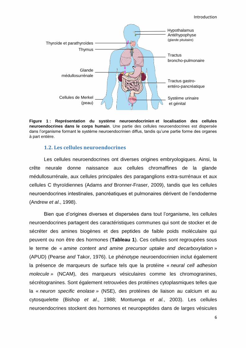
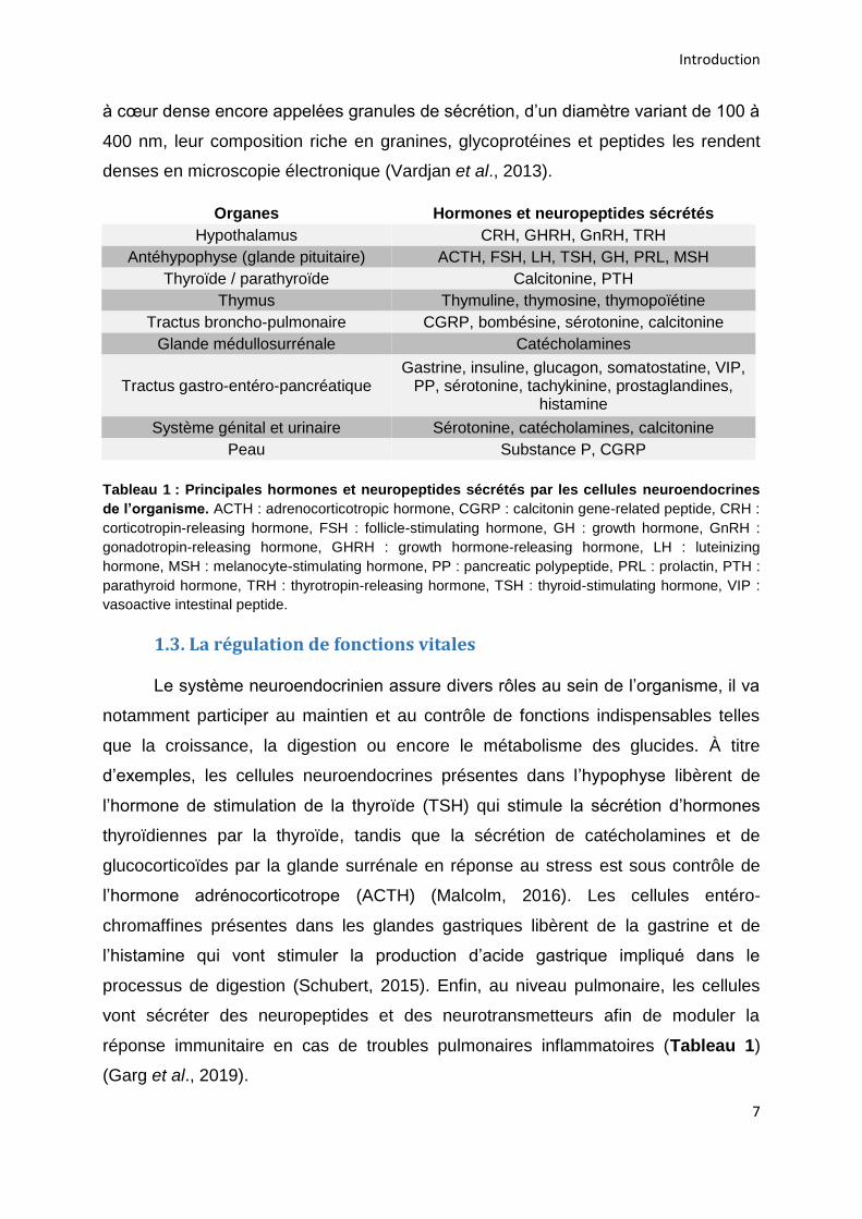
Organes Hormones et neuropeptides sécrétés
Hypothalamus CRH, GHRH, GnRH, TRH
Antéhypophyse (glande pituitaire) ACTH, FSH, LH, TSH, GH, PRL, MSH
Thyroïde / parathyroïde Calcitonine, PTH
Thymus Thymuline, thymosine, thymopoïétine
Tractus broncho-pulmonaire CGRP, bombésine, sérotonine, calcitonine
Glande médullosurrénale Catécholamines
Tractus gastro-entéro-pancréatique Gastrine, insuline, glucagon, somatostatine, VIP,
PP, sérotonine, tachykinine, prostaglandines, histamine
Système génital et urinaire Sérotonine, catécholamines, calcitonine
Peau Substance P, CGRP
Tableau 1 : Principales hormones et neuropeptides sécrétés par les cellules neuroendocrines
de l’organisme. ACTH : adrenocorticotropic hormone, CGRP : calcitonin gene-related peptide, CRH :
corticotropin-releasing hormone, FSH : follicle-stimulating hormone, GH : growth hormone, GnRH :
gonadotropin-releasing hormone, GHRH : growth hormone-releasing hormone, LH : luteinizing
hormone, MSH : melanocyte-stimulating hormone, PP : pancreatic polypeptide, PRL : prolactin, PTH :
parathyroid hormone, TRH : thyrotropin-releasing hormone, TSH : thyroid-stimulating hormone, VIP :
vasoactive intestinal peptide.
1.3. La régulation de fonctions vitales
Le système neuroendocrinien assure divers rôles au sein de l’organisme, il va
notamment participer au maintien et au contrôle de fonctions indispensables telles
que la croissance, la digestion ou encore le métabolisme des glucides. À titre
d’exemples, les cellules neuroendocrines présentes dans l’hypophyse libèrent de
l’hormone de stimulation de la thyroïde (TSH) qui stimule la sécrétion d’hormones
thyroïdiennes par la thyroïde, tandis que la sécrétion de catécholamines et de
glucocorticoïdes par la glande surrénale en réponse au stress est sous contrôle de
l’hormone adrénocorticotrope (ACTH) (Malcolm, 2016). Les cellules entéro-
chromaffines présentes dans les glandes gastriques libèrent de la gastrine et de
l’histamine qui vont stimuler la production d’acide gastrique impliqué dans le
processus de digestion (Schubert, 2015). Enfin, au niveau pulmonaire, les cellules
vont sécréter des neuropeptides et des neurotransmetteurs afin de moduler la
réponse immunitaire en cas de troubles pulmonaires inflammatoires (Tableau 1)
(Garg et al., 2019).
Introduction
8
1.4 Les tumeurs neuroendocrines et l’hypersécrétion
1.4.1. Généralités
L’ensemble des cellules neuroendocrines de l'organisme peuvent se
transformer en cellules tumorales et donner naissance à une tumeur neuroendocrine
(TNE). Ce sont des néoplasmes se formant à partir de ces cellules sécrétrices
d'hormones dispersées dans tout l’organisme. Diverses localisations des TNE sont
donc possibles : l’hypophyse, la thyroïde et les parathyroïdes, le thymus, les tractus
pulmonaire et gastro-entéro-pancréatique, la glande médullosurrénale, la peau et le
système uro-génital. L'incidence des TNE n'a cessé d'augmenter au cours des
dernières décennies avec 1,1 cas/10 000 personnes en 1973 à 7 cas/10 000
personnes en 2012 soit une augmentation de plus de 6 fois. Les TNE les plus
fréquentes sont retrouvées dans les poumons et le tractus gastro-entéro-
pancréatique (Dasari et al., 2017). L’incidence ainsi que le pronostic sont dépendants
des origines ethniques (Shen et al., 2019).
L’apparition des TNE est influencée par différents facteurs tels que l’histoire
familiale. Des mutations dans plusieurs gènes jouent un rôle prépondérant dans
l’apparition et le développement de ces tumeurs. D’autres facteurs comme le
tabagisme et la consommation d'alcool sont connus pour augmenter les risques de
cancers et sont également associés à l’apparition de certaines TNE. Par exemple, la
présence de TNE pancréatiques est associée positivement à la consommation
d’alcool et de tabac, tandis que pour l'intestin grêle seul le tabagisme reste associé
(Leoncini et al., 2016). L’évolution de ces tumeurs est également très variable, par
exemple le phéochromocytome qui est une tumeur originaire de la glande
médullosurrénale est métastatique dans 10 à 20% des cas, tandis que le carcinome
du poumon à petites cellules est hautement agressive, avec plus de 60% des
patients qui développent des métastases (Bernhardt and Jalal, 2016; Buffet et al.,
2020).
Introduction
9
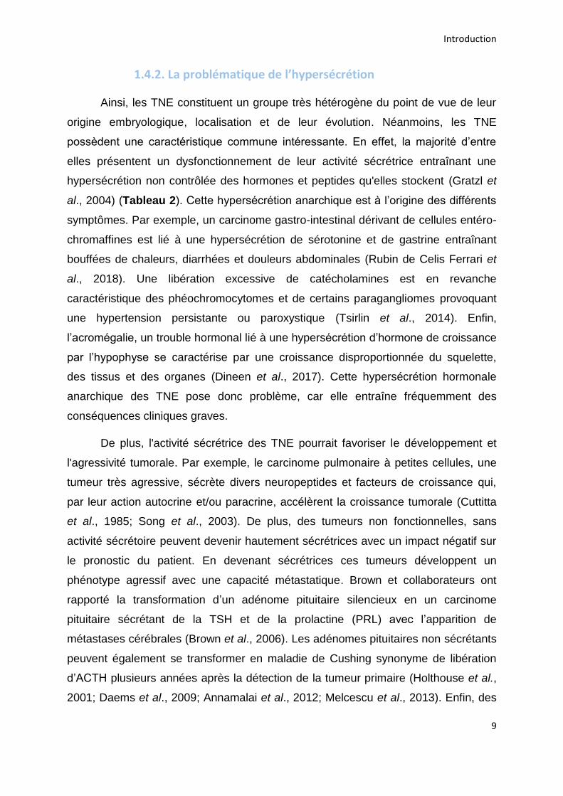
1.4.2. La problématique de l’hypersécrétion
Ainsi, les TNE constituent un groupe très hétérogène du point de vue de leur
origine embryologique, localisation et de leur évolution. Néanmoins, les TNE
possèdent une caractéristique commune intéressante. En effet, la majorité d’entre
elles présentent un dysfonctionnement de leur activité sécrétrice entraînant une
hypersécrétion non contrôlée des hormones et peptides qu'elles stockent (Gratzl et
al., 2004) (Tableau 2). Cette hypersécrétion anarchique est à l’origine des différents
symptômes. Par exemple, un carcinome gastro-intestinal dérivant de cellules entéro-
chromaffines est lié à une hypersécrétion de sérotonine et de gastrine entraînant
bouffées de chaleurs, diarrhées et douleurs abdominales (Rubin de Celis Ferrari et
al., 2018). Une libération excessive de catécholamines est en revanche
caractéristique des phéochromocytomes et de certains paragangliomes provoquant
une hypertension persistante ou paroxystique (Tsirlin et al., 2014). Enfin,
l’acromégalie, un trouble hormonal lié à une hypersécrétion d’hormone de croissance
par l’hypophyse se caractérise par une croissance disproportionnée du squelette,
des tissus et des organes (Dineen et al., 2017). Cette hypersécrétion hormonale
anarchique des TNE pose donc problème, car elle entraîne fréquemment des
conséquences cliniques graves.
De plus, l'activité sécrétrice des TNE pourrait favoriser le développement et
l'agressivité tumorale. Par exemple, le carcinome pulmonaire à petites cellules, une
tumeur très agressive, sécrète divers neuropeptides et facteurs de croissance qui,
par leur action autocrine et/ou paracrine, accélèrent la croissance tumorale (Cuttitta
et al., 1985; Song et al., 2003). De plus, des tumeurs non fonctionnelles, sans
activité sécrétoire peuvent devenir hautement sécrétrices avec un impact négatif sur
le pronostic du patient. En devenant sécrétrices ces tumeurs développent un
phénotype agressif avec une capacité métastatique. Brown et collaborateurs ont
rapporté la transformation d’un adénome pituitaire silencieux en un carcinome
pituitaire sécrétant de la TSH et de la prolactine (PRL) avec l’apparition de
métastases cérébrales (Brown et al., 2006). Les adénomes pituitaires non sécrétants
peuvent également se transformer en maladie de Cushing synonyme de libération
d’ACTH plusieurs années après la détection de la tumeur primaire (Holthouse et al.,
2001; Daems et al., 2009; Annamalai et al., 2012; Melcescu et al., 2013). Enfin, des
Introduction
10
TNE pancréatiques non fonctionnels peuvent se transformer en insulinomes malins
producteurs d’insuline plusieurs années après le diagnostic initial (Juhlin et al., 2019).
L’acquisition de ce phénotype sécrétoire a été associée à un comportement tumoral
plus agressif et la détection de métastases.
Tumeurs neuroendocrines Hormones et peptides principaux
sécrétés
Adénome ou carcinome pituitaire ACTH, GH, PRL, FSH, LH, TSH
Carcinome médullaire de la thyroïde, adénome ou carcinome parathyroïdien
Calcitonine, CEA, PTH
TNE du thymus ou tumeur carcinoïde thymique (tumeur carcinoïde atypique, carcinome à petites cellules,
carcinome neuroendocrinien à grandes cellules) ACTH, GHRH
TNE pulmonaire ou tumeur carcinoïde des poumons (tumeur carcinoïde pulmonaire typique ou atypique,
carcinome ou cancer du poumon à petites cellules, carcinome neuroendocrinien à grandes cellules)
ACTH, ADH
Phéochromocytome/paragangliome Catécholamines
TNE gastro-entéro-pancréatique (insulinome,
glucagome, gastrinome, somatostatinome, VIPome, PPome, carcinome neuroendocrinien à petites ou grandes cellules)
Insuline, glucagon, gastrine, sérotonine, somatostatine, VIP, PP
Carcinome neuroendocrinien de la vessie (tumeur
carcinoïde, carcinome à petites cellules, carcinome neuroendocrinien à grandes cellules)
/
TNE du col utérin ou carcinome neuroendocrinien du col de l'utérus (tumeur carcinoïde
typique ou atypique, carcinome à petites cellules, carcinome neuroendocrinien à grandes cellules)
/
Cancer neuroendocrinien de la prostate (carcinome
neuroendocrinien à petites ou grandes cellules, carcinome neuroendocrinien mixte, tumeur carcinoïde, adénocarcinome avec
différenciation neuroendocrinienne à cellules de Paneth)
CEA, PSA
Carcinome neuroendocrinien de la peau ou carcinome à cellules de Merkel
/
Tableau 2 : Catégories et sous catégories des différentes tumeurs neuroendocrines ainsi que
les principaux peptides et hormones sécrétés. En fonction de leur localisation les tumeurs
neuroendocrines sécrètent une variété de composés. ACTH : adrenocorticotropic hormone, ADH :
antidiuretic hormone, CEA : carcinoembryonic antigen, FSH : follicle-stimulating hormone, GH : growth
hormone, GHRH : growth hormone-releasing hormone, LH : luteinizing hormone, PP : pancreatic
polypeptide, PRL : prolactine, PSA : prostate-specific antigen, PTH : parathyroid hormone, TSH :
thyroid-stimulating hormone, VIP : vasoactive intestinal peptide.
Introduction
11
2. La glande médullosurrénale et le phéochromocytome
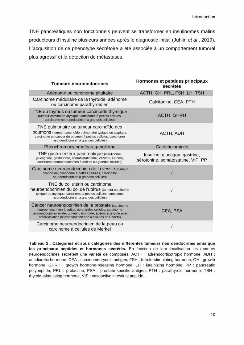
2.1. Anatomie et histologie de la glande surrénale
Le corps humain est constitué de deux glandes surrénales, non symétriques,
situées au-dessus de chaque rein dans l’espace périrénal. Ces glandes ont une
forme concave, et chacune est composée de deux unités fonctionnelles : la partie
externe appelée cortex ou corticosurrénale et la partie interne nommée médulla ou
médullosurrénale (Figure 2). Ces dernières sont enveloppées par un tissu fibreux
dense, la capsule. La médulla et le cortex ont des origines embryologiques et des
fonctions distinctes au sein de l’organisme.
La corticosurrénale dérive du mésoderme et est constituée de 3 couches au
rôle distinct. De l’extérieur vers l’intérieur de la structure : la zone glomérulée secrète
des minéralocorticoïdes comme l’aldostérone, la zone fasciculée libère des
glucocorticoïdes, tels que le cortisol et la corticostérone, enfin la zone réticulée
assure la production des androgènes. La médullosurrénale est originaire de
l’ectoderme ; elle appartient au système nerveux sympathique et libère des
catécholamines. La glande surrénale est constituée à 80% de la partie corticale et
20% de la partie médullaire (Mitty, 1988; Kemppainen and Behrend, 1997).
Figure 2 : Schéma de la glande surrénale avec ses deux unités fonctionnelles. La
corticosurrénale est en position externe et la médullosurrénale en position interne.
Glande surrénale
Rein
Corticosurrénale
Médullosurrénale
Introduction
12
2.2. Les cellules chromaffines
Au sein de la glande médullosurrénale, deux populations de cellules
chromaffines sont présentes, les cellules adrénergiques libèrent de l’adrénaline et
représentent 70 à 80% des cellules, et les cellules noradrénergiques sécrètent de la
noradrénaline et constituent 20 à 30% de la population. Ces deux populations
cellulaires se différencient par la présence de la phényléthanolamine N-méthyl-
transférase (PNMT), l’enzyme assurant la conversion de la noradrénaline en
adrénaline, qui est uniquement présente dans les cellules adrénergiques (Pérez-
Alvarez et al., 2010).
2.3. Processus de sécrétion par les cellules chromaffines
2.3.1 Les granules de sécrétion à cœur dense
2.3.1.1. Le contenu granulaire
Les granules de sécrétion occupent environ 13% du volume cytoplasmique
d’une cellule chromaffine, et chacune d’elle peut contenir environ 10 000 à 30 000
granules (Vitale et al., 1995; Plattner et al., 1997). La composition de ces organelles
est complexe et se caractérise par une forte concentration en protéines. En effet, en
plus des catécholamines, le contenu intragranulaire comprend des hormones
peptidiques, des protéines de la famille des granines telles que les chromogranines
A (CGA) et B (CGB), et les sécrétogranines II, III, V, VI mais aussi des
glycoprotéines, des enzymes comme des pro-hormones convertases et d’autres
molécules non protéiques (l’ATP, le calcium,…) (Crivellato et al., 2008; Takeuchi and
Hosaka, 2008; Estévez-Herrera et al., 2016) (Tableau 3). Représentant plus de 80%
des protéines solubles des granules, les granines sont largement majoritaires, la
CGA à elle seule représentant 40% (Crivellato et al., 2008).
Introduction
13
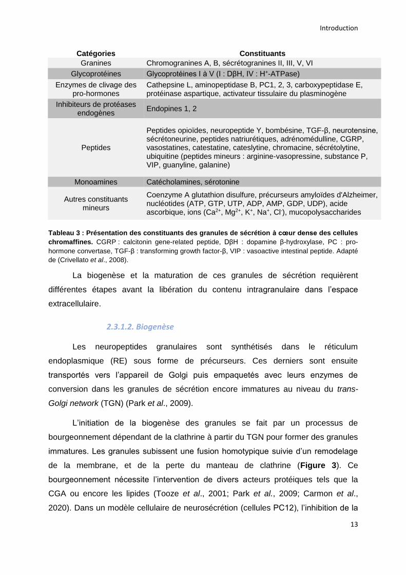
Catégories Constituants
Granines Chromogranines A, B, sécrétogranines II, III, V, VI
Glycoprotéines Glycoprotéines I à V (I : DβH, IV : H+-ATPase)
Enzymes de clivage des pro-hormones
Cathepsine L, aminopeptidase B, PC1, 2, 3, carboxypeptidase E, protéinase aspartique, activateur tissulaire du plasminogène
Inhibiteurs de protéases endogènes
Endopines 1, 2
Peptides
Peptides opioïdes, neuropeptide Y, bombésine, TGF-β, neurotensine, sécrétoneurine, peptides natriurétiques, adrénomédulline, CGRP, vasostatines, catestatine, cateslytine, chromacine, sécrétolytine, ubiquitine (peptides mineurs : arginine-vasopressine, substance P, VIP, guanyline, galanine)
Monoamines Catécholamines, sérotonine
Autres constituants mineurs
Coenzyme A glutathion disulfure, précurseurs amyloïdes d'Alzheimer, nucléotides (ATP, GTP, UTP, ADP, AMP, GDP, UDP), acide ascorbique, ions (Ca2+, Mg2+, K+, Na+, Cl-), mucopolysaccharides
Tableau 3 : Présentation des constituants des granules de sécrétion à cœur dense des cellules
chromaffines. CGRP : calcitonin gene-related peptide, DβH : dopamine β-hydroxylase, PC : pro-
hormone convertase, TGF-β : transforming growth factor-β, VIP : vasoactive intestinal peptide. Adapté
de (Crivellato et al., 2008).
La biogenèse et la maturation de ces granules de sécrétion requièrent
différentes étapes avant la libération du contenu intragranulaire dans l’espace
extracellulaire.
2.3.1.2. Biogenèse
Les neuropeptides granulaires sont synthétisés dans le réticulum
endoplasmique (RE) sous forme de précurseurs. Ces derniers sont ensuite
transportés vers l’appareil de Golgi puis empaquetés avec leurs enzymes de
conversion dans les granules de sécrétion encore immatures au niveau du trans-
Golgi network (TGN) (Park et al., 2009).
L’initiation de la biogenèse des granules se fait par un processus de
bourgeonnement dépendant de la clathrine à partir du TGN pour former des granules
immatures. Les granules subissent une fusion homotypique suivie d’un remodelage
de la membrane, et de la perte du manteau de clathrine (Figure 3). Ce
bourgeonnement nécessite l’intervention de divers acteurs protéiques tels que la
CGA ou encore les lipides (Tooze et al., 2001; Park et al., 2009; Carmon et al.,
2020). Dans un modèle cellulaire de neurosécrétion (cellules PC12), l’inhibition de la
Introduction
14
CGA entraîne une diminution du nombre de granules, tandis que son expression
dans des cellules non neuroendocrines (fibroblastes) induit la formation de granules
denses capables de libérer leur contenu (Kim et al., 2001; Loh et al., 2004; Montero-
Hadjadje et al., 2009). De la même façon, l’expression de la CGA dans des cellules
endocrines dépourvues d’un processus de sécrétion régulée et déficiente pour la
CGA restaure la formation de granules sécrétoires et le processus de sécrétion
régulé (Loh et al., 2004). In vivo, l’utilisation de souris transgéniques déficientes pour
la CGA a montré une diminution du nombre de granules formés et une altération de
leur morphologie (Kim et al., 2005). Le potentiel granulogénique de la CGA semble
médié par sa partie N-terminale puisque cette région est suffisante pour induire la
granulogenèse dans des cellules sympatho-surrénaliennes (Courel et al., 2006).
L’analyse morphologique de cellules chromaffines de souris dont le gène Chga a été
inactivé n’a en revanche montré aucune différence pour le nombre et la taille des
granules de sécrétion entre les souris sauvages et Chga-/-. L’analyse des autres
granines (CGB et sécrétogranines II à VI) a montré un augmentation de leur
expression de 2 à 3 fois, suggérant un rôle compensatoire des autres granines
(Hendy et al., 2006). Un rôle semblable à celui de la CGA dans la biogenèse des
granules a été mis en évidence pour la CGB (Huh et al., 2003; Beuret et al., 2004).
Dans le TGN où les granules de sécrétion se forment, l'environnement est acide et le
milieu contient une forte concentration de cations bivalents comme le Ca2+. Ces
conditions engendrent l’agrégation et la condensation des granines entre elles et à
d'autres composants tels que les catécholamines, l'ATP et d’autres protéines
(Crivellato et al., 2008).
Enfin, la formation des granules par bourgeonnement du TGN va nécessiter
des modifications de courbures membranaires et implique ainsi les lipides. Par
exemple, l’accumulation d’acide phosphatidique (PA) et de diacylglycérol (DAG) à la
membrane du TGN pourrait faciliter la courbure de la membrane conduisant au
bourgeonnement (Tanguy et al., 2016). De façon intéressante le PA interagit avec la
CGA et pourrait ainsi participer à ce remodelage membranaire. En effet, il a été
montré récemment que l’inhibition de la phospholipase D (PLD), l’enzyme
responsable de la synthèse du PA altère la biogenèse des granules (Carmon et al.,
2020).
Introduction
15
2.3.1.3. Maturation
Les granules immatures vont ensuite suivre un processus de maturation qui
inclut des modifications morphologiques et biochimiques. Les granules de sécrétion
vont ainsi perdre leur manteau de clathrine qui est présent lors du bourgeonnement
et voir leur taille augmenter (Tooze, 1991).
La lumière des granules immatures va s’acidifier progressivement, entraînant
une condensation supplémentaire des précurseurs. Cette acidification se fait grâce à
une augmentation de la densité de pompes à protons (V-ATPase) dépendantes de
l’ATP et une diminution de la perméabilité aux protons des membranes. Les protons
vont donc être amenés dans la lumière du granule ce qui conduit à une acidification
du pH intragranulaire qui passe de 6,3 à 5-5,5 (Park et al., 2009; Kögel and Gerdes,
2010). Dans les cellules PC12, il a été montré que l’acidification dure environ 90
minutes (Urbé et al., 1997). Ce gradient de protons va permettre l’activation des pro-
hormones convertases pour la conversion des précurseurs d’hormones et de
neuropeptides en molécules bioactives (Johnson, 1988; Kögel and Gerdes, 2010).
Tout en acquérant leur maturité, les granules se déplacent le long des
microtubules vers la partie corticale riche en actine où le granule va rester jusqu’à
son recrutement pour migrer vers la membrane plasmique (Rudolf et al., 2001).
Lors de sa maturation, le granule va acquérir son contenu en catécholamines.
La L-tyrosine est transformée en L-dopa par l’action de la tyrosine hydroxylase (TH)
dans le cytoplasme de la cellule. La décarboxylation de la L-dopa en dopamine est
ensuite catalysée par la dopamine décarboxylase (DD). Puis, le transporteur
vesicular monoamine transporter (VMAT) va prendre en charge la dopamine et la
transporter dans l’espace intragranulaire, où elle est alors hydroxylée en
noradrénaline par la dopamine β-hydroxylase (DβH). La voie de biosynthèse
s’achève à cette étape pour les cellules noradrénergiques. Pour les cellules
adrénergiques, la noradrénaline est exportée dans le cytoplasme de la cellule pour y
être méthylée en adrénaline par la PNMT, puis l’adrénaline obtenue est transportée
dans le granule (Figure 3).
Introduction
16
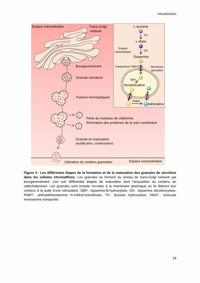
Figure 3 : Les différentes étapes de la formation et de la maturation des granules de sécrétion
dans les cellules chromaffines. Les granules se forment au niveau de trans-Golgi network par
bourgeonnement, s’en suit différentes étapes de maturation dont l’acquisition du contenu en
catécholamines. Les granules sont ensuite recrutés à la membrane plasmique où ils libèrent leur
contenu à la suite d’une stimulation. DβH : dopamine-β-hydroxylase, DD : dopamine décarboxylase,
PNMT : phényléthanolamine N-méthyl-transférase, TH : tyrosine hydroxylase, VMAT : vesicular
monoamine transporter.
Trans-Golgi network
Bourgeonnement
Granule immature
Fusions homotypiques
Perte du manteau de clatherine Elimination des protéines de la voie constitutive
Granule en maturation
(acidification, condensation)
TH
DD
L-tyrosine
L-dopa
Dopamine
Noradrénaline
DβH
Adrénaline PNMT
Espace
intracellulaire
Transporteur VMAT Membrane granulaire
Libération du contenu granulaire
Espace intracellulaire
Espace extracellulaire
Introduction
17
2.3.2. L’exocytose régulée
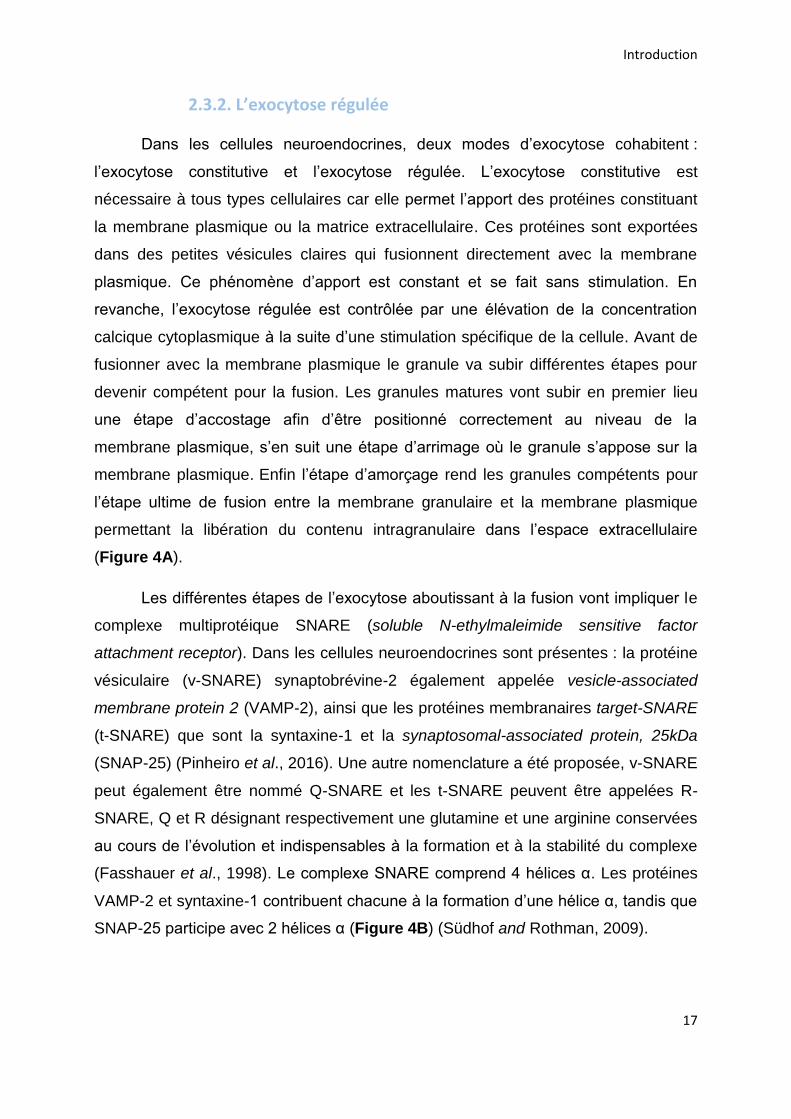
Dans les cellules neuroendocrines, deux modes d’exocytose cohabitent :
l’exocytose constitutive et l’exocytose régulée. L’exocytose constitutive est
nécessaire à tous types cellulaires car elle permet l’apport des protéines constituant
la membrane plasmique ou la matrice extracellulaire. Ces protéines sont exportées
dans des petites vésicules claires qui fusionnent directement avec la membrane
plasmique. Ce phénomène d’apport est constant et se fait sans stimulation. En
revanche, l’exocytose régulée est contrôlée par une élévation de la concentration
calcique cytoplasmique à la suite d’une stimulation spécifique de la cellule. Avant de
fusionner avec la membrane plasmique le granule va subir différentes étapes pour
devenir compétent pour la fusion. Les granules matures vont subir en premier lieu
une étape d’accostage afin d’être positionné correctement au niveau de la
membrane plasmique, s’en suit une étape d’arrimage où le granule s’appose sur la
membrane plasmique. Enfin l’étape d’amorçage rend les granules compétents pour
l’étape ultime de fusion entre la membrane granulaire et la membrane plasmique
permettant la libération du contenu intragranulaire dans l’espace extracellulaire
(Figure 4A).
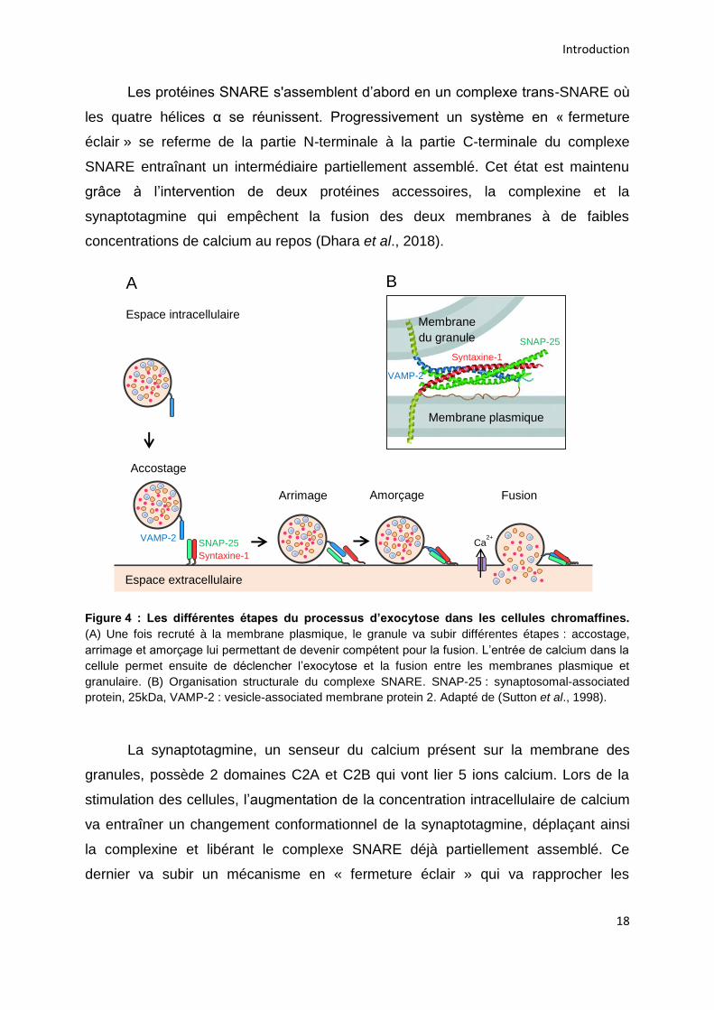
Les différentes étapes de l’exocytose aboutissant à la fusion vont impliquer le
complexe multiprotéique SNARE (soluble N-ethylmaleimide sensitive factor
attachment receptor). Dans les cellules neuroendocrines sont présentes : la protéine
vésiculaire (v-SNARE) synaptobrévine-2 également appelée vesicle-associated
membrane protein 2 (VAMP-2), ainsi que les protéines membranaires target-SNARE
(t-SNARE) que sont la syntaxine-1 et la synaptosomal-associated protein, 25kDa
(SNAP-25) (Pinheiro et al., 2016). Une autre nomenclature a été proposée, v-SNARE
peut également être nommé Q-SNARE et les t-SNARE peuvent être appelées R-
SNARE, Q et R désignant respectivement une glutamine et une arginine conservées
au cours de l’évolution et indispensables à la formation et à la stabilité du complexe
(Fasshauer et al., 1998). Le complexe SNARE comprend 4 hélices α. Les protéines
VAMP-2 et syntaxine-1 contribuent chacune à la formation d’une hélice α, tandis que
SNAP-25 participe avec 2 hélices α (Figure 4B) (Südhof and Rothman, 2009).
Introduction
18
Les protéines SNARE s'assemblent d’abord en un complexe trans-SNARE où
les quatre hélices α se réunissent. Progressivement un système en « fermeture
éclair » se referme de la partie N-terminale à la partie C-terminale du complexe
SNARE entraînant un intermédiaire partiellement assemblé. Cet état est maintenu
grâce à l’intervention de deux protéines accessoires, la complexine et la
synaptotagmine qui empêchent la fusion des deux membranes à de faibles
concentrations de calcium au repos (Dhara et al., 2018).
Figure 4 : Les différentes étapes du processus d’exocytose dans les cellules chromaffines.
(A) Une fois recruté à la membrane plasmique, le granule va subir différentes étapes : accostage,
arrimage et amorçage lui permettant de devenir compétent pour la fusion. L’entrée de calcium dans la
cellule permet ensuite de déclencher l’exocytose et la fusion entre les membranes plasmique et
granulaire. (B) Organisation structurale du complexe SNARE. SNAP-25 : synaptosomal-associated
protein, 25kDa, VAMP-2 : vesicle-associated membrane protein 2. Adapté de (Sutton et al., 1998).
La synaptotagmine, un senseur du calcium présent sur la membrane des
granules, possède 2 domaines C2A et C2B qui vont lier 5 ions calcium. Lors de la
stimulation des cellules, l’augmentation de la concentration intracellulaire de calcium
va entraîner un changement conformationnel de la synaptotagmine, déplaçant ainsi
la complexine et libérant le complexe SNARE déjà partiellement assemblé. Ce
dernier va subir un mécanisme en « fermeture éclair » qui va rapprocher les
VAMP-2 SNAP-25 Syntaxine-1
Accostage
Arrimage
Membrane plasmique
VAMP-2
Syntaxine-1 SNAP-25
Membrane du granule
Amorçage
Ca2+
Fusion
Espace extracellulaire
Espace intracellulaire
A B
Introduction
19
membranes et permettre la fusion entre les bicouches lipidiques vésiculaire et
plasmique permettant la libération du contenu intragranulaire (Pinheiro et al., 2016).
La disponibilité des protéines SNARE et leur engagement dans le complexe
est également étroitement régulé par les protéines Munc (Toonen and Verhage,
2007). Par exemple, la protéine Munc18-1 interagit avec la partie N-terminale de la
syntaxine-1 assurant ainsi son maintien dans une conformation fermée. Cette
configuration empêche l’interaction de la syntaxine-1 avec ses partenaires VAMP-2
et SNAP-25 et la formation finale du complexe SNARE. En se liant au complexe
syntaxine-1-Munc18-1, Munc13-1 va ensuite engendrer un changement de
conformation permettant à la syntaxine-1 d’adopter une conformation ouverte
favorable à l’assemblage du complexe SNARE (Gladycheva et al., 2004; Ma et al.,
2011).
Une fois les catécholamines et autres constituants des granules de sécrétion
libérés, le complexe SNARE va être désassemblé pour être recyclé et utilisé pour un
autre cycle de fusion. Ce processus est dépendant de l’ATP et implique la protéine
N-ethylmaleimide-sensitive fusion protein (NSF) et son cofacteur soluble NSF
attachment protein (SNAP). L'association de SNAP avec le complexe SNARE permet
la liaison de la protéine NSF au complexe SNAP–SNARE. Puis, la stimulation de
l'activité ATPase du NSF conduit au désassemblage du complexe et la libération des
acteurs pour d'autres cycles de fusion (Morgan and Burgoyne, 2004).
2.3.3. La régulation de l’exocytose
Les étapes successives de l’exocytose sont également régulées par différents
autres acteurs protéiques ou lipidiques. Il a été montré notamment que l’organisation
et le remodelage à la fois des lipides et du cytosquelette d’actine constituent des
processus clés dans la régulation de l’exocytose, et que plusieurs GTPases
monomériques sont impliquées.
Après leur synthèse au niveau du TGN, les granules de sécrétion sont
acheminés vers la périphérie cellulaire le long des microtubules, puis ils sont ancrés
dans un réseau cortical de filaments d'actine qui forme une barrière physique à
l'exocytose (Trifaró et al., 2008). Cette dernière est contrôlée par des GTPases Rho,
une sous-famille de la superfamille des Ras. Par exemple, l’activation de la protéine
Introduction
20
RhoA dans les cellules chromaffines contribue à la stabilisation du réseau cortical
d’actine empêchant le recrutement des granules à la membrane plasmique (Gasman
et al., 1998). Le mécanisme d’action n’est pas complément élucidé mais RhoA
activerait une phosphatidylinositol 4-kinase (PI 4-kinase) ce qui pourrait modifier le
niveau de synthèse de certains lipides comme le phosphatidylinositol-4,5-
bisphosphate (PIP2) connu pour réguler les interactions entre la membrane et le
cytosquelette (Gasman et al., 1998). La stimulation des cellules chromaffines par un
sécrétagogue entraîne une réorganisation complexe du cytosquelette d’actine. Dans
un premier temps, la barrière corticale d’actine est destructurée, un processus qui
nécessite très certainement l’inactivation de RhoA (Bader et al., 2004), puis les
granules sont acheminés au niveau de la membrane plasmique grâce à l’action de
Rab27A, une GTPase granulaire qui permet l’interaction des granules avec des
moteurs moléculaires comme la myosine-V via son effecteur MyRip (Desnos et al.,
2003). En parallèle, la GTPase Cdc42 (un autre membre de la famille Rho) est
recrutée à la membrane plasmique où elle active la synthèse de nouveaux filaments
d’actine, spécifiquement au niveau des sites d’exocytose, en activant la cascade
moléculaire incluant N-WASP et Arp2/3 (Gasman et al., 2004; Malacombe et al.,
2006). La protéine annexine-A2 régule l’organisation des filaments d’actine en
faisceaux reliant le granule à la membrane plasmique (Gabel et al., 2015). L’actine
semble donc être un acteur important de l’accostage et/ou de l’arrimage mais
pourrait également participer à des phases plus tardives en exerçant des forces
nécessaires à l’expansion du pore de fusion ou à l’expulsion de la matrice
intragranulaire.
Le déclenchement de l’exocytose entraîne également une réorganisation
lipidique indispensable au bon déroulement de la sécrétion (Gasman and Vitale,
2017). Par exemple, il a été montré que l’annexine-A2 participe à la formation des
sites d’exocytose en contrôlant la mise en place de microdomaines lipidique riches
en gangliosides GM1 et certains phosphoinositides comme le PIP2 (Chasserot-Golaz
et al., 2005; Umbrecht-Jenck et al., 2010). Un autre lipide impliqué de façon
importante dans le contrôle de l’exocytose est le PA. En effet, les travaux de l’équipe
proposent que la synthèse de PA induite par l’activation de phospholipase D1 soit
indispensable à l’exocytose (Vitale et al., 2001; Zeniou-Meyer et al., 2007). Plus
récemment il a été montré que les formes mono-insaturées du PA régulent l’arrimage
Introduction
21
des granules tandis que les formes poly-insaturées réguleraient plutôt l’étape de
fusion (Tanguy et al., 2020). De façon intéressante, l’activité de la PLD déclenchée
au cours de l’exocytose est sous le contrôle de plusieurs GTPases monomériques
comme Arf6, Rac1 et RalA (Vitale et al., 2005; Béglé et al., 2009; Momboisse et al.,
2009).
Pour plus de détails sur l’implication des GTPases Rho au cours de la
sécrétion neuroendocrine dans les cellules saines et tumorales, vous pouvez vous
référer à la revue que j’ai écrite pendant ma thèse (Annexe; Streit et al., 2020).
2.4. Effets physiologiques des molécules libérées par les cellules
chromaffines
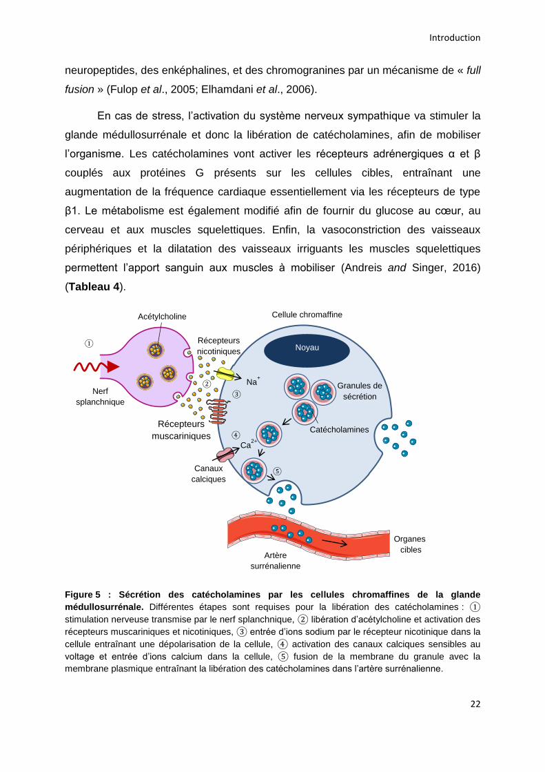
Les cellules chromaffines sécrètent des catécholamines telles que l’adrénaline
et la noradrénaline dont la libération est régulée par le système nerveux sympathique
grâce à une innervation par le nerf splanchnique. Ce dernier libère de l’acétylcholine
qui active les récepteurs nicotiniques et muscariniques présents sur les cellules
chromaffines (Pérez-Alvarez and Albillos, 2007). L’entrée d’ions sodium à travers le
récepteur canal nicotinique entraîne alors une dépolarisation de la membrane de la
cellule, et par conséquent une activation des canaux calciques dépendants du
voltage. Il s’en suit une entrée d’ions calcium qui constitue l’élément déclencheur de
l’exocytose et de la libération des catécholamines et neuropeptides dans la
circulation sanguine (Aunis and Langley, 1999) (Figure 5).
Dans des conditions physiologiques, les cellules chromaffines libèrent
uniquement des catécholamines à un taux basal qui permet de réguler les fonctions
homéostatiques telles que l’activité cardiaque, intestinale et le métabolisme. Cette
libération se fait par un mécanisme de « kiss and run » qui implique la libération de
petites molécules solubles comme les catécholamines et retient les molécules moins
mobiles telles que les neuropeptides et les chromogranines. Lors de stimulations
soutenues et répétitives en réponse au stress, le nerf splanchnique va libérer en plus
de l’acétylcholine, d’autres neuropeptides tels que le pituitary adenylate cyclase-
activating polypeptide (PACAP) qui vont contribuer à la libération des catécholamines
(Guérineau, 2020). L’activation sympathique médiée par le stress entraîne une
augmentation de la sécrétion des catécholamines, ainsi qu’une libération des
Introduction
22
neuropeptides, des enképhalines, et des chromogranines par un mécanisme de « full
fusion » (Fulop et al., 2005; Elhamdani et al., 2006).
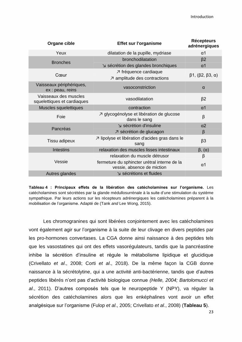
En cas de stress, l’activation du système nerveux sympathique va stimuler la
glande médullosurrénale et donc la libération de catécholamines, afin de mobiliser
l’organisme. Les catécholamines vont activer les récepteurs adrénergiques α et β
couplés aux protéines G présents sur les cellules cibles, entraînant une
augmentation de la fréquence cardiaque essentiellement via les récepteurs de type
β1. Le métabolisme est également modifié afin de fournir du glucose au cœur, au
cerveau et aux muscles squelettiques. Enfin, la vasoconstriction des vaisseaux
périphériques et la dilatation des vaisseaux irriguants les muscles squelettiques
permettent l’apport sanguin aux muscles à mobiliser (Andreis and Singer, 2016)
(Tableau 4).
Figure 5 : Sécrétion des catécholamines par les cellules chromaffines de la glande
médullosurrénale. Différentes étapes sont requises pour la libération des catécholamines : ①
stimulation nerveuse transmise par le nerf splanchnique, ② libération d’acétylcholine et activation des
récepteurs muscariniques et nicotiniques, ③ entrée d’ions sodium par le récepteur nicotinique dans la
cellule entraînant une dépolarisation de la cellule, ④ activation des canaux calciques sensibles au
voltage et entrée d’ions calcium dans la cellule, ⑤ fusion de la membrane du granule avec la
membrane plasmique entraînant la libération des catécholamines dans l’artère surrénalienne.
Acétylcholine
Nerf splanchnique
①
②
Récepteurs
nicotiniques
Na+
③
Ca2+
④
Noyau
Canaux calciques
Granules de
sécrétion
⑤
Catécholamines
Cellule chromaffine
Artère
surrénalienne
Organes cibles
Récepteurs muscariniques
Introduction
23
Organe cible Effet sur l'organisme Récepteurs
adrénergiques
Yeux dilatation de la pupille, mydriase α1
Bronches bronchodilatation β2
↘ sécrétion des glandes bronchiques α1
Cœur ↗ fréquence cardiaque
β1, (β2, β3, α) ↗ amplitude des contractions
Vaisseaux périphériques, ex : peau, reins
vasoconstriction α
Vaisseaux des muscles squelettiques et cardiaques
vasodilatation β2
Muscles squelettiques contraction α1
Foie ↗ glycogénolyse et libération de glucose
dans le sang β
Pancréas ↘ sécrétion d'insuline α2
↗ sécrétion de glucagon β
Tissu adipeux ↗ lipolyse et libération d'acides gras dans le
sang β3
Intestins relaxation des muscles lisses intestinaux β, (α)
Vessie
relaxation du muscle détrusor β
fermeture du sphincter urétral interne de la vessie, absence de miction
α1
Autres glandes ↘ sécrétions et fluides
Tableau 4 : Principaux effets de la libération des catécholamines sur l’organisme. Les
catécholamines sont sécrétées par la glande médullosurrénale à la suite d’une stimulation du système
sympathique. Par leurs actions sur les récepteurs adrénergiques les catécholamines préparent à la
mobilisation de l’organisme. Adapté de (Tank and Lee Wong, 2015).
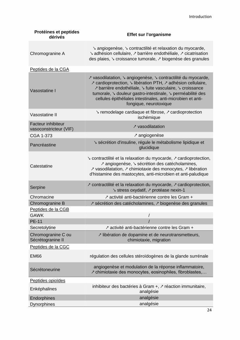
Les chromogranines qui sont libérées conjointement avec les catécholamines
vont également agir sur l’organisme à la suite de leur clivage en divers peptides par
les pro-hormones convertases. La CGA donne ainsi naissance à des peptides tels
que les vasostatines qui ont des effets vasorégulateurs, tandis que la pancréastine
inhibe la sécrétion d’insuline et régule le métabolisme lipidique et glucidique
(Crivellato et al., 2008; Corti et al., 2018). De la même façon la CGB donne
naissance à la sécrétolytine, qui a une activité anti-bactérienne, tandis que d’autres
peptides libérés n’ont pas d’activité biologique connue (Helle, 2004; Bartolomucci et
al., 2011). D’autres composés tels que le neuropeptide Y (NPY), va réguler la
sécrétion des catécholamines alors que les enképhalines vont avoir un effet
analgésique sur l’organisme (Fulop et al., 2005; Crivellato et al., 2008) (Tableau 5).
Introduction
24
Protéines et peptides dérivés
Effet sur l'organisme
Chromogranine A
↘ angiogenèse, ↘ contractilité et relaxation du myocarde, ↘ adhésion cellulaire, ↗ barrière endothéliale, ↗ cicatrisation
des plaies, ↘ croissance tumorale, ↗ biogenèse des granules
Peptides de la CGA
Vasostatine I
↗ vasodilatation, ↘ angiogenèse, ↘ contractilité du myocarde,
↗ cardioprotection, ↘ libération PTH, ↗ adhésion cellulaire, ↗ barrière endothéliale, ↘ fuite vasculaire, ↘ croissance
tumorale, ↘ douleur gastro-intestinale, ↘ perméabilité des cellules épithéliales intestinales, anti-microbien et anti-
fongique, neurotoxique
Vasostatine II ↘ remodelage cardiaque et fibrose, ↗ cardioprotection
ischémique
Facteur inhibiteur vasoconstricteur (VIF)
↗ vasodilatation
CGA 1-373 ↗ angiogenèse
Pancréastine ↘ sécrétion d'insuline, régule le métabolisme lipidique et
glucidique
Catestatine
↘ contractilité et la relaxation du myocarde, ↗ cardioprotection,
↗ angiogenèse, ↘ sécrétion des catécholamines, ↗ vasodilatation, ↗ chimiotaxie des monocytes, ↗ libération
d'histamine des mastocytes, anti-microbien et anti-paludique
Serpine ↗ contractilité et la relaxation du myocarde, ↗ cardioprotection,
↘ stress oxydatif, ↗ protéase nexin-1
Chromacine ↗ activité anti-bactérienne contre les Gram +
Chromogranine B ↗ sécrétion des catécholamines, ↗ biogenèse des granules
Peptides de la CGB
GAWK /
PE-11 /
Secretolytine ↗ activité anti-bactérienne contre les Gram +
Chromogranine C ou Sécrétogranine II
↗ libération de dopamine et de neurotransmetteurs, chimiotaxie, migration
Peptides de la CGC
EM66 régulation des cellules stéroïdogènes de la glande surrénale
Sécrétoneurine angiogenèse et modulation de la réponse inflammatoire, ↗ chimiotaxie des monocytes, eosinophiles, fibroblastes,…
Peptides opioïdes
Enképhalines inhibiteur des bactéries à Gram +, ↗ réaction immunitaire,
analgésie
Endorphines analgésie
Dynorphines analgésie
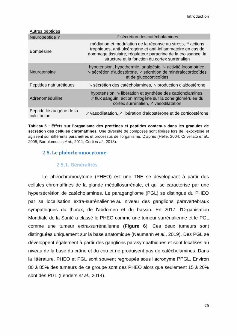
Introduction
25
Autres peptides
Neuropeptide Y ↗ sécrétion des catécholamines
Bombésine
médiation et modulation de la réponse au stress, ↗ actions trophiques, anti-ulcérogène et anti-inflammatoire en cas de
dommage tissulaire, régulateur paracrine de la croissance, la structure et la fonction du cortex surrénalien
Neurotensine hypotension, hypothermie, analgésie, ↘ activité locomotrice,
↘ sécrétion d'aldostérone, ↗ sécrétion de minéralocorticoїdes et de glucocorticoїdes
Peptides natriurétiques ↘ sécrétion des catécholamines, ↘ production d'aldostérone
Adrénomédulline hypotension, ↘ libération et synthèse des catécholamines,
↗ flux sanguin, action mitogène sur la zone glomérulée du cortex surrénalien, ↗ vasodilatation
Peptide lié au gène de la calcitonine
↗ vasodilatation, ↗ libération d'aldostérone et de corticostérone
Tableau 5 : Effets sur l’organisme des protéines et peptides contenus dans les granules de
sécrétion des cellules chromaffines. Une diversité de composés sont libérés lors de l’exocytose et
agissent sur différents paramètres et processus de l’organisme. D’après (Helle, 2004; Crivellato et al.,
2008; Bartolomucci et al., 2011; Corti et al., 2018).
2.5. Le phéochromocytome
2.5.1. Généralités
Le phéochromocytome (PHEO) est une TNE se développant à partir des
cellules chromaffines de la glande médullosurrénale, et qui se caractérise par une
hypersécrétion de catécholamines. Le paragangliome (PGL) se distingue du PHEO
par sa localisation extra-surrénalienne au niveau des ganglions paravertébraux
sympathiques du thorax, de l'abdomen et du bassin. En 2017, l'Organisation
Mondiale de la Santé a classé le PHEO comme une tumeur surrénalienne et le PGL
comme une tumeur extra-surrénalienne (Figure 6). Ces deux tumeurs sont
distinguées uniquement sur la base anatomique (Neumann et al., 2019). Des PGL se
développent également à partir des ganglions parasympathiques et sont localisés au
niveau de la base du crâne et du cou et ne produisent pas de catécholamines. Dans
la littérature, PHEO et PGL sont souvent regroupés sous l’acronyme PPGL. Environ
80 à 85% des tumeurs de ce groupe sont des PHEO alors que seulement 15 à 20%
sont des PGL (Lenders et al., 2014).
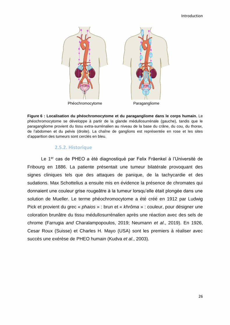
Introduction
26
Figure 6 : Localisation du phéochromocytome et du paragangliome dans le corps humain. Le
phéochromocytome se développe à partir de la glande médullosurrénale (gauche), tandis que le
paragangliome provient du tissu extra-surrénalien au niveau de la base du crâne, du cou, du thorax,
de l’abdomen et du pelvis (droite). La chaîne de ganglions est représentée en rose et les sites
d’apparition des tumeurs sont cerclés en bleu.
2.5.2. Historique
Le 1er cas de PHEO a été diagnostiqué par Felix Fräenkel à l’Université de
Fribourg en 1886. La patiente présentait une tumeur bilatérale provoquant des
signes cliniques tels que des attaques de panique, de la tachycardie et des
sudations. Max Schottelius a ensuite mis en évidence la présence de chromates qui
donnaient une couleur grise rougeâtre à la tumeur lorsqu’elle était plongée dans une
solution de Mueller. Le terme phéochromocytome a été créé en 1912 par Ludwig
Pick et provient du grec « phaios » : brun et « khrôma » : couleur, pour désigner une
coloration brunâtre du tissu médullosurrénalien après une réaction avec des sels de
chrome (Farrugia and Charalampopoulos, 2019; Neumann et al., 2019). En 1926,
Cesar Roux (Suisse) et Charles H. Mayo (USA) sont les premiers à réaliser avec
succès une exérèse de PHEO humain (Kudva et al., 2003).
Phéochromocytome Paragangliome
Introduction
27
2.5.3. Epidémiologie
Le nombre de patients atteints de PPGL a augmenté au cours des dernières
années avec une incidence qui est passée de 0,37 cas/100 000 personnes/an en
1995 à 0,57 et en 2015 (Berends et al., 2018). Néanmoins ce nombre reste
probablement sous-estimé puisque 0,05% des autopsies révèlent des PHEO (Lo et
al., 2000; McNeil et al., 2000). L’incidence de cette tumeur augmente avec l’âge des
sujets avec un pic d’incidence entre 40 et 60 ans, et touche autant les femmes que
les hommes (Berends et al., 2018). La prévalence des PPGL chez les patients
adultes souffrants d’hypertension est de 0,2 à 0,6%, et de 1,7% chez les enfants. À
noter que près de 5% des PPGL sont découverts de façon fortuite (Lenders et al.,
2014; Tsirlin et al., 2014).
2.5.4. Symptômes cliniques
Le PHEO est caractérisé par une hypersécrétion de catécholamines
(noradrénaline, adrénaline) qui peut entraîner une hypertension artérielle importante,
paroxystique ou continue. La triade classique de Menard décrit les symptômes
principaux que sont les céphalées, les palpitations et les sudations. Peuvent
également être présents une sensation de faiblesse, de la fatigue, des nausées et
une pâleur (Lenders et al., 2005; Tsirlin et al., 2014) (Tableau 6). Néanmoins
certains PHEO peuvent être asymptomatiques dans environ 10% des cas (Fall et al.,
2013). La tumeur est alors découverte de façon fortuite par imagerie réalisée pour
une autre suspicion de maladie ; on parle alors d’incidentalome surrénalien
(Fassnacht et al., 2016).
L’intensité de la sécrétion tumorale est responsable de l’intensité des signes
cliniques mais surtout de l’importance des modifications hémodynamiques
associées. En conséquence, les patients présentent un risque sévère d'accident
vasculaire, ce qui rend d'autant plus délicate la chirurgie. De plus, le PHEO peut être
à l’origine de différentes cardiomyopathies (Zhang et al., 2017; Y-Hassan and
Falhammar, 2020). L’exérèse de la tumeur permet d’améliorer les problèmes
cardiaques des patients dans 96% des cas, tandis que l’absence de chirurgie
engendre un taux de mortalité de 33%. Le PHEO conduit à des événements
indésirables graves dans 44% des cas (Zhang et al., 2017). Après opération, une
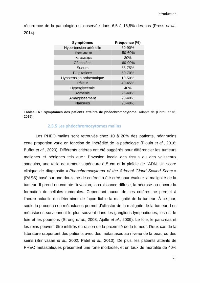
Introduction
28
récurrence de la pathologie est observée dans 6,5 à 16,5% des cas (Press et al.,
2014).
Symptômes Fréquence (%)
Hypertension artérielle 80-90%
- Permanente 50-60%
- Paroxystique 30%
Céphalées 60-90%
Sueurs 55-75%
Palpitations 50-70%
Hypotension orthostatique 10-50%
Pâleur 40-45%
Hyperglycémie 40%
Asthénie 25-40%
Amaigrissement 20-40%
Nausées 20-40%
Tableau 6 : Symptômes des patients atteints de phéochromocytome. Adapté de (Cornu et al.,
2019).
2.5.5 Les phéochromocytomes malins
Les PHEO malins sont retrouvés chez 10 à 20% des patients, néanmoins
cette proportion varie en fonction de l’hérédité de la pathologie (Plouin et al., 2016;
Buffet et al., 2020). Différents critères ont été suggérés pour différencier les tumeurs
malignes et bénignes tels que : l'invasion locale des tissus ou des vaisseaux
sanguins, une taille de tumeur supérieure à 5 cm et la ploïdie de l'ADN. Un score
clinique de diagnostic « Pheochromocytoma of the Adrenal Gland Scaled Score »
(PASS) basé sur une douzaine de critères a été créé pour évaluer la malignité de la
tumeur. Il prend en compte l'invasion, la croissance diffuse, la nécrose ou encore la
formation de cellules tumorales. Cependant aucun de ces critères ne permet à
l’heure actuelle de déterminer de façon fiable la malignité de la tumeur. À ce jour,
seule la présence de métastases permet d’attester de la malignité de la tumeur. Les
métastases surviennent le plus souvent dans les ganglions lymphatiques, les os, le
foie et les poumons (Strong et al., 2008; Ajallé et al., 2009). Le foie, le pancréas et
les reins peuvent être infiltrés en raison de la proximité de la tumeur. Deux cas de la
littérature rapportent des patients avec des métastases au niveau de la peau ou des
seins (Srinivasan et al., 2002; Patel et al., 2010). De plus, les patients atteints de
PHEO métastatiques présentent une forte morbidité, et un taux de mortalité de 40%
Introduction
29
à 5 ans (Jimenez et al., 2013). Environ 50% des patients atteints de PPGL malins
sont porteurs de mutations héréditaires de la lignée germinale dans le gène de la
sous-unité B de la succinate déshydrogénase (SDHB) (Jimenez et al., 2013). Ces
données suggèrent que la génétique joue un rôle primordial dans la détection des
PHEO malins, mais pourrait aussi à terme servir à l’identification de nouveaux
moyens thérapeutiques.
2.5.6. La génétique
2.5.6.1. Prédisposition
La connaissance de la génétique des PPGL s’est considérablement améliorée
ces dernières années, notamment avec l’expansion des techniques de séquençage.
Le dogme selon lequel 10% des PPGL étaient d’origine génétique a été largement
remis en cause par la découverte de nouveaux gènes (Elder et al., 2005). À ce jour,
plus d’une vingtaine de gènes de susceptibilité ont été découverts, dont les premiers
mis en évidence sont NF1 (neurofibromin 1), RET (rearranged during transfection) et
VHL (von Hippel-Lindau). Ce sont ensuite ajoutés les gènes de la SDH (succinate
dehydrogenase) avec l’implication des sous-unités A, B, C et D et le gène SDHAF2
(succinate dehydrogenase assembly factor 2), ainsi que d’autres gènes (Tableau 7).
Des mutations germinales pour des gènes sont retrouvées dans environ 40% des
PPGL qui sont donc marqués par un fort déterminisme génétique (Gupta et al., 2017;
Buffet et al., 2020).

Intr
od
uct
ion
30
Gèn
e
Syn
dro
me
asso
cié
A
ge
L
oc
ali
sati
on
p
rin
cip
ale
P
HE
O
bil
até
rau
x (
%)
Malig
nit
é (
%)
Ph
én
oty
pe
sécré
toir
e
Tu
meu
rs a
sso
cié
es
DLS
T
Dih
yd
rolip
oam
ide S
-succin
yltr
ansfe
rase
-
20-3
0
PG
L
thora
ciq
ue,
abdom
inal,
pelv
ien
- -
NA
-
DN
MT
3A
D
NA
meth
yltra
nsfe
rase 3
alp
ha
-
20-3
0
PG
L c
erv
ical,
thora
ciq
ue
- -
NA
-
EG
LN
1
(PH
D2)
Egl nin
e h
om
olo
g 1
-
- P
GL a
bdom
inal
- -
NA
-
EP
AS
1
(HIF
2α
) E
ndoth
elia
l P
AS
dom
ain
-co
nta
inin
g
pro
tein
1
/ 20-3
0
PH
EO
, P
GL
thora
ciq
ue,
abdom
inal,
pelv
ien
Possib
le
0,2
9
NA
S
om
ato
sta
tin
om
es, lé
sio
ns o
cula
ires, poly
cyth
ém
ie
FH
F
um
ara
te h
yd
roxyla
se
/
30-4
0
PG
L a
bdom
inal
Possib
le
3/8
cas
rapport
és
NA
Léio
myom
es c
uta
nés e
t uté
rin
s, carc
inom
es r
énaux
papill
aires d
e t
ype 2
GO
T2
Glu
tam
ic-o
xalo
acetic tra
nsam
inase 2
-
- -
- -
- -
H3F
3A
H
isto
ne f
am
ily m
em
be
r 3A
-
20-3
0
PG
L
abdom
inal,
PH
EO
-
- -
-
KIF
1β
K
inesin
fam
ily m
em
ber
1β
-
- P
HE
O
- -
- -
MA
X
Myc-a
ssocia
ted f
acto
r X
/
35-4
0
PH
EO
67%
7-2
5%
N
A, A
D
Oncocyto
me
s r
énaux
MD
H2
M
ala
te d
eh
yd
rogen
ase t
yp
e 2
-
30-4
0
PG
L
abdom
inal,
PH
EO
40%
40%
N
A,
NS
-
ME
RT
K
ME
R p
roto
-oncog
en
e, ty
rosin
e k
ina
se
-
- P
GL a
bdom
inal
- -
- C
arc
inom
es m
édulla
ires d
e la
thyro
ïde
ME
T
Mesench
ymal epithelia
l tr
ansitio
n
- -
PH
EO
-
- -
-
NF
1
Neuro
fibro
min
1
NF
1
40-5
0
PH
EO
0,1
6
~ 1
2%
A
D
Ta
ches c
afé
-au-la
it,
neuro
fib
rom
es, ta
ches d
e r
ousseur
cuta
nées,
glio
mes o
ptiq
ues,
ham
art
om
es d
e l'ir
is,
pseudoart
hro
se
RE
T
Rearr
ang
ed d
uring
tra
nsfe
ction
M
EN
2A
et
B
30-4
0
PH
EO
50-8
0%
<
5%
A
D
ME
N2A
: c
arc
inom
es m
édulla
ires d
e la
thyro
ïde,
hyperp
ara
thyro
ïdie
/ M
EN
2B
: c
arc
inom
es m
édulla
ires
de la
thyro
ïde,
ganglio
neuro
mes
SD
HA
S
uccin
ate
de
hyd
roge
nase
A
PG
L5
35-4
0
PG
L c
erv
ical,
abdom
inal
- 0-1
4%
N
A,
NA
+D
A,
NS
C
arc
inom
es r
énaux,
tum
eurs
neuro
endocrin
es g
astr
o-
inte
stin
ale
s,
adénom
es d
e l'h
ypophyse
SD
HA
F2
Succin
ate
de
hyd
roge
nase a
ssem
bly
fa
cto
r 2
P
GL2
30-4
0
PG
L c
erv
ical
- -
- -
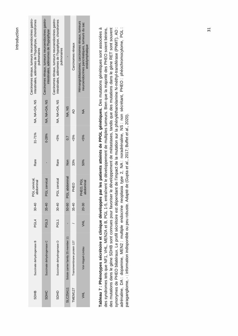
Intr
od
uct
ion
31
SD
HB
S
uccin
ate
de
hyd
roge
nase B
P
GL4
30-4
0
PG
L c
erv
ical,
abdom
inal
Rare
31-7
1%
N
A,
NA
+D
A,
NS
C
arc
inom
es r
énaux,
tum
eurs
neuro
endocrin
es g
astr
o-
inte
stin
ale
s,
adénom
es d
e l'h
ypophyse, chondro
mes
pulm
onaires
SD
HC
S
uccin
ate
de
hyd
roge
nase C
P
GL3
35-4
0
PG
L c
erv
ical
- 0-2
8%
N
A,
NA
+D
A,
NS
C
arc
inom
es r
énaux,
tum
eurs
neuro
endocrin
es g
astr
o-
inte
stin
ale
s,
adénom
es d
e l'h
ypophyse
SD
HD
S
uccin
ate
de
hyd
roge
nase D
P
GL1
30-4
0
PG
L c
erv
ical
Rare
<
5%
N
A,
NA
+D
A,
NS
C
arc
inom
es r
énaux,
tum
eurs
neuro
endocrin
es g
astr
o-
inte
stin
ale
s,
adénom
es d
e l'h
ypophyse, chondro
mes
pulm
onaires
SLC
25A
11
Solu
te c
arr
ier
fam
ily 2
5 m
em
be
r 1
1
- 50-6
0
PG
L a
bdom
inal
Non
0,7
N
A,
NS
-
TM
EM
127
T
ransm
em
bra
ne p
rote
in 1
27
/
35-4
0
PH
EO
33%
<
5%
A
D
Carc
inom
es r
énaux
VH
L
Von H
ippel-
Lin
dau
V
HL
20-3
0
PH
EO
, P
GL
abdom
inal
50%
<
5%
N
A
Hém
angio
bla
sto
me
s, carc
inom
es r
énau
x,
tum
eurs
neuro
endocrines p
ancré
atiq
ues, tu
meurs
du s
ac
endoly
mphatiq
ue
Tab
leau
7 :
Ph
én
oty
pe
s s
écré
toir
e e
t clin
iqu
e d
év
elo
pp
és p
ar
les p
ati
en
ts a
ttein
ts d
e P
PG
L g
én
éti
qu
es.
Des m
uta
tions g
énétiq
ues s
ont
associé
es à
des s
yn
dro
mes t
els
que N
F1,
VH
L,
ME
N2A
et
B,
PG
L 1
-5,
entr
aîn
en
t le
dévelo
ppem
ent
de m
ultip
les t
um
eurs
. B
ien q
ue la m
ajo
rité
des P
HE
O s
oie
nt
bén
ins,
des m
uta
tions d
ans le g
èn
e S
DH
B s
ont
conn
ues p
ou
r fa
vorise
r le
dé
ve
lop
pem
ent
de m
éta
sta
ses,
tandis
que d
es m
uta
tions d
ans le g
ène R
ET
sont
souvent
synon
ym
es d
e P
HE
O b
ilaté
raux.
Le p
rofil sécré
toire
est
dé
pen
dan
t d
e l’im
pact
de la
muta
tion
sur
la p
hé
nylé
thano
lam
ine N
-méth
yl-
tra
nsfé
rase
(P
NM
T).
AD
:
adré
nalin
e,
DA
:
do
pam
ine,
ME
N2 :
multip
le
endocrine
ne
opla
sia
ty
pe
2,
NA
:
nora
dré
na
line,
NS
:
non
sécré
tant,
P
HE
O :
ph
éochro
mocyto
me,
PG
L :
para
gang
liom
e,
- : in
form
ation ind
ispon
ible
ou p
eu r
ob
uste
. A
dap
té d
e (
Gupta
et
al.,
201
7;
Buff
et
et al., 2
020
).
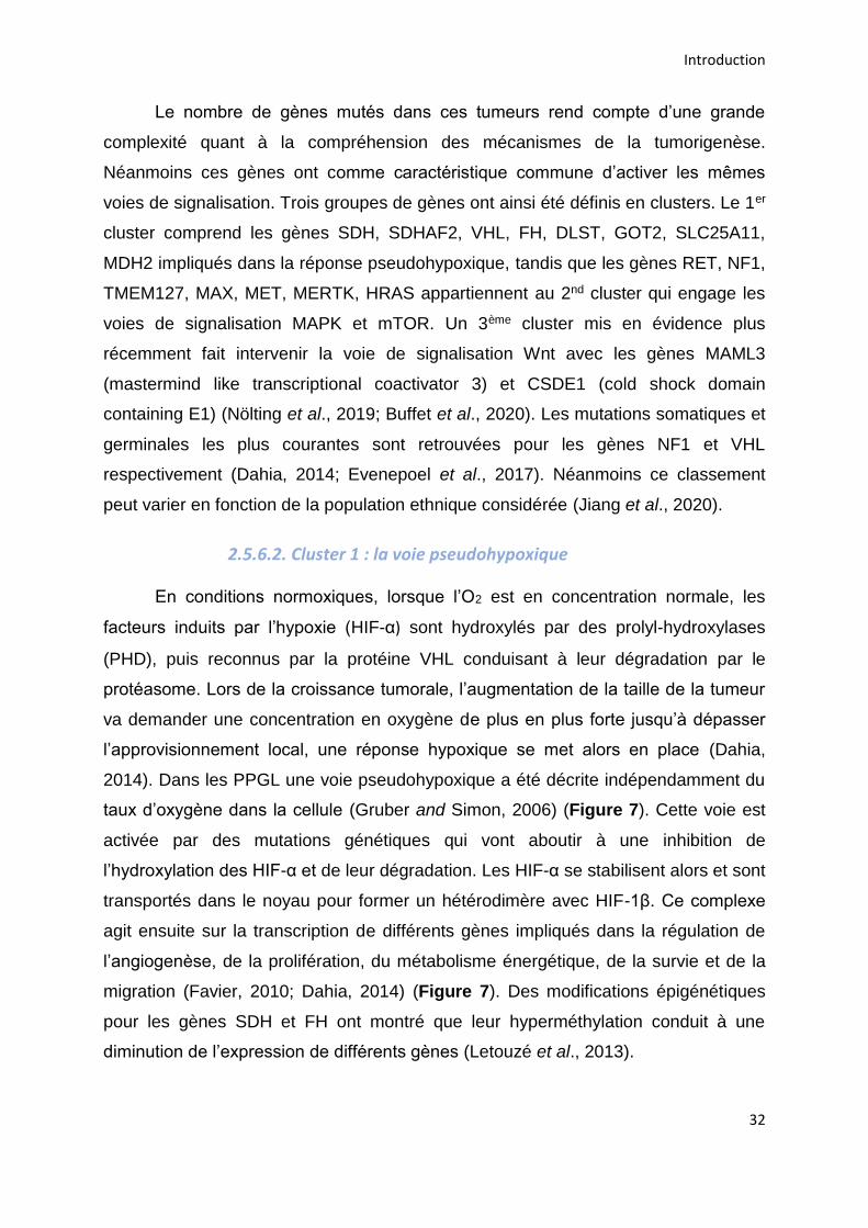
Introduction
32
Le nombre de gènes mutés dans ces tumeurs rend compte d’une grande
complexité quant à la compréhension des mécanismes de la tumorigenèse.
Néanmoins ces gènes ont comme caractéristique commune d’activer les mêmes
voies de signalisation. Trois groupes de gènes ont ainsi été définis en clusters. Le 1er
cluster comprend les gènes SDH, SDHAF2, VHL, FH, DLST, GOT2, SLC25A11,
MDH2 impliqués dans la réponse pseudohypoxique, tandis que les gènes RET, NF1,
TMEM127, MAX, MET, MERTK, HRAS appartiennent au 2nd cluster qui engage les
voies de signalisation MAPK et mTOR. Un 3ème cluster mis en évidence plus
récemment fait intervenir la voie de signalisation Wnt avec les gènes MAML3
(mastermind like transcriptional coactivator 3) et CSDE1 (cold shock domain
containing E1) (Nölting et al., 2019; Buffet et al., 2020). Les mutations somatiques et
germinales les plus courantes sont retrouvées pour les gènes NF1 et VHL
respectivement (Dahia, 2014; Evenepoel et al., 2017). Néanmoins ce classement
peut varier en fonction de la population ethnique considérée (Jiang et al., 2020).
2.5.6.2. Cluster 1 : la voie pseudohypoxique
En conditions normoxiques, lorsque l’O2 est en concentration normale, les
facteurs induits par l’hypoxie (HIF-α) sont hydroxylés par des prolyl-hydroxylases
(PHD), puis reconnus par la protéine VHL conduisant à leur dégradation par le
protéasome. Lors de la croissance tumorale, l’augmentation de la taille de la tumeur
va demander une concentration en oxygène de plus en plus forte jusqu’à dépasser
l’approvisionnement local, une réponse hypoxique se met alors en place (Dahia,
2014). Dans les PPGL une voie pseudohypoxique a été décrite indépendamment du
taux d’oxygène dans la cellule (Gruber and Simon, 2006) (Figure 7). Cette voie est
activée par des mutations génétiques qui vont aboutir à une inhibition de
l’hydroxylation des HIF-α et de leur dégradation. Les HIF-α se stabilisent alors et sont
transportés dans le noyau pour former un hétérodimère avec HIF-1β. Ce complexe
agit ensuite sur la transcription de différents gènes impliqués dans la régulation de
l’angiogenèse, de la prolifération, du métabolisme énergétique, de la survie et de la
migration (Favier, 2010; Dahia, 2014) (Figure 7). Des modifications épigénétiques
pour les gènes SDH et FH ont montré que leur hyperméthylation conduit à une
diminution de l’expression de différents gènes (Letouzé et al., 2013).
Introduction
33
Figure 7 : Voie pseudohypoxique dans les PPGL. Des mutations dans les gènes de susceptibilité
appartenant au cluster 1 entraînent une accumulation du facteur HIF-α. Les gènes SDHx, DLST, FH
ou MDH2 sont impliqués dans le cycle de Krebs, leurs mutations conduisent à une accumulation de
métabolites tels que le succinate, le fumarate, le pyruvate. Les gènes PHD et VHL sont quant à eux
responsables de l’hydroxylation et de la reconnaissance de HIF-α pour sa dégradation par le
protéasome. Les mutations dans les gènes décrits vont empêcher la dégradation de HIF-α, qui se
transloque alors dans le noyau pour interagir avec HIF-1β et activer la transcription de gènes
impliqués dans la tumorigenèse. L’accumulation des métabolites du cycle de Krebs va également
inhiber l’activité des enzymes ten-eleven translocation (TET) et des Jumonji-related histone
demethylases (JMJD) impliquées dans la déméthylation de l’ADN et des histones respectivement,
conduisant leur hyperméthylation et la régulation de gènes cibles. Les oncogènes impliqués dans la
tumorigenèse sont en violet et les gènes suppresseurs de tumeurs en rouge. HIF : hypoxia-inducible
factor, PHD : prolyl-hydroxylases.
2.5.6.3. Clusters 2 et 3 : les voies des récepteurs tyrosine kinases et
Wnt
Le second cluster implique une activation anormale des voies de signalisation
PI3K/mTOR et des récepteurs kinases régulant la croissance, la prolifération,
l’apoptose, le remodelage de la chromatine et certaines signalisations métaboliques.
Ce 2ème cluster inclut des mutations dans des gènes tels quel RET, NF1, H-RAS, K-
RAS, TMEM127, MAX, CSDE1 (Jochmanova and Pacak, 2018) (Figure 8). Les
mutations dans le gène RET engendrent une activation de la protéine qui va alors
activer RAS, tandis que la protéine NF1 mutée perd sa fonction d’activer une enzyme
(ras GTPase) qui inhibe la protéine RAS. Un arrêt de la régulation négative de la
HIF-α HIF-α
PHD
Dégradation par
le protéasome
GOT2
SCL2511A
DLST
MDH2
SDHx
FH ↗ Fumarate,
succinate,
pyruvate
Cytoplasme
Mitochondrie
VHL
HIF-α HIF-β
Activation de gènes
(angiogenèse,
prolifération,…)
JMJD TET
Méthylation de
l’ADN et des
histones Noyau
↗ Fumarate, succinate,
pyruvate
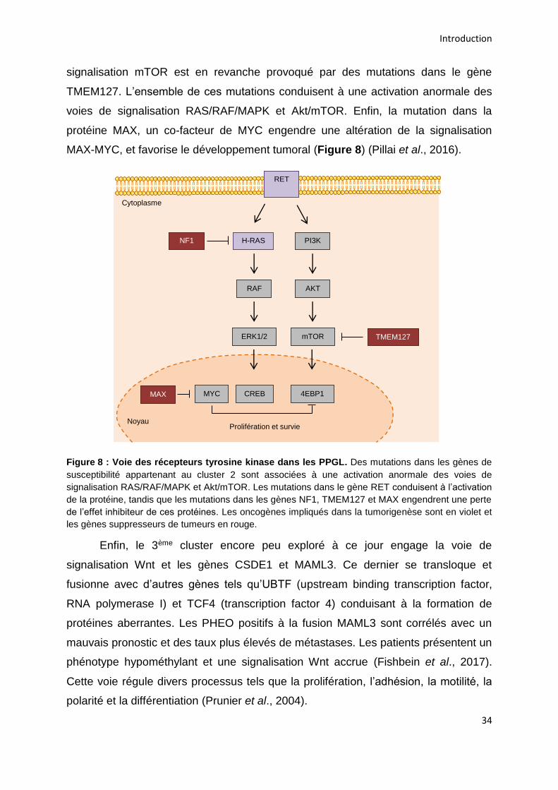
Introduction
34
signalisation mTOR est en revanche provoqué par des mutations dans le gène
TMEM127. L’ensemble de ces mutations conduisent à une activation anormale des
voies de signalisation RAS/RAF/MAPK et Akt/mTOR. Enfin, la mutation dans la
protéine MAX, un co-facteur de MYC engendre une altération de la signalisation
MAX-MYC, et favorise le développement tumoral (Figure 8) (Pillai et al., 2016).
Figure 8 : Voie des récepteurs tyrosine kinase dans les PPGL. Des mutations dans les gènes de
susceptibilité appartenant au cluster 2 sont associées à une activation anormale des voies de
signalisation RAS/RAF/MAPK et Akt/mTOR. Les mutations dans le gène RET conduisent à l’activation
de la protéine, tandis que les mutations dans les gènes NF1, TMEM127 et MAX engendrent une perte
de l’effet inhibiteur de ces protéines. Les oncogènes impliqués dans la tumorigenèse sont en violet et
les gènes suppresseurs de tumeurs en rouge.
Enfin, le 3ème cluster encore peu exploré à ce jour engage la voie de
signalisation Wnt et les gènes CSDE1 et MAML3. Ce dernier se transloque et
fusionne avec d’autres gènes tels qu’UBTF (upstream binding transcription factor,
RNA polymerase I) et TCF4 (transcription factor 4) conduisant à la formation de
protéines aberrantes. Les PHEO positifs à la fusion MAML3 sont corrélés avec un
mauvais pronostic et des taux plus élevés de métastases. Les patients présentent un
phénotype hypométhylant et une signalisation Wnt accrue (Fishbein et al., 2017).
Cette voie régule divers processus tels que la prolifération, l’adhésion, la motilité, la
polarité et la différentiation (Prunier et al., 2004).
Cytoplasme
RET
H-RAS NF1
RAF
ERK1/2
PI3K
AKT
mTOR TMEM127
CREB MYC MAX 4EBP1
Noyau Prolifération et survie
Introduction
35
2.5.6.4. Les multiples conséquences des mutations
Les altérations génétiques citées précédemment peuvent également être à
l’origine d’autres néoplasmes (Tableau 7). Les mutations des gènes SDH peuvent
ainsi provoquer des TNE gastro-intestinales, des carcinomes rénaux ainsi que des
adénomes de l’hypophyse (Gill, 2018). Ces altérations génétiques peuvent aussi être
à l’origine de syndromes associés aux PPGL, par exemple la néoplasie
endocrinienne de type 2 (MEN2) (mutation du gène RET) se caractérise par
l’association possible, chez un même patient, d’un cancer médullaire de la thyroïde,
d’une hyperparathyroïdie et d’un PHEO. Tandis que l’association
d’hémangioblastomes (tumeurs du système nerveux central), de carcinomes rénaux,
de TNE pancréatiques, et de PPGL sont caractéristiques de la maladie de von
Hippel-Lindau. Enfin des patients atteints de neurofibromatose de type 1 présentent
des neurofibromes, des lésions cutanées, des atteintes des nerfs optiques ou de la
rétine et des PHEO (Pawlu et al., 2005; Dahia, 2014).
Ces différents gènes et voies de signalisation altérés dans les PPGL et leurs
implications dans d’autres pathologies rendent compte de la complexité quant à la
compréhension des mécanismes à l’origine de ces tumeurs. D’autant plus qu’il existe
une hétérogénéité génétique intra- et inter-tumorale pour les PPGL (Crona et al.,
2015; Flynn et al., 2015).
2.5.6.5. De la génétique au phénotype
Cette hétérogénéité génétique des PPGL va donner lieu à différents
phénotypes en termes de localisation, de progression tumorale et de profil sécrétoire.
Les patients porteurs d’altérations de gènes du cluster 1 (SDH, VHL, FH, DLST,
SCL25A11, MDH2) présentent une localisation tumorale plutôt extra-surrénalienne.
Par exemple, les mutations SDH et FH engendrent majoritairement des tumeurs
cervicales et abdominales, alors que des tumeurs associées à une mutation VHL
peuvent être abdominales ou surrénaliennes. En revanche, les mutations de gènes
du cluster 2 (NF1, RET, TMEM127, MAX, MET) donnent lieu à des tumeurs
surrénaliennes exclusivement (Tableau 7). Le devenir tumoral est également
influencé par le profil génétique ; des mutations des gènes RET, MAX et VHL
favorisent le développement de PPGL multiples, tandis que des altérations des
Introduction
36
gènes SDHB, EPAS1, SLC25A11 et MDH2 prédisposent à une évolution
métastatique chez les sujets (Lenders et al., 2014; Buffet et al., 2020). Le profil
biochimique de la tumeur est aussi corrélé au génotype. Par exemple, des mutations
de gènes du cluster 2 tels que RET, NF1 et TMEM127 entraînent une sécrétion
tumorale adrénergique. Ce phénotype est lié à l’expression de la PNMT responsable
de la conversion de la noradrénaline en adrénaline (Figure 4) (Eisenhofer et al.,
2011; Gupta et al., 2017). Les PPGL avec un phénotype adrénergique sont localisés
dans la glande surrénale et sont très rarement extra-surrénaliens. En revanche, les
PPGL liés à des mutations de gènes du cluster 1 (VHL, SDH, FH, EPAS1) ont une
sécrétion noradrénergique et n’expriment pas la PNMT (Tableau 7). Un silençage
épigénétique du gène PNMT a été montré chez des patients porteurs de mutations
VHL et SDH. L’hyperméthylation de la région promotrice de ce gène entraîne une
réduction des taux de PNMT. Le même mécanisme a été montré pour les gènes FH
et MDH2 (Gupta et al., 2017; Ma et al., 2020).
2.5.7. Diagnostic biochimique et imagerie
À l’heure actuelle le diagnostic du PHEO demeure complexe car les
symptômes des patients comme l’hypertension artérielle, les céphalées, les
palpitations et les sudations ne sont pas spécifiques et n’orientent que peu le
diagnostic vers cette pathologie. Le dépistage repose uniquement sur la mise en
évidence de l’excès de catécholamines produit par la tumeur.
D’après les directives du « Endocrine Society Clinical Practice Guideline », les
tests biochimiques initiaux pour les PPGL comprennent les mesures plasmatiques et
urinaires des métabolites des catécholamines (métanéphrine et normétanéphrine)
(Lenders et al., 2014). La CGA, une protéine co-sécrétée avec les catécholamines
est également recherchée dans le sang. Une élévation de la CGA est retrouvée chez
90% des patients atteints de PPGL (Stridsberg and Husebye, 1997; Grossrubatscher
et al., 2006). De nombreuses études s’accordent sur l’utilisation de la CGA en
complément du dosage des métabolites des catécholamines (Algeciras-Schimnich et
al., 2008; Bílek et al., 2019).
Introduction
37
Après l’obtention d’une preuve biochimique de la sécrétion tumorale,
l’imagerie est utilisée afin de localiser la tumeur et d’éventuelles métastases. La
tomodensitométrie est recommandée en raison de son excellente résolution spatiale
pour le thorax, l'abdomen et le bassin. L’imagerie par résonnance magnétique (IRM)
est préconisée pour la détection des PGL de la base du crâne et du cou, ainsi que
pour les femmes enceintes et les enfants. L’imagerie fonctionnelle est utilisée dans
les cas de tumeurs malignes (Lenders et al., 2002). Dans les cas familiaux de PHEO,
l’histoire familiale constitue un élément de diagnostic important, puisque des tests
génétiques peuvent également être réalisés.
Malgré ces différents moyens, le diagnostic du PHEO reste difficile
compliquant la prise en charge des patients. Des facteurs environnementaux,
alimentaires et médicamenteux peuvent modifier les taux de catécholamines et de
CGA. De même la sensibilité et la spécificité des tests utilisés sont variables (cette
partie sera abordée plus en détails dans la discussion).
2.5.8. Prise en charge clinique du phéochromocytome
Actuellement, il n’existe pas de traitement permettant de cibler directement la
tumeur. Une prise en charge symptomatique et chirurgicale est donc mise en place.
2.5.8.1. Gestion préopératoire de la tumeur
La détection d’un PHEO va entraîner dans la majorité des cas une exérèse
totale de la glande surrénale. Cette opération nécessite une prise en charge en
amont du patient, notamment par l’introduction de traitements pharmacologiques en
préopératoire qui permettent de diminuer le taux de mortalité de 45% à moins de 2%
(Adler et al., 2008).
La gestion pharmacologique a pour but de réduire la pression artérielle induite
par l’anesthésie et par la manipulation de la tumeur, et de prévenir l’hypotension
pouvant survenir juste après l’ablation de la tumeur. En pré et peropératoire, des
antagonistes adrénergiques α et/ou β bloquants peuvent être administrés au patient,
le traitement est débuté 7 à 14 jours avant l’opération afin de normaliser les
paramètres physiologiques tels que la pression sanguine. En première intention les
antagonistes adrénergiques α sont utilisés. Les bloquants β peuvent également être
employés mais couplés à une autre molécule et uniquement après un traitement
Introduction
38
approprié avec des antihypertenseurs α. Des bloqueurs de canaux calciques et des
inhibiteurs de la TH sont aussi administrés dans certains cas (Adler et al., 2008; Jain
et al., 2020). Le patient suit également un régime riche en sodium et un apport
hydrique afin d’inverser la contraction du volume sanguin induite par les
catécholamines en préopératoire, et afin de prévenir une hypotension sévère après
l'ablation de la tumeur (Lenders et al., 2014).
Néanmoins ces traitements préopératoires ne sont pas sans conséquences
pour le patient. Par exemple, le phenoxybanzamine un des antagonistes α les plus
utilisés entraîne de l’hypotension orthostatique, une congestion nasale et un réflexe
de tachycardie. L’aténolol un bloquant des récepteurs adrénergiques β provoque de
la fatigue et des vertiges (Jain et al., 2020).
2.5.8.2. Approche chirurgicale
La surrénalectomie est le principal traitement du PHEO, elle consiste à réaliser
une exérèse chirurgicale de toute la glande surrénale (Figure 9). La technique
recommandée par le guide de pratique clinique de la société endocrinienne est la
laparoscopie (Lenders et al., 2014). Elle est utilisée dans plus de 90% des cas car
elle améliore la morbidité postopératoire et permet de réduire la durée et le coût de
l’hospitalisation pour des tumeurs de 6 à 10 cm de diamètre. Cette méthode permet
de retirer la tumeur de façon peu invasive pour le patient (Tsirlin et al., 2014). La
surrénalectomie peut être réalisée par robotique ce qui permet d’améliorer le devenir
postopératoire pour des cas compliqués et l’ergonomie pour le chirurgien (Nomine-
Criqui et al., 2017). En revanche, pour des PHEO de grandes tailles, métastatiques,
ou dans des cas plus complexes une résection ouverte est réalisée.
La chirurgie comporte des risques potentiels, une hypertension peut ainsi
provenir de sources spécifiques à l’opération telles que l’intubation du patient, mais
également de la libération de catécholamines lors de la manipulation de la tumeur
(Flávio Rocha et al., 2004; Tauzin-Fin et al., 2020). Une chirurgie longue pour retirer
la tumeur signifie généralement que le processus de résection est difficile, ou peut-
être accompagné d'une perte de sang massive, d'une lésion d'organe ou d'une
fluctuation de la pression artérielle élevée, ce qui entraîne une morbidité
supplémentaire pour le patient (Bai et al., 2018).
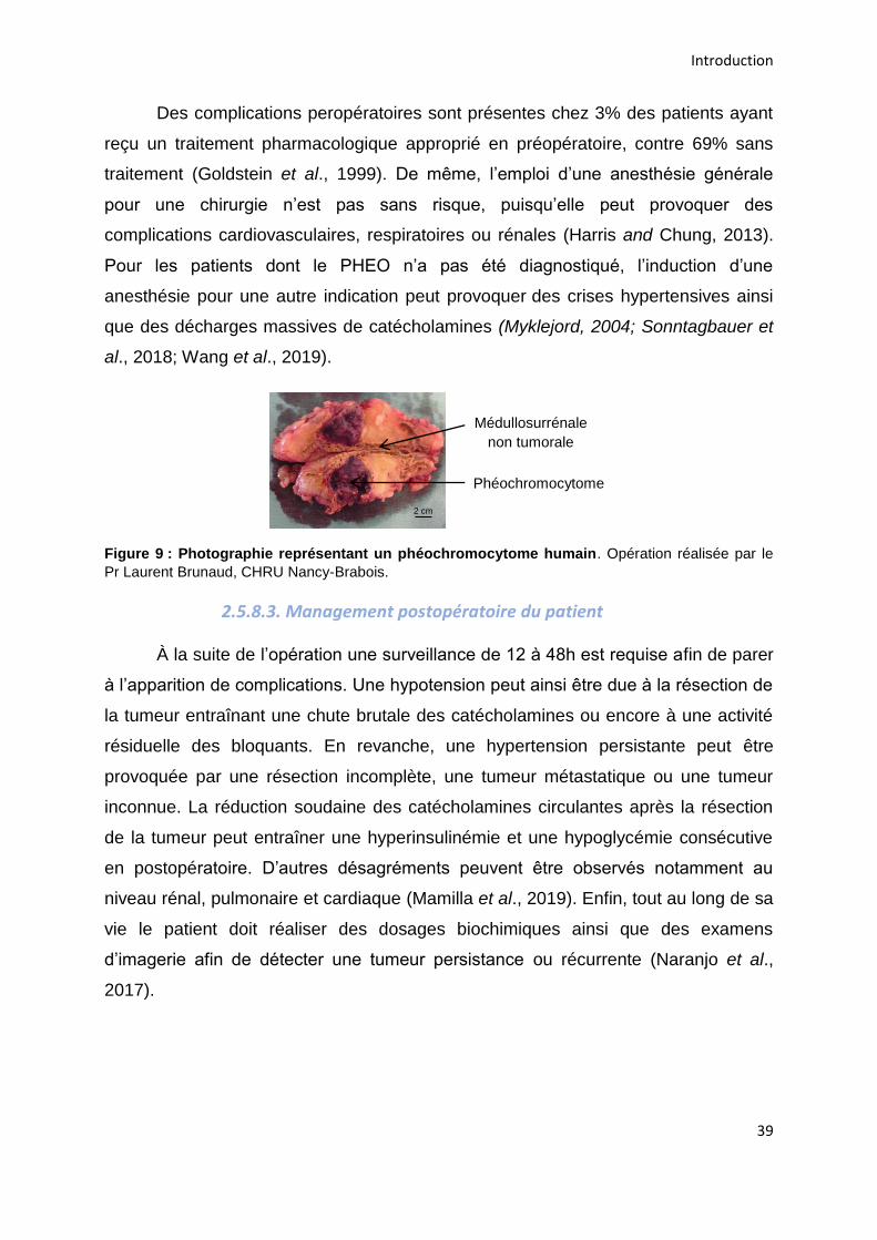
Introduction
39
Des complications peropératoires sont présentes chez 3% des patients ayant
reçu un traitement pharmacologique approprié en préopératoire, contre 69% sans
traitement (Goldstein et al., 1999). De même, l’emploi d’une anesthésie générale
pour une chirurgie n’est pas sans risque, puisqu’elle peut provoquer des
complications cardiovasculaires, respiratoires ou rénales (Harris and Chung, 2013).
Pour les patients dont le PHEO n’a pas été diagnostiqué, l’induction d’une
anesthésie pour une autre indication peut provoquer des crises hypertensives ainsi
que des décharges massives de catécholamines (Myklejord, 2004; Sonntagbauer et
al., 2018; Wang et al., 2019).
Figure 9 : Photographie représentant un phéochromocytome humain. Opération réalisée par le
Pr Laurent Brunaud, CHRU Nancy-Brabois.
2.5.8.3. Management postopératoire du patient
À la suite de l’opération une surveillance de 12 à 48h est requise afin de parer
à l’apparition de complications. Une hypotension peut ainsi être due à la résection de
la tumeur entraînant une chute brutale des catécholamines ou encore à une activité
résiduelle des bloquants. En revanche, une hypertension persistante peut être
provoquée par une résection incomplète, une tumeur métastatique ou une tumeur
inconnue. La réduction soudaine des catécholamines circulantes après la résection
de la tumeur peut entraîner une hyperinsulinémie et une hypoglycémie consécutive
en postopératoire. D’autres désagréments peuvent être observés notamment au
niveau rénal, pulmonaire et cardiaque (Mamilla et al., 2019). Enfin, tout au long de sa
vie le patient doit réaliser des dosages biochimiques ainsi que des examens
d’imagerie afin de détecter une tumeur persistance ou récurrente (Naranjo et al.,
2017).
2 cm
Médullosurrénale non tumorale
Phéochromocytome
Introduction
40
2.5.8.4. Les phéochromocytomes malins et inopérables : des cas
complexes pour la prise charge
Le PHEO malin est un véritable problème pour les chirurgiens puisqu’aucun
critère ne permet de prédire la malignité de la tumeur. Néanmoins des mutations
dans le gène de la SDHB sont retrouvées chez environ 40% des patients porteurs de
PHEO malins et sont associées à une faible survie (Amar et al., 2007). Les
métastases sont retrouvées majoritairement dans les ganglions lymphatiques, les os,
le foie et les poumons (Ajallé et al., 2009). Il n’existe actuellement aucun traitement
curatif pour les PHEO métastatiques. Une résection de la tumeur primaire permet de
diminuer l’excès de catécholamines et les symptômes associés, d’améliorer la qualité
de vie et la survie des patients. Néanmoins les récidives sont courantes et toutes les
métastases ne peuvent pas être retirées. La chimiothérapie et radiothérapie peuvent
ensuite être employées, néanmoins seule une fraction de patients répond à ces
traitements et souvent de façon partielle et limitée dans le temps, avec de nombreux
effets secondaires (Jimenez et al., 2013). D’autres traitements comme des inhibiteurs
de tyrosine kinase ou l’immunothérapie sont à l’étude ces dernières années (Roman-
Gonzalez and Jimenez, 2017).
Une difficulté de prise en charge est également rencontrée pour des patients
non opérables. La chirurgie par laparoscopie est ainsi contre-indiquée en cas de
risque de rupture de la capsule tumorale, d’invasion des structures adjacentes
rendant la tumeur non résécable ou d’incapacité à réaliser la procédure en toute
sécurité avec des techniques mini-invasives. De même, les maladies
cardiopulmonaires, l’obésité massive ou encore la coagulopathie persistante
constituent une barrière à la chirurgie (Gumbs and Gagner, 2006; Germain et al.,
2011).
De nombreux problèmes, risques et/ou contre-indications se posent donc sur
les différents niveaux de prise en charge et de gestion clinique du PHEO. Des
traitements symptomatiques sont également utilisés mais aucune molécule ne cible
directement l’origine des symptômes qui est liée à la sécrétion des catécholamines.
Introduction
41
3. Les analogues somatostatinergiques : un espoir de traitement anti-
sécrétoire
3.1. La somatostatine
Aujourd’hui il n’existe aucun traitement anti-sécrétoire permettant de stopper
ou de freiner directement l’hypersécrétion des PHEO, et donc d’empêcher les
manifestations cliniques qui en découlent. Cependant, une classe de molécules très
prometteuse est aujourd’hui utilisée en cancérologie et pourrait constituer une piste
sérieuse pour un traitement anti-sécrétoire. Il s’agit des analogues de la
somatostatine.
3.1.1. Découverte et historique
La somatostatine (SST) est une hormone peptidique cyclique. La première
preuve de son existence a été apportée par Krulich et collaborateurs en 1968. Ils ont
montré que des extraits d’hypothalamus de rats inhibaient la sécrétion d’hormone de
croissance par l’hypophyse. Néanmoins, l’étude n’est pas allée plus loin et a émis
l’hypothèse d’un artefact (Krulich et al., 1968). La SST a été isolée et caractérisée
par l’équipe du Dr Guillemin en 1972 et l’étude a été publiée en 1973 (Brazeau et al.,
1973). Enfin, sa localisation dans les neurones hypothalamiques de rats a été
démontrée par Pelletier et son équipe en 1977 (Désy and Pelletier, 1977; Pelletier et
al., 1977). La SST est également appelée « somatotropin release-inhibiting factor »
(SRIF) ou « growth hormone-inhibiting hormone » (GHIH) car découverte initialement
comme inhibiteur de la libération de l’hormone de croissance. Depuis, son rôle
inhibiteur général sur la sécrétion hormonale a été largement caractérisé et s'est
étendu à d’autres hormones, notamment dans les systèmes nerveux et digestif.
3.1.2. Biosynthèse
La SST est synthétisée sous forme d’un large précurseur de pré-pro-
somatostatine qui est rapidement clivé en pro-somatostatine de 92 acides aminés.
Cette dernière va ensuite donner naissance à deux formes actives de la SST grâce à
des enzymes convertases (Figures 10, 11). Un clivage de la pro-somatostatine au
niveau d’un site Arg-Lys va donner une forme courte de somatostatine de 14 acides
aminés majoritaire dans le système gastro-entéro-pancréatique tandis qu’un clivage
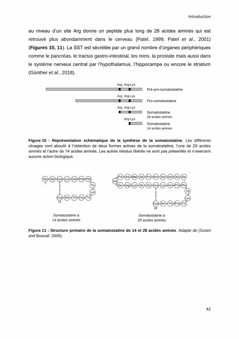
Introduction
42
au niveau d’un site Arg donne un peptide plus long de 28 acides aminés qui est
retrouvé plus abondamment dans le cerveau (Patel, 1999; Patel et al., 2001)
(Figures 10, 11). La SST est sécrétée par un grand nombre d’organes périphériques
comme le pancréas, le tractus gastro-intestinal, les reins, la prostate mais aussi dans
le système nerveux central par l’hypothalamus, l’hippocampe ou encore le striatum
(Günther et al., 2018).
Figure 10 : Représentation schématique de la synthèse de la somatostatine. Les différents
clivages vont aboutir à l’obtention de deux formes actives de la somatostatine, l’une de 28 acides
aminés et l’autre de 14 acides aminés. Les autres résidus libérés ne sont pas présentés et n’exercent
aucune action biologique.
Figure 11 : Structure primaire de la somatostatine de 14 et 28 acides aminés. Adapté de (Susini
and Buscail, 2006).
Somatostatine à 14 acides aminés
Somatostatine à 28 acides aminés
Pro-somatostatine Arg-Lys Arg
Pré-pro-somatostatine Arg-Lys Arg
Somatostatine 28 acides aminés
Arg-Lys Arg
Somatostatine 14 acides aminés
Arg-Lys
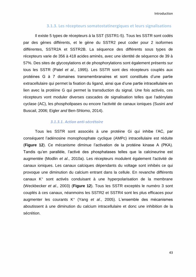
Introduction
43
3.1.3. Les récepteurs somatostatinergiques et leurs signalisations
Il existe 5 types de récepteurs à la SST (SSTR1-5). Tous les SSTR sont codés
par des gènes différents, et le gène du SSTR2 peut coder pour 2 isoformes
différentes, SSTR2A et SSTR2B. La séquence des différents sous types de
récepteurs varie de 356 à 418 acides aminés, avec une identité de séquence de 39 à
57%. Des sites de glycosylations et de phosphorylations sont également présents sur
tous les SSTR (Patel et al., 1995). Les SSTR sont des récepteurs couplés aux
protéines G à 7 domaines transmembranaires et sont constitués d’une partie
extracellulaire qui permet la fixation du ligand, ainsi que d’une partie intracellulaire en
lien avec la protéine G qui permet la transduction du signal. Une fois activés, ces
récepteurs vont moduler diverses cascades de signalisation telles que l'adénylate
cyclase (AC), les phospholipases ou encore l'activité de canaux ioniques (Susini and
Buscail, 2006; Eigler and Ben-Shlomo, 2014).
3.1.3.1. Action anti-sécrétoire
Tous les SSTR sont associés à une protéine Gi qui inhibe l’AC, par
conséquent l’adénosine monophosphate cyclique (AMPc) intracellulaire est réduite
(Figure 12). Ce mécanisme diminue l’activation de la protéine kinase A (PKA).
Tandis qu’en parallèle, l’activé des phosphatases telles que la calcineurine est
augmentée (Modlin et al., 2010a). Les récepteurs modulent également l'activité de
canaux ioniques. Les canaux calciques dépendants du voltage sont inhibés ce qui
provoque une diminution du calcium entrant dans la cellule. En revanche différents
canaux K+ sont activés conduisant à une hyperpolarisation de la membrane
(Weckbecker et al., 2003) (Figure 12). Tous les SSTR exceptés le numéro 3 sont
couplés à ces canaux, néanmoins les SSTR2 et SSTR4 sont les plus efficaces pour
augmenter les courants K+ (Yang et al., 2005). L’ensemble des mécanismes
aboutissent à une diminution du calcium intracellulaire et donc une inhibition de la
sécrétion.
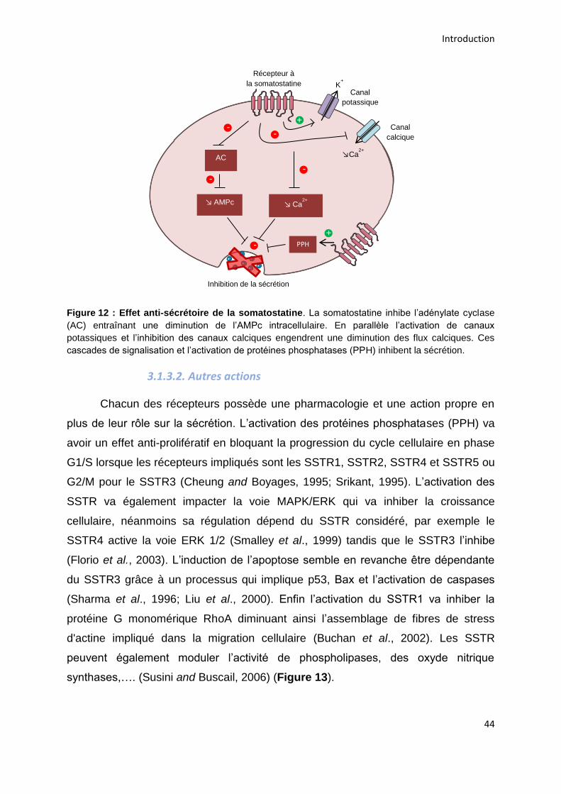
Introduction
44
Figure 12 : Effet anti-sécrétoire de la somatostatine. La somatostatine inhibe l’adénylate cyclase
(AC) entraînant une diminution de l’AMPc intracellulaire. En parallèle l’activation de canaux
potassiques et l’inhibition des canaux calciques engendrent une diminution des flux calciques. Ces
cascades de signalisation et l’activation de protéines phosphatases (PPH) inhibent la sécrétion.
3.1.3.2. Autres actions
Chacun des récepteurs possède une pharmacologie et une action propre en
plus de leur rôle sur la sécrétion. L’activation des protéines phosphatases (PPH) va
avoir un effet anti-prolifératif en bloquant la progression du cycle cellulaire en phase
G1/S lorsque les récepteurs impliqués sont les SSTR1, SSTR2, SSTR4 et SSTR5 ou
G2/M pour le SSTR3 (Cheung and Boyages, 1995; Srikant, 1995). L’activation des
SSTR va également impacter la voie MAPK/ERK qui va inhiber la croissance
cellulaire, néanmoins sa régulation dépend du SSTR considéré, par exemple le
SSTR4 active la voie ERK 1/2 (Smalley et al., 1999) tandis que le SSTR3 l’inhibe
(Florio et al., 2003). L’induction de l’apoptose semble en revanche être dépendante
du SSTR3 grâce à un processus qui implique p53, Bax et l’activation de caspases
(Sharma et al., 1996; Liu et al., 2000). Enfin l’activation du SSTR1 va inhiber la
protéine G monomérique RhoA diminuant ainsi l’assemblage de fibres de stress
d'actine impliqué dans la migration cellulaire (Buchan et al., 2002). Les SSTR
peuvent également moduler l’activité de phospholipases, des oxyde nitrique
synthases,…. (Susini and Buscail, 2006) (Figure 13).
AC
↘ AMPc ↘ Ca2+
PPH
Inhibition de la sécrétion
Récepteur à la somatostatine
-
+
- +
- -
Canal
potassique
Canal calcique
K+
↘Ca2+
-
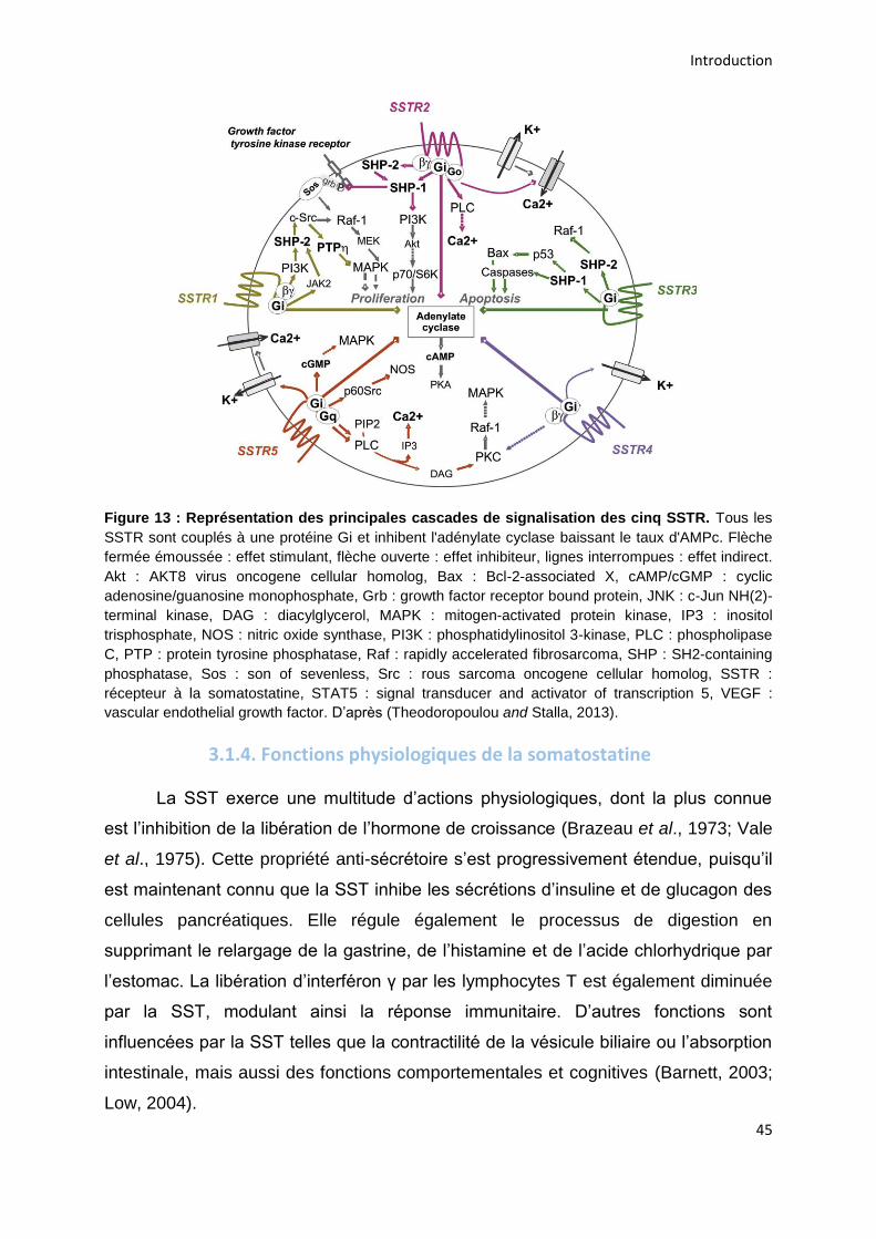
Introduction
45
Figure 13 : Représentation des principales cascades de signalisation des cinq SSTR. Tous les
SSTR sont couplés à une protéine Gi et inhibent l'adénylate cyclase baissant le taux d'AMPc. Flèche
fermée émoussée : effet stimulant, flèche ouverte : effet inhibiteur, lignes interrompues : effet indirect.
Akt : AKT8 virus oncogene cellular homolog, Bax : Bcl-2-associated X, cAMP/cGMP : cyclic
adenosine/guanosine monophosphate, Grb : growth factor receptor bound protein, JNK : c-Jun NH(2)-
terminal kinase, DAG : diacylglycerol, MAPK : mitogen-activated protein kinase, IP3 : inositol
trisphosphate, NOS : nitric oxide synthase, PI3K : phosphatidylinositol 3-kinase, PLC : phospholipase
C, PTP : protein tyrosine phosphatase, Raf : rapidly accelerated fibrosarcoma, SHP : SH2-containing
phosphatase, Sos : son of sevenless, Src : rous sarcoma oncogene cellular homolog, SSTR :
récepteur à la somatostatine, STAT5 : signal transducer and activator of transcription 5, VEGF :
vascular endothelial growth factor. D’après (Theodoropoulou and Stalla, 2013).
3.1.4. Fonctions physiologiques de la somatostatine
La SST exerce une multitude d’actions physiologiques, dont la plus connue
est l’inhibition de la libération de l’hormone de croissance (Brazeau et al., 1973; Vale
et al., 1975). Cette propriété anti-sécrétoire s’est progressivement étendue, puisqu’il
est maintenant connu que la SST inhibe les sécrétions d’insuline et de glucagon des
cellules pancréatiques. Elle régule également le processus de digestion en
supprimant le relargage de la gastrine, de l’histamine et de l’acide chlorhydrique par
l’estomac. La libération d’interféron γ par les lymphocytes T est également diminuée
par la SST, modulant ainsi la réponse immunitaire. D’autres fonctions sont
influencées par la SST telles que la contractilité de la vésicule biliaire ou l’absorption
intestinale, mais aussi des fonctions comportementales et cognitives (Barnett, 2003;
Low, 2004).
Introduction
46
3.2. Les analogues de la somatostatine : les nouvelles molécules
employées en cancérologie
3.2.1. Du développement à aujourd’hui
La SST possède une demi-vie courte variant de 1 à 3 min (Modlin et al.,
2010a). Après activation du récepteur elle est rapidement internalisée puis dégradée
par des endopeptidases endosomales, ou des peptidases présentes dans la
circulation sanguine (Dupont et al., 1978; Werle and Bernkop-Schnürch, 2006;
Roosterman et al., 2008). Cette propriété freine l’usage de la somatostatine en
clinique. Afin de palier à cette limitation, les entreprises pharmaceutiques ont
développé des analogues de la somatostatine plus stables, avec une demi-vie plus
longue, afin de les utiliser en cancérologie.
Dans les années 80, il a été mis en évidence que l’organisation en feuillet β
par 4 acides aminés (Phe7, Trp8, Lys9 et Thr10) était nécessaire pour l’activité
biologique de la SST (Tableau 8). Le Trp8 et la Lys9 sont indispensables tandis que
la Phe7 et la Thr10 peuvent être substituées (Modlin et al., 2010a). Cette avancée va
permettre une accélération dans le développement de nouvelles molécules et la
synthèse de l’octréotide (SMS 201–995). Cette molécule est prometteuse puisqu’elle
a montré un potentiel supérieur à la somatostatine pour inhiber les sécrétions
hormonales et elle possède une durée de vie plus longue (Bauer et al., 1982). Sa
structure a été modifiée par l’introduction de deux acides aminés non naturels en N-
terminal et C-terminal ainsi qu’une substitution du L-Trp en D-Trp. Ces modifications
rendent le peptide résistant à la dégradation, permettant une action de plusieurs
heures dans l’organisme. En 1983, l’octréotide (OCT) est introduit sur le marché et
est actuellement utilisé pour le traitement d’une TNE hypophysaire somatotrope
hypersécrétant de la GH à l’origine d’acromégalie, ainsi que pour des tumeurs
neuroendocrines gastro-entéro-pancréatiques (TNE-GEP) (Novartis
Pharmaceuticals, 2012 ; Lamberts and Hofland, 2019). Dans certains cas l’octréotide
peut également être utilisé dans le traitement d’adénomes thyréotropes (VIDAL). Il
est actuellement commercialisé sous le nom de Sandostatine® par Novartis. Le
lanréotide (BIM23014), un autre analogue de la SST avec les mêmes applications
thérapeutiques que l’OCT, est commercialisé sous l’appellation Somatuline®. Ces
Introduction
47
deux drogues présentent toutes les deux une affinité préférentielle pour le SSTR2
(Modlin et al., 2010b; Rai et al., 2015) (Tableau 8).
Au début des années 2000, une nouvelle génération d'analogues
somatostatinergiques voit le jour avec la mise au point du pasiréotide (SOM230), un
cyclohexapeptide commercialisé par Novartis sous l’appellation Signifor®. Sa
structure comprend les éléments de la SST mais avec des acides aminés artificiels
tels que la phénylglycine ou l’O-benzyl-tyrosine. Actuellement le SOM230 est utilisé
dans le traitement de la maladie de Cushing, un trouble endocrinien caractérisé par
un hypercortisolisme. Cette pathologie est causée par une TNE hypophysaire
corticotrope, qui hypersécrète de l’ACTH, induisant ensuite une libération excessive
de cortisol par la glande surrénale. Le SOM230 est aussi utilisé pour contrôler
l’hypersécrétion de patients atteints d’acromégalie (Novartis Pharmaceuticals, 2014 ;
Cuevas-Ramos and Fleseriu, 2016; Pivonello et al., 2020). Cette molécule possède
une demi-vie de plusieurs heures, avec une forte affinité pour le SSTR5. Le SOM230
possède également une affinité 30, 5 et 40 fois plus élevée pour les SSTR1, SSTR3
et SSTR5 respectivement en comparaison à l’OCT (Schmid, 2008) (Tableau 8). Un
4ème analogue de la SST est disponible sur le marché, le vapréotide qui est indiqué
dans le traitement des varices œsophagiennes saignantes secondaires à une
hypertension portale (Rai et al., 2015).
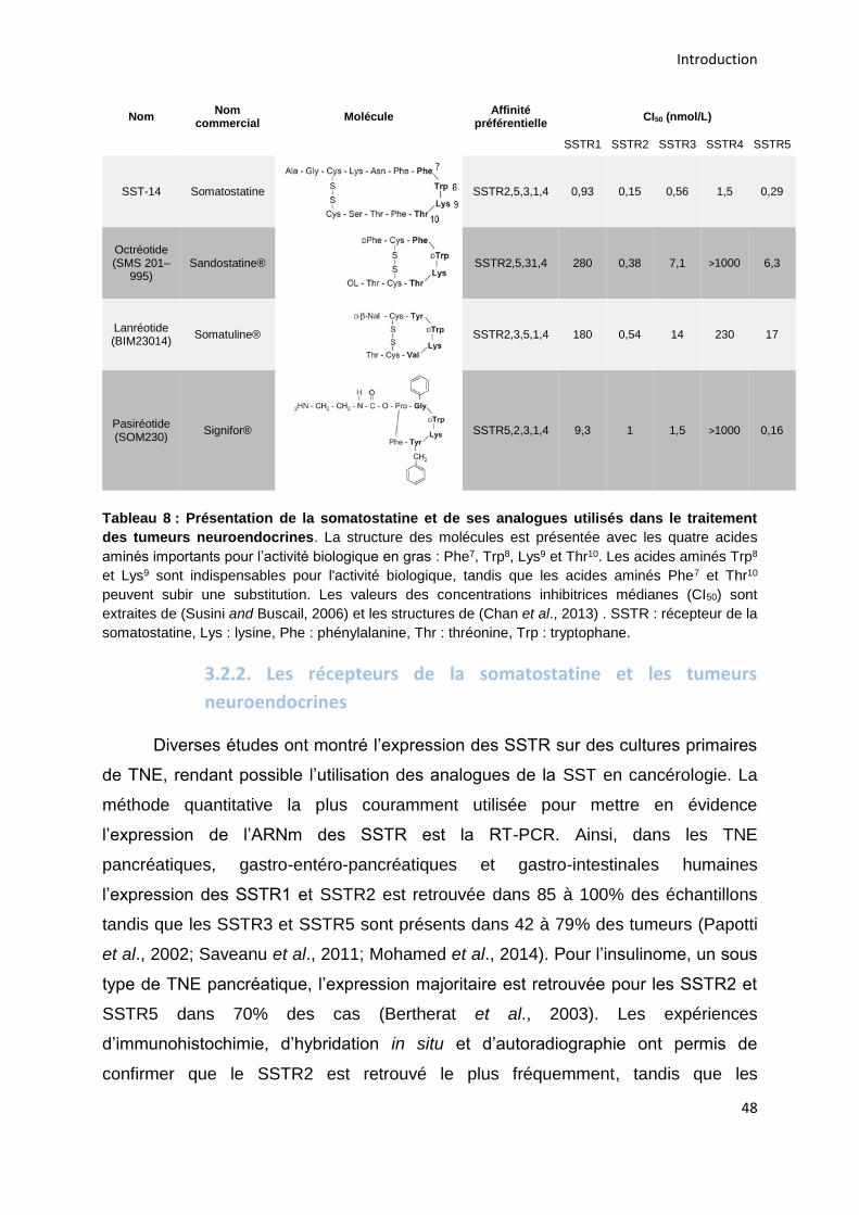
Introduction
48
Nom Nom
commercial Molécule
Affinité préférentielle
CI50 (nmol/L)
SSTR1 SSTR2 SSTR3 SSTR4 SSTR5
SST-14 Somatostatine
SSTR2,5,3,1,4 0,93 0,15 0,56 1,5 0,29
Octréotide (SMS 201–
995) Sandostatine® SSTR2,5,31,4 280 0,38 7,1 >1000 6,3
Lanréotide (BIM23014)
Somatuline®
SSTR2,3,5,1,4 180 0,54 14 230 17
Pasiréotide (SOM230)
Signifor®
SSTR5,2,3,1,4 9,3 1 1,5 >1000 0,16
Tableau 8 : Présentation de la somatostatine et de ses analogues utilisés dans le traitement
des tumeurs neuroendocrines. La structure des molécules est présentée avec les quatre acides
aminés importants pour l’activité biologique en gras : Phe7, Trp8, Lys9 et Thr10. Les acides aminés Trp8
et Lys9 sont indispensables pour l'activité biologique, tandis que les acides aminés Phe7 et Thr10
peuvent subir une substitution. Les valeurs des concentrations inhibitrices médianes (CI50) sont
extraites de (Susini and Buscail, 2006) et les structures de (Chan et al., 2013) . SSTR : récepteur de la
somatostatine, Lys : lysine, Phe : phénylalanine, Thr : thréonine, Trp : tryptophane.
3.2.2. Les récepteurs de la somatostatine et les tumeurs
neuroendocrines
Diverses études ont montré l’expression des SSTR sur des cultures primaires
de TNE, rendant possible l’utilisation des analogues de la SST en cancérologie. La
méthode quantitative la plus couramment utilisée pour mettre en évidence
l’expression de l’ARNm des SSTR est la RT-PCR. Ainsi, dans les TNE
pancréatiques, gastro-entéro-pancréatiques et gastro-intestinales humaines
l’expression des SSTR1 et SSTR2 est retrouvée dans 85 à 100% des échantillons
tandis que les SSTR3 et SSTR5 sont présents dans 42 à 79% des tumeurs (Papotti
et al., 2002; Saveanu et al., 2011; Mohamed et al., 2014). Pour l’insulinome, un sous
type de TNE pancréatique, l’expression majoritaire est retrouvée pour les SSTR2 et
SSTR5 dans 70% des cas (Bertherat et al., 2003). Les expériences
d’immunohistochimie, d’hybridation in situ et d’autoradiographie ont permis de
confirmer que le SSTR2 est retrouvé le plus fréquemment, tandis que les
Introduction
49
expressions sont plus variables et dépendent du sous type de TNE considérée pour
les SSTR1, SSTR3 et SSTR5. Mise en évidence dans moins de 25% des
échantillons, l’expression du SSTR4 semble peu répandue (Papotti et al., 2002;
Bertherat et al., 2003) (Tableau 9).
Pour les TNE pulmonaires, l’expression est également retrouvée en majorité
pour les SSTR1 et SSTR2 dans 88% des tumeurs (Kanakis et al., 2015). D’autres
méthodes confirment cette tendance lorsque tous les récepteurs sont recherchés
(Reubi and Waser, 2003; Righi et al., 2010; Tsuta et al., 2012; Kanakis et al., 2015).
L’expression des SSTR3 et SSTR5 est variable en fonction de la tumeur étudiée,
tandis que le SSTR4 est rarement exprimé (Tabeau 9).
Dans les PHEO et PGL, la recherche de l’ARNm des SSTR par RT-PCR
montre que le SSTR2 est présent chez 100% des patients (Pasquali et al., 2008;
Saveanu et al., 2011). L’étude de Pasquali et al., en 2008 montre également une
expression du SSTR5 dans 100% des cas, tandis qu’il n’est retrouvé que dans 54%
des échantillons par Saveanu et al. en 2011. Ces derniers montrent en revanche une
expression du SSTR1 dans plus de 90% des échantillons. Tandis que des
expériences d’immunocytochimie montrent une expression majoritaire du SSTR3
(Mundschenk et al., 2003; Unger et al., 2008; Leijon et al., 2019) (Tableau 9).
Enfin, pour les adénomes de pituitaires, l’ARNm des SSTR2 et SSTR5 est
majoritaire pour les tumeurs de type somatotropes ou corticotropes (Taboada et al.,
2007; de Bruin et al., 2009; Neto et al., 2009; Ibáñez-Costa et al., 2016). Tandis que
pour les prolactinomes, un autre sous type de TNE pituitaire, l’ARNm des SSTR1 et
SSTR2 est retrouvé le plus courramment et en quantité plus importante que les
autres SSTR (Miller et al., 1995; Ibáñez-Costa et al., 2016) (Tableau 9).
Malgré une variabilité de l’expression des SSTR en fonction des études et des
méthodes utilisées, toutes les TNE expriment une diversité de SSTR. Cette
observation a permis le développement et l’utilisation de nouveaux analogues de la
somatostatine, dont l’octréotide et le SOM230.
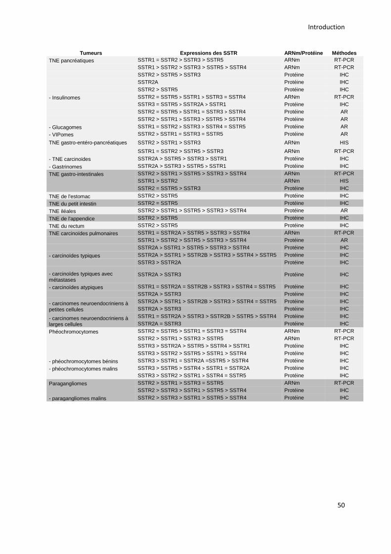
Introduction
50
Tumeurs Expressions des SSTR ARNm/Protéine Méthodes
TNE pancréatiques SSTR1 = SSTR2 > SSTR3 > SSTR5 ARNm RT-PCR
SSTR1 > SSTR2 > SSTR3 > SSTR5 > SSTR4 ARNm RT-PCR
SSTR2 > SSTR5 > SSTR3 Protéine IHC
SSTR2A Protéine IHC
SSTR2 > SSTR5 Protéine IHC
- Insulinomes SSTR2 = SSTR5 > SSTR1 > SSTR3 = SSTR4 ARNm RT-PCR
SSTR3 = SSTR5 > SSTR2A > SSTR1 Protéine IHC
SSTR2 = SSTR5 > SSTR1 = SSTR3 > SSTR4 Protéine AR
SSTR2 > SSTR1 > SSTR3 > SSTR5 > SSTR4 Protéine AR
- Glucagomes SSTR1 = SSTR2 > SSTR3 > SSTR4 = SSTR5 Protéine AR
- VIPomes SSTR2 > SSTR1 = SSTR3 = SSTR5 Protéine AR
TNE gastro-entéro-pancréatiques SSTR2 > SSTR1 > SSTR3 ARNm HIS
SSTR1 = SSTR2 > SSTR5 > SSTR3 ARNm RT-PCR
- TNE carcinoïdes SSTR2A > SSTR5 > SSTR3 > SSTR1 Protéine IHC
- Gastrinomes SSTR2A > SSTR3 > SSTR5 > SSTR1 Protéine IHC
TNE gastro-intestinales SSTR2 > SSTR1 > SSTR5 > SSTR3 > SSTR4 ARNm RT-PCR
SSTR1 > SSTR2 ARNm HIS
SSTR2 = SSTR5 > SSTR3 Protéine IHC
TNE de l'estomac SSTR2 > SSTR5 Protéine IHC
TNE du petit intestin SSTR2 = SSTR5 Protéine IHC
TNE iléales SSTR2 > SSTR1 > SSTR5 > SSTR3 > SSTR4 Protéine AR
TNE de l'appendice SSTR2 > SSTR5 Protéine IHC
TNE du rectum SSTR2 > SSTR5 Protéine IHC
TNE carcinoïdes pulmonaires SSTR1 = SSTR2A > SSTR5 > SSTR3 > SSTR4 ARNm RT-PCR
SSTR1 > SSTR2 > SSTR5 > SSTR3 > SSTR4 Protéine AR
SSTR2A > SSTR1 > SSTR5 > SSTR3 > SSTR4 Protéine IHC
- carcinoïdes typiques SSTR2A > SSTR1 > SSTR2B > SSTR3 > SSTR4 > SSTR5 Protéine IHC
SSTR3 > SSTR2A Protéine IHC
- carcinoïdes typiques avec métastases
SSTR2A > SSTR3 Protéine IHC
- carcinoïdes atypiques SSTR1 = SSTR2A = SSTR2B > SSTR3 > SSTR4 = SSTR5 Protéine IHC
SSTR2A > SSTR3 Protéine IHC
- carcinomes neuroendocriniens à petites cellules
SSTR2A > SSTR1 > SSTR2B > SSTR3 > SSTR4 = SSTR5 Protéine IHC
SSTR2A > SSTR3 Protéine IHC
- carcinomes neuroendocriniens à larges cellules
SSTR1 = SSTR2A > SSTR3 > SSTR2B > SSTR5 > SSTR4 Protéine IHC
SSTR2A = SSTR3 Protéine IHC
Phéochromocytomes SSTR2 = SSTR5 > SSTR1 = SSTR3 = SSTR4 ARNm RT-PCR
SSTR2 > SSTR1 > SSTR3 > SSTR5 ARNm RT-PCR
SSTR3 > SSTR2A > SSTR5 > SSTR4 > SSTR1 Protéine IHC
SSTR3 > SSTR2 > SSTR5 > SSTR1 > SSTR4 Protéine IHC
- phéochromocytomes bénins SSTR3 > SSTR1 = SSTR2A =SSTR5 > SSTR4 Protéine IHC
- phéochromocytomes malins SSTR3 > SSTR5 > SSTR4 > SSTR1 = SSTR2A Protéine IHC
SSTR3 > SSTR2 > SSTR1 > SSTR4 = SSTR5 Protéine IHC
Paragangliomes SSTR2 > SSTR1 > SSTR3 = SSTR5 ARNm RT-PCR
SSTR2 > SSTR3 > SSTR1 > SSTR5 > SSTR4 Protéine IHC
- paragangliomes malins SSTR2 > SSTR3 > SSTR1 > SSTR5 > SSTR4 Protéine IHC
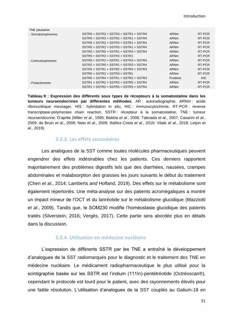
Introduction
51
TNE pituitaires
- Somatotropinomes SSTR5 > SSTR2 > SSTR3 > SSTR1 > SSTR4 ARNm RT-PCR
SSTR5 > SSTR3 > SSTR2 > SSTR1 > SSTR4 ARNm RT-PCR
SSTR5 > SSTR2 > SSTR3 > SSTR1 > SSTR4 ARNm RT-PCR
SSTR5 > SSTR2 > SSTR3 > SSTR1 > SSTR4 ARNm RT-PCR
SSTR2 = SSTR5 > SSTR1 > SSTR3 > SSTR4 ARNm RT-PCR
SSTR5 > SSTR2 > SSTR3 > SSTR1 ARNm RT-PCR
- Corticotropinomes SSTR5 > SSTR2 > SSTR1 > SSTR3 > SSTR4 ARNm RT-PCR
SSTR2 > SSTR5 > SSTR1 > SSTR3 = SSTR4 ARNm RT-PCR
SSTR5 > SSTR1 > SSTR2 > SSTR4 > SSTR3 ARNm RT-PCR
SSTR5 > SSTR2 > SSTR3 > SSTR1 ARNm RT-PCR
SSTR5 > SSTR4 = SSTR1 = SSTR2 = SSTR3 Protéine IHC
- Prolactinomes SSTR1 > SSTR2 > SSTR3 > SSTR5 > SSTR4 ARNm RT-PCR
SSTR1 > SSTR2 > SSTR5 > SSTR3 = SSTR4 ARNm RT-PCR
Tableau 9 : Expression des différents sous types de récepteurs à la somatostatine dans les
tumeurs neuroendocrines par différentes méthodes. AR : autoradiographie, ARNm : acide
ribonucléique messager, HIS : hybridation in situ, IHC : immunocytochimie, RT-PCR : reverse
transcriptase-polymerase chain reaction, SSTR : récepteur à la somatostatine, TNE : tumeur
neuroendocrine. D’après (Miller et al., 1995; Batista et al., 2006; Taboada et al., 2007; Casarini et al.,
2009; de Bruin et al., 2009; Neto et al., 2009; Ibáñez-Costa et al., 2016; Vitale et al., 2018; Leijon et
al., 2019).
3.2.3. Les effets secondaires
Les analogues de la SST comme toutes molécules pharmaceutiques peuvent
engendrer des effets indésirables chez les patients. Ces derniers rapportent
majoritairement des problèmes digestifs tels que des diarrhées, nausées, crampes
abdominales et malabsorption des graisses les jours suivants le début du traitement
(Chen et al., 2014; Lamberts and Hofland, 2019). Des effets sur le métabolisme sont
également répertoriés. Une méta-analyse sur des patients acromégaliques a montré
un impact mineur de l’OCT et du lanréotide sur le métabolisme glucidique (Mazziotti
et al., 2009). Tandis que, le SOM230 modifie l’homéostasie glucidique des patients
traités (Silverstein, 2016; Vergès, 2017). Cette partie sera abordée plus en détails
dans la discussion.
3.2.4. Utilisation en médecine nucléaire
L’expression de différents SSTR par les TNE a entraîné le développement
d’analogues de la SST radiomarqués pour le diagnostic et le traitement des TNE en
médecine nucléaire. Le médicament radiopharmaceutique le plus utilisé pour la
scintigraphie basée sur les SSTR est l’indium (111In)-pentétréotide (Octréoscan®),
cependant le protocole est lourd pour le patient, avec des rayonnements élevés pour
une faible résolution. L’utilisation d’analogues de la SST couplés au Galium-18 en
Introduction
52
tomographie par émission de positons a permis par la suite d’améliorer la résolution
et de diminuer les doses injectées. Trois radiopharmaceutiques ont été développés
avec des affinités variées pour les SSTR : le 68Ga-DOTATATE approuvé par la U.S.
Food and Drug Administration en 2016, le 68Ga-DOTATOC approuvé par l’Union
européenne et le 68Ga-DOTANOC. Ils sont utilisés pour le diagnostic et l’évaluation
des traitements de plusieurs TNE dont les TNE-GEP (Brabander et al., 2015;
Graham et al., 2017; Sanli et al., 2018; Ivanidze et al., 2019).
En thérapeutique, les analogues de la SST sont marqués avec des
radionucléides comme le Lutétium-177 ou le Yttrium-90 afin d’irradier les tumeurs
exprimant les SSTR. Le 90Y-DOTATOC (90Y-octréotide) et le 177Lu-DOTATATE
(177Lu-octreotate) sont utilisés en peptide receptor radionuclide therapy (PRRT) et
permettent le ciblage de TNE inopérables ou métastatiques. Leur utilisation permet
de contrôler la pathologie dans 68 à 94% des cas, de diminuer le volume tumoral et
les marqueurs biochimiques (Bodei et al., 2016; Stueven et al., 2019). Une étude
clinique de phase III a démontré que l’administration du 177Lu-DOTATATE améliore
la survie et le taux de réponse de patients atteints de TNE intestinales métastatiques
en comparaison à l’OCT à longue durée d’action (Strosberg et al., 2017). D’autres
approches sont à l’étude ces dernières années, notamment avec l’utilisation
d’antagonistes somatostatinergiques radiomarqués (Dalm et al., 2016).

RÉSULTATS

53
RÉSULTATS
1. Publication n°1 : Étude des mécanismes de l'exocytose régulée par le
calcium dans le phéochromocytome humain
1.1. Objectifs et déroulement de l’étude
La sécrétion excessive de catécholamines chez les patients atteints de
phéochromocytome est à l’origine de sérieux problèmes cliniques tels que de
l’hypertension, l’augmentation du risque d’accidents cardiovasculaires et le
développement potentiel de cardiomyopathies (Tsirlin et al., 2014). À ce jour les
mécanismes moléculaires qui induisent et maintiennent l’hypersécrétion ne sont pas
connus.
Découvrir les origines de l’hypersécrétion tumorale apparaît comme crucial et
permettrait à terme d’améliorer les connaissances sur ces tumeurs mais également
d’ouvrir des pistes de recherches pour développer de nouvelles thérapies. Notre
étude a pour objectif de caractériser la sécrétion catécholaminergique des
phéochromocytomes afin de déterminer quel(s) paramètre(s) de l’exocytose peuvent
être dérégulés dans cette tumeur. Pour cela j’ai mis en culture des échantillons de
phéchromocytomes humains et de médullosurrénales non tumorales afin de
comparer leur capacité sécrétoire par ampérométrie à fibre de carbone. Ces
expériences m’ont permis de voir que l’emballement de la sécrétion de ces tumeurs
provient d’une dérégulation à l’échelle unicellulaire, et que différents paramètres de
l’exocytose sont impactés. Grâce à une analyse par spectrométrie de masse réalisée
par l’entreprise Caprion, nous avons mis en évidence que ces tumeurs présentent
des changements d’expression de protéines spécifiquement impliquées dans
l’exocytose.
1.2. Résultats
Les résultats obtenus sont exposés dans le manuscrit intitulé « Systematic
investigation of calcium-regulated exocytosis in human pheochromocytoma »
que je signe en co-premier auteur et qui est encore en préparation.
Dans cette étude j’ai contribué à l’analyse de la sécrétion de plusieurs
phéochromocytomes par ampérométrie à fibre de carbone.

54
1
Amperometric and proteomic investigation of calcium-regulated exocytosis in human
pheochromocytoma
Sébastien Houy1*, Laura Streit1*, Marion Rame1, Charles Decraene1, Sophie Moog1, Laurent
Brunaud2, Joël Lanoix3, Rabie Chelbi1,6, Florence Bihain2, Stéphanie Lacomme4, Sandra
Lomazzi4, Michel Vix5, Didier Mutter5, Eustache Paramithiotis3, Nicolas Vitale1, Stéphane
Ory1# and Stéphane Gasman1#§
1Centre National de la Recherche Scientifique, Université de Strasbourg, Institut des
Neurosciences Cellulaires et Intégratives, F-67000 Strasbourg, FRANCE.
2Département de Chirurgie Viscérale, Métabolique et Cancérologique (CVMC), Unité
médico-chirurgicale de chirurgie métabolique, endocrinienne et thyroïdienne (UMET), Unité
médico-chirurgicale de chirurgie de l’obésité (UMCO), Université de Lorraine, CHRU
NANCY, Hôpital Brabois adultes, F-54511 Vandœuvre-lès-Nancy, FRANCE.
3Caprion Proteome, Inc., 201 avenue Président-Kennedy, suite 3900, Montréal, Québec,
CANADA H2X 3Y7.
4Centre de Ressources Biologiques Lorrain, CHRU Nancy, Hôpitaux de Brabois, F-54511
Vandœuvre-lès-Nancy, FRANCE.
5NHC Strasbourg, Service de Chirurgie Digestive et Endocrinienne des Hôpitaux
Universitaires de Strasbourg, Hôpital Civil, F-67000 Strasbourg, FRANCE.
6Inovarion, F-75005 Paris, FRANCE.
* S. Houy and L. Streit contributed equally to this paper
# S. Ory and S. Gasman contributed equally to this paper
§ Corresponding author: Stéphane Gasman, address as above.
e-mail: [email protected]
Short title: Secretory activity of human pheochromocytoma cells
Key words: calcium-regulated exocytosis, neuroendocrine tumor, pheochromocytoma,
carbon fiber amperometry
2
ABSTRACT
Neuroendocrine tumors constitute a heterogeneous group of tumors arising from hormone-
secreting cells and are generally associated with a dysfunction of secretion.
Pheochromocytoma (Pheo) is a neuroendocrine tumor that develops from chromaffin cells of
the adrenal medulla, and is responsible of an excess of catecholamine secretion leading to
severe clinical symptoms such as hypertension, elevated stroke risk and various
cardiovascular complications. Surprisingly, while the hypersecretory activity of Pheo is well
known to pathologists and clinicians, it has never been carefully explored at the cellular and
molecular levels from individual tumor cells. In the present study, we have combined
catecholamine secretion measurement by carbon fiber amperometry on human tumor
chromaffin cells directly cultured from freshly resected Pheo, with the analysis by mass
spectrometry of the exocytotic proteins differentially expressed between the tumor and the
matched adjacent non-tumor tissue. Catecholamine secretion recordings from individual Pheo
chromaffin cells obtained from most patients reveal a higher number of exocytic events per
cell associated with faster kinetic parameters. Accordingly, we unravel significant tumor-
associated modifications in the expression of key proteins involved in different steps of the
calcium-regulated exocytic pathway. Altogether, our findings indicate that the tumor-
associated hypersecretion of catecholamine is likely to be a consequence of a dysfunction of
the calcium-regulated exocytosis at the level of individual Pheo chromaffin cell.
3
INTRODUCTION
Through the secretion of hormones and neuropeptides, the neuroendocrine system controls
many vital functions such as metabolism, blood pressure, reproduction, growth and
development, stress and eating behavior, to cite only a few. Neoplasms can derive from all
kind of hormone secreting cells giving rise to a neuroendocrine tumor (NET). NETs constitute
a highly heterogeneous group of neoplasm, but share a common feature in that they are often
associated with a deregulation of hormone secretion, mainly hypersecretion, which can induce
symptoms and major clinical complications (Zandee, et al. 2017). Therefore, the secretory
pathways and their dysfunction appear as an important issue to be considered in NETs.
Surprisingly, while the hypersecretory activity of NETs is well known to pathologists and
clinicians, to our knowledge it has never been carefully explored at the cellular and molecular
levels.
In neuroendocrine cells, hormones and neuropeptides are stored in large dense core vesicles
(secretory granules) and are secreted through calcium-regulated exocytosis, a process that has
been intensively studied since many decades (Anantharam and Kreutzberger 2019). It
involves several tightly regulated trafficking steps including the recruitment of secretory
granules to the cell periphery, their docking to exocytic sites of the plasma membrane, their
priming and finally the fusion between the secretory granule membrane and the plasma
membrane leading to the release of the intra-granular content (Burgoyne and Morgan 2003;
Lang and Jahn 2008). Chromaffin cells of the adrenal medulla, which store and then secrete
catecholamines into the blood stream, have been widely used by us and others as an
experimental model to uncover the molecular mechanisms controlling calcium-regulated
exocytosis (Bader, et al. 2002; Gasman and Vitale 2017; Malacombe, et al. 2006).
The NET deriving from chromaffin cells of the adrenal medulla is called pheochromocytoma
(Pheo) (Lenders, et al. 2020). Most of the Pheos are responsible for catecholamine
hypersecretion leading to clinical symptoms such as permanent or paroxysmal hypertension or
to cardiovascular complications including myocarditis, Takotsubo syndrome and various
forms of cardiomyopathies (Lenders, et al. 2020; Pappachan, et al. 2018; Pourian, et al. 2015;
Y-Hassan and Falhammar 2020; Zhang, et al. 2017). The reason for this excess of secretion is
not known. One likely possibility could be an increase of the secretory capacity at the single
cell level. We therefore attempted to investigate the cellular mechanisms responsible for

4
specific secretion dysfunction in Pheo by applying on primary culture of human Pheo cells,
carbon fiber amperometry, a technique that enables precise measurement of individual
exocytotic event dynamics in real time and in single cell (Fathali and Cans 2018; Mosharov
and Sulzer 2005). Combined with the detection in human Pheo tissue of exocytotic protein
expression changes by mass spectrometry, it allowed us to reveal upregulated exocytosis at
the single cell level and to identify specific steps of the exocytotic process that are
dysregulated in the tumor as well as potential actors of the molecular machinery triggering
hypersecretion in Pheo.
5
MATERIAL AND METHODS
Subjects and samples
The medical files of patients with pheochromocytoma, followed-up between 2013 and 2020 in
2 French centers were retrospectively reviewed. Informed consents for genetics test and this
study were obtained from all patients. We collected the complete characterization made at the
time of initial diagnosis, including a clinical examination looking for hormonal- or tumoral-
related symptoms, biological analysis and genetic testing proposed to identify germline
mutations in the major susceptibility genes as recommended by the Endocrine Society clinical
practice guideline published in 2014 (Lenders, et al. 2014). Pathological evaluation was
reviewed, including tumor size, Ki-67 result and the PASS (Pheochromocytoma of the
Adrenal Gland Scaled Score) system as previously described (Thompson 2002). Biological
analysis comprised the measurement of metanephrine (MN) and normetanephrine (NMN)
levels (in urine and/or plasma). When available, chromogranin A (CGA) measurements were
also registered. Levels of free MN, NMN and CGA in plasma, as well as urinary levels of MN
and NMN are presented as ratios normalized by upper limits of normal (ULN). Plasma and
urinary MN or NMN levels reaching two-fold the upper limit of the normal range and/or CGA
exceeding the upper limit of the normal range was defined as the threshold of abnormal
hormonal secretion. The ULN of free metanephrine (MN) and normetanephrine (NM) were
4.05 and 9.8 nmol/L in plasma and 1625 and 2620 nmol/24 h in urine, respectively. The upper
reference limit for CGA was 100 mg/L. We have categorized the catecholamine-producing
phenotype of Pheo as previously described (Eisenhofer, et al. 2005): adrenergic (AD)
phenotype, in case the MN content exceeds 10% of the combined MN and NM contents or,
noradrenergic (NAD) phenotype in case the MN content remained below 10% of the
combined MN and NM contents.
Primary culture of human pheochromocytomas
Human chromaffin cells were cultured from freshly resected tumor adrenal gland following
surgery (Moog, et al. 2018). In the operating room and immediately after the resection, the
adrenal gland was cut longitudinally in two parts. Roughly 1 cm3 piece of tumoral tissue was
dissected and immediately plunged into ice cold transport medium (Ca2+- and Mg2+-free
Hank’s Balanced Salt Solution (CMF HBSS, Sigma) supplemented with 0.2% Fetal Bovine
Serum (FBS, Gibco) and 1% penicillin/streptomycin (Sigma) or MACS Medium Tissue
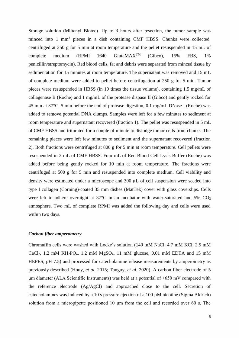
6
Storage solution (Miltenyi Biotec). Up to 3 hours after resection, the tumor sample was
minced into 1 mm3 pieces in a dish containing CMF HBSS. Chunks were collected,
centrifuged at 250 g for 5 min at room temperature and the pellet resuspended in 15 mL of
complete medium (RPMI 1640 GlutaMAXTM (Gibco), 15% FBS, 1%
penicillin/streptomycin). Red blood cells, fat and debris were separated from minced tissue by
sedimentation for 15 minutes at room temperature. The supernatant was removed and 15 mL
of complete medium were added to pellet before centrifugation at 250 g for 5 min. Tumor
pieces were resuspended in HBSS (in 10 times the tissue volume), containing 1.5 mg/mL of
collagenase B (Roche) and 1 mg/mL of the protease dispase II (Gibco) and gently rocked for
45 min at 37°C. 5 min before the end of protease digestion, 0.1 mg/mL DNase I (Roche) was
added to remove potential DNA clumps. Samples were left for a few minutes to sediment at
room temperature and supernatant recovered (fraction 1). The pellet was resuspended in 5 mL
of CMF HBSS and triturated for a couple of minute to dislodge tumor cells from chunks. The
remaining pieces were left few minutes to sediment and the supernatant recovered (fraction
2). Both fractions were centrifuged at 800 g for 5 min at room temperature. Cell pellets were
resuspended in 2 mL of CMF HBSS. Four mL of Red Blood Cell Lysis Buffer (Roche) was
added before being gently rocked for 10 min at room temperature. The fractions were
centrifuged at 500 g for 5 min and resuspended into complete medium. Cell viability and
density were estimated under a microscope and 300 L of cell suspension were seeded into
type I collagen (Corning)-coated 35 mm dishes (MatTek) cover with glass coverslips. Cells
were left to adhere overnight at 37°C in an incubator with water-saturated and 5% CO2
atmosphere. Two mL of complete RPMI was added the following day and cells were used
within two days.
Carbon fiber amperometry
Chromaffin cells were washed with Locke’s solution (140 mM NaCl, 4.7 mM KCl, 2.5 mM
CaCl2, 1.2 mM KH2PO4, 1.2 mM MgSO4, 11 mM glucose, 0.01 mM EDTA and 15 mM
HEPES, pH 7.5) and processed for catecholamine release measurements by amperometry as
previously described (Houy, et al. 2015; Tanguy, et al. 2020). A carbon fiber electrode of 5
μm diameter (ALA Scientific Instruments) was held at a potential of +650 mV compared with
the reference electrode (Ag/AgCl) and approached close to the cell. Secretion of
catecholamines was induced by a 10 s pressure ejection of a 100 µM nicotine (Sigma Aldrich)
solution from a micropipette positioned 10 μm from the cell and recorded over 60 s. The
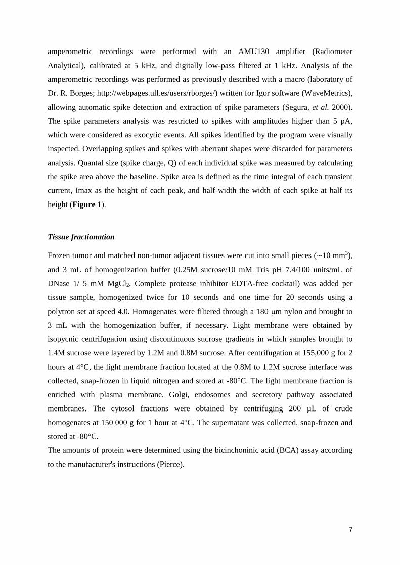
7
amperometric recordings were performed with an AMU130 amplifier (Radiometer
Analytical), calibrated at 5 kHz, and digitally low-pass filtered at 1 kHz. Analysis of the
amperometric recordings was performed as previously described with a macro (laboratory of
Dr. R. Borges; http://webpages.ull.es/users/rborges/) written for Igor software (WaveMetrics),
allowing automatic spike detection and extraction of spike parameters (Segura, et al. 2000).
The spike parameters analysis was restricted to spikes with amplitudes higher than 5 pA,
which were considered as exocytic events. All spikes identified by the program were visually
inspected. Overlapping spikes and spikes with aberrant shapes were discarded for parameters
analysis. Quantal size (spike charge, Q) of each individual spike was measured by calculating
the spike area above the baseline. Spike area is defined as the time integral of each transient
current, Imax as the height of each peak, and half-width the width of each spike at half its
height (Figure 1).
Tissue fractionation
Frozen tumor and matched non-tumor adjacent tissues were cut into small pieces (∼10 mm3),
and 3 mL of homogenization buffer (0.25M sucrose/10 mM Tris pH 7.4/100 units/mL of
DNase 1/ 5 mM MgCl2, Complete protease inhibitor EDTA-free cocktail) was added per
tissue sample, homogenized twice for 10 seconds and one time for 20 seconds using a
polytron set at speed 4.0. Homogenates were filtered through a 180 μm nylon and brought to
3 mL with the homogenization buffer, if necessary. Light membrane were obtained by
isopycnic centrifugation using discontinuous sucrose gradients in which samples brought to
1.4M sucrose were layered by 1.2M and 0.8M sucrose. After centrifugation at 155,000 g for 2
hours at 4°C, the light membrane fraction located at the 0.8M to 1.2M sucrose interface was
collected, snap-frozen in liquid nitrogen and stored at -80°C. The light membrane fraction is
enriched with plasma membrane, Golgi, endosomes and secretory pathway associated
membranes. The cytosol fractions were obtained by centrifuging 200 µL of crude
homogenates at 150 000 g for 1 hour at 4°C. The supernatant was collected, snap-frozen and
stored at -80°C.
The amounts of protein were determined using the bicinchoninic acid (BCA) assay according
to the manufacturer's instructions (Pierce).
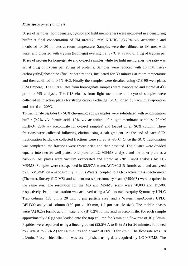
8
Mass spectrometry analysis
30 g of samples (homogenates, cytosol and light membranes) were incubated in a denaturing
buffer at final concentration of 7M urea/175 mM NH4HCO3/8.75% v/v acetonitrile and
incubated for 30 minutes at room temperature. Samples were then diluted to 1M urea with
water and digested with trypsin (Promega) overnight at 37°C at a ratio of 1g of trypsin per
10 g of protein for homogenate and cytosol samples while for light membranes, the ratio was
set at 1g of trypsin per 25 g of proteins. Samples were reduced with 10 mM tris(2-
carboxyethyl)phosphine (final concentration), incubated for 30 minutes at room temperature
and then acidified to 0.5N HCl. Finally the samples were desalted using C18 96-well plates
(3M Empore). The C18 eluates from homogenate samples were evaporated and stored at 4°C
prior to MS analysis. The C18 eluates from light membrane and cytosol samples were
collected in injection plates for strong cation exchange (SCX), dried by vacuum evaporation
and stored at -20°C.
To fractionate peptides by SCX chromatography, samples were solubilized with reconstitution
buffer (0.2% v/v formic acid, 10% v/v acetonitrile for light membrane samples; 20mM
K2HPO4, 25% v/v acetonitrile for cytosol samples) and loaded on an SCX column. Three
fractions were collected following elution using a salt gradient. At the end of each SCX
fractionation batch, the collected fractions were stored at -80°C. Once the SCX fractionation
was completed, the fractions were freeze-dried and then desalted. The eluates were divided
equally into two 96-well plates; one plate for LC-MS/MS analysis and the other plate as a
back-up. All plates were vacuum evaporated and stored at -20°C until analysis by LC-
MS/MS. Samples were resuspended in 92.5/7.5 water/ACN+0.2 % formic acid and analyzed
by LC-MS/MS on a nanoAcquity UPLC (Waters) coupled to a Q-Exactive mass spectrometer
(Thermo). Survey (LC-MS) and tandem mass spectrometry scans (MS/MS) were acquired in
the same run. The resolution for the MS and MS/MS scans were 70,000 and 17,500,
respectively. Peptide separation was achieved using a Waters nanoAcquity Symmetry UPLC
Trap column (180 µm x 20 mm, 5 µm particle size) and a Waters nanoAcquity UPLC
BEH300 analytical column (150 µm x 100 mm, 1.7 µm particle size). The mobile phases
were (A) 0.2% formic acid in water and (B) 0.2% formic acid in acetonitrile. For each sample
approximately 3.6 µg was loaded onto the trap column for 3 min at a flow rate of 10 µL/min.
Peptides were separated using a linear gradient (92.5% A to 84% A) for 26 minutes, followed
by (84% A to 75% A) for 14 minutes and a wash at 60% B for 2min. The flow rate was 1.8
µL/min. Protein identification was accomplished using data acquired by LC-MS/MS. The
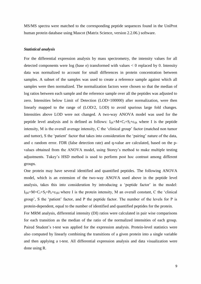
9
MS/MS spectra were matched to the corresponding peptide sequences found in the UniProt
human protein database using Mascot (Matrix Science, version 2.2.06.) software.
Statistical analysis
For the differential expression analysis by mass spectrometry, the intensity values for all
detected components were log (base e) transformed with values < 0 replaced by 0. Intensity
data was normalized to account for small differences in protein concentration between
samples. A subset of the samples was used to create a reference sample against which all
samples were then normalized. The normalization factors were chosen so that the median of
log ratios between each sample and the reference sample over all the peptides was adjusted to
zero. Intensities below Limit of Detection (LOD=100000) after normalization, were then
linearly mapped to the range of (LOD/2, LOD) to avoid spurious large fold changes.
Intensities above LOD were not changed. A two-way ANOVA model was used for the
peptide level analysis and is defined as follows: Iijk=M+Ci+Sj+ijk where I is the peptide
intensity, M is the overall average intensity, C the ‘clinical group’ factor (matched non tumor
and tumor), S the ‘patient’ factor that takes into consideration the ‘pairing’ nature of the data,
and ε random error. FDR (false detection rate) and q-value are calculated, based on the p-
values obtained from the ANOVA model, using Storey’s method to make multiple testing
adjustments. Tukey’s HSD method is used to perform post hoc contrast among different
groups.
One protein may have several identified and quantified peptides. The following ANOVA
model, which is an extension of the two-way ANOVA used above in the peptide level
analysis, takes this into consideration by introducing a ‘peptide factor’ in the model:
Iijkl=M+Ci+Sj+Pk+ijkl where I is the protein intensity, M an overall constant, C the ‘clinical
group’, S the ‘patient’ factor, and P the peptide factor. The number of the levels for P is
protein-dependent, equal to the number of identified and quantified peptides for the protein.
For MRM analysis, differential intensity (DI) ratios were calculated in pair wise comparisons
for each transition as the median of the ratio of the normalized intensities of each group.
Paired Student’s t-test was applied for the expression analysis. Protein-level statistics were
also computed by linearly combining the transitions of a given protein into a single variable
and then applying a t-test. All differential expression analysis and data visualization were
done using R.
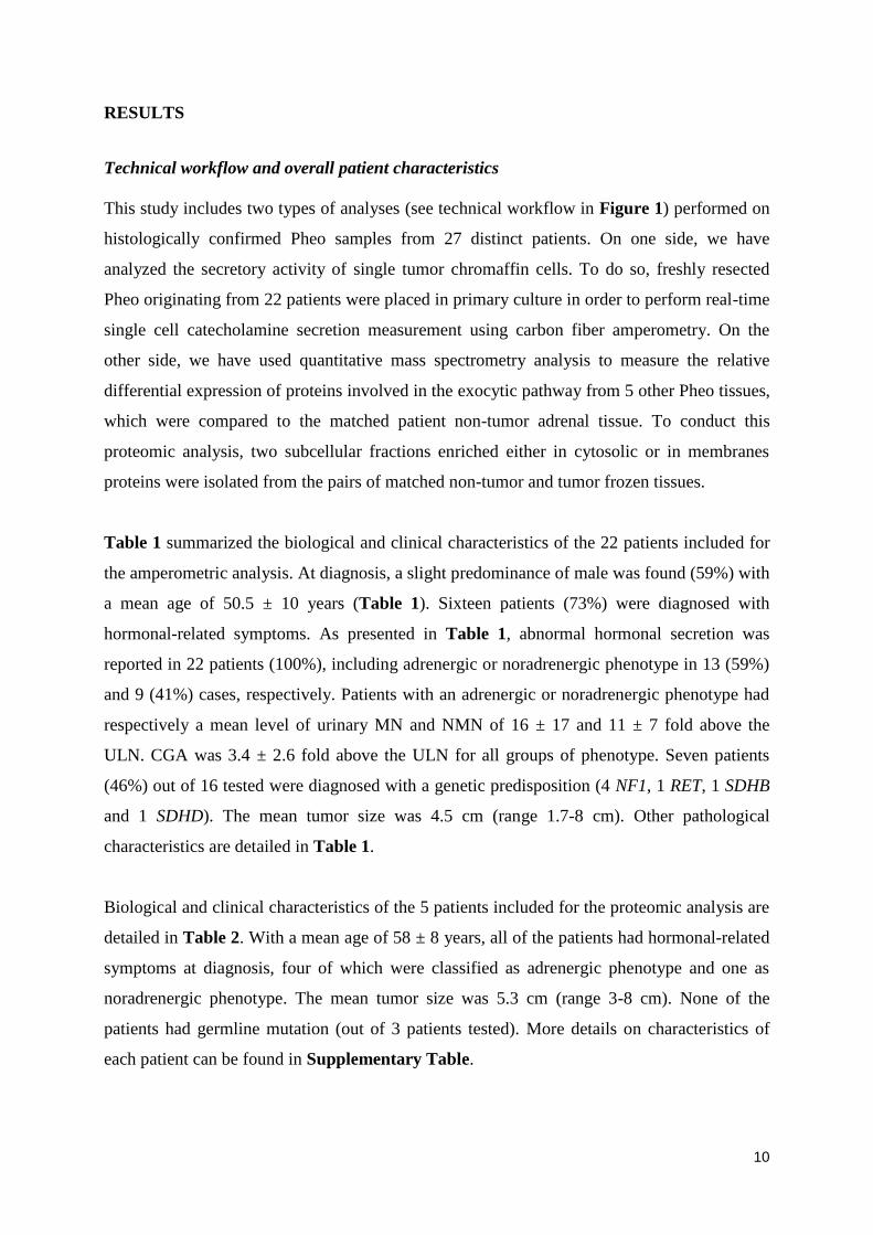
10
RESULTS
Technical workflow and overall patient characteristics
This study includes two types of analyses (see technical workflow in Figure 1) performed on
histologically confirmed Pheo samples from 27 distinct patients. On one side, we have
analyzed the secretory activity of single tumor chromaffin cells. To do so, freshly resected
Pheo originating from 22 patients were placed in primary culture in order to perform real-time
single cell catecholamine secretion measurement using carbon fiber amperometry. On the
other side, we have used quantitative mass spectrometry analysis to measure the relative
differential expression of proteins involved in the exocytic pathway from 5 other Pheo tissues,
which were compared to the matched patient non-tumor adrenal tissue. To conduct this
proteomic analysis, two subcellular fractions enriched either in cytosolic or in membranes
proteins were isolated from the pairs of matched non-tumor and tumor frozen tissues.
Table 1 summarized the biological and clinical characteristics of the 22 patients included for
the amperometric analysis. At diagnosis, a slight predominance of male was found (59%) with
a mean age of 50.5 ± 10 years (Table 1). Sixteen patients (73%) were diagnosed with
hormonal-related symptoms. As presented in Table 1, abnormal hormonal secretion was
reported in 22 patients (100%), including adrenergic or noradrenergic phenotype in 13 (59%)
and 9 (41%) cases, respectively. Patients with an adrenergic or noradrenergic phenotype had
respectively a mean level of urinary MN and NMN of 16 ± 17 and 11 ± 7 fold above the
ULN. CGA was 3.4 ± 2.6 fold above the ULN for all groups of phenotype. Seven patients
(46%) out of 16 tested were diagnosed with a genetic predisposition (4 NF1, 1 RET, 1 SDHB
and 1 SDHD). The mean tumor size was 4.5 cm (range 1.7-8 cm). Other pathological
characteristics are detailed in Table 1.
Biological and clinical characteristics of the 5 patients included for the proteomic analysis are
detailed in Table 2. With a mean age of 58 ± 8 years, all of the patients had hormonal-related
symptoms at diagnosis, four of which were classified as adrenergic phenotype and one as
noradrenergic phenotype. The mean tumor size was 5.3 cm (range 3-8 cm). None of the
patients had germline mutation (out of 3 patients tested). More details on characteristics of
each patient can be found in Supplementary Table.
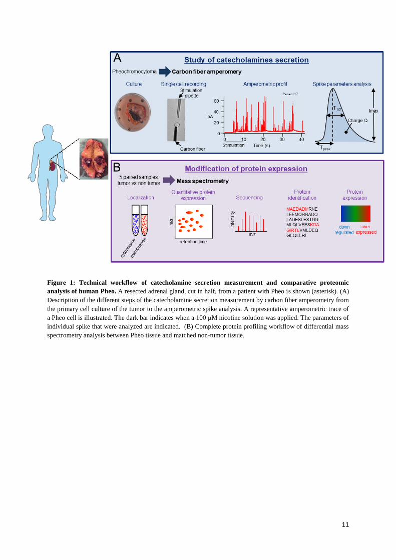
11
Figure 1: Technical workflow of catecholamine secretion measurement and comparative proteomic
analysis of human Pheo. A resected adrenal gland, cut in half, from a patient with Pheo is shown (asterisk). (A)
Description of the different steps of the catecholamine secretion measurement by carbon fiber amperometry from
the primary cell culture of the tumor to the amperometric spike analysis. A representative amperometric trace of
a Pheo cell is illustrated. The dark bar indicates when a 100 µM nicotine solution was applied. The parameters of
individual spike that were analyzed are indicated. (B) Complete protein profiling workflow of differential mass
spectrometry analysis between Pheo tissue and matched non-tumor tissue.
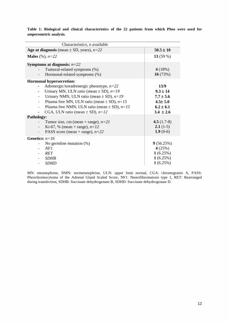
12
Table 1: Biological and clinical characteristics of the 22 patients from which Pheo were used for
amperometric analysis.
Characteristics, n available
Age at diagnosis (mean SD, years), n=22 50.5 10
Males (%), n=22 13 (59 %)
Symptoms at diagnosis: n=22
- Tumoral-related symptoms (%)
- Hormonal-related symptoms (%)
4 (18%)
16 (73%)
Hormonal hypersecretion:
- Adrenergic/noradrenergic phenotype, n=22
- Urinary MN, ULN ratio (mean SD), n=19
- Urinary NMN, ULN ratio (mean SD), n=19
- Plasma free MN, ULN ratio (mean SD), n=15
- Plasma free NMN, ULN ratio (mean SD), n=15
- CGA, ULN ratio (mean SD), n=12
13/9
9.3 14
7.7 5.6
4.5 5.0
6.2 6.1
3.4 2.6
Pathology:
- Tumor size, cm (mean + range), n=21
- Ki-67, % (mean + range), n=12
- PASS score (mean + range), n=22
4.5 (1.7-8)
2.1 (1-5)
1.9 (0-6)
Genetics: n=16
- No germline mutation (%)
- NF1
- RET
- SDHB
- SDHD
9 (56.25%)
4 (25%)
1 (6.25%)
1 (6.25%)
1 (6.25%)
MN: metanephrine, NMN: normetanephrine, ULN: upper limit normal, CGA: chromogranin A, PASS:
Pheochromocytoma of the Adrenal Gland Scaled Score, NF1: Neurofibromatosis type 1, RET: Rearranged
during transfection, SDHB: Succinate dehydrogenase B, SDHD: Succinate dehydrogenase D.
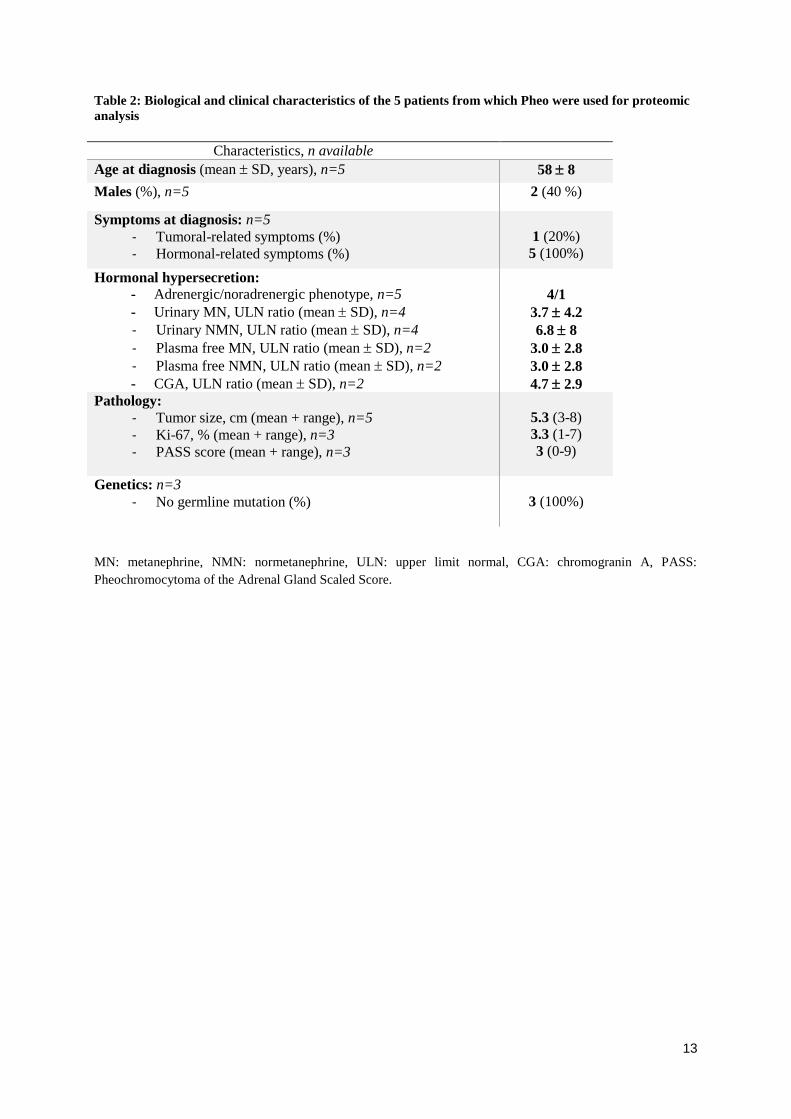
13
Table 2: Biological and clinical characteristics of the 5 patients from which Pheo were used for proteomic
analysis
Characteristics, n available
Age at diagnosis (mean SD, years), n=5 58 8
Males (%), n=5 2 (40 %)
Symptoms at diagnosis: n=5
- Tumoral-related symptoms (%)
- Hormonal-related symptoms (%)
1 (20%)
5 (100%)
Hormonal hypersecretion:
- Adrenergic/noradrenergic phenotype, n=5
- Urinary MN, ULN ratio (mean SD), n=4
- Urinary NMN, ULN ratio (mean SD), n=4
- Plasma free MN, ULN ratio (mean SD), n=2
- Plasma free NMN, ULN ratio (mean SD), n=2
- CGA, ULN ratio (mean SD), n=2
4/1
3.7 4.2
6.8 8
3.0 2.8
3.0 2.8
4.7 2.9
Pathology:
- Tumor size, cm (mean + range), n=5
- Ki-67, % (mean + range), n=3
- PASS score (mean + range), n=3
5.3 (3-8)
3.3 (1-7)
3 (0-9)
Genetics: n=3
- No germline mutation (%)
3 (100%)
MN: metanephrine, NMN: normetanephrine, ULN: upper limit normal, CGA: chromogranin A, PASS:
Pheochromocytoma of the Adrenal Gland Scaled Score.
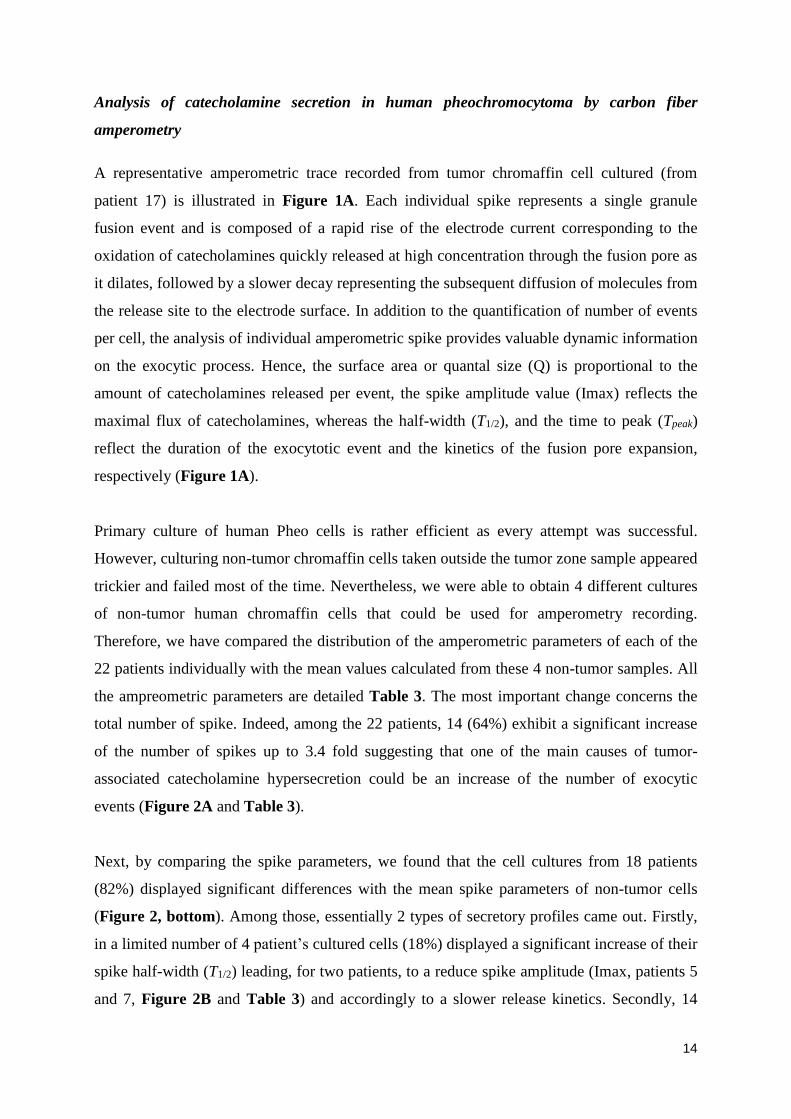
14
Analysis of catecholamine secretion in human pheochromocytoma by carbon fiber
amperometry
A representative amperometric trace recorded from tumor chromaffin cell cultured (from
patient 17) is illustrated in Figure 1A. Each individual spike represents a single granule
fusion event and is composed of a rapid rise of the electrode current corresponding to the
oxidation of catecholamines quickly released at high concentration through the fusion pore as
it dilates, followed by a slower decay representing the subsequent diffusion of molecules from
the release site to the electrode surface. In addition to the quantification of number of events
per cell, the analysis of individual amperometric spike provides valuable dynamic information
on the exocytic process. Hence, the surface area or quantal size (Q) is proportional to the
amount of catecholamines released per event, the spike amplitude value (Imax) reflects the
maximal flux of catecholamines, whereas the half-width (T1/2), and the time to peak (Tpeak)
reflect the duration of the exocytotic event and the kinetics of the fusion pore expansion,
respectively (Figure 1A).
Primary culture of human Pheo cells is rather efficient as every attempt was successful.
However, culturing non-tumor chromaffin cells taken outside the tumor zone sample appeared
trickier and failed most of the time. Nevertheless, we were able to obtain 4 different cultures
of non-tumor human chromaffin cells that could be used for amperometry recording.
Therefore, we have compared the distribution of the amperometric parameters of each of the
22 patients individually with the mean values calculated from these 4 non-tumor samples. All
the ampreometric parameters are detailed Table 3. The most important change concerns the
total number of spike. Indeed, among the 22 patients, 14 (64%) exhibit a significant increase
of the number of spikes up to 3.4 fold suggesting that one of the main causes of tumor-
associated catecholamine hypersecretion could be an increase of the number of exocytic
events (Figure 2A and Table 3).
Next, by comparing the spike parameters, we found that the cell cultures from 18 patients
(82%) displayed significant differences with the mean spike parameters of non-tumor cells
(Figure 2, bottom). Among those, essentially 2 types of secretory profiles came out. Firstly,
in a limited number of 4 patient’s cultured cells (18%) displayed a significant increase of their
spike half-width (T1/2) leading, for two patients, to a reduce spike amplitude (Imax, patients 5
and 7, Figure 2B and Table 3) and accordingly to a slower release kinetics. Secondly, 14

15
patients (64%) for which tumor cells presented a significant reduction of the spike half-width
T1/2 and/or the spike rise time (Tpeak), often accompanied (9 patients) with a reduction of the
quantal size (charge Q; Figure 2C, D and Table 3). The later type of amperometric profile
corresponds to exocytic events occurring with a faster kinetics compared to normal cells.
Altogether, our amperometric analysis indicate that catecholamine secretion in tumor
chromaffin cells from patients with Pheo often involves a high number and/or fast secretory
events which most likely contribute to the tumor-associated hypersecretion.
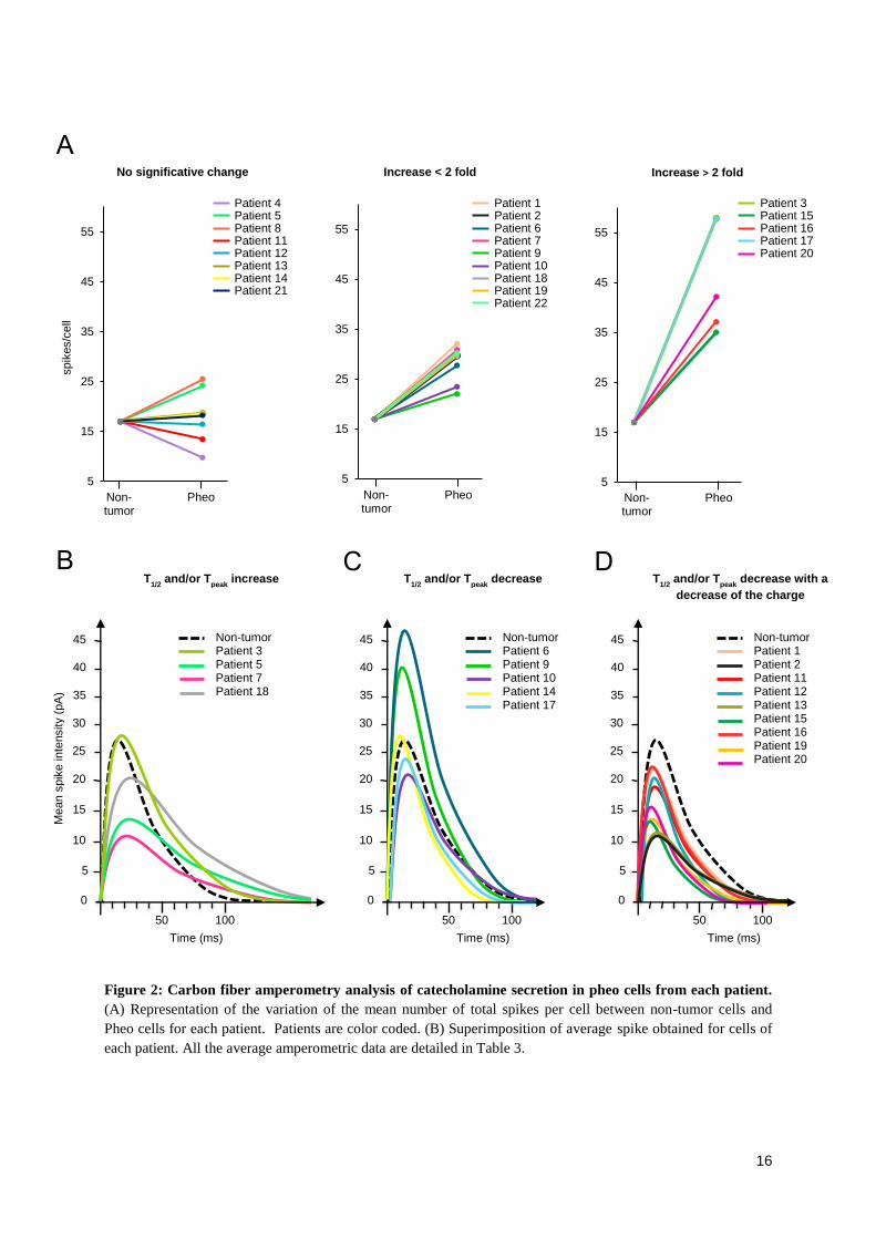
16
Figure 2: Carbon fiber amperometry analysis of catecholamine secretion in pheo cells from each patient.
(A) Representation of the variation of the mean number of total spikes per cell between non-tumor cells and
Pheo cells for each patient. Patients are color coded. (B) Superimposition of average spike obtained for cells of
each patient. All the average amperometric data are detailed in Table 3.
5
15
25
35
45
55
0,8 1,8
5
15
25
35
45
55
0,8 1,85
15
25
35
45
55
0,8 1,8
A
B
Patient 4 Patient 5 Patient 8 Patient 11 Patient 12 Patient 13 Patient 14 Patient 21
Patient 1 Patient 2 Patient 6 Patient 7 Patient 9 Patient 10 Patient 18 Patient 19 Patient 22
spik
es/c
ell
Non- tumor
Pheo
Non-tumor Patient 3 Patient 5 Patient 7 Patient 18
Mean s
pik
e in
tensity (
pA
)
Time (ms)
0
5
10
15
20
50 100
25
30
35
40
Time (ms)
0
5
10
15
20
50 100
25
30
35
40
45 45 Non-tumor Patient 6 Patient 9 Patient 10 Patient 14 Patient 17
Non-tumor Patient 1 Patient 2 Patient 11 Patient 12 Patient 13 Patient 15 Patient 16 Patient 19 Patient 20
Time (ms)
0
5
10
15
20
50 100
25
30
35
40
45
Non- tumor
Pheo Non- tumor
Pheo
Patient 3 Patient 15 Patient 16 Patient 17 Patient 20
C D
No significative change Increase < 2 fold Increase > 2 fold
T1/2
and/or Tpeak
increase T1/2
and/or Tpeak
decrease T1/2
and/or Tpeak
decrease with a
decrease of the charge
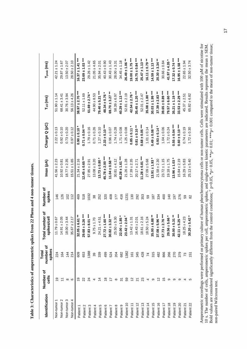
17
T
ab
le 3
: C
ha
ract
eris
tics
of
am
per
om
etri
c sp
ikes
fro
m 2
2 P
heo
s a
nd
4 n
on
-tu
mo
r ti
ssu
es.
Ide
nti
fica
tio
n
Nu
mb
er
of
ce
lls
To
tal
nu
mb
er
of
sp
ike
s
To
tal
nu
mb
er
of
sp
ike
s/c
ell
Nu
mb
er
of
sp
ike
s
Ima
x (
pA
) C
ha
rge
Q (
pC
) T
1/2
(m
s)
Tp
ea
k (
ms
)
Non-t
um
or
1
19
224
11.7
9 ±
0.6
7
146
41.5
6 ±
2.2
3
2.2
2 ±
0.1
3
51.9
0 ±
1.1
4
42.2
1 ±
1.2
4
Non-t
um
or
2
11
164
14.9
1 ±
2.0
5
126
10.9
2 ±
0.8
1
0.9
2 ±
0.1
0
68.4
1 ±
3.4
1
29.8
7 ±
1.6
7
Non-t
um
or
3
8
144
18.0
0 ±
3.4
4
102
18.7
7 ±
2.3
5
0.7
3 ±
0.2
0
30.7
6 ±
3.9
4
13.5
0 ±
2.0
7
Non-t
um
or
4
6
214
35.6
7 ±
2.1
7
112
15.5
1 ±
1.6
5
0.9
7 ±
0.1
2
56.1
3 ±
4.2
6
28.5
0 ±
2.1
0
Patie
nt 1
19
609
32.0
5 ±
6.6
1 *
* 469
21.3
4 ±
2.1
6
0.9
0 ±
0.1
0 *
38.9
7 ±
2.7
0 *
**
16.3
7 ±
1.4
1 *
**
Patie
nt 2
22
653
29.6
8 ±
3.2
6 *
**
518
12.4
5 ±
0.6
5 *
**
0.7
1 ±
0.0
4 *
**
52.1
1 ±
1.6
9
23.6
9 ±
1.0
3 *
**
Patie
nt 3
24
1388
57.8
3 ±
8.0
1 *
**
1032
27.8
5 ±
2.9
1
1.7
9 ±
0.1
6
61.6
9 ±
2.7
4 *
29.2
6 ±
1.4
2
Patie
nt 4
4
39
9.7
5 ±
1.7
0
38
13.0
8 ±
3.2
0
0.7
1 ±
0.2
6
44.9
0 ±
8.5
3
21.0
5 ±
4.6
5
Patie
nt 5
14
339
24.2
1 ±
4.5
1
252
13.7
3 ±
0.8
0 *
1.2
7 ±
0.1
0
79.4
0 ±
5.2
1 *
**
35.9
4 ±
2.0
1
Patie
nt 6
18
499
27.7
2 ±
1.6
0 *
**
320
45.7
8 ±
2.0
0 *
**
2.3
3 ±
0.1
0 *
**
48.3
4 ±
0.7
6 *
40.4
3 ±
0.9
0
Patie
nt 7
17
524
30.8
2 ±
3.3
2 *
**
408
11.0
4 ±
0.5
8 *
* 0.9
6 ±
0.0
7
69.7
0 ±
3.2
7 *
* 30.4
3 ±
1.4
3
Patie
nt 8
8
204
25.5
0 ±
5.3
0
177
30.8
1 ±
4.1
2
1.7
4 ±
0.2
8
58.3
9 ±
6.9
7
28.0
0 ±
3.3
1
Patie
nt 9
31
682
22.0
0 ±
1.8
5 *
416
40.2
8 ±
1.6
1 *
**
1.7
1 ±
0.0
8
40.2
9 ±
1.1
3 *
**
34.4
5 ±
1.1
8
Patie
nt 10
59
1382
23.4
2 ±
2.0
4 T
1231
21.2
6 ±
1.2
8
1.2
5 ±
0.0
8
52.8
1 ±
2.1
9
26.4
9 ±
1.0
6 *
*
Patie
nt 11
12
161
13.4
2 ±
1.3
1
130
17.3
9 ±
2.3
5
0.8
5 ±
0.1
1 *
42.5
4 ±
2.7
6 *
19.0
0 ±
1.7
5 *
**
Patie
nt 12
21
345
16.4
3 ±
2.0
1
292
20.1
7 ±
2.7
1
0.8
1 ±
0.1
0 *
* 35.4
5 ±
1.2
0 *
**
16.7
5 ±
0.8
4 *
**
Patie
nt 13
23
428
18.6
1 ±
2.1
6
353
11.2
8 ±
0.8
1 *
**
0.6
8 ±
0.0
5 *
**
52.0
1 ±
2.4
7
25.4
7 ±
1.1
3 *
*
Patie
nt 14
4
74
18.5
0 ±
5.2
4
59
27.0
9 ±
5.6
8
1.0
1 ±
0.1
4
35.0
8 ±
1.8
0 *
* 16.1
1 ±
0.7
9 *
*
Patie
nt 15
9
315
35.0
0 ±
6.8
8 *
* 187
13.9
1 ±
1.9
3 *
0.4
9 ±
0.0
8 *
**
30.0
3 ±
1.9
9 *
**
14.5
9 ±
1.1
3 *
**
Patie
nt 16
12
482
37.0
8 ±
5.9
6 *
**
334
21.5
8 ±
2.3
0
0.9
3 ±
0.1
4 *
37.3
9 ±
2.8
3 *
* 20.3
0 ±
3.3
4 *
*
Patie
nt 17
15
866
57.7
3 ±
2.7
6 *
**
400
23.7
2 ±
1.1
5
1.0
4 ±
0.0
6
39.6
5 ±
0.8
9 *
**
30.6
6 ±
0.8
4
Patie
nt 18
9
266
29.5
6 ±
5.6
6 *
140
19.1
8 ±
1.7
5
1.7
2 ±
0.2
4
79.5
7 ±
6.6
0 *
**
41.9
7 ±
4.3
5 *
Patie
nt 19
9
270
30.0
0 ±
5.3
1 *
* 197
13.9
6 ±
1.0
3 *
0.5
5 ±
0.0
4 *
**
35.2
1 ±
1.1
9 *
**
17.3
2 ±
0.7
4 *
**
Patie
nt 20
9
379
42.1
1 ±
5.2
5 *
**
276
15.8
4 ±
2.1
9
0.6
0 ±
0.1
0 *
**
33.5
3 ±
2.2
9 *
**
16.9
5 ±
1.5
8 *
**
Patie
nt 21
4
73
18.2
5 ±
4.2
3
59
16.2
9 ±
5.8
3
0.7
4 ±
0.1
9
45.3
7 ±
2.5
1
22.6
5 ±
1.9
4
Patie
nt 22
5
151
30.2
0 ±
5.4
2 *
82
25.1
3 ±
5.4
0
1.7
2 ±
0.3
0
65.8
2 ±
6.8
2
32.5
0 ±
3.7
4
Am
per
om
etri
c re
cord
ings
wer
e p
erfo
rmed
on p
rim
ary c
ult
ure
of
hu
man
pheo
chro
mo
cyto
ma
cell
s o
r no
n-t
um
or
cell
s. C
ells
wer
e st
imu
late
d w
ith 1
00
μM
of
nic
oti
ne
for
10
s.
The
nu
mb
er o
f ce
lls,
am
per
om
etri
c sp
ikes
per
cel
l, a
mp
ero
met
ric
spik
es,
and
sin
gle
-event
kin
etic
par
am
eter
s are
ind
icat
ed.
Res
ult
s re
pre
sent
the
mea
n ±
SE
M.
Bo
ld v
alues
are
co
nsi
der
ed s
ignif
icantl
y d
iffe
rent
fro
m t
he
contr
ol
cond
itio
ns;
T p
=0
.05,
*p
< 0
.05
, **p
< 0
.01
; ***p
< 0
.00
1 c
om
par
ed t
o t
he
mea
n o
f no
n-t
um
or
tiss
ue;
no
n-p
aire
d W
ilco
xo
n t
est.
18
Differential expression of the exocytic machinery in human pheochromocytoma
The molecular mechanism deregulating catecholamine secretion in human Pheo is currently
not known. To get a hint on the pathways dysregulated we performed a quantitative mass
spectrometry analysis comparing 5 pairs of human Pheo with their respective matched
adjacent non-tumor tissue. To increase detection and sensitivity, we conducted the proteomic
analysis on purified subcellular fraction in place of total homogenate. Two main subcellular
fractions were isolated from each tissue sample, a membrane-enriched fraction and a fraction
enriched in cytosolic proteins (see Materials and Methods section).
In order to focus on the core machinery of secretory granule exocytotic process, we selected
from our proteomic data set proteins specifically involved in the significant functional
enrichment GO terms regulated exocytosis (#BPGO:0045055) and secretory granule
(#CCGO:0030141). In total, we observed 166 proteins significantly up or downregulated in
the tumor by comparison with the corresponding non-tumor tissue, with fold change values
greater than or equal to 2. 62 were specifically deregulated in the cytosolic fraction, 54 in the
membrane-enriched fraction and 50 in both fractions (Figure 3A). The volcano plot (Figure
3B) shows the relationship between the p-values of a statistical test and the magnitude of the
difference in expression values of the samples. The hierarchical clustering of all differentially
expressed proteins in row according the paired samples and the corresponding cellular
compartment in columns, is presented in Figure 3C. This specific data set of proteins
corresponding to the proteins involved in the machinery of secretory granule exocytotic
process clearly differentiate, by their modulation of expressions, the Pheo samples from the
non-tumor samples and each cellular compartment in each tissue type. Table 4 details the
median value of the relative expression changes of the 5 pairs of Pheo samples for all the 166
selected proteins associated with their known function. The functions of the deregulated
proteins linked to secretory granule exocytosis include mainly: secretory granule organization
and biogenesis, hormone processing, vesicular trafficking, docking, priming, membrane
fusion, actin cytoskeleton organization, and small GTPases (Table 4 and Figure 4A). Hence,
we observed that numerous secretory granules cargo, as well as different proteins involved in
the control of the frequency and the dynamic of the exocytic events are up-regulated. We next
wanted to confirm the specific modulation of some of these proteins and focused on various
chromogranins (CHGA, CHGB, SCG2, SCG3), enzymes involved in catecholamine synthesis
or in hormone processing (DBH, PCSK1, PSCK2, CPE, PAM), different SNAREs or
SNAREs interacting proteins (STX1A, SYTL4, STXBP1, SYT1) and Rab GTPases (RAB3A,

19
RAB27A, RAB27B). To do so, we performed a multiplexed MRM MS assay in total protein
homogenates prepared from an independent cohort of 25 pairs of human Pheo and their
matched non-tumor tissue (Croise, et al. 2016). As MRM is a targeted MS approach that uses
synthetic peptide reference standards, it is used to confirm and quantify the presence of
proteins of interest on smaller amounts of sample with high sensitivity, which eliminates the
need for fractionation (Keshishian, et al. 2007). As observed for subcellular fraction, we
found that the expression of all these proteins were significantly increased in Pheo compared
with the matched adjacent non-tumor adrenal tissue (Figure 4B).
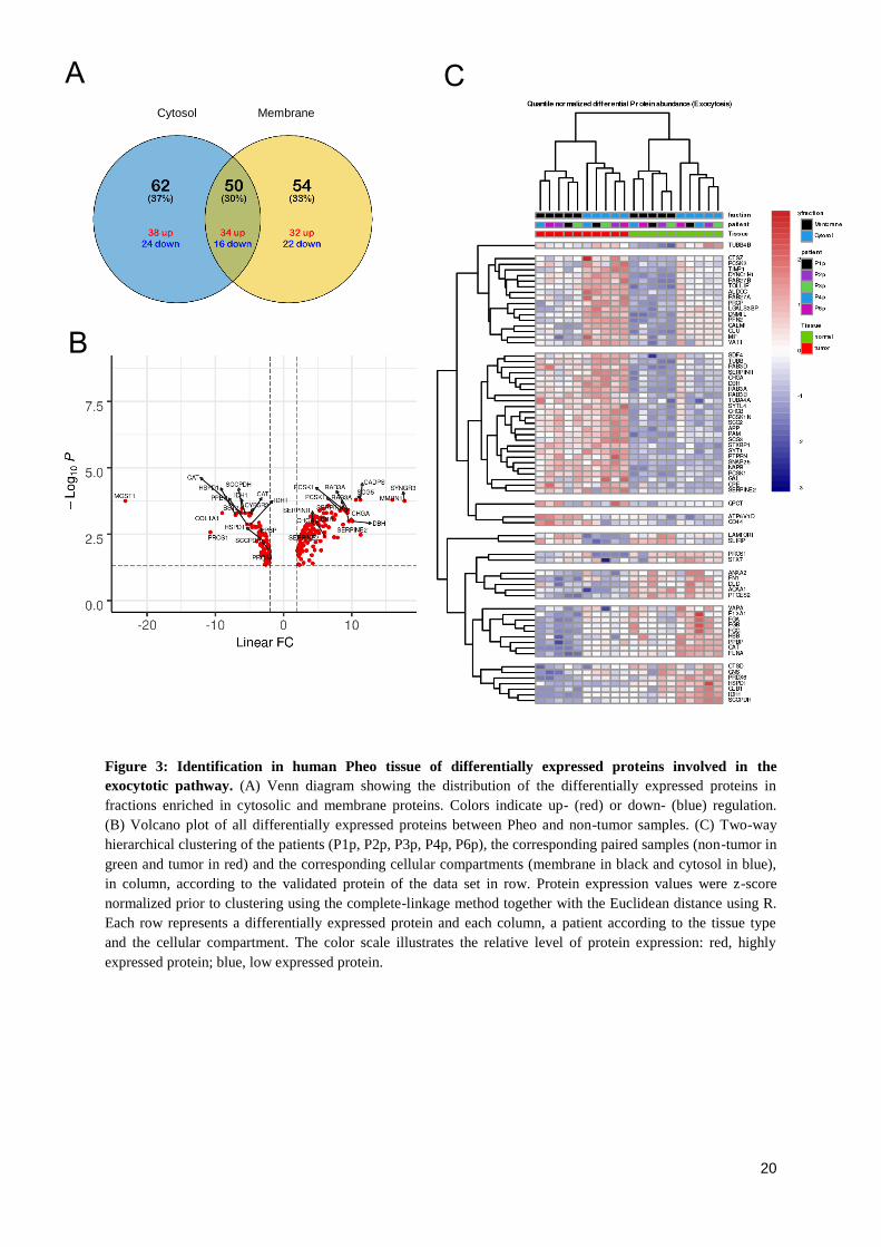
20
Figure 3: Identification in human Pheo tissue of differentially expressed proteins involved in the
exocytotic pathway. (A) Venn diagram showing the distribution of the differentially expressed proteins in
fractions enriched in cytosolic and membrane proteins. Colors indicate up- (red) or down- (blue) regulation.
(B) Volcano plot of all differentially expressed proteins between Pheo and non-tumor samples. (C) Two-way
hierarchical clustering of the patients (P1p, P2p, P3p, P4p, P6p), the corresponding paired samples (non-tumor in
green and tumor in red) and the corresponding cellular compartments (membrane in black and cytosol in blue),
in column, according to the validated protein of the data set in row. Protein expression values were z-score
normalized prior to clustering using the complete-linkage method together with the Euclidean distance using R.
Each row represents a differentially expressed protein and each column, a patient according to the tissue type
and the cellular compartment. The color scale illustrates the relative level of protein expression: red, highly
expressed protein; blue, low expressed protein.
A
B
C
Cytosol Membrane
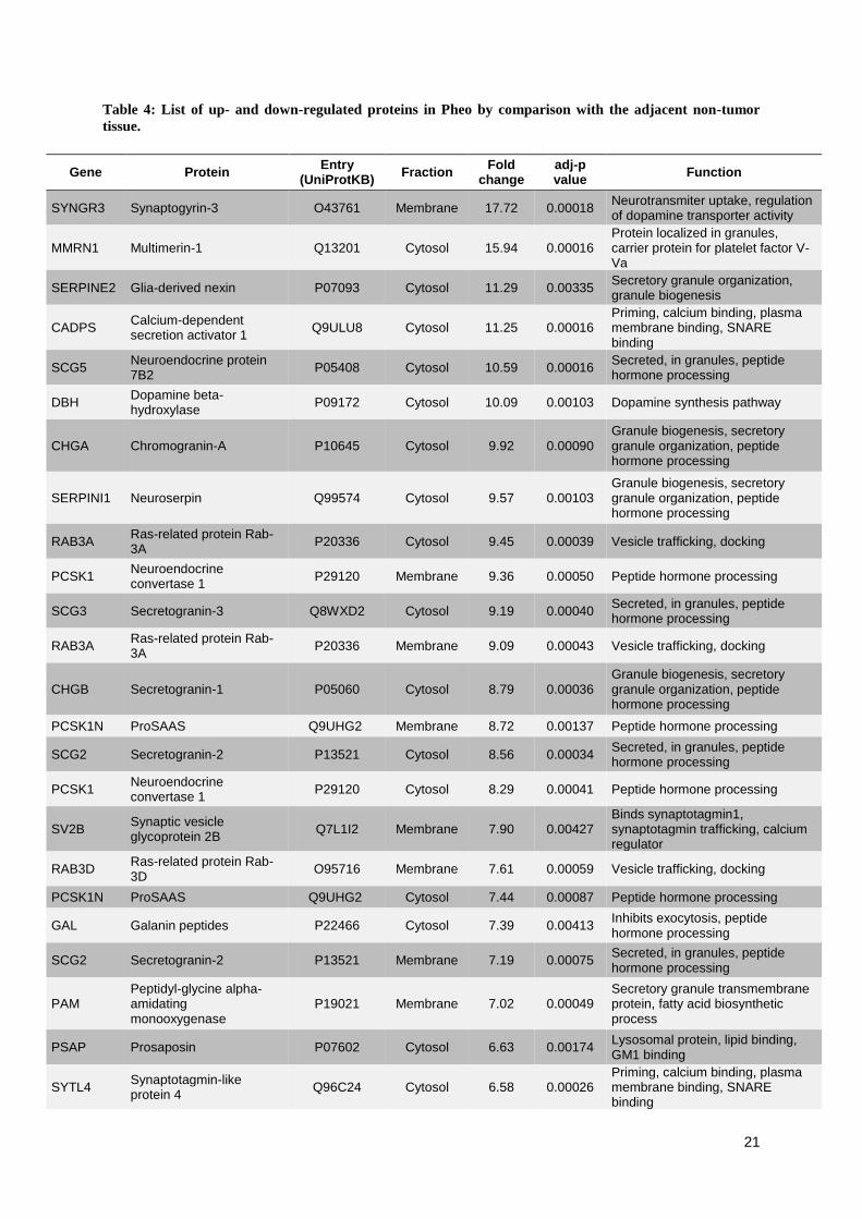
21
Table 4: List of up- and down-regulated proteins in Pheo by comparison with the adjacent non-tumor
tissue.
Gene Protein Entry
(UniProtKB) Fraction
Fold change
adj-p value
Function
SYNGR3 Synaptogyrin-3 O43761 Membrane 17.72 0.00018 Neurotransmiter uptake, regulation of dopamine transporter activity
MMRN1 Multimerin-1 Q13201 Cytosol 15.94 0.00016 Protein localized in granules, carrier protein for platelet factor V-Va
SERPINE2 Glia-derived nexin P07093 Cytosol 11.29 0.00335 Secretory granule organization, granule biogenesis
CADPS Calcium-dependent secretion activator 1
Q9ULU8 Cytosol 11.25 0.00016 Priming, calcium binding, plasma membrane binding, SNARE binding
SCG5 Neuroendocrine protein 7B2
P05408 Cytosol 10.59 0.00016 Secreted, in granules, peptide hormone processing
DBH Dopamine beta-hydroxylase
P09172 Cytosol 10.09 0.00103 Dopamine synthesis pathway
CHGA Chromogranin-A P10645 Cytosol 9.92 0.00090 Granule biogenesis, secretory granule organization, peptide hormone processing
SERPINI1 Neuroserpin Q99574 Cytosol 9.57 0.00103 Granule biogenesis, secretory granule organization, peptide hormone processing
RAB3A Ras-related protein Rab-3A
P20336 Cytosol 9.45 0.00039 Vesicle trafficking, docking
PCSK1 Neuroendocrine convertase 1
P29120 Membrane 9.36 0.00050 Peptide hormone processing
SCG3 Secretogranin-3 Q8WXD2 Cytosol 9.19 0.00040 Secreted, in granules, peptide hormone processing
RAB3A Ras-related protein Rab-3A
P20336 Membrane 9.09 0.00043 Vesicle trafficking, docking
CHGB Secretogranin-1 P05060 Cytosol 8.79 0.00036 Granule biogenesis, secretory granule organization, peptide hormone processing
PCSK1N ProSAAS Q9UHG2 Membrane 8.72 0.00137 Peptide hormone processing
SCG2 Secretogranin-2 P13521 Cytosol 8.56 0.00034 Secreted, in granules, peptide hormone processing
PCSK1 Neuroendocrine convertase 1
P29120 Cytosol 8.29 0.00041 Peptide hormone processing
SV2B Synaptic vesicle glycoprotein 2B
Q7L1I2 Membrane 7.90 0.00427 Binds synaptotagmin1, synaptotagmin trafficking, calcium regulator
RAB3D Ras-related protein Rab-3D
O95716 Membrane 7.61 0.00059 Vesicle trafficking, docking
PCSK1N ProSAAS Q9UHG2 Cytosol 7.44 0.00087 Peptide hormone processing
GAL Galanin peptides P22466 Cytosol 7.39 0.00413 Inhibits exocytosis, peptide hormone processing
SCG2 Secretogranin-2 P13521 Membrane 7.19 0.00075 Secreted, in granules, peptide hormone processing
PAM Peptidyl-glycine alpha-amidating monooxygenase
P19021 Membrane 7.02 0.00049 Secretory granule transmembrane protein, fatty acid biosynthetic process
PSAP Prosaposin P07602 Cytosol 6.63 0.00174 Lysosomal protein, lipid binding, GM1 binding
SYTL4 Synaptotagmin-like protein 4
Q96C24 Cytosol 6.58 0.00026 Priming, calcium binding, plasma membrane binding, SNARE binding
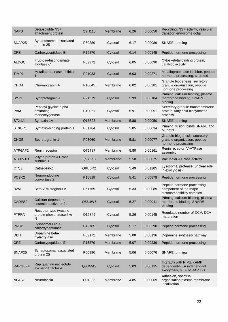
22
NAPB Beta-soluble NSF attachment protein
Q9H115 Membrane 6.26 0.00050 Recycling, NSF activity, vesicular transport endosome golgi
SNAP25 Synaptosomal-associated protein 25
P60880 Cytosol 6.17 0.00089 SNARE, priming
CPE Carboxypeptidase E P16870 Cytosol 6.14 0.00145 Peptide hormone processing
ALDOC Fructose-bisphosphate aldolase C
P09972 Cytosol 6.05 0.00090 Cytoskeletal binding protein, catalytic activity
TIMP1 Metalloproteinase inhibitor 1
P01033 Cytosol 6.03 0.00271 Metalloproteinase inhibitor, peptide hormone processing, secreted
CHGA Chromogranin-A P10645 Membrane 6.02 0.00381 Granule biogenesis, secretory granule organization, peptide hormone processing
SYT1 Synaptotagmin-1 P21579 Cytosol 5.93 0.00104 Priming, calcium binding, plasma membrane binding, SNARE binding
PAM Peptidyl-glycine alpha-amidating monooxygenase
P19021 Cytosol 5.91 0.00051 Secretory granule transmembrane protein, fatty acid biosynthetic process
STX1A Syntaxin-1A Q16623 Membrane 5.88 0.00050 SNARE, priming
STXBP1 Syntaxin-binding protein 1 P61764 Cytosol 5.85 0.00034 Priming, fusion, binds SNARE and Munc13
CHGB Secretogranin-1 P05060 Membrane 5.81 0.00077 Granule biogenesis, secretory granule organization, peptide hormone processing
ATP6AP2 Renin receptor O75787 Membrane 5.80 0.00161 Renin receptor, V-ATPase assembly
ATP6V1D V-type proton ATPase subunit D
Q9Y5K8 Membrane 5.50 0.00075 Vacuaolar ATPase activity
CTSZ Cathepsin-Z Q9UBR2 Cytosol 5.49 0.01285 Lysosomal protease (unclear role in exocytosis)
PCSK2 Neuroendocrine convertase 2
P16519 Cytosol 5.41 0.00578 Peptide hormone processing
B2M Beta-2-microglobulin P61769 Cytosol 5.33 0.00089 Peptide hormone processing, component of the major histocompatibility complex
CADPS2 Calcium-dependent secretion activator 2
Q86UW7 Cytosol 5.27 0.00041 Priming, calcium binding, plasma membrane binding, SNARE binding
PTPRN Receptor-type tyrosine-protein phosphatase-like N
Q16849 Cytosol 5.26 0.00145 Regulates number of DCV, DCV maturation
PRCP Lysosomal Pro-X carboxypeptidase
P42785 Cytosol 5.17 0.00290 Peptide hormone processing
DBH Dopamine beta-hydroxylase
P09172 Membrane 5.08 0.00136 Dopamine synthesis pathway
CPE Carboxypeptidase E P16870 Membrane 5.07 0.00239 Peptide hormone processing
SNAP25 Synaptosomal-associated protein 25
P60880 Membrane 5.06 0.00076 SNARE, priming
RAPGEF4 Rap guanine nucleotide exchange factor 4
Q8WZA2 Cytosol 5.03 0.00115 Interacts with RIM2, cAMP dependent-PKA independant exocytosis, GEF of RAP 1-3
NFASC Neurofascin O94856 Membrane 4.85 0.00069 Adhesion, spectrin-organisation,plasma membrane localization
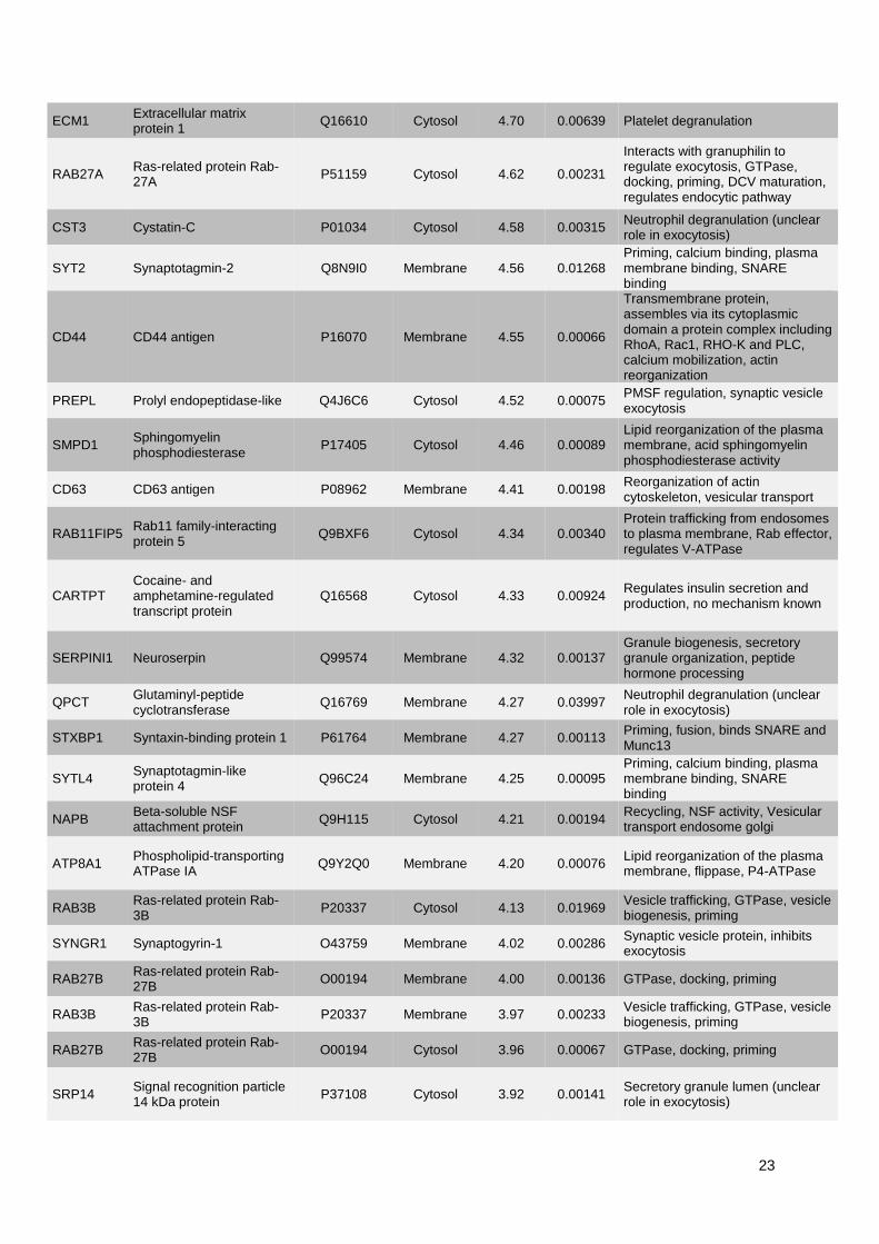
23
ECM1 Extracellular matrix protein 1
Q16610 Cytosol 4.70 0.00639 Platelet degranulation
RAB27A Ras-related protein Rab-27A
P51159 Cytosol 4.62 0.00231
Interacts with granuphilin to regulate exocytosis, GTPase, docking, priming, DCV maturation, regulates endocytic pathway
CST3 Cystatin-C P01034 Cytosol 4.58 0.00315 Neutrophil degranulation (unclear role in exocytosis)
SYT2 Synaptotagmin-2 Q8N9I0 Membrane 4.56 0.01268 Priming, calcium binding, plasma membrane binding, SNARE binding
CD44 CD44 antigen P16070 Membrane 4.55 0.00066
Transmembrane protein, assembles via its cytoplasmic domain a protein complex including RhoA, Rac1, RHO-K and PLC, calcium mobilization, actin reorganization
PREPL Prolyl endopeptidase-like Q4J6C6 Cytosol 4.52 0.00075 PMSF regulation, synaptic vesicle exocytosis
SMPD1 Sphingomyelin phosphodiesterase
P17405 Cytosol 4.46 0.00089 Lipid reorganization of the plasma membrane, acid sphingomyelin phosphodiesterase activity
CD63 CD63 antigen P08962 Membrane 4.41 0.00198 Reorganization of actin cytoskeleton, vesicular transport
RAB11FIP5 Rab11 family-interacting protein 5
Q9BXF6 Cytosol 4.34 0.00340 Protein trafficking from endosomes to plasma membrane, Rab effector, regulates V-ATPase
CARTPT Cocaine- and amphetamine-regulated transcript protein
Q16568 Cytosol 4.33 0.00924 Regulates insulin secretion and production, no mechanism known
SERPINI1 Neuroserpin Q99574 Membrane 4.32 0.00137 Granule biogenesis, secretory granule organization, peptide hormone processing
QPCT Glutaminyl-peptide cyclotransferase
Q16769 Membrane 4.27 0.03997 Neutrophil degranulation (unclear role in exocytosis)
STXBP1 Syntaxin-binding protein 1 P61764 Membrane 4.27 0.00113 Priming, fusion, binds SNARE and Munc13
SYTL4 Synaptotagmin-like protein 4
Q96C24 Membrane 4.25 0.00095 Priming, calcium binding, plasma membrane binding, SNARE binding
NAPB Beta-soluble NSF attachment protein
Q9H115 Cytosol 4.21 0.00194 Recycling, NSF activity, Vesicular transport endosome golgi
ATP8A1 Phospholipid-transporting ATPase IA
Q9Y2Q0 Membrane 4.20 0.00076 Lipid reorganization of the plasma membrane, flippase, P4-ATPase
RAB3B Ras-related protein Rab-3B
P20337 Cytosol 4.13 0.01969 Vesicle trafficking, GTPase, vesicle biogenesis, priming
SYNGR1 Synaptogyrin-1 O43759 Membrane 4.02 0.00286 Synaptic vesicle protein, inhibits exocytosis
RAB27B Ras-related protein Rab-27B
O00194 Membrane 4.00 0.00136 GTPase, docking, priming
RAB3B Ras-related protein Rab-3B
P20337 Membrane 3.97 0.00233 Vesicle trafficking, GTPase, vesicle biogenesis, priming
RAB27B Ras-related protein Rab-27B
O00194 Cytosol 3.96 0.00067 GTPase, docking, priming
SRP14 Signal recognition particle 14 kDa protein
P37108 Cytosol 3.92 0.00141 Secretory granule lumen (unclear role in exocytosis)
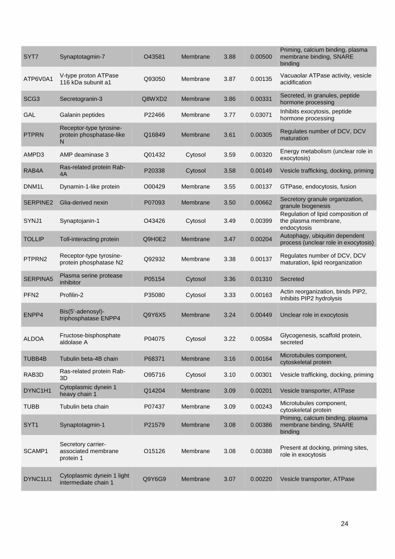
24
SYT7 Synaptotagmin-7 O43581 Membrane 3.88 0.00500 Priming, calcium binding, plasma membrane binding, SNARE binding
ATP6V0A1 V-type proton ATPase 116 kDa subunit a1
Q93050 Membrane 3.87 0.00135 Vacuaolar ATPase activity, vesicle acidification
SCG3 Secretogranin-3 Q8WXD2 Membrane 3.86 0.00331 Secreted, in granules, peptide hormone processing
GAL Galanin peptides P22466 Membrane 3.77 0.03071 Inhibits exocytosis, peptide hormone processing
PTPRN Receptor-type tyrosine-protein phosphatase-like N
Q16849 Membrane 3.61 0.00305 Regulates number of DCV, DCV maturation
AMPD3 AMP deaminase 3 Q01432 Cytosol 3.59 0.00320 Energy metabolism (unclear role in exocytosis)
RAB4A Ras-related protein Rab-4A
P20338 Cytosol 3.58 0.00149 Vesicle trafficking, docking, priming
DNM1L Dynamin-1-like protein O00429 Membrane 3.55 0.00137 GTPase, endocytosis, fusion
SERPINE2 Glia-derived nexin P07093 Membrane 3.50 0.00662 Secretory granule organization, granule biogenesis
SYNJ1 Synaptojanin-1 O43426 Cytosol 3.49 0.00399 Regulation of lipid composition of the plasma membrane, endocytosis
TOLLIP Toll-interacting protein Q9H0E2 Membrane 3.47 0.00204 Autophagy, ubiquitin dependent process (unclear role in exocytosis)
PTPRN2 Receptor-type tyrosine-protein phosphatase N2
Q92932 Membrane 3.38 0.00137 Regulates number of DCV, DCV maturation, lipid reorganization
SERPINA5 Plasma serine protease inhibitor
P05154 Cytosol 3.36 0.01310 Secreted
PFN2 Profilin-2 P35080 Cytosol 3.33 0.00163 Actin reorganization, binds PIP2, Inhibits PIP2 hydrolysis
ENPP4 Bis(5'-adenosyl)-triphosphatase ENPP4
Q9Y6X5 Membrane 3.24 0.00449 Unclear role in exocytosis
ALDOA Fructose-bisphosphate aldolase A
P04075 Cytosol 3.22 0.00584 Glycogenesis, scaffold protein, secreted
TUBB4B Tubulin beta-4B chain P68371 Membrane 3.16 0.00164 Microtubules component, cytoskeletal protein
RAB3D Ras-related protein Rab-3D
O95716 Cytosol 3.10 0.00301 Vesicle trafficking, docking, priming
DYNC1H1 Cytoplasmic dynein 1 heavy chain 1
Q14204 Membrane 3.09 0.00201 Vesicle transporter, ATPase
TUBB Tubulin beta chain P07437 Membrane 3.09 0.00243 Microtubules component, cytoskeletal protein
SYT1 Synaptotagmin-1 P21579 Membrane 3.08 0.00386 Priming, calcium binding, plasma membrane binding, SNARE binding
SCAMP1 Secretory carrier-associated membrane protein 1
O15126 Membrane 3.08 0.00388 Present at docking, priming sites, role in exocytosis
DYNC1LI1 Cytoplasmic dynein 1 light intermediate chain 1
Q9Y6G9 Membrane 3.07 0.00220 Vesicle transporter, ATPase
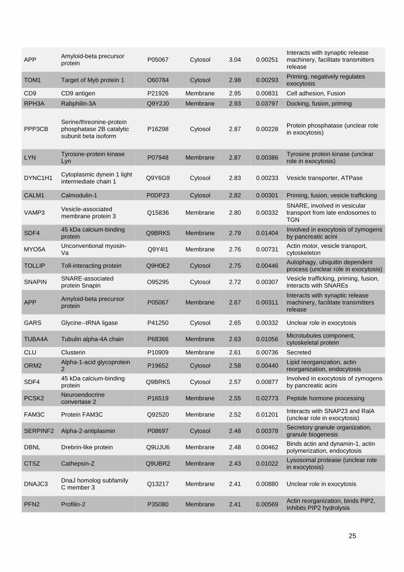
25
APP Amyloid-beta precursor protein
P05067 Cytosol 3.04 0.00251 Interacts with synaptic release machinery, facilitate transmitters release
TOM1 Target of Myb protein 1 O60784 Cytosol 2.98 0.00293 Priming, negatively regulates exocytosis
CD9 CD9 antigen P21926 Membrane 2.95 0.00831 Cell adhesion, Fusion
RPH3A Rabphilin-3A Q9Y2J0 Membrane 2.93 0.03797 Docking, fusion, priming
PPP3CB Serine/threonine-protein phosphatase 2B catalytic subunit beta isoform
P16298 Cytosol 2.87 0.00228 Protein phosphatase (unclear role in exocytosis)
LYN Tyrosine-protein kinase Lyn
P07948 Membrane 2.87 0.00386 Tyrosine protein kinase (unclear role in exocytosis)
DYNC1H1 Cytoplasmic dynein 1 light intermediate chain 1
Q9Y6G9 Cytosol 2.83 0.00233 Vesicle transporter, ATPase
CALM1 Calmodulin-1 P0DP23 Cytosol 2.82 0.00301 Priming, fusion, vesicle trafficking
VAMP3 Vesicle-associated membrane protein 3
Q15836 Membrane 2.80 0.00332 SNARE, involved in vesicular transport from late endosomes to TGN
SDF4 45 kDa calcium-binding protein
Q9BRK5 Membrane 2.79 0.01404 Involved in exocytosis of zymogens by pancreatic acini
MYO5A Unconventional myosin-Va
Q9Y4I1 Membrane 2.76 0.00731 Actin motor, vesicle transport, cytoskeleton
TOLLIP Toll-interacting protein Q9H0E2 Cytosol 2.75 0.00446 Autophagy, ubiquitin dependent process (unclear role in exocytosis)
SNAPIN SNARE-associated protein Snapin
O95295 Cytosol 2.72 0.00307 Vesicle trafficking, priming, fusion, interacts with SNAREs
APP Amyloid-beta precursor protein
P05067 Membrane 2.67 0.00311 Interacts with synaptic release machinery, facilitate transmitters release
GARS Glycine--tRNA ligase P41250 Cytosol 2.65 0.00332 Unclear role in exocytosis
TUBA4A Tubulin alpha-4A chain P68366 Membrane 2.63 0.01056 Microtubules component, cytoskeletal protein
CLU Clusterin P10909 Membrane 2.61 0.00736 Secreted
ORM2 Alpha-1-acid glycoprotein 2
P19652 Cytosol 2.58 0.00440 Lipid reorganization, actin reorganization, endocytosis
SDF4 45 kDa calcium-binding protein
Q9BRK5 Cytosol 2.57 0.00877 Involved in exocytosis of zymogens by pancreatic acini
PCSK2 Neuroendocrine convertase 2
P16519 Membrane 2.55 0.02773 Peptide hormone processing
FAM3C Protein FAM3C Q92520 Membrane 2.52 0.01201 Interacts with SNAP23 and RalA (unclear role in exocytosis)
SERPINF2 Alpha-2-antiplasmin P08697 Cytosol 2.48 0.00378 Secretory granule organization, granule biogenesis
DBNL Drebrin-like protein Q9UJU6 Membrane 2.48 0.00462 Binds actin and dynamin-1, actin polymerization, endocytosis
CTSZ Cathepsin-Z Q9UBR2 Membrane 2.43 0.01022 Lysosomal protease (unclear role in exocytosis)
DNAJC3 DnaJ homolog subfamily C member 3
Q13217 Membrane 2.41 0.00880 Unclear role in exocytosis
PFN2 Profilin-2 P35080 Membrane 2.41 0.00569 Actin reorganization, binds PIP2, Inhibits PIP2 hydrolysis
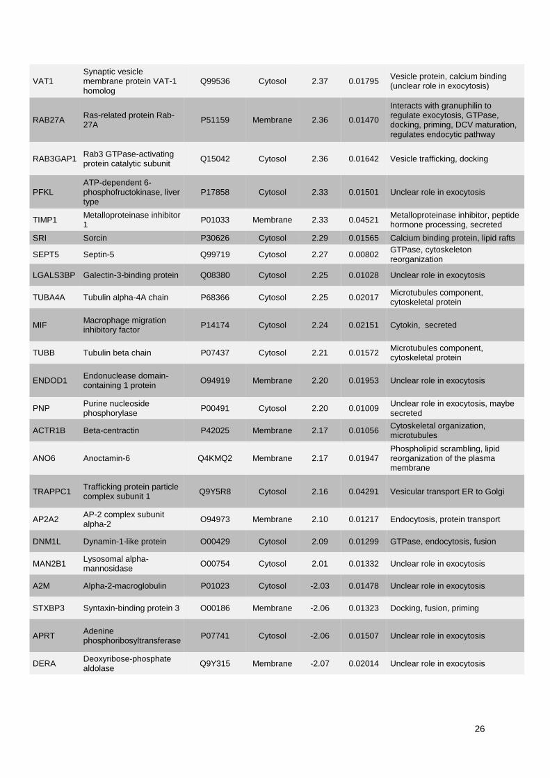
26
VAT1 Synaptic vesicle membrane protein VAT-1 homolog
Q99536 Cytosol 2.37 0.01795 Vesicle protein, calcium binding (unclear role in exocytosis)
RAB27A Ras-related protein Rab-27A
P51159 Membrane 2.36 0.01470
Interacts with granuphilin to regulate exocytosis, GTPase, docking, priming, DCV maturation, regulates endocytic pathway
RAB3GAP1 Rab3 GTPase-activating protein catalytic subunit
Q15042 Cytosol 2.36 0.01642 Vesicle trafficking, docking
PFKL ATP-dependent 6-phosphofructokinase, liver type
P17858 Cytosol 2.33 0.01501 Unclear role in exocytosis
TIMP1 Metalloproteinase inhibitor 1
P01033 Membrane 2.33 0.04521 Metalloproteinase inhibitor, peptide hormone processing, secreted
SRI Sorcin P30626 Cytosol 2.29 0.01565 Calcium binding protein, lipid rafts
SEPT5 Septin-5 Q99719 Cytosol 2.27 0.00802 GTPase, cytoskeleton reorganization
LGALS3BP Galectin-3-binding protein Q08380 Cytosol 2.25 0.01028 Unclear role in exocytosis
TUBA4A Tubulin alpha-4A chain P68366 Cytosol 2.25 0.02017 Microtubules component, cytoskeletal protein
MIF Macrophage migration inhibitory factor
P14174 Cytosol 2.24 0.02151 Cytokin, secreted
TUBB Tubulin beta chain P07437 Cytosol 2.21 0.01572 Microtubules component, cytoskeletal protein
ENDOD1 Endonuclease domain-containing 1 protein
O94919 Membrane 2.20 0.01953 Unclear role in exocytosis
PNP Purine nucleoside phosphorylase
P00491 Cytosol 2.20 0.01009 Unclear role in exocytosis, maybe secreted
ACTR1B Beta-centractin P42025 Membrane 2.17 0.01056 Cytoskeletal organization, microtubules
ANO6 Anoctamin-6 Q4KMQ2 Membrane 2.17 0.01947 Phospholipid scrambling, lipid reorganization of the plasma membrane
TRAPPC1 Trafficking protein particle complex subunit 1
Q9Y5R8 Cytosol 2.16 0.04291 Vesicular transport ER to Golgi
AP2A2 AP-2 complex subunit alpha-2
O94973 Membrane 2.10 0.01217 Endocytosis, protein transport
DNM1L Dynamin-1-like protein O00429 Cytosol 2.09 0.01299 GTPase, endocytosis, fusion
MAN2B1 Lysosomal alpha-mannosidase
O00754 Cytosol 2.01 0.01332 Unclear role in exocytosis
A2M Alpha-2-macroglobulin P01023 Cytosol -2.03 0.01478 Unclear role in exocytosis
STXBP3 Syntaxin-binding protein 3 O00186 Membrane -2.06 0.01323 Docking, fusion, priming
APRT Adenine phosphoribosyltransferase
P07741 Cytosol -2.06 0.01507 Unclear role in exocytosis
DERA Deoxyribose-phosphate aldolase
Q9Y315 Membrane -2.07 0.02014 Unclear role in exocytosis
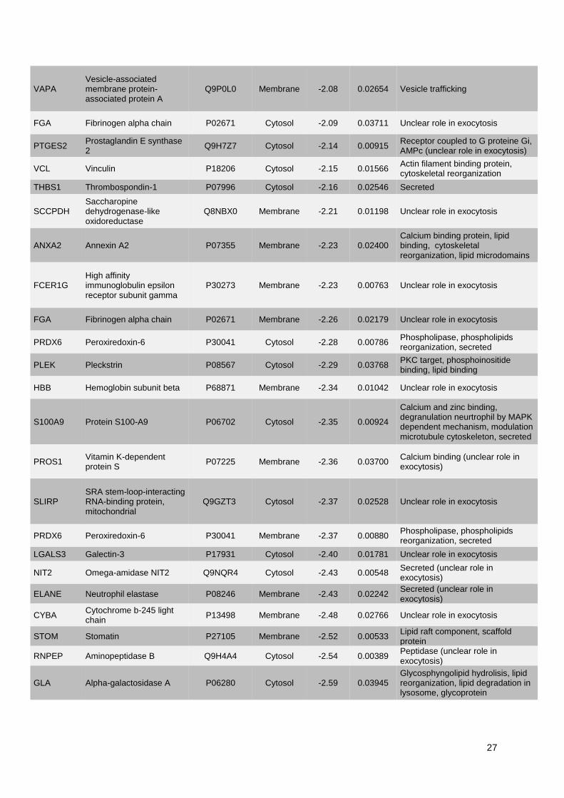
27
VAPA Vesicle-associated membrane protein-associated protein A
Q9P0L0 Membrane -2.08 0.02654 Vesicle trafficking
FGA Fibrinogen alpha chain P02671 Cytosol -2.09 0.03711 Unclear role in exocytosis
PTGES2 Prostaglandin E synthase 2
Q9H7Z7 Cytosol -2.14 0.00915 Receptor coupled to G proteine Gi, AMPc (unclear role in exocytosis)
VCL Vinculin P18206 Cytosol -2.15 0.01566 Actin filament binding protein, cytoskeletal reorganization
THBS1 Thrombospondin-1 P07996 Cytosol -2.16 0.02546 Secreted
SCCPDH Saccharopine dehydrogenase-like oxidoreductase
Q8NBX0 Membrane -2.21 0.01198 Unclear role in exocytosis
ANXA2 Annexin A2 P07355 Membrane -2.23 0.02400 Calcium binding protein, lipid binding, cytoskeletal reorganization, lipid microdomains
FCER1G High affinity immunoglobulin epsilon receptor subunit gamma
P30273 Membrane -2.23 0.00763 Unclear role in exocytosis
FGA Fibrinogen alpha chain P02671 Membrane -2.26 0.02179 Unclear role in exocytosis
PRDX6 Peroxiredoxin-6 P30041 Cytosol -2.28 0.00786 Phospholipase, phospholipids reorganization, secreted
PLEK Pleckstrin P08567 Cytosol -2.29 0.03768 PKC target, phosphoinositide binding, lipid binding
HBB Hemoglobin subunit beta P68871 Membrane -2.34 0.01042 Unclear role in exocytosis
S100A9 Protein S100-A9 P06702 Cytosol -2.35 0.00924
Calcium and zinc binding, degranulation neurtrophil by MAPK dependent mechanism, modulation microtubule cytoskeleton, secreted
PROS1 Vitamin K-dependent protein S
P07225 Membrane -2.36 0.03700 Calcium binding (unclear role in exocytosis)
SLIRP SRA stem-loop-interacting RNA-binding protein, mitochondrial
Q9GZT3 Cytosol -2.37 0.02528 Unclear role in exocytosis
PRDX6 Peroxiredoxin-6 P30041 Membrane -2.37 0.00880 Phospholipase, phospholipids reorganization, secreted
LGALS3 Galectin-3 P17931 Cytosol -2.40 0.01781 Unclear role in exocytosis
NIT2 Omega-amidase NIT2 Q9NQR4 Cytosol -2.43 0.00548 Secreted (unclear role in exocytosis)
ELANE Neutrophil elastase P08246 Membrane -2.43 0.02242 Secreted (unclear role in exocytosis)
CYBA Cytochrome b-245 light chain
P13498 Membrane -2.48 0.02766 Unclear role in exocytosis
STOM Stomatin P27105 Membrane -2.52 0.00533 Lipid raft component, scaffold protein
RNPEP Aminopeptidase B Q9H4A4 Cytosol -2.54 0.00389 Peptidase (unclear role in exocytosis)
GLA Alpha-galactosidase A P06280 Cytosol -2.59 0.03945 Glycosphyngolipid hydrolisis, lipid reorganization, lipid degradation in lysosome, glycoprotein
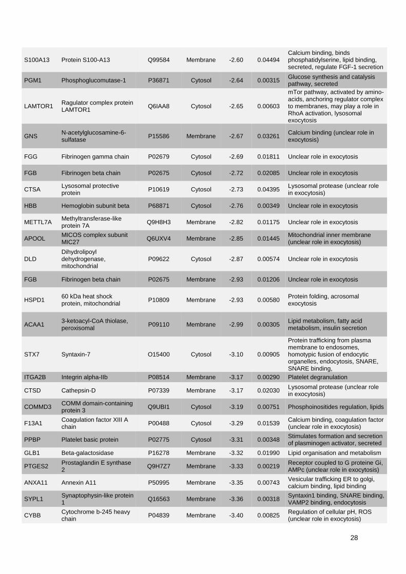
28
S100A13 Protein S100-A13 Q99584 Membrane -2.60 0.04494 Calcium binding, binds phosphatidylserine, lipid binding, secreted, regulate FGF-1 secretion
PGM1 Phosphoglucomutase-1 P36871 Cytosol -2.64 0.00315 Glucose synthesis and catalysis pathway, secreted
LAMTOR1 Ragulator complex protein LAMTOR1
Q6IAA8 Cytosol -2.65 0.00603
mTor pathway, activated by amino-acids, anchoring regulator complex to membranes, may play a role in RhoA activation, lysosomal exocytosis
GNS N-acetylglucosamine-6-sulfatase
P15586 Membrane -2.67 0.03261 Calcium binding (unclear role in exocytosis)
FGG Fibrinogen gamma chain P02679 Cytosol -2.69 0.01811 Unclear role in exocytosis
FGB Fibrinogen beta chain P02675 Cytosol -2.72 0.02085 Unclear role in exocytosis
CTSA Lysosomal protective protein
P10619 Cytosol -2.73 0.04395 Lysosomal protease (unclear role in exocytosis)
HBB Hemoglobin subunit beta P68871 Cytosol -2.76 0.00349 Unclear role in exocytosis
METTL7A Methyltransferase-like protein 7A
Q9H8H3 Membrane -2.82 0.01175 Unclear role in exocytosis
APOOL MICOS complex subunit MIC27
Q6UXV4 Membrane -2.85 0.01445 Mitochondrial inner membrane (unclear role in exocytosis)
DLD Dihydrolipoyl dehydrogenase, mitochondrial
P09622 Cytosol -2.87 0.00574 Unclear role in exocytosis
FGB Fibrinogen beta chain P02675 Membrane -2.93 0.01206 Unclear role in exocytosis
HSPD1 60 kDa heat shock protein, mitochondrial
P10809 Membrane -2.93 0.00580 Protein folding, acrosomal exocytosis
ACAA1 3-ketoacyl-CoA thiolase, peroxisomal
P09110 Membrane -2.99 0.00305 Lipid metabolism, fatty acid metabolism, insulin secretion
STX7 Syntaxin-7 O15400 Cytosol -3.10 0.00905
Protein trafficking from plasma membrane to endosomes, homotypic fusion of endocytic organelles, endocytosis, SNARE, SNARE binding,
ITGA2B Integrin alpha-IIb P08514 Membrane -3.17 0.00290 Platelet degranulation
CTSD Cathepsin-D P07339 Membrane -3.17 0.02030 Lysosomal protease (unclear role in exocytosis)
COMMD3 COMM domain-containing protein 3
Q9UBI1 Cytosol -3.19 0.00751 Phosphoinositides regulation, lipids
F13A1 Coagulation factor XIII A chain
P00488 Cytosol -3.29 0.01539 Calcium binding, coagulation factor (unclear role in exocytosis)
PPBP Platelet basic protein P02775 Cytosol -3.31 0.00348 Stimulates formation and secretion of plasminogen activator, secreted
GLB1 Beta-galactosidase P16278 Membrane -3.32 0.01990 Lipid organisation and metabolism
PTGES2 Prostaglandin E synthase 2
Q9H7Z7 Membrane -3.33 0.00219 Receptor coupled to G proteine Gi, AMPc (unclear role in exocytosis)
ANXA11 Annexin A11 P50995 Membrane -3.35 0.00743 Vesicular trafficking ER to golgi, calcium binding, lipid binding
SYPL1 Synaptophysin-like protein 1
Q16563 Membrane -3.36 0.00318 Syntaxin1 binding, SNARE binding, VAMP2 binding, endocytosis
CYBB Cytochrome b-245 heavy chain
P04839 Membrane -3.40 0.00825 Regulation of cellular pH, ROS (unclear role in exocytosis)
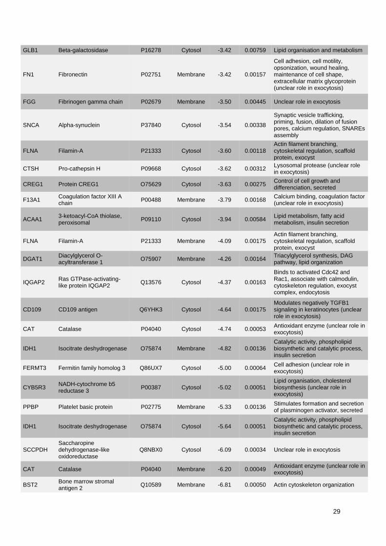
29
GLB1 Beta-galactosidase P16278 Cytosol -3.42 0.00759 Lipid organisation and metabolism
FN1 Fibronectin P02751 Membrane -3.42 0.00157
Cell adhesion, cell motility, opsonization, wound healing, maintenance of cell shape, extracellular matrix glycoprotein (unclear role in exocytosis)
FGG Fibrinogen gamma chain P02679 Membrane -3.50 0.00445 Unclear role in exocytosis
SNCA Alpha-synuclein P37840 Cytosol -3.54 0.00338
Synaptic vesicle trafficking, priming, fusion, dilation of fusion pores, calcium regulation, SNAREs assembly
FLNA Filamin-A P21333 Cytosol -3.60 0.00118 Actin filament branching, cytoskeletal regulation, scaffold protein, exocyst
CTSH Pro-cathepsin H P09668 Cytosol -3.62 0.00312 Lysosomal protease (unclear role in exocytosis)
CREG1 Protein CREG1 O75629 Cytosol -3.63 0.00275 Control of cell growth and differenciation, secreted
F13A1 Coagulation factor XIII A chain
P00488 Membrane -3.79 0.00168 Calcium binding, coagulation factor (unclear role in exocytosis)
ACAA1 3-ketoacyl-CoA thiolase, peroxisomal
P09110 Cytosol -3.94 0.00584 Lipid metabolism, fatty acid metabolism, insulin secretion
FLNA Filamin-A P21333 Membrane -4.09 0.00175 Actin filament branching, cytoskeletal regulation, scaffold protein, exocyst
DGAT1 Diacylglycerol O-acyltransferase 1
O75907 Membrane -4.26 0.00164 Triacylglycerol synthesis, DAG pathway, lipid organization
IQGAP2 Ras GTPase-activating-like protein IQGAP2
Q13576 Cytosol -4.37 0.00163
Binds to activated Cdc42 and Rac1, associate with calmodulin, cytoskeleton regulation, exocyst complex, endocytosis
CD109 CD109 antigen Q6YHK3 Cytosol -4.64 0.00175 Modulates negatively TGFB1 signaling in keratinocytes (unclear role in exocytosis)
CAT Catalase P04040 Cytosol -4.74 0.00053 Antioxidant enzyme (unclear role in exocytosis)
IDH1 Isocitrate deshydrogenase O75874 Membrane -4.82 0.00136 Catalytic activity, phospholipid biosynthetic and catalytic process, insulin secretion
FERMT3 Fermitin family homolog 3 Q86UX7 Cytosol -5.00 0.00064 Cell adhesion (unclear role in exocytosis)
CYB5R3 NADH-cytochrome b5 reductase 3
P00387 Cytosol -5.02 0.00051 Lipid organisation, cholesterol biosynthesis (unclear role in exocytosis)
PPBP Platelet basic protein P02775 Membrane -5.33 0.00136 Stimulates formation and secretion of plasminogen activator, secreted
IDH1 Isocitrate deshydrogenase O75874 Cytosol -5.64 0.00051 Catalytic activity, phospholipid biosynthetic and catalytic process, insulin secretion
SCCPDH Saccharopine dehydrogenase-like oxidoreductase
Q8NBX0 Cytosol -6.09 0.00034 Unclear role in exocytosis
CAT Catalase P04040 Membrane -6.20 0.00049 Antioxidant enzyme (unclear role in exocytosis)
BST2 Bone marrow stromal antigen 2
Q10589 Membrane -6.81 0.00050 Actin cytoskeleton organization
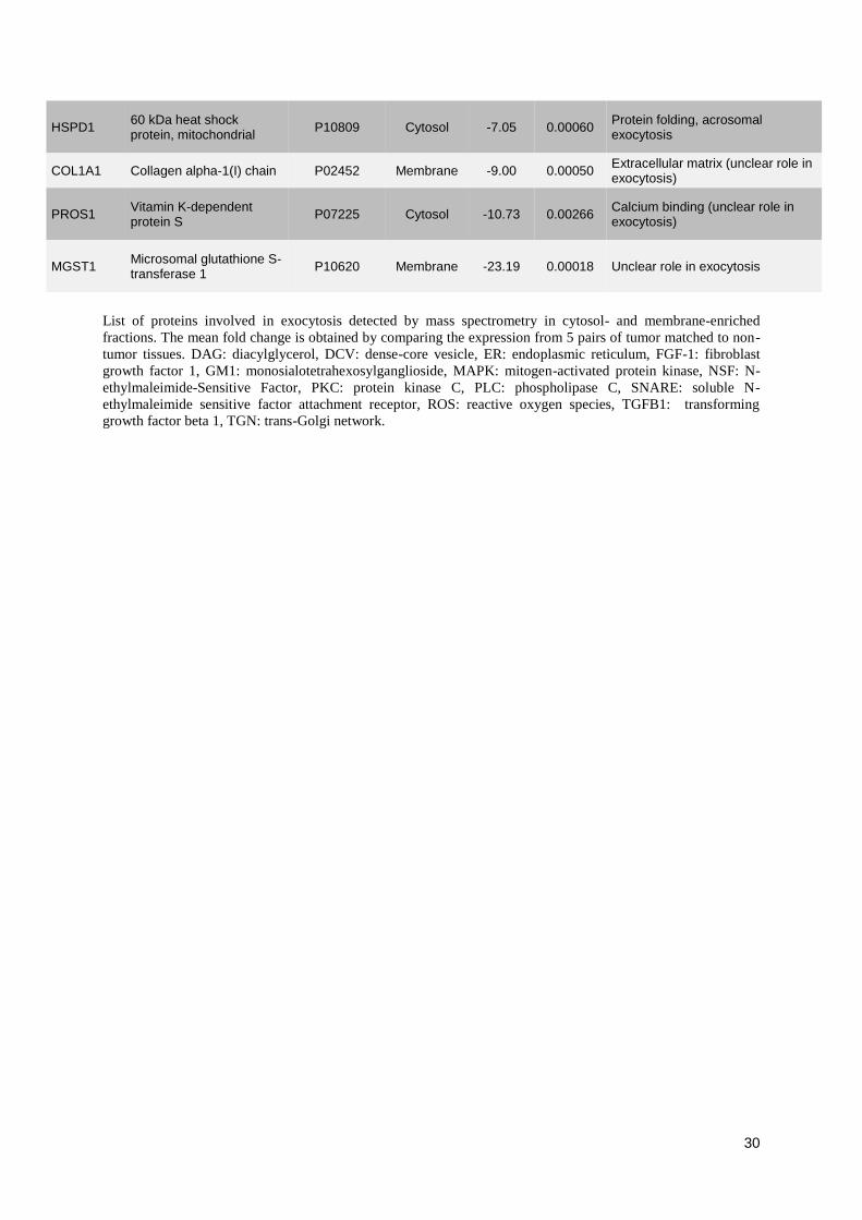
30
HSPD1 60 kDa heat shock protein, mitochondrial
P10809 Cytosol -7.05 0.00060 Protein folding, acrosomal exocytosis
COL1A1 Collagen alpha-1(I) chain P02452 Membrane -9.00 0.00050 Extracellular matrix (unclear role in exocytosis)
PROS1 Vitamin K-dependent protein S
P07225 Cytosol -10.73 0.00266 Calcium binding (unclear role in exocytosis)
MGST1 Microsomal glutathione S-transferase 1
P10620 Membrane -23.19 0.00018 Unclear role in exocytosis
List of proteins involved in exocytosis detected by mass spectrometry in cytosol- and membrane-enriched
fractions. The mean fold change is obtained by comparing the expression from 5 pairs of tumor matched to non-
tumor tissues. DAG: diacylglycerol, DCV: dense-core vesicle, ER: endoplasmic reticulum, FGF-1: fibroblast
growth factor 1, GM1: monosialotetrahexosylganglioside, MAPK: mitogen-activated protein kinase, NSF: N-
ethylmaleimide-Sensitive Factor, PKC: protein kinase C, PLC: phospholipase C, SNARE: soluble N-
ethylmaleimide sensitive factor attachment receptor, ROS: reactive oxygen species, TGFB1: transforming
growth factor beta 1, TGN: trans-Golgi network.
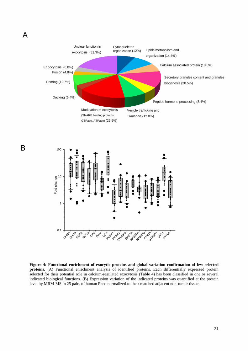
31
Figure 4: Functional enrichment of exocytic proteins and global variation confirmation of few selected
proteins. (A) Functional enrichment analysis of identified proteins. Each differentially expressed protein
selected for their potential role in calcium-regulated exocytosis (Table 4) has been classified in one or several
indicated biological functions. (B) Expression variation of the indicated proteins was quantified at the protein
level by MRM-MS in 25 pairs of human Pheo normalized to their matched adjacent non-tumor tissue.
B
A
Cytosqueleton organization (12%) Lipids metabolism and
organization (14.5%)
Calcium associated protein (10.8%)
Secretory granules content and granules
biogenesis (20.5%)
Peptide hormone processing (8.4%)
Vesicle trafficking and
Transport (12.0%)
Modulation of exocytosis (SNARE binding proteins, GTPase, ATPase) (25.9%)
Docking (5.4%)
Priming (12.7%)
Fusion (4.8%) Endocytosis (6.0%)
Unclear function in
exocytosis (31.3%)
2D Graph 1
Lo
g
0,1
1
10
100
Plot 1 Plot 2
Fo
ld c
hange
0.1
1
10
100
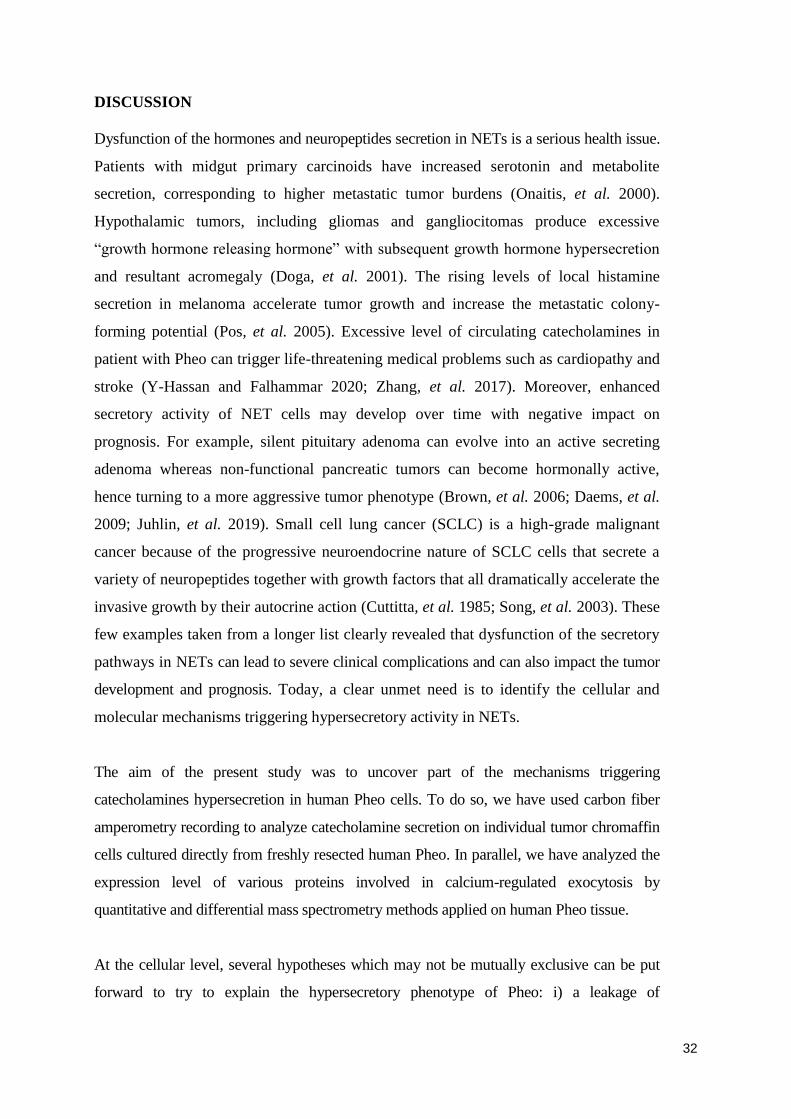
32
DISCUSSION
Dysfunction of the hormones and neuropeptides secretion in NETs is a serious health issue.
Patients with midgut primary carcinoids have increased serotonin and metabolite
secretion, corresponding to higher metastatic tumor burdens (Onaitis, et al. 2000).
Hypothalamic tumors, including gliomas and gangliocitomas produce excessive
“growth hormone releasing hormone” with subsequent growth hormone hypersecretion
and resultant acromegaly (Doga, et al. 2001). The rising levels of local histamine
secretion in melanoma accelerate tumor growth and increase the metastatic colony-
forming potential (Pos, et al. 2005). Excessive level of circulating catecholamines in
patient with Pheo can trigger life-threatening medical problems such as cardiopathy and
stroke (Y-Hassan and Falhammar 2020; Zhang, et al. 2017). Moreover, enhanced
secretory activity of NET cells may develop over time with negative impact on
prognosis. For example, silent pituitary adenoma can evolve into an active secreting
adenoma whereas non-functional pancreatic tumors can become hormonally active,
hence turning to a more aggressive tumor phenotype (Brown, et al. 2006; Daems, et al.
2009; Juhlin, et al. 2019). Small cell lung cancer (SCLC) is a high-grade malignant
cancer because of the progressive neuroendocrine nature of SCLC cells that secrete a
variety of neuropeptides together with growth factors that all dramatically accelerate the
invasive growth by their autocrine action (Cuttitta, et al. 1985; Song, et al. 2003). These
few examples taken from a longer list clearly revealed that dysfunction of the secretory
pathways in NETs can lead to severe clinical complications and can also impact the tumor
development and prognosis. Today, a clear unmet need is to identify the cellular and
molecular mechanisms triggering hypersecretory activity in NETs.
The aim of the present study was to uncover part of the mechanisms triggering
catecholamines hypersecretion in human Pheo cells. To do so, we have used carbon fiber
amperometry recording to analyze catecholamine secretion on individual tumor chromaffin
cells cultured directly from freshly resected human Pheo. In parallel, we have analyzed the
expression level of various proteins involved in calcium-regulated exocytosis by
quantitative and differential mass spectrometry methods applied on human Pheo tissue.
At the cellular level, several hypotheses which may not be mutually exclusive can be put
forward to try to explain the hypersecretory phenotype of Pheo: i) a leakage of
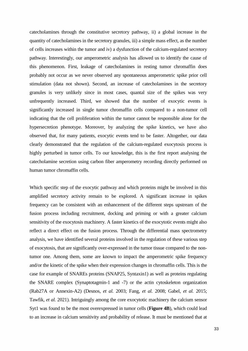
33
catecholamines through the constitutive secretory pathway, ii) a global increase in the
quantity of catecholamines in the secretory granules, iii) a simple mass effect, as the number
of cells increases within the tumor and iv) a dysfunction of the calcium-regulated secretory
pathway. Interestingly, our amperometric analysis has allowed us to identify the cause of
this phenomenon. First, leakage of catecholamines in resting tumor chromaffin does
probably not occur as we never observed any spontaneous amperometric spike prior cell
stimulation (data not shown). Second, an increase of catecholamines in the secretory
granules is very unlikely since in most cases, quantal size of the spikes was very
unfrequently increased. Third, we showed that the number of exocytic events is
significantly increased in single tumor chromaffin cells compared to a non-tumor cell
indicating that the cell proliferation within the tumor cannot be responsible alone for the
hypersecretion phenotype. Moreover, by analyzing the spike kinetics, we have also
observed that, for many patients, exocytic events tend to be faster. Altogether, our data
clearly demonstrated that the regulation of the calcium-regulated exocytosis process is
highly perturbed in tumor cells. To our knowledge, this is the first report analysing the
catecholamine secretion using carbon fiber amperometry recording directly performed on
human tumor chromaffin cells.
Which specific step of the exocytic pathway and which proteins might be involved in this
amplified secretory activity remain to be explored. A significant increase in spikes
frequency can be consistent with an enhancement of the different steps upstream of the
fusion process including recruitment, docking and priming or with a greater calcium
sensitivity of the exocytosis machinery. A faster kinetics of the exocytotic events might also
reflect a direct effect on the fusion process. Through the differential mass spectrometry
analysis, we have identified several proteins involved in the regulation of these various step
of exocytosis, that are significantly over-expressed in the tumor tissue compared to the non-
tumor one. Among them, some are known to impact the amperometric spike frequency
and/or the kinetic of the spike when their expression changes in chromaffin cells. This is the
case for example of SNAREs proteins (SNAP25, Syntaxin1) as well as proteins regulating
the SNARE complex (Synaptotagmin-1 and -7) or the actin cytoskeleton organization
(Rab27A or Annexin-A2) (Desnos, et al. 2003; Fang, et al. 2008; Gabel, et al. 2015;
Tawfik, et al. 2021). Intriguingly among the core exocytotic machinery the calcium sensor
Syt1 was found to be the most overexpressed in tumor cells (Figure 4B), which could lead
to an increase in calcium sensitivity and probability of release. It must be mentioned that at
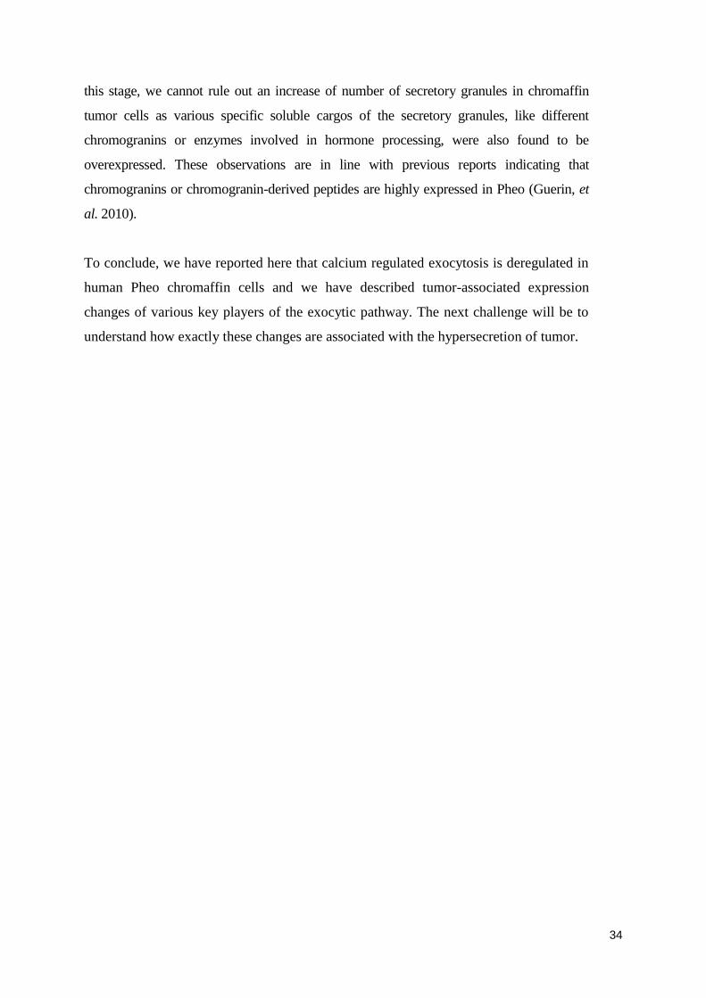
34
this stage, we cannot rule out an increase of number of secretory granules in chromaffin
tumor cells as various specific soluble cargos of the secretory granules, like different
chromogranins or enzymes involved in hormone processing, were also found to be
overexpressed. These observations are in line with previous reports indicating that
chromogranins or chromogranin-derived peptides are highly expressed in Pheo (Guerin, et
al. 2010).
To conclude, we have reported here that calcium regulated exocytosis is deregulated in
human Pheo chromaffin cells and we have described tumor-associated expression
changes of various key players of the exocytic pathway. The next challenge will be to
understand how exactly these changes are associated with the hypersecretion of tumor.
35
Ethics approval and consent to participate
The present study used the data and the human biological material of the biological collection
“Approche moléculaire des tumeurs corticosurrénaliennes” which was agreed by the “Comité
de Protection des Personnes Est III” ethical advisory committee, and was conducted according
to currently accepted ethical guidelines, including informed written consent approval signed
by all patients prior to inclusion.
Funding and acknowledgements
This work was financially supported by ITMO Cancer AVIESAN (Alliance Nationale pour
les Sciences de la Vie et de la Santé, National Alliance for Life Sciences & Health) within the
framework of the Cancer Plan to SG and LB (Single Cell 2018 N° 19CS004-00); by the
University of Strasbourg Institute for Advanced Study (USIAS) for a Fellowship, within the
French national programme “Investment for the future” (IdEx-Unistra) to SG; by grants from
the Agence Nationale pour la Recherche (“SecretoNET”, N° ANR-16-CE17-0022-01) and
from the Ligue contre le Cancer (CCIR Grand-Est) to SG; by a fellowship from la Fondation
pour la Recherche Médicale (FRM; FDM201806005916) to SM. INSERM is providing salary
to SG and NV.
Conflict of interest
The authors declare that there is no conflict of interest that could be perceived as prejudicing
the impartiality of the research reported.
36
REFERENCES
Anantharam A & Kreutzberger AJB 2019 Unraveling the mechanisms of calcium-dependent
secretion. J Gen Physiol 151 417-434.
Bader MF, Holz RW, Kumakura K & Vitale N 2002 Exocytosis: the chromaffin cell as a
model system. Ann N Y Acad Sci 971 178-183.
Brown RL, Muzzafar T, Wollman R & Weiss RE 2006 A pituitary carcinoma secreting TSH
and prolactin: a non-secreting adenoma gone awry. Eur J Endocrinol 154 639-643.
Burgoyne RD & Morgan A 2003 Secretory granule exocytosis. Physiol Rev 83 581-632.
Croise P, Houy S, Gand M, Lanoix J, Calco V, Toth P, Brunaud L, Lomazzi S, Paramithiotis
E, Chelsky D, et al. 2016 Cdc42 and Rac1 activity is reduced in human
pheochromocytoma and correlates with FARP1 and ARHGEF1 expression. Endocr Relat
Cancer 23 281-293.
Cuttitta F, Carney DN, Mulshine J, Moody TW, Fedorko J, Fischler A & Minna JD 1985
Autocrine growth factors in human small cell lung cancer. Cancer Surv 4 707-727.
Daems T, Verhelst J, Michotte A, Abrams P, De Ridder D & Abs R 2009 Modification of
hormonal secretion in clinically silent pituitary adenomas. Pituitary 12 80-86.
Desnos C, Schonn JS, Huet S, Tran VS, El-Amraoui A, Raposo G, Fanget I, Chapuis C,
Menasche G, de Saint Basile G, et al. 2003 Rab27A and its effector MyRIP link secretory
granules to F-actin and control their motion towards release sites. J Cell Biol 163 559-570.
Doga M, Bonadonna S, Burattin A & Giustina A 2001 Ectopic secretion of growth hormone-
releasing hormone (GHRH) in neuroendocrine tumors: relevant clinical aspects. Ann Oncol
12 Suppl 2 S89-94.
Eisenhofer G, Lenders JW, Goldstein DS, Mannelli M, Csako G, Walther MM, Brouwers FM
& Pacak K 2005 Pheochromocytoma catecholamine phenotypes and prediction of tumor
size and location by use of plasma free metanephrines. Clin Chem 51 735-744.
Fang Q, Berberian K, Gong LW, Hafez I, Sorensen JB & Lindau M 2008 The role of the C
terminus of the SNARE protein SNAP-25 in fusion pore opening and a model for fusion
pore mechanics. Proc Natl Acad Sci U S A 105 15388-15392.
Fathali H & Cans AS 2018 Amperometry methods for monitoring vesicular quantal size and
regulation of exocytosis release. Pflugers Arch 470 125-134.
Gabel M, Delavoie F, Demais V, Royer C, Bailly Y, Vitale N, Bader MF & Chasserot-Golaz
S 2015 Annexin A2-dependent actin bundling promotes secretory granule docking to the
plasma membrane and exocytosis. J Cell Biol 210 785-800.
Gasman S & Vitale N 2017 Lipid remodelling in neuroendocrine secretion. Biol Cell 109 381-
390.
Guerin M, Guillemot J, Thouennon E, Pierre A, El-Yamani FZ, Montero-Hadjadje M,
Dubessy C, Magoul R, Lihrmann I, Anouar Y, et al. 2010 Granins and their derived
peptides in normal and tumoral chromaffin tissue: Implications for the diagnosis and
prognosis of pheochromocytoma. Regul Pept 165 21-29.
Houy S, Estay-Ahumada C, Croise P, Calco V, Haeberle AM, Bailly Y, Billuart P, Vitale N,
Bader MF, Ory S, et al. 2015 Oligophrenin-1 Connects Exocytotic Fusion to
Compensatory Endocytosis in Neuroendocrine Cells. J Neurosci 35 11045-11055.
Juhlin CC, Skoglund S, Juntti-Berggren L, Karlberg M & Calissendorff J 2019 Non-
functioning neuroendocrine pancreatic tumors transforming to malignant insulinomas -
four cases and review of the literature. Neuro Endocrinol Lett 40 175-183.
Keshishian H, Addona T, Burgess M, Kuhn E & Carr SA 2007 Quantitative, multiplexed
assays for low abundance proteins in plasma by targeted mass spectrometry and stable
isotope dilution. Mol Cell Proteomics 6 2212-2229.
37
Lang T & Jahn R 2008 Core proteins of the secretory machinery. Handb Exp Pharmacol 107-
127.
Lenders JW, Duh QY, Eisenhofer G, Gimenez-Roqueplo AP, Grebe SK, Murad MH, Naruse
M, Pacak K, Young WF, Jr. & Endocrine S 2014 Pheochromocytoma and paraganglioma:
an endocrine society clinical practice guideline. J Clin Endocrinol Metab 99 1915-1942.
Lenders JWM, Kerstens MN, Amar L, Prejbisz A, Robledo M, Taieb D, Pacak K, Crona J,
Zelinka T, Mannelli M, et al. 2020 Genetics, diagnosis, management and future directions
of research of phaeochromocytoma and paraganglioma: a position statement and consensus
of the Working Group on Endocrine Hypertension of the European Society of
Hypertension. J Hypertens 38 1443-1456.
Malacombe M, Bader MF & Gasman S 2006 Exocytosis in neuroendocrine cells: new tasks
for actin. Biochim Biophys Acta 1763 1175-1183.
Moog S, Houy S, Chevalier E, Ory S, Weryha G, Rame M, Klein M, Brunaud L, Gasman S &
Cuny T 2018 18F-FDOPA PET/CT Uptake Parameters Correlate with Catecholamine
Secretion in Human Pheochromocytomas. Neuroendocrinology 107 228-236.
Mosharov EV & Sulzer D 2005 Analysis of exocytotic events recorded by amperometry. Nat
Methods 2 651-658.
Onaitis MW, Kirshbom PM, Hayward TZ, Quayle FJ, Feldman JM, Seigler HF & Tyler DS
2000 Gastrointestinal carcinoids: characterization by site of origin and hormone
production. Ann Surg 232 549-556.
Pappachan JM, Tun NN, Arunagirinathan G, Sodi R & Hanna FWF 2018
Pheochromocytomas and Hypertension. Curr Hypertens Rep 20 3.
Pos Z, Safrany G, Muller K, Toth S, Falus A & Hegyesi H 2005 Phenotypic profiling of
engineered mouse melanomas with manipulated histamine production identifies histamine
H2 receptor and rho-C as histamine-regulated melanoma progression markers. Cancer Res
65 4458-4466.
Pourian M, Mostafazadeh DB & Soltani A 2015 Does this patient have pheochromocytoma?
A systematic review of clinical signs and symptoms. J Diabetes Metab Disord 15 11.
Segura F, Brioso MA, Gomez JF, Machado JD & Borges R 2000 Automatic analysis for
amperometrical recordings of exocytosis. J Neurosci Methods 103 151-156.
Song P, Sekhon HS, Jia Y, Keller JA, Blusztajn JK, Mark GP & Spindel ER 2003
Acetylcholine is synthesized by and acts as an autocrine growth factor for small cell lung
carcinoma. Cancer Res 63 214-221.
Tanguy E, Coste de Bagneaux P, Kassas N, Ammar MR, Wang Q, Haeberle AM,
Raherindratsara J, Fouillen L, Renard PY, Montero-Hadjadje M, et al. 2020 Mono- and
Poly-unsaturated Phosphatidic Acid Regulate Distinct Steps of Regulated Exocytosis in
Neuroendocrine Cells. Cell Rep 32 108026.
Tawfik B, Martins JS, Houy S, Imig C, Pinheiro PS, Wojcik SM, Brose N, Cooper BH &
Sorensen JB 2021 Synaptotagmin-7 places dense-core vesicles at the cell membrane to
promote Munc13-2- and Ca(2+)-dependent priming. Elife 10.
Thompson LD 2002 Pheochromocytoma of the Adrenal gland Scaled Score (PASS) to
separate benign from malignant neoplasms: a clinicopathologic and immunophenotypic
study of 100 cases. Am J Surg Pathol 26 551-566.
Y-Hassan S & Falhammar H 2020 Cardiovascular Manifestations and Complications of
Pheochromocytomas and Paragangliomas. J Clin Med 9.
Zandee WT, Kamp K, van Adrichem RC, Feelders RA & de Herder WW 2017 Effect of
hormone secretory syndromes on neuroendocrine tumor prognosis. Endocr Relat Cancer
24 R261-R274.
Zhang R, Gupta D & Albert SG 2017 Pheochromocytoma as a reversible cause of
cardiomyopathy: Analysis and review of the literature. Int J Cardiol 249 319-323.
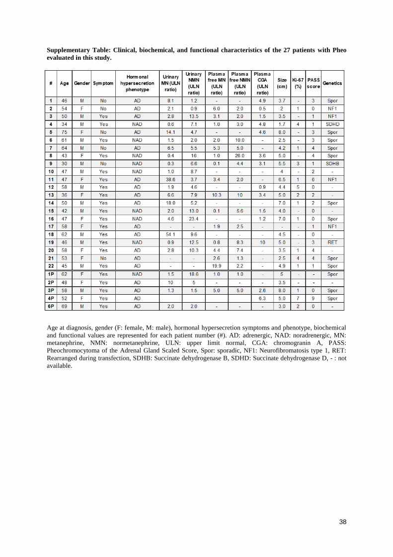
38
Supplementary Table: Clinical, biochemical, and functional characteristics of the 27 patients with Pheo
evaluated in this study.
Age at diagnosis, gender (F: female, M: male), hormonal hypersecretion symptoms and phenotype, biochemical
and functional values are represented for each patient number (#). AD: adrenergic, NAD: noradrenergic, MN:
metanephrine, NMN: normetanephrine, ULN: upper limit normal, CGA: chromogranin A, PASS:
Pheochromocytoma of the Adrenal Gland Scaled Score, Spor: sporadic, NF1: Neurofibromatosis type 1, RET:
Rearranged during transfection, SDHB: Succinate dehydrogenase B, SDHD: Succinate dehydrogenase D, - : not
available.
Résultats
55
2. Publication n°2 : Effet des analogues de la somatostatine sur la
sécrétion de catécholamines par les cellules de phéochromocytomes
humains
2.1. Objectifs et déroulement de l’étude
Aujourd’hui il n’existe aucune thérapie ciblée permettant de bloquer
directement cette hypersécrétion dans le phéochromocytome. Par leurs effets
inhibiteurs connus sur les voies de sécrétion et leurs applications cliniques sur
plusieurs tumeurs neuroendocrines, l’octréotide et le pasiréotide (SOM230), deux
analogues de la somatostatine apparaissent comme des candidats de choix pour
tenter d’inhiber la sécrétion incontrôlée du phéochromocytome. En effet, l’effet anti-
sécrétoire de ces molécules a été démontré par différentes études cliniques et sur
plusieurs tumeurs neuroendocrines (Mazziotti et al., 2017; Vitale et al., 2018;
Lamberts and Hofland, 2019). Cependant, leur impact sur la sécrétion des cellules
chromaffines tumorales n’a jamais été étudié.
L’objectif de cette étude fut donc de tester l’effet potentiel de l'octréotide et du
SOM230 sur l’activité sécrétrice des cellules chromaffines de phéochromocytomes
humains, et d’en étudier le mécanisme. Étant donné le caractère rare et précieux des
échantillons humains, j’ai tout d’abord utilisé des cultures primaires de cellules
chromaffines bovines afin de mettre en évidence un effet anti-sécrétoire potentiel de
ces analogues. J’ai ainsi réalisé des courbes dose-réponse pour trouver une
concentration efficace. J’ai poursuivi cette étude en utilisant la technique
d’ampérométrie à fibre de carbone pour mesurer la sécrétion de catécholamines sur
des cultures primaires de cellules chromaffines humaines directement cultivées à
partir de phéochromocytomes humains. Cette approche m’a permis de mesurer
précisément, à l’échelle unicellulaire, le nombre d'évènements d'exocytose, la
quantité de catécholamines libérées ainsi que divers paramètres cinétiques du
processus d’exocytose. Enfin, grâce à une collaboration avec Sylvain Hugel,
chercheur dans l’institut, nous avons tenté de mettre en évidence le mécanisme
d’action de ces drogues en utilisant des techniques d’imagerie calcique et
d’électrophysiologie.
Résultats
56
2.2. Résultats
Les résultats obtenus sont exposés dans l’article intitulé « Somatostatin
analogue pasireotide (SOM230) inhibits catecholamine secretion in human
pheochromocytoma cells » que je signe en premier auteur et que nous avons très
récemment soumis au journal « Cancer Letters » :
Dans cet article j’ai réalisé l’ensemble des expériences excepté l’imagerie
calcique et les enregistrements des courants nicotiniques. Des résultats
complémentaires à cette étude sont également présentés dans la partie discussion
pages 67 et 70.
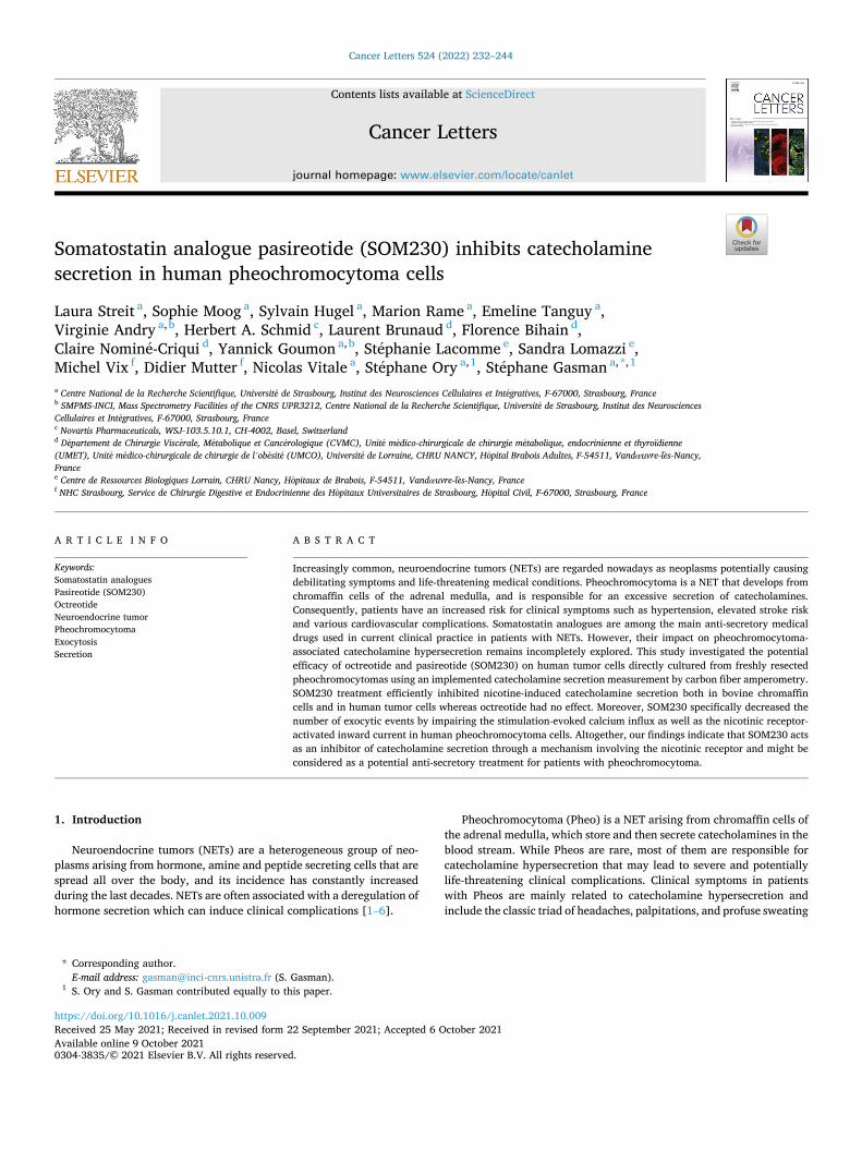
Cancer Letters 524 (2022) 232–244
Available online 9 October 20210304-3835/© 2021 Elsevier B.V. All rights reserved.
Somatostatin analogue pasireotide (SOM230) inhibits catecholamine secretion in human pheochromocytoma cells
Laura Streit a, Sophie Moog a, Sylvain Hugel a, Marion Rame a, Emeline Tanguy a, Virginie Andry a,b, Herbert A. Schmid c, Laurent Brunaud d, Florence Bihain d, Claire Nomine-Criqui d, Yannick Goumon a,b, Stephanie Lacomme e, Sandra Lomazzi e, Michel Vix f, Didier Mutter f, Nicolas Vitale a, Stephane Ory a,1, Stephane Gasman a,*,1
a Centre National de la Recherche Scientifique, Universite de Strasbourg, Institut des Neurosciences Cellulaires et Integratives, F-67000, Strasbourg, France b SMPMS-INCI, Mass Spectrometry Facilities of the CNRS UPR3212, Centre National de la Recherche Scientifique, Universite de Strasbourg, Institut des Neurosciences Cellulaires et Integratives, F-67000, Strasbourg, France c Novartis Pharmaceuticals, WSJ-103.5.10.1, CH-4002, Basel, Switzerland d Departement de Chirurgie Viscerale, Metabolique et Cancerologique (CVMC), Unite medico-chirurgicale de chirurgie metabolique, endocrinienne et thyroïdienne (UMET), Unite medico-chirurgicale de chirurgie de l’obesite (UMCO), Universite de Lorraine, CHRU NANCY, Hopital Brabois Adultes, F-54511, Vandœuvre-les-Nancy, France e Centre de Ressources Biologiques Lorrain, CHRU Nancy, Hopitaux de Brabois, F-54511, Vandœuvre-les-Nancy, France f NHC Strasbourg, Service de Chirurgie Digestive et Endocrinienne des Hopitaux Universitaires de Strasbourg, Hopital Civil, F-67000, Strasbourg, France
A R T I C L E I N F O
Keywords: Somatostatin analogues Pasireotide (SOM230) Octreotide Neuroendocrine tumor Pheochromocytoma Exocytosis Secretion
A B S T R A C T
Increasingly common, neuroendocrine tumors (NETs) are regarded nowadays as neoplasms potentially causing debilitating symptoms and life-threatening medical conditions. Pheochromocytoma is a NET that develops from chromaffin cells of the adrenal medulla, and is responsible for an excessive secretion of catecholamines. Consequently, patients have an increased risk for clinical symptoms such as hypertension, elevated stroke risk and various cardiovascular complications. Somatostatin analogues are among the main anti-secretory medical drugs used in current clinical practice in patients with NETs. However, their impact on pheochromocytoma- associated catecholamine hypersecretion remains incompletely explored. This study investigated the potential efficacy of octreotide and pasireotide (SOM230) on human tumor cells directly cultured from freshly resected pheochromocytomas using an implemented catecholamine secretion measurement by carbon fiber amperometry. SOM230 treatment efficiently inhibited nicotine-induced catecholamine secretion both in bovine chromaffin cells and in human tumor cells whereas octreotide had no effect. Moreover, SOM230 specifically decreased the number of exocytic events by impairing the stimulation-evoked calcium influx as well as the nicotinic receptor- activated inward current in human pheochromocytoma cells. Altogether, our findings indicate that SOM230 acts as an inhibitor of catecholamine secretion through a mechanism involving the nicotinic receptor and might be considered as a potential anti-secretory treatment for patients with pheochromocytoma.
1. Introduction
Neuroendocrine tumors (NETs) are a heterogeneous group of neo-plasms arising from hormone, amine and peptide secreting cells that are spread all over the body, and its incidence has constantly increased during the last decades. NETs are often associated with a deregulation of hormone secretion which can induce clinical complications [1–6].
Pheochromocytoma (Pheo) is a NET arising from chromaffin cells of the adrenal medulla, which store and then secrete catecholamines in the blood stream. While Pheos are rare, most of them are responsible for catecholamine hypersecretion that may lead to severe and potentially life-threatening clinical complications. Clinical symptoms in patients with Pheos are mainly related to catecholamine hypersecretion and include the classic triad of headaches, palpitations, and profuse sweating
* Corresponding author. E-mail address: [email protected] (S. Gasman).
1 S. Ory and S. Gasman contributed equally to this paper.
Contents lists available at ScienceDirect
Cancer Letters
journal homepage: www.elsevier.com/locate/canlet
https://doi.org/10.1016/j.canlet.2021.10.009 Received 25 May 2021; Received in revised form 22 September 2021; Accepted 6 October 2021
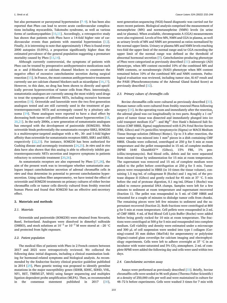
Cancer Letters 524 (2022) 232–244
233
but also permanent or paroxysmal hypertension [7–9]. It has been also reported that Pheo can lead to severe acute cardiovascular complica-tions including myocarditis, Takotsubo syndrome, as well as various forms of cardiomyopathies [10,11]. Accordingly, a retrospective study has shown that patients with Pheo have a 14-fold higher rate of car-diovascular events than patients with essential hypertension [12]. Finally, it is interesting to note that approximately 1 Pheo is found every 2000 autopsies (0.05%), a proportion significantly higher than the estimated prevalence of the general population suggesting a premature mortality caused by Pheo [13,14].
Although currently controversial, the symptoms of patients with Pheo can be treated by preoperative antihypertensive medications such as α- and β-blockers or calcium channel blockers to counteract the negative effect of excessive catecholamine secretion during surgical resection [15]. In France, the most common antihypertensive treatments currently use are calcium channel blockers such as nicardipine [16,17]. However, to this date, no drug has been shown to directly and specif-ically prevent hypersecretion of tumor cells from Pheo. Interestingly, somatostatin analogues are currently among the most widely used drugs to treat the symptoms of different NETs, including excessive hormone secretion [18]. Octreotide and lanreotide were the two first-generation analogues tested and are still currently used in the treatment of gas-troenteropancreatic NETs and acromegaly caused by a pituitary ade-noma [19]. These analogues were reported to have a dual effect by decreasing both tumor cell proliferation and tumor hypersecretion [18, 20,21]. In the early 2000s, a new generation of somatostatin analogues has emerged with the development of pasireotide (SOM230). While octreotide binds preferentially the somatostatin receptor SSR2, SOM230 is a multireceptor-targeted analogue with a 40-, 30- and 5-fold higher affinity than octreotide for somatostatin receptors SSR5, SSR1 and SSR3, respectively [22,23]. For instance, SOM230 has been authorized for Cushing disease and acromegaly treatments [24,25]. In vitro and in vivo data have also shown that this analog is able to effectively inhibit gas-troenteropancreatic NETs secretion and improve symptoms in patients refractory to octreotide treatment [20,26].
As somatostatin receptors are also expressed by Pheo [27,28], the aim of the present work was to investigate whether somatostatin ana-logues impact catecholamine secretion of adrenal chromaffin cells in vitro and thus determine its potential to prevent catecholamine hyper-secretion. Using carbon fiber amperometry, we have tested the effect of octreotide and SOM230 treatments on primary cultures of either bovine chromaffin cells or tumor cells directly cultured from freshly resected human Pheos and found that SOM230 has an effective anti-secretory effect.
2. Materials and methods
2.1. Materials
Octreotide and pasireotide (SOM230) were obtained from Novartis, Basel, Switzerland. Analogues were dissolved in dimethyl sulfoxide (DMSO), and stock solutions at 10− 2 or 10− 3 M were stored at − 20 ◦C and protected from light exposure.
2.2. Patient population
The medical files of patients with Pheo in 2 French centers between 2017 and 2021 were retrospectively reviewed. We collected the following data: initial diagnosis, including a clinical examination look-ing for hormonal-related symptoms and biological analysis. As recom-mended by the Endocrine Society clinical practice guideline published in 2014 [29], Pheo genetic testing was proposed to identify germline mutations in the major susceptibility genes (SDHB, SDHC, SDHD, VHL, NF1, RET, TMEM127, MAX) using Sanger sequencing and multiplex ligation-dependent probe amplification (MLPA). Then, as recommended in the consensus statement published in 2017 [30],
next-generation-sequencing (NGS)-based diagnostic was carried out for more recent patients. Biological analysis comprised the measurement of metanephrine (MN) and normetanephrine (NMN) levels (in urine and/or plasma). When available, chromogranin A (CGA) measurements were also registered. Levels of free MN, NMN and CGA in plasma, as well as urinary levels of MN and NMN are presented as ratios normalized by the normal upper limits. Urinary or plasma MN and NMN levels reaching two-fold the upper limit of the normal range and/or CGA exceeding the upper limit of the normal range was defined as the threshold of abnormal hormonal secretion [7]. Catecholamine-producing phenotype of Pheo were categorized as previously described [31]: adrenergic (AD) phenotype, when MN content exceeded 10% of the combined MN and NMN contents, or noradrenergic (NAD) phenotype when MN content remained below 10% of the combined MN and NMN contents. Patho-logical evaluation was reviewed, including tumor size, Ki-67 result and the PASS (Pheochromocytoma of the Adrenal Gland Scaled Score) as previously described [32].
2.3. Primary culture of chromaffin cells
Bovine chromaffin cells were cultured as previously described [33]. Human tumor cells were cultured from freshly resected Pheos following surgery [34]. In the operating room and immediately after the resection, the adrenal gland was cut longitudinally in two parts. Roughly a 1 cm3
piece of tumor tissue was dissected and immediately plunged into ice cold transport medium (Ca2+- and Mg2+-free Hank’s Balanced Salt So-lution (CMF HBSS, Sigma) supplemented with 0.2% Fetal Bovine Serum (FBS, Gibco) and 1% penicillin/streptomycin (Sigma) or MACS Medium Tissue Storage solution (Miltenyi Biotec). Up to 3 h after resection, the tumor sample was minced into 1 mm3 pieces in a dish containing CMF HBSS. Chunks were collected, centrifuged at 250 g for 5 min at room temperature and the pellet resuspended in 15 mL of complete medium (RPMI 1640 GlutaMAX™ (Gibco), 15% FBS, 1% pen-icillin/streptomycin). Red blood cells, debris and fat were separated from minced tissue by sedimentation for 15 min at room temperature. The supernatant was removed and 15 mL of complete medium were added to the pellet before centrifugation at 250 g for 5 min. Tumor pieces were resuspended in HBSS (in 10 times the tissue volume), con-taining 1.5 mg/mL of collagenase B (Roche) and 1 mg/mL of the pro-tease dispase II (Gibco) and gently rocked for 45 min at 37 ◦C. 5 min before the end of protease digestion, 0.1 mg/mL DNase I (Roche) was added to remove potential DNA clumps. Samples were left for a few minutes to sediment at room temperature and supernatant recovered (fraction 1). The pellet was resuspended in 5 mL of CMF HBSS and triturated for a couple of minutes to dislodge tumor cells from chunks. The remaining pieces were left few minutes to sediment and the su-pernatant recovered (fraction 2). Both fractions were centrifuged at 800 g for 5 min at room temperature. Cell pellets were resuspended in 2 mL of CMF HBSS. 4 mL of Red Blood Cell Lysis Buffer (Roche) were added before being gently rocked for 10 min at room temperature. The frac-tions were centrifuged at 500 g for 5 min and resuspended into complete medium. Cell viability and density were estimated under a microscope and 300 μL of cell suspension were seeded into type I collagen (Cor-ning)-coated 35 mm dishes (MatTek) for amperometry or polylysine (Sigma)-coated glass coverslips for calcium imaging and electrophysi-ology experiments. Cells were left to adhere overnight at 37 ◦C in an incubator with water-saturated and 5% CO2 atmosphere. 2 mL of com-plete RPMI were added the following day and cells were used within two days.
2.4. Catecholamine secretion assay
Assays were performed as previously described [33]. Briefly, bovine chromaffin cells were seeded in 96 well plates (Thermo Fisher Scientific) at a density of 250,000 cells per well and were maintained in culture for 48–72 h before experiments. Cells were washed 3 times for 7 min with
L. Streit et al.
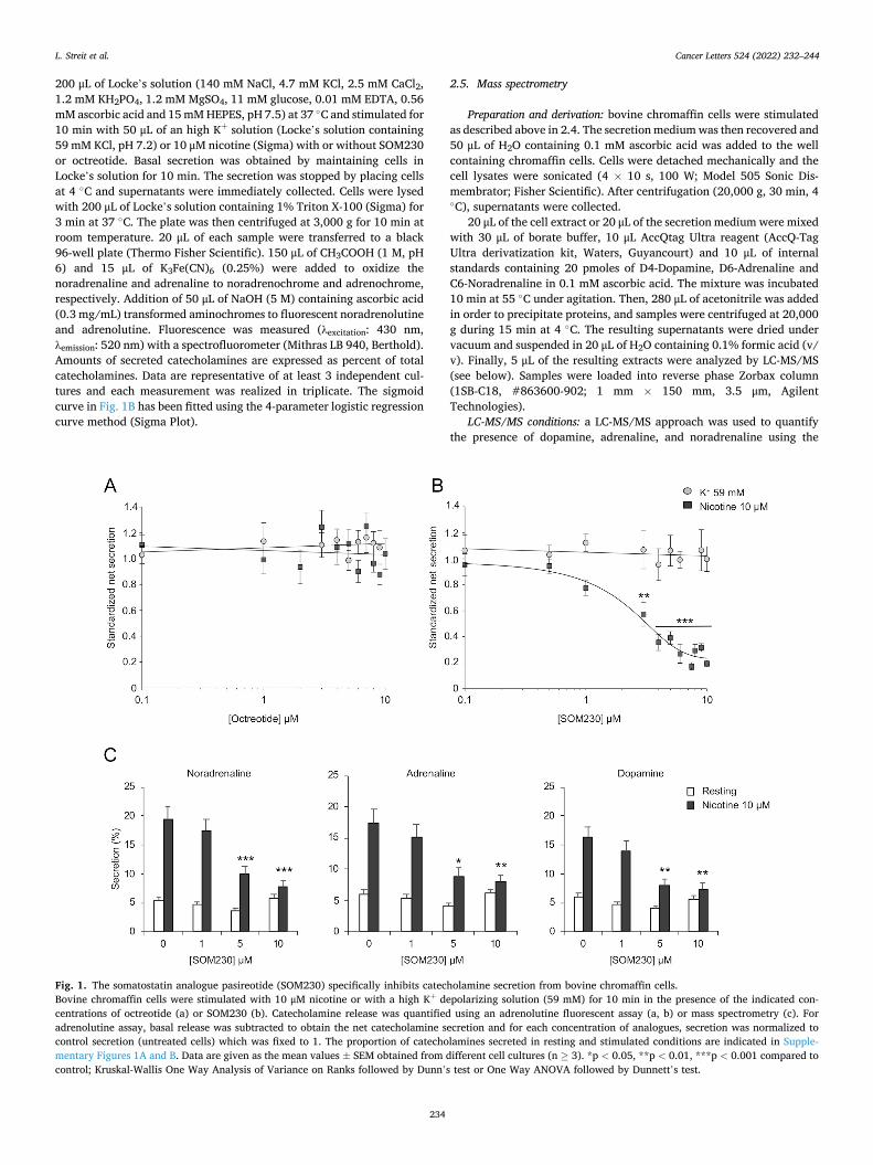
Cancer Letters 524 (2022) 232–244
234
200 μL of Locke’s solution (140 mM NaCl, 4.7 mM KCl, 2.5 mM CaCl2, 1.2 mM KH2PO4, 1.2 mM MgSO4, 11 mM glucose, 0.01 mM EDTA, 0.56 mM ascorbic acid and 15 mM HEPES, pH 7.5) at 37 ◦C and stimulated for 10 min with 50 μL of an high K+ solution (Locke’s solution containing 59 mM KCl, pH 7.2) or 10 μM nicotine (Sigma) with or without SOM230 or octreotide. Basal secretion was obtained by maintaining cells in Locke’s solution for 10 min. The secretion was stopped by placing cells at 4 ◦C and supernatants were immediately collected. Cells were lysed with 200 μL of Locke’s solution containing 1% Triton X-100 (Sigma) for 3 min at 37 ◦C. The plate was then centrifuged at 3,000 g for 10 min at room temperature. 20 μL of each sample were transferred to a black 96-well plate (Thermo Fisher Scientific). 150 μL of CH3COOH (1 M, pH 6) and 15 μL of K3Fe(CN)6 (0.25%) were added to oxidize the noradrenaline and adrenaline to noradrenochrome and adrenochrome, respectively. Addition of 50 μL of NaOH (5 M) containing ascorbic acid (0.3 mg/mL) transformed aminochromes to fluorescent noradrenolutine and adrenolutine. Fluorescence was measured (λexcitation: 430 nm, λemission: 520 nm) with a spectrofluorometer (Mithras LB 940, Berthold). Amounts of secreted catecholamines are expressed as percent of total catecholamines. Data are representative of at least 3 independent cul-tures and each measurement was realized in triplicate. The sigmoid curve in Fig. 1B has been fitted using the 4-parameter logistic regression curve method (Sigma Plot).
2.5. Mass spectrometry
Preparation and derivation: bovine chromaffin cells were stimulated as described above in 2.4. The secretion medium was then recovered and 50 μL of H2O containing 0.1 mM ascorbic acid was added to the well containing chromaffin cells. Cells were detached mechanically and the cell lysates were sonicated (4 × 10 s, 100 W; Model 505 Sonic Dis-membrator; Fisher Scientific). After centrifugation (20,000 g, 30 min, 4 ◦C), supernatants were collected.
20 μL of the cell extract or 20 μL of the secretion medium were mixed with 30 μL of borate buffer, 10 μL AccQtag Ultra reagent (AccQ-Tag Ultra derivatization kit, Waters, Guyancourt) and 10 μL of internal standards containing 20 pmoles of D4-Dopamine, D6-Adrenaline and C6-Noradrenaline in 0.1 mM ascorbic acid. The mixture was incubated 10 min at 55 ◦C under agitation. Then, 280 μL of acetonitrile was added in order to precipitate proteins, and samples were centrifuged at 20,000 g during 15 min at 4 ◦C. The resulting supernatants were dried under vacuum and suspended in 20 μL of H2O containing 0.1% formic acid (v/ v). Finally, 5 μL of the resulting extracts were analyzed by LC-MS/MS (see below). Samples were loaded into reverse phase Zorbax column (1SB-C18, #863600-902; 1 mm × 150 mm, 3.5 μm, Agilent Technologies).
LC-MS/MS conditions: a LC-MS/MS approach was used to quantify the presence of dopamine, adrenaline, and noradrenaline using the
Fig. 1. The somatostatin analogue pasireotide (SOM230) specifically inhibits catecholamine secretion from bovine chromaffin cells. Bovine chromaffin cells were stimulated with 10 μM nicotine or with a high K+ depolarizing solution (59 mM) for 10 min in the presence of the indicated con-centrations of octreotide (a) or SOM230 (b). Catecholamine release was quantified using an adrenolutine fluorescent assay (a, b) or mass spectrometry (c). For adrenolutine assay, basal release was subtracted to obtain the net catecholamine secretion and for each concentration of analogues, secretion was normalized to control secretion (untreated cells) which was fixed to 1. The proportion of catecholamines secreted in resting and stimulated conditions are indicated in Supple-mentary Figures 1A and B. Data are given as the mean values ± SEM obtained from different cell cultures (n ≥ 3). *p < 0.05, **p < 0.01, ***p < 0.001 compared to control; Kruskal-Wallis One Way Analysis of Variance on Ranks followed by Dunn’s test or One Way ANOVA followed by Dunnett’s test.
L. Streit et al.
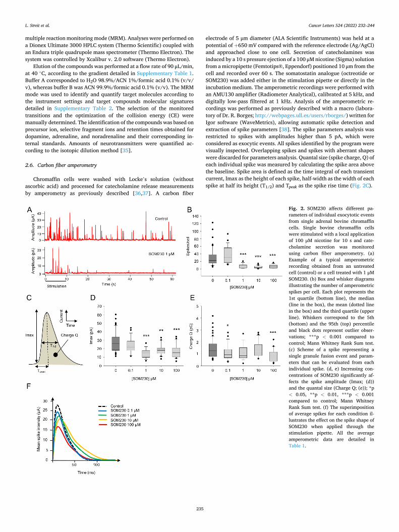
Cancer Letters 524 (2022) 232–244
235
multiple reaction monitoring mode (MRM). Analyses were performed on a Dionex Ultimate 3000 HPLC system (Thermo Scientific) coupled with an Endura triple quadrupole mass spectrometer (Thermo Electron). The system was controlled by Xcalibur v. 2.0 software (Thermo Electron).
Elution of the compounds was performed at a flow rate of 90 μL/min, at 40 ◦C, according to the gradient detailed in Supplementary Table 1. Buffer A corresponded to H2O 98.9%/ACN 1%/formic acid 0.1% (v/v/ v), whereas buffer B was ACN 99.9%/formic acid 0.1% (v/v). The MRM mode was used to identify and quantify target molecules according to the instrument settings and target compounds molecular signatures detailed in Supplementary Table 2. The selection of the monitored transitions and the optimization of the collision energy (CE) were manually determined. The identification of the compounds was based on precursor ion, selective fragment ions and retention times obtained for dopamine, adrenaline, and noradrenaline and their corresponding in-ternal standards. Amounts of neurotransmitters were quantified ac-cording to the isotopic dilution method [35].
2.6. Carbon fiber amperometry
Chromaffin cells were washed with Locke’s solution (without ascorbic acid) and processed for catecholamine release measurements by amperometry as previously described [36,37]. A carbon fiber
electrode of 5 μm diameter (ALA Scientific Instruments) was held at a potential of +650 mV compared with the reference electrode (Ag/AgCl) and approached close to one cell. Secretion of catecholamines was induced by a 10 s pressure ejection of a 100 μM nicotine (Sigma) solution from a micropipette (Femtotips®, Eppendorf) positioned 10 μm from the cell and recorded over 60 s. The somatostatin analogue (octreotide or SOM230) was added either in the stimulation pipette or directly in the incubation medium. The amperometric recordings were performed with an AMU130 amplifier (Radiometer Analytical), calibrated at 5 kHz, and digitally low-pass filtered at 1 kHz. Analysis of the amperometric re-cordings was performed as previously described with a macro (labora-tory of Dr. R. Borges; http://webpages.ull.es/users/rborges/) written for Igor software (WaveMetrics), allowing automatic spike detection and extraction of spike parameters [38]. The spike parameters analysis was restricted to spikes with amplitudes higher than 5 pA, which were considered as exocytic events. All spikes identified by the program were visually inspected. Overlapping spikes and spikes with aberrant shapes were discarded for parameters analysis. Quantal size (spike charge, Q) of each individual spike was measured by calculating the spike area above the baseline. Spike area is defined as the time integral of each transient current, Imax as the height of each spike, half-width as the width of each spike at half its height (T1/2) and Tpeak as the spike rise time (Fig. 2C).
Fig. 2. SOM230 affects different pa-rameters of individual exocytotic events from single adrenal bovine chromaffin cells. Single bovine chromaffin cells were stimulated with a local application of 100 μM nicotine for 10 s and cate-cholamine secretion was monitored using carbon fiber amperometry. (a) Example of a typical amperometric recording obtained from an untreated cell (control) or a cell treated with 1 μM SOM230. (b) Box and whisker diagrams illustrating the number of amperometric spikes per cell. Each plot represents the 1st quartile (bottom line), the median (line in the box), the mean (dotted line in the box) and the third quartile (upper line). Whiskers correspond to the 5th (bottom) and the 95th (top) percentile and black dots represent outlier obser-vations; ***p < 0.001 compared to control; Mann Whitney Rank Sum test. (c) Scheme of a spike representing a single granule fusion event and param-eters that can be evaluated from each individual spike. (d, e) Increasing con-centrations of SOM230 significantly af-fects the spike amplitude (Imax; (d)) and the quantal size (Charge Q; (e)); *p < 0.05, **p < 0.01, ***p < 0.001 compared to control; Mann Whitney Rank Sum test. (f) The superimposition of average spikes for each condition il-lustrates the effect on the spike shape of SOM230 when applied through the stimulation pipette. All the average amperometric data are detailed in Table 1.
L. Streit et al.
Cancer Letters 524 (2022) 232–244
236
2.7. Intracellular calcium imaging
Changes in the intracellular Ca2+ concentration were detected by the ratiometric fluorescent probe Fura-2. Cells were loaded during a 1 h incubation with 2 μM of the cell-permeant precursor Fura-2 acetox-ymethyl ester (Fura-2AM; Molecular Probes) and 0.001% (w/v) pluronic acid (Molecular Probes). The recording saline solution contained 135 mM NaCl, 5 mM KCl, 2 mM CaCl2, 2 mM MgCl2, 10 mM glucose and 10 mM HEPES, pH 7.4. Fluorescence measurements were performed on an inverted microscope (Axiovert 35; Zeiss) with an oil immersion 40 Nikon objective (Fluor 40, NA 1.30) and a cooled CCD camera (Cool-Snap HQ; Photometrics). The Imaging Workbench 4.0 software (Axon Instruments) was used for image acquisition. Fluorescence was excited alternately at 350 and 380 nm with a Lambda-10 filter wheel (Sutter Instruments), and emitted light was collected above 520 nm. Pairs of images were acquired every 2 s. Intracellular calcium level is expressed throughout as the fluorescence ratio F350 ⁄ F380, calculated after back-ground subtraction. Experiments were performed at 34 ◦C. During cal-cium measurements, cells were continuously perfused with saline solution: the whole dish by a bath perfusion of control medium and the recorded field by a single-tip multichannel gravity-fed system, allowing switching between various solutions. Analysis was performed using Clampfit 10.2 (MDS Analytical Technologies). Responses induced by 15 μM nicotine in presence of SOM230 were normalized with respect to the average of the previous and subsequent responses induced by 15 μM nicotine alone.
2.8. Electrophysiological recordings
Whole-cell patch-clamp recordings were performed with an Axo-patch 200A amplifier (Axon Instruments) and low resistance (3–4 MΩ) electrodes. The final series resistance during electrophysiological re-cordings was between 5 and 15 MΩ. Membrane currents were recorded under voltage-clamp at a steady holding potential of − 60 mV. The recording saline solution was the same as for calcium imaging experi-ments (see above). The culture dish was continuously perfused with extracellular solution at a rate of 3 mL.min− 1. Recording electrodes were filled with: 140 mM KCl, 2 mM MgCl2, 10 mM HEPES, 2 mM MgATP, pH 7.3. 1 mM QX314 (Alomone Labs) was added to the intrapipette solution to prevent spiking of the recorded cell. Experiments were performed at room temperature (20–22 ◦C). The different substances were applied or coapplied locally by means of a U-tube allowing a relatively fast solution exchange around the cell (<50 ms). Signals were low-pass filtered at 5 kHz, sampled at 20 kHz, digitized using a BNC-2110 data acquisition card (National Instruments) and acquired with the Strathclyde electro-physiology software (WinWCP, John Dempster, University of Strath-clyde). Analysis was performed using Clampfit 10.2 (MDS Analytical Technologies). Responses induced by 15 μM nicotine in presence of SOM230 were normalized with respect to the average of the previous and subsequent responses induced by 15 μM nicotine alone.
2.9. Statistical analysis
Statistical analyses were performed using SigmaPlot 13.0 (Ritme). One way ANOVA followed by Dunnett test were performed to assess the difference between multiple groups when data followed normal distri-bution and equal variance. If normality or equal variance failed, sig-nificance was estimated by non-parametric tests. Wilcoxon Rank Sum test or Mann-Whitney Rank Sum test was used for comparison between two groups and multiple comparisons Kruskal-Wallis One Way Analysis of Variance on Ranks followed by Dunn’s test were performed to assess the difference between multiple groups. A p value < 0.05 was consid-ered to be significant for all tests.
3. Results
3.1. Effect of octreotide and pasireotide on bovine chromaffin cells secretion
We first analyzed the potential effect of the first-generation so-matostatin analogue octreotide and the next-generation somatostatin analogue pasireotide (SOM230) on catecholamine secretion from pri-mary culture of bovine chromaffin cells. To analyze the global cate-cholamine release, we used an assay based on the oxidation of catecholamines into fluorescent aminolutin [33]. Secretion was evoked either by 10 μM nicotine or by a high K+ depolarizing solution (59 mM). As shown in Fig. 1A, increasing concentration of octreotide did not affect catecholamine secretion regardless of the stimulus type. On contrary, SOM230 treatment triggered a significant dose-dependent inhibition of the nicotine-induced catecholamine secretion (IC50 = 2.64 μM and maximum efficacy = 77% inhibition), whereas no effect was observed on K+-evoked secretion (Fig. 1B). At higher concentration, SOM230 inhibited catecholamine release up to 81.0% ± 2.4% (Fig. 1B). In agreement with previous studies [39,40], we also observed in our experimental conditions that somatostatin (SS14) inhibited catechol-amine secretion in a concentration range similar to SOM230 (data not shown).
To confirm that SOM230 inhibits secretagogue-evoked catechol-amine release and to check whether the secretion of different cate-cholamines is equally affected, we quantified noradrenaline, adrenaline and dopamine release using mass spectrometry. As shown in Fig. 1C, SOM230 similarly inhibited noradrenaline, adrenaline and dopamine secretion. In control experiments, we have shown that 10 μM octreotide does not modify the secretion of any of the catecholamines (Supple-mentary Fig. 1C).
To better investigate the effect of SOM230 on secretion, we next recorded catecholamine release by carbon fiber amperometry (Fig. 2). More sensitive than the adrenolutine assay, carbon fiber amperometry is an electrochemical method that allows to calculate frequency, quantal size and kinetics of individual exocytic events from single cells [41,42]. Cultured bovine chromaffin cells were stimulated by a puff application using a stimulation pipette containing 100 μM nicotine, with or without the indicated concentration of SOM230. As illustrated by the represen-tative amperometric traces in Fig. 2A, the most striking effect of SOM230 treatment is a reduction in the number of amperometric spikes. Indeed, increasing concentrations of SOM230 reduced substantially the spike number per cell (reduction from 76 to 82%) indicating a strong decrease of exocytic events (Fig. 2B and Table 1). These results clearly demonstrated that SOM230 treatment drastically inhibits exocytosis leading to a large reduction of catecholamine release.
Each individual spike represents a single granule fusion event and is composed of a rapid rise corresponding to the quick release of a high concentration of catecholamines through a fusion pore as it dilates, followed by a slower decay representing the subsequent diffusion of molecules from the release site to the electrode surface. Therefore, analyzing the individual amperometric spike provided valuable dy-namic information on the remaining exocytic process. The surface area or quantal size (Q) is proportional to the amount of catecholamines released per event, the spike amplitude value (Imax) reflects the maximal flux of catecholamines, whereas the half-width (T1/2) and the time to peak (Tpeak) reflect the duration of exocytotic events and the kinetics of the fusion pore expansion, respectively (Fig. 2C). The spike parameters means for each SOM230 concentration are listed in Table 1. From 1 μM concentration in the stimulation pipette, SOM230 reduced the spike amplitude with or without reducing the quantal size (Fig. 2D and E, Table 1). This effect on quantal size may depend on the capacity of SOM230 to change the spike kinetics and duration as a concomitant increase of T1/2 and Tpeak has been observed only at concentrations of 1 and 10 μM (Table 1). Accordingly, superimposing the average shape of spikes for each SOM230 treatment condition revealed that the spike
L. Streit et al.
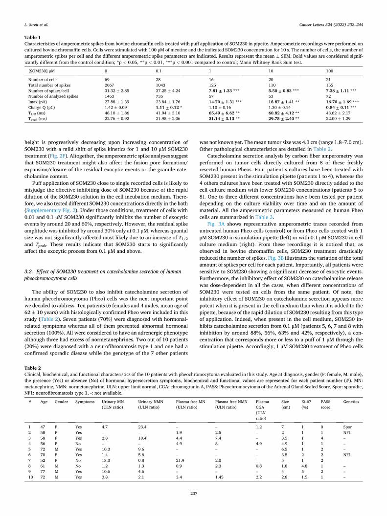
Cancer Letters 524 (2022) 232–244
237
height is progressively decreasing upon increasing concentration of SOM230 with a mild shift of spike kinetics for 1 and 10 μM SOM230 treatment (Fig. 2F). Altogether, the amperometric spike analyses suggest that SOM230 treatment might also affect the fusion pore formation/ expansion/closure of the residual exocytic events or the granule cate-cholamine content.
Puff application of SOM230 close to single recorded cells is likely to misjudge the effective inhibiting dose of SOM230 because of the rapid dilution of the SOM230 solution in the cell incubation medium. There-fore, we also tested different SOM230 concentrations directly in the bath (Supplementary Fig. 2). Under those conditions, treatment of cells with 0.01 and 0.1 μM SOM230 significantly inhibits the number of exocytic events by around 20 and 60%, respectively. However, the residual spike amplitude was inhibited by around 30% only at 0.1 μM, whereas quantal size was not significantly affected most likely due to an increase of T1/2 and Tpeak. These results indicate that SOM230 starts to significantly affect the exocytic process from 0.1 μM and above.
3.2. Effect of SOM230 treatment on catecholamine secretion of human pheochromocytoma cells
The ability of SOM230 to also inhibit catecholamine secretion of human pheochromocytoma (Pheo) cells was the next important point we decided to address. Ten patients (6 females and 4 males, mean age of 62 ± 10 years) with histologically confirmed Pheo were included in this study (Table 2). Seven patients (70%) were diagnosed with hormonal- related symptoms whereas all of them presented abnormal hormonal secretion (100%). All were considered to have an adrenergic phenotype although three had excess of normetanephrines. Two out of 10 patients (20%) were diagnosed with a neurofibromatosis type 1 and one had a confirmed sporadic disease while the genotype of the 7 other patients
was not known yet. The mean tumor size was 4.3 cm (range 1.8–7.0 cm). Other pathological characteristics are detailed in Table 2.
Catecholamine secretion analysis by carbon fiber amperometry was performed on tumor cells directly cultured from 8 of these freshly resected human Pheos. Four patient’s cultures have been treated with SOM230 present in the stimulation pipette (patients 1 to 4), whereas the 4 others cultures have been treated with SOM230 directly added to the cell culture medium with lower SOM230 concentrations (patients 5 to 8). One to three different concentrations have been tested per patient depending on the culture viability over time and on the amount of material. All the amperometric parameters measured on human Pheo cells are summarized in Table 3.
Fig. 3A shows representative amperometric traces recorded from untreated human Pheo cells (control) or from Pheo cells treated with 1 μM SOM230 in stimulation pipette (left) or with 0.1 μM SOM230 in cell culture medium (right). From these recordings it is noticed that, as observed in bovine chromaffin cells, SOM230 treatment drastically reduced the number of spikes. Fig. 3B illustrates the variation of the total amount of spikes per cell for each patient. Importantly, all patients were sensitive to SOM230 showing a significant decrease of exocytic events. Furthermore, the inhibitory effect of SOM230 on catecholamine release was dose-dependent in all the cases, when different concentrations of SOM230 were tested on cells from the same patient. Of note, the inhibitory effect of SOM230 on catecholamine secretion appears more potent when it is present in the cell medium than when it is added to the pipette, because of the rapid dilution of SOM230 resulting from this type of application. Indeed, when present in the cell medium, SOM230 in-hibits catecholamine secretion from 0.1 μM (patients 5, 6, 7 and 8 with inhibition by around 88%, 56%, 63% and 42%, respectively), a con-centration that corresponds more or less to a puff of 1 μM through the stimulation pipette. Accordingly, 1 μM SOM230 treatment of Pheo cells
Table 2 Clinical, biochemical, and functional characteristics of the 10 patients with pheochromocytoma evaluated in this study. Age at diagnosis, gender (F: female, M: male), the presence (Yes) or absence (No) of hormonal hypersecretion symptoms, biochemical and functional values are represented for each patient number (#). MN: metanephrine, NMN: normetanephrine, ULN: upper limit normal, CGA: chromogranin A, PASS: Pheochromocytoma of the Adrenal Gland Scaled Score, Spor: sporadic, NF1: neurofibromatosis type 1, -: not available.
# Age Gender Symptoms Urinary MN (ULN ratio)
Urinary NMN (ULN ratio)
Plasma free MN (ULN ratio)
Plasma free NMN (ULN ratio)
Plasma CGA (ULN ratio)
Size (cm)
Ki-67 (%)
PASS score
Genetics
1 47 F Yes 4.7 23.4 – – 1.2 7 1 0 Spor 2 58 F Yes – – 1.9 2.5 – 2 1 1 NF1 3 58 F Yes 2.8 10.4 4.4 7.4 – 3.5 1 4 – 4 56 F No – – 4.9 8 4.9 4.9 1 1 – 5 72 M Yes 10.3 9.6 – – – 6.5 1 2 – 6 70 F Yes 1.4 5.6 – – – 3.5 2 2 NF1 7 52 F No 13.3 0.8 21.9 2.0 – 5 1 2 – 8 61 M No 1.2 1.3 0.9 2.3 0.8 1.8 4.8 1 – 9 77 M Yes 10.6 4.6 – – – 4 5 2 – 10 72 M Yes 3.8 2.1 3.4 1.45 2.2 2.8 1.5 1 –
Table 1 Characteristics of amperometric spikes from bovine chromaffin cells treated with puff application of SOM230 in pipette. Amperometric recordings were performed on cultured bovine chromaffin cells. Cells were stimulated with 100 μM of nicotine and the indicated SOM230 concentration for 10 s. The number of cells, the number of amperometric spikes per cell and the different amperometric spike parameters are indicated. Results represent the mean ± SEM. Bold values are considered signif-icantly different from the control condition; *p < 0.05, **p < 0.01, ***p < 0.001 compared to control; Mann Whitney Rank Sum test.
[SOM230] μM 0 0.1 1 10 100
Number of cells 69 28 16 20 21 Total number of spikes 2067 1043 125 110 155 Number of spikes/cell 31.32 ± 2.85 37.25 ± 4.24 7.81 ± 1.33 *** 5.50 ± 0.83 *** 7.38 ± 1.11 *** Number of analyzed spikes 1463 735 57 53 72 Imax (pA) 27.88 ± 1.39 23.84 ± 1.76 14.70 ± 1.31 *** 18.87 ± 1.41 ** 16.70 ± 1.69 *** Charge Q (pC) 1.42 ± 0.09 1.11 ± 0.12 * 1.10 ± 0.16 1.30 ± 0.14 0.84 ± 0.11 *** T1/2 (ms) 46.10 ± 1.86 41.94 ± 3.10 65.49 ± 6.62 ** 60.82 ± 4.12 ** 43.62 ± 2.17 Tpeak (ms) 22.76 ± 0.92 21.95 ± 2.06 31.14 ± 3.13 ** 29.75 ± 2.40 ** 22.00 ± 1.29
L. Streit et al.
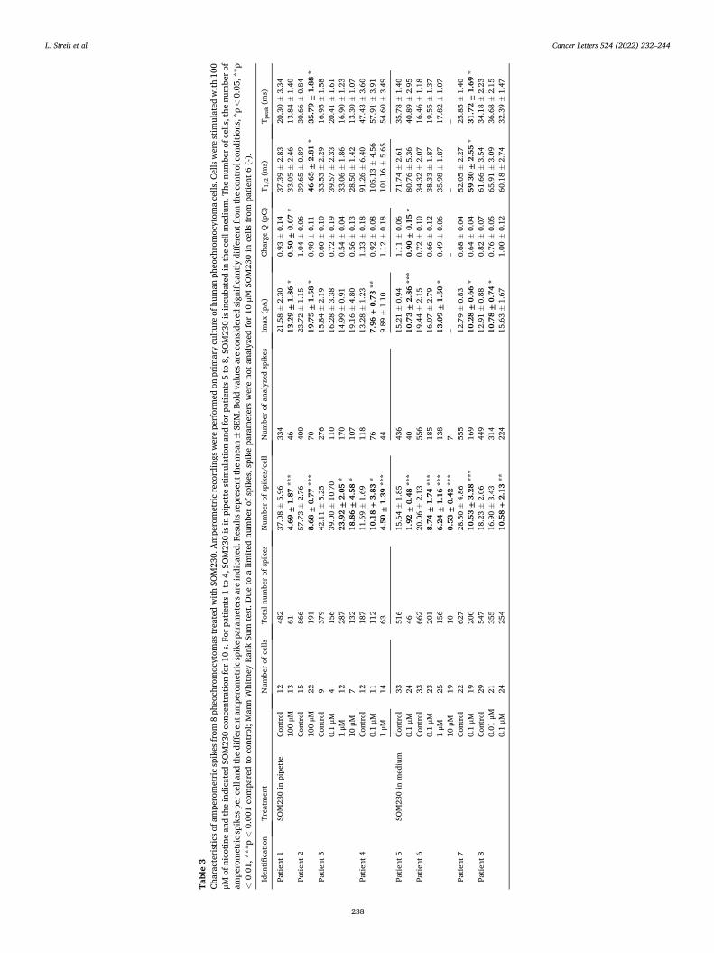
Cancer Letters 524 (2022) 232–244
238
Tabl
e 3
Char
acte
rist
ics o
f am
pero
met
ric
spik
es fr
om 8
phe
ochr
omoc
ytom
as tr
eate
d w
ith S
OM
230.
Am
pero
met
ric
reco
rdin
gs w
ere
perf
orm
ed o
n pr
imar
y cu
lture
of h
uman
phe
ochr
omoc
ytom
a ce
lls. C
ells
wer
e st
imul
ated
with
100
μM
of n
icot
ine
and
the
indi
cate
d SO
M23
0 co
ncen
trat
ion
for 1
0 s.
For
pat
ient
s 1 to
4, S
OM
230
is in
pip
ette
stim
ulat
ion
and
for p
atie
nts 5
to 8
, SO
M23
0 is
incu
bate
d in
the
cell
med
ium
. The
num
ber o
f cel
ls, t
he n
umbe
r of
ampe
rom
etri
c sp
ikes
per
cel
l and
the
diffe
rent
am
pero
met
ric
spik
e pa
ram
eter
s are
indi
cate
d. R
esul
ts re
pres
ent t
he m
ean ±
SEM
. Bol
d va
lues
are
con
side
red
sign
ifica
ntly
diff
eren
t fro
m th
e co
ntro
l con
ditio
ns; *
p <
0.05
, **p
<
0.01
, ***
p <
0.00
1 co
mpa
red
to c
ontr
ol; M
ann
Whi
tney
Ran
k Su
m te
st. D
ue to
a li
mite
d nu
mbe
r of
spi
kes,
spi
ke p
aram
eter
s w
ere
not a
naly
zed
for
10 µ
M S
OM
230
in c
ells
from
pat
ient
6 (
-).
Iden
tifica
tion
Trea
tmen
t N
umbe
r of
cel
ls
Tota
l num
ber
of s
pike
s N
umbe
r of
spi
kes/
cell
Num
ber
of a
naly
zed
spik
es
Imax
(pA
) Ch
arge
Q (
pC)
T 1/2
(m
s)
T pea
k (m
s)
Patie
nt 1
SO
M23
0 in
pip
ette
Co
ntro
l 12
48
2 37
.08 ±
5.96
33
4 21
.58 ±
2.30
0.
93 ±
0.14
37
.39 ±
2.83
20
.30 ±
3.34
10
0 μM
13
61
4.
69 ±
1.8
7 **
* 46
13
.29
± 1
.86
* 0.
50 ±
0.0
7 *
33.0
5 ±
2.46
13
.84 ±
1.40
Pa
tient
2
Cont
rol
15
866
57.7
3 ±
2.76
40
0 23
.72 ±
1.15
1.
04 ±
0.06
39
.65 ±
0.89
30
.66 ±
0.84
10
0 μM
22
19
1 8.
68 ±
0.7
7 **
* 70
19
.75
± 1
.58
* 0.
98 ±
0.11
46
.65
± 2
.81
* 35
.79
± 1
.88
* Pa
tient
3
Cont
rol
9 37
9 42
.11 ±
5.25
27
6 15
.84 ±
2.19
0.
60 ±
0.10
33
.53 ±
2.29
16
.95 ±
1.58
0.
1 μM
4
156
39.0
0 ±
10.7
0 11
0 16
.28 ±
3.38
0.
72 ±
0.19
39
.57 ±
2.33
20
.41 ±
1.61
1
μM
12
287
23.9
2 ±
2.0
5 *
170
14.9
9 ±
0.91
0.
54 ±
0.04
33
.06 ±
1.86
16
.90 ±
1.23
10
μM
7
132
18.8
6 ±
4.5
8 *
107
19.1
6 ±
4.80
0.
56 ±
0.13
28
.50 ±
1.42
13
.30 ±
1.07
Pa
tient
4
Cont
rol
12
187
11.6
9 ±
1.69
11
8 13
.28 ±
1.23
1.
33 ±
0.18
91
.26 ±
6.40
47
.43 ±
3.60
0.
1 μM
11
11
2 10
.18
± 3
.83
* 76
7.
96 ±
0.7
3 **
0.
92 ±
0.08
10
5.13
±4.
56
57.9
1 ±
3.91
1
μM
14
63
4.50
± 1
.39
***
44
9.89
±1.
10
1.12
±0.
18
101.
16 ±
5.65
54
.60 ±
3.49
Patie
nt 5
SO
M23
0 in
med
ium
Co
ntro
l 33
51
6 15
.64 ±
1.85
43
6 15
.21 ±
0.94
1.
11 ±
0.06
71
.74 ±
2.61
35
.78 ±
1.40
0.
1 μM
24
46
1.
92 ±
0.4
8 **
* 40
10
.73
± 2
.86
***
0.90
± 0
.15
* 80
.76 ±
5.36
40
.89 ±
2.95
Pa
tient
6
Cont
rol
33
662
20.0
6 ±
2.13
55
6 19
.44 ±
2.15
0.
72 ±
0.10
34
.32 ±
2.07
16
.46 ±
1.18
0.
1 μM
23
20
1 8.
74 ±
1.7
4 **
* 18
5 16
.07 ±
2.79
0.
66 ±
0.12
38
.33 ±
1.87
19
.55 ±
1.37
1
μM
25
156
6.24
± 1
.16
***
138
13.0
9 ±
1.5
0 *
0.49
±0.
06
35.9
8 ±
1.87
17
.82 ±
1.07
10
μM
19
10
0.
53 ±
0.4
2 **
* 7
– –
– –
Patie
nt 7
Co
ntro
l 22
62
7 28
.50 ±
4.86
55
5 12
.79 ±
0.83
0.
68 ±
0.04
52
.05 ±
2.27
25
.85 ±
1.40
0.
1 μM
19
20
0 10
.53
± 3
.28
***
169
10.2
8 ±
0.6
6 *
0.64
±0.
04
59.3
0 ±
2.5
5 *
31.7
2 ±
1.6
9 *
Patie
nt 8
Co
ntro
l 29
54
7 18
.23 ±
2.06
44
9 12
.91 ±
0.88
0.
82 ±
0.07
61
.66 ±
3.54
34
.18 ±
2.23
0.
01 μ
M
21
355
16.9
0 ±
3.43
31
4 10
.78
± 0
.74
* 0.
76 ±
0.05
65
.91 ±
3.09
36
.68 ±
2.15
0.
1 μM
24
25
4 10
.58
± 2
.13
**
224
15.6
3 ±
1.67
1.
00 ±
0.12
60
.18 ±
2.74
32
.39 ±
1.47
L. Streit et al.
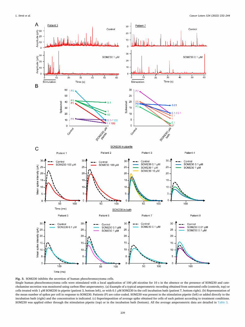
Cancer Letters 524 (2022) 232–244
239
Fig. 3. SOM230 inhibits the secretion of human pheochromocytoma cells. Single human pheochromocytoma cells were stimulated with a local application of 100 μM nicotine for 10 s in the absence or the presence of SOM230 and cate-cholamine secretion was monitored using carbon fiber amperometry. (a) Example of a typical amperometric recording obtained from untreated cells (controls, top) or cells treated with 1 μM SOM230 in pipette (patient 3, bottom left), or with 0.1 μM SOM230 in the cell incubation bath (patient 7, bottom right). (b) Representation of the mean number of spikes per cell in response to SOM230. Patients (P) are color-coded. SOM230 was present in the stimulation pipette (left) or added directly to the incubation bath (right) and the concentration is indicated. (c) Superimposition of average spike obtained for cells of each patient according to treatment conditions. SOM230 was applied either through the stimulation pipette (top) or in the incubation bath (bottom). All the average amperometric data are detailed in Table 3.
L. Streit et al.
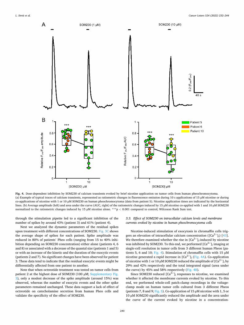
Cancer Letters 524 (2022) 232–244
240
through the stimulation pipette led to a significant inhibition of the number of spikes by around 43% (patient 3) and 61% (patient 4).
Next we analyzed the dynamic parameters of the residual spikes upon treatment with different concentrations of SOM230. Fig. 3C shows the average shape of spikes for each patient. Spike amplitude was reduced in 80% of patients’ Pheo cells (ranging from 15 to 40% inhi-bition depending on SOM230 concentration) either alone (patients 4, 6 and 8) or associated with a decrease of the quantal size (patients 1 and 5) or with an increase of the kinetic and the duration of the exocytic events (patients 2 and 7). No significant changes have been observed for patient 3. These data tend to indicate that the residual exocytic events might be differentially affected from one patient to another.
Note that when octreotide treatment was tested on tumor cells from patient 2 at the highest dose of SOM230 (100 μM; Supplementary Fig. 3), only a modest decrease of the spike amplitude (around 15%) was observed, whereas the number of exocytic events and the other spike parameters remained unchanged. These data support a lack of effect of octreotide on catecholamine secretion from human Pheo cells and validate the specificity of the effect of SOM230.
3.3. Effect of SOM230 on intracellular calcium levels and membrane currents evoked by nicotine in human pheochromocytoma cells
Nicotine-induced stimulation of exocytosis in chromaffin cells trig-gers an elevation of intracellular calcium concentration ([Ca2+]i) [43]. We therefore examined whether the rise in [Ca2+]i-induced by nicotine was inhibited by SOM230. To this end, we performed [Ca2+]i imaging at single-cell resolution in tumor cells from 3 different human Pheos (pa-tients 5, 6 and 10; Fig. 4). Stimulation of chromaffin cells with 15 μM nicotine generated a rapid increase in [Ca2+]i (Fig. 4A). Co-application of nicotine with 1 or 10 μM SOM230 reduced the amplitude of [Ca2+]i by 29% and 42% respectively and the total integrated signal (area under the curve) by 45% and 58% respectively (Fig. 4B).
Since SOM230 reduced [Ca2+]i responses to nicotine, we examined whether it affected the membrane currents evoked by nicotine. To this end, we performed whole-cell patch-clamp recordings in the voltage- clamp mode on human tumor cells cultured from 3 different Pheos (patients 7, 8 and 9; Fig. 5). Co-application of 15 μM nicotine with 1, 5 or 10 μM SOM230 significantly reduced the amplitude and the area under the curve of the current evoked by nicotine in a concentration-
Fig. 4. Dose-dependent inhibition by SOM230 of calcium transients evoked by brief nicotine application on tumor cells from human pheochromocytoma. (a) Example of typical traces of calcium transients, represented as ratiometric changes in fluorescence emission during 10 s applications of 15 μM nicotine or during co-applications of nicotine with 1 or 10 μM SOM230 on human pheochromocytoma (data from patient 5). Nicotine application times are indicated by the horizontal lines. (b) Average amplitude (left) and area under the curve (AUC, right) of the ratiometric changes induced by 15 μM nicotine co-applied with 1 and 10 μM SOM230 normalized to the ratiometric changes induced by 15 μM nicotine alone. ***p < 0.001 compared to control; Wilcoxon Rank Sum test.
L. Streit et al.
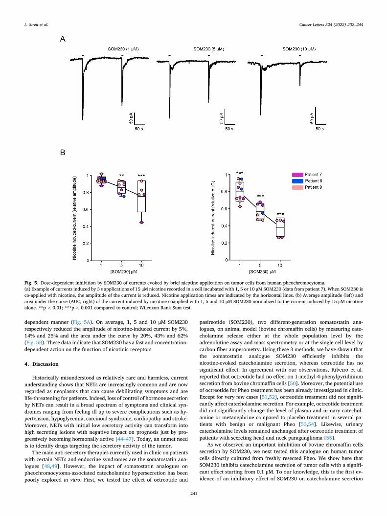
Cancer Letters 524 (2022) 232–244
241
dependent manner (Fig. 5A). On average, 1, 5 and 10 μM SOM230 respectively reduced the amplitude of nicotine-induced current by 5%, 14% and 25% and the area under the curve by 20%, 43% and 62% (Fig. 5B). These data indicate that SOM230 has a fast and concentration- dependent action on the function of nicotinic receptors.
4. Discussion
Historically misunderstood as relatively rare and harmless, current understanding shows that NETs are increasingly common and are now regarded as neoplasms that can cause debilitating symptoms and are life-threatening for patients. Indeed, loss of control of hormone secretion by NETs can result in a broad spectrum of symptoms and clinical syn-dromes ranging from feeling ill up to severe complications such as hy-pertension, hypoglycemia, carcinoid syndrome, cardiopathy and stroke. Moreover, NETs with initial low secretory activity can transform into high secreting lesions with negative impact on prognosis just by pro-gressively becoming hormonally active [44–47]. Today, an unmet need is to identify drugs targeting the secretory activity of the tumor.
The main anti-secretory therapies currently used in clinic on patients with certain NETs and endocrine syndromes are the somatostatin ana-logues [48,49]. However, the impact of somatostatin analogues on pheochromocytoma-associated catecholamine hypersecretion has been poorly explored in vitro. First, we tested the effect of octreotide and
pasireotide (SOM230), two different-generation somatostatin ana-logues, on animal model (bovine chromaffin cells) by measuring cate-cholamine release either at the whole population level by the adrenolutine assay and mass spectrometry or at the single cell level by carbon fiber amperometry. Using these 3 methods, we have shown that the somatostatin analogue SOM230 efficiently inhibits the nicotine-evoked catecholamine secretion, whereas octreotide has no significant effect. In agreement with our observations, Ribeiro et al. reported that octreotide had no effect on 1-methyl-4-phenylpyridinium secretion from bovine chromaffin cells [50]. Moreover, the potential use of octreotide for Pheo treatment has been already investigated in clinic. Except for very few cases [51,52], octreotide treatment did not signifi-cantly affect catecholamine secretion. For example, octreotide treatment did not significantly change the level of plasma and urinary catechol-amine or metanephrine compared to placebo treatment in several pa-tients with benign or malignant Pheo [53,54]. Likewise, urinary catecholamine levels remained unchanged after octreotide treatment of patients with secreting head and neck paraganglioma [55].
As we observed an important inhibition of bovine chromaffin cells secretion by SOM230, we next tested this analogue on human tumor cells directly cultured from freshly resected Pheo. We show here that SOM230 inhibits catecholamine secretion of tumor cells with a signifi-cant effect starting from 0.1 μM. To our knowledge, this is the first ev-idence of an inhibitory effect of SOM230 on catecholamine secretion
Fig. 5. Dose-dependent inhibition by SOM230 of currents evoked by brief nicotine application on tumor cells from human pheochromocytoma. (a) Example of currents induced by 3 s applications of 15 μM nicotine recorded in a cell incubated with 1, 5 or 10 μM SOM230 (data from patient 7). When SOM230 is co-applied with nicotine, the amplitude of the current is reduced. Nicotine application times are indicated by the horizontal lines. (b) Average amplitude (left) and area under the curve (AUC, right) of the current induced by nicotine coapplied with 1, 5 and 10 μM SOM230 normalized to the current induced by 15 μM nicotine alone. **p < 0.01; ***p < 0.001 compared to control; Wilcoxon Rank Sum test.
L. Streit et al.
Cancer Letters 524 (2022) 232–244
242
from human Pheo cells. Nevertheless, in agreement with our observa-tions, the effect of SOM230 on hormone secretion has been tested on various types of other tumor cells in vitro. For example, SOM230 effi-ciently inhibits prolactine, growth hormone and adrenocorticotropic hormone secretion from primary cultures of human secreting pituitary adenomas [56,57]. It also significantly decreases secretion of chro-mogranin A in cells cultured from human gastroenteropancreatic NETs [20]. Moreover, Quinn et al. demonstrated in vivo, in a mouse model with insulinoma, that SOM230 injection inhibits hypersecretion of in-sulin thus preventing hypoglycemia [58].
Using carbon fiber amperometry, we have further dissected the anti- secretory effect of SOM230 on the exocytic pathway. The main effect observed after the application of 0.1 μM SOM230 was an important inhibition of the number of exocytic events on both bovine chromaffin cells (decreased by around 60%) and human tumor cells (decrease ranging from 42 to 88%; patients 5 to 8). Note that, due to the dilution factor, the effect seen with 0.1 μM in bath of SOM230 corresponds more or less to the one seen with 1 μM co-incubated with nicotine in the stimulation pipette. Accordingly, under the latter experimental condi-tion, the number of exocytic events was reduced by SOM230 with a comparable efficiency (by 43 and 61% in patients 3 and 4, respectively and by 75% in average for bovine model). These results support the notion that SOM230 affects early steps in the exocytotic process, such as the recruitment of the secretory granules or their docking at the plasma membrane. Moreover, the analysis of the amperometric parameters revealed that the amplitude of the remaining exocytic events tends to be lower upon SOM230 treatment, an effect sometimes accompanied by a decrease of the kinetics. This suggests that, even when secretory granule fusion is engaged, the release might be disturbed, being incomplete and/ or slowed down. To our knowledge, only a single study investigated the in vitro effect of SOM230 on tumor cells cultured from human Pheos. Hence, Pasquali et al. showed that a long-lasting (24 h) treatment of 0.1 and 1 μM SOM230 significantly decreased the intracellular content of dopamine and noradrenaline [28]. However, in contrast to our study, they didn’t measure the secretory activity of the tumor cells. A reduction of cellular catecholamine levels can be the consequence of a decrease in the number of secretory granules and/or the level of catecholamines per granule. In our study, the total amount of catecholamines in cells re-mains unchanged which was expected because relatively short SOM230 treatments were performed. This indicates that the effect we observed on secretion is necessarily independent from its potential long-term ef-fect on catecholamine metabolism. It is also of note that the inhibitory effect of SOM230 on catecholamine release was seen only when the drug was present during the stimulation period and a pre-incubation period did not significantly further increase this inhibition of secretion (data not shown). These data show that the effect of SOM230 is rapid and that a long pre-incubation is not required.
In our experimental conditions, we found that SOM230 drastically inhibited catecholamine secretion of tumor cells at 100 nM but not at 10 nM. Accordingly, inhibitory effects with similar concentrations have been previously observed in cells cultured from human cancer such as growth hormone-secreting pituitary adenoma and growth hormone- releasing hormone-producing bronchial carcinoid [59,60]. However, this concentration is relatively high compared to the IC50 (ranging from 0.2 to 1 nM) reported after long lasting treatment of SOM230 on the secretory activity of rat pituitary cells, rat pancreatic islets, mouse cor-ticotroph adenoma cells and human pituitary adenoma cells [56,57,61, 62], raising the question of the mechanisms by which SOM230 inhibits catecholamine secretion in human Pheo cells. As frequency and ampli-tude of exocytic events depend on the level of secretagogue-evoked intracellular calcium elevation [63,64], it was tempting to imagine that SOM230 might affect secretory granule exocytosis upstream of calcium entry. Indeed, we showed that SOM230 significantly inhibited nicotine-evoked increase of intracellular calcium in human tumor cells. One other interesting aspect is that SOM230 acts instantly and exclu-sively on secretion induced by nicotine without affecting secretion
induced by high K+ solution. In agreement with our results, several studies demonstrated that somatostatin itself was able to inhibit cate-cholamine release when chromaffin cells are stimulated with nicotine or acetylcholine but not upon membrane depolarization [39,40,65,66]. In fact, we show here by whole cell recordings in the voltage-clamp mode that SOM230 is able to reduce the nicotinic current. Since membrane potential remains constant under these recording conditions, this effect most likely involves an effect on the function of nicotinic receptors and not on voltage-dependent Na+ and Ca2+ channels. Accordingly, co-application of somatostatin with nicotine reversibly inhibits the nicotine-induced inward current in guinea pig chromaffin cells [67]. Whether SOM230 indirectly affects the function of the nicotinic receptor via its own somatostatin receptor or via a direct action on the nicotinic receptor is currently unknown. Interestingly, several evidences indi-cated that somatostatin receptors are able to either homo-oligomerize or to hetero-oligomerize between different subtypes of somatostatin re-ceptors or with other types of receptor like the dopamine receptor D2 or the μ-opioid receptor MOR1 [68–72]. Potential oligomerization between somatostatin and nicotinic receptors has never been reported and will require further investigations.
This pre-clinical study allowed us to uncover a novel anti-secretory role of SOM230 on tumor adrenal medulla cells from patients with functional Pheo and abnormal secretion. If SOM230 treatment could actually improve the management of patients remains a key question that will require further clinical studies. As SOM230 efficiently inhibits catecholamine secretion by directly acting on the tumor secretion, it might be considered for preoperative medical preparation or intra-operative management of highly secreting Pheo. SOM230 might also be useful to treat some metastatic Pheos resistant to conventional antihy-pertensive medications, especially as high hormonal secretion impacts overall survival in patient with metastatic Pheo [73]. Moreover, as long lasting effect of SOM230 is known to reduce catecholamine synthesis in Pheo cells [28], treatment of patients may lead to a combination of reduced catecholamine synthesis and secretion which is in fact likely to boost the effect of SOM230 on lowering catecholamine levels in patients with Pheo.
In conclusion, a long-acting release formulation of SOM230 might be a potential lead to consider a therapy aimed at treating the symptoms induced by excessive catecholamine secretion of Pheo. This new data may be used to better control catecholamine hypersecretion when needed and should be evaluated in clinical practice.
Ethics approval and consent to participate
The present study used the data and the human biological material of the biological collection “Approche moleculaire des tumeurs cortico-surrenaliennes” which was agreed by the “Comite de Protection des Personnes Est III” ethical advisory committee, and was conducted ac-cording to currently accepted ethical guidelines, including informed written consent approval signed by all patients prior to inclusion.
Fundings and acknowledgements
This work was financially supported by ITMO Cancer AVIESAN (Alliance Nationale pour les Sciences de la Vie et de la Sante, National Alliance for Life Sciences & Health) within the framework of the Cancer Plan to SG and LB (Single Cell 2018 N◦ 19CS004-00); by grants from the Agence Nationale pour la Recherche (“SecretoNET”, N◦ ANR-16-CE17- 0022-01) and from the Ligue contre le Cancer (CCIR Grand-Est) to SG; by the University of Strasbourg Institute for Advanced Study (USIAS) for a Fellowship to SG, within the French national programme “Investment for the future” (IdEx-Unistra); by a fellowship from la Fondation pour la Recherche Medicale (FRM; FDM201806005916) to SM. INSERM is providing salary to YG, NV and SG. We acknowledge the municipal slaughterhouse of Haguenau (France) for providing the bovine adrenal glands. The authors are also grateful to Dr. Nan Qin (University of
L. Streit et al.
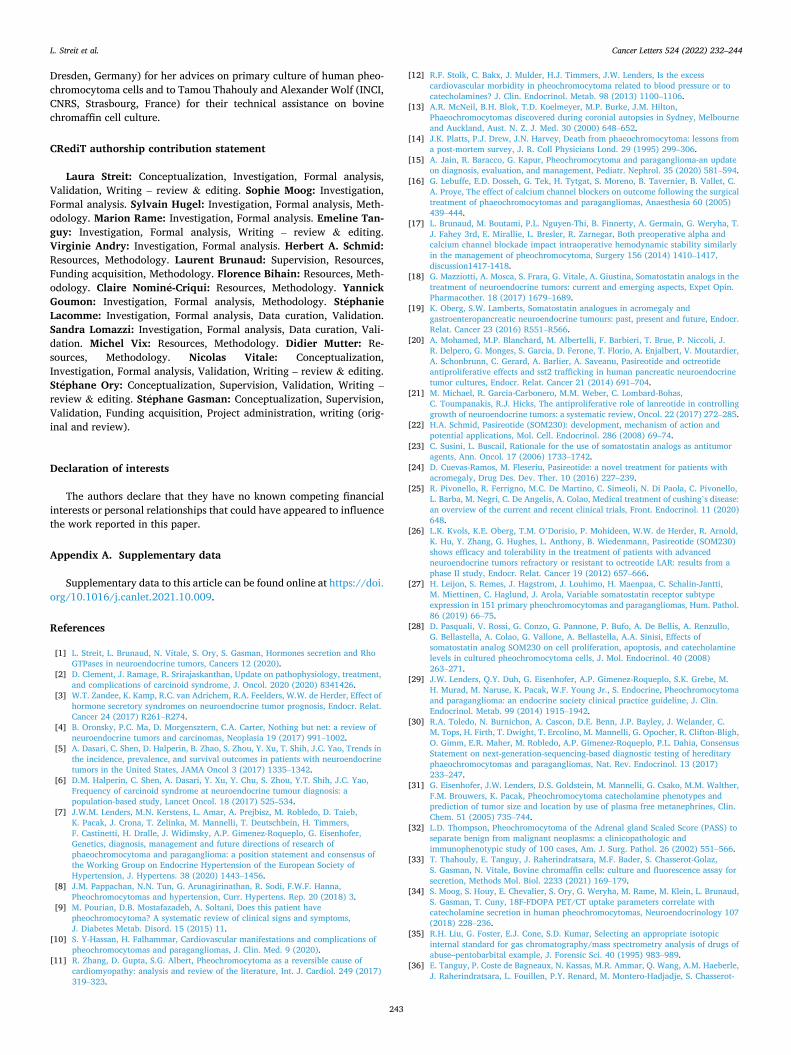
Cancer Letters 524 (2022) 232–244
243
Dresden, Germany) for her advices on primary culture of human pheo-chromocytoma cells and to Tamou Thahouly and Alexander Wolf (INCI, CNRS, Strasbourg, France) for their technical assistance on bovine chromaffin cell culture.
CRediT authorship contribution statement
Laura Streit: Conceptualization, Investigation, Formal analysis, Validation, Writing – review & editing. Sophie Moog: Investigation, Formal analysis. Sylvain Hugel: Investigation, Formal analysis, Meth-odology. Marion Rame: Investigation, Formal analysis. Emeline Tan-guy: Investigation, Formal analysis, Writing – review & editing. Virginie Andry: Investigation, Formal analysis. Herbert A. Schmid: Resources, Methodology. Laurent Brunaud: Supervision, Resources, Funding acquisition, Methodology. Florence Bihain: Resources, Meth-odology. Claire Nomine-Criqui: Resources, Methodology. Yannick Goumon: Investigation, Formal analysis, Methodology. Stephanie Lacomme: Investigation, Formal analysis, Data curation, Validation. Sandra Lomazzi: Investigation, Formal analysis, Data curation, Vali-dation. Michel Vix: Resources, Methodology. Didier Mutter: Re-sources, Methodology. Nicolas Vitale: Conceptualization, Investigation, Formal analysis, Validation, Writing – review & editing. Stephane Ory: Conceptualization, Supervision, Validation, Writing – review & editing. Stephane Gasman: Conceptualization, Supervision, Validation, Funding acquisition, Project administration, writing (orig-inal and review).
Declaration of interests
The authors declare that they have no known competing financial interests or personal relationships that could have appeared to influence the work reported in this paper.
Appendix A. Supplementary data
Supplementary data to this article can be found online at https://doi. org/10.1016/j.canlet.2021.10.009.
References
[1] L. Streit, L. Brunaud, N. Vitale, S. Ory, S. Gasman, Hormones secretion and Rho GTPases in neuroendocrine tumors, Cancers 12 (2020).
[2] D. Clement, J. Ramage, R. Srirajaskanthan, Update on pathophysiology, treatment, and complications of carcinoid syndrome, J. Oncol. 2020 (2020) 8341426.
[3] W.T. Zandee, K. Kamp, R.C. van Adrichem, R.A. Feelders, W.W. de Herder, Effect of hormone secretory syndromes on neuroendocrine tumor prognosis, Endocr. Relat. Cancer 24 (2017) R261–R274.
[4] B. Oronsky, P.C. Ma, D. Morgensztern, C.A. Carter, Nothing but net: a review of neuroendocrine tumors and carcinomas, Neoplasia 19 (2017) 991–1002.
[5] A. Dasari, C. Shen, D. Halperin, B. Zhao, S. Zhou, Y. Xu, T. Shih, J.C. Yao, Trends in the incidence, prevalence, and survival outcomes in patients with neuroendocrine tumors in the United States, JAMA Oncol 3 (2017) 1335–1342.
[6] D.M. Halperin, C. Shen, A. Dasari, Y. Xu, Y. Chu, S. Zhou, Y.T. Shih, J.C. Yao, Frequency of carcinoid syndrome at neuroendocrine tumour diagnosis: a population-based study, Lancet Oncol. 18 (2017) 525–534.
[7] J.W.M. Lenders, M.N. Kerstens, L. Amar, A. Prejbisz, M. Robledo, D. Taieb, K. Pacak, J. Crona, T. Zelinka, M. Mannelli, T. Deutschbein, H. Timmers, F. Castinetti, H. Dralle, J. Widimsky, A.P. Gimenez-Roqueplo, G. Eisenhofer, Genetics, diagnosis, management and future directions of research of phaeochromocytoma and paraganglioma: a position statement and consensus of the Working Group on Endocrine Hypertension of the European Society of Hypertension, J. Hypertens. 38 (2020) 1443–1456.
[8] J.M. Pappachan, N.N. Tun, G. Arunagirinathan, R. Sodi, F.W.F. Hanna, Pheochromocytomas and hypertension, Curr. Hypertens. Rep. 20 (2018) 3.
[9] M. Pourian, D.B. Mostafazadeh, A. Soltani, Does this patient have pheochromocytoma? A systematic review of clinical signs and symptoms, J. Diabetes Metab. Disord. 15 (2015) 11.
[10] S. Y-Hassan, H. Falhammar, Cardiovascular manifestations and complications of pheochromocytomas and paragangliomas, J. Clin. Med. 9 (2020).
[11] R. Zhang, D. Gupta, S.G. Albert, Pheochromocytoma as a reversible cause of cardiomyopathy: analysis and review of the literature, Int. J. Cardiol. 249 (2017) 319–323.
[12] R.F. Stolk, C. Bakx, J. Mulder, H.J. Timmers, J.W. Lenders, Is the excess cardiovascular morbidity in pheochromocytoma related to blood pressure or to catecholamines? J. Clin. Endocrinol. Metab. 98 (2013) 1100–1106.
[13] A.R. McNeil, B.H. Blok, T.D. Koelmeyer, M.P. Burke, J.M. Hilton, Phaeochromocytomas discovered during coronial autopsies in Sydney, Melbourne and Auckland, Aust. N. Z. J. Med. 30 (2000) 648–652.
[14] J.K. Platts, P.J. Drew, J.N. Harvey, Death from phaeochromocytoma: lessons from a post-mortem survey, J. R. Coll Physicians Lond. 29 (1995) 299–306.
[15] A. Jain, R. Baracco, G. Kapur, Pheochromocytoma and paraganglioma-an update on diagnosis, evaluation, and management, Pediatr. Nephrol. 35 (2020) 581–594.
[16] G. Lebuffe, E.D. Dosseh, G. Tek, H. Tytgat, S. Moreno, B. Tavernier, B. Vallet, C. A. Proye, The effect of calcium channel blockers on outcome following the surgical treatment of phaeochromocytomas and paragangliomas, Anaesthesia 60 (2005) 439–444.
[17] L. Brunaud, M. Boutami, P.L. Nguyen-Thi, B. Finnerty, A. Germain, G. Weryha, T. J. Fahey 3rd, E. Mirallie, L. Bresler, R. Zarnegar, Both preoperative alpha and calcium channel blockade impact intraoperative hemodynamic stability similarly in the management of pheochromocytoma, Surgery 156 (2014) 1410–1417, discussion1417-1418.
[18] G. Mazziotti, A. Mosca, S. Frara, G. Vitale, A. Giustina, Somatostatin analogs in the treatment of neuroendocrine tumors: current and emerging aspects, Expet Opin. Pharmacother. 18 (2017) 1679–1689.
[19] K. Oberg, S.W. Lamberts, Somatostatin analogues in acromegaly and gastroenteropancreatic neuroendocrine tumours: past, present and future, Endocr. Relat. Cancer 23 (2016) R551–R566.
[20] A. Mohamed, M.P. Blanchard, M. Albertelli, F. Barbieri, T. Brue, P. Niccoli, J. R. Delpero, G. Monges, S. Garcia, D. Ferone, T. Florio, A. Enjalbert, V. Moutardier, A. Schonbrunn, C. Gerard, A. Barlier, A. Saveanu, Pasireotide and octreotide antiproliferative effects and sst2 trafficking in human pancreatic neuroendocrine tumor cultures, Endocr. Relat. Cancer 21 (2014) 691–704.
[21] M. Michael, R. Garcia-Carbonero, M.M. Weber, C. Lombard-Bohas, C. Toumpanakis, R.J. Hicks, The antiproliferative role of lanreotide in controlling growth of neuroendocrine tumors: a systematic review, Oncol. 22 (2017) 272–285.
[22] H.A. Schmid, Pasireotide (SOM230): development, mechanism of action and potential applications, Mol. Cell. Endocrinol. 286 (2008) 69–74.
[23] C. Susini, L. Buscail, Rationale for the use of somatostatin analogs as antitumor agents, Ann. Oncol. 17 (2006) 1733–1742.
[24] D. Cuevas-Ramos, M. Fleseriu, Pasireotide: a novel treatment for patients with acromegaly, Drug Des. Dev. Ther. 10 (2016) 227–239.
[25] R. Pivonello, R. Ferrigno, M.C. De Martino, C. Simeoli, N. Di Paola, C. Pivonello, L. Barba, M. Negri, C. De Angelis, A. Colao, Medical treatment of cushing’s disease: an overview of the current and recent clinical trials, Front. Endocrinol. 11 (2020) 648.
[26] L.K. Kvols, K.E. Oberg, T.M. O’Dorisio, P. Mohideen, W.W. de Herder, R. Arnold, K. Hu, Y. Zhang, G. Hughes, L. Anthony, B. Wiedenmann, Pasireotide (SOM230) shows efficacy and tolerability in the treatment of patients with advanced neuroendocrine tumors refractory or resistant to octreotide LAR: results from a phase II study, Endocr. Relat. Cancer 19 (2012) 657–666.
[27] H. Leijon, S. Remes, J. Hagstrom, J. Louhimo, H. Maenpaa, C. Schalin-Jantti, M. Miettinen, C. Haglund, J. Arola, Variable somatostatin receptor subtype expression in 151 primary pheochromocytomas and paragangliomas, Hum. Pathol. 86 (2019) 66–75.
[28] D. Pasquali, V. Rossi, G. Conzo, G. Pannone, P. Bufo, A. De Bellis, A. Renzullo, G. Bellastella, A. Colao, G. Vallone, A. Bellastella, A.A. Sinisi, Effects of somatostatin analog SOM230 on cell proliferation, apoptosis, and catecholamine levels in cultured pheochromocytoma cells, J. Mol. Endocrinol. 40 (2008) 263–271.
[29] J.W. Lenders, Q.Y. Duh, G. Eisenhofer, A.P. Gimenez-Roqueplo, S.K. Grebe, M. H. Murad, M. Naruse, K. Pacak, W.F. Young Jr., S. Endocrine, Pheochromocytoma and paraganglioma: an endocrine society clinical practice guideline, J. Clin. Endocrinol. Metab. 99 (2014) 1915–1942.
[30] R.A. Toledo, N. Burnichon, A. Cascon, D.E. Benn, J.P. Bayley, J. Welander, C. M. Tops, H. Firth, T. Dwight, T. Ercolino, M. Mannelli, G. Opocher, R. Clifton-Bligh, O. Gimm, E.R. Maher, M. Robledo, A.P. Gimenez-Roqueplo, P.L. Dahia, Consensus Statement on next-generation-sequencing-based diagnostic testing of hereditary phaeochromocytomas and paragangliomas, Nat. Rev. Endocrinol. 13 (2017) 233–247.
[31] G. Eisenhofer, J.W. Lenders, D.S. Goldstein, M. Mannelli, G. Csako, M.M. Walther, F.M. Brouwers, K. Pacak, Pheochromocytoma catecholamine phenotypes and prediction of tumor size and location by use of plasma free metanephrines, Clin. Chem. 51 (2005) 735–744.
[32] L.D. Thompson, Pheochromocytoma of the Adrenal gland Scaled Score (PASS) to separate benign from malignant neoplasms: a clinicopathologic and immunophenotypic study of 100 cases, Am. J. Surg. Pathol. 26 (2002) 551–566.
[33] T. Thahouly, E. Tanguy, J. Raherindratsara, M.F. Bader, S. Chasserot-Golaz, S. Gasman, N. Vitale, Bovine chromaffin cells: culture and fluorescence assay for secretion, Methods Mol. Biol. 2233 (2021) 169–179.
[34] S. Moog, S. Houy, E. Chevalier, S. Ory, G. Weryha, M. Rame, M. Klein, L. Brunaud, S. Gasman, T. Cuny, 18F-FDOPA PET/CT uptake parameters correlate with catecholamine secretion in human pheochromocytomas, Neuroendocrinology 107 (2018) 228–236.
[35] R.H. Liu, G. Foster, E.J. Cone, S.D. Kumar, Selecting an appropriate isotopic internal standard for gas chromatography/mass spectrometry analysis of drugs of abuse–pentobarbital example, J. Forensic Sci. 40 (1995) 983–989.
[36] E. Tanguy, P. Coste de Bagneaux, N. Kassas, M.R. Ammar, Q. Wang, A.M. Haeberle, J. Raherindratsara, L. Fouillen, P.Y. Renard, M. Montero-Hadjadje, S. Chasserot-
L. Streit et al.
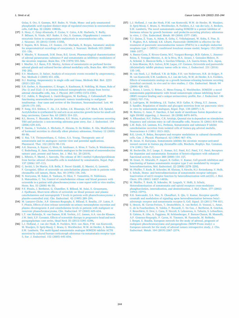
Cancer Letters 524 (2022) 232–244
244
Golaz, S. Ory, S. Gasman, M.F. Bader, N. Vitale, Mono- and poly-unsaturated phosphatidic acid regulate distinct steps of regulated exocytosis in neuroendocrine cells, Cell Rep. 32 (2020) 108026.
[37] S. Houy, C. Estay-Ahumada, P. Croise, V. Calco, A.M. Haeberle, Y. Bailly, P. Billuart, N. Vitale, M.F. Bader, S. Ory, S. Gasman, Oligophrenin-1 connects exocytotic fusion to compensatory endocytosis in neuroendocrine cells, J. Neurosci. 35 (2015) 11045–11055.
[38] F. Segura, M.A. Brioso, J.F. Gomez, J.D. Machado, R. Borges, Automatic analysis for amperometrical recordings of exocytosis, J. Neurosci. Methods 103 (2000) 151–156.
[39] F. Mizobe, V. Kozousek, D.M. Dean, B.G. Livett, Pharmacological characterization of adrenal paraneurons: substance P and somatostatin as inhibitory modulators of the nicotinic response, Brain Res. 178 (1979) 555–566.
[40] I. Moeller, S.J. Bunn, P.D. Marley, Actions of somatostatin on perfused bovine adrenal glands and cultured bovine adrenal medullary cells, Brain Res. 484 (1989) 192–202.
[41] E.V. Mosharov, D. Sulzer, Analysis of exocytotic events recorded by amperometry, Nat. Methods 2 (2005) 651–658.
[42] D.J. Keating, Amperometry in single cells and tissue, Methods Mol. Biol. 2233 (2021) 223–231.
[43] S.H. Gerber, A. Haunstetter, C. Kruger, A. Kaufmann, R. Nobiling, M. Haass, Role of [Na+]i and [Ca2+]i in nicotine-induced norepinephrine release from bovine adrenal chromaffin cells, Am. J. Physiol. 269 (1995) C572–C581.
[44] C.C. Juhlin, S. Skoglund, L. Juntti-Berggren, M. Karlberg, J. Calissendorff, Non- functioning neuroendocrine pancreatic tumors transforming to malignant insulinomas - four cases and review of the literature, Neuroendocrinol. Lett. 40 (2019) 175–183.
[45] P. Song, H.S. Sekhon, Y. Jia, J.A. Keller, J.K. Blusztajn, G.P. Mark, E.R. Spindel, Acetylcholine is synthesized by and acts as an autocrine growth factor for small cell lung carcinoma, Cancer Res. 63 (2003) 214–221.
[46] R.L. Brown, T. Muzzafar, R. Wollman, R.E. Weiss, A pituitary carcinoma secreting TSH and prolactin: a non-secreting adenoma gone awry, Eur. J. Endocrinol. 154 (2006) 639–643.
[47] T. Daems, J. Verhelst, A. Michotte, P. Abrams, D. De Ridder, R. Abs, Modification of hormonal secretion in clinically silent pituitary adenomas, Pituitary 12 (2009) 80–86.
[48] U. Rai, T.R. Thrimawithana, C. Valery, S.A. Young, Therapeutic uses of somatostatin and its analogues: current view and potential applications, Pharmacol. Ther. 152 (2015) 98–110.
[49] A.K. Stueven, A. Kayser, C. Wetz, H. Amthauer, A. Wree, F. Tacke, B. Wiedenmann, C. Roderburg, H. Jann, Somatostatin analogues in the treatment of neuroendocrine tumors: past, present and future, Int. J. Mol. Sci. 20 (2019).
[50] L. Ribeiro, F. Martel, I. Azevedo, The release of 3H-1-methyl-4-phenylpyridinium from bovine adrenal chromaffin cells is modulated by somatostatin, Regul. Pept. 137 (2006) 107–113.
[51] C. Invitti, I. De Martin, G.B. Bolla, F. Pecori Giraldi, E. Maestri, G. Leonetti, F. Cavagnini, Effect of octreotide on catecholamine plasma levels in patients with chromaffin cell tumors, Horm. Res. 40 (1993) 156–160.
[52] N. Koriyama, M. Kakei, K. Yaekura, H. Okui, T. Yamashita, H. Nishimura, S. Matsushita, C. Tei, Control of catecholamine release and blood pressure with octreotide in a patient with pheochromocytoma: a case report with in vitro studies, Horm. Res. 53 (2000) 46–50.
[53] P.F. Plouin, J. Bertherat, G. Chatellier, E. Billaud, M. Azizi, E. Grouzmann, J. Epelbaum, Short-term effects of octreotide on blood pressure and plasma catecholamines and neuropeptide Y levels in patients with phaeochromocytoma: a placebo-controlled trial, Clin. Endocrinol. 42 (1995) 289–294.
[54] M. Lamarre-Cliche, A.P. Gimenez-Roqueplo, E. Billaud, E. Baudin, J.P. Luton, P. F. Plouin, Effects of slow-release octreotide on urinary metanephrine excretion and plasma chromogranin A and catecholamine levels in patients with malignant or recurrent phaeochromocytoma, Clin. Endocrinol. 57 (2002) 629–634.
[55] L.T. van Hulsteijn, N. van Duinen, B.M. Verbist, J.C. Jansen, A.A. van der Klaauw, J.W. Smit, E.P. Corssmit, Effects of octreotide therapy in progressive head and neck paragangliomas: case series, Head Neck 35 (2013) E391–E396.
[56] L.J. Hofland, J. van der Hoek, R. Feelders, M.O. van Aken, P.M. van Koetsveld, M. Waaijers, D. Sprij-Mooij, C. Bruns, G. Weckbecker, W.W. de Herder, A. Beckers, S.W. Lamberts, The multi-ligand somatostatin analogue SOM230 inhibits ACTH secretion by cultured human corticotroph adenomas via somatostatin receptor type 5, Eur. J. Endocrinol. 152 (2005) 645–654.
[57] L.J. Hofland, J. van der Hoek, P.M. van Koetsveld, W.W. de Herder, M. Waaijers, D. Sprij-Mooij, C. Bruns, G. Weckbecker, R. Feelders, A.J. van der Lely, A. Beckers, S.W. Lamberts, The novel somatostatin analog SOM230 is a potent inhibitor of hormone release by growth hormone- and prolactin-secreting pituitary adenomas in vitro, J. Clin. Endocrinol. Metab. 89 (2004) 1577–1585.
[58] T.J. Quinn, Z. Yuan, A. Adem, R. Geha, C. Vrikshajanani, W. Koba, E. Fine, D. T. Hughes, H.A. Schmid, S.K. Libutti, Pasireotide (SOM230) is effective for the treatment of pancreatic neuroendocrine tumors (PNETs) in a multiple endocrine neoplasia type 1 (MEN1) conditional knockout mouse model, Surgery 152 (2012) 1068–1077.
[59] A. Ibanez-Costa, E. Rivero-Cortes, M.C. Vazquez-Borrego, M.D. Gahete, L. Jimenez- Reina, E. Venegas-Moreno, A. de la Riva, M.A. Arraez, I. Gonzalez-Molero, H. A. Schmid, S. Maraver-Selfa, I. Gavilan-Villarejo, J.A. Garcia-Arnes, M.A. Japon, A. Soto-Moreno, M.A. Galvez, R.M. Luque, J.P. Castano, Octreotide and pasireotide (dis)similarly inhibit pituitary tumor cells in vitro, J. Endocrinol. 231 (2016) 135–145.
[60] M. van Hoek, L.J. Hofland, Y.B. de Rijke, F.H. van Nederveen, R.R. de Krijger, P. M. van Koetsveld, S.W. Lamberts, A.J. van der Lely, W.W. de Herder, R.A. Feelders, Effects of somatostatin analogs on a growth hormone-releasing hormone secreting bronchial carcinoid, in vivo and in vitro studies, J. Clin. Endocrinol. Metab. 94 (2009) 428–433.
[61] C. Bruns, I. Lewis, U. Briner, G. Meno-Tetang, G. Weckbecker, SOM230: a novel somatostatin peptidomimetic with broad somatotropin release inhibiting factor (SRIF) receptor binding and a unique antisecretory profile, Eur. J. Endocrinol. 146 (2002) 707–716.
[62] E. Ludvigsen, M. Stridsberg, J.E. Taylor, M.D. Culler, K. Oberg, E.T. Janson, S. Sandler, Regulation of insulin and glucagon secretion from rat pancreatic islets in vitro by somatostatin analogues, Regul. Pept. 138 (2007) 1–9.
[63] M. Bretou, C. Anne, F. Darchen, A fast mode of membrane fusion dependent on tight SNARE zippering, J. Neurosci. 28 (2008) 8470–8476.
[64] A. Elhamdani, H.C. Palfrey, C.R. Artalejo, Quantal size is dependent on stimulation frequency and calcium entry in calf chromaffin cells, Neuron 31 (2001) 819–830.
[65] L.W. Role, S.E. Leeman, R.L. Perlman, Somatostatin and substance P inhibit catecholamine secretion from isolated cells of Guinea-pig adrenal medulla, Neuroscience 6 (1981) 1813–1821.
[66] B.G. Livett, P. Boksa, Receptors and receptor modulation in cultured chromaffin cells, Can. J. Physiol. Pharmacol. 62 (1984) 467–476.
[67] M. Inoue, H. Kuriyama, Somatostatin inhibits the nicotinic receptor-activated inward current in Guinea pig chromaffin cells, Biochem. Biophys. Res. Commun. 174 (1991) 750–757.
[68] M. Rocheville, D.C. Lange, U. Kumar, S.C. Patel, R.C. Patel, Y.C. Patel, Receptors for dopamine and somatostatin: formation of hetero-oligomers with enhanced functional activity, Science 288 (2000) 154–157.
[69] M. Grant, H. Alturaihi, P. Jaquet, B. Collier, U. Kumar, Cell growth inhibition and functioning of human somatostatin receptor type 2 are modulated by receptor heterodimerization, Mol. Endocrinol. 22 (2008) 2278–2292.
[70] M. Pfeiffer, T. Koch, H. Schroder, M. Klutzny, S. Kirscht, H.J. Kreienkamp, V. Hollt, S. Schulz, Homo- and heterodimerization of somatostatin receptor subtypes. Inactivation of sst(3) receptor function by heterodimerization with sst(2A), J. Biol. Chem. 276 (2001) 14027–14036.
[71] M. Pfeiffer, T. Koch, H. Schroder, M. Laugsch, V. Hollt, S. Schulz, Heterodimerization of somatostatin and opioid receptors cross-modulates phosphorylation, internalization, and desensitization, J. Biol. Chem. 277 (2002) 19762–19772.
[72] R.K. Somvanshi, S.A. War, N. Chaudhari, X. Qiu, U. Kumar, Receptor specific crosstalk and modulation of signaling upon heterodimerization between beta1- adrenergic receptor and somatostatin receptor-5, Cell. Signal. 23 (2011) 794–811.
[73] S. Hescot, M. Curras-Freixes, T. Deutschbein, A. van Berkel, D. Vezzosi, L. Amar, C. de la Fouchardiere, N. Valdes, F. Riccardi, C. Do Cao, J. Bertherat, B. Goichot, F. Beuschlein, D. Drui, L. Canu, P. Niccoli, S. Laboureau, A. Tabarin, S. Leboulleux, B. Calsina, R. Libe, A. Faggiano, M. Schlumberger, F. Borson-Chazot, M. Mannelli, A.P. Gimenez-Roqueplo, P. Caron, H. Timmers, M. Fassnacht, M. Robledo, I. Borget, E. Baudin, European network for the study of adrenal, prognosis of malignant pheochromocytoma and paraganglioma (MAPP-Prono study): a European network for the study of adrenal tumors retrospective study, J. Clin. Endocrinol. Metab. 104 (2019) 2367–2374.
L. Streit et al.
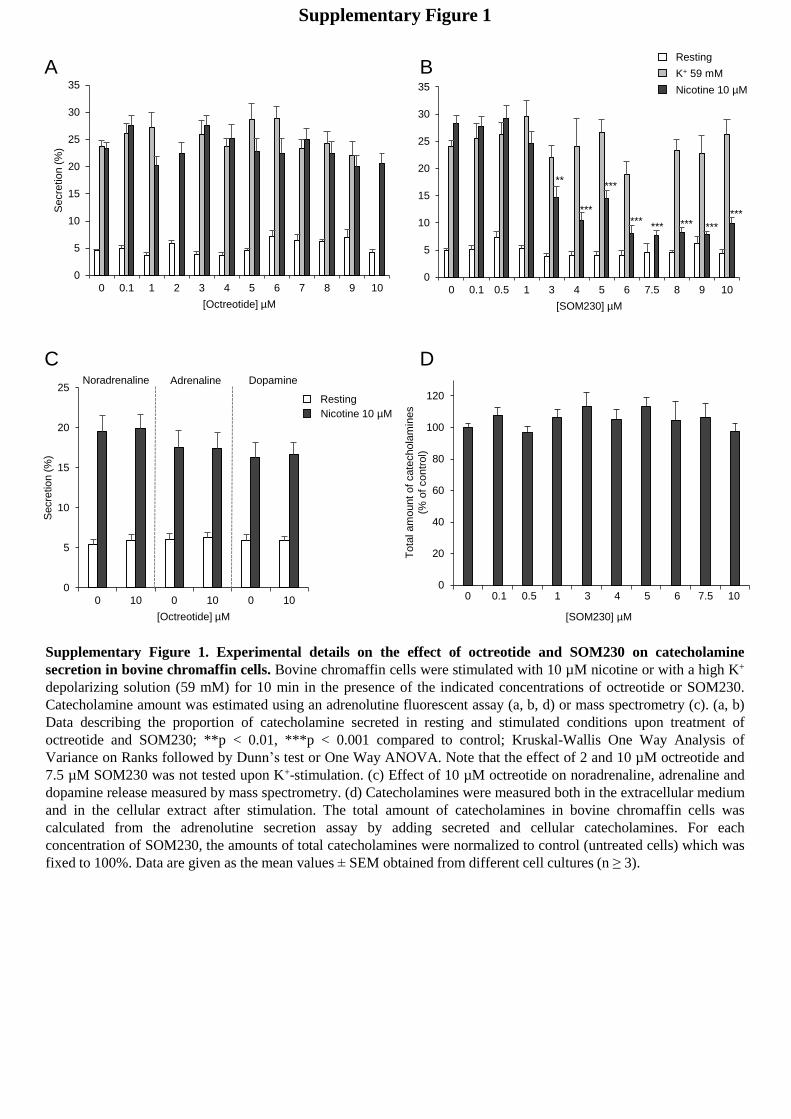
Supplementary Figure 1. Experimental details on the effect of octreotide and SOM230 on catecholamine
secretion in bovine chromaffin cells. Bovine chromaffin cells were stimulated with 10 µM nicotine or with a high K+
depolarizing solution (59 mM) for 10 min in the presence of the indicated concentrations of octreotide or SOM230.
Catecholamine amount was estimated using an adrenolutine fluorescent assay (a, b, d) or mass spectrometry (c). (a, b)
Data describing the proportion of catecholamine secreted in resting and stimulated conditions upon treatment of
octreotide and SOM230; **p < 0.01, ***p < 0.001 compared to control; Kruskal-Wallis One Way Analysis of
Variance on Ranks followed by Dunn’s test or One Way ANOVA. Note that the effect of 2 and 10 µM octreotide and
7.5 µM SOM230 was not tested upon K+-stimulation. (c) Effect of 10 µM octreotide on noradrenaline, adrenaline and
dopamine release measured by mass spectrometry. (d) Catecholamines were measured both in the extracellular medium
and in the cellular extract after stimulation. The total amount of catecholamines in bovine chromaffin cells was
calculated from the adrenolutine secretion assay by adding secreted and cellular catecholamines. For each
concentration of SOM230, the amounts of total catecholamines were normalized to control (untreated cells) which was
fixed to 100%. Data are given as the mean values ± SEM obtained from different cell cultures (n ≥ 3).
0
20
40
60
80
100
120
0,0 0,1 0,5 1,0 3,0 4,0 5,0 6,0 7,5 10,0[SOM230] µM
Tota
l am
ountof
cate
chola
min
es
(%of
contr
ol)
0 0.1 ,0.5 1 3 4 5 6 7.5 10
0
5
10
15
20
25
30
35
0 0.1 0.5 1 3 4 5 6 7.5 8 9 10
Secre
tio
n (
%)
[SOM230] µM
Nicotine 10 µM
K+ 59 mM
Resting
******
*** ***
***
***
***
**
0
5
10
15
20
25
30
35
0 0.1 1 2 3 4 5 6 7 8 9 10
Secre
tio
n (
%)
[Octreotide] µM
A B
D
0
5
10
15
20
25
0 10 0 10 0 10
Secre
tio
n (
%)
[Octreotide] µM
C
Nicotine 10 µM
Resting
Noradrenaline Adrenaline Dopamine
Supplementary Figure 1
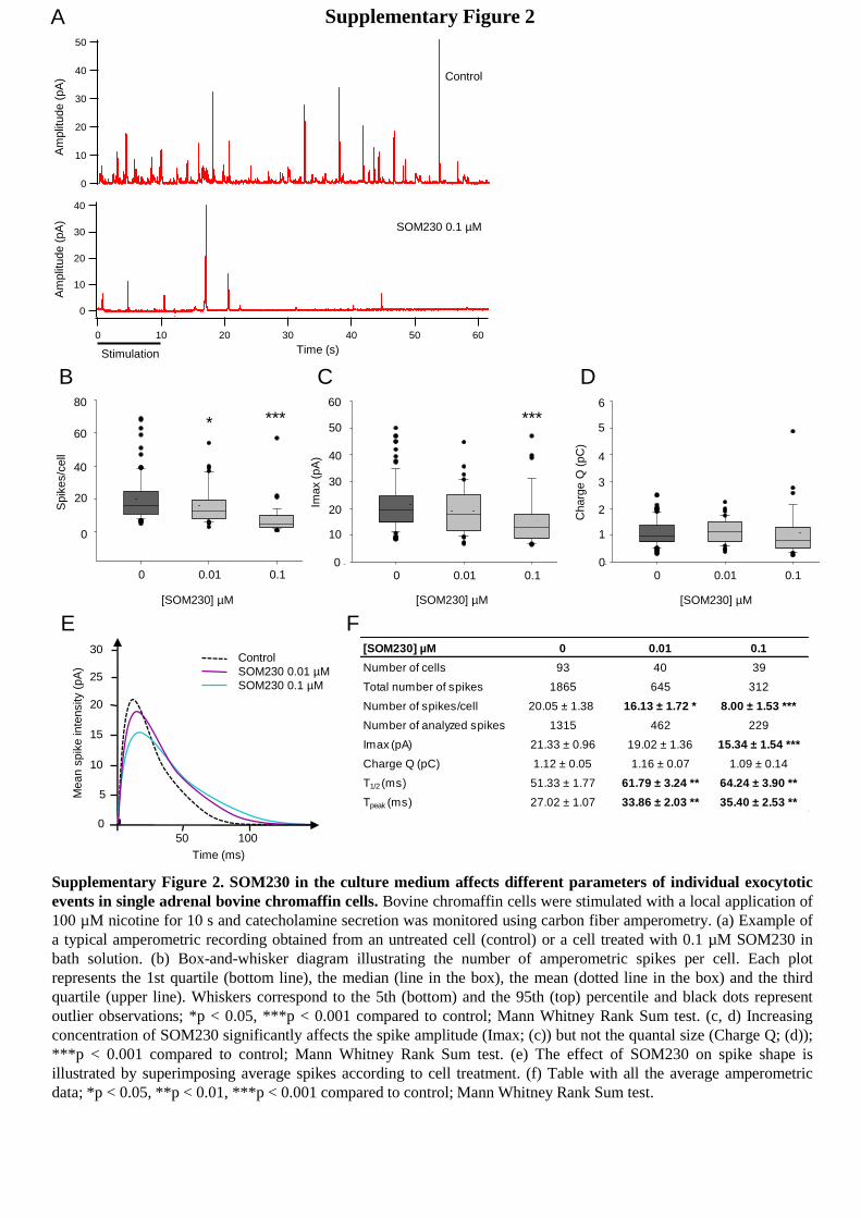
Supplementary Figure 2. SOM230 in the culture medium affects different parameters of individual exocytotic
events in single adrenal bovine chromaffin cells. Bovine chromaffin cells were stimulated with a local application of
100 µM nicotine for 10 s and catecholamine secretion was monitored using carbon fiber amperometry. (a) Example of
a typical amperometric recording obtained from an untreated cell (control) or a cell treated with 0.1 µM SOM230 in
bath solution. (b) Box-and-whisker diagram illustrating the number of amperometric spikes per cell. Each plot
represents the 1st quartile (bottom line), the median (line in the box), the mean (dotted line in the box) and the third
quartile (upper line). Whiskers correspond to the 5th (bottom) and the 95th (top) percentile and black dots represent
outlier observations; *p < 0.05, ***p < 0.001 compared to control; Mann Whitney Rank Sum test. (c, d) Increasing
concentration of SOM230 significantly affects the spike amplitude (Imax; (c)) but not the quantal size (Charge Q; (d));
***p < 0.001 compared to control; Mann Whitney Rank Sum test. (e) The effect of SOM230 on spike shape is
illustrated by superimposing average spikes according to cell treatment. (f) Table with all the average amperometric
data; *p < 0.05, **p < 0.01, ***p < 0.001 compared to control; Mann Whitney Rank Sum test.
B
A50
40
30
20
10
0
pA
6050403020100
s
Control
40
30
20
10
0
pA
6050403020100
s
Am
plit
ude (
pA
)
Time (s)Stimulation
SOM230 0.1 µM
C D
Am
plit
ude (
pA
)
2D Graph 3
Charg
e (
pC
)
0
1
2
3
4
5
6
DSMO 0,1 % SOM230 0,01 µM SOM230 0,1 µM
Charg
e Q
(pC
)
[SOM230] µM
0 0.01 0.10
1
2
3
4
5
6
2D Graph 2
Imax (
pA
)
0
10
20
30
40
50
60
DMSO 0,1 % SOM230 0,01 µM SOM230 0,1 µM
Imax (
pA
)
***
0 0.01 0.1
[SOM230] µM
60
50
40
30
20
10
0
2D Graph 1
Tota
l spik
es
0
20
40
60
80
Control SOM230 0,01 µM SOM230 0,1 µM
Spik
es/c
ell
0 0.01 0.1
****
[SOM230] µM
80
60
40
20
00
F
Control
SOM230 0.01 µM
SOM230 0.1 µM
Mean s
pik
e in
tensity (
pA
)
Time (ms)
0
5
10
15
20
50 100
25
30 [SOM230] µM 0 0.01 0.1
Number of cells 93 40 39
Total number of spikes 1865 645 312
Number of spikes/cell 20.05 ± 1.38 16.13 ± 1.72 * 8.00 ± 1.53 ***
Number of analyzed spikes 1315 462 229
Imax (pA) 21.33 ± 0.96 19.02 ± 1.36 15.34 ± 1.54 ***
Charge Q (pC) 1.12 ± 0.05 1.16 ± 0.07 1.09 ± 0.14
T1/2 (ms) 51.33 ± 1.77 61.79 ± 3.24 ** 64.24 ± 3.90 **
Tpeak (ms) 27.02 ± 1.07 33.86 ± 2.03 ** 35.40 ± 2.53 **
E
Supplementary Figure 2
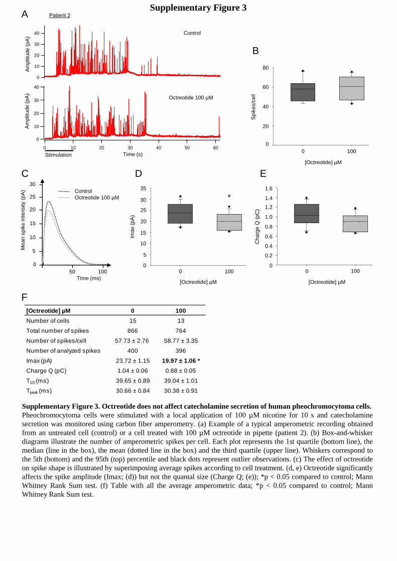
Am
plit
ude (
pA
)
Time (s)Stimulation
A
C D E
F
Control
Octreotide 100 µM
Mean s
pik
e in
tensity (
pA
)
Time (ms)
0
5
10
15
20
50 100
25
30
Patient 2
40
30
20
10
0
pA
6050403020100
s
40
30
20
10
0
pA
6050403020100
s
Control
Octreotide 100 µM
B
Am
plit
ude (
pA
)
Spik
es/c
ell
[Octreotide] µM
2D Graph 1
To
tal sp
ike
s
0
20
40
60
80
Control OCT 100 µM
0 100
80
60
40
20
00
Charg
e Q
(pC
)
[Octreotide] µM
2D Graph 3
Charg
e Q
(pC
)0,0
0,2
0,4
0,6
0,8
1,0
1,2
1,4
1,6
Control OCT 100 µM
0 100
1.6
1.4
1.2
1.0
0.8
0.6
0.4
0.2
0.0
Imax (
pA
)
0 100
*
[Octreotide] µM
2D Graph 2
Ima
x (
pA
)
0
5
10
15
20
25
30
35
Control OCT 100 µM
35
30
25
20
15
10
05
00
*
[Octreotide] µM 0 100
Number of cells 15 13
Total number of spikes 866 764
Number of spikes/cell 57.73 ± 2.76 58.77 ± 3.35
Number of analyzed spikes 400 396
Imax (pA) 23.72 ± 1.15 19.97 ± 1.06 *
Charge Q (pC) 1.04 ± 0.06 0.88 ± 0.05
T1/2 (ms) 39.65 ± 0.89 39.04 ± 1.01
Tpeak (ms) 30.66 ± 0.84 30.38 ± 0.91
Supplementary Figure 3. Octreotide does not affect catecholamine secretion of human pheochromocytoma cells.
Pheochromocytoma cells were stimulated with a local application of 100 µM nicotine for 10 s and catecholamine
secretion was monitored using carbon fiber amperometry. (a) Example of a typical amperometric recording obtained
from an untreated cell (control) or a cell treated with 100 µM octreotide in pipette (patient 2). (b) Box-and-whisker
diagrams illustrate the number of amperometric spikes per cell. Each plot represents the 1st quartile (bottom line), the
median (line in the box), the mean (dotted line in the box) and the third quartile (upper line). Whiskers correspond to
the 5th (bottom) and the 95th (top) percentile and black dots represent outlier observations. (c) The effect of octreotide
on spike shape is illustrated by superimposing average spikes according to cell treatment. (d, e) Octreotide significantly
affects the spike amplitude (Imax; (d)) but not the quantal size (Charge Q; (e)); *p < 0.05 compared to control; Mann
Whitney Rank Sum test. (f) Table with all the average amperometric data; *p < 0.05 compared to control; Mann
Whitney Rank Sum test.
Supplementary Figure 3
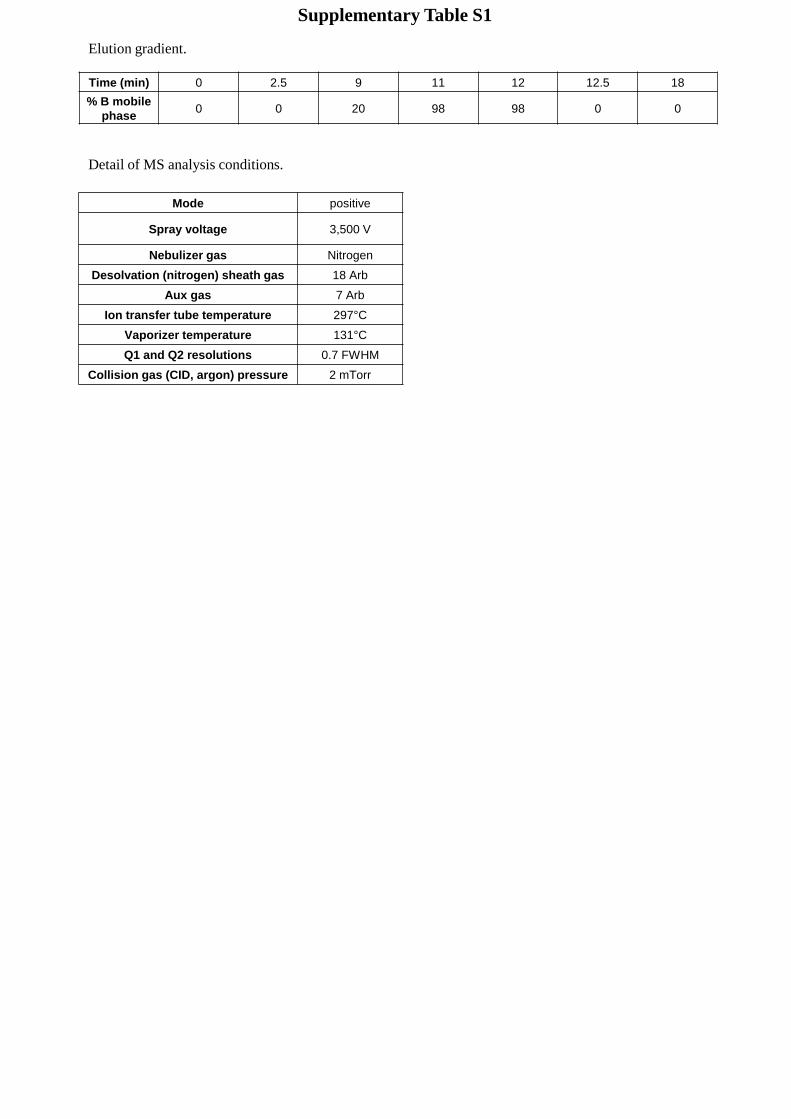
Elution gradient.
Time (min) 0 2.5 9 11 12 12.5 18
% B mobile
phase0 0 20 98 98 0 0
Detail of MS analysis conditions.
Mode positive
Spray voltage 3,500 V
Nebulizer gas Nitrogen
Desolvation (nitrogen) sheath gas 18 Arb
Aux gas 7 Arb
Ion transfer tube temperature 297°C
Vaporizer temperature 131°C
Q1 and Q2 resolutions 0.7 FWHM
Collision gas (CID, argon) pressure 2 mTorr
Supplementary Table S1
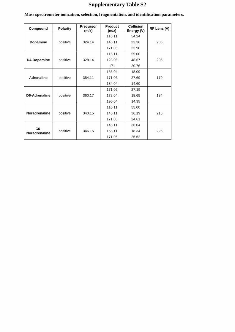
Compound PolarityPrecursor
(m/z)
Product
(m/z)
Collision
Energy (V)RF Lens (V)
Dopamine positive 324.14
116.11 54.24
206145.11 33.36
171.05 23.90
D4-Dopamine positive 328.14
116.11 55.00
206128.05 48.67
171 20.76
Adrenaline positive 354.11
166.04 18.09
179171.06 27.69
184.04 14.60
D6-Adrenaline positive 360.17
171.06 27.19
184172.04 18.65
190.04 14.35
Noradrenaline positive 340.15
116.11 55.00
215145.11 36.19
171.06 24.61
C6-
Noradrenalinepositive 346.15
145.11 36.04
226158.11 18.34
171.06 25.62
Mass spectrometer ionization, selection, fragmentation, and identification parameters.
Supplementary Table S2

DISCUSSION GÉNÉRALE
ET RÉSULTATS
COMPLÉMENTAIRES

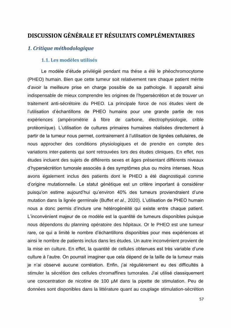
57
DISCUSSION GÉNÉRALE ET RÉSULTATS COMPLÉMENTAIRES
1. Critique méthodologique
1.1. Les modèles utilisés
Le modèle d’étude privilégié pendant ma thèse a été le phéochromocytome
(PHEO) humain. Bien que cette tumeur soit relativement rare chaque patient mérite
d’avoir la meilleure prise en charge possible de sa pathologie. Il apparaît ainsi
indispensable de mieux comprendre les origines de l’hypersécrétion et de trouver un
traitement anti-sécrétoire du PHEO. La principale force de nos études vient de
l’utilisation d’échantillons de PHEO humains pour une grande partie de nos
expériences (ampérométrie à fibre de carbone, électrophysiologie, crible
protéomique). L’utilisation de cultures primaires humaines réalisées directement à
partir de la tumeur nous permet, contrairement à l’utilisation de lignées cellulaires, de
nous approcher des conditions physiologiques et de prendre en compte des
variations inter-patients qui sont retrouvées lors des études cliniques. En effet, nos
études incluent des sujets de différents sexes et âges présentant différents niveaux
d’hypersécrétion tumorale associés à des symptômes plus ou moins intenses. Nous
avons également inclus des patients dont le PHEO a été diagnostiqué comme
d’origine mutationnelle. Le statut génétique est un critère important à considérer
puisqu’on estime aujourd’hui qu’environ 40% des tumeurs proviendraient d’une
mutation dans la lignée germinale (Buffet et al., 2020). L’utilisation de PHEO humain
nous a donc permis d’inclure une hétérogénéité qui existe entre chaque patient.
L’inconvénient majeur de ce modèle est la quantité de tumeurs disponibles puisque
nous dépendons du planning opératoire des hôpitaux. Or le PHEO est une tumeur
rare, ce qui a limité le nombre d’échantillons disponibles pour mes expériences et
ainsi le nombre de patients inclus dans les études. Un autre inconvénient provient de
la mise en culture. En effet, la quantité de cellules obtenues est très variable d’une
culture à l’autre. On pourrait imaginer que cela dépend de la taille de la tumeur mais
je n’ai observé aucune corrélation. Enfin, j’ai régulièrement eu des difficultés à
stimuler la sécrétion des cellules chromaffines tumorales. J’ai utilisé classiquement
une concentration de nicotine de 100 µM dans la pipette de stimulation. Peu de
données sont disponibles dans la littérature quant au couplage stimulation-sécrétion
Discussion générale et résultats complémentaires
58
des cellules chromaffines humaines. L’équipe du Dr. Albillos (Alicante, Espagne)
utilise jusqu’à 300 µM d’acétylcholine avec un système de perfusion très rapide pour
stimuler ces cellules (Hone et al., 2017). Je n’ai jamais testé l’acétylcholine mais
quelques fois, en augmentant la concentration de nicotine (de 300 µM à 1 mM), j’ai
pu obtenir plus de réponses. Malheureusement, cela n’a souvent pas été suffisant
pour obtenir un nombre robuste de cellules répondantes et des résultats exploitables.
Une variabilité de la sécrétion des cellules chromaffines humaines en réponse à
l’acétylcholine a déjà été montrée (Jaques and Tobes, 1986). Ces différences de
réponse à la stimulation peuvent venir soit de la qualité de la culture, soit du fait qu’il
s’agisse de tumeurs hétérogènes avec des phénotypes sécrétoires propres à chaque
patient. Un nouveau mode de stimulation avec de la nicotine cagée est en train d’être
mis en place au laboratoire, afin d’avoir un meilleur contrôle spatial et temporel lors
de la stimulation des cellules. La nicotine cagée placée dans le milieu des cellules
pourra être décagée grâce à un flash lumineux à un temps et une localisation précise
sur la cellule. Cet outil nous permettra de nous affranchir de la dilution du
sécrétagogue et d’une variabilité du placement de la pipette de stimulation (Filevich
et al., 2010).
Le second modèle que j’ai employé est la culture primaire de cellules
chromaffines bovines, qui m’a permis de réaliser les courbes doses-réponses des
analogues de la somatostatine et de déterminer une gamme de concentrations à
tester sur le PHEO. Les cellules chromaffines bovines ont l’avantage d’exprimer les
récepteurs nicotiniques α3β4 que l’on retrouve également majoritairement sur les
cellules chromaffines de la glande médullosurrénale humaine (Hone et al., 2015;
Criado, 2018). Le modèle bovin présente également plusieurs avantages techniques.
La partie médullaire de la glande surrénale est facilement dissociable de la partie
corticale ce qui facilite la culture et améliore les rendements. De plus, il permet
d’obtenir une grande quantité de cellules pour effectuer des expériences de
biochimie comme le dosage fluorimétrique des catécholamines.
Discussion générale et résultats complémentaires
59
1.2. La sécrétion mesurée sur des cellules en culture primaire est-elle
le reflet de la sécrétion tumorale ?
L’utilisation de cultures cellulaires constitue une approche utile et efficace pour
tester des drogues, mettre en évidence des mécanismes ou découvrir de nouvelles
fonctions. Cependant, je suis consciente que les données in vitro ne peuvent pas
être extrapolées directement à la physiologie, que ce soit dans un contexte sain ou
pathologique. Il faut donc rester prudent quant à l’interprétation des données, et se
questionner sur les limites des techniques et approches qui sont employées.
Une fois en culture les cellules tumorales conservent-elles les mêmes
capacités sécrétoires qu’in vivo ? Plusieurs études ont ainsi mis en évidence que la
quantité de catécholamines diminue au cours du temps dans les cultures de PHEO,
et ce malgré l’utilisation de différents milieux pour tenter de prévenir cette baisse
(Tischler et al., 1984; Jaques and Tobes, 1986). De même, les changements de
milieux impactent les taux de catécholamines (Finnegan et al., 1996). Ces
changements de taux de noradrénaline et d’adrénaline pour les PHEO en culture
pourrait impacter notre mesure ampérométrique. Dans notre protocole nous utilisons
les cellules chromaffines tumorales dans un délai de 48h en moyenne (parfois 72h
au maximum) après la mise en culture, or les changements décrits précédemment
sont observés à partir de 3-4 jours. De même notre mode de culture cellulaire en 2D
ne pourrait-il pas impacter la sécrétion que nous enregistrons ? En effet, en mettant
les cellules de PHEO en culture nous perdons les interactions inter-cellulaires et
avec le microenvironnement tumoral. Une étude a ainsi comparé une culture en 2D à
une culture en 3D de lignées humaines de tumeurs neuroendocrines (TNE)
pancréatiques, et a montré de meilleurs effets des drogues en culture 3D (Herrera-
Martínez et al., 2019). Pourrait-on envisager de tester nos drogues dans une modèle
de culture en 3D du PHEO ? De façon intéressante, l’utilisation de sphéroïdes
générés à partir d’une lignée cellulaire de PHEO de souris (MTT) a permis d’évaluer
l’effet de molécules sur la migration et la croissance tumorale (Bechmann et al.,
2018). Il serait intéressant d’évaluer l’effet des analogues de la somatostatine sur des
cultures de PHEO en 3D, afin d’inclure ces interactions inter-cellulaires et le
microenvironnement tumoral. Néanmoins, ce mode de culture ne me semble pas
compatible avec l’utilisation de l’ampérométrie à fibre de carbone qui requière la
Discussion générale et résultats complémentaires
60
présence de cellules isolées. Nous pourrions envisager de mesurer la sécrétion des
catécholamines avec une autre méthode telle que l’ELISA.
On peut également s’interroger sur l’impact de la technique d’ampérométrie à
fibre de carbone : quels sont les avantages et les inconvénients de cette technique
pour mesurer la sécrétion des cellules chromaffines ? L’ampérométrie à fibre de
carbone permet de mesurer la sécrétion des catécholamines sur cellule unique, et
nécessite ainsi un rendement de culture faible. L’utilisation d’un nombre restreint de
cellules convient parfaitement à notre modèle d’étude qu’est le PHEO, puisque je ne
récupère qu’une petite partie de la tumeur après exérèse chez le patient. De plus, la
mesure de la sécrétion sur cellule unique m’a permis de montrer que l’hypersécrétion
des PHEO provient probablement d’un dérèglement à l’échelle unicellulaire, et ne
peut pas être simplement attribuée à un effet de masse de la tumeur. Enfin, cette
technique donne accès à des mesures précises de la cinétique des évènements
d’exocytose.
L’ampérométrie à fibre de carbone repose sur l’oxydation des catécholamines
qui vont chacune libérer des électrons qui sont ensuite détectés par la fibre de
carbone (Mosharov and Sulzer, 2005). Cependant, l'oxydation électrochimique des
catéchols produit également des dérivés de quinone et des protons. En
conséquence, à moins que des mécanismes spécifiques puissent être adoptés par la
cellule pour réguler le pH près de sa membrane, le pH local entre la membrane
cellulaire et l'électrode chute dans la fente électrode/cellule. Lors de la stimulation,
une grande quantité de catécholamines est libérée générant beaucoup de protons, le
pH pouvant ainsi chuter à des valeurs en dessous de 6 dans la fente
électrode/cellule (Amatore et al., 2010).
Je me suis ensuite demandée si le nombre d’évènements d’exocytose que je
mesure peut être mis en lien avec la sécrétion tumorale en clinique. De façon
intéressante, une étude du laboratoire s’est intéressée à la corrélation entre des
paramètres de diagnostic mesurés en imagerie nucléaire (PET/CT : positron
emission tomography/computed tomography) par le 18F-FDOPA et la sécrétion des
PHEO. Une corrélation positive et significative a été montrée entre la radioactivité
maximale (SUVmax) mesurée dans la tumeur et le nombre de pics enregistrés en
ampérométrie. Ces résultats suggèrent que plus le nombre d’évènements
Discussion générale et résultats complémentaires
61
d’exocytose est élevé, plus la tumeur va absorber le traceur, conduisant à des
valeurs de SUVmax élevées. Il existerait donc bien des corrélations entre les
données ampérométriques mesurées in vitro et le comportement de la tumeur in
vivo. Malheureusement le taux d’incorporation du 18F-FDOPA n’a pu être corrélé aux
taux de métanéphrines et normétanéphrines (Moog et al., 2018). Dans nos études
nous n’avons pas trouvé de lien évident entre le nombre d’évènements d’exocytose
enregistrés et la sécrétion de catécholamines des PHEO. Un frein important à la
mise en évidence de corrélation est le nombre limité de patients, ainsi que le nombre
important de paramètres cliniques et biochimiques manquants pour chaque sujet. Il
serait intéressant de recruter une cohorte plus importante de patients présentant des
données biochimiques et cliniques complètes.
2. L’hypersécrétion tumorale : quels mécanismes possibles ?
Quelle est l’origine de l’hypersécrétion dans les PHEO ? Une des premières
idées qui vient à l’esprit est que l’hypersécrétion pourrait simplement être la
conséquence d’un effet de masse. En effet, la prolifération cellulaire importante au
cours du développement tumoral accroît significativement le nombre de cellules
chromaffines tumorales. Est-ce que cette augmentation de la masse cellulaire va
forcément entraîner une sécrétion plus importante ? Pour cela, il faudrait qu’une
même stimulation de la glande surrénale recrute plus de cellules chromaffines. À ma
connaissance aucune donnée n’est disponible dans la littérature à ce sujet. D’un
point de vue purement cellulaire, plusieurs hypothèses peuvent être formulées pour
tenter d’expliquer l’hypersécrétion des PHEO. Une fuite de catécholamines vers la
voie de l’exocytose constitutive pourrait engendrer une sécrétion excessive et surtout
non régulée. Une granulogenèse accrue augmentant significativement le nombre de
granules dans les cellules, ou bien simplement une augmentation de la quantité de
catécholamines par granule pourraient contribuer à une sécrétion plus importante
des PHEO. Enfin, on peut aisément penser qu’un dérèglement d’une des étapes
tardives de l’exocytose (recrutement, accostage, arrimage, amorçage, fusion)
pourrait aboutir à une plus grande libération de catécholamines.
Discussion générale et résultats complémentaires
62
Afin de vérifier qu’il n’y a pas une libération de catécholamines due à la voie
d’exocytose constitutive, j’ai mesuré la sécrétion par ampérométrie à fibre de
carbone sur des cellules chromaffines de PHEO sans les stimuler. Je n’ai jamais
détecté de sécrétion, tandis que des pics ampérométriques apparaissaient dès que
je stimulais ces cellules (résultats non publiés). Ces résultats montrent qu’il n’y a pas
de libération spontanée de catécholamines par les cellules tumorales et donc qu’il n’y
pas de fuite vers la voie d’exocytose constitutive. L’analyse protéomique réalisée sur
des tissus de PHEO en comparaison au tissu non tumoral indique une augmentation
de l’expression de plusieurs marqueurs granulaires dans les PHEO. On ne peut pas
exclure à ce stade qu’il n’y ait pas une augmentation de la granulogenèse dans les
cellules tumorales. Une analyse en microscopie électronique de la densité des
granules dans les cellules de PHEO est en cours dans l’équipe. Grâce à
l’ampérométrie à fibre de carbone, j’ai montré qu’une cellule chromaffine tumorale
présente une capacité sécrétrice plus importante, avec une augmentation très
significative du nombre d’évènements d’exocytose par rapport à une cellule
chromaffine non tumorale. Ces données sont intéressantes car elles montrent dans
un premier temps que la sécrétion est augmentée au niveau unicellulaire, ce qui n’est
pas en faveur d’un simple effet de masse dû à la prolifération cellulaire. Dans un
second temps, cela suggère bien un dérèglement du contrôle de la sécrétion par la
cellule. De plus, en analysant les paramètres des évènements d’exocytose, j’ai
observé pour de nombreux patients une cinétique de libération accélérée suggérant
que la sécrétion se faisait plus rapidement. Ainsi, pour une même stimulation, une
cellule chromaffine tumorale sécrèterait une quantité plus importante de
catécholamines en sollicitant plus de granules et en accélérant leur exocytose.
Reste à découvrir quelle(s) étape(s) et quel(s) acteur(s) de l’exocytose
pourraient être impliqués dans la dérégulation de la sécrétion des PHEO. Une
analyse par spectrométrie de masse réalisée par l’entreprise Caprion (Montréal,
Canada) en collaboration avec l’équipe a permis de générer une liste de protéines
potentiellement impliquées dans l’exocytose et dont l’expression est modifiée dans
les PHEO en comparaison au tissu non tumoral. Plusieurs protéines granulaires
telles que les chromogranines A et B (CGA, CGB), les sécrétogranines 2 et 3 (SCG2,
SCG3) sont surexprimées dans les PHEO. À noter qu’une augmentation de la CGA
plasmatique est retrouvée chez 90% des patients atteints de phéochromocytomes ou
Discussion générale et résultats complémentaires
63
de paragangliomes (PPGL), et a conduit à son utilisation pour le diagnostic de cette
tumeur (Stridsberg and Husebye, 1997; Grossrubatscher et al., 2006). La CGB n’est
pas utilisée en diagnostic, néanmoins sa concentration plasmatique est élevée chez
81% des patients atteints de PHEO (Stridsberg and Husebye, 1997). Parmi les
protéines les plus augmentées dans le PHEO sont retrouvées des enzymes
impliquées dans la synthèse et le traitement des composés granulaires (dopamine-β-
hydroxylase (DβH), les convertases neuroendocrines (PCSK), peptidylglycine α-
amidating monooxygenase (PAM),…), suggérant une dérégulation ces voies.
Notre analyse protéomique a également mis en évidence une augmentation
de plusieurs régulateurs de l’exocytose comme des protéines qui composent ou qui
régulent le complexe SNARE, des protéines qui régulent l’organisation du
cytosquelette d’actine ou encore certains régulateurs clés du processus d’exocytose.
Parmi les régulateurs impactés dans le PHEO on retrouve notamment des GTPase.
Par exemple, notre étude montre que la protéine Rab4A est augmentée de plus de 9
fois dans la condition tumorale. De façon intéressante, Rab4A semble impliquée
dans la sécrétion de procathepsine (une pro-enzyme) conférant un phénotype
tumoral agressif et métastatique dans le mélanome. L’expression de Rab4A est
corrélée avec la quantité de procathepsine sécrétée, et l’inhibition de cette GTPase
diminue la sécrétion tumorale de 80% (Barbarin and Frade, 2011). Dans notre étude
nous avons également montré que les deux isoformes Rab27A et Rab27B sont
surexprimées dans les PHEO. Des données de la littérature montrent que la
surexpression de Rab27A engendre une augmentation de la sécrétion de prostate-
specific antigen (PSA) et de prostatic-specific acid phosphatase (PSAP) dans une
lignée de carcinome de la prostate (LNCPa) (Johnson et al., 2005). De la même
façon, la surexpression de Rab27A favorise la sécrétion d’insulin-like growth factor-II
(IGF-II) ce qui confère un phénotype invasif et métastatique à des cellules de cancer
du sein (Wang et al., 2008). Enfin, la surexpression d’un mutant inactif de Rab27B
dans une lignée de tumeur pituitaire (AtT20) inhibe la sécrétion d’ACTH des cellules
(Zhao et al., 2002). Ces deux exemples montrent que des protéines régulatrices de
l’exocytose comme les GTPases semblent jouer un rôle dans le processus de
contrôle de la sécrétion tumorale. Nous avons montré qu’un grand nombre de
protéines sont impactées dans les PHEO, suggérant une dérégulation de différentes
étapes et processus de l’exocytose. De façon intéressante, une étude sur des PHEO
Discussion générale et résultats complémentaires
64
d’origine génétique (VHL et MEN2) a montré des variations d’expression de
nombreuses protéines de l’exocytose entre ces deux conditions. Ces résultats
suggèrent que l’origine génétique et le phénotype sécrétoire sont à l’origine de
signatures protéiques distinctes (Eisenhofer et al., 2008).
Nos résultats montrent que l’expression de protéines impliquées dans
l’exocytose ainsi qu’un certain nombre de régulateurs est modifiée dans les PHEO,
renforçant l’hypothèse d’une dérégulation de ce processus de sécrétion. Ces
variations d’expressions pourraient peut-être expliquer le dérèglement de la sécrétion
des PHEO. Il serait intéressant de confirmer le rôle de certaines de ces protéines
dans la sécrétion des PHEO en utilisant par exemple des approches de siRNA ou de
surexpression dans les cellules PC12 (PHEO de rat) et de suivre la capacité
sécrétrice des cellules.
Comme nous l’avons vu, l’hypersécrétion est un véritable problème pour les
patients puisqu’elle engendre de nombreux symptômes, il apparaît donc crucial de la
freiner.
3. Effet fonctionnel des analogues de la somatostatine
3.1. Octréotide et SOM230, un impact différent sur la sécrétion
catécholaminergique
Le premier constat que j’observe est la différence de résultats obtenus entre
l’octréotide (OCT) et le SOM230. En effet, contrairement au traitement par le
SOM230, un traitement par l’OCT ne modifie pas l’activité sécrétrice des cellules
chromaffines bovines et tumorales quelles que soient les concentrations. J’ai tenté
d’incuber les cellules chromaffines bovines avec de l’OCT pendant un temps plus
long, jusqu’à 24h, mais je n’ai observé aucun effet inhibiteur. Ces résultats sont en
accord avec la plupart des données de la littérature. Pour exemple, les travaux de
Ribeiro et collaborateurs en 2006 démontrent qu’à des concentrations de 1 et 10 µM
l’OCT n’a pas d’effet sur la sécrétion de cellules chromaffines de bœuf (Ribeiro et al.,
2006). In vivo, l’administration d'OCT aux patients atteints de PHEO ne semble pas
améliorer les symptômes et ne modifie pas la quantité de catécholamines urinaires et
plasmatiques (Plouin et al., 1995). De même, chez des patients porteurs de PHEO
malin/récurrent ou de paragangliome (PGL) en progression, l’injection d’OCT n’a pas
Discussion générale et résultats complémentaires
65
ou très peu d’effet sur les concentrations de catécholamines (Kopf et al., 1997;
Lamarre-Cliche et al., 2002; van Hulsteijn et al., 2013). Quelques cas dans la
littérature ont néanmoins rapporté un effet inhibiteur de l’OCT sur des tumeurs issues
des cellules chromaffines. Ainsi, une perfusion de 2h d’OCT à 6 patients atteints de
PPGL permet de diminuer le taux de noradrénaline. Néanmoins les valeurs
remontent au seuil initial dès que la perfusion est arrêtée (Invitti et al., 1993). Un
patient atteint de PHEO dont la sécrétion est incontrôlable par d’autres thérapies et
un patient porteur de PGL présentent également une diminution du taux de
catécholamines suite à l’injection d’OCT (Koriyama et al., 2000; Elshafie et al., 2014).
Néanmoins ces études portent sur un nombre très restreint de patients.
Concernant l’effet du SOM230, une seule étude publiée a utilisé cette
molécule sur des cellules chromaffines tumorales. Dans cet article, les auteurs
proposent qu’un traitement prolongé (24h) des cellules de PHEO humain par le
SOM230 diminuerait la quantité de catécholamines stockées dans les cellules de
PHEO (Pasquali et al., 2008). Il est difficile de comparer ces données aux nôtres
étant donné que nos conditions expérimentales sont très différentes. En effet dans
mes protocoles, soit le SOM230 est libéré en puff pendant 10 s et agit donc très
rapidement, soit il est présent en bain dans le milieu cellulaire pendant 1h maximum.
Au cours de mes expériences avec les cellules de PHEO, j’ai pu observer une
diminution de la charge Q des pics ampérométriques sur 2 cultures seulement
(patients 1 et 5). Cependant, il est difficile d’imaginer qu’en quelques secondes le
SOM230 puisse agir sur la concentration catécholaminergique des granules. Une
hypothèse serait que, pour certains patients, un traitement des cellules par le
SOM230 favoriserait des évènements de sécrétion partiels.
Le SOM230 possède donc un effet inhibiteur sur la sécrétion de
catécholamines tandis que l’OCT non. Qu’est ce qui pourrait expliquer cette
différence d’effet entre les deux molécules ? On sait que les affinités de ces deux
analogues pour les récepteurs à la somatostatine (SSTR) sont différentes. L’OCT a
une affinité préférentielle pour le SSTR2, tandis que le SSTR5 est la cible privilégiée
du SOM230. À noter que le SOM230 possède une affinité 30, 5 et 40 fois plus élevée
pour les SSTR1, SSTR3 et SSTR5 respectivement en comparaison à l’OCT (Schmid,
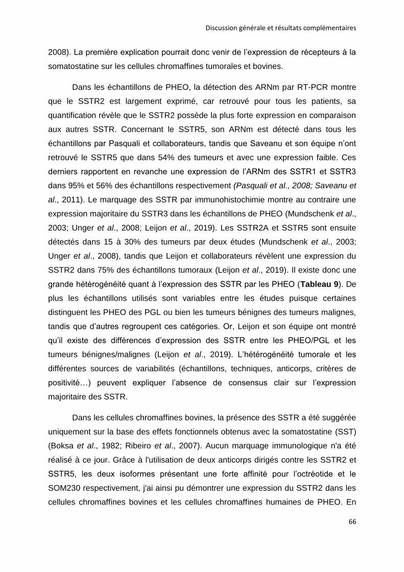
Discussion générale et résultats complémentaires
66
2008). La première explication pourrait donc venir de l’expression de récepteurs à la
somatostatine sur les cellules chromaffines tumorales et bovines.
Dans les échantillons de PHEO, la détection des ARNm par RT-PCR montre
que le SSTR2 est largement exprimé, car retrouvé pour tous les patients, sa
quantification révèle que le SSTR2 possède la plus forte expression en comparaison
aux autres SSTR. Concernant le SSTR5, son ARNm est détecté dans tous les
échantillons par Pasquali et collaborateurs, tandis que Saveanu et son équipe n’ont
retrouvé le SSTR5 que dans 54% des tumeurs et avec une expression faible. Ces
derniers rapportent en revanche une expression de l’ARNm des SSTR1 et SSTR3
dans 95% et 56% des échantillons respectivement (Pasquali et al., 2008; Saveanu et
al., 2011). Le marquage des SSTR par immunohistochimie montre au contraire une
expression majoritaire du SSTR3 dans les échantillons de PHEO (Mundschenk et al.,
2003; Unger et al., 2008; Leijon et al., 2019). Les SSTR2A et SSTR5 sont ensuite
détectés dans 15 à 30% des tumeurs par deux études (Mundschenk et al., 2003;
Unger et al., 2008), tandis que Leijon et collaborateurs révèlent une expression du
SSTR2 dans 75% des échantillons tumoraux (Leijon et al., 2019). Il existe donc une
grande hétérogénéité quant à l’expression des SSTR par les PHEO (Tableau 9). De
plus les échantillons utilisés sont variables entre les études puisque certaines
distinguent les PHEO des PGL ou bien les tumeurs bénignes des tumeurs malignes,
tandis que d’autres regroupent ces catégories. Or, Leijon et son équipe ont montré
qu’il existe des différences d’expression des SSTR entre les PHEO/PGL et les
tumeurs bénignes/malignes (Leijon et al., 2019). L’hétérogénéité tumorale et les
différentes sources de variabilités (échantillons, techniques, anticorps, critères de
positivité…) peuvent expliquer l’absence de consensus clair sur l’expression
majoritaire des SSTR.
Dans les cellules chromaffines bovines, la présence des SSTR a été suggérée
uniquement sur la base des effets fonctionnels obtenus avec la somatostatine (SST)
(Boksa et al., 1982; Ribeiro et al., 2007). Aucun marquage immunologique n'a été
réalisé à ce jour. Grâce à l'utilisation de deux anticorps dirigés contre les SSTR2 et
SSTR5, les deux isoformes présentant une forte affinité pour l’octréotide et le
SOM230 respectivement, j'ai ainsi pu démontrer une expression du SSTR2 dans les
cellules chromaffines bovines et les cellules chromaffines humaines de PHEO. En
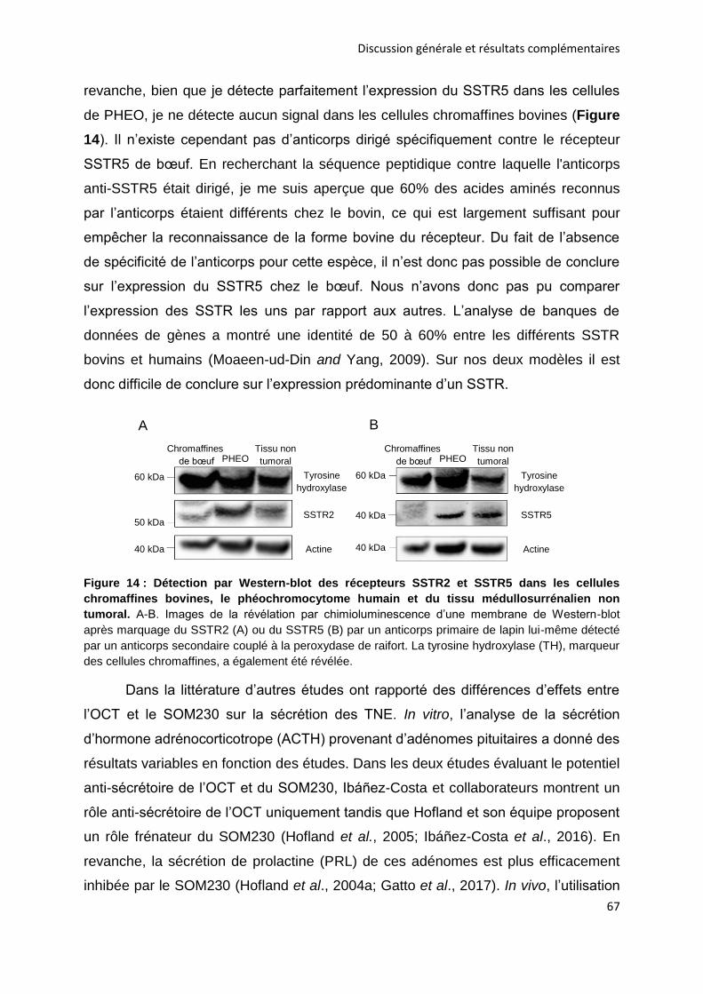
Discussion générale et résultats complémentaires
67
revanche, bien que je détecte parfaitement l’expression du SSTR5 dans les cellules
de PHEO, je ne détecte aucun signal dans les cellules chromaffines bovines (Figure
14). Il n’existe cependant pas d’anticorps dirigé spécifiquement contre le récepteur
SSTR5 de bœuf. En recherchant la séquence peptidique contre laquelle l'anticorps
anti-SSTR5 était dirigé, je me suis aperçue que 60% des acides aminés reconnus
par l’anticorps étaient différents chez le bovin, ce qui est largement suffisant pour
empêcher la reconnaissance de la forme bovine du récepteur. Du fait de l’absence
de spécificité de l’anticorps pour cette espèce, il n’est donc pas possible de conclure
sur l’expression du SSTR5 chez le bœuf. Nous n’avons donc pas pu comparer
l’expression des SSTR les uns par rapport aux autres. L’analyse de banques de
données de gènes a montré une identité de 50 à 60% entre les différents SSTR
bovins et humains (Moaeen-ud-Din and Yang, 2009). Sur nos deux modèles il est
donc difficile de conclure sur l’expression prédominante d’un SSTR.
Figure 14 : Détection par Western-blot des récepteurs SSTR2 et SSTR5 dans les cellules
chromaffines bovines, le phéochromocytome humain et du tissu médullosurrénalien non
tumoral. A-B. Images de la révélation par chimioluminescence d’une membrane de Western-blot
après marquage du SSTR2 (A) ou du SSTR5 (B) par un anticorps primaire de lapin lui-même détecté
par un anticorps secondaire couplé à la peroxydase de raifort. La tyrosine hydroxylase (TH), marqueur
des cellules chromaffines, a également été révélée.
Dans la littérature d’autres études ont rapporté des différences d’effets entre
l’OCT et le SOM230 sur la sécrétion des TNE. In vitro, l’analyse de la sécrétion
d’hormone adrénocorticotrope (ACTH) provenant d’adénomes pituitaires a donné des
résultats variables en fonction des études. Dans les deux études évaluant le potentiel
anti-sécrétoire de l’OCT et du SOM230, Ibáñez-Costa et collaborateurs montrent un
rôle anti-sécrétoire de l’OCT uniquement tandis que Hofland et son équipe proposent
un rôle frénateur du SOM230 (Hofland et al., 2005; Ibáñez-Costa et al., 2016). En
revanche, la sécrétion de prolactine (PRL) de ces adénomes est plus efficacement
inhibée par le SOM230 (Hofland et al., 2004a; Gatto et al., 2017). In vivo, l’utilisation
Chromaffines de bœuf PHEO
Tissu non
tumoral 60 kDa
50 kDa
40 kDa
Tyrosine
hydroxylase
SSTR2
Actine
A
Chromaffines de bœuf PHEO
Tissu non
tumoral 60 kDa
40 kDa
40 kDa
Tyrosine
hydroxylase
SSTR5
Actine
B
Discussion générale et résultats complémentaires
68
de souris développant des adénomes pituitaires montre que la sécrétion de PRL est
diminuée uniquement par une forte dose de SOM230 tandis qu’un traitement par
l’OCT est sans effet (Fedele et al., 2007). Ces divergences d’effets pourraient-elles
être en lien avec l’expression des SSTR, notamment les SSTR2 et SSTR5, sur ces
tumeurs ? L’ARNm du SSTR5 a été montré comme prédominant dans les adénomes
pituitaires sécrétant de l’ACTH, pourtant une étude a montré un effet inhibiteur de
l’OCT et l’autre du SOM230 (Hofland et al., 2005; Ibáñez-Costa et al., 2016). En
revanche, sur un modèle de souris porteuses d’adénomes pituitaires, l’ARNm du
SSTR2 est majoritaire et 100 fois plus important que le SSTR5, et l’étude a rapporté
un effet inhibiteur du SOM230 et aucun effet de l’OCT (Fedele et al., 2007). Il a été
monté que l'efficacité anti-sécrétoire de l'OCT et du SOM230 est corrélée
positivement avec le taux d'ARNm du SSTR2, mais pas du SSTR5, sur des
adénomes pituitaires humains sécrétant de l’hormone de croissance (GH) (Hofland et
al., 2004a; Gatto et al., 2017). Néanmoins, dans ces études c’est le niveau d’ARNm
qui est évalué et non l’expression de la protéine. De façon intéressante, une étude a
montré que la réponse au SOM230 chez des patients atteints d’adénomes
sécrétants de la GH est dépendante de l’expression du SSTR2 mais pas du SSTR5.
Après 24 semaines de traitement une corrélation positive entre la réponse hormonale
et la quantité de la protéine SSTR2 a été trouvée : plus le SSTR2 était exprimé dans
l’échantillon tumoral plus la diminution de la sécrétion était importante. En revanche
aucune corrélation n’a été montrée avec l’expression du SSTR5 (Muhammad et al.,
2019).
Ces corrélations sont en adéquation avec l’affinité préférentielle de l’OCT pour
le SSTR2. En revanche, dans ces tumeurs, l’effet anti-sécrétoire du SOM230 ne
semble pas dépendre du SSTR5 qui est pourtant son récepteur préférentiel. Dans les
PHEO il n’existe pas de consensus clair sur l’expression majoritaire d’un sous-type
de SSTR, néanmoins l’ARNm ou la protéine des SSTR2, SSTR3 et SSTR5 semblent
retrouvés le plus fréquemment (Tableau 9). Le SOM230 se fixe respectivement avec
30, 5, et 40 fois plus d’affinité aux SSTR1, SSTR3 et SSTR5 que l’OCT tandis que ce
dernier ne se fixe qu’avec 2,5 fois plus d’affinité au SSTR2 que le SOM230 (Schmid,
2008). On peut imaginer que l’affinité préférentielle de l’OCT pour le SSTR2 et sa
moindre affinité pour les autres récepteurs ne lui permettent pas d’exercer un effet
anti-sécrétoire sur les cellules chromaffines contrairement au SOM230.
Discussion générale et résultats complémentaires
69
3.2. Mode d’action du SOM230
Au fur et à mesure des expériences, j’ai commencé à m’interroger sur le mode
d’action par lequel le SOM230 pouvait inhiber la sécrétion. En effet, le SOM230
inhibe exclusivement la sécrétion induite par une stimulation nicotinique, alors que je
n’observe aucun effet quand la sécrétion est déclenchée par une solution
dépolarisante de potassium. De manière intéressante, des auteurs ont fait la même
constatation mais en utilisant de la SST. En effet, ils démontrent que la SST inhibe la
sécrétion de cellules chromaffines bovines ou de cobaye uniquement avec une
solution de nicotine ou d’acétylcholine, tandis que l’utilisation du potassium et
d’autres sécrétagogues tels que la pilocarpine (agoniste muscarinique), l’histamine
ou l’angiotensine n’ont pas d’effet (Mizobe et al., 1979; Role et al., 1981; Livett and
Boksa, 1984; Moeller et al., 1989).
Nous avons montré que le SOM230 est capable de diminuer l’amplitude des
courants nicotiniques. Une des hypothèses soulevées par ces données serait que
l’action frénatrice du SOM230 pourrait s’exercer via le récepteur nicotinique.
Certaines données de la littérature obtenues avec la SST semblent corroborer cette
hypothèse. Plusieurs auteurs ont suggéré que la SST pourrait agir comme un
modulateur du récepteur nicotinique notamment en impactant sa désensibilisation
(Mizobe et al., 1979; Ribeiro et al., 2006). De façon intéressante, l’incubation de
cellules chromaffines de cobaye avec de la SST a pour effet de diminuer les courants
nicotiniques de façon dose dépendante et réversible. Les auteurs montrent
également que la SST facilite la désensibilisation du récepteur (Inoue and Kuriyama,
1991).
La question est maintenant de savoir par quel(s) mécanisme(s) le SOM230
agirait-il sur le récepteur nicotinique ? Une hypothèse possible est que le SOM230
pourrait être un antagoniste du récepteur nicotinique. Pour tester cette possibilité j’ai
testé différentes doses de SOM230 sur les cellules chromaffines bovines. Pour
chaque dose d’analogue, j’ai fait varier la concentration de nicotine lors de la
stimulation avant de mesurer la sécrétion des catécholamines par dosage
fluorimétrique. Les courbes doses-réponses obtenues et leurs déplacements me
permettent de déterminer si le SOM230 est un antagoniste compétitif ou non
compétitif du récepteur nicotinique.
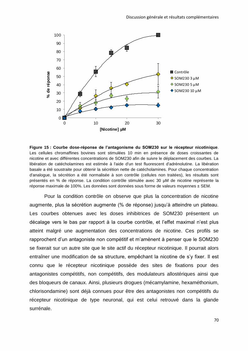
Discussion générale et résultats complémentaires
70
0
10
20
30
40
50
60
70
80
90
100
0 10 20 30
% d
e r
ép
on
se
[Nicotine] µM
Contrôle
SOM230 3 µM
SOM230 5 µM
SOM230 10 µM
Figure 15 : Courbe dose-réponse de l’antagonisme du SOM230 sur le récepteur nicotinique.
Les cellules chromaffines bovines sont stimulées 10 min en présence de doses croissantes de
nicotine et avec différentes concentrations de SOM230 afin de suivre le déplacement des courbes. La
libération de catécholamines est estimée à l'aide d'un test fluorescent d'adrénolutine. La libération
basale a été soustraite pour obtenir la sécrétion nette de catécholamines. Pour chaque concentration
d'analogue, la sécrétion a été normalisée à son contrôle (cellules non traitées), les résultats sont
présentés en % de réponse. La condition contrôle stimulée avec 30 µM de nicotine représente la
réponse maximale de 100%. Les données sont données sous forme de valeurs moyennes ± SEM.
Pour la condition contrôle on observe que plus la concentration de nicotine
augmente, plus la sécrétion augmente (% de réponse) jusqu’à atteindre un plateau.
Les courbes obtenues avec les doses inhibitrices de SOM230 présentent un
décalage vers le bas par rapport à la courbe contrôle, et l’effet maximal n’est plus
atteint malgré une augmentation des concentrations de nicotine. Ces profils se
rapprochent d’un antagoniste non compétitif et m’amènent à penser que le SOM230
se fixerait sur un autre site que le site actif du récepteur nicotinique. Il pourrait alors
entraîner une modification de sa structure, empêchant la nicotine de s’y fixer. Il est
connu que le récepteur nicotinique possède des sites de fixations pour des
antagonistes compétitifs, non compétitifs, des modulateurs allostériques ainsi que
des bloqueurs de canaux. Ainsi, plusieurs drogues (mécamylamine, hexaméthonium,
chlorisondamine) sont déjà connues pour être des antagonistes non compétitifs du
récepteur nicotinique de type neuronal, qui est celui retrouvé dans la glande
surrénale.
Discussion générale et résultats complémentaires
71
Quelles autres hypothèses peuvent être envisagées ? À ce stade nous ne
pouvons pas exclure totalement qu’au moins une partie de l’effet du SOM230 passe
par son propre récepteur à la somatostatine. Les SSTR sont des récepteurs couplés
aux protéines G qui pourraient activer une cascade de signalisation aboutissant à
l’inhibition du récepteur nicotinique. Afin de vérifier cette hypothèse, il serait
intéressant de tester l’effet du SOM230 sur les courants nicotiniques en présence ou
en absence des récepteurs à la somatostatine en utilisant un système d’expression
hétérologue tel que l’ovocyte de Xénope. De façon intéressante des auteurs ont
exprimé un ou des SSTR dans ce modèle (White and Reisine, 1990; Kreienkamp et
al., 1998). Néanmoins cette expérience est complexe du fait du nombre important de
sous-types de récepteurs à la somatostatine. Afin de déterminer si le SOM230
exerce son action exclusivement via le récepteur nicotinique ou en partie via ses
propres récepteurs, j’ai réalisé des expériences de sécrétion en utilisant un
antagoniste des SSTR (cycloSST). Je souhaitais voir si le SOM230 conservait son
effet inhibiteur sur la sécrétion des catécholamines en bloquant les SSTR. Dans ce
cas nous aurions montré que l’action du SOM230 passe uniquement par le récepteur
nicotinique. Malheureusement l’utilisation de cette drogue s’est avérée inefficace car,
à certaines doses, elle avait elle-même un effet inhibiteur sur la sécrétion, et en
utilisant des doses trop faibles je n’étais pas certaine de bloquer tous les récepteurs,
ni tous les sous-types. De façon intéressante cette expérience a été menée par un
autre laboratoire avec la SST sur des cellules chromaffines bovines. Les auteurs ont
montré que l’utilisation du cycloSST permettait de reverser l’effet inhibiteur de la
SST, sous tendant que l’action inhibitrice de la SST passerait par ses propres
récepteurs somatostatinergiques (Ribeiro et al., 2007). Dans cette étude la dose de
cycloSST utilisée était 10 fois supérieure à celle de la SST, condition qu’il ne m’était
pas possible de reproduire avec le SOM230. Néanmoins, du fait des différences
structurales et d’affinité entre la SST et le SOM230, il est possible que l’effet
inhibiteur observé sur la sécrétion des catécholamines ne passe pas par les mêmes
récepteurs (Tableau 8).
Enfin, plusieurs études ont mis en évidence que les SSTR sont capables de
former des homo- ou des hétéro-oligomères entre les sous-types de SSTR ou avec
d’autres récepteurs couplés aux protéines G. Par exemple, le SSTR5 peut former
des dimères avec les SSTR1 et SSTR3, mais pas avec le SSTR4 (Rocheville et al.,
Discussion générale et résultats complémentaires
72
2000b; Pfeiffer et al., 2001; Grant et al., 2008). De façon intéressante, le SSTR5 peut
former des hétéromères avec le récepteur à la dopamine D2 pour créer un nouveau
récepteur avec une pharmacologie différente puisque l’affinité à la dopamine et ses
agonistes est modifiée (Rocheville et al., 2000a). L’interaction entre le SSTR2A et le
récepteur µ opioïde 1 quant à elle modifie le trafic des récepteurs puisque la
phosphorylation, l’internalisation et la désensibilisation des récepteurs sont affectés.
En revanche, la fixation des ligands n’est pas affectée (Pfeiffer et al., 2002). Une
oligomérisation potentielle entre les SSTR et les récepteurs nicotiniques n'a jamais
été rapportée et serait, selon moi, très intéressante à étudier.
4. Le SOM230 : vers les prémices d’un traitement de l’hypersécrétion ?
4.1. Dose utilisée
Dans notre étude nous avons démontré pour la première fois que le SOM230
a un effet inhibiteur sur la sécrétion de catécholamines de cellules chromaffines
bovines et tumorales humaines in vitro. Il est donc tentant d’imaginer l’utilisation du
SOM230 comme un potentiel traitement anti-sécrétoire pour cette tumeur. Si l'on
réfléchit en termes d'application clinique potentielle du SOM230 pour le PHEO, que
révèlent nos données ?
En utilisant les cellules chromaffines bovines j’ai déterminé une concentration
inhibitrice médiane (CI50) de 2,64 µM pour le SOM230. Dans la littérature, l’utilisation
de modèles de sécrétion tels que les cellules pancréatiques et pituitaires de rats ont
montré un effet inhibiteur de cette drogue à partir de concentrations de l’ordre du nM
(Bruns et al., 2002; Ludvigsen et al., 2007). De la même façon, l’incubation de
SOM230 sur des lignées ou cultures primaires tumorales a montré un effet anti-
sécrétoire à partir de 1 nM voire à des doses plus faibles, avec des CI50 de 0,06 à 0,5
nM (Hofland et al., 2004b, 2005; van der Hoek et al., 2005; Batista et al., 2006;
Mariniello et al., 2011; Murasawa et al., 2014). La CI50 que nous avons déterminée
est donc bien supérieure à celles retrouvées dans la littérature. Mes résultats se
rapprochent en revanche de ceux obtenus avec la SST, puisqu’un effet anti-
sécrétoire sur les cellules chromaffines est obtenu avec des concentrations de l’ordre
du µM (Mizobe et al., 1979; Boksa et al., 1982; Livett and Boksa, 1984). Une CI50
d’environ 2 µM a été calculée par Role et collaborateurs (Role et al., 1981). Grâce à
Discussion générale et résultats complémentaires
73
l’ampérométrie à fibre de carbone, nous avons ensuite montré que le SOM230 inhibe
significativement la sécrétion de cellules bovines et de PHEO humains à partir de 0,1
µM, abaissant ainsi la concentration utilisée. En accord avec mes résultats, des
études sur des tumeurs humaines ont montré que le SOM230 avait un effet inhibiteur
à 0,1 µM (van Hoek et al., 2009; Ibáñez-Costa et al., 2016). La seule étude menée
sur le PHEO humain a montré un effet du SOM230 à partir de 0,1 µM sur l’apoptose
et la quantité de catécholamines intracellulaires (Pasquali et al., 2008). Dans la
plupart des études, les cellules ont été incubées pendant plusieurs heures et jusqu’à
96h. L’incubation de cellules chromaffines bovines pendant 24 et 48h avec du
SOM230 ne m’a pas permis de montrer un effet à de plus faibles doses (résultats
non montrés).
La question centrale est maintenant de savoir si l’on peut envisager une
application clinique pour le PHEO ? Actuellement le SOM230 est utilisé dans le
traitement de deux TNE pituitaires : la première hypersécrète de l’ACTH, entraînant
une augmentation de la sécrétion de cortisol par la glande surrénale, et ainsi un
hypercortisolisme à l’origine de la maladie de Cushing (Pivonello et al., 2020), tandis
que la seconde libère un excès de GH à l’origine d’acromégalie (Cuevas-Ramos and
Fleseriu, 2016). On peut donc envisager que l’application clinique du SOM230 soit
étendue à une autre TNE. Mais est-ce compatible en termes de dose chez
l’Homme ?
Deux formulations ont été évaluées en clinique pour le SOM230, une à courte
durée d’action dont les injections sont requises plusieurs fois par jour en sous-
cutanée (s.c), et une à longue durée d’action injectée une fois par mois en
intramusculaire (i.m). Les taux circulants de SOM230 après son injection ont été
mesurés dans plusieurs études. Les concentrations plasmatiques minimum (Cmin) et
maximum (Cmax) de SOM230 ont été mesurées chez des patients atteints de TNE
avancées, réfractaires à l’OCT et traités avec 150 à 1200 µg (s.c) de SOM230. Une
Cmin de 1,4 ng/mL a été obtenue à la suite de l’injection de 150 µg de SOM230,
tandis que la Cmax était de 74 ng/mL avec 1200 µg de SOM230. Chez les patients
répondant au traitement, la concentration plasmatique effective médiane était de 10
ng/mL (Kvols et al., 2012). Dans une seconde étude, des patients atteints de TNE
gastro-entéro-pancréatiques (GEP) résistants à l’OCT ou au lanréotide ont été traités
Discussion générale et résultats complémentaires
74
avec le SOM230 à longue durée d’action. Les sujets ont reçu des doses de 20, 40 ou
60 mg de l’analogue en i.m une fois/mois pendant 3 mois. 24h après l’injection de 20
et 60 mg de SOM230, la Cmax était de 4,5 et 7 ng/mL respectivement, tandis qu’après
3 mois de traitement la Cmax était de 5,5 et 25 ng/mL pour chacune des doses (Wolin
et al., 2013). Enfin, l’injection de SOM230 à des volontaires sains a montré des
concentrations plasmatiques circulantes proches des études précédentes, de 10 à
40 ng/mL (Dietrich et al., 2012; Petersenn et al., 2012; Chen et al., 2014).
Ces données montrent que les doses circulantes de SOM230 après injection
sont variables en fonction des doses, des voies d’injections et des délais considérés.
Mes résultats montrent un effet inhibiteur du SOM230 sur la sécrétion des PHEO à
partir de 0,1 µM soit 143 ng/mL. Dans les études les doses circulantes de SOM230
se situent entre 10 et 40 ng/mL environ. Kvols et collaborateurs ont déterminé une
concentration effectrice médiane de 10 ng/mL, soit 14 fois plus faible que la dose
que j’ai déterminée. Cependant, nous ne pouvons pas prévoir la dose à laquelle le
SOM230 pourrait exercer son action anti-sécrétoire in vivo s’il était injecté à des
patients atteints de PHEO. Mes résultats ont permis de montrer pour la première fois
que le SOM230 pourrait être considéré comme un potentiel traitement anti-sécrétoire
pour le PHEO, et permettent d’ouvrir des perspectives pour d’éventuelles études
cliniques.
4.2. Effet anti-sécrétoire du SOM230 en clinique
Mes résultats démontrent un effet inhibiteur du SOM230 sur la sécrétion
catécholaminergique du PHEO. Ces résultats sont en accord avec l’efficacité de
cette molécule qui a été démontrée en clinique chez l’Homme. 17 études cliniques
utilisant le SOM230 en diagnostic ou en thérapeutique sur des TNE sont recensées
sur le site de référence « clinical trials » (https://www.clinicaltrials.gov/), dont une
douzaine sont achevées.
Le SOM230 est actuellement utilisé pour traiter la maladie de Cushing, une
tumeur pituitaire hypersécrétant de l’ACTH. Le SOM230 diminue le taux de cortisol,
d’ACTH et la taille de la tumeur, améliorant ainsi les symptômes des patients
(Pivonello et al., 2020). Cette efficacité anti-tumorale a amené les scientifiques à
tester le SOM230 sur d’autres TNE. Après 6 mois de traitement par le SOM230, 21%
Discussion générale et résultats complémentaires
75
des patients souffrant de TNE gastro-intestinales métastatiques incontrôlées par
d’autres analogues de la somatostatine présentent un contrôle de leurs symptômes
(Wolin et al., 2015). Une proportion similaire est retrouvée par Kvols et
collaborateurs. Des marqueurs de sécrétion comme la chromogranine A (CGA) ou
l’acide 5-hydroxyindolacétique (5-HIAA) diminuent rapidement après l’injection de
SOM230, et une stabilisation de la pathologie est enregistrée chez 57% des patients
(Kvols et al., 2012). Une stabilisation est également enregistrée chez 60% des sujets
atteints de TNE métastatiques, avec une réduction du taux de CGA pour 44% d’entre
eux (Cives et al., 2015). Les administrations de SOM230 à des patients porteurs de
TNE de la thyroïde permettent de diminuer les taux sanguins de la calcitonine et de
l’antigène carcino-embryonnaire (CEA) chez 26% et 10% des sujets respectivement
(Faggiano et al., 2018).
Plusieurs études cliniques se sont également intéressées à l’acromégalie, une
TNE de la glande pituitaire, traitée également par l’OCT. Néanmoins, environ 50%
des sujets sont partiellement répondants ou résistants aux analogues de première
génération (Freda, 2002; Gola et al., 2006). L’injection de SOM230 pendant 3 mois à
des patients acromégales normalise le taux de GH pour 27% d’entre eux (Petersenn
et al., 2010). Une extension de 6 mois de l’étude montre que la proportion de
patients avec un contrôle biochimique reste identique (Petersenn et al., 2014b). En
accord avec les données précédentes, une 3ème étude a montré que 18% des
patients présentent un contrôle des marqueurs biochimiques après traitement au
SOM230 (Tahara et al., 2017). Une étude a rapporté que 51% et 57% des patients
présentent respectivement des taux normaux de GH et IGF-1 après traitement au
SOM230 ; ce taux est plus élevé que celui retrouvé dans les études précédentes
(Petersenn et al., 2014a). Enfin, pour des patients résistants aux autres analogues,
50% d’entre eux ont une diminution des marqueurs suite au traitement avec le
SOM230 (Shimon et al., 2018; Colao et al., 2020).
Discussion générale et résultats complémentaires
76
Mes résultats obtenus sur le PHEO humain sont donc en adéquation avec de
nombreuses études qui ont montré un effet anti-sécrétoire du SOM230 sur plusieurs
TNE. En bloquant la libération catécholaminergique du PHEO, on peut aisément
imaginer que le SOM230 serait capable de réduire les symptômes liés à
l’hypersécrétion. Mais l’inhibition de la sécrétion des catécholamines pourrait-elle
également impacter le développement tumoral ?
4.3. Effet pro-tumoral des catécholamines et molécules sécrétées par
les phéochromocytomes
Les données de la littérature suggèrent que la sécrétion tumorale pourrait
influencer le développement des tumeurs par leur action autocrine et/ou paracrine.
Par exemple, le carcinome pulmonaire à petites cellules, une tumeur très agressive,
est capable de synthétiser et sécréter de l’acétylcholine qui se fixe sur les récepteurs
cholinergiques de la tumeur, stimulant sa propre croissance tumorale (Song et al.,
2003). Le même mode d’action auto-stimulant a été mis en évidence pour la
bombésine et ses peptides associés (Cuttitta et al., 1985). En conséquence, ces
cellules sécrètent divers neuropeptides et facteurs de croissance qui, par leur action
locale, accélèrent dramatiquement leur propre caractère invasif. On peut ainsi se
demander si la sécrétion catécholaminergique des PHEO pourrait impacter la
croissance tumorale ? Au laboratoire, une doctorante a tenté de mettre en évidence
l’impact potentiel de l’activité sécrétrice de cellules de PHEO sur l’initiation et le
développement tumoral. Pour se faire elle a réalisé des expériences de xénogreffes
de cellules de phéochromocytome de rat (PC12) sur des souris nude
immunodéficientes. La croissance des tumeurs induites par la lignée originale de
PC12 a été comparée à la croissance induite par les clones PC12 C-27 et A35C,
deux variants spontanés caractérisés par une perte de leurs capacités sécrétrices
due à la répression transcriptionnelle, par le facteur REST (RE-1 silencing
transcription factor) de plusieurs gènes impliqués dans le processus d’exocytose
régulé (Corradi et al., 1996; Pance et al., 1999; D’Alessandro et al., 2008). De façon
intéressante, les résultats ont montré que le développement et l’initiation de la
tumeur dérivant des clones C-27 et A35C sont fortement ralentis par rapport à la
croissance induite par les PC12 sauvages (Croisé P, thèse, Université de
Strasbourg, 2015). De même, une corrélation entre l’activité sécrétrice et le
Discussion générale et résultats complémentaires
77
développement tumoral a été établie : plus la sécrétion des cellules greffées est
élevée, plus le délai d’initiation de la tumeur est court et son développement rapide.
Ces résultats suggèrent que l’hypersécrétion des PHEO pourrait accélérer leur
initiation et leur développement. Cependant, le modèle des cellules PC12 exprimant
REST n’est pas idéal car il a été montré que ce facteur de transcription est
responsable de la variation d’une grande diversité de gènes (Grundschober et al.,
2002). Il serait intéressant d’utiliser un modèle de lignée de PHEO dépourvu
exclusivement de la voie de sécrétion régulée par le calcium. L’équipe y réfléchit
depuis de nombreuses années.
Plusieurs études ont montré que les taux de produits de dégradation des
catécholamines (métanéphrines, normétanephrines) urinaires ou plasmatiques,
étaient corrélés positivement au diamètre tumoral chez des patients atteints de
PHEO (Stenström and Waldenström, 1985; Eisenhofer et al., 2005; Amar et al.,
2006). La même corrélation a été établie pour l’acide vanylmandélique (VMA)
urinaire, métabolite terminal de la dégradation des catécholamines (Farndon et al.,
1980; Stenström and Waldenström, 1985). Enfin, pour des patients atteints de PHEO
malins, le diamètre tumoral et les taux de métanéphrines et normétanéphrines
urinaires sont plus élevés que pour des patients porteurs de PHEO bénins, et ce taux
est corrélé significativement avec le temps, suggérant une augmentation
exponentielle de l’activité tumorale (Amar et al., 2006). Il a également été établi que
l’adrénaline et la noradrénaline jouent un rôle dans l’angiogenèse tumorale en
activant des facteurs pro-angiogéniques (Chakroborty et al., 2009). La noradrénaline
peut ainsi stimuler le potentiel agressif de cellules tumorales de mélanomes en
induisant la production de facteur de croissance de l’endothélium vasculaire (VEGF),
d’interleukine 6 et d’interleukine 8 (Yang et al., 2009). L’augmentation du taux
sanguin d’adrénaline, quant à elle, est assimilée à une faible survie chez des patients
atteints de cancer pancréatiques et promeut la migration tumorale (Pu et al., 2017).
En plus des catécholamines, d’autres molécules pourraient avoir un rôle sur le
développement tumoral. Par exemple, le neuropeptide Y (NPY) et son récepteur sont
retrouvés en abondance chez les patients atteints de PHEO. Les données de la
littérature montrent que ce peptide aurait un rôle autocrine sur la production et la
sécrétion des catécholamines, néanmoins toutes les études ne s’accordent pas sur
Discussion générale et résultats complémentaires
78
une action stimulatrice ou inhibitrice (Thouennon et al., 2010). En revanche, le NPY
peut potentiellement augmenter la croissance tumorale des PHEO via son effet
angiogénique (Kitlinska, 2007). Le PACAP (pituitary adenylate cyclase-activating
polypeptide), un autre neuropeptide exprimé dans les PHEO exercerait des effets
trophiques et anti-apoptotiques sur les cellules tumorales et augmenterait la
biosynthèse et la sécrétion de catécholamines et d'autres peptides trophiques
(Thouennon et al., 2010). Enfin, l’adrénomodulline exerce un effet anti-apoptotique
sur des cellules de PHEO, notamment via le récepteur RDC1 qui est surexprimé
dans le PHEO et dans de nombreuses tumeurs (Thouennon et al., 2010; Thouënnon
et al., 2010). D’autres peptides tels que les peptides natriurétique et opioïdes
semblent également impliqués dans la sécrétion et/ou la tumorigenèse des PHEO,
néanmoins leurs rôles ne sont pas toujours clairement établis.
Ces molécules libérées en même temps que les catécholamines auraient
donc des rôles potentiels dans le développement et le devenir tumoral des PHEO,
notamment via des actions autocrines. L’inhibition de la sécrétion apparaît donc
d’autant plus cruciale puisqu’en plus de stopper l’action des catécholamines, d’autres
molécules verraient leur potentiel tumoral contrecarré.
4.4. Le SOM230 une molécule aux multiples actions anti-tumorale
En plus de son rôle frénateur sur la sécrétion, le SOM230 a des effets
inhibiteurs sur la croissance, la prolifération et la viabilité tumorale, des propriétés
très recherchées en cancérologie. Le SOM230 inhibe efficacement la prolifération et
la croissance des cellules d’adénomes pituitaires à l’origine de la maladie de Cushing
(Batista et al., 2006; Treppiedi et al., 2019), ainsi que des cellules de carcinomes
médullaires de la thyroïde (Giardino et al., 2021). Le SOM230 réduit également la
viabilité de TNE-GEP et d’adénomes pituitaires (Mohamed et al., 2014; Ibáñez-Costa
et al., 2016). Enfin, l’utilisation de SOM230 sur des adénomes pituitaires non
fonctionnels a donné des résultats variables. Une étude montre une réduction de la
viabilité pour le groupe répondant à cette molécule, tandis qu’aucun effet n’est
rapporté dans la seconde étude (Zatelli et al., 2007; Ibáñez-Costa et al., 2016).
L’effet sur la viabilité cellulaire du SOM230 est dû à son potentiel pro-apoptotique. En
effet, le SOM230 est connu pour augmenter l’activité des caspases 3/7 dans
différentes TNE comme les TNE-GEP, les adénomes pituitaires et les carcinomes
Discussion générale et résultats complémentaires
79
médullaires de la thyroïde (Ferrante et al., 2006; Mohamed et al., 2014; Treppiedi et
al., 2019; Giardino et al., 2021). In vivo, l’effet anti-tumoral du SOM230 a été étudié
sur divers modèles animaux. Le SOM230 induit une régression de la tumeur chez
des souris atteintes d’adénomes pituitaires (Fedele et al., 2007). De même, des
souris atteintes de TNE-GEP et traitées par le SOM230 ont une tumeur environ 3 fois
plus petite que les animaux non traités. Les auteurs ont également noté une
amélioration de la survie des animaux malgré un faible nombre d’échantillons (Quinn
et al., 2012). L’administration de SOM230 à des souris atteintes de TNE
pancréatiques et pituitaires montre que le SOM230 augmente la survie des animaux
et inhibe la croissance tumorale grâce à ses propriétés anti-prolifératives et pro-
apoptotiques (Walls et al., 2016). En revanche, l’utilisation d’un modèle murin de
carcinome neuroendocrine de la prostate n’a montré aucun effet anti-tumoral de
l’analogue (Hashimoto et al., 2013).
Dans les études cliniques, il a été montré que le SOM230 diminue la taille de
la tumeur chez des patients atteints de la maladie de Cushing (Pivonello et al., 2020).
De la même façon l’injection de SOM230 pendant 3 mois à des patients acromégales
diminue le volume tumoral pour 39% d’entre eux (Petersenn et al., 2010). Une
extension de 6 mois de l’étude montre l’intérêt de l’utilisation à long terme du
SOM230 puisqu’un plus grand nombre de patients présentaient une régression
tumorale (Petersenn et al., 2014b). La taille de la tumeur est également diminuée
pour 46% des patients atteints de TNE métastatiques (Cives et al., 2015). Pour des
patients atteints de tumeurs carcinoïdes avancées des poumons et du thymus,
l’administration de SOM230 réduit la taille de la tumeur pour 31% des patients
(Ferolla et al., 2017). Une stabilisation de la pathologie a également été enregistrée
pour plusieurs tumeurs.
Une seule étude a mis en évidence un effet du SOM230 sur des cellules
chromaffines tumorales. Dans cette étude, les auteurs montrent in vitro qu’un
traitement prolongé (24h) des cellules de PHEO humains par le SOM230 engendre
une diminution significative de la prolifération et une augmentation de l'apoptose
(Pasquali et al., 2008).
Discussion générale et résultats complémentaires
80
Le SOM230 possède donc, en plus d’une propriété anti-sécrétoire, des
capacités anti-tumorales qui permettent d’envisager d’inhiber l’hypersécrétion, mais
également de ralentir le développement tumoral du PHEO. Cette perspective semble
prometteuse et est appuyée par les résultats de Pasquali et collaborateurs.
4.5. Quelles sont les limites de l’utilisation du SOM230 ?
4.5.1. Le récepteur nicotinique
Nos résultats posent la question de l’utilisation du récepteur nicotinique
comme cible pharmacologique. En effet, nous avons vu que le mode d’action du
SOM230 passe probablement par le récepteur nicotinique. Ce dernier est étudié
depuis plusieurs années en cancérologie. Par exemple, des niveaux élevés de
l’expression de la sous-unité α7 de récepteur nicotinique favorise la prolifération
tumorale et les métastases pour plusieurs tumeurs (Wu et al., 2011). Les sous-unités
prédominantes du récepteur nicotinique dans les cellules chromaffines humaines
sont de type α3β4 (Hone et al., 2015). De façon intéressante, le crible protéomique
réalisé sur 5 échantillons de PHEO humains montre une surexpression d’environ 6
fois de la sous-unité α3 du récepteur nicotinique en comparaison au tissu non
tumoral (résultats non montrés). De même, les gènes des sous-unités α3 et β4 du
récepteur nicotinique sont surexprimés dans le carcinome pulmonaire à petites
cellules, une TNE agressive (Improgo et al., 2010). Il est connu que les récepteurs
nicotiniques jouent un rôle important dans la pathogenèse des cancers notamment
pulmonaires (Dang et al., 2016).
Mes résultats suggèrent plus précisément que le SOM230 agirait comme un
antagoniste non compétitif des récepteurs nicotiniques. De fait, des antagonistes ont
été étudiés en cancérologie notamment pour leur action anti-proliférative. Par
exemple, la mécamylamine, un antagoniste non compétitif des récepteurs
nicotiniques avec une affinité préférentielle pour les sous-unités α3β4 (Jensen et al.,
2005; Wu et al., 2011), diminue l’angiogenèse et la croissance tumorale dans un
modèle murin de cancer du poumon (Zhu et al., 2003). Une diminution de la tumeur
est également observée après l’injection de cette drogue à un modèle murin de
cancer du sein (Jimenez et al., 2020). Le récepteur nicotinique apparaît ainsi comme
une cible de choix en cancérologie. La mécamylamine étant un antagoniste des
Discussion générale et résultats complémentaires
81
récepteurs nicotiniques, on peut tout à fait imaginer qu’elle exerce une action anti-
sécrétoire sur les cellules chromaffines. Ainsi, l’incubation d’une lignée de PHEO de
rat (PC12) ou de neuroblastome humain (une tumeur avec la même origine
embryologique que le PHEO) avec de la mécamylamine entraîne effectivement une
inhibition de la sécrétion des catécholamines (Greene and Rein, 1977; Vaughan et
al., 1993).
Si on établit un parallèle avec la mécamylamine, on peut imaginer qu’en se
fixant sur le récepteur nicotinique le SOM230 pourrait exercer les mêmes effets anti-
tumoraux. Le SOM230 pourrait ainsi avoir un double effet anti-tumoral via ses
propres récepteurs somatostatinergiques et via le récepteur nicotinique, augmentant
ainsi son potentiel en cancérologie. De plus, les doses d’antagonistes des récepteurs
nicotiniques utilisées in vitro sont de l’ordre de 1 à 10 voire 100 µM (Wonnacott and
Barik, 2007), ce qui est en adéquation avec les doses que nous avons utilisées dans
notre étude avec le SOM230. Le principal problème de mes résultats viendrait des
effets secondaires provoqués par la fixation du SOM230 sur le récepteur nicotinique.
Les évènements indésirables des antagonistes nicotiniques viennent de leur non
spécificité pour un sous-type de récepteur. Par exemple, la mécamylamine entraîne
constipation, rétention urinaire, sécheresse buccale et cutanée, dilatation des
pupilles et perte d'accommodation visuelle chez certains patients. Actuellement cette
molécule est appouvée par la Food and Drug Administration (FDA) pour la prise en
charge de l'hypertension modérément sévère à sévère et dans les cas non
compliqués d'hypertension maligne (Young et al., 2001; Nickell et al., 2013). On peut
imaginer qu’en agissant sur le récepteur nicotinique le SOM230 pourrait avoir des
effets secondaires semblables. Néanmoins, nous ne savons pas si le SOM230 se
fixe préférentiellement sur une ou plusieurs sous-unités des récepteurs nicotiniques,
même si le sous-type α3β4 est majoritaire dans nos modèles.
4.5.2. Les effets secondaires du SOM230
Le SOM230 comme la plupart des molécules pharmaceutiques provoque des
effets secondaires. L’injection de SOM230 à des patients engendre des problèmes
digestifs tels que des diarrhées, des nausées, des maux de ventre, et une
malabsorption des graisses les jours suivants le début du traitement (Chen et al.,
2014). Le SOM230 modifie fortement l’homéostasie glucidique. Des patients atteints
Discussion générale et résultats complémentaires
82
de la maladie de Cushing ou d’acromégalie présentent une hyperglycémie avec une
incidence de 58 à 76% à la suite d’un traitement par le SOM230. De même, du
diabète est retrouvé chez 19 à 52% des malades. Ce déséquilibre métabolique peut
s’expliquer par l’affinité préférentielle du SOM230 pour le SSTR5 qui est retrouvé
majoritairement sur les cellules β pancréatiques produisant l’insuline, tandis que les
cellules α pancréatiques produisant le glucagon expriment majoritairement le SSTR2.
La sécrétion d’insuline est donc inhibée plus fortement que celle du glucagon,
provoquant une augmentation du taux de glucose sanguin. Néanmoins,
l'hyperglycémie induite par le SOM230 peut être traitée efficacement grâce à d’autres
molécules pharmacologiques (Silverstein, 2016; Vergès, 2017).
Malgré ces effets secondaires, le SOM230 semble être une piste
thérapeutique prometteuse pour le PHEO. Cette drogue a démontré une action anti-
sécrétoire efficace sur les PHEO en inhibant le nombre d’évènements d’exocytose,
réduisant ainsi la quantité de catécholamines libérées par la tumeur. De plus, son
potentiel anti-tumoral sur la croissance, la prolifération et l’apoptose constitue un
atout majeur en cancérologie.
4.6. Les drogues anti-sécrétoire pour le traitement du
phéochromocytome
En clinique le traitement chirurgical du PHEO inclut l’administration
d’antagonistes adrénergiques α et β en préopératoire afin de traiter les
conséquences de l’hypersécrétion (Jain et al., 2020) (Voir chapitre 2.5.8.1 de
l’introduction). Mais existe-il des molécules pharmacologiques avec un potentiel anti-
sécrétoire pour le PHEO ?
En deuxième intention, des inhibiteurs calciques dihydropyridiniques sont
utilisés pour contrôler la tension artérielle, mais ils sont moins efficaces que les
bloquants adrénergiques α et β. Les inhibiteurs calciques (amlodipine, nifédipine,
nicardipine) sont utilisés lorsque la pression artérielle n'est pas contrôlée par les
antagonistes adrénergiques α et β et/ou lorsque les effets secondaires des bloquants
de type α empêchent leur utilisation (Pacak, 2007; Jain et al., 2020). Ces inhibiteurs
bloquent les canaux calciques dépendants du voltage de type L, et ainsi l'afflux de
calcium induit par les catécholamines dans les cellules musculaires lisses
Discussion générale et résultats complémentaires
83
vasculaires. Ces drogues entraînent ainsi une relaxation des artères coronaires et
périphériques neutralisant l'hypertension, la tachyarythmie et éventuellement le
vasospasme coronarien. Elles peuvent être associées à des antagonistes
adrénergiques α (Bravo, 2002). Dans les cellules chromaffines et le PHEO humain
différents canaux calciques dépendants du voltage (L, N, P/Q, R et T) participent à
l’exocytose (Hernández-Guijo et al., 2000; Mahapatra et al., 2012). Les bloqueurs
calciques dihydropyridiniques n’ont pas été classés comme des drogues
interférantes sur les taux de métanéphrines et normétanéphrines plasmatiques et
urinaires dans le cadre du diagnostic du PHEO (Mañas-Martínez et al., 2016). Ces
molécules ne semblent donc pas capables de bloquer l’hypersécrétion tumorale.
La metyrosine (DEMSER®), une autre molécule pharmaceutique, est utilisée
dans le traitement de PHEO aux Etats-Unis (approuvée par la FDA). Cette drogue
est un inhibiteur compétitif de la tyrosine hydroxylase (TH) et bloque la synthèse des
catécholamines. La metyrosine est utilisée en préopératoire pour traiter une
hypertension persistante et incontrôlée par les bloquants adrénergiques α ou β, pour
des patients intolérants aux antagonistes adrénergiques α, ou encore pour certaines
résections difficiles ou des grandes tumeurs à forte décharge de catécholamines
(Gruber et al., 2021). L’administration de metyrosine (en moyenne 1 028 mg/jour) à
des patients atteints de PPGL montre qu’environ 30% des patients ont une réduction
d’au moins 50% des taux de métabolites urinaires des catécholamines
(métanéphrines et normétanéphrines). De plus, 62% des patients ont une
amélioration de leurs symptômes (Naruse et al., 2018). Dans une seconde étude la
prise de cette molécule (600-4 000 mg/jour) réduit le taux de métanéphrines et
normétanéphrines urinaires de 20 à 79%, et 68% des patients présentent une
diminution d’au moins 50%. Enfin, 82% des patients rapportent une amélioration de
leurs symptômes (Engelman et al., 1968). La metyrosine apparaît donc comme un
traitement anti-sécrétoire intéressant pour le PHEO puisqu’elle permet de diminuer
les taux de catécholamines ainsi que les symptômes. Cependant, cette molécule
engendre des effets secondaires importants tels que de la fatigue et de la
somnolence, de l’anxiété et de la dépression, des diarrhées, des tremblements et
une prise de poids. Un autre point très limitant pour l’utilisation de cette molécule est
son coût qui varie de 118 $ à 328 $ par comprimé de 250 mg. Un traitement
préopératoire dont la durée est de 4 jours coûte ainsi de 5 700 $ à 14 000 $. Ce
Discussion générale et résultats complémentaires
84
médicament est plus compliqué à obtenir, nécessite d’être commandé auprès de
pharmacies spécialisées, et n’est pas disponible partout (Gruber et al., 2021). À titre
de comparaison un traitement avec le SOM230 en sous-cutanée (plusieurs
injections/jour) ou en intramusculaire (1 injection/mois) valent respectivement 15 300
et 15 500 $ pour 1 mois, soit environ 2 000 $ pour 4 jours (Drugs.com). Le coût, la
disponibilité limitée, les effets secondaires et l’absence d’autorisation de mise sur le
marché notamment en Europe pour la metyrosine constituent des barrières
importantes pour un usage courant. D’autant que dans la plupart des études elle est
combinée à d’autres drogues, notamment des bloquants adrénergiques α et β, et son
utilisation seule est peu courante (Gruber et al., 2021).
Le sunitinib, un inhibiteur des récepteurs tyrosine kinase, est également
capable d’agir sur la TH en diminuant son activité et son expression dans une lignée
cellulaire de PHEO de rat (PC12). Cette molécule est capable d’inhiber
significativement la sécrétion de catécholamines d’environ 60% en diminuant la
quantité de catécholamines intracellulaire (Aita et al., 2012). De façon intéressante,
le sunitinib permet également d’inhiber la croissance tumorale et la néo-angiogenèse
notamment en promouvant l’apoptose sur un modèle de souris porteuses de
xénogreffes de cellules de PHEO (Denorme et al., 2014). Plusieurs cas rapportés
dans la littérature ont également mis en évidence un effet bénéfique du sunitinib
chez des patients atteints de PHEO ou PGL métastatiques (Joshua et al., 2009;
Ayala-Ramirez et al., 2012; Bourcier and Vinik, 2013; Hata et al., 2014; Leibowitz-
Amit et al., 2014). Néanmoins ces études ont porté sur peu de patients, l’action de
cette molécule mérite donc d’être étudiée sur une cohorte plus importante.
Enfin, les analogues de la somatostatine semblent être une piste prometteuse,
puisqu’un essai clinique est en cours aux Etats-Unis (NCT03946527) et s’intéresse à
l’effet du lanréotide, un autre analogue de la somatostatine sur les PHEO et PGL
métastatiques (LAnreotide in Metastatic Pheochromocytoma / PARAganglioma
(LAMPARA)). L’étude évaluera le taux de croissance tumorale, la survie globale, le
taux de réponse global, la survie sans progression et la réponse biochimique
(catécholamines et métanéphrines urinaires et la CGA plasmatique).
Discussion générale et résultats complémentaires
85
5. Vers une amélioration du diagnostic du phéochromocytome humain ?
5.1. Problématique
Même si des traitements prometteurs pour le PHEO se développent, une autre
limitation est le diagnostic souvent tardif. Le diagnostic actuel du PHEO repose
essentiellement sur la mise en évidence de l’excès de catécholamines en dosant
leurs produits de dégradation (métanéphrines et normétanéphrines) dans les urines
des patients récoltées pendant 24h, une procédure assez lourde. Les métanéphrines
et normétanéphrines peuvent également être dosées dans le sang mais cette
mesure ne reflète que la concentration circulante à un temps donné et est donc
moins fiable que le taux urinaire sur 24h. Enfin, la CGA (protéine co-sécrétée avec
les catécholamines) plasmatique peut également être recherchée. Des études ont
mis en évidence que les paramètres mesurés dans ces tests biochimiques peuvent
être influencés par divers éléments. De plus, ces tests présentent une sensibilité et
une spécificité limitées.
Ainsi les taux de catécholamines sont impactés par des facteurs tels que le
régime alimentaire, les médicaments, le stress. De nombreux produits alimentaires
comme les fruits et les noix contiennent des quantités importantes d’amines
biogènes. Des molécules pharmacologiques telles que la cocaïne et certains
antidépresseurs augmentent la libération d’adrénaline et de noradrénaline. Enfin, un
stress émotionnel est également connu pour stimuler la libération des
catécholamines. En revanche, des patients avec des tumeurs peu ou pas sécrétrices
et asymptomatiques ne seront pas détectées, entraînant des faux négatifs.
L’ensemble de ces facteurs vont impacter les taux de catécholamines et donc le taux
de leurs produits de dégradation. De plus, la mesure des métabolites des
catécholamines offre une spécificité et une sensibilité qui varient entre 80 et 100%
selon les études (Lenders et al., 2002; van Berkel et al., 2014). Les concentrations
de catécholamines et de leurs métabolites sont également soumises aux variations
saisonnières, avec des taux plus élevés en hiver qu’en été. Il y a donc une
augmentation des cas de faux positifs en hiver, associée à une spécificité réduite.
Néanmoins, aucune variation saisonnière des concentrations plasmatiques de
normétanéphrine n'a été observée chez des patients atteints de PHEO (Radke and
Izzo, 2010; Pamporaki et al., 2014).
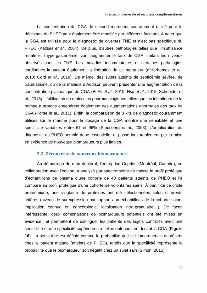
Discussion générale et résultats complémentaires
86
La concentration de CGA, le second marqueur couramment utilisé pour le
dépistage du PHEO peut également être modifiée par différents facteurs. À noter que
la CGA est utilisée pour le diagnostic de diverses TNE et n’est pas spécifique du
PHEO (Kaltsas et al., 2004). De plus, d’autres pathologies telles que l'insuffisance
rénale et l'hypergastrinémie, vont augmenter le taux de CGA, imitant les niveaux
observés pour les TNE. Les maladies inflammatoires et certaines pathologies
cardiaques impactent également la libération de ce marqueur (d’Herbomez et al.,
2010; Corti et al., 2018). De même, des sujets atteints de septicémie sévère, de
traumatisme, ou de la maladie d’Addison peuvent présenter une augmentation de la
concentration plasmatique de CGA (El Ali et al., 2010; Hsu et al., 2015; Schneider et
al., 2018). L’utilisation de molécules pharmacologiques telles que les inhibiteurs de la
pompe à protons engendrent également des augmentations anormales des taux de
CGA (Korse et al., 2011). Enfin, la comparaison de 3 kits de diagnostic couramment
utilisés sur le marché pour le dosage de la CGA montre une sensibilité et une
spécificité variables entre 67 et 96% (Stridsberg et al., 2003). L’amélioration du
diagnostic du PHEO semble donc essentielle, et passe inexorablement par la mise
en évidence de nouveaux biomarqueurs plus fiables.
5.2. Découverte de nouveaux biomarqueurs
Au démarrage de mon doctorat, l’entreprise Caprion (Montréal, Canada), en
collaboration avec l’équipe, a analysé par spectrométrie de masse le profil protéique
d’échantillons de plasma d’une cohorte de 40 patients atteints de PHEO et l’a
comparé au profil protéique d’une cohorte de volontaires sains. À partir de ce crible
protéomique, une vingtaine de protéines ont été sélectionnées selon différents
critères (niveau de surexpression par rapport aux échantillons de la cohorte saine,
implication connue en cancérologie, localisation intra-granulaire…). De façon
intéressante, deux combinaisons de biomarqueurs potentiels ont été mises en
évidence ; et permettent de distinguer les patients des sujets contrôles avec une
sensibilité et une spécificité supérieures à celles obtenues en dosant la CGA (Figure
16). La sensibilité est définie comme la probabilité que le biomarqueur soit présent
chez le patient malade (atteints de PHEO), tandis que la spécificité représente la
probabilité que le biomarqueur soit négatif chez un sujet sain (Simon, 2015).
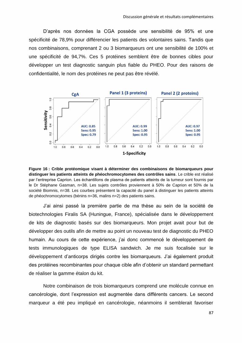
Discussion générale et résultats complémentaires
87
D’après nos données la CGA possède une sensibilité de 95% et une
spécificité de 78,9% pour différencier les patients des volontaires sains. Tandis que
nos combinaisons, comprenant 2 ou 3 biomarqueurs ont une sensibilité de 100% et
une spécificité de 94,7%. Ces 5 protéines semblent être de bonnes cibles pour
développer un test diagnostic sanguin plus fiable du PHEO. Pour des raisons de
confidentialité, le nom des protéines ne peut pas être révélé.
Figure 16 : Crible protéomique visant à déterminer des combinaisons de biomarqueurs pour
distinguer les patients atteints de phéochromocytomes des contrôles sains. Le crible est réalisé
par l’entreprise Caprion. Les échantillons de plasma de patients atteints de la tumeur sont fournis par
le Dr Stéphane Gasman, n=38. Les sujets contrôles proviennent à 50% de Caprion et 50% de la
société Biomnis, n=38. Les courbes présentent la capacité du panel à distinguer les patients atteints
de phéochromocytomes (bénins n=36, malins n=2) des patients sains.
J’ai ainsi passé la première partie de ma thèse au sein de la société de
biotechnologies Firalis SA (Huningue, France), spécialisée dans le développement
de kits de diagnostic basés sur des biomarqueurs. Mon projet avait pour but de
développer des outils afin de mettre au point un nouveau test de diagnostic du PHEO
humain. Au cours de cette expérience, j’ai donc commencé le développement de
tests immunologiques de type ELISA sandwich. Je me suis focalisée sur le
développement d’anticorps dirigés contre les biomarqueurs. J’ai également produit
des protéines recombinantes pour chaque cible afin d’obtenir un standard permettant
de réaliser la gamme étalon du kit.
Notre combinaison de trois biomarqueurs comprend une molécule connue en
cancérologie, dont l’expression est augmentée dans différents cancers. Le second
marqueur a été peu impliqué en cancérologie, néanmoins il semblerait favoriser
Discussion générale et résultats complémentaires
88
l’invasion dans le cancer du sein. Enfin le dernier marqueur de ce panel est une
enzyme qui n’a pas d’implication connue en cancérologie, ni dans d’autres
pathologies. De façon intéressante, notre panel de deux biomarqueurs comprend un
peptide dérivé d’une granine, tandis que le second marqueur est très connu en
cancérologie et a été montré comme augmenté dans le sérum de patients atteints de
diverses tumeurs dont le PHEO. Ce panel de deux biomarqueurs me semble très
prometteur puisque les deux molécules ont une forte implication en cancérologie.
Pour le développement d’un nouveau test de diagnostic du PHEO nous avons donc
décidé de nous focaliser sur la combinaison de deux biomarqueurs, étant donné leur
implication en cancérologie.
Dans la littérature les seules avancées sur des biomarqueurs plasmatiques
ont été réalisées sur la sécrétion de dérivés des chromogranines et sécrétogranines
par les PHEO. Il a ainsi été montré que le peptide EM66, dérivé de la sécrétogranine
est augmenté de 10 fois dans le plasma de patients atteints de PHEO par rapport à
des volontaires sains. La quantité urinaire d’EM66 n’est en revanche pas modifiée.
L’étude suggère que ce peptide pourrait être dosé en complémentarité des autres
marqueurs déjà utilisés en clinique (Guillemot et al., 2006). De même, le peptide
WE14 dérivé de la CGA est augmenté de 5,4 fois chez les patients porteurs de
PHEO par rapports aux sujets contrôles. La combinaison de la CGA, et d’un ou deux
de ces peptides permet d’augmenter la sensibilité de la détection de la tumeur à
environ 90% (Guillemot et al., 2014). Ces résultats restent en dessous de la
sensibilité obtenue avec nos biomarqueurs (de 100%), d’autant plus qu’aucune
indication n’est donnée pour la spécificité.
Ces dernières années les recherches se sont plus focalisées sur des
marqueurs moléculaires qui permettraient de distinguer les PHEO bénins et malins
tels que des ARN long non codants ou les miRNA (Meyer-Rochow et al., 2010; Ruff
et al., 2019; Ghosal et al., 2020; Job et al., 2020).

CONCLUSION

89
CONCLUSION
Mes travaux de thèse permettent de mieux comprendre pourquoi et comment
les phéochromocytomes présentent une hypersécrétion de catécholamines. J’ai
montré pour la première fois que les cellules tumorales ont une sécrétion accrue à
l’échelle unicellulaire, invalidant ainsi l’hypothèse selon laquelle l’hypersécrétion
proviendrait uniquement d’un effet de masse de la tumeur. Une analyse protéomique
de tissus tumoraux a également permis de mettre en évidence des dérégulations de
nombreuses protéines impliquées dans l’exocytose régulée par le calcium.
L’altération de divers acteurs du processus d’exocytose pourrait ainsi expliquer
l’augmentation du nombre d’évènements d’exocytose, ainsi qu’une cinétique de
libération plus rapide dans les cellules tumorales. Il serait à présent intéressant de
faire le lien entre l’hypersécrétion de la tumeur et les protéines de l’exocytose dont
l’expression varie. Découvrir les origines et les mécanismes de la sécrétion
incontrôlée des phéochromocytomes pourrait conduire à développer des stratégies
thérapeutiques visant à prévenir l’hypersécrétion et à diminuer ainsi les risques
cliniques. Mes résultats permettent également d’envisager que des mécanismes
similaires puissent être à l’origine de l’hypersécrétion d’autres tumeurs
neuroendocrines et ouvrent ainsi la voie à de nouvelles études.
Dans une seconde partie, mes recherches se sont focalisées sur les
analogues de la somatostatine grâce auxquels j’espérais inhiber directement
l’hypersécrétion des phéochromocytomes. Ces molécules sont déjà utilisées en
clinique pour leur effet anti-sécrétoire sur plusieurs tumeurs neuroendocrines. Mes
résultats montrent pour la première fois que le pasiréotide (SOM230), un analogue
de dernière génération, bloque efficacement la sécrétion de cellules tumorales
provenant d’exérèses de phéochromocytomes humains. L’ensemble de mes
données suggèrent également une nouvelle piste de recherche, puisque l’effet anti-
sécrétoire du SOM230 serait lié à une inhibition du récepteur nicotinique, une action
dont les mécanismes exacts restent à élucider. Mes résultats très prometteurs
obtenus avec le SOM230 permettent d’envisager à plus long terme des essais pré-
cliniques et cliniques pour valider une utilisation thérapeutique sur des patients
atteints de phéochromocytomes.

90

MATÉRIELS ET
MÉTHODES

91
MATÉRIELS ET MÉTHODES
1. Réactifs
Les analogues de la somatostatine : octréotide et pasiréotide (SOM230) ont
été fournis par Novartis (Basel, Switzerland). Ces drogues sont dissoutes dans du
DMSO, puis des solutions stocks à 10-2 ou 10-3 M sont conservées à -20°C, à l’abri
de la lumière.
2. Échantillons de phéochromocytomes humains
Les prélèvements de phéochromocytomes humains sont récupérés après
exérèse réalisée par le Pr Laurent Brunaud au CHRU de Nancy Brabois, par le Dr
Michel Vix ou Pr Didier Mutter à l’Institut de chirurgie guidée par l'image (IHU) de
Strasbourg, tous les trois sont chirurgiens viscéral et digestif. L’accord des patients a
été recueilli pour qu’une partie de la tumeur soit prélevée et utilisée en recherche.
Différentes données cliniques et biologiques ont été recueilles, notamment les
niveaux de métanéphrines et normétanéphrines urinaires et plasmatiques, et quand
la donnée était disponible, le niveau de chromogranine A plasmatique.
3. Culture cellulaire
3.1. Culture primaire de cellules chromaffines bovines
La culture primaire de cellules chromaffines obtenues à partir de glandes
surrénales bovines a été décrite par Thahouly et al., 2021.
Immédiatement après le sacrifice de l’animal, les glandes surrénales sont
prélevées et placées dans une solution de Krebs (154 mM NaCl, 5,6 mM KCl, 3,6
mM NaHCO3, 5,6 mM glucose, 5 mM d’HEPES, pH 7,4) contenant de la primocine à
100 μg/mL. Les prélèvements sont maintenus à 4°C pendant le transport. Les
glandes sont dégraissées puis perfusées pendant 10 min avec la solution de Krebs
(50 mL/glande) afin d’éliminer le plasma et les érythrocytes des vaisseaux. Les
glandes sont ensuite digérées par perfusion d’une solution de Krebs contenant 0,4%
de collagénase A et 0,5% de bovine serum albumin (BSA). Les parties médullaires et
corticales sont séparées par dissection, puis les cellules de la médullosurrénale sont
Matériels et méthodes
92
dissociées par passage sur un tamis de 217 μm. Après 10 min de centrifugation à
100 g et à température ambiante, le culot est repris dans la solution de Krebs puis
passé sur un tamis de 70 μm afin de dissocier les cellules plus finement. S’en suit
une centrifugation de 20 min à 20 000 g sans frein, à température ambiante, sur un
gradient continu de densité de Percoll à 47,5% (GE Healthcare), afin d’isoler les
cellules chromaffines des érythrocytes et des débris cellulaires. Les cellules
chromaffines sont ensuite collectées et lavées, avant d’être centrifugées 10 min à
100 g dans du milieu Dulbecco’s Modified Eagle Medium (DMEM) contenant 4 mM
de glutamine et de la primocine à 100 μg/mL. Après comptage avec une cellule de
Neubauer, 250 000 cellules/puits sont ensemencées dans des plaques 96 puits pour
le dosage fluorimérique ou dans des boites de Petri de 35 mm pour l’ampérométrie à
fibre de carbone, dans du milieu complet DMEM contenant 10% de sérum de veau
fœtal (SVF), 100 µg/mL de primocine et des antimitotiques (cytosine arabinoside à
10-5 M et fluorodéoxyrudine à 10-5 M). Les cellules sont maintenues à 37°C, avec 5%
de CO2, 95% d’air saturé en vapeur d’eau, pendant au moins 2 jours avant utilisation.
3.2. Culture primaire de cellules chromaffines humaines issues de
phéochromocytome
Les cellules chromaffines tumorales sont obtenues à partir de
phéochromocytomes humains récupérés après opération chirurgicale.
Immédiatement après résection, un morceau de la tumeur (1 cm3 environ) est
prélevé puis plongé dans un milieu de transport à 4°C : Hanks’ Balanced Salt
Solution (HBSS) sans calcium ni magnésium (HBSS CMF) (Sigma) supplémenté par
0,2% de SVF (Gibco) et 1% de pénicilline/streptomycine (Sigma), ou Medium Tissue
Storage Solution (MACS) (Miltenyibiotec). La biopsie est maintenue à 4°C pendant le
transport. 1 à 3h après la résection, la tumeur est découpée en petits fragments
d’environ 1 mm3 dans une boite contenant de l’HBSS CMF, avant d’être centrifugés 5
min à 250 g à température ambiante. Le culot est resuspendu dans 15 mL de milieu
complet (Roswell Park Memorial Institute medium (RPMI) 1640 GlutaMAXTM (Gibco)
supplémenté de 15% de SVF et 1% de pénicilline/streptomycine), et les fragments
laissés sédimenter 15 min à température ambiante afin d’éliminer les cellules
sanguines et le gras. Le surnageant est éliminé puis 15 mL de milieu complet sont
utilisés pour resuspendre le culot avant centrifugation 5 min à 250 g à température
Matériels et méthodes
93
ambiante. Les fragments sont ensuite digérés dans du HBSS contenant du Ca2+ et
du Mg2+ et les enzymes de digestion : 1,5 mg/mL de collagénase B (Roche) et 1
mg/mL de dispase 2 (Gibco), pendant 45 min à 37°C sous agitation modérée. Le
volume de la solution de digestion est d’environ 10 fois le volume de tissu. 5 min
avant la fin de la digestion, 0,1 mg/mL de DNase I (Roche) est ajoutée afin d’éviter
les agglomérats d’ADN. Après sédimentation à température ambiante, le surnageant
est récupéré (fraction P1). Le culot est ensuite trituré dans 5 mL de HBSS CMF.
Après sédimentation, le surnageant est conservé (fraction P2). Les deux fractions
sont centrifugées 5 min à 800 g à température ambiante. Les culots sont
resuspendus dans 2 mL de HBSS CMF et les hématies restantes lysées en ajoutant
4 mL de Red Blood Cell Lysis Buffer (Roche), le mélange est placé 10 min sous
agitation modérée à température ambiante. Les fractions sont centrifugées 5 min à
500 g, puis resuspendues dans du milieu complet. La viabilité et la densité cellulaire
sont évaluées au microscope optique puis le volume final pour chaque fraction est
ajusté. 300 µL de suspension cellulaire sont ensemencées dans des boites de Petri
de 35 mm contenant une lamelle de verre (MatTek) couverte de collagène I (Corning)
pour les expériences d’ampérométrie ou couverte de polylysine (Sigma) pour les
expériences d’imagerie calcique et d’électrophysiologie. Les cellules sont maintenues
à 37°C, avec 5% de CO2, 95% d’air saturé en vapeur d’eau. Le lendemain 2 mL de
milieu complet sont ajoutés. Les cellules sont utilisées 2 à 3 jours après la culture.
4. Techniques biochimiques
4.1. Détection des récepteurs à la somatostatine par Western-blot
L’extraction des protéines de biopsies humaines (phéochromocytome et tissu
non tumoral adjacent) a été réalisée sur glace, en broyant avec un mini Potter les
fragments de tissu dans un tampon de lyse (10 mM Tris pH 7,4, 100 mM NaCl, 1 mM
EDTA, 1 mM EGTA, 1 mM NaF, 20 mM Na4P2O7, 2 mM Na3VO4, 1% Triton X-100,
10% glycérol, 0,1% SDS, 0,5% deoxycholate, cocktail d’anti-protéases). 20 µL de
tampon de lyse est utilisé pour 10 mg de tissu. Lorsque la solution est homogène, le
lysat est incubé 15 min supplémentaire sur glace en vortexant à intervalle régulier.
Les cellules chromaffines de bœuf sont lysées dans du Triton à 1%, 15 min à 4°C, en
vortexant plusieurs fois. Les lysats sont clarifiés par centrifugation à 20 0000 g,
pendant 10 min, à 4°C, et les surnageants sont récupérés.
Matériels et méthodes
94
La concentration en protéines des échantillons est déterminée grâce à la
technique de Bradford (Bio-Rad protein assay). Les échantillons sont repris dans du
tampon de charge (10 mM Tris HCl pH 7, 1 mM EDTA, 20 mM DTT, 3% SDS (m/v),
10 % glycérol (v/v), 0,01% bromophénol (m/v)) de façon à avoir 30 µg de protéines
par échantillon puis dénaturés pendant 5 min à 95°C. Les échantillons sont déposés
sur un gel de polyacrylamide en gradient continu de 4 à 12% Bis-Tris (Invitrogen) et
les protéines séparées par migration pendant 45 min à 180 V dans un tampon MES
SDS (Invitrogen). Les protéines sont transférées sur membrane de nitrocellulose
(MES transfer buffer, Invitrogen supplémenté de 20% d’éthanol) à 30 V pendant 1h.
L’efficacité de transfert est vérifiée par coloration de la membrane au rouge de
Ponceau. Les sites non spécifiques sont saturés en incubant la membrane dans un
tampon de blocage (Tris Buffered Saline (Euromedex), 0,1% Tween20 (TBS-T), 3%
BSA) à température ambiante, sous agitation pendant 1h. La membrane est ensuite
incubée avec l’anticorps primaire dilué dans la solution de blocage, sur la nuit, à 4°C,
sous agitation. Après lavages dans le TBS-T, la membrane est incubée avec un
anticorps secondaire couplé à la peroxydase de raifort (HRP) dilué dans la solution
de blocage pendant 30 min, à température ambiante, sous agitation. La membrane
est lavée, puis l’activité de la HRP révélée par chimiluminescence (SuperSignal™,
Invitrogen) et détectée grâce au système d’acquisition d’images Chemi-smart 5000
(Vilbert Lourmat). Dilution des anticorps primaires : polyclonaux de lapin anti-SSTR2
(ASR-006, Alomone labs) et anti-SSTR5 (ASR-005, Alomone labs) : 1/300,
monoclonal de souris anti-tyrosine hydroxylase (TH) (MAB31, Millipore) : 1/5 000.
Les anticorps secondaires polyclonaux de chèvre contre les Ig de lapin (31460,
Thermo scientific) ou contre les Ig de souris (31430, Thermo scientific) couplés à la
HRP sont dilués au 1/50 000.
4.2. Dosage des catécholamines par méthode fluorimétrique
Le dosage fluorimétrique des catécholamines a été décrit par Thahouly et al.,
2021. Les cellules chromaffines bovines sont préalablement ensemencées dans une
plaque 96 puits (Thermo Fisher Scientific) à 250 000 cellules/puits et maintenues
environ 48 h en culture avant la mesure de sécrétion. Le test est réalisé sur plaque
chauffante à 37°C. Les cellules sont lavées 3 fois 7 min avec 200 µL d’une solution
de Locke Normal (LN) (140 mM NaCl, 4,7 mM KCl, 2,5 mM CaCl2, 1,2 mM KH2PO4,
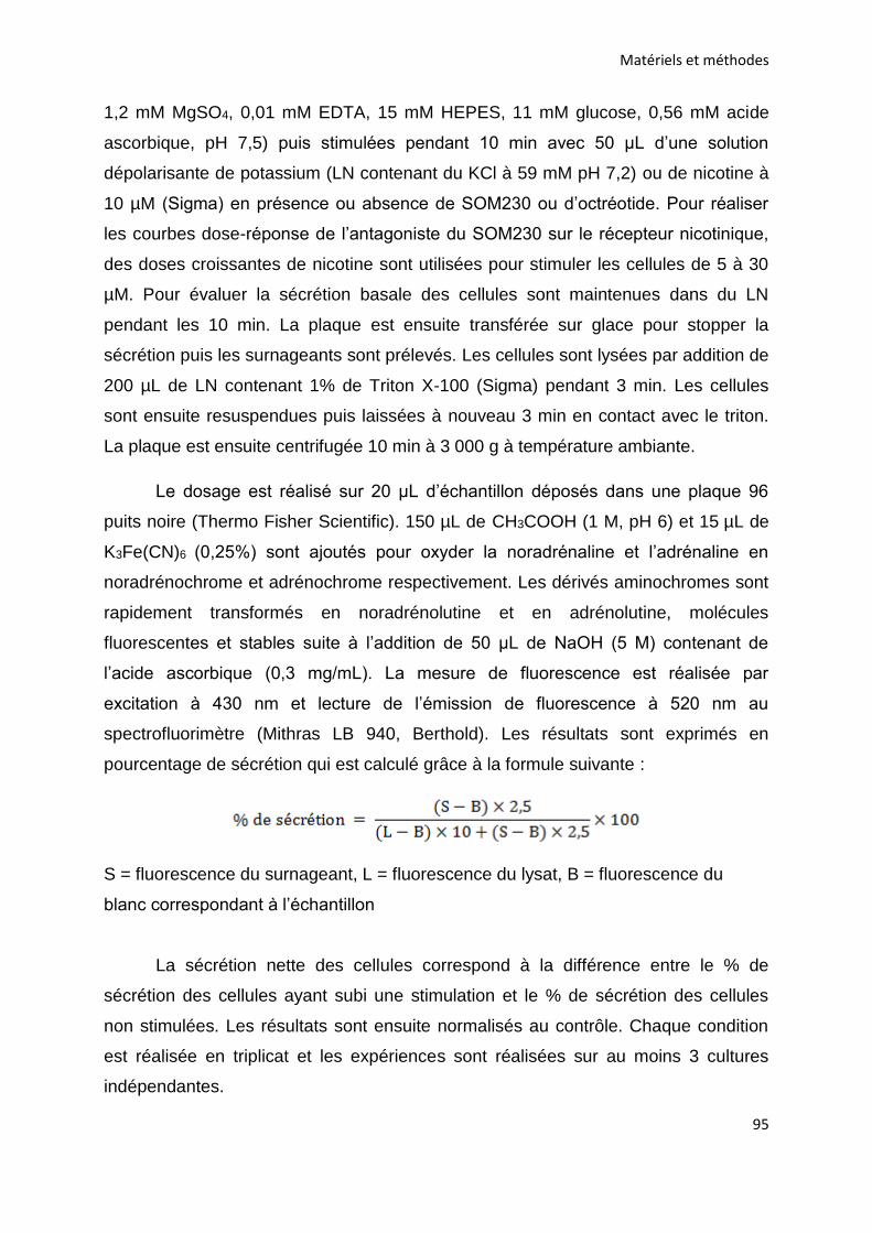
Matériels et méthodes
95
1,2 mM MgSO4, 0,01 mM EDTA, 15 mM HEPES, 11 mM glucose, 0,56 mM acide
ascorbique, pH 7,5) puis stimulées pendant 10 min avec 50 µL d’une solution
dépolarisante de potassium (LN contenant du KCl à 59 mM pH 7,2) ou de nicotine à
10 µM (Sigma) en présence ou absence de SOM230 ou d’octréotide. Pour réaliser
les courbes dose-réponse de l’antagoniste du SOM230 sur le récepteur nicotinique,
des doses croissantes de nicotine sont utilisées pour stimuler les cellules de 5 à 30
µM. Pour évaluer la sécrétion basale des cellules sont maintenues dans du LN
pendant les 10 min. La plaque est ensuite transférée sur glace pour stopper la
sécrétion puis les surnageants sont prélevés. Les cellules sont lysées par addition de
200 µL de LN contenant 1% de Triton X-100 (Sigma) pendant 3 min. Les cellules
sont ensuite resuspendues puis laissées à nouveau 3 min en contact avec le triton.
La plaque est ensuite centrifugée 10 min à 3 000 g à température ambiante.
Le dosage est réalisé sur 20 µL d’échantillon déposés dans une plaque 96
puits noire (Thermo Fisher Scientific). 150 µL de CH3COOH (1 M, pH 6) et 15 µL de
K3Fe(CN)6 (0,25%) sont ajoutés pour oxyder la noradrénaline et l’adrénaline en
noradrénochrome et adrénochrome respectivement. Les dérivés aminochromes sont
rapidement transformés en noradrénolutine et en adrénolutine, molécules
fluorescentes et stables suite à l’addition de 50 µL de NaOH (5 M) contenant de
l’acide ascorbique (0,3 mg/mL). La mesure de fluorescence est réalisée par
excitation à 430 nm et lecture de l’émission de fluorescence à 520 nm au
spectrofluorimètre (Mithras LB 940, Berthold). Les résultats sont exprimés en
pourcentage de sécrétion qui est calculé grâce à la formule suivante :
S = fluorescence du surnageant, L = fluorescence du lysat, B = fluorescence du
blanc correspondant à l’échantillon
La sécrétion nette des cellules correspond à la différence entre le % de
sécrétion des cellules ayant subi une stimulation et le % de sécrétion des cellules
non stimulées. Les résultats sont ensuite normalisés au contrôle. Chaque condition
est réalisée en triplicat et les expériences sont réalisées sur au moins 3 cultures
indépendantes.
Matériels et méthodes
96
5. Mesure de la sécrétion des catécholamines par ampérométrie à fibre
de carbone
L’ampérométrie à fibre de carbone est une technique électrochimique
permettant d’enregistrer des évènements d’exocytose sur cellule unique. Elle est
basée sur l’oxydation des catécholamines libérées par la cellule à la surface de
l’électrode.
Les cellules sont maintenues en culture pendant 48 à 72 h avant l’expérience.
Les cellules sont lavées dans 2 mL de LN sans acide ascorbique à 37°C. Les
enregistrements ampérométriques sont réalisés à l’aide d’une fibre en carbone
commerciale de 5 µm de diamètre (CFE-1, ALA Scientific instruments), placée au
contact de la cellule et maintenue au potentiel d’oxydation des catécholamines de
+650 mV. Une électrode de référence Ag/AgCl est également positionnée dans la
boite. Une micropipette (Femtotips, Eppendorf) placée à environ 10 µm de la cellule
permet de stimuler les cellules avec une solution de nicotine à 100 µM (Sigma),
pendant 10 s. En fonction des expériences, l’analogue de la somatostatine
(octréotide ou SOM230) est présent dans la pipette de stimulation ou dans le milieu
d’incubation, au contact direct des cellules. Les signaux sont enregistrés pendant 60
s grâce à un amplificateur AMU130 (Radiometer Analytical), calibré à 5 kHz et au
logiciel WinEDR. Les signaux sont ensuite filtrés à 1000 Hz et analysés à l’aide du
logiciel Igor Pro (Wavemetrics). L’analyse automatique des pics ainsi que l’extraction
des différents paramètres sont réalisés grâce à une macro développée par l’équipe
du Dr R. Borges (http://webpages.ull.es/users/rborges/) (Segura et al., 2000; Brioso
et al.,). L’ensemble des pics est vérifié manuellement et seuls ceux dépassant 5 pA
sont considérés comme des évènements d’exocytose. Les pics superposés et
aberrants sont comptabilisés pour l’analyse du nombre de pics totaux, puis retirés
pour l’analyse des paramètres ampérométriques. La charge Q représente l’aire sous
le pic et indique la quantité de catécholamines libérées. L’amplitude du pic est
représentée par l’Imax qui est le flux maximal de catécholamines libérées par
granule. Enfin, le T1/2 et le Tpeak sont représentatifs de la cinétique de libération des
catécholamines.

RÉFÉRENCES
BIBLIOGRAPHIQUES

97
RÉFÉRENCES BIBLIOGRAPHIQUES
A
Adams MS, Bronner-Fraser M (2009) Review: the role of neural crest cells in the endocrine system. Endocr Pathol 20:92–100.
Adler JT, Meyer-Rochow GY, Chen H, Benn DE, Robinson BG, Sippel RS, Sidhu SB (2008) Pheochromocytoma: current approaches and future directions. The Oncologist 13:779–793.
Aita Y, Ishii K, Saito Y, Ikeda T, Kawakami Y, Shimano H, Hara H, Takekoshi K (2012) Sunitinib inhibits catecholamine synthesis and secretion in pheochromocytoma tumor cells by blocking VEGF receptor 2 via PLC-γ-related pathways. Am J Physiol Endocrinol Metab 303:E1006-1014.
Ajallé R, Plouin PF, Pacak K, Lehnert H (2009) Treatment of Malignant Pheochromocytoma. Horm Metab Res Horm Stoffwechselforschung Horm Metab 41:687–696.
Algeciras-Schimnich A, Preissner CM, Young WF, Singh RJ, Grebe SKG (2008) Plasma chromogranin A or urine fractionated metanephrines follow-up testing improves the diagnostic accuracy of plasma fractionated metanephrines for pheochromocytoma. J Clin Endocrinol Metab 93:91–95.
Amar L, Baudin E, Burnichon N, Peyrard S, Silvera S, Bertherat J, Bertagna X, Schlumberger M, Jeunemaitre X, Gimenez-Roqueplo A-P, Plouin P-F (2007) Succinate dehydrogenase B gene mutations predict survival in patients with malignant pheochromocytomas or paragangliomas. J Clin Endocrinol Metab 92:3822–3828.
Amar L, Peyrard S, Rossignol P, Zinzindohoue F, Gimenez-Roqueplo A-P, Plouin P-F (2006) Changes in urinary total metanephrine excretion in recurrent and malignant pheochromocytomas and secreting paragangliomas. Ann N Y Acad Sci 1073:383–391.
Amatore C, Arbault S, Bouret Y, Guille M, Lemaître F (2010) Prediction of local pH variations during amperometric monitoring of vesicular exocytotic events at chromaffin cells. Chemphyschem Eur J Chem Phys Phys Chem 11:2931–2941.
Andreis DT, Singer M (2016) Catecholamines for inflammatory shock: a Jekyll-and-Hyde conundrum. Intensive Care Med 42:1387–1397.
Andrew A, Kramer B, Rawdon BB (1998) The origin of gut and pancreatic neuroendocrine (APUD) cells--the last word? J Pathol 186:117–118.
Annamalai AK, Dean AF, Kandasamy N, Kovacs K, Burton H, Halsall DJ, Shaw AS, Antoun NM, Cheow HK, Kirollos RW, Pickard JD, Simpson HL, Jefferies SJ,
Références bibliographiques
98
Burnet NG, Gurnell M (2012) Temozolomide responsiveness in aggressive corticotroph tumours: a case report and review of the literature. Pituitary 15:276–287.
Aunis D, Langley K (1999) Physiological aspects of exocytosis in chromaffin cells of the adrenal medulla. Acta Physiol Scand 167:89–97.
Ayala-Ramirez M, Chougnet CN, Habra MA, Palmer JL, Leboulleux S, Cabanillas ME, Caramella C, Anderson P, Al Ghuzlan A, Waguespack SG, Deandreis D, Baudin E, Jimenez C (2012) Treatment with sunitinib for patients with progressive metastatic pheochromocytomas and sympathetic paragangliomas. J Clin Endocrinol Metab 97:4040–4050.
B
Bader M-F, Doussau F, Chasserot-Golaz S, Vitale N, Gasman S (2004) Coupling actin and membrane dynamics during calcium-regulated exocytosis: a role for Rho and ARF GTPases. Biochim Biophys Acta 1742:37–49.
Bai S, Yao Z, Zhu X, Li Z, Jiang Y, Wang R, Wu B (2018) Risk factors for postoperative severe morbidity after pheochromocytoma surgery: A single center retrospective analysis of 262 patients. Int J Surg Lond Engl 60:188–193.
Barbarin A, Frade R (2011) Procathepsin L secretion, which triggers tumour progression, is regulated by Rab4a in human melanoma cells. Biochem J 437:97–107.
Barnett P (2003) Somatostatin and somatostatin receptor physiology. Endocrine 20:255–264.
Bartolomucci A, Possenti R, Mahata SK, Fischer-Colbrie R, Loh YP, Salton SRJ (2011) The extended granin family: structure, function, and biomedical implications. Endocr Rev 32:755–797.
Batista DL, Zhang X, Gejman R, Ansell PJ, Zhou Y, Johnson SA, Swearingen B, Hedley-Whyte ET, Stratakis CA, Klibanski A (2006) The effects of SOM230 on cell proliferation and adrenocorticotropin secretion in human corticotroph pituitary adenomas. J Clin Endocrinol Metab 91:4482–4488.
Bauer W, Briner U, Doepfner W, Haller R, Huguenin R, Marbach P, Petcher TJ, Pless null (1982) SMS 201-995: a very potent and selective octapeptide analogue of somatostatin with prolonged action. Life Sci 31:1133–1140.
Bechmann N, Ehrlich H, Eisenhofer G, Ehrlich A, Meschke S, Ziegler CG, Bornstein SR (2018) Anti-Tumorigenic and Anti-Metastatic Activity of the Sponge-Derived Marine Drugs Aeroplysinin-1 and Isofistularin-3 against Pheochromocytoma In Vitro. Mar Drugs 16.
Références bibliographiques
99
Béglé A, Tryoen-Tóth P, de Barry J, Bader M-F, Vitale N (2009) ARF6 regulates the synthesis of fusogenic lipids for calcium-regulated exocytosis in neuroendocrine cells. J Biol Chem 284:4836–4845.
Berends AMA, Buitenwerf E, de Krijger RR, Veeger NJGM, van der Horst-Schrivers ANA, Links TP, Kerstens MN (2018) Incidence of pheochromocytoma and sympathetic paraganglioma in the Netherlands: A nationwide study and systematic review. Eur J Intern Med 51:68–73.
Bernhardt EB, Jalal SI (2016) Small Cell Lung Cancer. Cancer Treat Res 170:301–322.
Bertherat J, Tenenbaum F, Perlemoine K, Videau C, Alberini JL, Richard B, Dousset B, Bertagna X, Epelbaum J (2003) Somatostatin receptors 2 and 5 are the major somatostatin receptors in insulinomas: an in vivo and in vitro study. J Clin Endocrinol Metab 88:5353–5360.
Beuret N, Stettler H, Renold A, Rutishauser J, Spiess M (2004) Expression of regulated secretory proteins is sufficient to generate granule-like structures in constitutively secreting cells. J Biol Chem 279:20242–20249.
Bílek R, Vlček P, Šafařík L, Michalský D, Novák K, Dušková J, Václavíková E, Widimský J, Zelinka T (2019) Chromogranin A in the Laboratory Diagnosis of Pheochromocytoma and Paraganglioma. Cancers 11.
Bishop AE, Power RF, Polak JM (1988) Markers for neuroendocrine differentiation. Pathol Res Pract 183:119–128.
Bodei L, Kwekkeboom DJ, Kidd M, Modlin IM, Krenning EP (2016) Radiolabeled Somatostatin Analogue Therapy Of Gastroenteropancreatic Cancer. Semin Nucl Med 46:225–238.
Boksa P, St-Pierre S, Livett BG (1982) Characterization of substance P and somatostatin receptors on adrenal chromaffin cells using structural analogues. Brain Res 245:275–283.
Bourcier ME, Vinik AI (2013) Sunitinib for the treatment of metastatic paraganglioma and vasoactive intestinal polypeptide-producing tumor (VIPoma). Pancreas 42:348–352.
Brabander T, Kwekkeboom DJ, Feelders RA, Brouwers AH, Teunissen JJM (2015) Nuclear Medicine Imaging of Neuroendocrine Tumors. Front Horm Res 44:73–87.
Bravo EL (2002) Pheochromocytoma: an approach to antihypertensive management. Ann N Y Acad Sci 970:1–10.
Brazeau P, Vale W, Burgus R, Ling N, Butcher M, Rivier J, Guillemin R (1973) Hypothalamic polypeptide that inhibits the secretion of immunoreactive pituitary growth hormone. Science 179:77–79.

Références bibliographiques
100
Brioso MA, Segura F, Gómez JF, Machado JD, Borges R (n.d.) Program on Igor Pro for the Automatical Analysis of Amperometric Spikes. :21.
Brown RL, Muzzafar T, Wollman R, Weiss RE (2006) A pituitary carcinoma secreting TSH and prolactin: a non-secreting adenoma gone awry. Eur J Endocrinol 154:639–643.
Bruns C, Lewis I, Briner U, Meno-Tetang G, Weckbecker G (2002) SOM230: a novel somatostatin peptidomimetic with broad somatotropin release inhibiting factor (SRIF) receptor binding and a unique antisecretory profile. Eur J Endocrinol 146:707–716.
Buchan AMJ, Lin C-Y, Choi J, Barber DL (2002) Somatostatin, acting at receptor subtype 1, inhibits Rho activity, the assembly of actin stress fibers, and cell migration. J Biol Chem 277:28431–28438.
Buffet A, Burnichon N, Favier J, Gimenez-Roqueplo A-P (2020) An overview of 20 years of genetic studies in pheochromocytoma and paraganglioma. Best Pract Res Clin Endocrinol Metab 34:101416.
C
Carmon O et al. (2020) Chromogranin A preferential interaction with Golgi phosphatidic acid induces membrane deformation and contributes to secretory granule biogenesis. FASEB J Off Publ Fed Am Soc Exp Biol 34:6769–6790.
Casarini APM, Jallad RS, Pinto EM, Soares IC, Nonogaki S, Giannella-Neto D, Musolino NR, Alves VAF, Bronstein MD (2009) Acromegaly: correlation between expression of somatostatin receptor subtypes and response to octreotide-lar treatment. Pituitary 12:297–303.
Chakroborty D, Sarkar C, Basu B, Dasgupta PS, Basu S (2009) Catecholamines regulate tumor angiogenesis. Cancer Res 69:3727–3730.
Chan MM, Chan MM, Mengshol JA, Fish DN, Chan ED (2013) Octreotide: a drug often used in the critical care setting but not well understood. Chest 144:1937–1945.
Chasserot-Golaz S, Vitale N, Umbrecht-Jenck E, Knight D, Gerke V, Bader M-F (2005) Annexin 2 promotes the formation of lipid microdomains required for calcium-regulated exocytosis of dense-core vesicles. Mol Biol Cell 16:1108–1119.
Chen X, Shen G, Jiang J, Liu H, Hu K, Darstein C, Lasher J, Hu P (2014) Pharmacokinetics and safety of subcutaneous pasireotide and intramuscular pasireotide long-acting release in Chinese male healthy volunteers: a phase I, single-center, open-label, randomized study. Clin Ther 36:1196–1210.
Références bibliographiques
101
Cheung NW, Boyages SC (1995) Somatostatin-14 and its analog octreotide exert a cytostatic effect on GH3 rat pituitary tumor cell proliferation via a transient G0/G1 cell cycle block. Endocrinology 136:4174–4181.
Cives M, Kunz PL, Morse B, Coppola D, Schell MJ, Campos T, Nguyen PT, Nandoskar P, Khandelwal V, Strosberg JR (2015) Phase II clinical trial of pasireotide long-acting repeatable in patients with metastatic neuroendocrine tumors. Endocr Relat Cancer 22:1–9.
Colao A, Bronstein MD, Brue T, De Marinis L, Fleseriu M, Guitelman M, Raverot G, Shimon I, Fleck J, Gupta P, Pedroncelli AM, Gadelha MR (2020) Pasireotide for acromegaly: long-term outcomes from an extension to the Phase III PAOLA study. Eur J Endocrinol 182:583.
Cornu E, Belmihoub I, Burnichon N, Grataloup C, Zinzindohoué F, Baron S, Billaud E, Azizi M, Gimenez-Roqueplo A-P, Amar L (2019) [Phaeochromocytoma and paraganglioma]. Rev Med Interne 40:733–741.
Corradi N, Borgonovo B, Clementi E, Bassetti M, Racchetti G, Consalez GG, Huttner WB, Meldolesi J, Rosa P (1996) Overall lack of regulated secretion in a PC12 variant cell clone. J Biol Chem 271:27116–27124.
Corti A, Marcucci F, Bachetti T (2018) Circulating chromogranin A and its fragments as diagnostic and prognostic disease markers. Pflugers Arch 470:199–210.
Courel M, Rodemer C, Nguyen ST, Pance A, Jackson AP, O’connor DT, Taupenot L (2006) Secretory granule biogenesis in sympathoadrenal cells: identification of a granulogenic determinant in the secretory prohormone chromogranin A. J Biol Chem 281:38038–38051.
Criado M (2018) Acetylcholine nicotinic receptor subtypes in chromaffin cells. Pflugers Arch 470:13–20.
Crivellato E, Nico B, Ribatti D (2008) The chromaffin vesicle: advances in understanding the composition of a versatile, multifunctional secretory organelle. Anat Rec Hoboken NJ 2007 291:1587–1602.
Crona J, Backman S, Maharjan R, Mayrhofer M, Stålberg P, Isaksson A, Hellman P, Björklund P (2015) Spatiotemporal Heterogeneity Characterizes the Genetic Landscape of Pheochromocytoma and Defines Early Events in Tumorigenesis. Clin Cancer Res Off J Am Assoc Cancer Res 21:4451–4460.
Croisé P (2015) Sécrétion des phéochromocytomes : impact sur le développement tumoral et rôle des GTPases Rho. Thèse de l’Université de Strasbourg.
Cuevas-Ramos D, Fleseriu M (2016) Pasireotide: a novel treatment for patients with
acromegaly. Drug Des Devel Ther 10:227–239.
Cuttitta F, Carney DN, Mulshine J, Moody TW, Fedorko J, Fischler A, Minna JD (1985) Bombesin-like peptides can function as autocrine growth factors in human small-cell lung cancer. Nature 316:823–826.
Références bibliographiques
102
D
d’Herbomez M, Do Cao C, Vezzosi D, Borzon-Chasot F, Baudin E, groupe des tumeurs endocrines (GTE France) (2010) Chromogranin A assay in clinical practice. Ann Endocrinol 71:274–280.
Daems T, Verhelst J, Michotte A, Abrams P, De Ridder D, Abs R (2009) Modification of hormonal secretion in clinically silent pituitary adenomas. Pituitary 12:80–86.
Dahia PLM (2014) Pheochromocytoma and paraganglioma pathogenesis: learning from genetic heterogeneity. Nat Rev Cancer 14:108–119.
D’Alessandro R, Klajn A, Stucchi L, Podini P, Malosio ML, Meldolesi J (2008) Expression of the neurosecretory process in PC12 cells is governed by REST. J Neurochem 105:1369–1383.
Dalm SU, Nonnekens J, Doeswijk GN, de Blois E, van Gent DC, Konijnenberg MW, de Jong M (2016) Comparison of the Therapeutic Response to Treatment with a 177Lu-Labeled Somatostatin Receptor Agonist and Antagonist in Preclinical Models. J Nucl Med Off Publ Soc Nucl Med 57:260–265.
Dang N, Meng X, Song H (2016) Nicotinic acetylcholine receptors and cancer. Biomed Rep 4:515–518.
Dasari A, Shen C, Halperin D, Zhao B, Zhou S, Xu Y, Shih T, Yao JC (2017) Trends in the Incidence, Prevalence, and Survival Outcomes in Patients With Neuroendocrine Tumors in the United States. JAMA Oncol 3:1335–1342.
de Bruin C, Feelders RA, Lamberts SWJ, Hofland LJ (2009) Somatostatin and dopamine receptors as targets for medical treatment of Cushing’s Syndrome. Rev Endocr Metab Disord 10:91–102.
de Herder WW, Rehfeld JF, Kidd M, Modlin IM (2016) A short history of neuroendocrine tumours and their peptide hormones. Best Pract Res Clin Endocrinol Metab 30:3–17.
Denorme M, Yon L, Roux C, Gonzalez BJ, Baudin E, Anouar Y, Dubessy C (2014) Both sunitinib and sorafenib are effective treatments for pheochromocytoma in a xenograft model. Cancer Lett 352:236–244.
Desnos C, Schonn J-S, Huet S, Tran VS, El-Amraoui A, Raposo G, Fanget I, Chapuis C, Ménasché G, de Saint Basile G, Petit C, Cribier S, Henry J-P, Darchen F (2003) Rab27A and its effector MyRIP link secretory granules to F-actin and control their motion towards release sites. J Cell Biol 163:559–570.
Désy L, Pelletier G (1977) Immunohistochemical localization of somatostatin in the human hypothalamus. Cell Tissue Res 184:491–497.
Références bibliographiques
103
Dhara M, Mohrmann R, Bruns D (2018) v-SNARE function in chromaffin cells. Pflugers Arch 470:169–180.
Dietrich H, Hu K, Ruffin M, Song D, Bouillaud E, Wang Y, Hasskarl J (2012) Safety, tolerability, and pharmacokinetics of a single dose of pasireotide long-acting release in healthy volunteers: a single-center Phase I study. Eur J Endocrinol 166:821–828.
Dineen R, Stewart PM, Sherlock M (2017) Acromegaly. QJM Mon J Assoc Physicians 110:411–420.
Drugs.com. Signifor Prices, Coupons and Patient Assistance Programs. Accessed May 12, 2021 https://www.drugs.com/price-guide/signifor
Drugs.com. Signifor LAR Prices, Coupons and Patient Assistance Programs.
Accessed May 12, 2021 https://www.drugs.com/price-guide/signifor-lar
Dupont A, Alvarado-Urbina G, Côté J, Labrie F (1978) Enzymatic degradation of somatostatin by rat plasma and hypothalamus. Can J Physiol Pharmacol 56:840–843.
E
Eigler T, Ben-Shlomo A (2014) Somatostatin system: molecular mechanisms regulating anterior pituitary hormones. J Mol Endocrinol 53:R1-19.
Eisenhofer G, Huynh T-T, Elkahloun A, Morris JC, Bratslavsky G, Linehan WM, Zhuang Z, Balgley BM, Lee CS, Mannelli M, Lenders JWM, Bornstein SR, Pacak K (2008) Differential expression of the regulated catecholamine secretory pathway in different hereditary forms of pheochromocytoma. Am J Physiol Endocrinol Metab 295:E1223-1233.
Eisenhofer G, Lenders JWM, Goldstein DS, Mannelli M, Csako G, Walther MM, Brouwers FM, Pacak K (2005) Pheochromocytoma catecholamine phenotypes and prediction of tumor size and location by use of plasma free metanephrines. Clin Chem 51:735–744.
Eisenhofer G, Lenders JWM, Timmers H, Mannelli M, Grebe SK, Hofbauer LC, Bornstein SR, Tiebel O, Adams K, Bratslavsky G, Linehan WM, Pacak K (2011) Measurements of plasma methoxytyramine, normetanephrine, and metanephrine as discriminators of different hereditary forms of pheochromocytoma. Clin Chem 57:411–420.
El Ali Z, Fichna M, Piniewska J, Kosowicz J, Grzymisławski M (2010) Chromogranin A as a useful neuroendocrine marker in patients with autoimmune Addison’s disease. J Endocrinol Invest 33:186–191.
Références bibliographiques
104
Elder EE, Elder G, Larsson C (2005) Pheochromocytoma and functional paraganglioma syndrome: no longer the 10% tumor. J Surg Oncol 89:193–201.
Elhamdani A, Azizi F, Artalejo CR (2006) Double Patch Clamp Reveals That Transient Fusion (Kiss-and-Run) Is a Major Mechanism of Secretion in Calf Adrenal Chromaffin Cells: High Calcium Shifts the Mechanism from Kiss-and-Run to Complete Fusion. J Neurosci 26:3030–3036.
Elshafie O, Al Badaai Y, Alwahaibi K, Qureshi A, Hussein S, Al Azzri F, Almamari A, Woodhouse N (2014) Catecholamine-secreting carotid body paraganglioma: successful preoperative control of hypertension and clinical symptoms using high-dose long-acting octreotide. Endocrinol Diabetes Metab Case Rep 2014:140051.
Engelman K, Horwitz D, Jéquier E, Sjoerdsma A (1968) Biochemical and pharmacologic effects of alpha-methyltyrosine in man. J Clin Invest 47:577–594.
Estévez-Herrera J, González-Santana A, Baz-Dávila R, Machado JD, Borges R (2016) The intravesicular cocktail and its role in the regulation of exocytosis. J Neurochem 137:897–903.
Evenepoel L, Helaers R, Vroonen L, Aydin S, Hamoir M, Maiter D, Vikkula M, Persu A (2017) KIF1B and NF1 are the most frequently mutated genes in paraganglioma and pheochromocytoma tumors. Endocr Relat Cancer 24:L57–L61.
F
Faggiano A, Modica R, Severino R, Camera L, Fonti R, Del Prete M, Chiofalo MG, Aria M, Ferolla P, Vitale G, Pezzullo L, Colao A (2018) The antiproliferative effect of pasireotide LAR alone and in combination with everolimus in patients with medullary thyroid cancer: a single-center, open-label, phase II, proof-of-concept study. Endocrine 62:46–56.
Fall CA, Dupuy O, Garcia C, Giraud L, Almehrezi Y, Bordier L, Mayaudon H (2013) [Asymptomatic secretory phaeochromocytoma: the interest of a systematic evaluation of adrenal incidentalomas]. Presse Medicale Paris Fr 1983 42:1145–1147.
Farndon JR, Davidson HA, Johnston ID, Wells SA (1980) VMA excretion in patients with pheochromocytoma. Ann Surg 191:259–263.
Farrugia F-A, Charalampopoulos A (2019) Pheochromocytoma. Endocr Regul 53:191–212.
Fasshauer D, Sutton RB, Brunger AT, Jahn R (1998) Conserved structural features of the synaptic fusion complex: SNARE proteins reclassified as Q- and R-SNAREs. Proc Natl Acad Sci U S A 95:15781–15786.
Références bibliographiques
105
Fassnacht M, Arlt W, Bancos I, Dralle H, Newell-Price J, Sahdev A, Tabarin A, Terzolo M, Tsagarakis S, Dekkers OM (2016) Management of adrenal incidentalomas: European Society of Endocrinology Clinical Practice Guideline in collaboration with the European Network for the Study of Adrenal Tumors. Eur J Endocrinol 175:G1–G34.
Favier J (2010) Hypoxie et pseudo-hypoxie dans les tumeurs endocrines : l’exemple du phéochromocytome – Hypoxia and pseudo-hypoxia in endocrine tumors: the pheochromocytoma example. Corresp En Métabolismes Horm Diabètes Nutr:7.
Fedele M, De Martino I, Pivonello R, Ciarmiello A, Del Basso De Caro ML, Visone R, Palmieri D, Pierantoni GM, Arra C, Schmid HA, Hofland L, Lombardi G, Colao A, Fusco A (2007) SOM230, a new somatostatin analogue, is highly effective in the therapy of growth hormone/prolactin-secreting pituitary adenomas. Clin Cancer Res Off J Am Assoc Cancer Res 13:2738–2744.
Ferolla P et al. (2017) Efficacy and safety of long-acting pasireotide or everolimus alone or in combination in patients with advanced carcinoids of the lung and thymus (LUNA): an open-label, multicentre, randomised, phase 2 trial. Lancet Oncol 18:1652–1664.
Ferrante E, Pellegrini C, Bondioni S, Peverelli E, Locatelli M, Gelmini P, Luciani P, Peri A, Mantovani G, Bosari S, Beck-Peccoz P, Spada A, Lania A (2006) Octreotide promotes apoptosis in human somatotroph tumor cells by activating somatostatin receptor type 2. Endocr Relat Cancer 13:955–962.
Filevich O, Salierno M, Etchenique R (2010) A caged nicotine with nanosecond range kinetics and visible light sensitivity. J Inorg Biochem 104:1248–1251.
Finnegan JM, Pihel K, Cahill PS, Huang L, Zerby SE, Ewing AG, Kennedy RT, Wightman RM (1996) Vesicular quantal size measured by amperometry at chromaffin, mast, pheochromocytoma, and pancreatic beta-cells. J Neurochem 66:1914–1923.
Fishbein L et al. (2017) Comprehensive Molecular Characterization of Pheochromocytoma and Paraganglioma. Cancer Cell 31:181–193.
Flávio Rocha M, Faramarzi-Roques R, Tauzin-Fin P, Vallee V, Leitao de Vasconcelos PR, Ballanger P (2004) Laparoscopic surgery for pheochromocytoma. Eur Urol 45:226–232.
Florio T, Morini M, Villa V, Arena S, Corsaro A, Thellung S, Culler MD, Pfeffer U, Noonan DM, Schettini G, Albini A (2003) Somatostatin inhibits tumor angiogenesis and growth via somatostatin receptor-3-mediated regulation of endothelial nitric oxide synthase and mitogen-activated protein kinase activities. Endocrinology 144:1574–1584.
Flynn A, Benn D, Clifton-Bligh R, Robinson B, Trainer AH, James P, Hogg A, Waldeck K, George J, Li J, Fox SB, Gill AJ, McArthur G, Hicks RJ, Tothill RW (2015) The genomic landscape of phaeochromocytoma. J Pathol 236:78–89.
Références bibliographiques
106
Freda PU (2002) Somatostatin analogs in acromegaly. J Clin Endocrinol Metab 87:3013–3018.
Fulop T, Radabaugh S, Smith C (2005) Activity-dependent differential transmitter release in mouse adrenal chromaffin cells. J Neurosci Off J Soc Neurosci 25:7324–7332.
G
Gabel M, Delavoie F, Demais V, Royer C, Bailly Y, Vitale N, Bader M-F, Chasserot-Golaz S (2015) Annexin A2-dependent actin bundling promotes secretory granule docking to the plasma membrane and exocytosis. J Cell Biol 210:785–800.
Garg A, Sui P, Verheyden JM, Young LR, Sun X (2019) Consider the lung as a sensory organ: A tip from pulmonary neuroendocrine cells. Curr Top Dev Biol 132:67–89.
Gasman S, Chasserot-Golaz S, Hubert P, Aunis D, Bader MF (1998) Identification of a potential effector pathway for the trimeric Go protein associated with secretory granules. Go stimulates a granule-bound phosphatidylinositol 4-kinase by activating RhoA in chromaffin cells. J Biol Chem 273:16913–16920.
Gasman S, Chasserot-Golaz S, Malacombe M, Way M, Bader M-F (2004) Regulated exocytosis in neuroendocrine cells: a role for subplasmalemmal Cdc42/N-WASP-induced actin filaments. Mol Biol Cell 15:520–531.
Gasman S, Vitale N (2017) Lipid remodelling in neuroendocrine secretion. Biol Cell 109:381–390.
Gatto F, Feelders RA, Franck SE, van Koetsveld PM, Dogan F, Kros JM, Neggers SJCMM, van der Lely A-J, Lamberts SWJ, Ferone D, Hofland LJ (2017) In vitro head-to-head comparison between octreotide and pasireotide in GH-secreting pituitary adenomas. J Clin Endocrinol Metab.
Germain A, Klein M, Brunaud L (2011) Surgical management of adrenal tumors. J Visc Surg 148:e250-261.
Ghosal S, Das S, Pang Y, Gonzales MK, Huynh T-T, Yang Y, Taieb D, Crona J, Shankavaram UT, Pacak K (2020) Long intergenic noncoding RNA profiles of pheochromocytoma and paraganglioma: A novel prognostic biomarker. Int J Cancer 146:2326–2335.
Giardino E, Catalano R, Mangili F, Barbieri AM, Treppiedi D, Elli FM, Dolci A, Contarino A, Spada A, Arosio M, Mantovani G, Peverelli E (2021) Octreotide and pasireotide effects on medullary thyroid carcinoma (MTC) cells growth, migration and invasion. Mol Cell Endocrinol 520:111092.
Gill AJ (2018) Succinate dehydrogenase (SDH)-deficient neoplasia. Histopathology 72:106–116.
Références bibliographiques
107
Gladycheva SE, Ho CS, Lee YYF, Stuenkel EL (2004) Regulation of syntaxin1A-munc18 complex for SNARE pairing in HEK293 cells. J Physiol 558:857–871.
Gola M, Bonadonna S, Mazziotti G, Amato G, Giustina A (2006) Resistance to somatostatin analogs in acromegaly: an evolving concept? J Endocrinol Invest 29:86–93.
Goldstein RE, O’Neill JA, Holcomb GW, Morgan WM, Neblett WW, Oates JA, Brown N, Nadeau J, Smith B, Page DL, Abumrad NN, Scott HW (1999) Clinical experience over 48 years with pheochromocytoma. Ann Surg 229:755–764; discussion 764-766.
Graham MM, Gu X, Ginader T, Breheny P, Sunderland JJ (2017) 68Ga-DOTATOC Imaging of Neuroendocrine Tumors: A Systematic Review and Metaanalysis. J Nucl Med Off Publ Soc Nucl Med 58:1452–1458.
Grant M, Alturaihi H, Jaquet P, Collier B, Kumar U (2008) Cell growth inhibition and functioning of human somatostatin receptor type 2 are modulated by receptor heterodimerization. Mol Endocrinol Baltim Md 22:2278–2292.
Gratzl M, Breckner M, Prinz C (2004) Mechanisms of storage and exocytosis in neuroendocrine tumors. Endocr Pathol 15:1–16.
Greene LA, Rein G (1977) Release of (3H)norepinephrine from a clonal line of pheochromocytoma cells (PC12) by nicotinic cholinergic stimulation. Brain Res 138:521–528.
Grossrubatscher E, Dalino P, Vignati F, Gambacorta M, Pugliese R, Boniardi M, Rossetti O, Marocchi A, Bertuzzi M, Loli P (2006) The role of chromogranin A in the management of patients with phaeochromocytoma. Clin Endocrinol (Oxf) 65:287–293.
Gruber LM, Jasim S, Ducharme-Smith A, Weingarten T, Young WF, Bancos I (2021) The Role for Metyrosine in the Treatment of Patients With Pheochromocytoma and Paraganglioma. J Clin Endocrinol Metab 106:e2393–e2401.
Gruber M, Simon MC (2006) Hypoxia-inducible factors, hypoxia, and tumor angiogenesis. Curr Opin Hematol 13:169–174.
Grundschober C, Malosio ML, Astolfi L, Giordano T, Nef P, Meldolesi J (2002) Neurosecretion competence. A comprehensive gene expression program identified in PC12 cells. J Biol Chem 277:36715–36724.
Guérineau NC (2020) Cholinergic and peptidergic neurotransmission in the adrenal medulla: A dynamic control of stimulus-secretion coupling. IUBMB Life 72:553–567.
Guillemot J, Anouar Y, Montero-Hadjadje M, Grouzmann E, Grumolato L, Roshmaninho-Salgado J, Turquier V, Duparc C, Lefebvre H, Plouin P-F, Klein M, Muresan M, Chow BKC, Vaudry H, Yon L (2006) Circulating EM66 is a
Références bibliographiques
108
highly sensitive marker for the diagnosis and follow-up of pheochromocytoma. Int J Cancer 118:2003–2012.
Guillemot J, Guérin M, Thouënnon E, Montéro-Hadjadje M, Leprince J, Lefebvre H, Klein M, Muresan M, Anouar Y, Yon L (2014) Characterization and plasma measurement of the WE-14 peptide in patients with pheochromocytoma. PloS One 9:e88698.
Gumbs AA, Gagner M (2006) Laparoscopic adrenalectomy. Best Pract Res Clin Endocrinol Metab 20:483–499.
Günther T, Tulipano G, Dournaud P, Bousquet C, Csaba Z, Kreienkamp H-J, Lupp A, Korbonits M, Castaño JP, Wester H-J, Culler M, Melmed S, Schulz S (2018) International Union of Basic and Clinical Pharmacology. CV. Somatostatin Receptors: Structure, Function, Ligands, and New Nomenclature. Pharmacol Rev 70:763–835.
Gupta G, Pacak K, AACE Adrenal Scientific Committee (2017) PRECISION MEDICINE: AN UPDATE ON GENOTYPE/BIOCHEMICAL PHENOTYPE RELATIONSHIPS IN PHEOCHROMOCYTOMA/PARAGANGLIOMA PATIENTS. Endocr Pract Off J Am Coll Endocrinol Am Assoc Clin Endocrinol 23:690–704.
H
Harris M, Chung F (2013) Complications of general anesthesia. Clin Plast Surg 40:503–513.
Hashimoto K, Masumori N, Tanaka T, Maeda T, Kobayashi K, Kitamura H, Hirata K, Tsukamoto T (2013) Zoledronic acid but not somatostatin analogs exerts anti-tumor effects in a model of murine prostatic neuroendocrine carcinoma of the development of castration-resistant prostate cancer. The Prostate 73:500–511.
Hata J, Haga N, Ishibashi K, Takahashi N, Ogawa S, Kataoka M, Akaihata H, Satoh Y, Koguchi T, Kojima Y (2014) Sunitinib for refractory malignant pheochromocytoma: two case reports. Int Urol Nephrol 46:1309–1312.
Helle KB (2004) The granin family of uniquely acidic proteins of the diffuse neuroendocrine system: comparative and functional aspects. Biol Rev Camb Philos Soc 79:769–794.
Hendy GN, Li T, Girard M, Feldstein RC, Mulay S, Desjardins R, Day R, Karaplis AC, Tremblay ML, Canaff L (2006) Targeted ablation of the chromogranin a (Chga) gene: normal neuroendocrine dense-core secretory granules and increased expression of other granins. Mol Endocrinol Baltim Md 20:1935–1947.
Hernández-Guijo JM, Gandía L, Cuchillo-Ibáñez I, Albillos A, Novalbos J, Gilsanz F, Larrañaga E, de Pascual R, Abad F, García AG (2000) Altered regulation of
Références bibliographiques
109
calcium channels and exocytosis in single human pheochromocytoma cells. Pflugers Arch 440:253–263.
Herrera-Martínez AD, van den Dungen R, Dogan-Oruc F, van Koetsveld PM, Culler MD, de Herder WW, Luque RM, Feelders RA, Hofland LJ (2019) Effects of novel somatostatin-dopamine chimeric drugs in 2D and 3D cell culture models of neuroendocrine tumors. Endocr Relat Cancer 26:585–599.
Hofland LJ, van der Hoek J, Feelders R, van Aken MO, van Koetsveld PM, Waaijers M, Sprij-Mooij D, Bruns C, Weckbecker G, de Herder WW, Beckers A, Lamberts SWJ (2005) The multi-ligand somatostatin analogue SOM230 inhibits ACTH secretion by cultured human corticotroph adenomas via somatostatin receptor type 5. Eur J Endocrinol 152:645–654.
Hofland LJ, van der Hoek J, van Koetsveld PM, de Herder WW, Waaijers M, Sprij-Mooij D, Bruns C, Weckbecker G, Feelders R, van der Lely A-J, Beckers A, Lamberts SWJ (2004a) The novel somatostatin analog SOM230 is a potent inhibitor of hormone release by growth hormone- and prolactin-secreting pituitary adenomas in vitro. J Clin Endocrinol Metab 89:1577–1585.
Hofland LJ, van der Hoek J, van Koetsveld PM, de Herder WW, Waaijers M, Sprij-Mooij D, Bruns C, Weckbecker G, Feelders R, van der Lely A-J, Beckers A, Lamberts SWJ (2004b) The novel somatostatin analog SOM230 is a potent inhibitor of hormone release by growth hormone- and prolactin-secreting pituitary adenomas in vitro. J Clin Endocrinol Metab 89:1577–1585.
Holthouse DJ, Robbins PD, Kahler R, Knuckey N, Pullan P (2001) Corticotroph pituitary carcinoma: case report and literature review. Endocr Pathol 12:329–341.
Hone AJ, McIntosh JM, Azam L, Lindstrom J, Lucero L, Whiteaker P, Passas J, Blázquez J, Albillos A (2015) α-Conotoxins Identify the α3β4* Subtype as the Predominant Nicotinic Acetylcholine Receptor Expressed in Human Adrenal Chromaffin Cells. Mol Pharmacol 88:881–893.
Hone AJ, Michael McIntosh J, Rueda-Ruzafa L, Passas J, de Castro-Guerín C, Blázquez J, González-Enguita C, Albillos A (2017) Therapeutic concentrations of varenicline in the presence of nicotine increase action potential firing in human adrenal chromaffin cells. J Neurochem 140:37–52.
Hsu C-H et al. (2015) Chromogranin A levels and mortality in patients with severe sepsis. Biomark Biochem Indic Expo Response Susceptibility Chem 20:171–176.
Huh YH, Jeon SH, Yoo SH (2003) Chromogranin B-induced secretory granule biogenesis: comparison with the similar role of chromogranin A. J Biol Chem 278:40581–40589.
Références bibliographiques
110
I
Ibáñez-Costa A, Rivero-Cortés E, Vázquez-Borrego MC, Gahete MD, Jiménez-Reina L, Venegas-Moreno E, de la Riva A, Arráez MÁ, González-Molero I, Schmid HA, Maraver-Selfa S, Gavilán-Villarejo I, García-Arnés JA, Japón MA, Soto-Moreno A, Gálvez MA, Luque RM, Castaño JP (2016) Octreotide and pasireotide (dis)similarly inhibit pituitary tumor cells in vitro. J Endocrinol 231:135–145.
Improgo MRD, Schlichting NA, Cortes RY, Zhao-Shea R, Tapper AR, Gardner PD (2010) ASCL1 regulates the expression of the CHRNA5/A3/B4 lung cancer susceptibility locus. Mol Cancer Res MCR 8:194–203.
Inoue M, Kuriyama H (1991) Somatostatin inhibits the nicotinic receptor-activated inward current in guinea pig chromaffin cells. Biochem Biophys Res Commun 174:750–757.
Invitti C, De Martin I, Bolla GB, Pecori Giraldi F, Maestri E, Leonetti G, Cavagnini F (1993) Effect of octreotide on catecholamine plasma levels in patients with chromaffin cell tumors. Horm Res 40:156–160.
Ivanidze J, Roytman M, Sasson A, Skafida M, Fahey TJ, Osborne JR, Dutruel SP (2019) Molecular imaging and therapy of somatostatin receptor positive tumors. Clin Imaging 56:146–154.
J
Jain A, Baracco R, Kapur G (2020) Pheochromocytoma and paraganglioma-an update on diagnosis, evaluation, and management. Pediatr Nephrol Berl Ger 35:581–594.
Jaques S, Tobes MC (1986) Morphologic and biochemical variability of tissue and cultured cells from human pheochromocytoma. J Cell Physiol 128:261–270.
Jensen AA, Frølund B, Liljefors T, Krogsgaard-Larsen P (2005) Neuronal nicotinic acetylcholine receptors: structural revelations, target identifications, and therapeutic inspirations. J Med Chem 48:4705–4745.
Jiang J et al. (2020) Sino-European Differences in the Genetic Landscape and Clinical Presentation of Pheochromocytoma and Paraganglioma. J Clin Endocrinol Metab 105.
Jimenez C, Rohren E, Habra MA, Rich T, Jimenez P, Ayala-Ramirez M, Baudin E (2013) Current and future treatments for malignant pheochromocytoma and sympathetic paraganglioma. Curr Oncol Rep 15:356–371.
Jimenez T, Friedman T, Vadgama J, Singh V, Tucker A, Collazo J, Sinha S, Hikim AS, Singh R, Pervin S (2020) Nicotine Synergizes with High-Fat Diet to Induce
Références bibliographiques
111
an Anti-Inflammatory Microenvironment to Promote Breast Tumor Growth. Mediators Inflamm 2020:5239419.
Job S, Georges A, Burnichon N, Buffet A, Amar L, Bertherat J, Bouatia-Naji N, de Reyniès A, Drui D, Lussey-Lepoutre C, Favier J, Gimenez-Roqueplo A-P, Castro-Vega LJ (2020) Transcriptome Analysis of lncRNAs in Pheochromocytomas and Paragangliomas. J Clin Endocrinol Metab 105.
Jochmanova I, Pacak K (2018) Genomic Landscape of Pheochromocytoma and Paraganglioma. Trends Cancer 4:6–9.
Johnson JL, Ellis BA, Noack D, Seabra MC, Catz SD (2005) The Rab27a-binding protein, JFC1, regulates androgen-dependent secretion of prostate-specific antigen and prostatic-specific acid phosphatase. Biochem J 391:699–710.
Johnson RG (1988) Accumulation of biological amines into chromaffin granules: a model for hormone and neurotransmitter transport. Physiol Rev 68:232–307.
Joshua AM, Ezzat S, Asa SL, Evans A, Broom R, Freeman M, Knox JJ (2009) Rationale and evidence for sunitinib in the treatment of malignant paraganglioma/pheochromocytoma. J Clin Endocrinol Metab 94:5–9.
Juhlin CC, Skoglund S, Juntti-Berggren L, Karlberg M, Calissendorff J (2019) Non-functioning neuroendocrine pancreatic tumors transforming to malignant insulinomas - four cases and review of the literature. Neuro Endocrinol Lett 40:175–183.
K
Kaltsas GA, Besser GM, Grossman AB (2004) The diagnosis and medical management of advanced neuroendocrine tumors. Endocr Rev 25:458–511.
Kanakis G, Grimelius L, Spathis A, Tringidou R, Rassidakis GZ, Öberg K, Kaltsas G, Tsolakis AV (2015) Expression of Somatostatin Receptors 1-5 and Dopamine Receptor 2 in Lung Carcinoids: Implications for a Therapeutic Role. Neuroendocrinology 101:211–222.
Kemppainen RJ, Behrend EN (1997) Adrenal physiology. Vet Clin North Am Small Anim Pract 27:173–186.
Kim T, Tao-Cheng JH, Eiden LE, Loh YP (2001) Chromogranin A, an “on/off” switch controlling dense-core secretory granule biogenesis. Cell 106:499–509.
Kim T, Zhang C, Sun Z, Wu H, Loh YP (2005) Chromogranin A deficiency in transgenic mice leads to aberrant chromaffin granule biogenesis. J Neurosci Off J Soc Neurosci 25:6958–6961.
Kitlinska J (2007) Neuropeptide Y in neural crest-derived tumors: effect on growth and vascularization. Cancer Lett 245:293–302.
Références bibliographiques
112
Kögel T, Gerdes H-H (2010) Maturation of secretory granules. Results Probl Cell Differ 50:1–20.
Kopf D, Bockisch A, Steinert H, Hahn K, Beyer J, Neumann HP, Hensen J, Lehnert H (1997) Octreotide scintigraphy and catecholamine response to an octreotide challenge in malignant phaeochromocytoma. Clin Endocrinol (Oxf) 46:39–44.
Koriyama N, Kakei M, Yaekura K, Okui H, Yamashita T, Nishimura H, Matsushita S, Tei C (2000) Control of catecholamine release and blood pressure with octreotide in a patient with pheochromocytoma: a case report with in vitro studies. Horm Res 53:46–50.
Korse CM, Muller M, Taal BG (2011) Discontinuation of proton pump inhibitors during assessment of chromogranin A levels in patients with neuroendocrine tumours. Br J Cancer 105:1173–1175.
Kreienkamp HJ, Roth A, Richter D (1998) Rat somatostatin receptor subtype 4 can be made sensitive to agonist-induced internalization by mutation of a single threonine (residue 331). DNA Cell Biol 17:869–878.
Krulich L, Dhariwal AP, McCann SM (1968) Stimulatory and inhibitory effects of purified hypothalamic extracts on growth hormone release from rat pituitary in vitro. Endocrinology 83:783–790.
Kudva YC, Sawka AM, Young WF (2003) Clinical review 164: The laboratory diagnosis of adrenal pheochromocytoma: the Mayo Clinic experience. J Clin Endocrinol Metab 88:4533–4539.
Kvols LK, Oberg KE, O’Dorisio TM, Mohideen P, de Herder WW, Arnold R, Hu K, Zhang Y, Hughes G, Anthony L, Wiedenmann B (2012) Pasireotide (SOM230) shows efficacy and tolerability in the treatment of patients with advanced neuroendocrine tumors refractory or resistant to octreotide LAR: results from a phase II study. Endocr Relat Cancer 19:657–666.
L
Lamarre-Cliche M, Gimenez-Roqueplo A-P, Billaud E, Baudin E, Luton J-P, Plouin P-F (2002) Effects of slow-release octreotide on urinary metanephrine excretion and plasma chromogranin A and catecholamine levels in patients with malignant or recurrent phaeochromocytoma. Clin Endocrinol (Oxf) 57:629–634.
Lamberts SWJ, Hofland LJ (2019) ANNIVERSARY REVIEW: Octreotide, 40 years later. Eur J Endocrinol 181:R173–R183.
Leibowitz-Amit R, Lebowitz-Amit R, Mete O, Asa SL, Ezzat S, Joshua AM (2014) Malignant pheochromocytoma secreting vasoactive intestinal peptide and response to sunitinib: a case report and literature review. Endocr Pract Off J Am Coll Endocrinol Am Assoc Clin Endocrinol 20:e145-150.
Références bibliographiques
113
Leijon H, Remes S, Hagström J, Louhimo J, Mäenpää H, Schalin-Jäntti C, Miettinen M, Haglund C, Arola J (2019) Variable somatostatin receptor subtype expression in 151 primary pheochromocytomas and paragangliomas. Hum Pathol 86:66–75.
Lenders JWM, Duh Q-Y, Eisenhofer G, Gimenez-Roqueplo A-P, Grebe SKG, Murad MH, Naruse M, Pacak K, Young WF, Endocrine Society (2014) Pheochromocytoma and paraganglioma: an endocrine society clinical practice guideline. J Clin Endocrinol Metab 99:1915–1942.
Lenders JWM, Eisenhofer G, Mannelli M, Pacak K (2005) Phaeochromocytoma. Lancet Lond Engl 366:665–675.
Lenders JWM, Pacak K, Walther MM, Linehan WM, Mannelli M, Friberg P, Keiser HR, Goldstein DS, Eisenhofer G (2002) Biochemical diagnosis of pheochromocytoma: which test is best? JAMA 287:1427–1434.
Leoncini E, Carioli G, La Vecchia C, Boccia S, Rindi G (2016) Risk factors for neuroendocrine neoplasms: a systematic review and meta-analysis. Ann Oncol Off J Eur Soc Med Oncol 27:68–81.
Letouzé E, Martinelli C, Loriot C, Burnichon N, Abermil N, Ottolenghi C, Janin M, Menara M, Nguyen AT, Benit P, Buffet A, Marcaillou C, Bertherat J, Amar L, Rustin P, De Reyniès A, Gimenez-Roqueplo A-P, Favier J (2013) SDH mutations establish a hypermethylator phenotype in paraganglioma. Cancer Cell 23:739–752.
Liu D, Martino G, Thangaraju M, Sharma M, Halwani F, Shen SH, Patel YC, Srikant CB (2000) Caspase-8-mediated intracellular acidification precedes mitochondrial dysfunction in somatostatin-induced apoptosis. J Biol Chem 275:9244–9250.
Livett BG, Boksa P (1984) Receptors and receptor modulation in cultured chromaffin cells. Can J Physiol Pharmacol 62:467–476.
Lo CY, Lam KY, Wat MS, Lam KS (2000) Adrenal pheochromocytoma remains a frequently overlooked diagnosis. Am J Surg 179:212–215.
Loh YP, Kim T, Rodriguez YM, Cawley NX (2004) Secretory granule biogenesis and neuropeptide sorting to the regulated secretory pathway in neuroendocrine cells. J Mol Neurosci MN 22:63–71.
Low MJ (2004) The somatostatin neuroendocrine system: physiology and clinical relevance in gastrointestinal and pancreatic disorders. Best Pract Res Clin Endocrinol Metab 18:607–622.
Ludvigsen E, Stridsberg M, Taylor JE, Culler MD, Oberg K, Janson ET, Sandler S (2007) Regulation of insulin and glucagon secretion from rat pancreatic islets in vitro by somatostatin analogues. Regul Pept 138:1–9.
Références bibliographiques
114
M
Ma C, Li W, Xu Y, Rizo J (2011) Munc13 mediates the transition from the closed syntaxin-Munc18 complex to the SNARE complex. Nat Struct Mol Biol 18:542–549.
Ma X, Li M, Tong A, Wang F, Cui Y, Zhang X, Zhang Y, Chen S, Li Y (2020) Genetic and Clinical Profiles of Pheochromocytoma and Paraganglioma: A Single Center Study. Front Endocrinol 11:574662.
Mahapatra S, Calorio C, Vandael DHF, Marcantoni A, Carabelli V, Carbone E (2012) Calcium channel types contributing to chromaffin cell excitability, exocytosis and endocytosis. Cell Calcium 51:321–330.
Malacombe M, Bader M-F, Gasman S (2006) Exocytosis in neuroendocrine cells: new tasks for actin. Biochim Biophys Acta 1763:1175–1183.
Malcolm (2016) Neuroendocrinology. :109–175.
Mamilla D, Araque KA, Brofferio A, Gonzales MK, Sullivan JN, Nilubol N, Pacak K (2019) Postoperative Management in Patients with Pheochromocytoma and Paraganglioma. Cancers 11.
Mañas-Martínez AB, Aragoneses-Calvo A, Matei A, Ojeda-Rodríguez S, García-Durruti P (2016) Venlafaxine drug interaction in the diagnosis of pheochromocytoma. Endocrinol Nutr Organo Soc Espanola Endocrinol Nutr 63:569–570.
Mariniello B, Finco I, Sartorato P, Patalano A, Iacobone M, Guzzardo V, Fassina A, Mantero F (2011) Somatostatin receptor expression in adrenocortical tumors and effect of a new somatostatin analog SOM230 on hormone secretion in vitro and in ex vivo adrenal cells. J Endocrinol Invest 34:e131-138.
Mazziotti G, Floriani I, Bonadonna S, Torri V, Chanson P, Giustina A (2009) Effects of somatostatin analogs on glucose homeostasis: a metaanalysis of acromegaly studies. J Clin Endocrinol Metab 94:1500–1508.
Mazziotti G, Mosca A, Frara S, Vitale G, Giustina A (2017) Somatostatin analogs in the treatment of neuroendocrine tumors: current and emerging aspects. Expert Opin Pharmacother 18:1679–1689.
McNeil AR, Blok BH, Koelmeyer TD, Burke MP, Hilton JM (2000) Phaeochromocytomas discovered during coronial autopsies in Sydney, Melbourne and Auckland. Aust N Z J Med 30:648–652.
Melcescu E, Gannon AW, Parent AD, Fratkin JF, Nicholas WC, Koch CA, Galhom A (2013) Silent or Subclinical Corticotroph Pituitary Macroadenoma Transforming Into Cushing Disease: 11-Year Follow-up. Neurosurgery 72:E144-146.
Références bibliographiques
115
Meyer-Rochow GY, Jackson NE, Conaglen JV, Whittle DE, Kunnimalaiyaan M, Chen H, Westin G, Sandgren J, Stålberg P, Khanafshar E, Shibru D, Duh Q-Y, Clark OH, Kebebew E, Gill AJ, Clifton-Bligh R, Robinson BG, Benn DE, Sidhu SB (2010) MicroRNA profiling of benign and malignant pheochromocytomas identifies novel diagnostic and therapeutic targets. Endocr Relat Cancer 17:835–846.
Miller GM, Alexander JM, Bikkal HA, Katznelson L, Zervas NT, Klibanski A (1995) Somatostatin receptor subtype gene expression in pituitary adenomas. J Clin Endocrinol Metab 80:1386–1392.
Mitty HA (1988) Embryology, anatomy, and anomalies of the adrenal gland. Semin Roentgenol 23:271–279.
Mizobe F, Kozousek V, Dean DM, Livett BG (1979) Pharmacological characterization of adrenal paraneurons: substance P and somatostatin as inhibitory modulators of the nicotinic response. Brain Res 178:555–566.
Moaeen-ud-Din M, Yang LG (2009) Evolutionary history of the somatostatin and somatostatin receptors. J Genet 88:41–53.
Modlin IM, Pavel M, Kidd M, Gustafsson BI (2010a) Review article: somatostatin analogues in the treatment of gastroenteropancreatic neuroendocrine (carcinoid) tumours. Aliment Pharmacol Ther 31:169–188.
Modlin IM, Pavel M, Kidd M, Gustafsson BI (2010b) Review article: somatostatin analogues in the treatment of gastroenteropancreatic neuroendocrine (carcinoid) tumours. Aliment Pharmacol Ther 31:169–188.
Moeller I, Bunn SJ, Marley PD (1989) Actions of somatostatin on perfused bovine adrenal glands and cultured bovine adrenal medullary cells. Brain Res 484:192–202.
Mohamed A, Blanchard M-P, Albertelli M, Barbieri F, Brue T, Niccoli P, Delpero J-R, Monges G, Garcia S, Ferone D, Florio T, Enjalbert A, Moutardier V, Schonbrunn A, Gerard C, Barlier A, Saveanu A (2014) Pasireotide and octreotide antiproliferative effects and sst2 trafficking in human pancreatic neuroendocrine tumor cultures. Endocr Relat Cancer 21:691–704.
Momboisse F, Lonchamp E, Calco V, Ceridono M, Vitale N, Bader M-F, Gasman S (2009) betaPIX-activated Rac1 stimulates the activation of phospholipase D, which is associated with exocytosis in neuroendocrine cells. J Cell Sci 122:798–806.
Montero-Hadjadje M, Elias S, Chevalier L, Benard M, Tanguy Y, Turquier V, Galas L, Yon L, Malagon MM, Driouich A, Gasman S, Anouar Y (2009) Chromogranin A promotes peptide hormone sorting to mobile granules in constitutively and regulated secreting cells: role of conserved N- and C-terminal peptides. J Biol Chem 284:12420–12431.
Références bibliographiques
116
Montuenga LM, Guembe L, Burrell MA, Bodegas ME, Calvo A, Sola JJ, Sesma P, Villaro AC (2003) The diffuse endocrine system: from embryogenesis to carcinogenesis. Prog Histochem Cytochem 38:155–272.
Moog S, Houy S, Chevalier E, Ory S, Weryha G, Rame M, Klein M, Brunaud L, Gasman S, Cuny T (2018) 18F-FDOPA PET/CT Uptake Parameters Correlate with Catecholamine Secretion in Human Pheochromocytomas. Neuroendocrinology 107:228–236.
Morgan A, Burgoyne RD (2004) Membrane traffic: controlling membrane fusion by modifying NSF. Curr Biol CB 14:R968-970.
Mosharov EV, Sulzer D (2005) Analysis of exocytotic events recorded by amperometry. Nat Methods 2:651–658.
Muhammad A, Coopmans EC, Gatto F, Franck SE, Janssen JAMJL, van der Lely AJ, Hofland LJ, Neggers SJCMM (2019) Pasireotide Responsiveness in Acromegaly Is Mainly Driven by Somatostatin Receptor Subtype 2 Expression. J Clin Endocrinol Metab 104:915–924.
Mundschenk J, Unger N, Schulz S, Höllt V, Schulz S, Steinke R, Lehnert H (2003) Somatostatin receptor subtypes in human pheochromocytoma: subcellular expression pattern and functional relevance for octreotide scintigraphy. J Clin Endocrinol Metab 88:5150–5157.
Murasawa S, Kageyama K, Sugiyama A, Ishigame N, Niioka K, Suda T, Daimon M (2014) Inhibitory effects of SOM230 on adrenocorticotropic hormone production and corticotroph tumor cell proliferation in vitro and in vivo. Mol Cell Endocrinol 394:37–46.
Myklejord DJ (2004) Undiagnosed pheochromocytoma: the anesthesiologist nightmare. Clin Med Res 2:59–62.
N
Naranjo J, Dodd S, Martin YN (2017) Perioperative Management of Pheochromocytoma. J Cardiothorac Vasc Anesth 31:1427–1439.
Naruse M, Satoh F, Tanabe A, Okamoto T, Ichihara A, Tsuiki M, Katabami T, Nomura M, Tanaka T, Matsuda T, Imai T, Yamada M, Harada T, Kawata N, Takekoshi K (2018) Efficacy and safety of metyrosine in pheochromocytoma/paraganglioma: a multi-center trial in Japan. Endocr J 65:359–371.
Neto LV, Machado E de O, Luque RM, Taboada GF, Marcondes JB, Chimelli LMC, Quintella LP, Niemeyer P, de Carvalho DP, Kineman RD, Gadelha MR (2009) Expression analysis of dopamine receptor subtypes in normal human pituitaries, nonfunctioning pituitary adenomas and somatotropinomas, and the association between dopamine and somatostatin receptors with clinical
Références bibliographiques
117
response to octreotide-LAR in acromegaly. J Clin Endocrinol Metab 94:1931–1937.
Neumann HPH, Young WF, Eng C (2019) Pheochromocytoma and Paraganglioma. N Engl J Med 381:552–565.
Nickell JR, Grinevich VP, Siripurapu KB, Smith AM, Dwoskin LP (2013) Potential therapeutic uses of mecamylamine and its stereoisomers. Pharmacol Biochem Behav 108:28–43.
Nölting S, Ullrich M, Pietzsch J, Ziegler CG, Eisenhofer G, Grossman A, Pacak K (2019) Current Management of Pheochromocytoma/Paraganglioma: A Guide for the Practicing Clinician in the Era of Precision Medicine. Cancers 11.
Nomine-Criqui C, Germain A, Ayav A, Bresler L, Brunaud L (2017) Robot-assisted adrenalectomy: indications and drawbacks. Updat Surg 69:127–133.
Novartis Pharmaceuticals. Sandostatin LAR (Octreotide Acetate) Prescribing Information. 2012. Available from: https://www.novartis.us/sites/www.novartis.us/files/sandostatin_lar.pdf Accessed May 05, 2021.
Novartis Pharmaceuticals. Signifor LAR Prescribing Information. 2014. Available from: https://www.novartis.us/sites/www.novartis.us/files/signifor_lar.pdf Accessed May 05, 2021.
P
Pacak K (2007) Preoperative management of the pheochromocytoma patient. J Clin Endocrinol Metab 92:4069–4079.
Pamporaki C, Bursztyn M, Reimann M, Ziemssen T, Bornstein SR, Sweep FCGJ, Timmers H, Lenders JWM, Eisenhofer G (2014) Seasonal variation in plasma free normetanephrine concentrations: implications for biochemical diagnosis of pheochromocytoma. Eur J Endocrinol 170:349–357.
Pance A, Morgan K, Guest PC, Bowers K, Dean GE, Cutler DF, Jackson AP (1999) A PC12 variant lacking regulated secretory organelles: aberrant protein targeting and evidence for a factor inhibiting neuroendocrine gene expression. J Neurochem 73:21–30.
Papotti M, Bongiovanni M, Volante M, Allìa E, Landolfi S, Helboe L, Schindler M, Cole SL, Bussolati G (2002) Expression of somatostatin receptor types 1-5 in 81 cases of gastrointestinal and pancreatic endocrine tumors. A correlative immunohistochemical and reverse-transcriptase polymerase chain reaction analysis. Virchows Arch Int J Pathol 440:461–475.
Park JJ, Koshimizu H, Loh YP (2009) Biogenesis and transport of secretory granules to release site in neuroendocrine cells. J Mol Neurosci MN 37:151–159.
Références bibliographiques
118
Pasquali D, Rossi V, Conzo G, Pannone G, Bufo P, De Bellis A, Renzullo A, Bellastella G, Colao A, Vallone G, Bellastella A, Sinisi AA (2008) Effects of somatostatin analog SOM230 on cell proliferation, apoptosis, and catecholamine levels in cultured pheochromocytoma cells. J Mol Endocrinol 40:263–271.
Patel M, Santos P, Jong I, Nandurkar D, McKay J (2010) Malignant pheochromocytoma metastasis to the breast shown on I-123 MIBG scan. Clin Nucl Med 35:816–817.
Patel YC (1999) Somatostatin and its receptor family. Front Neuroendocrinol 20:157–198.
Patel YC, Greenwood MT, Panetta R, Demchyshyn L, Niznik H, Srikant CB (1995) The somatostatin receptor family. Life Sci 57:1249–1265.
Patel YC, Liu J-L, Galanopoulou A, Papachristou DN (2001) Production, Action, and Degradation of Somatostatin. In: Handbook of Physiology - Section 7: The Endocrine System - Volume II: The Endocrine Pancreas and Regulation of Metabolism (Terjung R, ed), pp 267–302. Oxford University Press.
Pawlu C, Bausch B, Reisch N, Neumann HPH (2005) Genetic testing for pheochromocytoma-associated syndromes. Ann Endocrinol 66:178–185.
Pearse AG, Takor TT (1976) Neuroendocrine embryology and the APUD concept. Clin Endocrinol (Oxf) 5 Suppl:229S-244S.
Pelletier G, Dubé D, Puviani R (1977) Somatostatin: electron microscope immunohistochemical localization in secretory neurons of rat hypothalamus. Science 196:1469–1470.
Pérez-Alvarez A, Albillos A (2007) Key role of the nicotinic receptor in neurotransmitter exocytosis in human chromaffin cells. J Neurochem 103:2281–2290.
Pérez-Alvarez A, Hernández-Vivanco A, Albillos A (2010) Past, present and future of human chromaffin cells: role in physiology and therapeutics. Cell Mol Neurobiol 30:1407–1415.
Petersenn S, Bollerslev J, Arafat AM, Schopohl J, Serri O, Katznelson L, Lasher J, Hughes G, Hu K, Shen G, Reséndiz KH, Giannone V, Beckers A (2014a) Pharmacokinetics, pharmacodynamics, and safety of pasireotide LAR in patients with acromegaly: a randomized, multicenter, open-label, phase I study. J Clin Pharmacol 54:1308–1317.
Petersenn S, Farrall AJ, De Block C, Block C, Melmed S, Schopohl J, Caron P, Cuneo R, Kleinberg D, Colao A, Ruffin M, Hermosillo Reséndiz K, Hughes G, Hu K, Barkan A (2014b) Long-term efficacy and safety of subcutaneous pasireotide in acromegaly: results from an open-ended, multicenter, Phase II extension study. Pituitary 17:132–140.
Références bibliographiques
119
Petersenn S, Schopohl J, Barkan A, Mohideen P, Colao A, Abs R, Buchelt A, Ho Y-Y, Hu K, Farrall AJ, Melmed S, Biller BMK, Pasireotide Acromegaly Study Group (2010) Pasireotide (SOM230) demonstrates efficacy and safety in patients with acromegaly: a randomized, multicenter, phase II trial. J Clin Endocrinol Metab 95:2781–2789.
Petersenn S, Unger N, Hu K, Weisshaar B, Zhang Y, Bouillaud E, Reséndiz KH, Wang Y, Mann K (2012) Pasireotide (SOM230), a novel multireceptor-targeted somatostatin analogue, is well tolerated when administered as a continuous 7-day subcutaneous infusion in healthy male volunteers. J Clin Pharmacol 52:1017–1027.
Pfeiffer M, Koch T, Schröder H, Klutzny M, Kirscht S, Kreienkamp HJ, Höllt V, Schulz S (2001) Homo- and heterodimerization of somatostatin receptor subtypes. Inactivation of sst(3) receptor function by heterodimerization with sst(2A). J Biol Chem 276:14027–14036.
Pfeiffer M, Koch T, Schröder H, Laugsch M, Höllt V, Schulz S (2002) Heterodimerization of somatostatin and opioid receptors cross-modulates phosphorylation, internalization, and desensitization. J Biol Chem 277:19762–19772.
Pillai S, Gopalan V, Smith RA, Lam AK-Y (2016) Updates on the genetics and the clinical impacts on phaeochromocytoma and paraganglioma in the new era. Crit Rev Oncol Hematol 100:190–208.
Pinheiro PS, Houy S, Sørensen JB (2016) C2-domain containing calcium sensors in neuroendocrine secretion. J Neurochem 139:943–958.
Pivonello R, Ferrigno R, De Martino MC, Simeoli C, Di Paola N, Pivonello C, Barba L, Negri M, De Angelis C, Colao A (2020) Medical Treatment of Cushing’s Disease: An Overview of the Current and Recent Clinical Trials. Front Endocrinol 11:648.
Plattner H, Artalejo AR, Neher E (1997) Ultrastructural organization of bovine chromaffin cell cortex-analysis by cryofixation and morphometry of aspects pertinent to exocytosis. J Cell Biol 139:1709–1717.
Plouin PF, Amar L, Dekkers OM, Fassnacht M, Gimenez-Roqueplo AP, Lenders JWM, Lussey-Lepoutre C, Steichen O, Guideline Working Group (2016) European Society of Endocrinology Clinical Practice Guideline for long-term follow-up of patients operated on for a phaeochromocytoma or a paraganglioma. Eur J Endocrinol 174:G1–G10.
Plouin PF, Bertherat J, Chatellier G, Billaud E, Azizi M, Grouzmann E, Epelbaum J (1995) Short-term effects of octreotide on blood pressure and plasma catecholamines and neuropeptide Y levels in patients with phaeochromocytoma: a placebo-controlled trial. Clin Endocrinol (Oxf) 42:289–294.
Références bibliographiques
120
Press D, Akyuz M, Dural C, Aliyev S, Monteiro R, Mino J, Mitchell J, Hamrahian A, Siperstein A, Berber E (2014) Predictors of recurrence in pheochromocytoma. Surgery 156:1523–1527; discussion 1527-1528.
Prunier C, Hocevar BA, Howe PH (2004) Wnt Signaling: Physiology and Pathology. Growth Factors 22:141–150.
Pu J, Zhang X, Luo H, Xu L, Lu X, Lu J (2017) Adrenaline promotes epithelial-to-mesenchymal transition via HuR-TGFβ regulatory axis in pancreatic cancer cells and the implication in cancer prognosis. Biochem Biophys Res Commun 493:1273–1279.
Q
Quinn TJ, Yuan Z, Adem A, Geha R, Vrikshajanani C, Koba W, Fine E, Hughes DT, Schmid HA, Libutti SK (2012) Pasireotide (SOM230) is effective for the treatment of pancreatic neuroendocrine tumors (PNETs) in a multiple endocrine neoplasia type 1 (MEN1) conditional knockout mouse model. Surgery 152:1068–1077.
R
Radke KJ, Izzo JL (2010) Seasonal variation in haemodynamics and blood pressure-regulating hormones. J Hum Hypertens 24:410–416.
Rai U, Thrimawithana TR, Valery C, Young SA (2015) Therapeutic uses of somatostatin and its analogues: Current view and potential applications. Pharmacol Ther 152:98–110.
Reubi JC, Waser B (2003) Concomitant expression of several peptide receptors in neuroendocrine tumours: molecular basis for in vivo multireceptor tumour targeting. Eur J Nucl Med Mol Imaging 30:781–793.
Ribeiro L, Martel F, Azevedo I (2006) The release of 3H-1-methyl-4-phenylpyridinium from bovine adrenal chromaffin cells is modulated by somatostatin. Regul Pept 137:107–113.
Ribeiro L, Martel F, Azevedo I (2007) Short-term exposure to somatostatin or muscarinic agonists reduce acetylcholine-induced 3H-MPP+ release from bovine adrenal medullary cells. J Biomed Sci 14:347–355.
Righi L, Volante M, Tavaglione V, Billè A, Daniele L, Angusti T, Inzani F, Pelosi G, Rindi G, Papotti M (2010) Somatostatin receptor tissue distribution in lung neuroendocrine tumours: a clinicopathologic and immunohistochemical study of 218 “clinically aggressive” cases. Ann Oncol Off J Eur Soc Med Oncol 21:548–555.
Références bibliographiques
121
Rocheville M, Lange DC, Kumar U, Patel SC, Patel RC, Patel YC (2000a) Receptors for dopamine and somatostatin: formation of hetero-oligomers with enhanced functional activity. Science 288:154–157.
Rocheville M, Lange DC, Kumar U, Sasi R, Patel RC, Patel YC (2000b) Subtypes of the somatostatin receptor assemble as functional homo- and heterodimers. J Biol Chem 275:7862–7869.
Role LW, Leeman SE, Perlman RL (1981) Somatostatin and substance P inhibit catecholamine secretion from isolated cells of guinea-pig adrenal medulla. Neuroscience 6:1813–1821.
Roman-Gonzalez A, Jimenez C (2017) Malignant pheochromocytoma-paraganglioma: pathogenesis, TNM staging, and current clinical trials. Curr Opin Endocrinol Diabetes Obes 24:174–183.
Roosterman D, Kempkes C, Cottrell GS, Padilla BE, Bunnett NW, Turck CW, Steinhoff M (2008) Endothelin-converting enzyme-1 degrades internalized somatostatin-14. Endocrinology 149:2200–2207.
Rubin de Celis Ferrari AC, Glasberg J, Riechelmann RP (2018) Carcinoid syndrome: update on the pathophysiology and treatment. Clin Sao Paulo Braz 73:e490s.
Rudolf R, Salm T, Rustom A, Gerdes HH (2001) Dynamics of immature secretory granules: role of cytoskeletal elements during transport, cortical restriction, and F-actin-dependent tethering. Mol Biol Cell 12:1353–1365.
Ruff SM, Ayabe RI, Malekzadeh P, Good ML, Wach MM, Gonzales MK, Tirosh A, Nilubol N, Pacak K, Kebebew E, Patel D (2019) MicroRNA-210 May Be a Preoperative Biomarker of Malignant Pheochromocytomas and Paragangliomas. J Surg Res 243:1–7.
S
Sanli Y, Garg I, Kandathil A, Kendi T, Zanetti MJB, Kuyumcu S, Subramaniam RM (2018) Neuroendocrine Tumor Diagnosis and Management: 68Ga-DOTATATE PET/CT. AJR Am J Roentgenol 211:267–277.
Saveanu A, Muresan M, De Micco C, Taieb D, Germanetti A-L, Sebag F, Henry J-F, Brunaud L, Enjalbert A, Weryha G, Barlier A (2011) Expression of
somatostatin receptors, dopamine D₂ receptors, noradrenaline transporters, and vesicular monoamine transporters in 52 pheochromocytomas and paragangliomas. Endocr Relat Cancer 18:287–300.
Schmid HA (2008) Pasireotide (SOM230): development, mechanism of action and potential applications. Mol Cell Endocrinol 286:69–74.
Schneider F, Marban C, Ajob G, Helle S, Guillot M, Launoy A, Maestraggi Q, Scavello F, Rohr O, Metz-Boutigue M-H (2018) In Trauma Patients, the Occurrence of Early-Onset Nosocomial Infections is Associated With
Références bibliographiques
122
Increased Plasma Concentrations of Chromogranin A. Shock Augusta Ga 49:522–528.
Schubert ML (2015) Functional anatomy and physiology of gastric secretion. Curr Opin Gastroenterol 31:479–485.
Segura F, Brioso MA, Gómez JF, Machado JD, Borges R (2000) Automatic analysis for amperometrical recordings of exocytosis. J Neurosci Methods 103:151–156.
Sharma K, Patel YC, Srikant CB (1996) Subtype-selective induction of wild-type p53 and apoptosis, but not cell cycle arrest, by human somatostatin receptor 3. Mol Endocrinol Baltim Md 10:1688–1696.
Shen C, Gu D, Zhou S, Xu Y, Sarshekeh AM, Halperin D, Shih Y-CT, Yao JC, Dasari A (2019) Racial Differences in the Incidence and Survival of Patients With Neuroendocrine Tumors. Pancreas 48:1373–1379.
Shimon I, Adnan Z, Gorshtein A, Baraf L, Saba Khazen N, Gershinsky M, Pauker Y, Abid A, Niven MJ, Shechner C, Greenman Y (2018) Efficacy and safety of long-acting pasireotide in patients with somatostatin-resistant acromegaly: a multicenter study. Endocrine 62:448–455.
Silverstein JM (2016) Hyperglycemia induced by pasireotide in patients with Cushing’s disease or acromegaly. Pituitary 19:536–543.
Simon R (2015) Sensitivity, Specificity, PPV, and NPV for Predictive Biomarkers. J Natl Cancer Inst 107.
Smalley KS, Feniuk W, Sellers LA, Humphrey PP (1999) The pivotal role of phosphoinositide-3 kinase in the human somatostatin sst(4) receptor-mediated stimulation of p44/p42 mitogen-activated protein kinase and extracellular acidification. Biochem Biophys Res Commun 263:239–243.
Song P, Sekhon HS, Jia Y, Keller JA, Blusztajn JK, Mark GP, Spindel ER (2003) Acetylcholine is synthesized by and acts as an autocrine growth factor for small cell lung carcinoma. Cancer Res 63:214–221.
Sonntagbauer M, Koch A, Strouhal U, Zacharowski K, Weber CF (2018) Catecholamine crisis during induction of general anesthesia : A case report. Anaesthesist 67:209–215.
Srikant CB (1995) Cell cycle dependent induction of apoptosis by somatostatin analog SMS 201-995 in AtT-20 mouse pituitary cells. Biochem Biophys Res Commun 209:400–406.
Srinivasan R, Kini U, Babu MK, Jayaseelan E, Pradeep R (2002) Malignant pheochromocytoma with cutaneous metastases presenting with hemolytic anemia and pyrexia of unknown origin. J Assoc Physicians India 50:731–733.
Références bibliographiques
123
Stenström G, Waldenström J (1985) Positive correlation between urinary excretion of catecholamine metabolites and tumour mass in pheochromocytoma. Results in patients with sustained and paroxysmal hypertension and multiple endocrine neoplasia. Acta Med Scand 217:73–77.
Streit L, Brunaud L, Vitale N, Ory S, Gasman S (2020) Hormones Secretion and Rho GTPases in Neuroendocrine Tumors. Cancers 12.
Stridsberg M, Eriksson B, Oberg K, Janson ET (2003) A comparison between three commercial kits for chromogranin A measurements. J Endocrinol 177:337–341.
Stridsberg M, Husebye ES (1997) Chromogranin A and chromogranin B are sensitive circulating markers for phaeochromocytoma. Eur J Endocrinol 136:67–73.
Strong VE, Kennedy T, Al-Ahmadie H, Tang L, Coleman J, Fong Y, Brennan M, Ghossein RA (2008) Prognostic indicators of malignancy in adrenal pheochromocytomas: clinical, histopathologic, and cell cycle/apoptosis gene expression analysis. Surgery 143:759–768.
Strosberg J et al. (2017) Phase 3 Trial of 177Lu-Dotatate for Midgut Neuroendocrine Tumors. N Engl J Med 376:125–135.
Stueven AK, Kayser A, Wetz C, Amthauer H, Wree A, Tacke F, Wiedenmann B, Roderburg C, Jann H (2019) Somatostatin Analogues in the Treatment of Neuroendocrine Tumors: Past, Present and Future. Int J Mol Sci 20.
Südhof TC, Rothman JE (2009) Membrane fusion: grappling with SNARE and SM proteins. Science 323:474–477.
Susini C, Buscail L (2006) Rationale for the use of somatostatin analogs as antitumor agents. Ann Oncol Off J Eur Soc Med Oncol 17:1733–1742.
Sutton RB, Fasshauer D, Jahn R, Brunger AT (1998) Crystal structure of a SNARE complex involved in synaptic exocytosis at 2.4 A resolution. Nature 395:347–353.
T
Taboada GF, Luque RM, Bastos W, Guimarães RFC, Marcondes JB, Chimelli LMC, Fontes R, Mata PJP, Filho PN, Carvalho DP, Kineman RD, Gadelha MR (2007) Quantitative analysis of somatostatin receptor subtype (SSTR1-5) gene expression levels in somatotropinomas and non-functioning pituitary adenomas. Eur J Endocrinol 156:65–74.
Tahara S, Murakami M, Kaneko T, Shimatsu A (2017) Efficacy and safety of long-acting pasireotide in Japanese patients with acromegaly or pituitary gigantism: results from a multicenter, open-label, randomized, phase 2 study. Endocr J 64:735–747.
Références bibliographiques
124
Takeuchi T, Hosaka M (2008) Sorting mechanism of peptide hormones and biogenesis mechanism of secretory granules by secretogranin III, a cholesterol-binding protein, in endocrine cells. Curr Diabetes Rev 4:31–38.
Tanguy E, Carmon O, Wang Q, Jeandel L, Chasserot-Golaz S, Montero-Hadjadje M, Vitale N (2016) Lipids implicated in the journey of a secretory granule: from biogenesis to fusion. J Neurochem 137:904–912.
Tanguy E, Costé de Bagneaux P, Kassas N, Ammar M-R, Wang Q, Haeberlé A-M, Raherindratsara J, Fouillen L, Renard P-Y, Montero-Hadjadje M, Chasserot-Golaz S, Ory S, Gasman S, Bader M-F, Vitale N (2020) Mono- and Poly-unsaturated Phosphatidic Acid Regulate Distinct Steps of Regulated Exocytosis in Neuroendocrine Cells. Cell Rep 32:108026.
Tank AW, Lee Wong D (2015) Peripheral and central effects of circulating catecholamines. Compr Physiol 5:1–15.
Tauzin-Fin P, Barrucand K, Sesay M, Roullet S, Gosse P, Bernhard J-C, Robert G, Sztark F (2020) Peri-operative management of pheochromocytoma with intravenous urapidil to prevent hemodynamic instability: A 17-year experience. J Anaesthesiol Clin Pharmacol 36:49–54.
Thahouly T, Tanguy E, Raherindratsara J, Bader M-F, Chasserot-Golaz S, Gasman S, Vitale N (2021) Bovine Chromaffin Cells: Culture and Fluorescence Assay for Secretion. Methods Mol Biol Clifton NJ 2233:169–179.
Theodoropoulou M, Stalla GK (2013) Somatostatin receptors: from signaling to clinical practice. Front Neuroendocrinol 34:228–252.
Thouënnon E, Pierre A, Tanguy Y, Guillemot J, Manecka D-L, Guérin M, Ouafik L, Muresan M, Klein M, Bertherat J, Lefebvre H, Plouin P-F, Yon L, Anouar Y (2010) Expression of trophic amidated peptides and their receptors in benign and malignant pheochromocytomas: high expression of adrenomedullin RDC1 receptor and implication in tumoral cell survival. Endocr Relat Cancer 17:637–651.
Thouennon E, Pierre A, Yon L, Anouar Y (2010) Expression of trophic peptides and their receptors in chromaffin cells and pheochromocytoma. Cell Mol Neurobiol 30:1383–1389.
Tischler AS, Lee YC, Perlman RL, Costopoulos D, Slayton VW, Bloom SR (1984) Production of “ectopic” vasoactive intestinal peptide-like and neurotensin-like immunoreactivity in human pheochromocytoma cell cultures. J Neurosci Off J Soc Neurosci 4:1398–1404.
Toonen RFG, Verhage M (2007) Munc18-1 in secretion: lonely Munc joins SNARE team and takes control. Trends Neurosci 30:564–572.
Tooze J (1991) Secretory granule formation. The morphologist’s view. Cell Biophys 19:117–130.
Références bibliographiques
125
Tooze SA, Martens GJ, Huttner WB (2001) Secretory granule biogenesis: rafting to the SNARE. Trends Cell Biol 11:116–122.
Treppiedi D, Giardino E, Catalano R, Mangili F, Vercesi P, Sala E, Locatelli M, Arosio M, Spada A, Mantovani G, Peverelli E (2019) Somatostatin analogs regulate tumor corticotrophs growth by reducing ERK1/2 activity. Mol Cell Endocrinol 483:31–38.
Trifaró J-M, Gasman S, Gutiérrez LM (2008) Cytoskeletal control of vesicle transport and exocytosis in chromaffin cells. Acta Physiol Oxf Engl 192:165–172.
Tsirlin A, Oo Y, Sharma R, Kansara A, Gliwa A, Banerji MA (2014) Pheochromocytoma: a review. Maturitas 77:229–238.
Tsuta K, Wistuba II, Moran CA (2012) Differential expression of somatostatin receptors 1-5 in neuroendocrine carcinoma of the lung. Pathol Res Pract 208:470–474.
U
Umbrecht-Jenck E, Demais V, Calco V, Bailly Y, Bader M-F, Chasserot-Golaz S (2010) S100A10-mediated translocation of annexin-A2 to SNARE proteins in adrenergic chromaffin cells undergoing exocytosis. Traffic Cph Den 11:958–971.
Unger N, Serdiuk I, Sheu S-Y, Walz MK, Schulz S, Saeger W, Schmid KW, Mann K, Petersenn S (2008) Immunohistochemical localization of somatostatin receptor subtypes in benign and malignant adrenal tumours. Clin Endocrinol (Oxf) 68:850–857.
Urbé S, Dittié AS, Tooze SA (1997) pH-dependent processing of secretogranin II by the endopeptidase PC2 in isolated immature secretory granules. Biochem J 321 ( Pt 1):65–74.
V
Vale W, Brazeau P, Rivier C, Brown M, Boss B, Rivier J, Burgus R, Ling N, Guillemin R (1975) Somatostatin. Recent Prog Horm Res 31:365–397.
van Berkel A, Lenders JWM, Timmers HJLM (2014) Diagnosis of endocrine disease: Biochemical diagnosis of phaeochromocytoma and paraganglioma. Eur J Endocrinol 170:R109-119.
van der Hoek J, Waaijers M, van Koetsveld PM, Sprij-Mooij D, Feelders RA, Schmid HA, Schoeffter P, Hoyer D, Cervia D, Taylor JE, Culler MD, Lamberts SWJ, Hofland LJ (2005) Distinct functional properties of native somatostatin receptor subtype 5 compared with subtype 2 in the regulation of ACTH release by corticotroph tumor cells. Am J Physiol Endocrinol Metab 289:E278-287.
Références bibliographiques
126
van Hoek M, Hofland LJ, de Rijke YB, van Nederveen FH, de Krijger RR, van Koetsveld PM, Lamberts SWJ, van der Lely AJ, de Herder WW, Feelders RA (2009) Effects of somatostatin analogs on a growth hormone-releasing hormone secreting bronchial carcinoid, in vivo and in vitro studies. J Clin Endocrinol Metab 94:428–433.
van Hulsteijn LT, van Duinen N, Verbist BM, Jansen JC, van der Klaauw AA, Smit JWA, Corssmit EPM (2013) Effects of octreotide therapy in progressive head and neck paragangliomas: case series. Head Neck 35:E391-396.
Vardjan N, Jorgacevski J, Zorec R (2013) Fusion pores, SNAREs, and exocytosis. Neurosci Rev J Bringing Neurobiol Neurol Psychiatry 19:160–174.
Vaughan PF, Kaye DF, Reeve HL, Ball SG, Peers C (1993) Nicotinic receptor-mediated release of noradrenaline in the human neuroblastoma SH-SY5Y. J Neurochem 60:2159–2166.
Vergès B (2017) Effects of anti-somatostatin agents on glucose metabolism. Diabetes Metab 43:411–415.
Vitale G, Dicitore A, Sciammarella C, Di Molfetta S, Rubino M, Faggiano A, Colao A (2018) Pasireotide in the treatment of neuroendocrine tumors: a review of the literature. Endocr Relat Cancer 25:R351–R364.
Vitale ML, Seward EP, Trifaró JM (1995) Chromaffin cell cortical actin network dynamics control the size of the release-ready vesicle pool and the initial rate of exocytosis. Neuron 14:353–363.
Vitale N, Caumont AS, Chasserot-Golaz S, Du G, Wu S, Sciorra VA, Morris AJ, Frohman MA, Bader MF (2001) Phospholipase D1: a key factor for the exocytotic machinery in neuroendocrine cells. EMBO J 20:2424–2434.
Vitale N, Mawet J, Camonis J, Regazzi R, Bader M-F, Chasserot-Golaz S (2005) The Small GTPase RalA controls exocytosis of large dense core secretory granules by interacting with ARF6-dependent phospholipase D1. J Biol Chem 280:29921–29928.
W
Walls GV, Stevenson M, Soukup BS, Lines KE, Grossman AB, Schmid HA, Thakker RV (2016) Pasireotide Therapy of Multiple Endocrine Neoplasia Type 1–Associated Neuroendocrine Tumors in Female Mice Deleted for an Men1 Allele Improves Survival and Reduces Tumor Progression. Endocrinology 157:1789–1798.
Wang J, Wei G, Wang Z, Huang H (2019) Detection of severe hypertension in a patient with neurofibromatosis type 1 during anesthesia induction: a case report. J Med Case Reports 13:349.
Références bibliographiques
127
Wang J-S, Wang F-B, Zhang Q-G, Shen Z-Z, Shao Z-M (2008) Enhanced expression of Rab27A gene by breast cancer cells promoting invasiveness and the metastasis potential by secretion of insulin-like growth factor-II. Mol Cancer Res MCR 6:372–382.
Weckbecker G, Lewis I, Albert R, Schmid HA, Hoyer D, Bruns C (2003) Opportunities in somatostatin research: biological, chemical and therapeutic aspects. Nat Rev Drug Discov 2:999–1017.
Werle M, Bernkop-Schnürch A (2006) Strategies to improve plasma half life time of peptide and protein drugs. Amino Acids 30:351–367.
White MM, Reisine T (1990) Expression of functional pituitary somatostatin receptors in Xenopus oocytes. Proc Natl Acad Sci U S A 87:133–136.
Wolin EM, Hu K, Hughes G, Bouillaud E, Giannone V, Resendiz KH (2013) Safety, tolerability, pharmacokinetics, and pharmacodynamics of a long-acting release (LAR) formulation of pasireotide (SOM230) in patients with gastroenteropancreatic neuroendocrine tumors: results from a randomized, multicenter, open-label, phase I study. Cancer Chemother Pharmacol 72:387–395.
Wolin EM, Jarzab B, Eriksson B, Walter T, Toumpanakis C, Morse MA, Tomassetti P, Weber MM, Fogelman DR, Ramage J, Poon D, Gadbaw B, Li J, Pasieka JL, Mahamat A, Swahn F, Newell-Price J, Mansoor W, Öberg K (2015) Phase III study of pasireotide long-acting release in patients with metastatic neuroendocrine tumors and carcinoid symptoms refractory to available somatostatin analogues. Drug Des Devel Ther 9:5075–5086.
Wonnacott S, Barik J 2007 Nicotinic Ach receptors, Toris biosciences, Reviews No.28.
Wu C-H, Lee C-H, Ho Y-S (2011) Nicotinic acetylcholine receptor-based blockade:
applications of molecular targets for cancer therapy. Clin Cancer Res Off J Am Assoc Cancer Res 17:3533–3541.
Y
Yang EV, Kim S, Donovan EL, Chen M, Gross AC, Webster Marketon JI, Barsky SH, Glaser R (2009) Norepinephrine upregulates VEGF, IL-8, and IL-6 expression in human melanoma tumor cell lines: implications for stress-related enhancement of tumor progression. Brain Behav Immun 23:267–275.
Yang S-K, Parkington HC, Blake AD, Keating DJ, Chen C (2005) Somatostatin increases voltage-gated K+ currents in GH3 cells through activation of multiple somatostatin receptors. Endocrinology 146:4975–4984.
Y-Hassan S, Falhammar H (2020) Cardiovascular Manifestations and Complications of Pheochromocytomas and Paragangliomas. J Clin Med 9 Available at:
Références bibliographiques
128
https://www.ncbi.nlm.nih.gov/pmc/articles/PMC7465968/ [Accessed May 3, 2021].
Young JM, Shytle RD, Sanberg PR, George TP (2001) Mecamylamine: new therapeutic uses and toxicity/risk profile. Clin Ther 23:532–565.
Z
Zatelli MC, Piccin D, Vignali C, Tagliati F, Ambrosio MR, Bondanelli M, Cimino V, Bianchi A, Schmid HA, Scanarini M, Pontecorvi A, De Marinis L, Maira G, degli Uberti EC (2007) Pasireotide, a multiple somatostatin receptor subtypes ligand, reduces cell viability in non-functioning pituitary adenomas by inhibiting vascular endothelial growth factor secretion. Endocr Relat Cancer 14:91–102.
Zeniou-Meyer M, Zabari N, Ashery U, Chasserot-Golaz S, Haeberlé A-M, Demais V, Bailly Y, Gottfried I, Nakanishi H, Neiman AM, Du G, Frohman MA, Bader M-F, Vitale N (2007) Phospholipase D1 production of phosphatidic acid at the plasma membrane promotes exocytosis of large dense-core granules at a late stage. J Biol Chem 282:21746–21757.
Zhang R, Gupta D, Albert SG (2017) Pheochromocytoma as a reversible cause of cardiomyopathy: Analysis and review of the literature. Int J Cardiol 249:319–323.
Zhao S, Torii S, Yokota-Hashimoto H, Takeuchi T, Izumi T (2002) Involvement of Rab27b in the regulated secretion of pituitary hormones. Endocrinology 143:1817–1824.
Zhu B, Heeschen C, Sievers RE, Karliner JS, Parmley WW, Glantz SA, Cooke JP (2003) Second hand smoke stimulates tumor angiogenesis and growth. Cancer Cell 4:191–196.

ANNEXES

129
ANNEXES
Listes des communications scientifiques
Streit L, Rame M, Houy S, Moog S, Brunaud L, Ory S, Vitale N, Gasman S
Molecular mechanisms of hypersecretion and treatment of human
pheochromocytoma.
20ème Journée scientifique régionale de la Ligue contre le cancer (France), novembre
2019
Streit L, Houy S, Moog S, Rame M, Hugel S, Brunaud L, Lomazzi S, Vix M, Mutter D,
Lanoix J, Paramithiotis E, Vitale N, Ory S, Gasman S
Analysis of uncontrolled calcium-regulated exocytosis in human pheochromocytoma
cells.
23ème Congrès annuel du Club Exocytose-Endocytose (en virtuel), juin 2021
(Obtention du prix de la meilleure présentation flash)
Publication annexe
Revue
Streit L, Brunaud L, Vitale N, Ory S, Gasman S
Hormones Secretion and Rho GTPases in Neuroendocrine Tumors (2020)
Cancers (Basel), 10;12(7):1859.

130
cancers
Review
Hormones Secretion and Rho GTPases inNeuroendocrine Tumors
Laura Streit 1, Laurent Brunaud 2, Nicolas Vitale 1 , Stéphane Ory 1 and Stéphane Gasman 1,*1 Institut des Neurosciences Cellulaires et Intégratives, Centre National de la Recherche Scientifique,
Université de Strasbourg, F-67000 Strasbourg, France; [email protected] (L.S.);[email protected] (N.V.); [email protected] (S.O.)
2 Département de Chirurgie Viscérale, Métabolique et Cancérologique (CVMC), Unité Médico-chirurgicale deChirurgie Métabolique, Endocrinienne et Thyroïdienne (UMET), Unité Médico-chirurgicale de Chirurgie deL’obésité (UMCO), Université de Lorraine, CHRU NANCY, Hôpital Brabois Adultes,F-54511 Vandœuvre-lès-Nancy, France; [email protected]
* Correspondence: [email protected] or [email protected]
Received: 5 June 2020; Accepted: 6 July 2020; Published: 10 July 2020�����������������
Abstract: Neuroendocrine tumors (NETs) belong to a heterogeneous group of neoplasms arisingfrom hormone secreting cells. These tumors are often associated with a dysfunction of their secretoryactivity. Neuroendocrine secretion occurs through calcium-regulated exocytosis, a process that istightly controlled by Rho GTPases family members. In this review, we compiled the numerousmutations and modification of expression levels of Rho GTPases or their regulators (Rho guaninenucleotide-exchange factors and Rho GTPase-activating proteins) that have been identified in NETs.We discussed how they might regulate neuroendocrine secretion.
Keywords: neuroendocrine tumors; Rho GTPases; hormone secretion; vesicular trafficking; mutations;expression changes
1. Introduction
Neuroendocrine tumors (NETs) constitute a group of neoplasms that arise from cells secretinghormones, amines, or peptides. This family of tumors is highly heterogeneous in terms of morphologyand function mainly because neuroendocrine cells are spread all over the body (Figure 1). The diffuseneuroendocrine system includes the neuroendocrine cells dispersed in various organs such as thethyroid (C cells), gastrointestinal tract, gallbladder, pancreas (islet cells), the respiratory tract, lungs,thymus, kidneys, liver, prostate, skin, cervix, ovaries, and the testicles. Few types of neuroendocrinecells actually constitute full organs such as the pituitary, paraganglia, parathyroid, and adrenal gland.From a cell biology perspective, the main common features of all these specialized cells are their abilityto synthetize, stock in vesicles, and secrete, through calcium-regulated exocytosis, hormones, peptides,or amines. NETs are often associated with a deregulation of hormones secretion mainly leading tohypersecretion [1,2]. NETs with initial low secretory activity can evolve to high secreting lesions havinga negative impact on prognosis simply by progressively becoming hormonally active [3–8]. Hence,cellular secretory activity appears to be a key controller of tumor behavior. However, how secretionbecomes uncontrolled in NETs remains poorly understood.
Cancers 2020, 12, 1859; doi:10.3390/cancers12071859 www.mdpi.com/journal/cancers
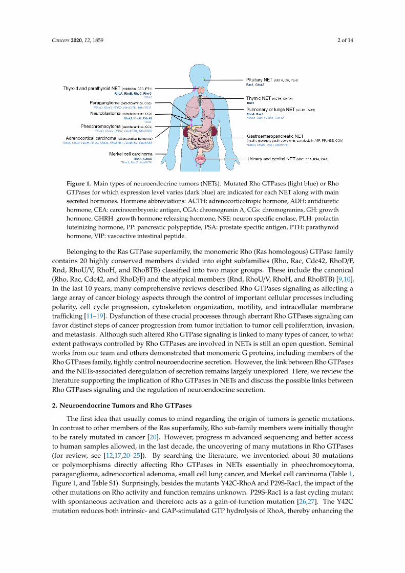
Cancers 2020, 12, 1859 2 of 14
Cancers 2020, 12, x FOR PEER REVIEW
Figure 1. Main types of neuroendocrine tumors (NET). Mutated Rho GTPases (light blue) or Rho GTPases for which expression level varies (dark blue) are indicated for each NET along with main secreted hormones. Hormone abbreviations: ACTH: adrenocorticotropic hormone, ADH: antidiuretic hormone, CEA: carcinoembryonic antigen, CGA: chromogranin A, CGs: chromogranins, GH: growth hormone, GHRH: growth hormone releasing-hormone, NSE: neuron specific enolase, PLH: prolactin luteinizing hormone, PP: pancreatic polypeptide, PSA: prostate specific antigen, PTH: parathyroid hormone, VIP: vasoactive intestinal peptide.
Belonging to the Ras GTPase superfamily, the monomeric Rho (Ras homologous) GTPase family contains 20 highly conserved members divided into eight subfamilies (Rho, Rac, Cdc42, RhoD/F, Rnd, RhoU/V, RhoH, and RhoBTB) classified into two major groups. These include the canonical (Rho, Rac, Cdc42, and RhoD/F) and the atypical members (Rnd, RhoU/V, RhoH, and RhoBTB) [9,10]. In the last 10 years, many comprehensive reviews described Rho GTPases signaling as affecting a large array of cancer biology aspects through the control of important cellular processes including polarity, cell cycle progression, cytoskeleton organization, motility, and intracellular membrane trafficking [11–19]. Dysfunction of these crucial processes through aberrant Rho GTPases signaling can favor distinct steps of cancer progression from tumor initiation to tumor cell proliferation, invasion, and metastasis. Although such altered Rho GTPase signaling is linked to many types of cancer, to what extent pathways controlled by Rho GTPases are involved in NETs is still an open question. Seminal works from our team and others demonstrated that monomeric G proteins, including members of the Rho GTPases family, tightly control neuroendocrine secretion. However, the link between Rho GTPases and the NETs-associated deregulation of secretion remains largely unexplored. Here, we review the literature supporting the implication of Rho GTPases in NETs and discuss the possible links between Rho GTPases signaling and the regulation of neuroendocrine secretion.
2. Neuroendocrine Tumors and Rho GTPases
The first idea that usually comes to mind regarding the origin of tumors is genetic mutations. In contrast to other members of the Ras superfamily, Rho sub-family members were initially thought to be rarely mutated in cancer [20]. However, progress in advanced sequencing and better access to human samples allowed, in the last decade, the uncovering of many mutations in Rho GTPases (for review, see [12,17,20–25]). By searching the literature, we inventoried about 30 mutations or polymorphisms directly affecting Rho GTPases in NETs essentially in pheochromocytoma, paraganglioma, adrenocortical adenoma, small cell lung cancer, and Merkel cell carcinoma (Table 1, Figure 1, and Data S1). Surprisingly, besides the mutants Y42C-RhoA and P29S-Rac1, the impact of the other mutations on Rho activity and function remains unknown. P29S-Rac1 is a fast cycling mutant with spontaneous activation and therefore acts as a gain-of-function mutation [26,27]. The Y42C mutation reduces both intrinsic- and GAP-stimulated GTP hydrolysis of RhoA, thereby enhancing the active GTP-bound form [28]. On the contrary, Wang et al. proposed that the Y42C mutation decreased the level of the activated GTP-associated form of RhoA [29].
Figure 1. Main types of neuroendocrine tumors (NETs). Mutated Rho GTPases (light blue) or RhoGTPases for which expression level varies (dark blue) are indicated for each NET along with mainsecreted hormones. Hormone abbreviations: ACTH: adrenocorticotropic hormone, ADH: antidiuretichormone, CEA: carcinoembryonic antigen, CGA: chromogranin A, CGs: chromogranins, GH: growthhormone, GHRH: growth hormone releasing-hormone, NSE: neuron specific enolase, PLH: prolactinluteinizing hormone, PP: pancreatic polypeptide, PSA: prostate specific antigen, PTH: parathyroidhormone, VIP: vasoactive intestinal peptide.
Belonging to the Ras GTPase superfamily, the monomeric Rho (Ras homologous) GTPase familycontains 20 highly conserved members divided into eight subfamilies (Rho, Rac, Cdc42, RhoD/F,Rnd, RhoU/V, RhoH, and RhoBTB) classified into two major groups. These include the canonical(Rho, Rac, Cdc42, and RhoD/F) and the atypical members (Rnd, RhoU/V, RhoH, and RhoBTB) [9,10].In the last 10 years, many comprehensive reviews described Rho GTPases signaling as affecting alarge array of cancer biology aspects through the control of important cellular processes includingpolarity, cell cycle progression, cytoskeleton organization, motility, and intracellular membranetrafficking [11–19]. Dysfunction of these crucial processes through aberrant Rho GTPases signaling canfavor distinct steps of cancer progression from tumor initiation to tumor cell proliferation, invasion,and metastasis. Although such altered Rho GTPase signaling is linked to many types of cancer, to whatextent pathways controlled by Rho GTPases are involved in NETs is still an open question. Seminalworks from our team and others demonstrated that monomeric G proteins, including members of theRho GTPases family, tightly control neuroendocrine secretion. However, the link between Rho GTPasesand the NETs-associated deregulation of secretion remains largely unexplored. Here, we review theliterature supporting the implication of Rho GTPases in NETs and discuss the possible links betweenRho GTPases signaling and the regulation of neuroendocrine secretion.
2. Neuroendocrine Tumors and Rho GTPases
The first idea that usually comes to mind regarding the origin of tumors is genetic mutations.In contrast to other members of the Ras superfamily, Rho sub-family members were initially thoughtto be rarely mutated in cancer [20]. However, progress in advanced sequencing and better accessto human samples allowed, in the last decade, the uncovering of many mutations in Rho GTPases(for review, see [12,17,20–25]). By searching the literature, we inventoried about 30 mutationsor polymorphisms directly affecting Rho GTPases in NETs essentially in pheochromocytoma,paraganglioma, adrenocortical adenoma, small cell lung cancer, and Merkel cell carcinoma (Table 1,Figure 1, and Table S1). Surprisingly, besides the mutants Y42C-RhoA and P29S-Rac1, the impact of theother mutations on Rho activity and function remains unknown. P29S-Rac1 is a fast cycling mutantwith spontaneous activation and therefore acts as a gain-of-function mutation [26,27]. The Y42Cmutation reduces both intrinsic- and GAP-stimulated GTP hydrolysis of RhoA, thereby enhancing the
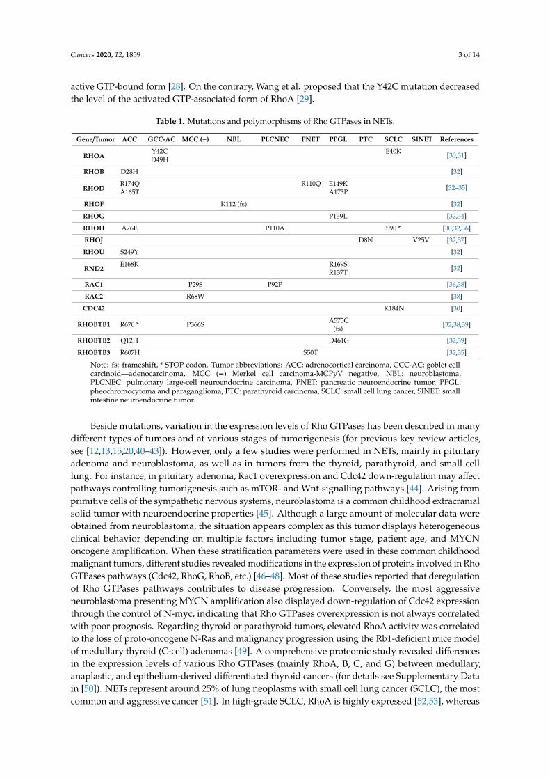
Cancers 2020, 12, 1859 3 of 14
active GTP-bound form [28]. On the contrary, Wang et al. proposed that the Y42C mutation decreasedthe level of the activated GTP-associated form of RhoA [29].
Table 1. Mutations and polymorphisms of Rho GTPases in NETs.
Gene/Tumor ACC GCC-AC MCC (−) NBL PLCNEC PNET PPGL PTC SCLC SINET References
RHOAY42C E40K [30,31]D49H
RHOB D28H [32]
RHODR174Q R110Q E149K [32–35]A165T A173P
RHOF K112 (fs) [32]
RHOG P139L [32,34]
RHOH A76E P110A S90 * [30,32,36]
RHOJ D8N V25V [32,37]
RHOU S249Y [32]
RND2E168K R169S [32]
R137T
RAC1 P29S P92P [36,38]
RAC2 R68W [38]
CDC42 K184N [30]
RHOBTB1 R670 * P366S A575C(fs) [32,38,39]
RHOBTB2 Q12H D461G [32,39]
RHOBTB3 R607H S50T [32,35]
Note: fs: frameshift, * STOP codon. Tumor abbreviations: ACC: adrenocortical carcinoma, GCC-AC: goblet cellcarcinoid—adenocarcinoma, MCC (−) Merkel cell carcinoma-MCPyV negative, NBL: neuroblastoma,PLCNEC: pulmonary large-cell neuroendocrine carcinoma, PNET: pancreatic neuroendocrine tumor, PPGL:pheochromocytoma and paraganglioma, PTC: parathyroid carcinoma, SCLC: small cell lung cancer, SINET: smallintestine neuroendocrine tumor.
Beside mutations, variation in the expression levels of Rho GTPases has been described in manydifferent types of tumors and at various stages of tumorigenesis (for previous key review articles,see [12,13,15,20,40–43]). However, only a few studies were performed in NETs, mainly in pituitaryadenoma and neuroblastoma, as well as in tumors from the thyroid, parathyroid, and small celllung. For instance, in pituitary adenoma, Rac1 overexpression and Cdc42 down-regulation may affectpathways controlling tumorigenesis such as mTOR- and Wnt-signalling pathways [44]. Arising fromprimitive cells of the sympathetic nervous systems, neuroblastoma is a common childhood extracranialsolid tumor with neuroendocrine properties [45]. Although a large amount of molecular data wereobtained from neuroblastoma, the situation appears complex as this tumor displays heterogeneousclinical behavior depending on multiple factors including tumor stage, patient age, and MYCNoncogene amplification. When these stratification parameters were used in these common childhoodmalignant tumors, different studies revealed modifications in the expression of proteins involved in RhoGTPases pathways (Cdc42, RhoG, RhoB, etc.) [46–48]. Most of these studies reported that deregulationof Rho GTPases pathways contributes to disease progression. Conversely, the most aggressiveneuroblastoma presenting MYCN amplification also displayed down-regulation of Cdc42 expressionthrough the control of N-myc, indicating that Rho GTPases overexpression is not always correlatedwith poor prognosis. Regarding thyroid or parathyroid tumors, elevated RhoA activity was correlatedto the loss of proto-oncogene N-Ras and malignancy progression using the Rb1-deficient mice modelof medullary thyroid (C-cell) adenomas [49]. A comprehensive proteomic study revealed differencesin the expression levels of various Rho GTPases (mainly RhoA, B, C, and G) between medullary,anaplastic, and epithelium-derived differentiated thyroid cancers (for details see Supplementary Datain [50]). NETs represent around 25% of lung neoplasms with small cell lung cancer (SCLC), the mostcommon and aggressive cancer [51]. In high-grade SCLC, RhoA is highly expressed [52,53], whereas
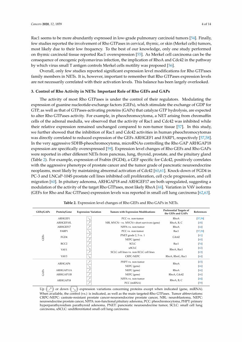
Cancers 2020, 12, 1859 4 of 14
Rac1 seems to be more abundantly expressed in low-grade pulmonary carcinoid tumors [54]. Finally,few studies reported the involvement of Rho GTPases in cervical, thymic, or skin (Merkel cells) tumors,most likely due to their low frequency. To the best of our knowledge, only one study performedon thymic carcinoid tissue reported Rac1 overexpression [55]. As Merkel cell carcinoma can be theconsequence of oncogenic polyomavirus infection, the implication of RhoA and Cdc42 in the pathwayby which virus small T antigen controls Merkel cells motility was proposed [56].
Overall, only few studies reported significant expression level modifications for Rho GTPasesfamily members in NETs. It is, however, important to remember that Rho GTPases expression levelsare not necessarily correlated with their activation levels. This balance has been largely overlooked.
3. Control of Rho Activity in NETs: Important Role of Rho GEFs and GAPs
The activity of most Rho GTPases is under the control of their regulators. Modulating theexpression of guanine nucleotide-exchange factors (GEFs), which stimulate the exchange of GDP forGTP, as well as that of GTPases-activating proteins (GAPs) that catalyze GTP hydrolysis, are expectedto alter Rho GTPases activity. For example, in pheochromocytoma, a NET arising from chromaffincells of the adrenal medulla, we observed that the activity of Rac1 and Cdc42 was inhibited whiletheir relative expression remained unchanged compared to non-tumor tissue [57]. In this study,we further showed that the inhibition of Rac1 and Cdc42 activities in human pheochromocytomaswas directly correlated to reduced expression of the GEFs ARHGEF1 and FARP1, respectively [57,58].In the very aggressive SDHB-pheochromocytoma, microRNAs controlling the Rho GAP ARHGAP18expression are specifically overexpressed [59]. Expression level changes of Rho GEFs and Rho GAPswere reported in other different NETs from pancreas, lung, thyroid, prostate, and the pituitary gland(Table 2). For example, expression of Frabin (FGD4), a GEF specific for Cdc42, positively correlateswith the aggressive phenotype of prostate cancer and the tumor grade of pancreatic neuroendocrineneoplasms, most likely by maintaining abnormal activation of Cdc42 [60,61]. Knock-down of FGD4 inPC-3 and LNCaP-104S prostate cell lines inhibited cell proliferation, cell cycle progression, and cellmigration [60]. In pituitary adenoma, ARHGAP18 and ARHGEF17 are both upregulated, suggesting amodulation of the activity of the target Rho GTPases, most likely RhoA [44]. Variation in VAV isoforms(GEFs for Rho and Rac GTPases) expression levels was reported in small cell lung carcinoma [62,63].
Table 2. Expression level changes of Rho GEFs and Rho GAPs in NETs.
GEFs/GAPs Protein/Gene Expression Variation Tumors with Expression Modifications Preferential Targets ofthe GEFs and GAPs References
GEF
s
ARHGEF1 ↘ PCC vs. non-tumor RhoA [57,58]
ARHGEF10L ↗ NBL MYCN− vs. MYCN+ short survivors (gene) RhoA, B, C [48]
ARHGEF17 ↗ NFPA vs. non-tumor RhoA [44]
FARP1 ↘ PCC vs. non-tumor Rac1 [57,58]
FGD4↗ PNET grade 2, 3 vs. 1
Cdc42[61]
↗ NEPC (gene) [60]
RCC2 ↗ SCLC Rac1 [54]
VAV1↘ uSCLC RhoA, Rac1 [62]
↗ SCLC cell lines vs. non-SCLC cell lines [63]
VAV3 ↘ CRPC-NEPC RhoA, RhoG, Rac1 [64]
GA
Ps
ARHGAP6↗ PHPT vs. non-tumor
RhoA[65]
↘ NEPC (gene) [66]
ARHGAP11A ↗ NEPC (gene) RhoA [66]
ARHGAP11B ↗ NEPC (gene) RhoA, Cdc42 [66]
ARHGAP18↗ NFPA vs. non-tumor RhoA, B, C [44]
↗ PCC (miRNA) [59]
Up ( ↗ ) or down ( ↘ ) expression variations concerning proteins except when indicated (gene, miRNA).When available, the control (vs.) is indicated, as well as the main targeted-Rho GTPases. Tumor abbreviations:CRPC-NEPC: castrate-resistant prostate cancer-neuroendocrine prostate cancer, NBL: neuroblastoma, NEPC:neuroendocrine prostate cancer, NFPA: non-functional pituitary adenoma, PCC: pheochromocytoma, PHPT: primaryhyperparathyroidism parathyroid adenoma, PNET: pancreatic neuroendocrine tumor, SCLC: small cell lungcarcinoma, uSCLC: undifferentiated small cell lung carcinoma.
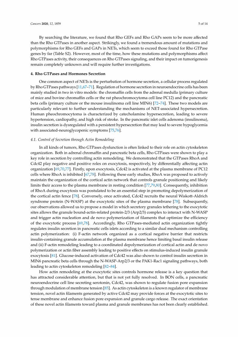
Cancers 2020, 12, 1859 5 of 14
By searching the literature, we found that Rho GEFs and Rho GAPs seem to be more affectedthan the Rho GTPases in another aspect. Strikingly, we found a tremendous amount of mutations andpolymorphisms for Rho GEFs and GAPs in NETs, which seem to exceed those found for Rho GTPasegenes by far (Table S2). However, most of the time, how these mutations and polymorphisms affectRho GTPases activity, their consequences on Rho GTPases signaling, and their impact on tumorigenesisremain completely unknown and will require further investigations.
4. Rho GTPases and Hormones Secretion
One common aspect of NETs is the perturbation of hormone secretion, a cellular process regulatedby Rho GTPases pathways [11,67–71]. Regulation of hormone secretion in neuroendocrine cells has beenmainly studied in two in vitro models: the chromaffin cells from the adrenal medulla (primary cultureof mice and bovine chromaffin cells or the rat pheochromocytoma cell line PC12) and the pancreaticbeta cells (primary culture or the mouse insulinoma cell line MIN6) [72–74]. These two models areparticularly relevant to further understanding the mechanisms of NET-associated hypersecretion.Human pheochromocytoma is characterized by catecholamine hypersecretion, leading to severehypertension, cardiopathy, and high risk of stroke. In the pancreatic islet cells adenoma (insulinoma),insulin secretion is dysregulated with a persistent hypersecretion that may lead to severe hypoglycemiawith associated-neuroglycopenic symptoms [75,76].
4.1. Control of Secretion through Actin Remodeling
In all kinds of tumors, Rho GTPases dysfunction is often linked to their role on actin cytoskeletonorganization. Both in adrenal chromaffin and pancreatic beta cells, Rho GTPases were shown to play akey role in secretion by controlling actin remodeling. We demonstrated that the GTPases RhoA andCdc42 play negative and positive roles on exocytosis, respectively, by differentially affecting actinorganization [69,70,77]. Firstly, upon exocytosis, Cdc42 is activated at the plasma membrane of PC12cells where RhoA is inhibited [67,78]. Following these early studies, RhoA was proposed to activelymaintain the organization of the cortical actin network that controls granule positioning and likelylimits their access to the plasma membrane in resting condition [77,79,80]. Consequently, inhibitionof RhoA during exocytosis was postulated to be an essential step in promoting depolymerization ofthe cortical actin fence [78]. Conversely, once activated, Cdc42 recruits the neural Wiskott-Aldrichsyndrome protein (N-WASP) at the exocytotic sites of the plasma membrane [78]. Subsequently,our observations allowed us to propose a model in which secretory granules tethering to the exocytoticsites allows the granule bound-actin-related protein-2/3 (Arp2/3) complex to interact with N-WASPand trigger actin nucleation and de novo polymerization of filaments that optimize the efficiencyof the exocytotic process [69,78]. Accordingly, Rho GTPases-mediated actin organization tightlyregulates insulin secretion in pancreatic cells islets according to a similar dual mechanism controllingactin polymerization: (i) F-actin network organized as a cortical negative barrier that restrictsinsulin-containing granule accumulation at the plasma membrane hence limiting basal insulin releaseand (ii) F-actin remodeling leading to a coordinated depolymerization of cortical actin and de novopolymerization or actin fiber assembly leading to positive effects on stimulus-induced insulin granuleexocytosis [81]. Glucose-induced activation of Cdc42 was also shown to control insulin secretion inMIN6 pancreatic beta cells through the N-WASP-Arp2/3 or the PAK1-Rac1 signaling pathways, bothleading to actin cytoskeleton remodeling [82–84].
How actin remodeling at the exocytotic sites controls hormone release is a key question thathas attracted considerable attention, but that is not yet fully resolved. In BON cells, a pancreaticneuroendocrine cell line secreting serotonin, Cdc42, was shown to regulate fusion pore expansionthrough modulation of membrane tension [85]. As actin cytoskeleton is a known regulator of membranetension, novel actin filaments generated by active Cdc42 may provide forces at the exocytotic sites totense membrane and enhance fusion pore expansion and granule cargo release. The exact orientationof these novel actin filaments toward plasma and granule membranes has not been clearly established.
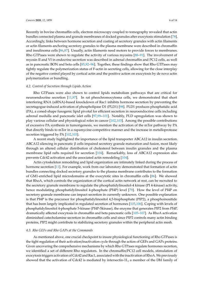
Cancers 2020, 12, 1859 6 of 14
Recently in bovine chromaffin cells, electron microscopy coupled to tomography revealed that actinbundles connected plasma and granule membranes of docked granules after exocytosis stimulation [78].Accordingly, links between hormone secretion and coating of secretory granules with actin filamentsor actin filaments anchoring secretory granules to the plasma membrane were described in chromaffinand insulinoma cells [86,87]. Usually, actin filaments need motors to provide forces to membranes.Rho GTPases were shown to regulate the activity of various myosins [88–91]. The involvement ofmyosin II and VI in endocrine secretion was described in adrenal chromaffin and PC12 cells, as wellas in pancreatic BON and beta cells [85,92–96]. Together, these findings show that Rho GTPases maytightly regulate the polymerization status of F-actin in secreting cells, allowing for the close interplayof the negative control played by cortical actin and the positive action on exocytosis by de novo actinpolymerization or bundling.
4.2. Control of Secretion through Lipids Action
Rho GTPases were also shown to control lipids metabolism pathways that are critical forneuroendocrine secretion [11,97]. In rat pheochromocytoma cells, we demonstrated that shortinterfering RNA (siRNA)-based knockdown of Rac1 inhibits hormone secretion by preventing thesecretagogue-induced activation of phospholipase D1 (PLD1) [98]. PLD1 produces phosphatidic acid(PA), a coned-shape fusogenic lipid pivotal for efficient secretion in neuroendocrine cells includingadrenal medulla and pancreatic islet cells [97,99–101]. Notably, PLD upregulation was shown toplay various cellular and physiological roles in cancer [102,103]. Among the possible contributionsof excessive PA synthesis in tumorigenesis, we mention the activation of the mTor pathway by PAthat directly binds to mTor in a rapamycine-competitive manner and the increase in metalloproteasesecretion triggered by PA [102,103].
A recent study highlighted the importance of the lipid transporter ABCA12 in insulin secretion.ABCA12 silencing in pancreatic β cells impaired secretory granule maturation and fusion, most likelythrough an altered cellular distribution of cholesterol between insulin granules and the plasmamembrane lipid rafts required for secretion [104]. Remarkably, loss of ABCA12 expression alsoprevents Cdc42 activation and the associated actin remodeling [104].
Actin cytoskeleton remodeling and lipid organization are intimately linked during the process ofhormone secretion [11]. For example, work from our laboratory demonstrated that formation of actinbundles connecting docked secretory granules to the plasma membrane contributes to the formationof GM1-enriched lipid microdomains at the exocytotic sites in chromaffin cells [86]. We showedthat RhoA, which controls the organization of the cortical actin network at rest, can be recruited tothe secretory granule membrane to regulate the phosphatidylinositol-4 kinase (PI 4-kinase) activity,hence modulating phosphatidylinositol 4-phosphate (PI4P) level [79]. How the level of PI4P onsecretory granule membrane can impact secretion in currently unknown. One possible explanationis that PI4P is the precursor for phosphatidylinositol 4,5-bisphosphate (PIP2), a phosphoinositidethat has been largely implicated in regulated secretion of hormones [105,106]. Coping with levels ofphosphatidylinositol 4-phosphate 5-kinase (PI4P-5kinase), the enzyme that generates PIP2 from PI4P,dramatically affected exocytosis in chromaffin and beta pancreatic cells [105–107]. As RhoA activationdiminished catecholamine secretion in chromaffin cells and since PIP2 controls many actin bindingproteins, PIP2 might contribute to stabilizing secretory granules within the peripheral actin mesh.
4.3. Rho GEFs and Rho GAPs at the Commands
As mentioned above, one crucial checkpoint to insure physiological functioning of Rho GTPases isthe tight regulation of their activation/inactivation cycle through the action of GEFs and GAPs proteins.Given uncovering the comprehensive mechanisms by which Rho GTPases regulate hormones secretion,we identified a set of different Rho regulators. In the chromaffin/PC12 cell models, stimulation ofexocytosis triggers activation of Cdc42 and Rac1, associated with the inactivation of RhoA. We previouslyshowed that the activation of Cdc42 is mediated by intersectin-1L, a member of the Dbl family of
Cancers 2020, 12, 1859 7 of 14
GEFs that also interacts with N-WASP and participates in actin organization [108–110]. In parallel,Rac1 is activated by β-PIX, a member of the Cool/Pix Rho GEFs family, which is recruited to the plasmamembrane of stimulated-PC12 cells through its interaction with Scrib, the mammalian homologue ofthe Drosophila neoplasic tumor suppressor Scribble [98,111,112].
In pituitary and pancreatic cells, different GEFs have also been proposed to control hormonesecretion. The transient activation of Rac1 required for glucose-induced insulin secretion was proposedto be under the control of VAV2, Tiam1, and Trio/Kalirin in pancreatic cells [113–116]. How these threeGEFs coordinate spatially and temporally Rac activation needs further investigation. In the pituitarygland, the GEF trio has been also proposed to control hormone release [117].
Regarding RhoA, we proposed that Oligophrenin-1, a multi-domains GAP protein involved invarious membrane trafficking events linked to synaptic functions (plasticity, post-synaptic receptortrafficking, and synaptic vesicle recycling [118–122]), might be responsible for the secretagogue-inducedinactivation of RhoA [123]. Along the same line, inhibition of the RhoA/Rock pathway reducedneurotensin secretion in BON cells [124].
5. Conclusions
In comparison to other types of tumors, the role of Rho GTPases in NETs is not well documented.However, the high amount of genetic mutations and polymorphisms discovered in Rho GEFs andGAPs indicates that pathways controlled by Rho GTPases are likely affected in NETs. Today, a cleareffort has to be directed toward understanding how mutations or variations in expression levels ofRho GTPases, GEFs, and GAPs identified in NETs favor tumorigenesis. Comparative genomic andproteomic analyses of human tumor samples remain among the most suitable general strategies touncover new actors involved in Rho GTPases signaling.
Besides being a predictive factor for tumor occurrence or for its progression, whether RhoGTPases pathways could be used as therapeutic targets is clearly an aspect that needs to be developedin the near future. Several drugs directly targeting Rho GTPases have been recently designedand different strategies such as preventing Rho GEF interaction or inhibiting effectors have beenproposed [125–141]. However, based on the complex involvement of Rho GTPases and their regulatorsin NETs hypersecretion, as reviewed here, the development of proper strategies to target each specifictumor will be critical and will require a perfect knowledge of the mechanisms leading to the deregulationof the Rho pathways, as well as their consequences on tumorigenesis.
Supplementary Materials: The following are available online at http://www.mdpi.com/2072-6694/12/7/1859/s1,Table S1: Mutations and polymorphisms of Rho GTPases in NETs, Table S2: Mutations and polymorphisms ofRho GTPases GEFs and GAPs in NETs.
Funding: Part of the author’s work discussed here was supported by grants from Ligue Contre le Cancer (CCIRGE)and from the ANR (SecretoNET) to SG.
Acknowledgments: Institut National de la Santé et de la Recherche Médicale (INSERM) is providing a salary toS.G. and N.V.
Conflicts of Interest: The authors declare no conflict of interest.
References
1. Kulke, M.H.; Shah, M.H.; Benson, A.B., 3rd; Bergsland, E.; Berlin, J.D.; Blaszkowsky, L.S.; Emerson, L.;Engstrom, P.F.; Fanta, P.; Giordano, T.; et al. Neuroendocrine tumors, version 1.2015. J. Natl. Compr. Canc.Netw. 2015, 13, 78–108. [CrossRef] [PubMed]
2. Zandee, W.T.; Kamp, K.; van Adrichem, R.C.; Feelders, R.A.; de Herder, W.W. Effect of hormone secretorysyndromes on neuroendocrine tumor prognosis. Endocr. Relat. Cancer 2017, 24, R261–R274. [CrossRef][PubMed]
3. Juhlin, C.C.; Skoglund, S.; Juntti-Berggren, L.; Karlberg, M.; Calissendorff, J. Non-functioning neuroendocrinepancreatic tumors transforming to malignant insulinomas - four cases and review of the literature.Neuro. Endocrinol. Lett. 2019, 40, 175–183. [PubMed]
Cancers 2020, 12, 1859 8 of 14
4. Song, P.; Sekhon, H.S.; Jia, Y.; Keller, J.A.; Blusztajn, J.K.; Mark, G.P.; Spindel, E.R. Acetylcholine is synthesizedby and acts as an autocrine growth factor for small cell lung carcinoma. Cancer Res. 2003, 63, 214–221.
5. Brown, R.L.; Muzzafar, T.; Wollman, R.; Weiss, R.E. A pituitary carcinoma secreting TSH and prolactin:A non-secreting adenoma gone awry. Eur. J. Endocrinol. 2006, 154, 639–643. [CrossRef]
6. Daems, T.; Verhelst, J.; Michotte, A.; Abrams, P.; De Ridder, D.; Abs, R. Modification of hormonal secretion inclinically silent pituitary adenomas. Pituitary 2009, 12, 80–86. [CrossRef]
7. Lenders, N.; McCormack, A. Malignant transformation in non-functioning pituitary adenomas (pituitarycarcinoma). Pituitary 2018, 21, 217–229. [CrossRef]
8. Neumann, H.P.H.; Young, W.F., Jr.; Eng, C. Pheochromocytoma and Paraganglioma. N. Engl. J. Med. 2019,381, 552–565. [CrossRef]
9. Aspenstrom, P.; Ruusala, A.; Pacholsky, D. Taking Rho GTPases to the next level: The cellular functions ofatypical Rho GTPases. Exp. Cell Res. 2007, 313, 3673–3679. [CrossRef]
10. Boureux, A.; Vignal, E.; Faure, S.; Fort, P. Evolution of the Rho family of ras-like GTPases in eukaryotes.Mol. Biol. Evol. 2007, 24, 203–216. [CrossRef]
11. Croise, P.; Estay-Ahumada, C.; Gasman, S.; Ory, S. Rho GTPases, phosphoinositides, and actin: A tripartiteframework for efficient vesicular trafficking. Small GTPases 2014, 5, e29469. [CrossRef] [PubMed]
12. Haga, R.B.; Ridley, A.J. Rho GTPases: Regulation and roles in cancer cell biology. Small GTPases 2016, 7,207–221. [CrossRef] [PubMed]
13. Li, H.; Peyrollier, K.; Kilic, G.; Brakebusch, C. Rho GTPases and cancer. Biofactors 2014, 40, 226–235. [CrossRef][PubMed]
14. Olayioye, M.A.; Noll, B.; Hausser, A. Spatiotemporal Control of Intracellular Membrane Trafficking by RhoGTPases. Cells 2019, 8, 1478. [CrossRef] [PubMed]
15. Orgaz, J.L.; Herraiz, C.; Sanz-Moreno, V. Rho GTPases modulate malignant transformation of tumor cells.Small GTPases 2014, 5, e29019. [CrossRef]
16. Phuyal, S.; Farhan, H. Multifaceted Rho GTPase Signaling at the Endomembranes. Front. Cell Dev. Biol. 2019,7, 127. [CrossRef]
17. Porter, A.P.; Papaioannou, A.; Malliri, A. Deregulation of Rho GTPases in cancer. Small GTPases 2016, 7,123–138. [CrossRef]
18. Warner, H.; Wilson, B.J.; Caswell, P.T. Control of adhesion and protrusion in cell migration by Rho GTPases.Curr. Opin. Cell. Biol. 2019, 56, 64–70. [CrossRef]
19. Wilson, K.F.; Erickson, J.W.; Antonyak, M.A.; Cerione, R.A. Rho GTPases and their roles in cancer metabolism.Trends Mol. Med. 2013, 19, 74–82. [CrossRef]
20. Alan, J.K.; Lundquist, E.A. Mutationally activated Rho GTPases in cancer. Small GTPases 2013, 4, 159–163.[CrossRef]
21. Bustelo, X.R. RHO GTPases in cancer: Known facts, open questions, and therapeutic challenges. Biochem. Soc.Trans. 2018, 46, 741–760. [CrossRef] [PubMed]
22. Kataoka, K.; Ogawa, S. Variegated RHOA mutations in human cancers. Exp. Hematol. 2016, 44, 1123–1129.[CrossRef] [PubMed]
23. Olson, M.F. Rho GTPases, their post-translational modifications, disease-associated mutations andpharmacological inhibitors. Small GTPases 2018, 9, 203–215. [CrossRef] [PubMed]
24. Svensmark, J.H.; Brakebusch, C. Rho GTPases in cancer: Friend or foe? Oncogene 2019, 38, 7447–7456.[CrossRef] [PubMed]
25. Zandvakili, I.; Lin, Y.; Morris, J.C.; Zheng, Y. Rho GTPases: Anti- or pro-neoplastic targets? Oncogene 2017,36, 3213–3222. [CrossRef]
26. Davis, M.J.; Ha, B.H.; Holman, E.C.; Halaban, R.; Schlessinger, J.; Boggon, T.J. RAC1P29S is a spontaneouslyactivating cancer-associated GTPase. Proc. Natl. Acad. Sci. USA 2013, 110, 912–917. [CrossRef]
27. Kawazu, M.; Ueno, T.; Kontani, K.; Ogita, Y.; Ando, M.; Fukumura, K.; Yamato, A.; Soda, M.; Takeuchi, K.;Miki, Y.; et al. Transforming mutations of RAC guanosine triphosphatases in human cancers. Proc. Natl.Acad. Sci. USA 2013, 110, 3029–3034. [CrossRef]
28. Zhang, H.; Schaefer, A.; Wang, Y.; Hodge, R.G.; Blake, D.R.; Diehl, J.N.; Papageorge, A.G.; Stachler, M.D.;Liao, J.; Zhou, J.; et al. Gain-of-Function RHOA Mutations Promote Focal Adhesion Kinase Activation andDependency in Diffuse Gastric Cancer. Cancer Discov. 2020, 10, 288–305. [CrossRef]
Cancers 2020, 12, 1859 9 of 14
29. Wang, K.; Yuen, S.T.; Xu, J.; Lee, S.P.; Yan, H.H.; Shi, S.T.; Siu, H.C.; Deng, S.; Chu, K.M.; Law, S.; et al.Whole-genome sequencing and comprehensive molecular profiling identify new driver mutations in gastriccancer. Nat. Genet. 2014, 46, 573–582. [CrossRef]
30. Augert, A.; Zhang, Q.; Bates, B.; Cui, M.; Wang, X.; Wildey, G.; Dowlati, A.; MacPherson, D. Small Cell LungCancer Exhibits Frequent Inactivating Mutations in the Histone Methyltransferase KMT2D/MLL2: CALGB151111 (Alliance). J. Thorac. Oncol. 2017, 12, 704–713. [CrossRef]
31. Wen, K.W.; Grenert, J.P.; Joseph, N.M.; Shafizadeh, N.; Huang, A.; Hosseini, M.; Kakar, S. Genomic profile ofappendiceal goblet cell carcinoid is distinct compared to appendiceal neuroendocrine tumor and conventionaladenocarcinoma. Hum. Pathol. 2018, 77, 166–174. [CrossRef] [PubMed]
32. Cao, Y.; Zhou, W.; Li, L.; Wang, J.; Gao, Z.; Jiang, Y.; Jiang, X.; Shan, A.; Bailey, M.H.; Huang, K.L.; et al.Pan-cancer analysis of somatic mutations across 21 neuroendocrine tumor types. Cell Res. 2018, 28, 601–604.[CrossRef]
33. Wilzen, A.; Rehammar, A.; Muth, A.; Nilsson, O.; Tesan Tomic, T.; Wangberg, B.; Kristiansson, E.; Abel, F.Malignant pheochromocytomas/paragangliomas harbor mutations in transport and cell adhesion genes.Int. J. Cancer 2016, 138, 2201–2211. [CrossRef] [PubMed]
34. Flynn, A.; Benn, D.; Clifton-Bligh, R.; Robinson, B.; Trainer, A.H.; James, P.; Hogg, A.; Waldeck, K.; George, J.;Li, J.; et al. The genomic landscape of phaeochromocytoma. J. Pathol. 2015, 236, 78–89. [CrossRef]
35. Scarpa, A.; Chang, D.K.; Nones, K.; Corbo, V.; Patch, A.M.; Bailey, P.; Lawlor, R.T.; Johns, A.L.; Miller, D.K.;Mafficini, A.; et al. Whole-genome landscape of pancreatic neuroendocrine tumours. Nature 2017, 543, 65–71.[CrossRef] [PubMed]
36. George, J.; Walter, V.; Peifer, M.; Alexandrov, L.B.; Seidel, D.; Leenders, F.; Maas, L.; Muller, C.; Dahmen, I.;Delhomme, T.M.; et al. Integrative genomic profiling of large-cell neuroendocrine carcinomas reveals distinctsubtypes of high-grade neuroendocrine lung tumors. Nat. Commun. 2018, 9, 1048. [CrossRef]
37. Francis, J.M.; Kiezun, A.; Ramos, A.H.; Serra, S.; Pedamallu, C.S.; Qian, Z.R.; Banck, M.S.; Kanwar, R.;Kulkarni, A.A.; Karpathakis, A.; et al. Somatic mutation of CDKN1B in small intestine neuroendocrinetumors. Nat. Genet. 2013, 45, 1483–1486. [CrossRef]
38. Harms, P.W.; Vats, P.; Verhaegen, M.E.; Robinson, D.R.; Wu, Y.M.; Dhanasekaran, S.M.; Palanisamy, N.;Siddiqui, J.; Cao, X.; Su, F.; et al. The Distinctive Mutational Spectra of Polyomavirus-Negative Merkel CellCarcinoma. Cancer Res. 2015, 75, 3720–3727. [CrossRef]
39. Castro-Vega, L.J.; Letouze, E.; Burnichon, N.; Buffet, A.; Disderot, P.H.; Khalifa, E.; Loriot, C.; Elarouci, N.;Morin, A.; Menara, M.; et al. Multi-omics analysis defines core genomic alterations in pheochromocytomasand paragangliomas. Nat. Commun. 2015, 6, 6044. [CrossRef]
40. Ellenbroek, S.I.; Collard, J.G. Rho GTPases: Functions and association with cancer. Clin. Exp. Metastasis 2007,24, 657–672. [CrossRef]
41. Karlsson, R.; Pedersen, E.D.; Wang, Z.; Brakebusch, C. Rho GTPase function in tumorigenesis. Biochim.Biophys. Acta 2009, 1796, 91–98. [CrossRef]
42. Gomez del Pulgar, T.; Benitah, S.A.; Valeron, P.F.; Espina, C.; Lacal, J.C. Rho GTPase expression intumourigenesis: Evidence for a significant link. Bioessays 2005, 27, 602–613. [CrossRef] [PubMed]
43. Sahai, E.; Marshall, C.J. RHO-GTPases and cancer. Nat. Rev. Cancer 2002, 2, 133–142. [CrossRef] [PubMed]44. Long, Y.; Lu, M.; Cheng, T.; Zhan, X.; Zhan, X. Multiomics-Based Signaling Pathway Network Alterations
in Human Non-functional Pituitary Adenomas. Front. Endocrinol. (Lausanne) 2019, 10, 835. [CrossRef][PubMed]
45. Veschi, V.; Verona, F.; Thiele, C.J. Cancer Stem Cells and Neuroblastoma: Characteristics and TherapeuticTargeting Options. Front. Endocrinol. (Lausanne) 2019, 10, 782. [CrossRef] [PubMed]
46. Chen, Q.R.; Song, Y.K.; Yu, L.R.; Wei, J.S.; Chung, J.Y.; Hewitt, S.M.; Veenstra, T.D.; Khan, J. Global genomicand proteomic analysis identifies biological pathways related to high-risk neuroblastoma. J. Proteome Res.2010, 9, 373–382. [CrossRef]
47. Coco, S.; Theissen, J.; Scaruffi, P.; Stigliani, S.; Moretti, S.; Oberthuer, A.; Valdora, F.; Fischer, M.; Gallo, F.;Hero, B.; et al. Age-dependent accumulation of genomic aberrations and deregulation of cell cycle andtelomerase genes in metastatic neuroblastoma. Int. J. Cancer 2012, 131, 1591–1600. [CrossRef]
48. Stigliani, S.; Coco, S.; Moretti, S.; Oberthuer, A.; Fischer, M.; Theissen, J.; Gallo, F.; Garavent, A.; Berthold, F.;Bonassi, S.; et al. High genomic instability predicts survival in metastatic high-risk neuroblastoma. Neoplasia2012, 14, 823–832. [CrossRef] [PubMed]
Cancers 2020, 12, 1859 10 of 14
49. Takahashi, C.; Contreras, B.; Iwanaga, T.; Takegami, Y.; Bakker, A.; Bronson, R.T.; Noda, M.; Loda, M.;Hunt, J.L.; Ewen, M.E. Nras loss induces metastatic conversion of Rb1-deficient neuroendocrine thyroidtumor. Nat. Genet. 2006, 38, 118–123. [CrossRef]
50. Gawin, M.; Wojakowska, A.; Pietrowska, M.; Marczak, L.; Chekan, M.; Jelonek, K.; Lange, D.; Jaksik, R.;Gruca, A.; Widlak, P. Proteome profiles of different types of thyroid cancers. Mol. Cell. Endocrinol. 2018, 472,68–79. [CrossRef]
51. Rekhtman, N. Neuroendocrine tumors of the lung: An update. Arch. Pathol. Lab. Med. 2010, 134, 1628–1638.[CrossRef]
52. Touge, H.; Chikumi, H.; Igishi, T.; Kurai, J.; Makino, H.; Tamura, Y.; Takata, M.; Yoneda, K.; Nakamoto, M.;Suyama, H.; et al. Diverse activation states of RhoA in human lung cancer cells: Contribution of G proteincoupled receptors. Int. J. Oncol. 2007, 30, 709–715. [CrossRef] [PubMed]
53. Varker, K.A.; Phelps, S.H.; King, M.M.; Williams, C.L. The small GTPase RhoA has greater expression in smallcell lung carcinoma than in non-small cell lung carcinoma and contributes to their unique morphologies.Int. J. Oncol. 2003, 22, 671–681. [PubMed]
54. Fujii, K.; Miyata, Y.; Takahashi, I.; Koizumi, H.; Saji, H.; Hoshikawa, M.; Takagi, M.; Nishimura, T.;Nakamura, H. Differential Proteomic Analysis between Small Cell Lung Carcinoma (SCLC) and PulmonaryCarcinoid Tumors Reveals Molecular Signatures for Malignancy in Lung Cancer. Proteom. Clin. Appl. 2018,12, e1800015. [CrossRef] [PubMed]
55. Liu, R.X.; Wang, W.Q.; Ye, L.; Bi, Y.F.; Fang, H.; Cui, B.; Zhou, W.W.; Dai, M.; Zhang, J.; Li, X.Y.; et al.p21-activated kinase 3 is overexpressed in thymic neuroendocrine tumors (carcinoids) with ectopic ACTHsyndrome and participates in cell migration. Endocrine 2010, 38, 38–47. [CrossRef]
56. Stakaityte, G.; Nwogu, N.; Dobson, S.J.; Knight, L.M.; Wasson, C.W.; Salguero, F.J.; Blackbourn, D.J.; Blair, G.E.;Mankouri, J.; Macdonald, A.; et al. Merkel Cell Polyomavirus Small T Antigen Drives Cell Motility viaRho-GTPase-Induced Filopodium Formation. J. Virol. 2018, 92. [CrossRef]
57. Croise, P.; Houy, S.; Gand, M.; Lanoix, J.; Calco, V.; Toth, P.; Brunaud, L.; Lomazzi, S.; Paramithiotis, E.;Chelsky, D.; et al. Cdc42 and Rac1 activity is reduced in human pheochromocytoma and correlates withFARP1 and ARHGEF1 expression. Endocr. Relat. Cancer 2016, 23, 281–293. [CrossRef]
58. Croise, P.; Brunaud, L.; Toth, P.; Gasman, S.; Ory, S. Inhibition of Cdc42 and Rac1 activities inpheochromocytoma, the adrenal medulla tumor. Small GTPases 2017, 8, 122–127. [CrossRef]
59. de Cubas, A.A.; Leandro-Garcia, L.J.; Schiavi, F.; Mancikova, V.; Comino-Mendez, I.; Inglada-Perez, L.;Perez-Martinez, M.; Ibarz, N.; Ximenez-Embun, P.; Lopez-Jimenez, E.; et al. Integrative analysis of miRNAand mRNA expression profiles in pheochromocytoma and paraganglioma identifies genotype-specificmarkers and potentially regulated pathways. Endocr. Relat. Cancer 2013, 20, 477–493. [CrossRef]
60. Bossan, A.; Ottman, R.; Andl, T.; Hasan, M.F.; Mahajan, N.; Coppola, D.; Chakrabarti, R. Expression of FGD4positively correlates with the aggressive phenotype of prostate cancer. BMC Cancer 2018, 18, 1257. [CrossRef]
61. Shahid, M.; George, T.B.; Saller, J.; Haija, M.; Sayegh, Z.; Boulware, D.; Strosberg, J.; Chakrabarti, R.;Coppola, D. FGD4 (Frabin) Overexpression in Pancreatic Neuroendocrine Neoplasms. Pancreas 2019, 48,1307–1311. [CrossRef]
62. Lazer, G.; Idelchuk, Y.; Schapira, V.; Pikarsky, E.; Katzav, S. The haematopoietic specific signal transducerVav1 is aberrantly expressed in lung cancer and plays a role in tumourigenesis. J. Pathol. 2009, 219, 25–34.[CrossRef] [PubMed]
63. Ziv, T.; Barnea, E.; Segal, H.; Sharon, R.; Beer, I.; Admon, A. Comparative proteomics of small cell lungcarcinoma. Cancer Biomark. 2006, 2, 219–234. [CrossRef] [PubMed]
64. Weissenrieder, J.S.; Reilly, J.E.; Neighbors, J.D.; Hohl, R.J. Inhibiting geranylgeranyl diphosphate synthesisreduces nuclear androgen receptor signaling and neuroendocrine differentiation in prostate cancer cellmodels. Prostate 2019, 79, 21–30. [CrossRef]
65. Arya, A.K.; Bhadada, S.K.; Singh, P.; Dahiya, D.; Kaur, G.; Sharma, S.; Saikia, U.N.; Behera, A.; Rao, S.D.;Bhasin, M. Quantitative proteomics analysis of sporadic parathyroid adenoma tissue samples. J. Endocrinol.Investig. 2019, 42, 577–590. [CrossRef] [PubMed]
66. Tsai, H.K.; Lehrer, J.; Alshalalfa, M.; Erho, N.; Davicioni, E.; Lotan, T.L. Gene expression signatures ofneuroendocrine prostate cancer and primary small cell prostatic carcinoma. BMC Cancer 2017, 17, 759.[CrossRef] [PubMed]
Cancers 2020, 12, 1859 11 of 14
67. Bader, M.F.; Doussau, F.; Chasserot-Golaz, S.; Vitale, N.; Gasman, S. Coupling actin and membrane dynamicsduring calcium-regulated exocytosis: A role for Rho and ARF GTPases. Biochim. Biophys. Acta 2004, 1742,37–49. [CrossRef]
68. Gasman, S.; Chasserot-Golaz, S.; Bader, M.F.; Vitale, N. Regulation of exocytosis in adrenal chromaffin cells:Focus on ARF and Rho GTPases. Cell Signal. 2003, 15, 893–899. [CrossRef]
69. Malacombe, M.; Bader, M.F.; Gasman, S. Exocytosis in neuroendocrine cells: New tasks for actin. Biochim.Biophys. Acta 2006, 1763, 1175–1183. [CrossRef]
70. Momboisse, F.; Houy, S.; Ory, S.; Calco, V.; Bader, M.F.; Gasman, S. How important are Rho GTPases inneurosecretion? J. Neurochem. 2011, 117, 623–631. [CrossRef]
71. Ory, S.; Gasman, S. Rho GTPases and exocytosis: What are the molecular links? Semin. Cell. Dev. Biol. 2011,22, 27–32. [CrossRef] [PubMed]
72. Bader, M.F.; Holz, R.W.; Kumakura, K.; Vitale, N. Exocytosis: The chromaffin cell as a model system. Ann. N.Y. Acad. Sci. 2002, 971, 178–183. [CrossRef]
73. Westerink, R.H.; Ewing, A.G. The PC12 cell as model for neurosecretion. Acta. Physiol. (Oxf.) 2008, 192,273–285. [CrossRef] [PubMed]
74. Miyazaki, J.; Araki, K.; Yamato, E.; Ikegami, H.; Asano, T.; Shibasaki, Y.; Oka, Y.; Yamamura, K. Establishmentof a pancreatic beta cell line that retains glucose-inducible insulin secretion: Special reference to expressionof glucose transporter isoforms. Endocrinology 1990, 127, 126–132. [CrossRef]
75. Flatt, P.R. Defective regulation of insulin secretion in diabetes and insulinoma. Biochem. Soc. Trans. 1990, 18,124–127. [CrossRef] [PubMed]
76. Okabayashi, T.; Shima, Y.; Sumiyoshi, T.; Kozuki, A.; Ito, S.; Ogawa, Y.; Kobayashi, M.; Hanazaki, K. Diagnosisand management of insulinoma. World J. Gastroenterol. 2013, 19, 829–837. [CrossRef] [PubMed]
77. Gasman, S.; Chasserot-Golaz, S.; Popoff, M.R.; Aunis, D.; Bader, M.F. Involvement of Rho GTPases incalcium-regulated exocytosis from adrenal chromaffin cells. J. Cell. Sci. 1999, 112 Pt. 24, 4763–4771.
78. Gasman, S.; Chasserot-Golaz, S.; Malacombe, M.; Way, M.; Bader, M.F. Regulated exocytosis in neuroendocrinecells: A role for subplasmalemmal Cdc42/N-WASP-induced actin filaments. Mol. Biol. Cell. 2004, 15, 520–531.[CrossRef] [PubMed]
79. Gasman, S.; Chasserot-Golaz, S.; Hubert, P.; Aunis, D.; Bader, M.F. Identification of a potential effectorpathway for the trimeric Go protein associated with secretory granules. Go stimulates a granule-boundphosphatidylinositol 4-kinase by activating RhoA in chromaffin cells. J. Biol. Chem. 1998, 273, 16913–16920.[CrossRef] [PubMed]
80. Gasman, S.; Chasserot-Golaz, S.; Popoff, M.R.; Aunis, D.; Bader, M.F. Trimeric G proteins control exocytosisin chromaffin cells. Go regulates the peripheral actin network and catecholamine secretion by a mechanisminvolving the small GTP-binding protein Rho. J. Biol. Chem. 1997, 272, 20564–20571. [CrossRef] [PubMed]
81. Kalwat, M.A.; Thurmond, D.C. Signaling mechanisms of glucose-induced F-actin remodeling in pancreaticislet beta cells. Exp. Mol. Med. 2013, 45, e37. [CrossRef] [PubMed]
82. Wang, Z.; Oh, E.; Thurmond, D.C. Glucose-stimulated Cdc42 signaling is essential for the second phase ofinsulin secretion. J. Biol. Chem. 2007, 282, 9536–9546. [CrossRef] [PubMed]
83. Uenishi, E.; Shibasaki, T.; Takahashi, H.; Seki, C.; Hamaguchi, H.; Yasuda, T.; Tatebe, M.; Oiso, Y.; Takenawa, T.;Seino, S. Actin dynamics regulated by the balance of neuronal Wiskott-Aldrich syndrome protein (N-WASP)and cofilin activities determines the biphasic response of glucose-induced insulin secretion. J. Biol. Chem.2013, 288, 25851–25864. [CrossRef] [PubMed]
84. Kalwat, M.A.; Yoder, S.M.; Wang, Z.; Thurmond, D.C. A p21-activated kinase (PAK1) signaling cascadecoordinately regulates F-actin remodeling and insulin granule exocytosis in pancreatic beta cells. Biochem.Pharmacol. 2013, 85, 808–816. [CrossRef] [PubMed]
85. Bretou, M.; Jouannot, O.; Fanget, I.; Pierobon, P.; Larochette, N.; Gestraud, P.; Guillon, M.; Emiliani, V.;Gasman, S.; Desnos, C.; et al. Cdc42 controls the dilation of the exocytotic fusion pore by regulatingmembrane tension. Mol. Biol. Cell. 2014, 25, 3195–3209. [CrossRef]
86. Gabel, M.; Delavoie, F.; Demais, V.; Royer, C.; Bailly, Y.; Vitale, N.; Bader, M.F.; Chasserot-Golaz, S. AnnexinA2-dependent actin bundling promotes secretory granule docking to the plasma membrane and exocytosis.J. Cell. Biol. 2015, 210, 785–800. [CrossRef]
87. Ma, W.; Chang, J.; Tong, J.; Ho, U.; Yau, B.; Kebede, M.A.; Thorn, P. Arp2/3 nucleates F-actin coating of fusinginsulin granules in pancreatic beta cells to control insulin secretion. J. Cell. Sci. 2020, 133. [CrossRef]
Cancers 2020, 12, 1859 12 of 14
88. Gomes, E.R.; Jani, S.; Gundersen, G.G. Nuclear movement regulated by Cdc42, MRCK, myosin, and actinflow establishes MTOC polarization in migrating cells. Cell 2005, 121, 451–463. [CrossRef]
89. Brooks, A.B.; Humphreys, D.; Singh, V.; Davidson, A.C.; Arden, S.D.; Buss, F.; Koronakis, V. MYO6 istargeted by Salmonella virulence effectors to trigger PI3-kinase signaling and pathogen invasion into hostcells. Proc. Natl. Acad. Sci. USA 2017, 114, 3915–3920. [CrossRef]
90. Kimura, K.; Ito, M.; Amano, M.; Chihara, K.; Fukata, Y.; Nakafuku, M.; Yamamori, B.; Feng, J.; Nakano, T.;Okawa, K.; et al. Regulation of myosin phosphatase by Rho and Rho-associated kinase (Rho-kinase). Science1996, 273, 245–248. [CrossRef]
91. Wilkinson, S.; Paterson, H.F.; Marshall, C.J. Cdc42-MRCK and Rho-ROCK signalling cooperate in myosinphosphorylation and cell invasion. Nat. Cell. Biol. 2005, 7, 255–261. [CrossRef] [PubMed]
92. Arous, C.; Rondas, D.; Halban, P.A. Non-muscle myosin IIA is involved in focal adhesion and actinremodelling controlling glucose-stimulated insulin secretion. Diabetologia 2013, 56, 792–802. [CrossRef][PubMed]
93. Berberian, K.; Torres, A.J.; Fang, Q.; Kisler, K.; Lindau, M. F-actin and myosin II accelerate catecholaminerelease from chromaffin granules. J. Neurosci. 2009, 29, 863–870. [CrossRef] [PubMed]
94. Neco, P.; Fernandez-Peruchena, C.; Navas, S.; Gutierrez, L.M.; de Toledo, G.A.; Ales, E. Myosin II contributesto fusion pore expansion during exocytosis. J. Biol. Chem. 2008, 283, 10949–10957. [CrossRef] [PubMed]
95. Papadopulos, A.; Gomez, G.A.; Martin, S.; Jackson, J.; Gormal, R.S.; Keating, D.J.; Yap, A.S.; Meunier, F.A.Activity-driven relaxation of the cortical actomyosin II network synchronizes Munc18-1-dependentneurosecretory vesicle docking. Nat. Commun. 2015, 6, 6297. [CrossRef]
96. Tomatis, V.M.; Josh, P.; Papadopulos, A.; Gormal, R.S.; Lanoue, V.; Martin, S.; Meunier, F.A. ENA/VASPproteins regulate exocytosis by mediating myosin VI-dependent recruitment of secretory granules to thecortical actin network. Mol. Cell. Neurosci. 2017, 84, 100–111. [CrossRef]
97. Gasman, S.; Vitale, N. Lipid remodelling in neuroendocrine secretion. Biol. Cell 2017. [CrossRef]98. Momboisse, F.; Lonchamp, E.; Calco, V.; Ceridono, M.; Vitale, N.; Bader, M.F.; Gasman, S. betaPIX-activated
Rac1 stimulates the activation of phospholipase D, which is associated with exocytosis in neuroendocrinecells. J. Cell. Sci. 2009, 122, 798–806. [CrossRef]
99. Zeniou-Meyer, M.; Zabari, N.; Ashery, U.; Chasserot-Golaz, S.; Haeberle, A.M.; Demais, V.; Bailly, Y.;Gottfried, I.; Nakanishi, H.; Neiman, A.M.; et al. Phospholipase D1 production of phosphatidic acid at theplasma membrane promotes exocytosis of large dense-core granules at a late stage. J. Biol. Chem. 2007, 282,21746–21757. [CrossRef]
100. Hughes, W.E.; Elgundi, Z.; Huang, P.; Frohman, M.A.; Biden, T.J. Phospholipase D1 regulatessecretagogue-stimulated insulin release in pancreatic beta-cells. J. Biol. Chem. 2004, 279, 27534–27541.[CrossRef]
101. Metz, S.A.; Dunlop, M. Stimulation of insulin release by phospholipase D. A potential role for endogenousphosphatidic acid in pancreatic islet function. Biochem. J. 1990, 270, 427–435. [CrossRef] [PubMed]
102. Gomez-Cambronero, J. Phospholipase D in cell signaling: From a myriad of cell functions to cancer growthand metastasis. J. Biol. Chem. 2014, 289, 22557–22566. [CrossRef] [PubMed]
103. Zhang, Y.; Frohman, M.A. Cellular and physiological roles for phospholipase D1 in cancer. J. Biol. Chem.2014, 289, 22567–22574. [CrossRef] [PubMed]
104. Ursino, G.M.; Fu, Y.; Cottle, D.L.; Mukhamedova, N.; Jones, L.K.; Low, H.; Tham, M.S.; Gan, W.J.; Mellett, N.A.;Das, P.P.; et al. ABCA12 regulates insulin secretion from beta-cells. EMBO Rep. 2020, 21, e48692. [CrossRef]
105. Gong, L.W.; Di Paolo, G.; Diaz, E.; Cestra, G.; Diaz, M.E.; Lindau, M.; De Camilli, P.; Toomre, D.Phosphatidylinositol phosphate kinase type I gamma regulates dynamics of large dense-core vesiclefusion. Proc. Natl. Acad. Sci. USA 2005, 102, 5204–5209. [CrossRef]
106. Milosevic, I.; Sorensen, J.B.; Lang, T.; Krauss, M.; Nagy, G.; Haucke, V.; Jahn, R.; Neher, E. Plasmalemmalphosphatidylinositol-4,5-bisphosphate level regulates the releasable vesicle pool size in chromaffin cells.J. Neurosci. 2005, 25, 2557–2565. [CrossRef]
107. Waselle, L.; Gerona, R.R.; Vitale, N.; Martin, T.F.; Bader, M.F.; Regazzi, R. Role of phosphoinositide signalingin the control of insulin exocytosis. Mol. Endocrinol. 2005, 19, 3097–3106. [CrossRef]
108. Gubar, O.; Morderer, D.; Tsyba, L.; Croise, P.; Houy, S.; Ory, S.; Gasman, S.; Rynditch, A. Intersectin:The Crossroad between Vesicle Exocytosis and Endocytosis. Front. Endocrinol. (Lausanne) 2013, 4, 109.[CrossRef]
Cancers 2020, 12, 1859 13 of 14
109. Malacombe, M.; Ceridono, M.; Calco, V.; Chasserot-Golaz, S.; McPherson, P.S.; Bader, M.F.; Gasman, S.Intersectin-1L nucleotide exchange factor regulates secretory granule exocytosis by activating Cdc42. Embo. J.2006, 25, 3494–3503. [CrossRef]
110. Zheng, Y. Dbl family guanine nucleotide exchange factors. Trends Biochem. Sci. 2001, 26, 724–732. [CrossRef]111. Audebert, S.; Navarro, C.; Nourry, C.; Chasserot-Golaz, S.; Lecine, P.; Bellaiche, Y.; Dupont, J.L.; Premont, R.T.;
Sempere, C.; Strub, J.M.; et al. Mammalian Scribble Forms a Tight Complex with the betaPIX ExchangeFactor. Curr. Biol. 2004, 14, 987–995. [CrossRef]
112. Zhou, W.; Li, X.; Premont, R.T. Expanding functions of GIT Arf GTPase-activating proteins, PIX Rho guaninenucleotide exchange factors and GIT-PIX complexes. J. Cell Sci. 2016, 129, 1963–1974. [CrossRef] [PubMed]
113. Dufurrena, Q.; Back, N.; Mains, R.E.; Hodgson, L.; Tanowitz, H.; Mandela, P.; Eipper, E.; Kuliawat, R.Kalirin/Trio Rho GDP/GTP exchange factors regulate proinsulin and insulin secretion. J. Mol. Endocrinol.2018. [CrossRef] [PubMed]
114. Kowluru, A. GPCRs, G Proteins, and Their Impact on beta-cell Function. Compr. Physiol. 2020, 10, 453–490.[CrossRef] [PubMed]
115. Veluthakal, R.; Madathilparambil, S.V.; McDonald, P.; Olson, L.K.; Kowluru, A. Regulatory roles for Tiam1, aguanine nucleotide exchange factor for Rac1, in glucose-stimulated insulin secretion in pancreatic beta-cells.Biochem. Pharmacol. 2009, 77, 101–113. [CrossRef] [PubMed]
116. Veluthakal, R.; Tunduguru, R.; Arora, D.K.; Sidarala, V.; Syeda, K.; Vlaar, C.P.; Thurmond, D.C.; Kowluru, A.VAV2, a guanine nucleotide exchange factor for Rac1, regulates glucose-stimulated insulin secretion inpancreatic beta cells. Diabetologia 2015, 58, 2573–2581. [CrossRef] [PubMed]
117. Xin, X.; Ferraro, F.; Back, N.; Eipper, B.A.; Mains, R.E. Cdk5 and Trio modulate endocrine cell exocytosis.J. Cell Sci. 2004, 117, 4739–4748. [CrossRef] [PubMed]
118. Khelfaoui, M.; Gambino, F.; Houbaert, X.; Ragazzon, B.; Muller, C.; Carta, M.; Lanore, F.; Srikumar, B.N.;Gastrein, P.; Lepleux, M.; et al. Lack of the presynaptic RhoGAP protein oligophrenin1 leads to cognitivedisabilities through dysregulation of the cAMP/PKA signalling pathway. Philos. Trans. R Soc. Lond. B Biol.Sci. 2014, 369, 20130160. [CrossRef]
119. Khelfaoui, M.; Pavlowsky, A.; Powell, A.D.; Valnegri, P.; Cheong, K.W.; Blandin, Y.; Passafaro, M.; Jefferys, J.G.;Chelly, J.; Billuart, P. Inhibition of RhoA pathway rescues the endocytosis defects in Oligophrenin1 mousemodel of mental retardation. Hum. Mol. Genet. 2009, 18, 2575–2583. [CrossRef]
120. Nadif Kasri, N.; Nakano-Kobayashi, A.; Malinow, R.; Li, B.; Van Aelst, L. The Rho-linked mental retardationprotein oligophrenin-1 controls synapse maturation and plasticity by stabilizing AMPA receptors. Genes Dev.2009, 23, 1289–1302. [CrossRef]
121. Nakano-Kobayashi, A.; Kasri, N.N.; Newey, S.E.; Van Aelst, L. The Rho-linked mental retardation proteinOPHN1 controls synaptic vesicle endocytosis via endophilin A1. Curr. Biol. 2009, 19, 1133–1139. [CrossRef][PubMed]
122. Powell, A.D.; Gill, K.K.; Saintot, P.P.; Jiruska, P.; Chelly, J.; Billuart, P.; Jefferys, J.G. Rapid reversal of impairedinhibitory and excitatory transmission but not spine dysgenesis in a mouse model of mental retardation.J. Physiol. 2012, 590, 763–776. [CrossRef] [PubMed]
123. Houy, S.; Estay-Ahumada, C.; Croise, P.; Calco, V.; Haeberle, A.M.; Bailly, Y.; Billuart, P.; Vitale, N.; Bader, M.F.;Ory, S.; et al. Oligophrenin-1 Connects Exocytotic Fusion to Compensatory Endocytosis in NeuroendocrineCells. J. Neurosci. 2015, 35, 11045–11055. [CrossRef] [PubMed]
124. Li, J.; O’Connor, K.L.; Hellmich, M.R.; Greeley, G.H., Jr.; Townsend, C.M., Jr.; Evers, B.M. The role of proteinkinase D in neurotensin secretion mediated by protein kinase C-alpha/-delta and Rho/Rho kinase. J. Biol.Chem. 2004, 279, 28466–28474. [CrossRef] [PubMed]
125. Arias-Romero, L.E.; Chernoff, J. Targeting Cdc42 in cancer. Expert. Opin. Ther. Targets 2013, 17, 1263–1273.[CrossRef]
126. Cardama, G.A.; Gonzalez, N.; Maggio, J.; Menna, P.L.; Gomez, D.E. Rho GTPases as therapeutic targets incancer (Review). Int. J. Oncol. 2017, 51, 1025–1034. [CrossRef]
127. Evelyn, C.R.; Ferng, T.; Rojas, R.J.; Larsen, M.J.; Sondek, J.; Neubig, R.R. High-throughput screening forsmall-molecule inhibitors of LARG-stimulated RhoA nucleotide binding via a novel fluorescence polarizationassay. J. Biomol. Screen 2009, 14, 161–172. [CrossRef]
Cancers 2020, 12, 1859 14 of 14
128. Friesland, A.; Zhao, Y.; Chen, Y.H.; Wang, L.; Zhou, H.; Lu, Q. Small molecule targeting Cdc42-intersectininteraction disrupts Golgi organization and suppresses cell motility. Proc. Natl. Acad. Sci. USA 2013, 110,1261–1266. [CrossRef]
129. Gao, Y.; Dickerson, J.B.; Guo, F.; Zheng, J.; Zheng, Y. Rational design and characterization of a RacGTPase-specific small molecule inhibitor. Proc. Natl. Acad. Sci. USA 2004, 101, 7618–7623. [CrossRef]
130. Lee, M.H.; Kundu, J.K.; Chae, J.I.; Shim, J.H. Targeting ROCK/LIMK/cofilin signaling pathway in cancer.Arch. Pharm. Res. 2019, 42, 481–491. [CrossRef]
131. Lin, Y.; Zheng, Y. Approaches of targeting Rho GTPases in cancer drug discovery. Expert Opin. Drug Discov.2015, 10, 991–1010. [CrossRef] [PubMed]
132. Liu, W.; Du, W.; Shang, X.; Wang, L.; Evelyn, C.; Florian, M.C.; Ryan, M.A.; Rayes, A.; Zhao, X.; Setchell, K.; et al.Rational identification of a Cdc42 inhibitor presents a new regimen for long-term hematopoietic stem cellmobilization. Leukemia 2019, 33, 749–761. [CrossRef] [PubMed]
133. Mardilovich, K.; Olson, M.F.; Baugh, M. Targeting Rho GTPase signaling for cancer therapy. Future Oncol.2012, 8, 165–177. [CrossRef]
134. Montalvo-Ortiz, B.L.; Castillo-Pichardo, L.; Hernandez, E.; Humphries-Bickley, T.; De la Mota-Peynado, A.;Cubano, L.A.; Vlaar, C.P.; Dharmawardhane, S. Characterization of EHop-016, novel small molecule inhibitorof Rac GTPase. J. Biol. Chem. 2012, 287, 13228–13238. [CrossRef]
135. Nassar, N.; Cancelas, J.; Zheng, J.; Williams, D.A.; Zheng, Y. Structure-function based design of small moleculeinhibitors targeting Rho family GTPases. Curr. Top. Med. Chem. 2006, 6, 1109–1116. [CrossRef] [PubMed]
136. Pajic, M.; Herrmann, D.; Vennin, C.; Conway, J.R.; Chin, V.T.; Johnsson, A.K.; Welch, H.C.; Timpson, P.The dynamics of Rho GTPase signaling and implications for targeting cancer and the tumor microenvironment.Small GTPases 2015, 6, 123–133. [CrossRef]
137. Shang, X.; Marchioni, F.; Sipes, N.; Evelyn, C.R.; Jerabek-Willemsen, M.; Duhr, S.; Seibel, W.; Wortman, M.;Zheng, Y. Rational design of small molecule inhibitors targeting RhoA subfamily Rho GTPases. Chem. Biol.2012, 19, 699–710. [CrossRef]
138. Xiao, X.H.; Lv, L.C.; Duan, J.; Wu, Y.M.; He, S.J.; Hu, Z.Z.; Xiong, L.X. Regulating Cdc42 and Its SignalingPathways in Cancer: Small Molecules and MicroRNA as New Treatment Candidates. Molecules 2018, 23, 787.[CrossRef]
139. Zhang, Y.; Li, J.; Lai, X.N.; Jiao, X.Q.; Xiong, J.P.; Xiong, L.X. Focus on Cdc42 in Breast Cancer: New Insights,Target Therapy Development and Non-Coding RNAs. Cells 2019, 8, 146. [CrossRef]
140. Rath, N.; Olson, M.F. Rho-associated kinases in tumorigenesis: Re-considering ROCK inhibition for cancertherapy. EMBO Rep. 2012, 13, 900–908. [CrossRef]
141. Wang, Y.; Lei, R.; Zhuang, X.; Zhang, N.; Pan, H.; Li, G.; Hu, J.; Pan, X.; Tao, Q.; Fu, D.; et al. DLC1-dependentparathyroid hormone-like hormone inhibition suppresses breast cancer bone metastasis. J. Clin. Investig.2014, 124, 1646–1659. [CrossRef] [PubMed]
© 2020 by the authors. Licensee MDPI, Basel, Switzerland. This article is an open accessarticle distributed under the terms and conditions of the Creative Commons Attribution(CC BY) license (http://creativecommons.org/licenses/by/4.0/).

Laura STREIT
Résumé
Les cellules neuroendocrines sécrètent des hormones et des neuropeptides par un processus d'exocytose régulé par le
calcium. Malheureusement, toutes les cellules neuroendocrines de l'organisme peuvent se transformer en cellules
tumorales et potentiellement engendrer un cancer. Bien que constituant un groupe très hétérogène, les tumeurs
neuroendocrines possèdent une caractéristique commune intéressante qui est un dysfonctionnement de leur activité
sécrétrice entraînant, dans la majorité des cas, une hypersécrétion des hormones et peptides qu'elles stockent. Cette
sécrétion anarchique pose problème car elle peut engendrer des conséquences cliniques graves chez les patients. À ce
jour, les mécanismes moléculaires qui induisent et maintiennent l’hypersécrétion de ces tumeurs ne sont pas connus
et il n’existe aucune thérapie ciblée permettant de l’empêcher. Mes travaux de thèse montrent que l’hypersécrétion
du phéochromocytome est la conséquence directe d’un dérèglement des phases tardives de l’exocytose et qu’un
traitement par le pasiréotide, un analogue de la somatostatine inhibe efficacement l’hypersécrétion.
Mots clés : phéochromocytome, sécrétion, exocytose régulée par le calcium, analogue de la somatostatine
Abstract
Neuroendocrine cells secrete hormones and neuropeptides through calcium-regulated exocytosis. Unfortunately, all
neuroendocrine cells in the body can develop into tumor cells and potentially into cancer. Although a very
heterogeneous group, neuroendocrine tumors have one interesting feature in common, which is a dysfunction of their
secretory activity leading, in the majority of cases, to hypersecretion of the hormones and neuropeptides. This anarchic
secretion is problematic as it can lead to serious clinical consequences in patients. To date, the molecular mechanisms
that induce and maintain hypersecretion in these tumors are not known and there is no targeted therapy to prevent
it. My thesis work shows that hypersecretion in pheochromocytoma is a direct consequence of a deregulation of the
late phases of exocytosis and that treatment with the somatostatin analogue pasireotide effectively inhibits
hypersecretion.
Key words: pheochromocytoma, secretion, calcium-regulated exocytosis, somatostatin analogue
Mécanismes moléculaires de la sécrétion hormonale et traitement anti-sécrétoire du
phéochromocytome humain
Related Documents