Measuring shape complexity of breast lesions on ultrasound images Wei Yang, Su Zhang, Yazhu Chen Dept. of Biomedical Engineering, Shanghai Jiao Tong Univ., Shanghai, China Wenying Li, Yaqing Chen Shanghai Sixth People’s Hospital, Shanghai Jiao Tong Univ., Shanghai, China Medical Imaging 2008: Ultrasonic Imaging and Signal Processing

Measuring shape complexity of breast lesions on ultrasound images Wei Yang, Su Zhang, Yazhu Chen Dept. of Biomedical Engineering, Shanghai Jiao Tong Univ.,
Dec 30, 2015
Welcome message from author
This document is posted to help you gain knowledge. Please leave a comment to let me know what you think about it! Share it to your friends and learn new things together.
Transcript
Measuring shape complexity of breast lesions on ultrasound images
Wei Yang, Su Zhang, Yazhu Chen Dept. of Biomedical Engineering, Shanghai Jiao Tong Univ., Shanghai, China
Wenying Li, Yaqing ChenShanghai Sixth People’s Hospital, Shanghai Jiao Tong Univ., Shanghai, China
Medical Imaging 2008: Ultrasonic Imaging and Signal Processing

Introduction
• The purpose of this study is to find the effective shape measures which can characterize the shape complexity of the breast tumors.
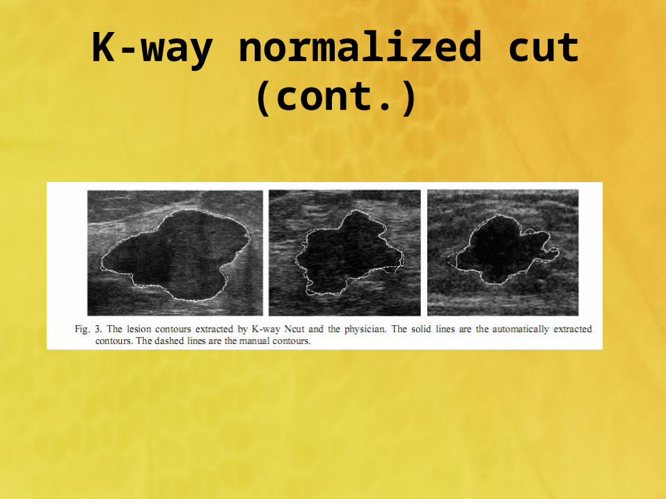
• First, the breast tumors are segmented by K-way normalized cut with the proposed empirical rules.
• Then, the polygonal model are used to represent the lesion contours.– shape complexity measure (SCM)
• Finally, the performance of a linear classifier using the shape complexity and the measures of margin feature as the input vector is evaluated.

Methodology
• Segmentation of breast lesions on ultrasound images by K-way normalized cut
• Shape analysis of lesion contour– Polygonal approximation of lesion contour and shape complexity
measure– Local integral invariant signatures

K-way normalized cut
• Rectangular ROI of the lesion area is first segmented out manually
• The segmentation of images to separate the lesion from the surrounding tissues is performed by means of the filter
K-way normalized cut (cont.)
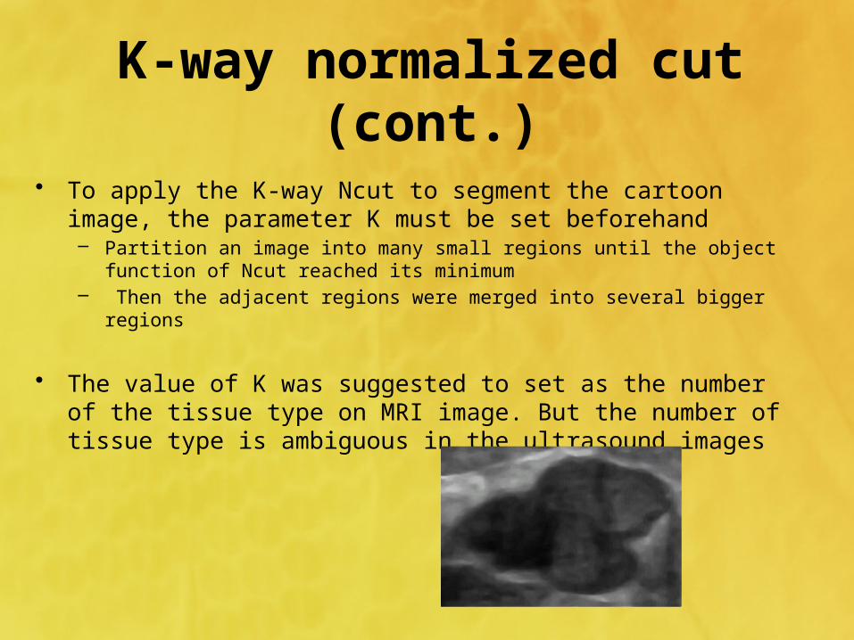
• To apply the K-way Ncut to segment the cartoon image, the parameter K must be set beforehand– Partition an image into many small regions until the object function of
Ncut reached its minimum– Then the adjacent regions were merged into several bigger regions
• The value of K was suggested to set as the number of the tissue type on MRI image. But the number of tissue type is ambiguous in the ultrasound images
K-way normalized cut (cont.)
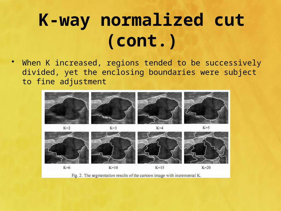
• When K increased, regions tended to be successively divided, yet the enclosing boundaries were subject to fine adjustment
K-way normalized cut (cont.)
• There is no common rule to determine the optimal K
• This study choose K through the following empirical rules
– The lesion region which is closest to the center of ROI is isolated to the margin of ROI with certain k
– Set k’ = k+1 and segment the image with k’ if k satisfies rule1. The optimal K is selected as k’ if the overlap area of the lesion regions corresponding to k’ and k is larger than 95% of the lesion area corresponding to k
– The segmentation process is failed if there is no k satisfies rule1 and rule2
K-way normalized cut (cont.)
Polygonal approximation
• Reduce the amount of data to be processed subsequently
• Preserve most of the shape information of the initial contours
• The aim of polygonalisation in this paper is to obtain the compact represent of digitized contour and preserve the structures of spicules and lobes along the tumor contour
Polygonal approximation (cont.)
• The adaptive dominant point detection method computes the suitable length of support region for each point to find the best approximated curvature and the dominant points are identified as the points with local maximum curvatures
• The merging and splitting schemes are iteratively implemented until convergence using the dominant points as the initial polygonal vertices.
• In the merging scheme– The line segments with length less than the threshold (set to 3 pixels in
the experiments) are merged to one of its adjacent line segments– The two adjacent line segments with the angle greater than a given
threshold (set to 170°) are merged to one line segment
Polygonal approximation (cont.)
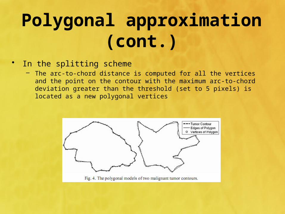
• In the splitting scheme– The arc-to-chord distance is computed for all the vertices and the point
on the contour with the maximum arc-to-chord deviation greater than the threshold (set to 5 pixels) is located as a new polygonal vertices

Polygonal approximation (cont.)
• Shape complexity measure(SCM)– Characterize the complexity of contour
• The complexity of a polygon pol is defined as:
• , : The weight coefficient, set to 0.9 and 0.1• : The amplitude• : The frequency of the vibration of the polygon• : The global shape convexity measure
)()()()( 21 polconvwpolfreqpolamplwpolSCM
1w 2w)( polampl)( polfreq)( polconv
Polygonal approximation (cont.)
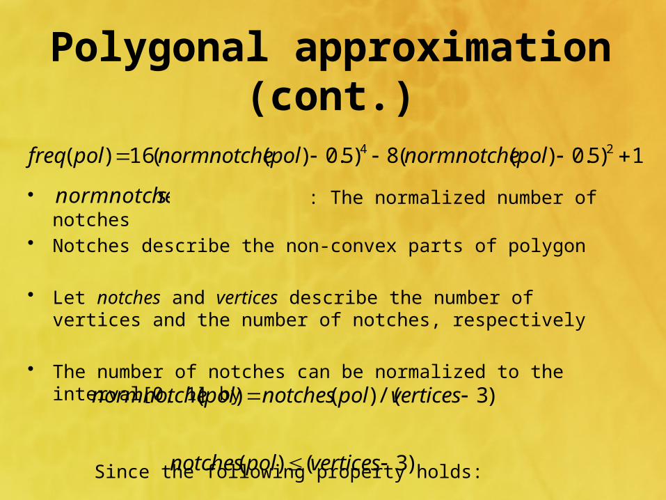
• : The normalized number of notches• Notches describe the non-convex parts of polygon • Let notches and vertices describe the number of vertices and
the number of notches, respectively
• The number of notches can be normalized to the interval[0, 1] by
Since the following property holds:
1)5.0)((8)5.0)((16)( 24 polnormnotchepolnormnotchepolfreq
snormnotche
)3/()()( verticespolnotchespolnormnotche
)3()( verticespolnotches
Polygonal approximation (cont.)
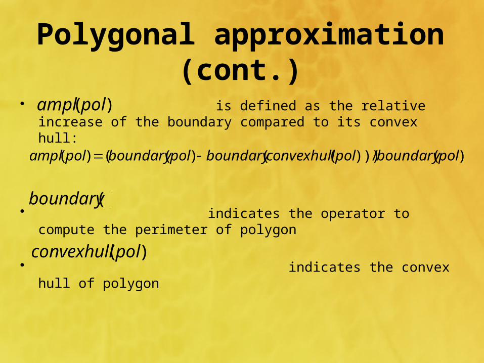
• is defined as the relative increase of the boundary compared to its convex hull:
• indicates the operator to compute the
perimeter of polygon
• indicates the convex hull of polygon
)( polampl
))(/)))(()(()( polboundarypolconvexhullboundarypolboundarypolampl
()boundary
)( polconvexhull
Polygonal approximation (cont.)
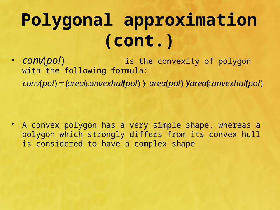
• is the convexity of polygon with the following formula:
• A convex polygon has a very simple shape, whereas a polygon which strongly differs from its convex hull is considered to have a complex shape
)( polconv
))((/))())((()( polconvexhullareapolareapolconvexhullareapolconv
Polygonal approximation (cont.)
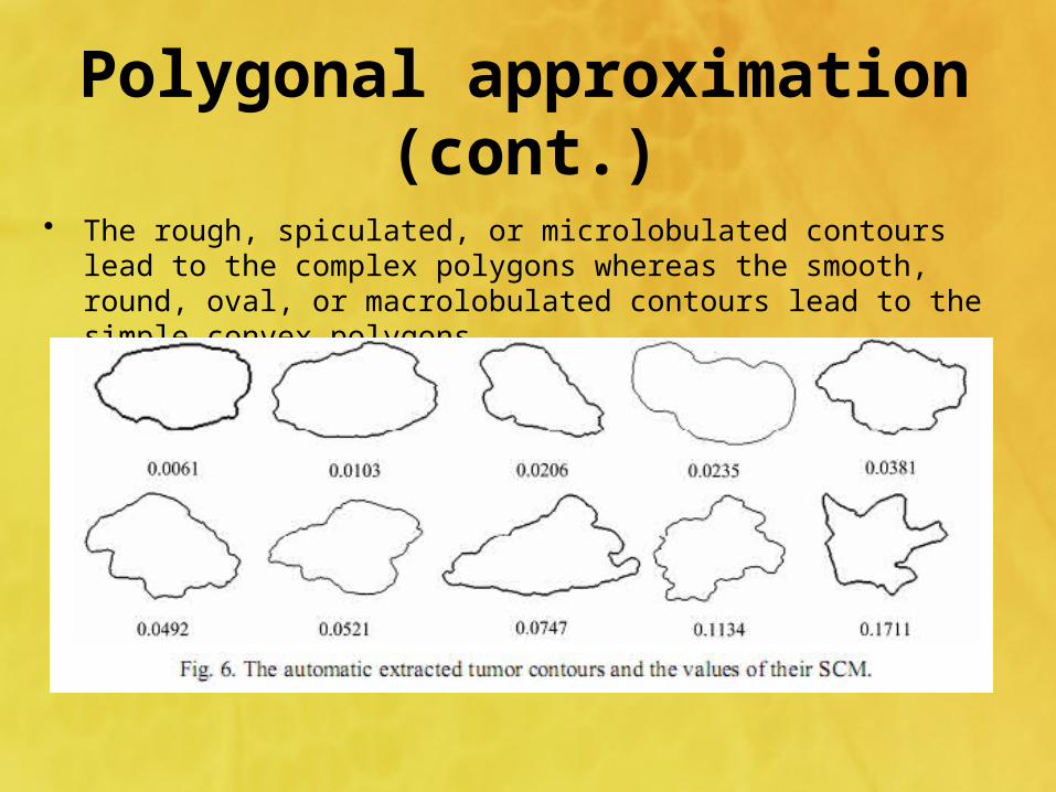
Polygonal approximation (cont.)
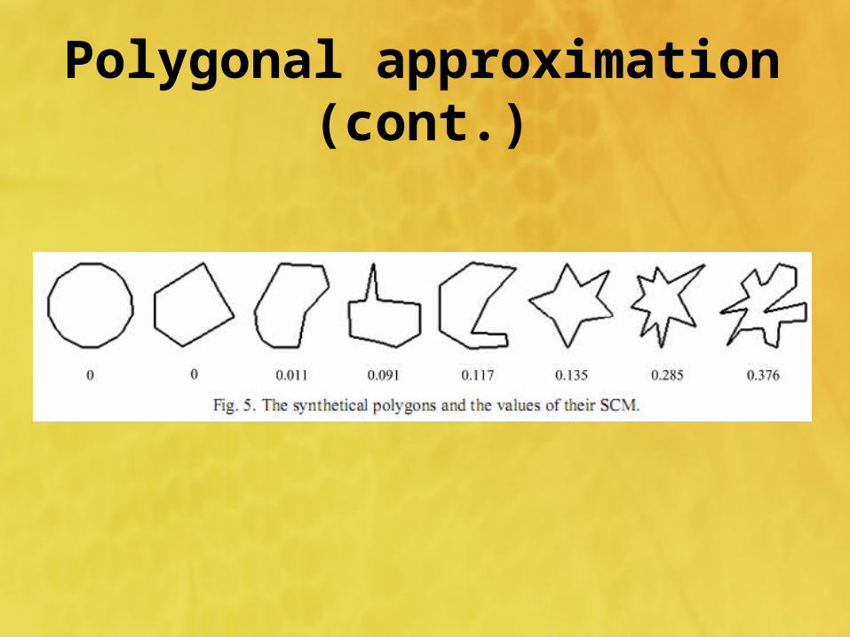
• The rough, spiculated, or microlobulated contours lead to the complex polygons whereas the smooth, round, oval, or macrolobulated contours lead to the simple convex polygons
Other shape measures
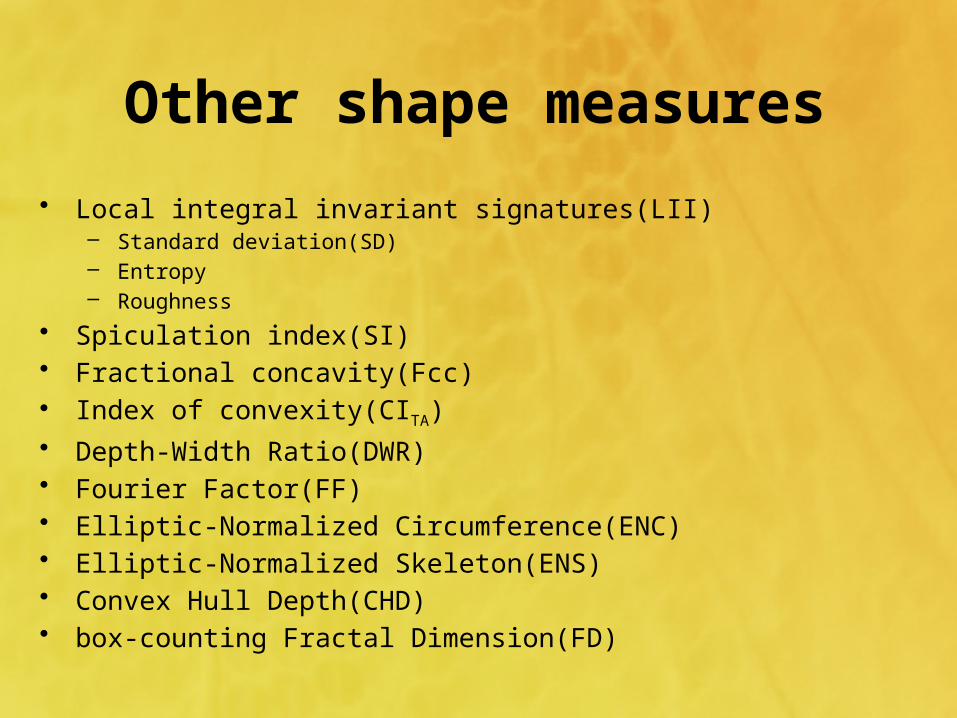
• Local integral invariant signatures(LII)– Standard deviation(SD)– Entropy– Roughness
• Spiculation index(SI)• Fractional concavity(Fcc)• Index of convexity(CITA)• Depth-Width Ratio(DWR)• Fourier Factor(FF)• Elliptic-Normalized Circumference(ENC)• Elliptic-Normalized Skeleton(ENS)• Convex Hull Depth(CHD)• box-counting Fractal Dimension(FD)
Other shape measures (cont.)
• Irregularity• Compactness• Solidity

Results
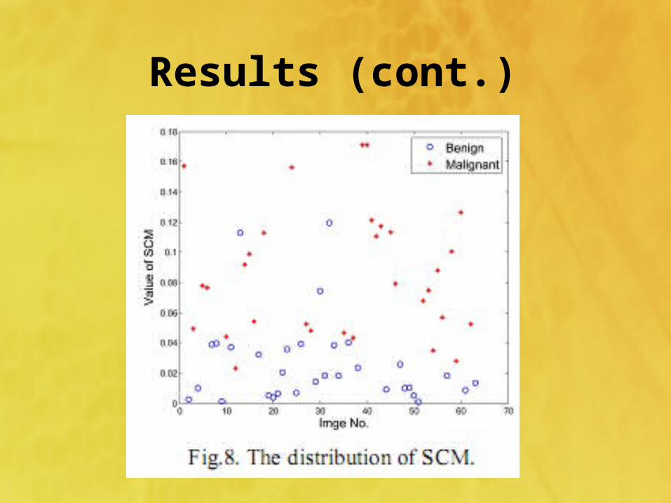
• The ultrasound image database comprises 63 images of pathologically proven benign breast lesions from 32 patients and carcinomas from 31 patients
• For each ROI, the lesion contour was extracted using K-way Ncut
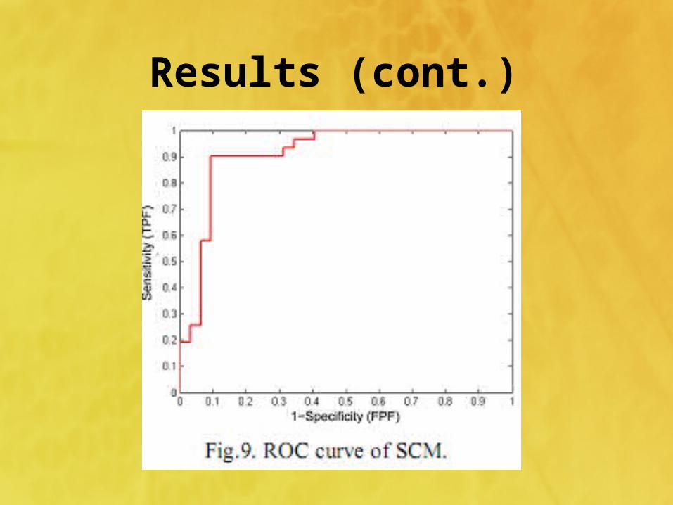
• To evaluate the performance of each individual shape feature, a receiver operating characteristic (ROC) curve was generated by using a sliding threshold on each feature and computing the sensitivity and specificity for each threshold
• The overall classification performance is summarized by the area under the ROC curve (AUC)
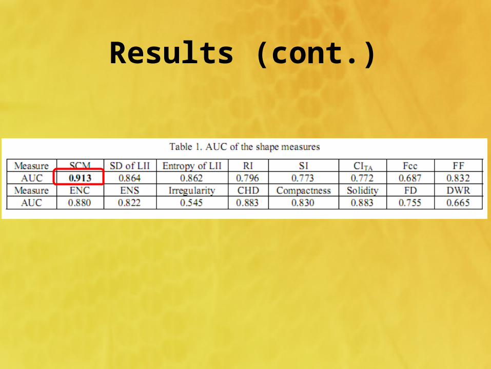
Results (cont.)
Results (cont.)
Results (cont.)
Linear discriminant analysis
• Atypical cases of macrolobulated or spiculated benign lesions, as well as microlobulated or well-circumscribed malignant tumors create difficulties in pattern classification
• This study used linear discriminant analysis (LDA) to merge the computer-extracted features
• ROC analysis was also used to evaluate the performance of LDA classifier in the task of distinguishing benign from malignant lesions– The resubstitution method, which has an optimistic bias– The leave-one-out method,which has a pessimistic bias

Conclusion• This paper introduced a new measure of shape complexity to
characterize the automatic extracted tumor contours, which outperformed the other measures of shape features
• The significant difference of SCM between the benign and malignant lesions suggested that it would be a useful index for the clinical diagnosis to reduce the number of unnecessary biopsies
• However, the conclusions of this paper are limited by the size of the study samples. The proposed system only used measures of shape and margin features
• The methods to extract effective measures which can quantity echo pattern, surrounding tissue, and calcifications should be developed

Thank You!
Related Documents