MEASUREMENT OF ALBUMINURIA WITH SIZE-EXCLUSION CHROMATOGRAPHY CHARACTERIZATION AND NEW PERSPECTIVES PHD THESES SUMMARY LAJOS MARKÓ MD HEAD OF THE DOCTORAL SCHOOL: PROF. DR. SÁMUEL KOMOLY MD, DSC HEAD OF THE PROGRAM: PROF. DR. JUDIT NAGY MD, DSC SUPERVISOR: PROF. DR. ISTVÁN WITTMANN MD, PHD UNIVERSITY OF PÉCS, FACULTY OF MEDICINE 2 ND DEPARTMENT OF MEDICINE AND NEPHROLOGICAL CENTER PÉCS, HUNGARY 2011

Welcome message from author
This document is posted to help you gain knowledge. Please leave a comment to let me know what you think about it! Share it to your friends and learn new things together.
Transcript

MEASUREMENT OF ALBUMINURIA WITH SIZE-EXCLUSION
CHROMATOGRAPHY
CHARACTERIZATION AND NEW PERSPECTIVES
PHD THESES
SUMMARY
LAJOS MARKÓ MD
HEAD OF THE DOCTORAL SCHOOL: PROF. DR. SÁMUEL KOMOLY MD, DSC
HEAD OF THE PROGRAM: PROF. DR. JUDIT NAGY MD, DSC
SUPERVISOR: PROF. DR. ISTVÁN WITTMANN MD, PHD
UNIVERSITY OF PÉCS, FACULTY OF MEDICINE
2ND DEPARTMENT OF MEDICINE AND NEPHROLOGICAL CENTER
PÉCS, HUNGARY
2011

2
ABBREVIATIONS
ACN .......................... acetonitrile
ACR .......................... albumin-creatinine ratio
AusDiab .................... Australian Diabetes, Obesity and Lifestyle
CD ............................ Crohn’s disease
CV ............................ coefficient of variation
DMR ......................... dimeric to monomeric ratio of urinary albumin
DTNB ....................... 5, 5'-dithio-bis (2-nitrobenzoic acid)
FDA .......................... Food and Drug Administration
GSA .......................... glycated human serum albumin
GSH .......................... reduced glutathione
HDL .......................... high-density lipoprotein
HPLC ........................ high-performance liquid chromatography
HSA .......................... human serum albumin
IN .............................. immunonephelometry
ir-uAlb ...................... immunoreactive urinary albumin
IT .............................. immunoturbidimetry
LDL .......................... low-density lipoprotein
MALDI-TOF/MS ..... matrix-assisted laser desorption/ionization time-of-flight mass spectrometry
MGO-HSA ............... human serum albumin modified with methylglyoxal
MM ............................ patients microalbuminuric using both IN and HPLC methods
MS ............................ mass spectrometry
NM ........................... patients normoalbuminuric by IN, microalbuminuric by HPLC method
NN ............................ normoalbuminuric patients using both IN and HPLC methods
PMF .......................... peptide mass fingerprinting
RF ............................. relative fluorescence
RP ............................. reversed-phase
SD ............................. standard deviation of mean
SDS-PAGE ............... sodiumdodecylsulphate polyacrylamide gel-electrophoresis
SE ............................. size-exclusion
TFA .......................... trifluoroacetic acid
TFSG ........................ total free sulfhydryl groups
t-uAlb ........................ total urinary albumin
UAC .......................... urinary albumin concentration
uAlb .......................... urinary albumin

3
1. INTRODUCTION
Accurate measurement of the urinary excretion of albumin (albuminuria) is of great
importance to be able to identify those at risk in order to be able to start treatment.
Recently, a new method has been developed to measure albuminuria and some aspect of
this method has been investigated in this thesis. This introduction aimed to give a short
overview about albumin, its role as a risk marker and its measurement.
1.1. DEFINITION AND PROPERTIES OF ALBUMIN
Albumin is one of the longest known and probably the most studied of all proteins. By
definition, the term “albumin” refers to any proteins that are soluble in water and in
moderately concentrated salt solution, and that are coagulable on heating. The human
serum albumin (further referred as albumin) is the most abundant protein in human
blood plasma, synthesized by the liver. Constituting almost 60% of the total plasma
protein, albumin is responsible for approximately 70% of the colloid osmotic pressure,
and binds a variety of ligands such as fatty acids, metal ions, pharmaceuticals, and
metabolites, playing a significant role in drug delivery, efficacy and detoxification.
Because of its only free cysteine residue albumin is the major extracellular source of
thiols and acts as scavenger of reactive oxygen and nitrogen species.
1.2. ALBUMINURIA AS A WELL-ESTABLISHED RISK MARKER
Under physiological conditions albumin is excreted in the urine in very small amounts
of less than 30 mg per day. Persistent albuminuria in the range of 30-300 mg/day
(microalbuminuria) is recognized as one of the earliest indicators of nephropathy in
patients with type 1 or type 2 diabetes mellitus and a marker of progressive kidney
disease. Moreover, it has been recognized as a powerful marker and predictor for
cardiovascular disease and overall mortality in diabetes and in the general population, as
well.
Given the fact that diabetes mellitus and cardiovascular disease are the leading
cause of death in industrialized countries, accurate measurement of albuminuria is of
great importance.
1.3. MEASUREMENT OF ALBUMINURIA
The very first laboratory tests developed to detect urinary albumin (dipstick tests) could
only estimate concentrations of 300 mg/24 hour and above. The first analytical test that

4
could measure lower albumin concentrations was a radioimmunoassay, using 125I
labeled albumin which is based on immune reaction. Unfortunately, this method was
time-consuming and too expensive for routine laboratory measurement. Therefore other
immuno-based (immunonephelometry (IN) and immunoturbidimetry (IT)) automatic
assays have been developed where the albumin containing sample (serum or urine) is
mixed with albumin-antibody, resulting in small aggregates. These aggregates will
scatter light and the amount of scatter is measured. In the clinical setting, assessment of
microalbuminuria (30-300 mg/day by immuno-based methods) has been established as
a valuable risk marker.
Recently, a high-performance liquid chromatography (HPLC) method based on
size-exclusion has been developed to detect albuminuria. The very first study using this
new method has shown that urinary albumin concentration in diabetic patients is
significantly higher compared with conventional assays. Urinary albumin measured by
HPLC is referred as total urinary albumin. The fraction of albumin which is not
detectable by conventional immunochemical methods, but which can be measured by
HPLC is referred as immuno-unreactive, nonimmunoreactive or immunochemically
nonreactive albumin.
2. AIMS
2.1. MEASUREMENT OF MODIFICATION AND INTERFERENCE RATE OF
URINARY ALBUMIN DETECTED BY SIZE-EXCLUSION HPLC (PART I OF THIS
THESIS)
After the introduction of the new HPLC method for the measurement of albuminuria
some authors proposed that oxidative stress-induced modification of albumin could be
one of the reasons for immuno-unreactivity, while other authors proposed that the size-
exclusion HPLC method does not have sufficient resolution to separate albumin from
other similar molecules of similar size. First aim of the PhD thesis was to address these
questions.
• Therefore a HPLC-based method has been worked out and applied for studying
the relation between the proposed oxidative stress-induced modification and the
immuno-unreactivity.
• The role of interference with other substances affecting the detection has also
been considered.

5
• Our aim has also been to measure glycoxidative modifications of total urinary
albumin in samples of patients with diabetes mellitus and reveal possible
connection with clinical parameters.
2.2. HPLC-MEASURED ALBUMINURIA AND STORAGE OF SPECIMENS
(PART II OF THIS THESIS)
Since the introduction of the new HPLC-based urinary albumin measurement, several
studies proved that HPLC detects more albumin (firstly only in diabetic patients, later in
the general population, as well) than the immuno-based methods. However, the clinical
significance of the measurement of the total albumin remained unclear. The first paper
which aimed to address this question was the reevaluation of the longitudinal Australian
Diabetes, Obesity and Lifestyle (AusDiab) study. The authors tested the hypothesis
whether HPLC-detected albuminuria identifies more patients at risk of mortality than IN
and they found that each test has a similar ability to predict mortality. For the
calculation they used the data for IN-measured albuminuria what were measured in
fresh urine at the time of the original collection (1999-2000) and for HPLC what were
measured in stored urine (at first thaw after storage at -80°C) in 2007.
However, it was already questioned by conventional immuno-based assays
whether storage of samples at -20°C, but also at -80°C, is permissible for the correct
assessment of albumin in the urine. Moreover, it was not even known how HPLC-
detected total albumin affected by long-term storage and if so what factors could play a
role. Therefore the second aim of the PhD thesis was to elucidate these open questions.
• We aimed to determine changes of HPLC-detected albuminuria - regarding both
HPLC-detectable dimeric and monomeric albumin forms - in 2.5 years deep-
frozen (-80°C) urine samples.
• Since it has been suggested that urinary pH is a determinant of urinary albumin
decrease we aimed to examine possible pH-dependency of decline of albumin
concentration.
• And since it was also proposed that non-immunoreactive form of albumin is a
partially cleaved form of albumin which is maintained in an intact relative
molecular mass (66 kDa) by the help of the disulfide bonds we hypothesized that
the reduction of these disulfide bonds could also play a role in the measurement
of total urinary albumin by HPLC. Therefore we aimed to assess the reducing

6
capacity of stored and fresh urines by measuring the total sulfhydryl groups of
the urine samples
2.3. NEW POTENTIAL BIOMARKERS DISCOVERED BY MEASURING
ALBUMINURIA WITH HPLC IN A CROHN’S DISEASE PATIENT (PART III OF
THIS THESIS)
Although clinical application of albuminuria is still largely limited to the area of
diabetes it has been shown in several other clinical disorders that measurement of
albuminuria can be a valuable marker. Measurement of albuminuria by immuno-based
methods has been shown to have the potential to be an objective marker in the
monitoring of disease activity and response to treatment in inflammatory bowel
diseases. However, the HPLC-measured total albuminuria was not yet addressed. As a
third part of this thesis we followed up a young Crohn’s disease patient with frequent
exacerbation phases.
• We aimed to measure the changes of the concentration of total albumin in the
course of his disease compared to the measured concentration by immuno-based
methods.
• The surprising high difference between the two methods led us to further
analyze the albumin peak of the size-exclusion chromatography of the Crohn’s
disease patient. Therefore we further aimed to apply techniques (reversed-phase
HPLC, sodiumdodecylsulphate polyacrylamide gel-electrophoresis (SDS-
PAGE) and matrix-assisted laser desorption/ionization time-of-flight mass
spectrometry (MALDI-TOF/MS)) that allow us the identification of possible
biomarkers.
3. METHODS
3.1. MEASUREMENT OF MODIFICATION AND INTERFERENCE RATE OF
URINARY ALBUMIN DETECTED BY SIZE-EXCLUSION HPLC (PART I OF THIS
THESIS)
3.1.1. PREPARATION OF THE DIFFERENT FORMS OF ALBUMIN IN VITRO
In order to decide whether glycoxidative modification alters albumin immunoreactivity
we used in our experiments different forms of albumin, namely human serum albumin
(HSA; A9511, Sigma-Aldrich Co., St. Louis, MO, USA), glycated human serum
7
albumin (GSA; A8301, Sigma-Aldrich Co., St. Louis, MO, USA) and human serum
albumin modified with methylglyoxal (MGO-HSA). We applied MGO since it is
proven to be the most important advanced glycation end product forming agent. MGO-
HSA was prepared as follows: 6.6 mg/ml HSA was incubated with 1 mM
methylglyoxal (M0252, Sigma-Aldrich Co., St. Louis, MO, USA) in sodium phosphate
buffer, pH=7.4, at 37°C for 24 hours, under aseptic conditions. After the incubation
time MGO-modified albumin was dialyzed against ammonium bicarbonate buffer (pH
7.9) at 4 °C for 72 hours to remove excessive MGO. A solution of 6.6 mg/ml of HSA
and GSA were prepared, as well. The solutions of HSA, GSA and MGO-HSA were 50-
fold diluted, then serially diluted to get the following concentrations: 132, 66, 33, 16.5
and 8.25 mg/l.
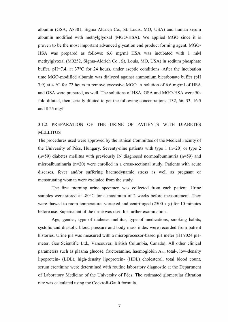
3.1.2. PREPARATION OF THE URINE OF PATIENTS WITH DIABETES
MELLITUS
The procedures used were approved by the Ethical Committee of the Medical Faculty of
the University of Pécs, Hungary. Seventy-nine patients with type 1 (n=20) or type 2
(n=59) diabetes mellitus with previously IN diagnosed normoalbuminuria (n=59) and
microalbuminuria (n=20) were enrolled in a cross-sectional study. Patients with acute
diseases, fever and/or suffering haemodynamic stress as well as pregnant or
menstruating woman were excluded from the study.
The first morning urine specimen was collected from each patient. Urine
samples were stored at -80°C for a maximum of 2 weeks before measurement. They
were thawed to room temperature, vortexed and centrifuged (2500 x g) for 10 minutes
before use. Supernatant of the urine was used for further examination.
Age, gender, type of diabetes mellitus, type of medications, smoking habits,
systolic and diastolic blood pressure and body mass index were recorded from patient
histories. Urine pH was measured with a microprocessor-based pH meter (HI 9024 pH-
meter, Geo Scientific Ltd., Vancouver, British Columbia, Canada). All other clinical
parameters such as plasma glucose, fructosamine, haemoglobin A1c, total-, low-density
lipoprotein- (LDL), high-density lipoprotein- (HDL) cholesterol, total blood count,
serum creatinine were determined with routine laboratory diagnostic at the Department
of Laboratory Medicine of the University of Pécs. The estimated glomerular filtration
rate was calculated using the Cockroft-Gault formula.

8
Because of the fact that first morning urine samples were used, urinary
creatinine levels were measured as well as part of routine laboratory work by buffered
kinetic Jaffé reaction without deproteinization. (Cobas Integra 400, Roche, Germany),
and albumin-creatinine ratios were calculated for both IN and HPLC-measured albumin
concentrations.
3.1.3. MEASUREMENT OF THE CONCENTRATION OF ALBUMIN
The in vitro prepared different forms of albumin as well as urinary albumin
concentrations were measured in duplicate by means of IN (IMMAGE
Immunochemistry Systems, Beckman Coulter Inc., Fullerton, CA, USA, sensitivity
(quantitation limit): 2 mg/l, linearity: 2-8640 mg/l, inter-assay and intra-assay precision
(percentage coefficient of variation) 8 % and 5 % respectively) in the routine laboratory
diagnostic, and by means of the size-exclusion HPLC method (Shimadzu SPD 10AVvp,
Shimadzu Corp., Japan) using a Food and Drug Administration (FDA) approved
AccuminTM kit (Accumin Diagnostics Inc., New York, NY, USA, sensitivity
(quantitation limit): 3 mg/l, linearity: 3-2000 mg/l, inter-assay and intra-assay precision
(percentage coefficient of variation) 5.8 % and 2.5 % respectively). The AccuminTM kit
contained a Zorbax Bio-Series GF 250 column and Zorbax Diol guard column (both
from Agilent Technologies Inc., Santa Clara, CA, USA). The mobile phase was
phosphate buffer saline (pH=6.93, provided with the kit). The HPLC system used for
the measurements was consisted of DGU-14A four-line vacuum membrane degasser, a
FCV-10ALvp solvent proportioning valve, a LC-10ADvp solvent delivery unit, a SIL-
10ADvp autosampler, a SPD-10AVvp UV-VIS detector and a SCL-10Avp system
controller (all parts purchased from Shimadzu Corp., Kyoto, Japan). During the HPLC
measurements 25 µl of the samples (in vitro prepared albumin or centrifuged urine)
were used. Absorbance was measured at 214 nm. The time program included 6 min at
flow rate of 0.5 ml/min, then a ramp up to 2 ml/min and washing time of 6.5-11.5 min.
Then ramping down to 0.5 ml/min in 0.5 min and washing were employed until a steady
baseline was observed (usually until 22 min). The peak retention time of albumin was
within ± 2 % of the elution time of the monomer albumin under the circumstances
recommended by the manufacturer. Data acquisition was carried out with LCSolution
software (Ver.: 1.11 SP1, Shimadzu, Japan).
9
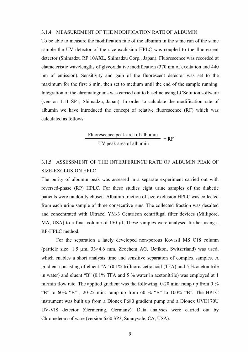
3.1.4. MEASUREMENT OF THE MODIFICATION RATE OF ALBUMIN
To be able to measure the modification rate of the albumin in the same run of the same
sample the UV detector of the size-exclusion HPLC was coupled to the fluorescent
detector (Shimadzu RF 10AXL, Shimadzu Corp., Japan). Fluorescence was recorded at
characteristic wavelengths of glycoxidative modification (370 nm of excitation and 440
nm of emission). Sensitivity and gain of the fluorescent detector was set to the
maximum for the first 6 min, then set to medium until the end of the sample running.
Integration of the chromatograms was carried out to baseline using LCSolution software
(version 1.11 SP1, Shimadzu, Japan). In order to calculate the modification rate of
albumin we have introduced the concept of relative fluorescence (RF) which was
calculated as follows:
3.1.5. ASSESSMENT OF THE INTERFERENCE RATE OF ALBUMIN PEAK OF
SIZE-EXCLUSION HPLC
The purity of albumin peak was assessed in a separate experiment carried out with
reversed-phase (RP) HPLC. For these studies eight urine samples of the diabetic
patients were randomly chosen. Albumin fraction of size-exclusion HPLC was collected
from each urine sample of three consecutive runs. The collected fraction was desalted
and concentrated with Ultracel YM-3 Centricon centrifugal filter devices (Millipore,
MA, USA) to a final volume of 150 µl. These samples were analysed further using a
RP-HPLC method.
For the separation a lately developed non-porous Kovasil MS C18 column
(particle size: 1.5 µm, 33×4.6 mm, Zeochem AG, Uetikon, Switzerland) was used,
which enables a short analysis time and sensitive separation of complex samples. A
gradient consisting of eluent “A” (0.1% trifluoroacetic acid (TFA) and 5 % acetonitrile
in water) and eluent “B” (0.1% TFA and 5 % water in acetonitrile) was employed at 1
ml/min flow rate. The applied gradient was the following: 0-20 min: ramp up from 0 %
“B” to 60% “B” , 20-25 min: ramp up from 60 % “B” to 100% “B”. The HPLC
instrument was built up from a Dionex P680 gradient pump and a Dionex UVD170U
UV-VIS detector (Germering, Germany). Data analyses were carried out by
Chromeleon software (version 6.60 SP3, Sunnyvale, CA, USA).
Fluorescence peak area of albumin
UV peak area of albumin = RF= RF

10
Chromatograms obtained during RP-HPLC presented two to three peaks with a
very small elution time difference. The albumin peak was identified in each case with
external albumin standard. Due to the small elution time difference of the peaks
interference could be assessed by calculating the ratio of non-albumin peak area to the
total peak area.
3.1.6. STATISTICAL ANALYSIS
Statistical analysis was performed using SPSS 13.0 (SPSS Inc., Chicago, IL, USA) and
MedCalc (MedCalc Software, Mariakerke, Belgium) programs. The Bland-Altman bias
plot was used to compare the IN and HPLC methods. Data of normal distribution were
analyzed by one-way ANOVA and Pearson’s correlation. Data of non-normal
distribution were analyzed with the Kruskall-Wallis test, the Mann-Whitney U test and
Spearman’s rho correlation. Chi-square tests were used to compare categorical data.
Data with normal distribution are presented as mean±SEM., while data with non-normal
distribution are presented as median and interquartile ranges. P values <0.05 were
considered to be statistically significant. Forward multivariate stepwise linear regression
analyses were performed to determine the independent predictors of the RF of urinary
albumin.
3.2. HPLC-MEASURED ALBUMINURIA AND STORAGE OF SPECIMENS
(PART II OF THIS THESIS)
3.2.1. STUDY POPULATION
In 2005 patients with type 2 diabetes mellitus (n=30), attending the 2nd Department of
Medicine and Nephrological Center, Pécs, Hungary with previously IN diagnosed
normo- and microalbuminuria, were enrolled in a cross-sectional study. Patients with
acute diseases, a fever and/or suffering haemodynamic stress as well as pregnant or
menstruating woman were excluded from the study. To assess total free sulfhydryl
groups (TFSG) of fresh urine samples, another 30 IN diagnosed normo- and
microalbuminuric type 2 diabetic patients, attending the Department, were included in
the study in 2008. The clinical characteristics of these patients did not differ from those
patients with stored urine. Both studies were approved by the Ethical Committee of the
Medical Faculty of the University of Pécs, Hungary.

11
3.2.2. LABORATORY METHODS
Urinary albumin concentration (UAC) of fresh urine (first morning urine, centrifuged at
2500xg for 10 min, separated in three polypropylene aliquots and kept at -80°C for a
maximum of 2 weeks before use) was assessed by the previously in detail described
(3.1.3), FDA-approved HPLC AccuminTM kit at the time of the original collection
(2005) and in 2008. Routine laboratory parameters of patients were measured as well as
urinary pH by a microprocessor-based pH meter (HI 9024 pH-meter, Geo Scientific
Ltd., Vancouver, British Columbia, Canada) and both dimeric and monomeric forms of
urinary albumin (assessed with AccuminTM kit according to the guidelines of the
manufacturer as the peak immediately preceding the albumin peak is that of albumin
dimer) and dimeric to monomeric ratio of urinary albumin (DMR) was calculated.
Presence and accuracy of elution time of dimeric form were verified using the spike
recovery method by adding external human albumin standard (containing both forms of
albumin) to the samples.
After 2.5 years of -80°C storage one of the two never used aliquots of the
patients’ urine was thawed and UAC was measured by the same HPLC method. We
have measured both dimeric and monomeric form of urinary albumin and DMR was
calculated again.
3.2.3. MEASUREMENT OF THE CONCENTRATION OF THE TOTAL FREE
SULFHYDRYL GROUPS
TFSG of the stored and of newly collected fresh urine samples were also measured.
Urine preparation was the same as for the UAC measurements. Briefly, in excess (final
concentration of 100 µM) 10 µl of colorimetric Ellman’s reagent, 5, 5'-dithio-bis (2-
nitrobenzoic acid) (DTNB) (Sigma-Aldrich, Schnelldorf, Germany) was added to 0.98
ml of urine in a 3 ml quartz cuvette. Maximum absorbance was measured against urine
not containing DTNB at 412 nm with Hitachi U-2001 double-beam Spectrophotometer,
Tokyo, Japan during a 3600 sec time scan. As baseline was reached (reaction
completed) 10 µl (final concentration of 10 µM) of freshly prepared reduced glutathione
(GSH) (Sigma-Aldrich, Schnelldorf, Germany) was added to the samples and
absorbance elevation was measured again. From these data TFSG of urine (in GSH
equivalent unit) could be calculated as follows: maximum absorbance with GSH minus
maximum absorbance with DTNB (delta), then the maximum absorbance with DTNB
divided by the delta and multiplied by 10 to get µM equivalent. Both stored and freshly

12
collected urine samples were measured at room temperature. Measurement of TFSG in
the fresh urine samples was performed in 1 hour.
3.2.4. STATISTICAL ANALYSES
Statistical analysis was performed using the SPSS 13.0 (SPSS Inc., IE, USA) software.
Wilcoxon tests were used to test changes in stored urine and paired-samples t-test to test
changes in DMR. Independent samples t-tests were used to test differences between
fresh and stored urine and to compare the clinical characteristics of the two study
populations. Correlation analyses were carried out using Pearson’s correlation. Chi-
square tests were used to compare qualitative data. Data are presented as mean±SD. P
values <0.05 were considered as statistically significant.
3.3. NEW POTENTIAL BIOMARKERS DISCOVERED BY MEASURING
ALBUMINURIA WITH HPLC IN A CROHN’S DISEASE PATIENT (PART III OF
THIS THESIS)
3.3.1. STUDY PATIENT
A 23-year-old non-smoker Hungarian male patient suffering frequent exacerbations
from CD was involved in a pilot study. CD was previously (2006) diagnosed on the
basis of endoscopy (Montreal classification A2, L1, B1) and histology. The patient
attended the 2nd Department of Medicine and Nephrological Center, Pécs, Hungary and
suffered from no other disease than CD. His regular medication included oral
mesalamine (3x1000 mg/day) and azathioprine (2.5 mg/kg/day). During acute phase
regular medication was supplemented with parenteral steroid (methylprednisolon 1
mg/kg/day). To assess disease activity, the Crohn's Disease Activity Index was used.
Scores ≥150 are defined as active.
First morning urine samples were obtained from the patient at the time of
clinical visits. Urine samples were vortexed and centrifuged (2500xg for 10 min) and
were used for analysis immediately. At the time of his clinical visits samples were taken
for routine biochemistry. All routine laboratory measurements were carried out at the
Institute of Laboratory Medicine of the University of Pécs. Aliquots of urine and serum
samples were reserved at -80°C for later examinations, as well. The study was
performed in accordance with the ethical standards as formulated in the Helsinki
13
Declaration and was approved by the Ethical Committee of the Medical Faculty of the
University of Pécs, Hungary.
3.3.2. URINARY ALBUMIN ASSAYS
Concentrations of immunoreactive urinary albumin (ir-uAlb) were measured in
duplicates by means of IT (Roche Diagnostics GmbH, Mannheim, Germany) using
Roche/Hitachi 812 Modular P analyzer (sensitivity: 3 mg/l, linearity: 3-3000 mg/l, inter-
assay and intra-assay precision 4.3% and 2.6% respectively). Concentrations of total
urinary albumin (t-uAlb) were measured in triplicates by the previously described
(3.1.3) SE-HPLC protocol.
3.3.3. REVERSED-PHASE HPLC ANALYSIS OF THE ALBUMIN PEAK OF SIZE-
EXCLUSION HPLC
Central fractions of albumin peaks of SE-HPLC were collected and prepared as
previously described (3.1.5). Eluted peaks were collected, evaporated to dryness and
were analyzed with MALDI-TOF/MS directly after taken up in 5 µl bidistilled water or
after in solution digestion according to Shevchenko.
3.3.4. GEL-ELECTROPHORETIC STUDIES
Central fractions of albumin peaks from SE-HPLC were collected and prepared as
described earlier. Due to the high concentration of salt of the size-exclusion fraction,
additional desalting prior to sodiumdodecylsulphate polyacrylamide gel-electrophoresis
(SDS-PAGE) was performed. The salt-free sample was evaporated to dryness and the
proteins were taken up in 5 µl bidistilled water.
Thus prepared samples were separated by SDS-PAGE according to Laemmli.
Two µg protein per lane was analyzed in a 12.5 % gel. Detection of protein fractions
was performed by silver post-intensification according to Willoughby following the
traditional Coomassie brilliant blue R-250 staining. Proteins identified were excised
from gel and after in-gel digestion according to Shevchenko were analyzed by MALDI-
TOF/MS.
3.3.5. MALDI-TOF/MS MEASUREMENTS
An Autoflex II MALDI instrument (Bruker Daltonics, Bremen, Germany) was
employed for the mass spectrometric measurements. For the measurement of the
14
digested proteins 8 mg of α-cyano-4-hydroxycinnamic acid was dissolved in 1 ml of 50
% ACN and 0.1 % TFA in water. For the measurement of intact proteins a saturated
sinapinic acid matrix was prepared in 50 % v/v ACN and 0.1 % TFA in water. In each
case 1 µl of the matrix was deposited on a stainless steel target together with 1 µl of the
sample. All mass spectra were monitored in positive mode with pulsed ionization (λ =
337 nm; nitrogen laser, maximum pulse rate: 50 Hz; maximal intensity 20-30 % of the
laser for peptides). Peptides of the digests were measured in reflectron mode using a
delayed extraction of 120 nsec and proteins were measured in linear mode at a delayed
extraction of 550 nsec. The accelerating voltage was set to +19 kV, the reflectron
voltage was set to + 20 kV. Spectra of peptides and proteins were the sum of 1000
shots, external calibration has been implemented. Data processing was executed with
Flex Analysis software packages (version: 2.4.). For the analysis of in solution digestion
Sequence Editor software (Bruker Daltonics, Bremen, Germany) was used with the
following criteria: 1. All cysteines were supposed to be treated with iodoacetamide 2.
Monoisotopic masses were allowed 3. The maximum number of missed cleavage sites
was two.
4. RESULTS
4.1. MEASUREMENT OF MODIFICATION AND INTERFERENCE RATE OF
URINARY ALBUMIN DETECTED BY SIZE-EXCLUSION HPLC (PART I OF THIS
THESIS)
4.1.1. CHARACTERIZATION OF THE UV-FLUORESCENT HPLC SYSTEM
To calculate between-day imprecision of the measurements with UV and fluorescent
detectors five samples (concentrations: 8.25, 16.5, 33, 66 and 132 mg/l) of each kind of
albumin form (HSA, GSA and MGO-HSA) were tested 5 times in one week. The
between-day imprecision (expressed as the percent coefficient of variation (%CV)) of
the lowest concentration (8.25 mg/l) were as follows: 3.5% and 11.8% for HSA, 3.7%
and 11.6% for GSA and 5.9% and 5.6% for MGO-HSA respectively for the UV and
fluorescent measurements. The %CVs of the highest concentration (132 mg/l) were as
follows: 1.1% and 5.1% for HSA, 1.5% and 3.0% for GSA and 1.8% and 2.0% for
MGO-HSA respectively for the UV and fluorescent measurements. To investigate
reproducibility of the measurements over time of the UV and fluorescent detections, the
same samples after 12 months of freezing at -80°C were thawed and were measured the

15
same way as for the between-day imprecision using a new kit. The total imprecision of
the two between-day imprecision measurements of the lowest concentration (8.25 mg/l)
were as follows: 10.7% and 13.9% for HSA, 12.5% and 10.9% for GSA and 11.7% and
11.5% for MGO-HSA respectively for the UV and fluorescent peak areas; and of the
highest concentration (132 mg/l) were as follows: 2.6% and 8.9% for HSA, 7.5% and
9.1% for GSA and 3.4% and 7.8% for MGO-HSA respectively for the of the UV and
fluorescent peak areas.
Between-day imprecision was calculated for the urine samples as well. To make
the calculations, 10 samples were randomly chosen and measurements were repeated
one week after the first measurement. The between-day imprecision expressed as the
percent CV of UV and fluorescent peak areas of the urine samples of patients with
diabetes mellitus were 6.1% and 8.8% respectively. To investigate reproducibility of the
measurements over time the urine samples were also re-analyzed after 12 months.
Interestingly, we have found a significant decrease in the UV signal of the albumin (-
25±9%, p<0.05) and a non-significant increase in the fluorescent signal (11±20%,
mean±SD, p=0.093).
4.1.2. COMPARISON OF THE CONCENTRATION OF THE DIFFERENT FORMS
OF IN VITRO PREPARED ALBUMIN BY IN AND BY HPLC
The different forms of albumin (HSA, GSA and MGO-HSA) prepared in the
concentrations of 8.25, 16.5, 33, 66 and 132 mg/l were measured by HPLC and IN in
triplicate. Then the albumin concentrations measured by HPLC were divided by the
concentrations measured by IN. These quotients of HSA, GSA and MGO-HSA were
compared by one-way ANOVA. The test failed to find a significant difference
(p=0.210, HSA: 132±10%, GSA: 120±8% and MGO-HSA: 142±8%).
4.1.3. RELATIVE FLUORESCENCE OF THE DIFFERENT FORMS OF IN VITRO
ALBUMIN
To avoid any possible confounding effect of fluorescent measurement, such as non-
linear changes in the peak area of fluorescence with concentration, correlation analysis
of UV and fluorescence signal of the different albumin forms were tested in the
examined concentration range and were as follow: HSA, r=0.9998, GSA=0.9999,
MGO-HSA, r=0.9997.

16
Relative fluorescence (RF) of the in vitro prepared albumin forms was
determined. The average RF of HSA was considered to be 100 %. RF of GSA and of
MGO-HSA was higher (p<0.001 for both) compared to HSA and RF of MGO-HSA was
also higher (p<0.01) compared to RF of GSA which indicates extensive changes in the
albumin structure of both GSA and MGO-HSA.
4.1.4. CHARACTERISTICS OF THE PATIENTS WITH DIABETES MELLITUS
Using the conventionally accepted cut-offs for albumin-creatinine ratio (ACR) for
microalbuminuria (male: 2.5-25 mg/mmol, female: 3.5-35 mg/mmol) the diabetic
patients were grouped as follow: normoalbuminuric using both IN and HPLC (NN,
n=47), normoalbuminuric by IN but microalbuminuric by HPLC (NM, n=12), and
microalbuminuric by both methods (MM, n=20). Classical ACR cut-off values were
used for HPLC measured urinary albumin concentrations as well, since there are no
accepted ACR cut-off values for HPLC yet. Of the clinical characteristics of the groups
of patients only serum creatinine was higher (and consequently eGFR lower) in NM and
MM groups compared to NN; however there was no difference between the NM and
MM groups. More patients took angiotensin converting enzyme inhibitors in the MM
group than in the NN group. There was no further difference between the groups.
Bland-Altman bias plot for both assays showed that in the majority of cases
HPLC measured a higher concentration of urinary albumin than IN and also that the
amount of bias increases as urinary albumin decreases.
4.1.5. RELATIVE FLUORESCENCE OF URINARY ALBUMIN IN DIABETIC
PATIENTS
We found a higher RF of albumin in the urine of the MM group compared to the NN
and NM groups (p<0.001 and p=0.007, respectively) but there was no difference
between the NN and NM groups (p=0.201). RF of urinary albumin showed significant
positive correlation with the serum creatinine levels (r=0.295; p=0.009) and significant
negative correlation with the estimated glomerular filtration rate eGFR levels (r=-0.255;
p=0.026), but not with glycaemic parameters (concentration of plasma glucose,
p=0.766; concentration of fructosamine, p=0.979; levels of hemoglobin A1c, p=0.442).
By forward stepwise multivariate linear regression analyses, both serum creatinine and
eGFR levels proved to be independent predictors of urinary albumin RF (β=0.397;
p=0.014 and β=-0.337; p=0.039, respectively). The first model included age, plasma

17
glucose, fructosamine, hemoglobin A1c, systolic and diastolic blood pressure,
triglycerides, LDL- and HDL-cholesterol, haemoglobin and serum creatinine; the
second model included the same parameters with the exception of ln eGFR in place of
serum creatinine.
4.1.6. INTERFERENCE RATE OF ALBUMIN PEAK OF SIZE-EXCLUSION HPLC
Carrying out our albumin peak purity test of size-exclusion HPLC using RP-HPLC it
was found that non-albumin material (calculated as non-albumin peak area to total peak
area) was present in 12.7±1.9% in the albumin peak of size-exclusion HPLC.
4.2. HPLC-MEASURED ALBUMINURIA AND STORAGE OF SPECIMENS
(PART II OF THIS THESIS)
4.2.1. EFFECT OF STORAGE ON THE CONCENTRATION OF URINARY
ALBUMIN
Mean decrease±SD in HPLC-detected albuminuria after 2.5 years at -80°C storage was
24±9% (UAC: 88±259 vs. 55±187 mg/l, p=0.002). When patients were categorized
according to their decrease of UAC to higher and lower than interassay imprecision and
their urinary pH (above and under mean pH), we found a significant relationship
between under mean urinary pH and higher UAC-decrease (p=0.030).
On the other hand, a significant increase could be observed in the DMR
(p<0.001). However, only peak areas of the monomeric form of albumin changed
significantly (p<0.001), while peak areas of the dimeric form of albumin did not
(p=0.275).
4.2.2. REDUCING CAPACITY OF URINE
We found an exponential correlation between urinary pH and the TFSG of fresh urine
samples (r=-0.795; p<0.001 for linear correlation), but not in 2.5 year stored urine
samples (r=-0.216; p=0.261 for linear correlation). Average TFSG was significantly
lower in stored urine compared to the fresh urine (6.6±7.7 vs. 22.7±14.3 in µM GSH
equivalent, p<0.001). Moreover, we found a significant correlation between increase of
DMR and pH (r=-0.382, p=0.041).

18
4.3. NEW POTENTIAL BIOMARKERS DISCOVERED BY MEASURING
ALBUMINURIA WITH HPLC IN A CROHN’S DISEASE PATIENT (PART III OF
THIS THESIS)
4.3.1. ALBUMIN ASSAYS
Total uAlb measured by SE-HPLC showed a marked increase during active phase
comparing with the measured value of IT. The difference between the uAlb
concentrations measured by the two methods during active phase was almost 15-fold
which difference decreased to 6-10-fold during inactive phase. This unexpectedly high
difference between the t-uAlb and ir-uAlb led us to analyze further our results.
4.3.2. REVERSED-PHASE HPLC AND SDS-PAGE ANALYSIS OF THE
ALBUMIN PEAK BY SIZE-EXCLUSION HPLC
Chromatogram of RP-HPLC of albumin fraction of SE-HPLC obtained during acute
phase clearly showed the presence of co-eluted proteins. Two fractions were collected
from the RP-separation. First fraction included those proteins eluted at 12.40 min and
12.69 min, being recognized as two partially resolved constituents, while the second
fraction contained actually uAlb that was verified by spike recovery studies and later by
MALDI-TOF/MS. Considerable decrease of first-fraction-proteins but not albumin
could be observed in the urine obtained in remission. Presence of two co-eluting
proteins was proven by SDS-PAGE, as well.
4.3.3. MALDI-TOF/MS MEASUREMENTS
Mass spectrum measured from the first fraction of RP-HPLC showed peaks appearing
at 23.5 kDa, 34.7 kDa and at 70.3 kDa (which can be considered to be the dimer of the
protein with a mass of 34.7 kDa). The resulted peptide mass fingerprinting (PMF) and
all the peptides of the PMF recognized by Mascot data base search engine were
analyzed. Three proteins, α1-acid-glycoprotein-1, α1-acid-glycoprotein-2 and Zn-α2-
glycoprotein have been identified with high scores and sequence coverage values of
39.3%, 56.2% and 48.1%, respectively. Identification of these proteins was also
corroborated by post-source decay spectra of the corresponding tryptic peptides.
Proteins identified from the excised gel slabs also confirmed these results.
Investigating control urine from healthy individual allowed only the identification of
albumin.

19
5. DISCUSSION AND CONCLUSIONS
Conventional urinary albumin assays, used in every-day laboratory medicine, are based
on immunochemical methods using antibodies raised against serum albumin rather than
urinary albumin. These assays detect immunoreactive albumin and other albumin
compounds such as albumin aggregates and albumin fragments with a molecular weight
of >12kDa. In 2003 a new method has been introduced for the measurement of albumin
in the urine, using size-exclusion high performance liquid chromatography. Early
studies using this method have shown that concentration of albumin is higher as
measured by conventional, immuno-based assays; with other words there is a portion of
albumin which is not immunoreactive. As an expected consequence, the nature of
albumin measured by high performance liquid chromatography has been addressed.
Moreover, some authors proposed that the method simply does not have sufficient
resolution.
As a first part of this thesis we wanted to address these questions. Firstly, we
have established a high performance liquid chromatography method equipped with
tandem UV and fluorescent detection to assess the changes of detectability of albumin
with the rate of modification. For this measurement in-vitro differently modified forms
of albumin were used. As a part of these measurements we have also aimed to measure
the modification rate of the total urinary albumin of diabetic patients to find a potential
connection between the modification rate and clinical parameters. We concluded that
albumin modification does not affect immunoreactivity. Interestingly, we found that the
modification rate of total urinary albumin in diabetic patients correlates with the renal
function and not with the parameters of glycaemia. Secondly, we have established a
reversed-phase high performance liquid chromatography method to assess the
interference rate of the albumin peak of size-exclusion high performance liquid
chromatography. With the help of this method the interference rate of the albumin peak
was found to be 12.7% on average, which does not explain the measured concentration
difference between the immuno-based and high performance liquid chromatography
methods.
In only 4 years after the publication of this new method for the measurement of
albuminuria, reevaluation of big studies such as the Australian Diabetes, Obesity and
Lifestyle study has been published to address the question if there is any clinical
significance of high performance liquid chromatography-measured albuminuria. They

20
found that both traditional immunonephelometry and the new high performance liquid
chromatography method have the same power for predicting mortality. However, for the
HPLC measurements stored urine was used.
Based on some publications which showed that storage could strongly decrease
the concentration of immunoreactive urinary albumin as a second part of this thesis we
wanted to investigate the effect of storage on the concentration of high performance
liquid chromatography-detected urinary albumin and we aimed to find possible
mechanisms for the results we have found. We found that measurement of the
concentration of albumin by high performance liquid chromatography in urine, stored
for long periods at -80°C gives unreliable results, as we have found a significant 24%
decrease in urinary albumin concentration after 2.5 years of storage. We found this
decrease pH-dependent. As it was suggested by one study, the nonimmunoreactive form
of urinary albumin is a partially cleaved form of albumin which is maintained with an
intact relative molecular mass by the help of the disulfide bonds and which form
fragments into smaller parts to reducing agents. That is why we have measured total
sulfhydryl groups of our urine samples, in an attempt to assess whether this free
sulfhydryl group capacity could play a role in the decrease of high performance liquid
chromatography-detected albuminuria, by reducing disulfide bonds of albumin. We
found a strong correlation between free sulfhydryl groups and urinary pH in fresh urine
samples, which could not be observed, in stored urine and concentration of free
sulfhydryl groups significantly decreased during the storage. We interpreted these
results as urine has a potentially high level of reducing activity which is pH-dependent,
and so it may play a role in the decrease of high performance liquid chromatography-
detected albuminuria by breaking up the cleaved nonimmunoreactive form of urinary
albumin.
Although clinical application of albuminuria is still largely limited to the area of
diabetes it has been shown in several other clinical disorders that measurement of
albuminuria can be a valuable marker. For instance, measurement of albuminuria has
been shown to have the potential to be an objective marker in the monitoring of disease
activity and response to treatment in inflammatory bowel diseases. As a third part of
this thesis we followed up a young Crohn’s disease patient with frequent exacerbation
phases to measure the changes of the concentration of total albumin in the course of his
disease compared to the measured concentration by immuno-based methods. The
21
surprisingly high difference between the two methods led us to further analyze the
albumin peak of the size-exclusion chromatography of the Crohn’s disease patient using
techniques that allowed us the identification of possible biomarkers. We concluded
from this study that urinary albumin measured by size-exclusion chromatography
method in acute phase of Crohn’s disease is not reliable since it measures a high amount
of other proteins. On the other side, the identified coeluting urinary proteins, the α-1
acid glycoprotein and the Zn-α-2 glycoprotein, showed a perfect association with the
clinical status, which let them candidature as a novel, non-invasive, easy-to-access
activity biomarkers in Crohn’s disease.
6. LIST OF PHD THESES
1) Glycoxidative modification of the albumin does not affect immunoreactivity.
2) Glycoxidative modification rate of total urinary albumin in patients with
diabetes mellitus reflects renal pathophysiology.
3) Coeluating proteins in the peak of albumin by size-exclusion chromatography
are present less than 20% on average in the urine of diabetic patients. This
interference rate does not explain the difference between the concentration of
albumin measured by immuno-based and size-exclusion chromatography
methods.
4) Concentration of albumin by high performance liquid chromatography in stored
urine decreases despite storage at -80°C which decrease is pH dependent.
5) Fresh urine has a potentially high level of reducing activity. This reducing
capacity is pH dependent and disappears with storage.
6) Urinary albumin measured by size-exclusion chromatography method in acute
phase of Crohn’s disease is not reliable.
7) The urinary α-1 acid glycoprotein and the urinary Zn-α-2 glycoprotein are
possible new biomarkers of disease activity in Crohn’s disease.
22
7. LIST OF PUBLICATIONS
Cumlative impact factor: full papers 32.159, abstracts: 43.311 Cumulative impact factor of publications used in this thesis: full papers: 4.766 abstracts: 3.154 This thesis is based on the following publications:
1. Markó L, Cseh J, Kőszegi T, Szabó Z, Molnár GA, Mohás M, Szigeti N,
Wittmann I. Storage at -80 degrees C decreases the concentration of HPLC-detected urinary albumin: possible mechanisms and implications. J Nephrol 2009:22(3):397-402. IF: 1.252
2. Markó L, Molnár GA, Wagner Z, Böddi K, Koszegi T, Szabó Z, Matus Z,
Szijártó I, Mérei A, Nagy G, Wittmann I. Measurement of the modification and interference rate of urinary albumin detected by size-exclusion HPLC. Physiol Meas 2009:30(10):1137-1150. IF: 1.430
3. Markó L, Szigeti N, Szabó Z, Böddi K, Takátsy A, Ludány A, Kőszegi T,
Molnár GA, WittmannI. Potential urinary biomarkers of disease activity in Crohn’s disease. Scand J Gastroenterol 2010 Jul 26. [Epub ahead of print] IF: 2.084
4. Markó L., Mikolás E., Molnár G. A., Wagner Z., Kőszegi T., Szijártó I. A.,
Mohás M., Matus Z., Szabó Z., Böddi K., Mérei Á., Wittmann I. Normo- és microalbuminuriás cukorbetegekben a HLCP-vel mért vizeletalbumin-fluoreszcencia a vesefunkciós paraméterekkel függ össze, nem a glikémiás értékkel. Diab Hung 2009:17(3):229-238.
5. Markó L., Szijártó I. A., Cseh J., Kőszegi T., Szabó Z., Molnár G. A., Matus Z.,
Mérei Á., Wittmann I. A HPLC-vel mérhető vizeletalbumin koncentrációja -80 °C-os tárolás során jelentősen csökken: lehetséges mechanizmusok és következmények. Hypertonia és Nephrologia 2009:13(2):88-93.
This thesis is based on the following congress presentations and abstracts:
1. Markó L., Molnár G. A., Wagner Z., Wagner L., Kőszegi T., Nagy J., Wittmann I.: Determination of protein glycoxidation-products in the urine of diabetic patients. Nephrol Dial Transplant 2006:21(S5):v84-85. IF: 3.154 Place of presentation: XLIII ERA-EDTA (Eurpoean Renal Association-European Dialysis and Transplant Association) Congress, July 15-18, 2006, Glasgow, United Kingdom
2. Markó L., Molnár G.A., Wagner Z., Wagner L., Matus Z., Kőszegi T., Laczy
B., Tamaskó M., Mohás M., Cseh J., Nagy J., Wittmann I.: Vizelet fehérje glikoxidációs termékek meghatározása diabeteses betegekben. Magyar
Belorvosi Archivum 2006:59(S2):111-112. Place of presentation: Magyar Belgyógyász Társaság Dunántúli Szekciójának LIII. Vándorgyűlése, Sopron, 2006. június: Legjobb fiatal előadók díja: 3. helyezés

23
3. Markó L., Molnár G. A., Wagner Z., Wagner L., Matus Z., Kőszegi T., Laczy
B., Tamaskó M., Mohás M., Cseh J., Nagy J., Wittmann I.: Determination of protein glycoxidation-products in the urine of diabetic patients. Acta Physiologia Hungarica 2006:93(2-3):210. Place of presentation: Magyar Élettani Társaság LXX. Vándorgyűlése, Szeged, 2006. június
4. Markó L., Wagner Z., Cseh J., Kőszegi T., Matus Z., Wittmann I.: HPLC és nephelometria (NM) összehasonlítása a mikroalbuminuria diagnózisában. Hypertonia és Nephrologia 2006:10(S5):107. Place of posterpresentation: Magyar Nephrológiai Társaság XXIII. Nagygyűlése, Eger, 2006. október
5. Markó L., Molnár G. A., Kőszegi T., Cseh J., Mohás M., Matus Z., Wittmann I. Analysis of albuminuria with high performance liquid chromatography (HPLC) and immunonephelometry (IN) in diabetic and/or hypertonic patients. Acta Physiologica Hungarica 2009:96(1):101. Place of posterpresentation: A Magyar Élettani Társaság LXXII. Vándorgyűlése és a Magyar Kísérletes és Klinikai Farmakológiai Társaság közös konferenciája, Debrecen, 2008. június
6. Markó L., Cseh J., Kőszegi T., Szabó Z., Molnár G. A., Mohás M., Szigeti N., Szijártó I., Wittmann I. A HPLC-vel mérhető vizelet albumin mennyisége a -80°C-os tárolás során jelentősen csökken. Lehetséges mechanizmusok és következmények. Hypertonia és Nephrologia 2008:12(S5):222. Place of posterpresentation: Magyar Nephrológiai Társaság XXIII. Nagygyűlése, Szeged, 2008. szeptember
24
ACKNOWLEDGEMENT
I would like to dedicate this work to my Grandparents most of them who passed away. The work
involved in this thesis could not have been carried out without help from a number of persons, to whom I
owe a great debt of gratitude and whom I would like to thank for their valuable contribution.
I am first of all thankful to my Parents, Grandparents and my Sister, who have supported me
throughout my life and made it possible to reach my goals.
I am thankful to my supervisor Prof. István Wittmann for inviting me to his excellent research group
as a medical student, for teaching the way of thinking in the research and in the clinic and for all his
support, time and energy he invested in me. I would also like emphasize my deep gratitude towards Prof.
Judit Nagy who helped me to carry out the work for my Ph.D. thesis at the 2nd Department of Internal
Medicine and Nephrology, University of Pécs.
I am thankful to all of the former and current Ph.D. students in the lab who helped me in my
research: from all of these fellows I am extremely thankful for Gergő A. Molnár who helped me a lot as a
young medical student researcher and a beginner Ph.D. student. I am grateful for Ilona Sámikné, who
helped my work with more than just her excellent technical assistance and for Enikő Bodor for her
administrative help.
I am thankful for the help of the colleagues of the Department of Biochemistry and Medical
Chemistry, University of Pécs: especially I owe my gratitude to Zoltán Szabó, Katalin Böddi, and Anikó
Takátsy. Without them critical parts of my thesis would be unanswered. I am thankful to my former
medical chemistry teacher, Zoltán Matus for his chemical knowledge and help in HPLC-issues.
I am thankful for the help of the colleagues of the Institute of Laboratory Medicine, University of
Pécs: especially for Tamás Kőszegi and Andrea Ludány for analyzing hundreds of urine samples and for
the expertise in the gel-electrophoretic studies.
I am thankful to all of the colleagues and patients of the 2nd Department of Internal Medicine and
Nephrology, University of Pécs for making possible to answer the scientific questions raised not only in
this thesis, and for their help in the duties and clinical work. I am especially grateful to Nóra Szigeti for
her excellent idea to measure albuminuria in Crohn’s disease. I am also especially grateful to the nurses
of the 2nd Department of Internal Medicine and Nephrology, University of Pécs who were always to my
help.
I also owe my gratitude to Prof. Friedrich C. Luft and Dominik Müller, who made it possible to work
further as a Ph.D. student in Berlin, Germany at the Experimental and Clinical Research Center.
I am thankful to everyone who is not listed above but contributed to my research or my life.
And last but not least I am thankful to my Love, Anett Melis who supported me with all of her love.
The research described in this thesis was supported by the following Hungarian national grants: T043788
(István Wittmann), PD 76395 (Zoltán Szabó) and PD 78599 (Anikó Takátsy) and by Sanofi-Aventis.
Related Documents