MSCT AN INTRODUCTION TO A NOVEL TECHNIQUE Dr. Hazem Abu Zeid Yousef Lecturer of Radiodiagnosis Faculty of Medicine Assiut University

MDCT AN INTRODUCTION TO CLINICAL APPLICATIONS
Jul 30, 2015
Welcome message from author
This document is posted to help you gain knowledge. Please leave a comment to let me know what you think about it! Share it to your friends and learn new things together.
Transcript

MSCT AN INTRODUCTION TO A NOVEL TECHNIQUE
Dr. Hazem Abu Zeid YousefLecturer of Radiodiagnosis
Faculty of MedicineAssiut University

INTRODUCTION
The introduction of spiral CT in the early 1990s resulted in fundamental and far-reaching improvement of CT imaging.
For the first time volume data could be acquired without mis-registration of anatomical details, which initiated the development of 3D image processing techniques such as multi-planar reformations (MPRs), maximum intensity projections (MIPs), surface-shaded displays (SSDs) or volume renderings.
As an important application CT angiography (CTA) has been established in clinical practice. As a consequence of increasing clinical demands, single-slice spiral CT with 1-s gantry rotation time soon encountered its limitations.
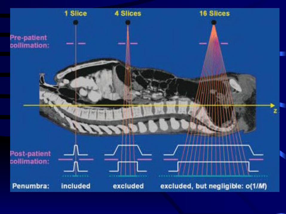
Larger volume coverage and improved transverse resolution can be achieved by simultaneous acquisition of more than one slice and by a shorter gantry rotation time. The first step towards multi-slice acquisition was a two-slice CT scanner introduced
in 1993 (Elscint Twin). In 1998 all major CT manufacturers introduced multi-slice CT systems which brought about considerable improvements of scan speed, transverse resolution and utilization of the
tube output.

SOME CLINICAL APPLICATIONS OF MDCT

MDCT ANGIOGRAPHY

CEREBRAL ANGIOGRAPHY


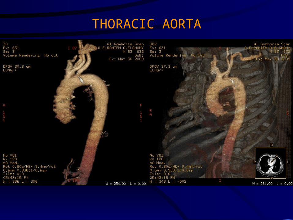
THORACIC AORTA

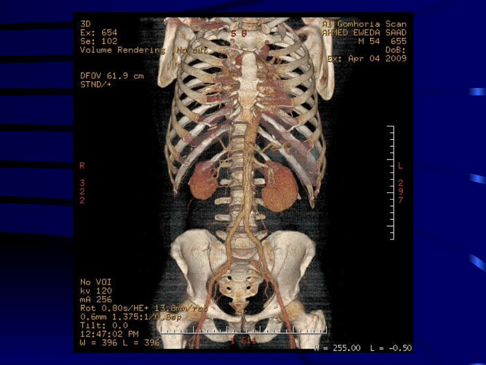
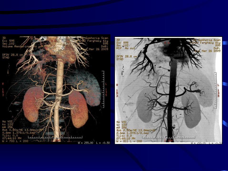
ABDOMINAL AORTA

LOWER LIMBARTERIOGRAPHY

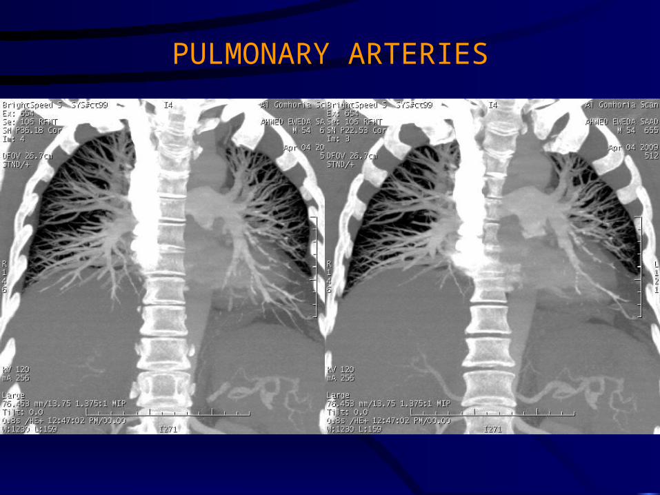
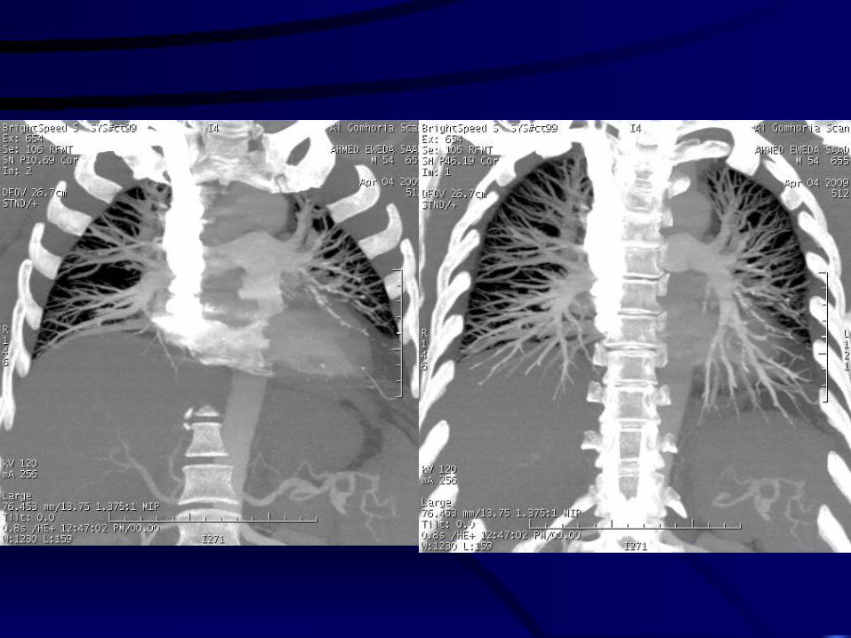
PULMONARY ARTERIES
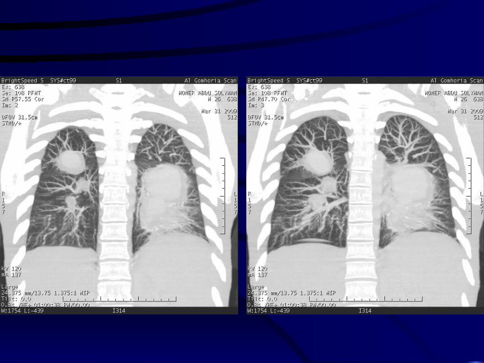
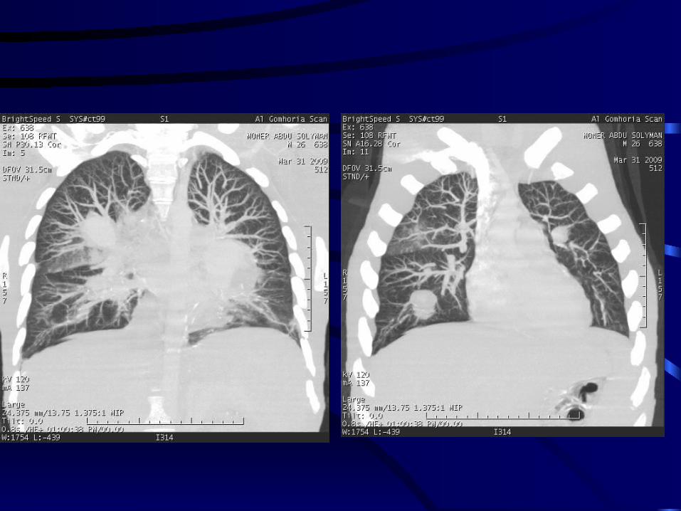

COMPERHENSIVE MSCT OF THE ABDOMEN
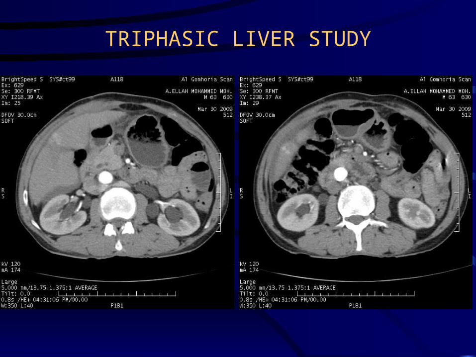
TRIPHASIC LIVER STUDY


HEPATOBILIARY SYSTEM


MDCT UROGRAPHY

NORMAL ANATOMY

NORMAL ANATOMY
FRONTAL (ANTERIOR) VIEW OF VR IMAGES

MIP IMAGE (POSTERIOR VIEW) VR DOUBLE DENSITY IMAGE
(POSTERIOR VIEW)

NORMAL VARIANTS AND CONGENITAL ANOMALIES

NORMAL PAPILLARY BLUSH


PROMINENT RENAL PAPILLA

COMPOUND CALYX

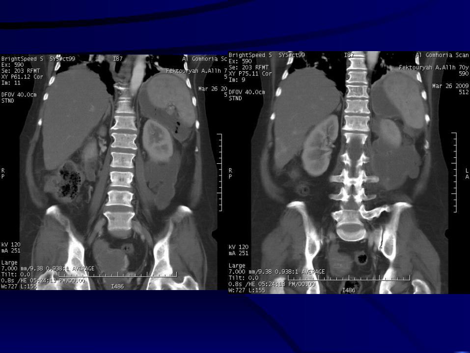
PTOTIC KIDNEY
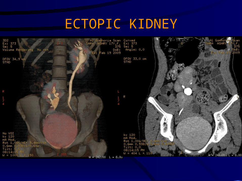
ECTOPIC KIDNEY
VR IMAGE MIP IMAGE


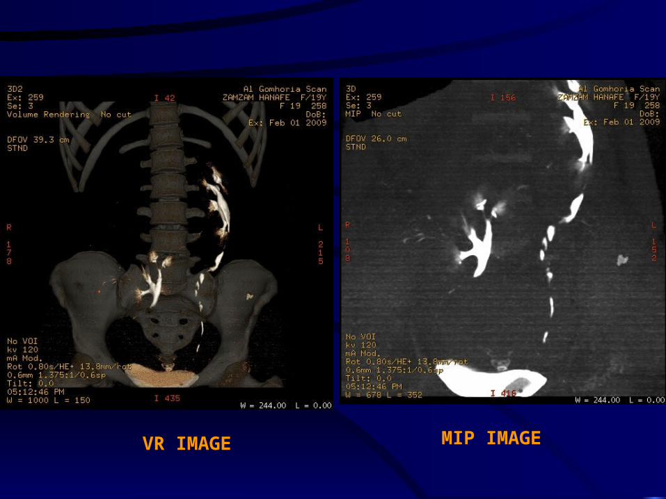
HORSESHOE KIDNEY
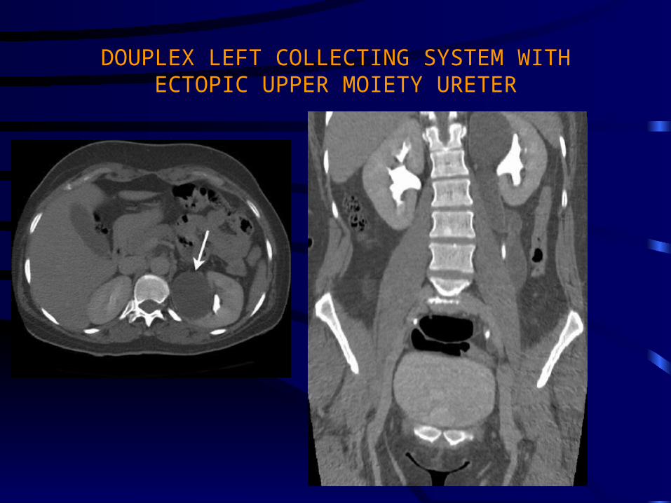
DOUPLEX LEFT COLLECTING SYSTEM WITH ECTOPIC UPPER MOIETY URETER


UROLITHIASIS

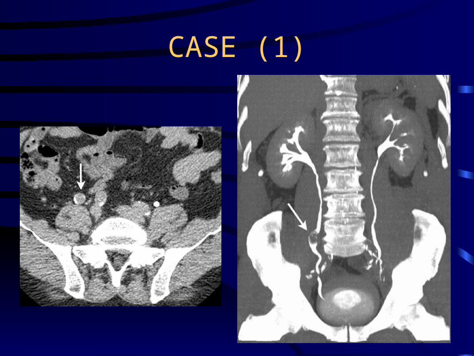
CASE (1)
NON ENHANCED CT SHOWING BILATERAL RENAL PELVIS CALCULI WITH MARKED PYELITIS.
ENHANCED CT SHOWING GOOD ENHANCEMENT.

MIP; THE STONES ARE WELL-SEEN WITHIN THE OPACIFIED RENAL PELVIS.

CASE (2)
THICK SLAP MIPBILATERAL RENAL AND UB STONES
CORONAL IMAGESSHOWING MARKED PYELITIS OF THE LEFT KIDNEY

MIP; THE STONES ARE WELL-SEEN WITHIN THE OPACIFIED RENAL PELVIS. MULTIPLE UB STONES.
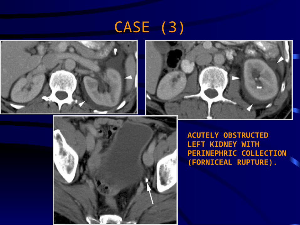
CASE (3)
ACUTELY OBSTRUCTEDLEFT KIDNEY WITH PERINEPHRIC COLLECTION (FORNICEAL RUPTURE).

CURVED REFORMATSSHOWING 3 LOWER URETERIC STONES.

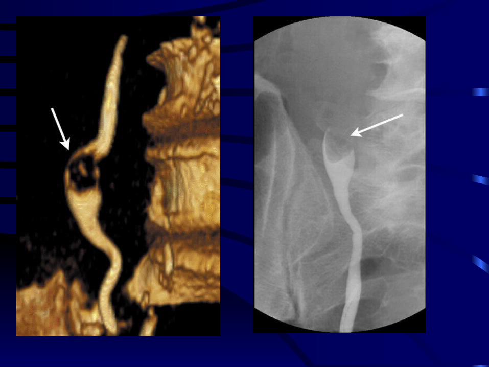
CASE (4)
CURVED REFORMATLOWER URETERIC STONE
CAUSING MILD HYDRONEPHROSIS
DOUBLE DENSITY VR IMAGETHE STONE IS DEMONSTRATED
AGAINST THE UNDERLYINGBONE

CASE (5)
BILHARZIAL CALCIFICATION OF THE LEFT LOWER URETER WITH LOWER URETERIC STONE.
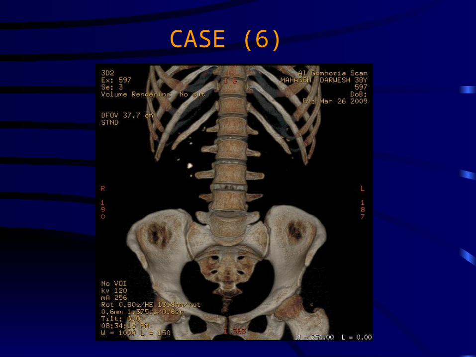
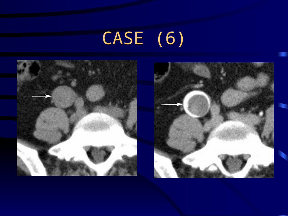
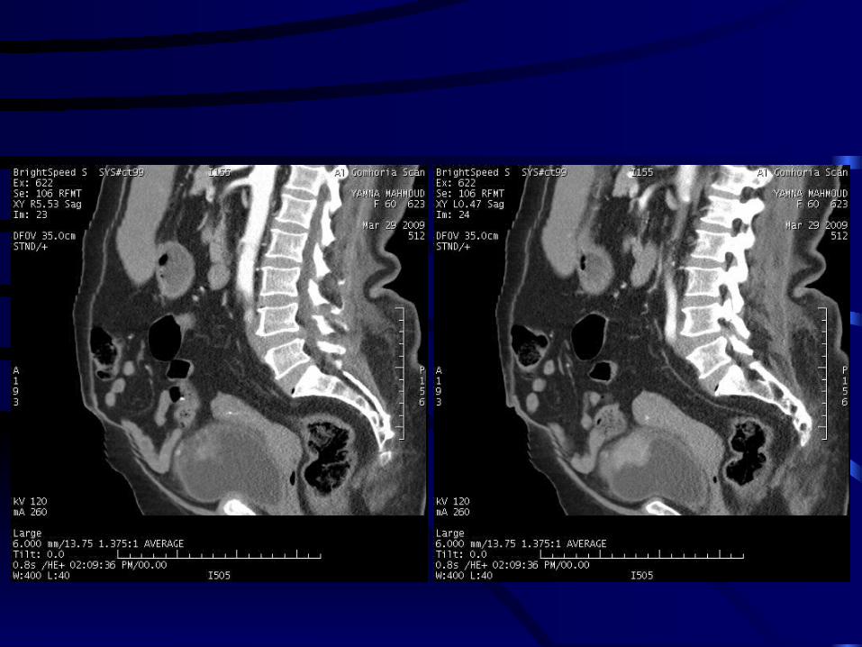
CASE (6)

CASE (7)
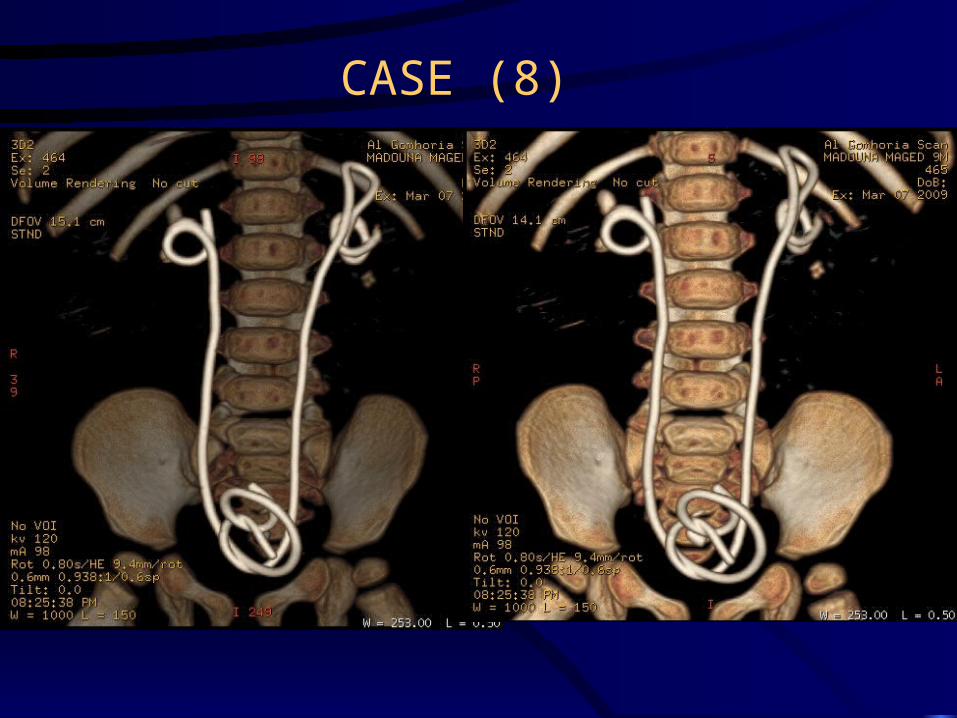
CASE (8)

RENAL INFECTIONS
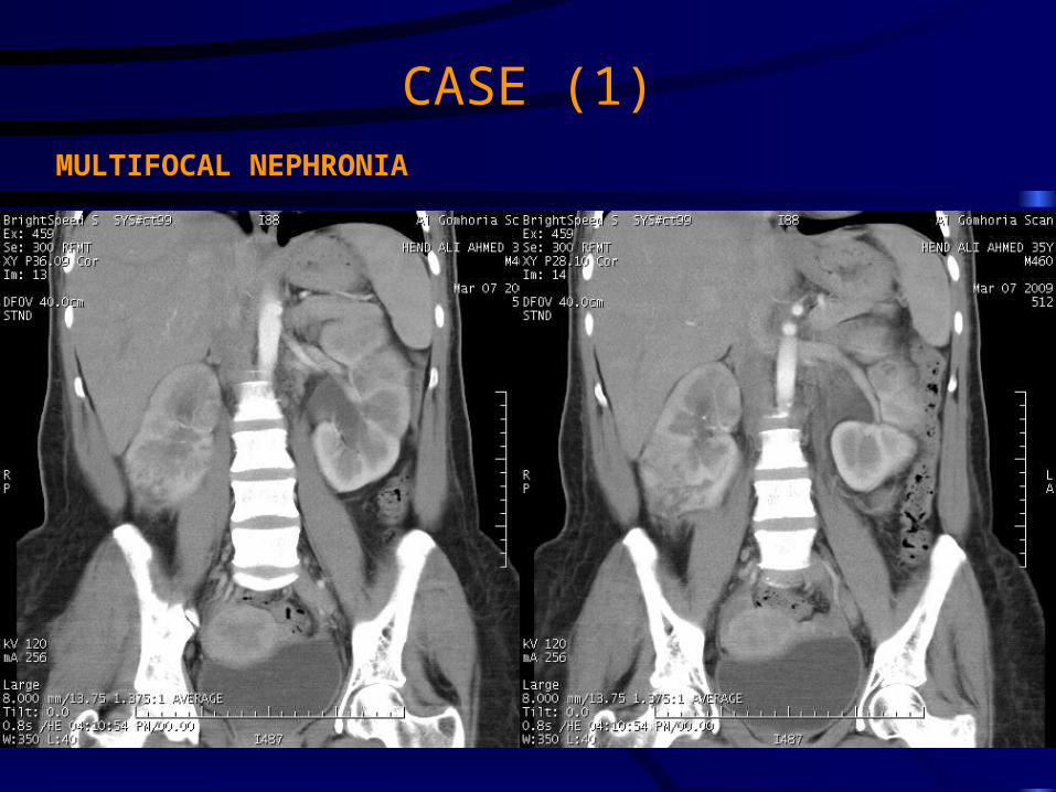
CASE (1)MULTIFOCAL NEPHRONIA


CASE (2)
OBSTRUCTED INFECTED KIDNEYENLARGED LEFT KIDNEY WITH MARKED STRANDING OF THE
PERINEPHRIC FAT AND OBSTRUCTING PELVIC CALCULUS

DOUBLE DENSITY VR IMAGE SHOWING THE OBSTRUCTING CALCULUS

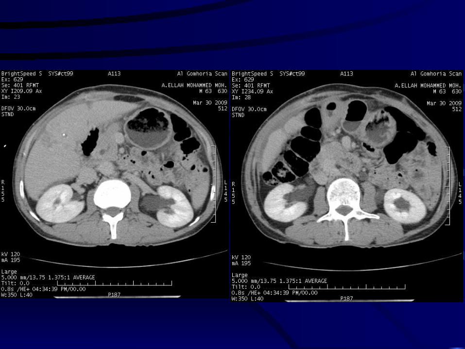
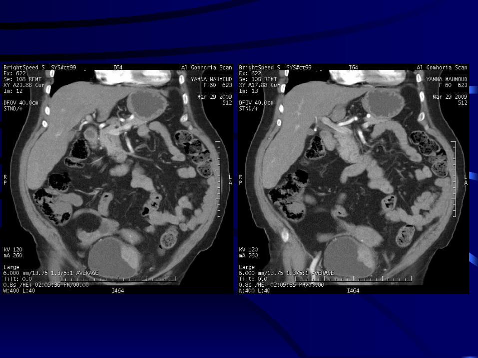
RENAL SOLs

CASE (1)
NON ENHANCED CT ENHANCED CT

DELAYED FILLING OF CALYCEAL DIVERTICULUM

CASE (2)
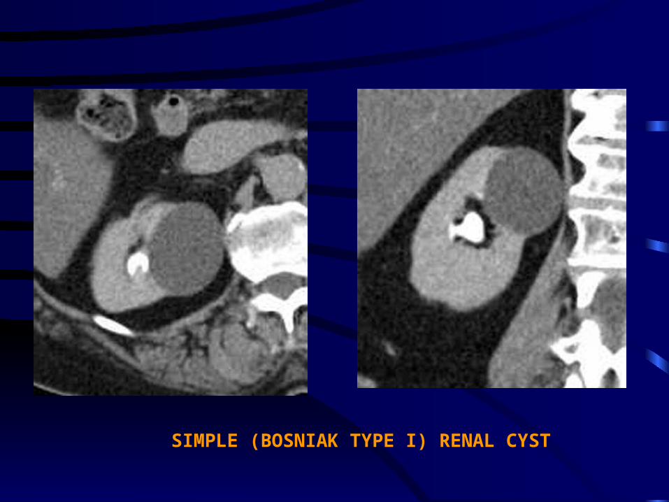
SIMPLE (BOSNIAK TYPE I) RENAL CYST
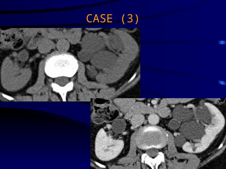
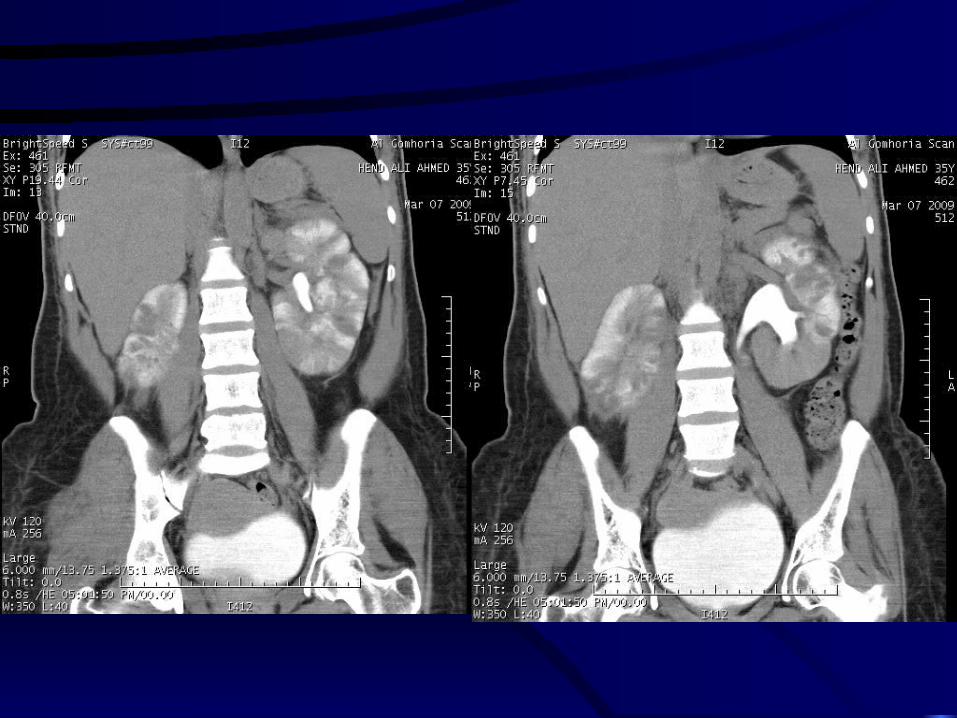
CASE (3)

MULTILOCULAR PARAPELVIC CYST WITH STRETCHING OF THE MAJOR CALYCES
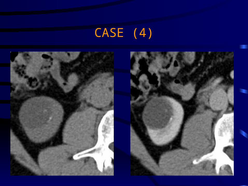
CASE (4)

BOSNIOAK TYPE II CYST WITH THIN CALCIFIED RIM AND INTRACYSTIC SEPTUM
(THANKS FOR THE SUBMILLEMETRIC SLICE THICKNESS)

CASE (5)
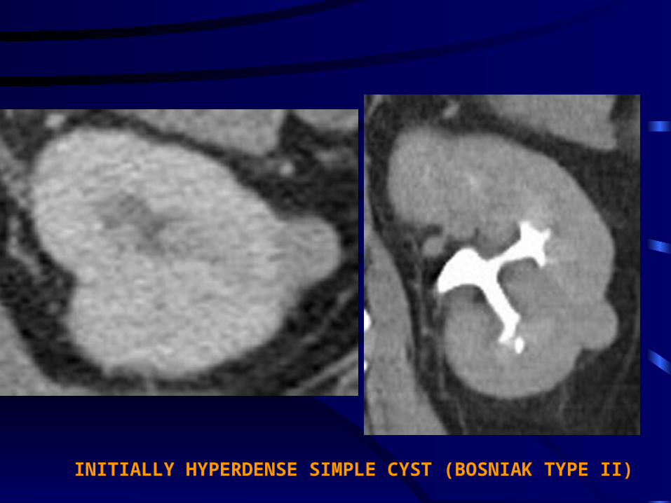
INITIALLY HYPERDENSE SIMPLE CYST (BOSNIAK TYPE II)

CASE (6)
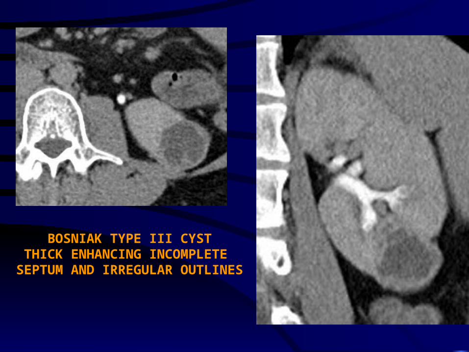
BOSNIAK TYPE III CYSTTHICK ENHANCING INCOMPLETE
SEPTUM AND IRREGULAR OUTLINES

CASE (7)

BOSNIAK TYPE IV CYSTTHICK ENHANCING MURAL NODULE

CASE (8)

SOLID PARAPELVIC MASS CLEARLY DEMONSTRATED IN CORONAL IMAGES

CASE (9)


MALIGNANT LOWER POLAR LEFT RENAL MASS WITH ENHANCING MALIGNANT THROMBUS WITHIN THE IVC
AND SECONDARY VARICOSITIES OF THE LEFT TESTICULAR VEIN.
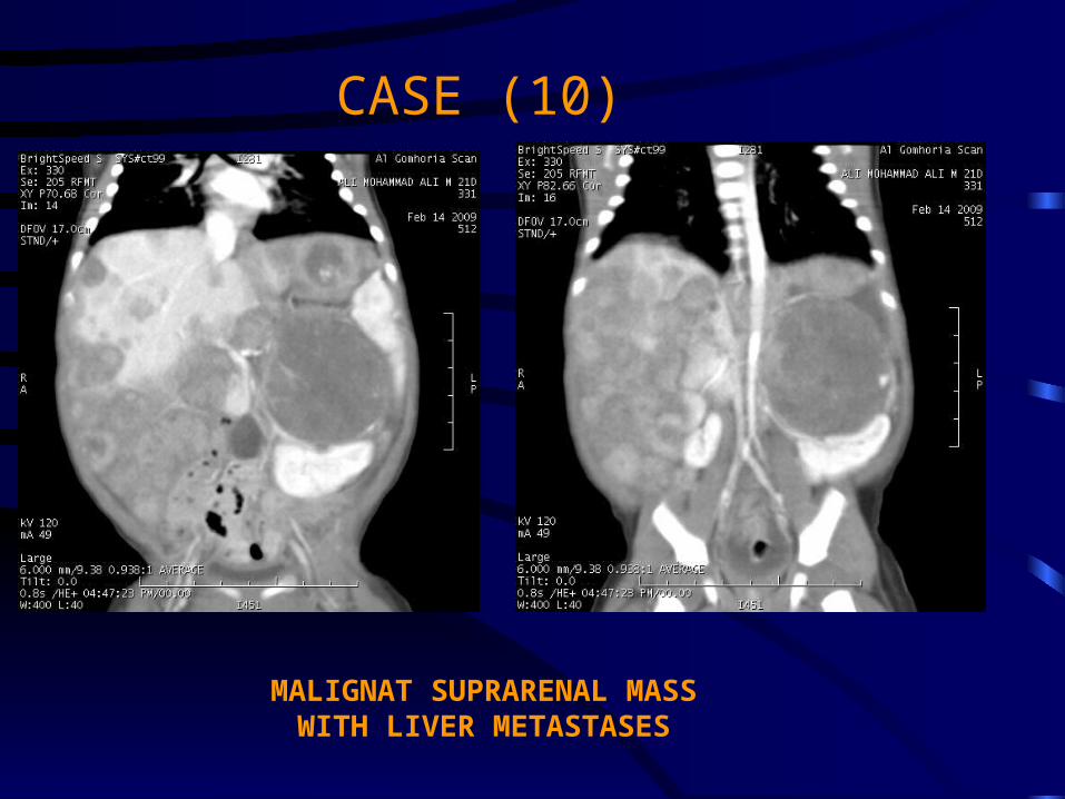
CASE (10)
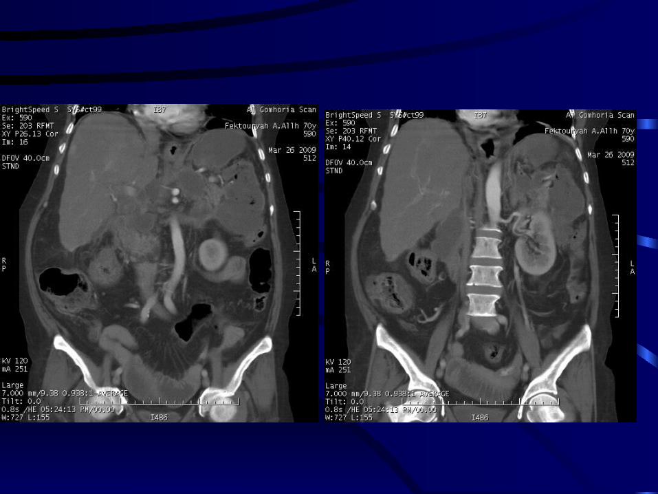
MALIGNAT SUPRARENAL MASSWITH LIVER METASTASES


DISPLACED LEFT KIDNEY WITH DOUPLEX RIGHT COLLECTING SYSTEM
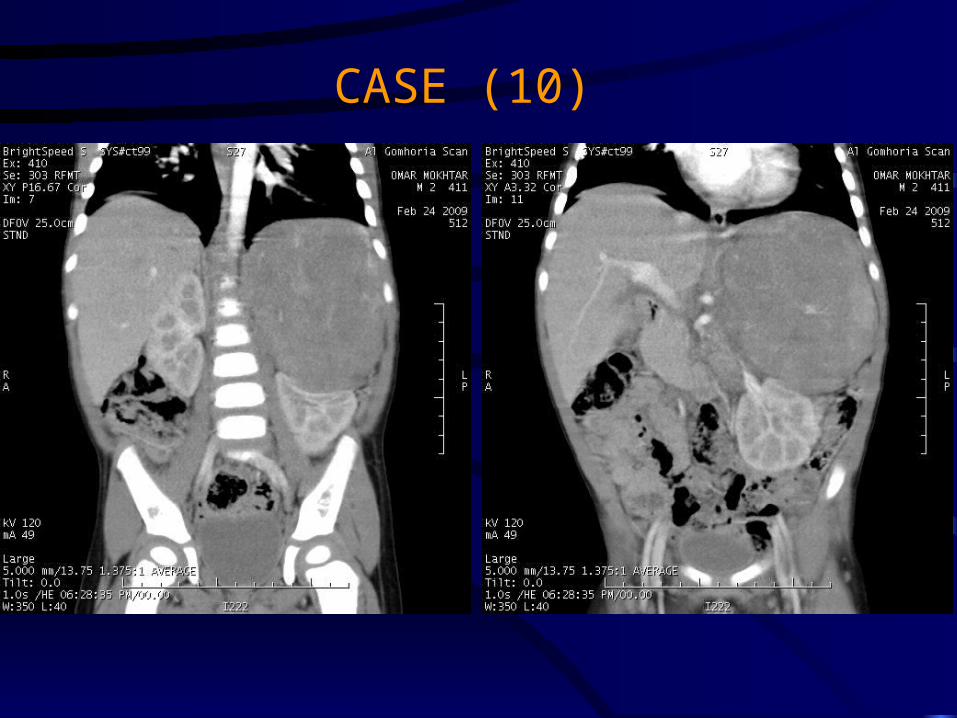
CASE (10)


URETERS
AS A RULE;MALIGNANT URETERIC NEOPLASMS CHARACTERISTICALLY CAUSE DILATATION OF THE URETER BOTH PROXIMAL AND DISTAL TO THE LESION.
CASE (1)

CASE (2)


CASE (3)

CASE (6)

FIBROVASCULAR POLYP OF THE URETER

URINARY BLADDER

CASE (1)


CASE (2)
EXTRAVESICAL PARARECTAL MASS


CASE (3)

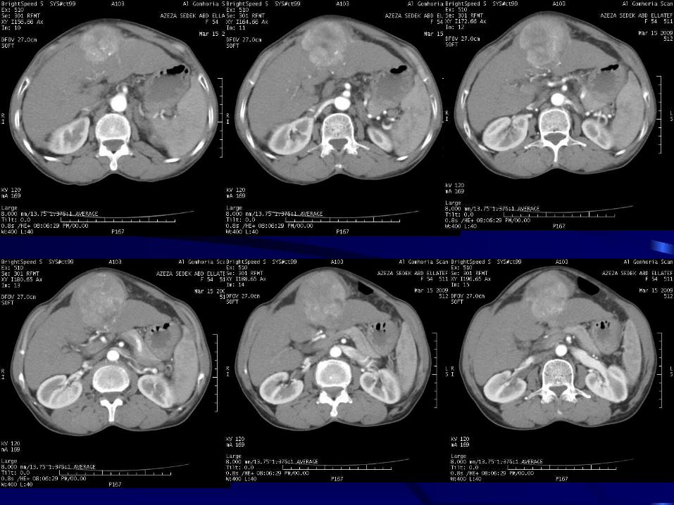
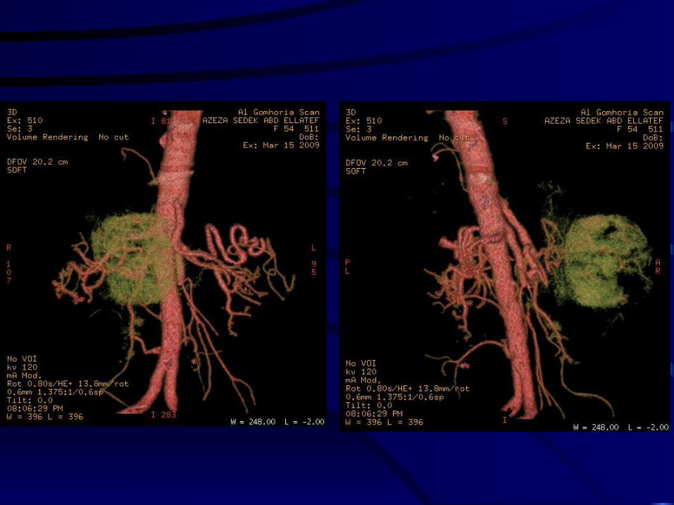
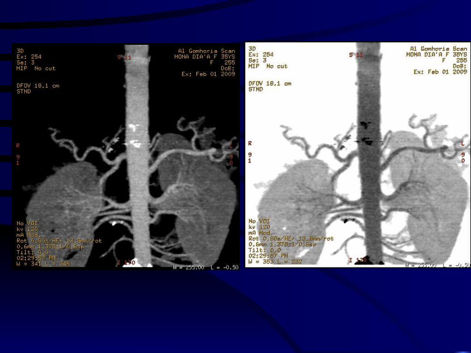
RENAL ARTERY ASSESSMENT

SOME SPECIAL TECHNIQUES
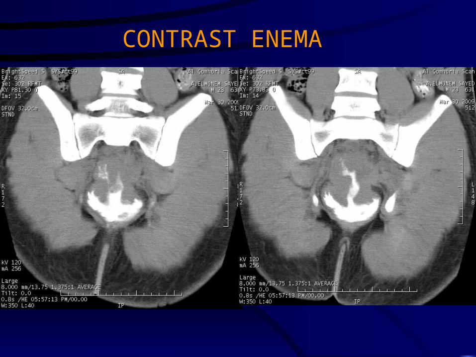
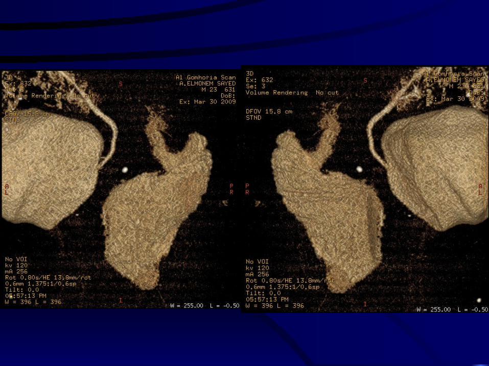
CONTRAST ENEMA


MDCT FISTULOGRAPHY

ASSESSMENT OF POLYTRAUMA
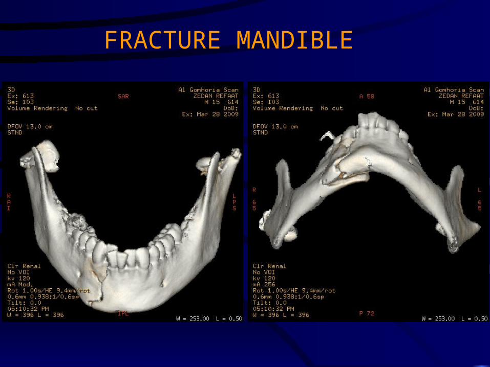
FRACTURE MANDIBLE


FRACTURE CX SPINE


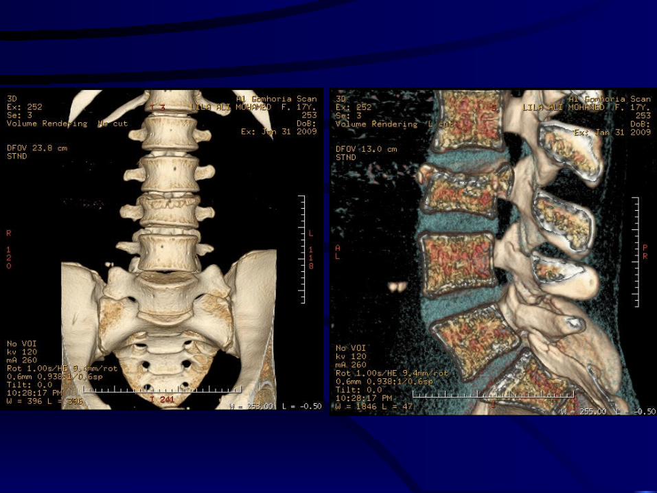
FRACTURE L.S.S.
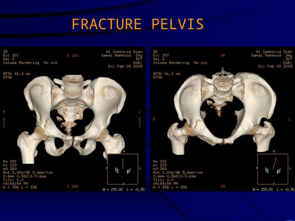
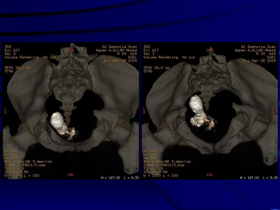
FRACTURE PELVIS


THANK YOUTHANK YOU
Related Documents