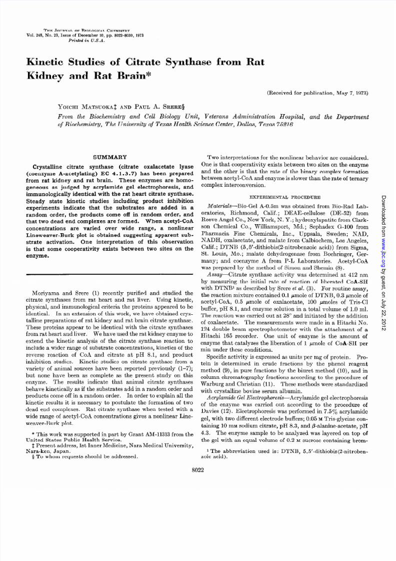
THE J OU R N A L OF BIOLOGICAL CHEMISTRY Vol. 248, No. 23, Issue of December 10, pp. 8022-8030, 1973 Printed in U.S .A. Kinetic Studies of Citrate Sy-nthase from Rat Kidney and Rat Brain* (Received for publication, May 7, 1973 ) YOICHI MATSUOKA ~ AND PAUL A. SRERE~ From the Biochemistry and Cell Biology Unit, Veterans Administration Hospital, and the Department of Biochemistry, The University of Texas Health Science Center, Dallas , Texas 75216 SUMMARY Crystalline citrate synthase (citrate oxalacetate lyase (coenzyme A-acet ylating ) EC 4.1.3.7 ) has been prepared from rat kidney and rat brain. These enzymes are homo- geneous as judged by acryla mide gel electropho resis, and immunologically identical with the rat heart citrate synt hase. Steady state kinetic studies includ ing product inhibi tion experiments indicate that the substr ates are added in a rando m order, the products come of f in rando m order, and that two dead end complexes are formed. When acetyl-CoA concentra tions are varie d over wide range, a nonlinear Lineweaver-Burk plot is obtained suggesting apparent sub- strate activatio n. One interpreta tion of this observation is that some coopera tivity exists between two sites on the enzyme. Moriyama and Srere (1) rece ntly purifie d and studied the citrate synthase s fro m rat heart an d rat liver. Using kinetic, phys ical , and immunological criteria the proteins appeared to be identical. In an extension of this work, we have obtai ned crys - talline prepara tions of rat kidney and rat brain citrate synthas e. These proteins appear to be identical with the citrat e syn thas es from rat heart and liver. We have us ed the rat kidney enzyme to extend the kinetic analysis of the citrate synthase reaction to include a wider range of substrate concentrations, kinetics of the reverse reaction of CoA and citrate at pH 8.1, a nd product inhibition studies. Kinetic studies on citrate synthase from a vari ety of animal sources have been report ed previous ly (l-7); but none have been as complete as the pre sent stud y on this enzyme. Th e results indicate that animal citrate sy nthases behave kinetic ally as if the substrates add in a rando m order and products come of f in a rando m order. In order to explain all the kinetic results i t is necessary to postulate the formation of two dead end complexes. Rat citrate synthase when tested with a wid e r ange of acetyl-CoA concentrations gives a nonli near Line- weaver-Burk plot. * This work was supported in part b y Grant AM-113 13 fro m the United States Public-H ealth Service. i Present address. 1st Inner Medicine. NaraM edica l Unive rsitv . Nara-ken, Japan. ’ 5 To w hom requests should be addressed. ” , Two interpretat ions for the nonlinear behavior are considered, One is that coo perativi ty exists between two sites on the enzyme and the other is that the rate of the binary complex formation between acetyl-CoA and enzyme is slow er than the ra te of ternary complex interconversion. EXPERIMENTAL PROCEDURE Materials-Bio-Gel A-0.5m was obtained fro m Bio-Rad Lab- oratories, Richmond, Ca lif.; DEAE-cel lulose (DE-52 ) from Reeve Ange l C o., New York, N . Y. ; hydroxylapatite from C lark- son Chemical Co., Williamsport, Md.; Sephadex G-100 from Pharma cia Fine Chemicals, Inc., Uppsala, Swed en; NAD, NADH, oxalacetate, and malate from Calbiochem, Los Angeles, Calif .; DTNB (5 ,5’-dit hiobis( 2-nitrobenzoic acid)) from Sigma, St. Louis, MO.; malate dehydrogenase fro m Boehringer, Ger- many; and coenzyme A from P-L Laboratories. Acetyl-CoA was prepared by the method of Simon and Shemin (8). Assay-Citrate synthase activ ity was determi ned at 412 nm by measuring the initial rate of reaction of liberat ed CoA-SH with DTNBl as described by Srere et ~2. (3). For routine assa y, the reaction mixture containe d 0.1 pmole of DTNB, 0.3 pmole of acetyl-CoA, 0.5 Imole of oxalacetate, 100 pmoles of Tris-Cl buff er, pH 8.1, and enzyme solution in a total volume of 1.0 ml. The reaction was carried out at 28” and initiated by the addition of oxalac etate. The measureme nts were made in a Hitachi No. 124 double beam spectroph otometer with the attach ment of a Hitachi 165 recorder. One unit o f enzyme is the amount of enzyme that catalyzes the liberat ion of 1 pmole of CoA-SH per min unde r these conditio ns. Specific ac tiv ity is expressed as units per mg of protein. Pro- tein is determined in crude fractions by the phenol reagen t method (9), in pure fractions by the biuret method (lo), and in column chromatography fractions accordi ng to the proce dure o f Warburg and Christia n (11). These methods were standardized with crystalline bovine serum albumin. Acrylamide Gel Electrophoresis -Acrylamide gel electrophor esis of the enzyme was carried out according to the proced ure of Davies (12). Electrophor esis was performed in 7.5% acrylamide gel, with two differ ent electrod e buff ers; 0.05 M Tris-glycine con- taining 10 m M sodium citrate, pH 8.3, and p-alanine -acetate, pH 4.3. The enzyme sam ple to be analyzed was layered on top o f the gel with an equal volume of 0.2 M sucrose containing brom- 1 The abbreviation used is: DTNB, 5.5’~dithiobis(2-nitroben- zoic acid). 8022 b y g u e s t , o J u l y 2 2 , 2 0 1 2 w w w j b c o r g D o w l o a d e d f r o

Welcome message from author
This document is posted to help you gain knowledge. Please leave a comment to let me know what you think about it! Share it to your friends and learn new things together.
Transcript
7/28/2019 Matsuoka Srere 1973
http://slidepdf.com/reader/full/matsuoka-srere-1973 1/9
THE JOURNAL OF BIOLOGICAL CHEMISTRY
Vol. 248, No. 23, Issue of December 10, pp. 8022-8030, 1973
Printed in U.S .A.
Kinetic Studies of Citrate Sy-nthase from Rat
Kidney and Rat Brain*
(Received for publication, May 7, 1973)
YOICHI MATSUOKA~ AND PAUL A. SRERE~
From the Biochemistry and Cell Biology Unit, Veterans Administration Hospital, and the Department
of Biochemistry, The University of Texas Health Science Center, Dallas, Texas 75216
SUMMARY
Crystalline citrate synthase (citrate oxalacetate lyase
(coenzyme A-acetylating) EC 4.1.3.7) has been prepared
from rat kidney and rat brain. These enzymes are homo-
geneous as judged by acrylamide gel electrophoresis, and
immunologically identical with the rat heart citrate synthase.
Steady state kinetic studies including product inhibition
experiments indicate that the substrates are added in a
random order, the products come of f in random order, and
that two dead end complexes are formed. When acetyl-CoA
concentrations are varied over wide range, a nonlinear
Lineweaver-Burk plot is obtained suggesting apparent sub-
strate activation. One interpretation of this observation
is that some cooperativity exists between two sites on the
enzyme.
Moriyama and Srere (1) recently purified and studied the
citrate synthases from rat heart and rat liver. Using kinetic,
phys ical, and immunological criteria the proteins appeared to be
identical. In an extension of this work, we have obtained crys-
talline preparations of rat kidney and rat brain citrate synthase.
These proteins appear to be identical with the citrate synthases
from rat heart and liver. We have used the rat kidney enzyme to
extend the kinetic analysis of the citrate synthase reaction to
include a wider range of substrate concentrations, kinetics of the
reverse reaction of CoA and citrate at pH 8.1, and product
inhibition studies. Kinetic studies on citrate synthase from a
variety of animal sources have been reported previous ly (l-7);but none have been as complete as the present study on this
enzyme. The results indicate that animal citrate synthases
behave kinetically as if the substrates add in a random order and
products come of f in a random order. In order to explain all the
kinetic results i t is necessary to postulate the formation of two
dead end complexes. Rat citrate synthase when tested with a
wide range of acetyl-CoA concentrations gives a nonlinear Line-
weaver-Burk plot.
* This work was supported in part by Grant AM-11313 from the
United States Public-Health Service.i Present address. 1st Inner Medicine. NaraMedical Unive rsitv .
Nara-ken, Japan. ’5 To whom requests should be addressed.
” ,
Two interpretat ions for the nonlinear behavior are considered,
One is that cooperativi ty exists between two sites on the enzyme
and the other is that the rate of the binary complex formation
between acetyl-CoA and enzyme is slower than the rate of ternary
complex interconversion.
EXPERIMENTAL PROCEDURE
Materials-Bio-Gel A-0.5m was obtained from Bio-Rad Lab-
oratories, Richmond, Calif.; DEAE-cellulose (DE-52) from
Reeve Angel Co., New York, N. Y. ; hydroxylapatite from Clark-
son Chemical Co., Williamsport, Md.; Sephadex G-100 from
Pharmacia Fine Chemicals, Inc., Uppsala, Sweden; NAD,
NADH, oxalacetate, and malate from Calbiochem, Los Angeles,
Calif .; DTNB (5,5’-dithiobis(2-nitrobenzoic acid)) from Sigma,
St. Louis, MO.; malate dehydrogenase from Boehringer, Ger-
many; and coenzyme A from P-L Laboratories. Acetyl-CoA
was prepared by the method of Simon and Shemin (8).
Assay-Citrate synthase activ ity was determined at 412 nm
by measuring the initial rate of reaction of liberated CoA-SH
with DTNBl as described by Srere et ~2. (3). For routine assay,
the reaction mixture contained 0.1 pmole of DTNB, 0.3 pmole of
acetyl-CoA, 0.5 Imole of oxalacetate, 100 pmoles of Tris-Cl
buffer, pH 8.1, and enzyme solution in a total volume of 1.0 ml.
The reaction was carried out at 28” and initiated by the addition
of oxalacetate. The measurements were made in a Hitachi No.
124 double beam spectrophotometer with the attachment of a
Hitachi 165 recorder. One unit o f enzyme is the amount of
enzyme that catalyzes the liberation of 1 pmole of CoA-SH per
min under these conditions.
Specific ac tiv ity is expressed as units per mg of protein. Pro-
tein is determined in crude fractions by the phenol reagent
method (9), in pure fractions by the biuret method (lo), and incolumn chromatography fractions according to the procedure o f
Warburg and Christian (11). These methods were standardized
with crystalline bovine serum albumin.
Acrylamide Gel Electrophoresis-Acrylamide gel electrophoresis
of the enzyme was carried out according to the procedure of
Davies (12). Electrophoresis was performed in 7.5% acrylamide
gel, with two different electrode buffers; 0.05 MTris-glycine con-
taining 10 m M sodium citrate, pH 8.3, and p-alanine-acetate, pH
4.3. The enzyme sample to be analyzed was layered on top of
the gel with an equal volume of 0.2 M sucrose containing brom-
1 The abbreviation used is: DTNB, 5.5’~dithiobis(2-nitroben-zoic acid).
8022
y g ,
y
,
j g
7/28/2019 Matsuoka Srere 1973
http://slidepdf.com/reader/full/matsuoka-srere-1973 2/9
8023
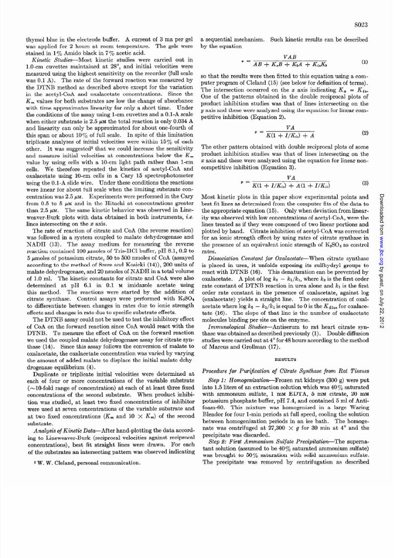
thymol blue in the electrodebuffer. A current of 3 ma per gelwas applied for 2 hours at room temperature. The gels werestained n 1y0 Amido black in 7 To acetic acid.
Kinetic Xtudies-Most kinetic studies were carried out inl.O-cm cuvettes maintained at 28”, and initial velocities weremeasured sing he highest sensitivity on the recorder (full scalewas 0.1 A). The rate of the forward reaction wasmeasured ythe DTNB method as describedabove except for the variation
in the acetyl-CoA and oxalacetate concentrations. Since theK, values or both substratesare low the changeof absorbancewith time approximates inearity for only a short time. Underthe conditionsof the assayusing -cm cuvettes and a 0.1-A scalewhen either substrate s 2.5 pM the total reaction s only 0.034Aand linearity can only be approximated for about one-fourth ofthis span or about 10% of full scale. In spite of this limitationtriplicate analysesof initial velocities were within 15% of eachother. It was suggested2hat we could increase he sensitivityand measure nitial velocities at concentrations below the K,
value by using cells with a lo-cm light path rather than l-cmcells. We therefore repeated the kinetics of acetyl-CoA andoxalacetate using IO-cm cells n a Cary 15 spectrophotometer
using he 0.1-A slidewire. Under theseconditions he reactionswere linear for about full scalewhen the limiting substratecon-centration was2.5PM . Experiments wereperformed n the Garyfrom 0.5 to 5 PM and in the Hitachi at concentrationsgreaterthan 2.5 pM. The samekinetic behavior was observed n Line-weaver-Burk plots with data obtained in both instruments, .e.lines ntersecting on the z axis.
The rate of reaction of citrate and CoA (the reverse eaction)was followed n a systemcoupled to malate dehydrogenase ndNADH (13). The assay medium for measuring the reversereaction contained 100pmolesof Tris-HCl buffer, pH 8.1, 0.2 to5 pmolesof potassium itrate, 50 to 500nmolesof CoA (assayedaccording o the method of Srere and Kosicki (14)), 200units ofmalate dehydrogenase, nd20 nmoles f NADH in a total volume
of 1.0 ml. The kinetic constants or citrate and CoA were alsodetermined at pH 6.1 in 0.1 M imidazole acetate usingthis method. The reactions were started by the addition ofcitrate synthase. Control assayswere performed with K&04to differentiate between changesn rates due to ionic strengtheffects and changesn rate due o specificsubstrateeffects.
The DTNB assaycouldnot be used o test the inhibitory effectof CoA on the forward reaction since CoA would react with theDTNB. To measure he effect of CoA on the forward reactionwe used he coupledmalate dehydrogenase ssay or citrate syn-thase (14). Since his assay ollows the conversionof malate tooxalacetate, the oxalacetateconcentrationwasvaried by varyingthe amount of addedmalate to displace he initial malate dehy-
drogenase quilibrium (4).Duplicate or triplicate initial velocities were determined ateach of four or more concentrations of the variable substrate(-lo-fold range of concentration) at eachof at least three fixedconcentrations of the secondsubstrate. When product inhibi-tion was studied, at least two fixed concentrationsof inhibitorwere usedat seven concentrationsof the variable substrateandat two fixed concentrations (Knz and 10 x K,) of the secondsubstrate.
Analysis of Kinetic Data-After hand-plotting the data accord-ing to Lineweaver-Burk (reciprocal velocities against reciprocalconcentrations), best fit straight lines were drawn. For eachof the substratesan intersectingpattern wasobserved ndicating
a sequentialmechanism. Such kinetic results can be describedby the equation
VAB
Y=dB+&B+&,d +&&, (1)
so that the resultswere then fitted to this equation using a com-puter program of Cleland (15) (see elow or definition of terms).
The intersection occurred on the x axis indicating K, = Ki,.One of the patterns obtained in the double reciprocal plots ofproduct inhibition studieswas that of lines ntersecting on they axis and thesewereanalyzed using he equation or linear com-petitive inhibition (Equation 2).
VA
’ = K(1 + I/KJ + A
The other pattern obtained with double eciprocal plots of someproduct inhibition studies was that of lines ntersecting on thex axis and thesewere analyzed using the equation for linear non-competitive inhibition (Equation 3).
VA
’ = KC1 + W-&s) + A(1 + W&i)
Most kinetic plots in this paper show experimental points andbest fit linesasdetermined rom the computer fits of the data tothe appropriate equation (15). Only whendeviation from linear-ity wasobservedwith low concentrationsof acetyl-CoA, were helines reated as f they were composed f two linear portions andplotted by hand. Citrate inhibition of acetyl-CoA wascorrectedfor an ionic strength effect by using rates of citrate synthase nthe presence f an equivalent ionic strength of K&04 as controlrates.
DissociationConstant for Oxalacetate-When citrate synthaseis placed in urea, it unfolds exposing ts sulfhydryl groups toreact with DTNB (16). This denaturation can be prevented by
oxalacetate. A plot of log ko - ki/ki, whereko is the first orderrate constant of DTNB reaction in urea aloneand kl is the firstorder rate constant in the presenceof oxalacetate, against log(oxalacetate) yields a straight line. The concentration of oxal-acetatewhere og ko - kl/kl is equal o 0 is the Kdiss for oxalace-tate (16). The slopeof that line is the number of oxalacetatemolecules inding per site on the enzyme.
Immunological&&es-Antiserum to rat heart citrate syn-thasewasobtainedasdescribed reviously (1). Double diffusionstudieswerecarried out at 4” for 48hoursaccording o the methodof Marcus and Grollman (17).
RESULTS
Procedure for Puri$cation of Citrate Synthase from Rat Tissues
Step 1: Homogenization-Frozen rat kidneys (300 g) wereputinto 1.5 iters of an extraction solution which was40 %saturatedwith ammonium sulfate, 1 mu EDTA, 5 m M citrate, 20 rnwpotassiumphosphatebuffer, pH 7.4, and contained 5 ml of Anti-foam-60. This mixture was homogenized n a large WaringBlendor for four 1-min periodsat full speed,cooling he solutionbetweenhomogenizationperiods n an ice bath. The homoge-nate was centrifuged at 27,300 x g for 30 min at 4’ and theprecipitate wasdiscarded.
Step d: First AmmoniumSulfate Precipitation-The superna-tant solution (assumedo be 40% saturated ammoniumsulfate)was brought to 50% saturation with solid ammoniumsulfate.
2W. W. Cleland,personalcommunication. The precipitate was removed by centrifugation as described
y g ,
y
,
j g
7/28/2019 Matsuoka Srere 1973
http://slidepdf.com/reader/full/matsuoka-srere-1973 3/9
8024
above and discarded. The supernatant solution was brought to
75% saturation of ammonium sulfate with solid ammonium sul-
fate , stirred for an hour, and centrifuged at 27,300 x g for an
hour. The precipitate was dissolved in 20 mM potassium phos-
phate buffe r, pH 7.4, containing 1 mu EDTA.
Step S: Bio-Gel A-0.6m Column Chromatography-The solution
of the 75% ammonium sulfate precipitate was centrifuged at
27,300 x g for 15 min to remove any insoluble material. The
supernatant solution was applied to a‘ Bio-Gel A-0.5m column
(5.0 x 80 cm) previous ly equilibrated with 20 mM potassium
phosphate buffer, pH 7.4, containing 1 mM EDTA, and the pro-
tein was eluted with the same buffer. Citrate synthase was
eluted at a buffer volume from 1000 to 1300 ml.
Step 4: Second Ammonium Sulfate Precipitation-The eluate
from Bio-Gel A-0.5m column was brought to 50% saturation
with solid ammonium sulfate. After stirring for 30 min, the
solution was centri fuged at 27,300 x g for 30 min. The precipi-
tate was discarded, and the supernatant solution was brought to
75% saturation with solid ammonium sulfate. The precipitate
was dissolved in 20 mu potassium phosphate buf fer, pH 7.4,
containing 1 mM EDTA. Insoluble protein was removed by
centrifugation at 12,100 x g for 15 min, and the supernatantsolution was dialyzed for 24 hours against 10 liters of 10 MM
potassium phosphate buffer, pH 7.4, containing 1 mM EDTA.
The external solution was changed four times.
Step 5: DEAE-cellulose Column Chromatography-Insoluble
protein formed during dialysis was removed by centrifugation
at 12,100 x g for 15 mm, and the supernatant solution was
applied to a DE-52 cellulose column (5.0 X 35 cm) previous ly
equilibrated with 10 InM potassium phosphate buffer, pH 7.5,
containing 1 mu EDTA. The column was washed with the same
buffer followed by a linear gradient of 0 to 0.2 M KC1 in the same
buffer. Citrate synthase was eluted from the column at approxi-
mately 0.1 M KCI. The fractions containing the enzyme were
combined and the protein precipitated with solid ammonium
sulfate (80% saturation) and collected by centrifugation at
30,900 x g for an hour. The precipitate was dissolved in 20 mM
potassium phosphate buffer, pH 7.5, containing 1 mM EDTA.
Step 6: Hydroxylapatite Column Chromatography-The enzyme
solution was dialyzed as described previously, centrifuged to
remove insoluble protein, and the supernatant solution applied
to a hydroxylapatite column (2.5 X 35 cm) previously equili-
brated with 20 mu potassium phosphate buffer, pH 7.4. The
column was washed with the same buf fer , and eluted with a
linear gradient between 5 MM and 0.2 M potassium phosphate
buffer, pH 7.4. Citrate synthase was eluted from the column at
approximately 0.1 M buffer concentration. Citrate synthase
fractions were collected and precipitated with solid ammonium
sulfate as before. The precipitate was collected by centrifuga-tion at 30,900 x g for an hour and dissolved in 20 mM potassium
phosphate buffer, pH 7.4, containing 1 mu EDTA.
Step 7: Sephadex G-100 Column Chromatography-The citrate
synthase preparation was then applied to a Sephadex G-100
column (2.0 X 100 cm) previously equilibrated with 20 mM
potassium phosphate buffer, pH 7.4, containing 1 mM EDTA,
and eluted with the same buffer. Citrate synthase was eluted
from the column between 400 to 460 ml of buffer eluent. Citrate
synthase in these fractions were precipitated by addition of solid
ammonium sulfate. The precipitate was collected by cen-
trifugation and dissolved in a small amount of 20 mM potassium
phosphate buffe r, pH 7.5, containing 1 MM EDTA.
Step 8: First Crystallization-A small amount of solid am-
monium sulfate was added slowly to the enzyme solution with
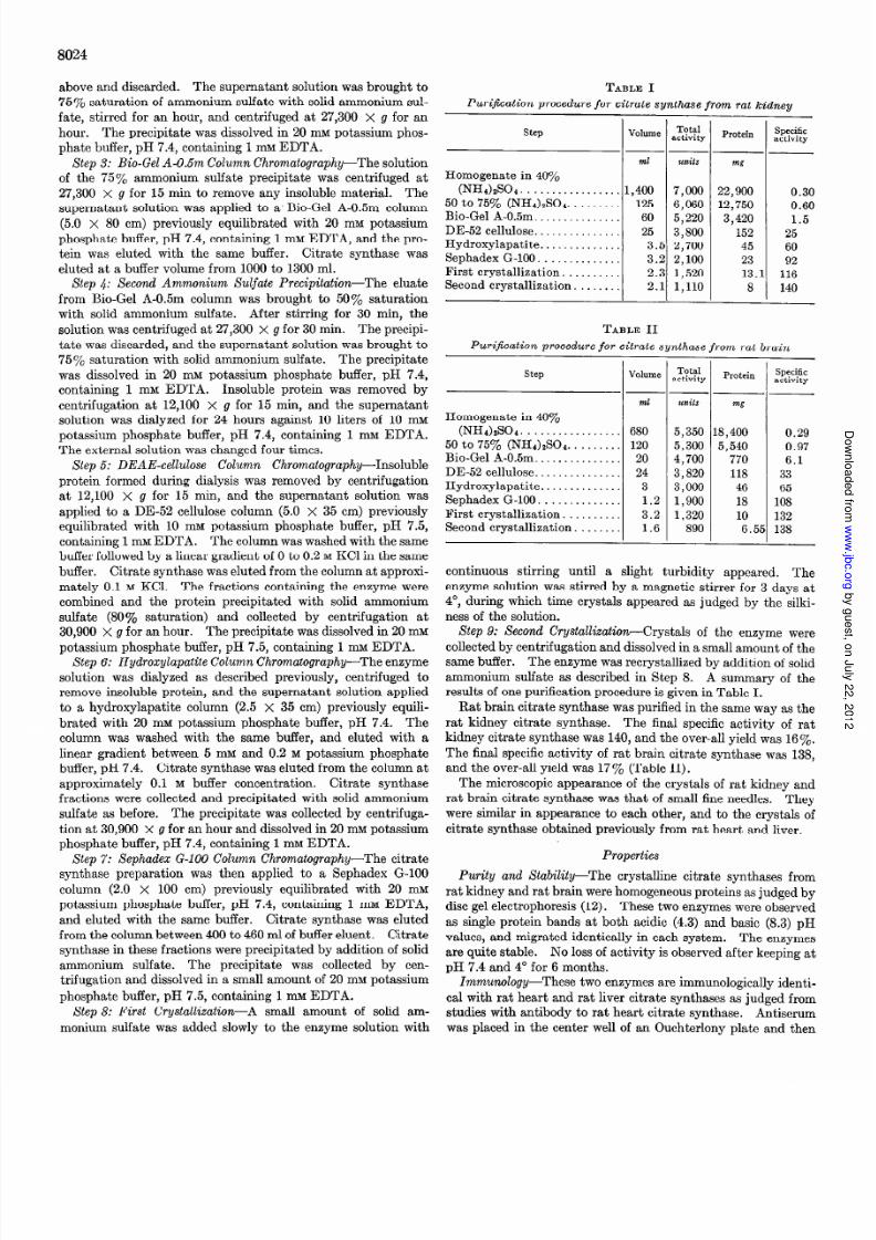
TABLE I
PuriJication procedure for citrate sj dhase from rat kidneu
step VolumeTotal
activity Protein Specificactivity
ml units mg
Homogenate in 40%
(NH&SOa. . . . . . . . . . 1,400 7,000 22,900 0.30
50 to 75% (NH&S04. . . . . . . 125 6,060 12,750 0.60Bio-Gel A-0.5m. . . . . . . 60 5,220 3,420 1.5DE-52 cellulose.. . . 25 3,800 152 25Hydroxylapatite. . . . . . 3.1 2,700 45 60Sephadex G-100. . . . . . . . . 3.: 2,100 23 92First crystallization. . . . . . . 2.: 1,520 13. 116Second crystallization. . . . . . 2.1 1,110 8 140
TABLE II
Purijkation procedure for citrate synthase from rat brain
step
Homogenate in 40%
(NHWO4. ...............
50 to 75% (NH&SO4 ... ... ..
Bio-Gel A-0.5m ...... ...... ..
DE-52 cellulose ..............
Hydroxylapatite. ............
Sephadex G-100. ............
First crystallization. ...... ..
Second crystallization. ......
-
-_
-
Volume
ml
680
120
20
24
3
1.2
3.2
1.6
-
--
-
7-
Totalactivity
WzilS
5,350 8,400 0.295,300 5,540 0.974,700 770 6.1
3,820 118 33
3,000 46 651,900 18 108
1,320 10 132
890 6.5: 138
Protein Specificactivity
continuous stirring until a slight turbidity appeared. The
enzyme solution was stirred by a magnetic stirrer for 3 days at
4’, during which time crystals appeared as judged by the silki-
ness of the solution.
Step 9: Second C’rystallization-Crystals of the enzyme were
collected by centrifugation and dissolved in a small amount of the
same buffer. The enzyme was recrystallized by addition of solid
ammonium sulfate as described in Step 8. A summary of the
results of one purification procedure is given in Table I.
Rat brain citrate synthase was purified in the same way as the
rat kidney citrate synthase. The final specific activity of rat
kidney citrate synthase was 140, and the over-all yield was 16%.
The final specific activ ity of rat brain citrate synthase was 138,
and the over-all yield was 17 y. (Table II).
The microscopic appearance of the crystals o f rat kidney and
rat brain citrate synthase was that of small fine needles. They
were similar in appearance to each other, and to the crysta ls ofcitrate synthase obtained previously from rat heart and liver.
Properties
Purity and Stalrility-The crystalline citrate synthases from
rat kidney and rat brain were homogeneous proteins as judged by
disc gel electrophoresis (12). These two enzymes were observed
as single protein bands at both acidic (4.3) and basic (8.3) pH
values, and migrated identically in each system. The enzymes
are quite stable. No loss of act ivi ty is observed after keeping at
pH 7.4 and 4” fo r 6 months.
Immunology-These two enzymes are immunologically identi-
cal with rat heart and rat liver citrate synthases as judged from
studies with antibody to rat heart citrate synthase. Antiserum
was placed in the center well of an Ouchterlony plate and then
y g ,
y
,
j g
7/28/2019 Matsuoka Srere 1973
http://slidepdf.com/reader/full/matsuoka-srere-1973 4/9
the diluted solution of rat liver, rat heart, rat kidney, and rat
brain crystalline citrate synthases were added to each outer well.
Diffusion was allowed to proceed at 4” for 24 hours. On the
stained plate, a single precipitin band was visible against all
citrate synthases, and the precipitin bands were fused to each
other at their ends, with no spur formation observed between the
bands.
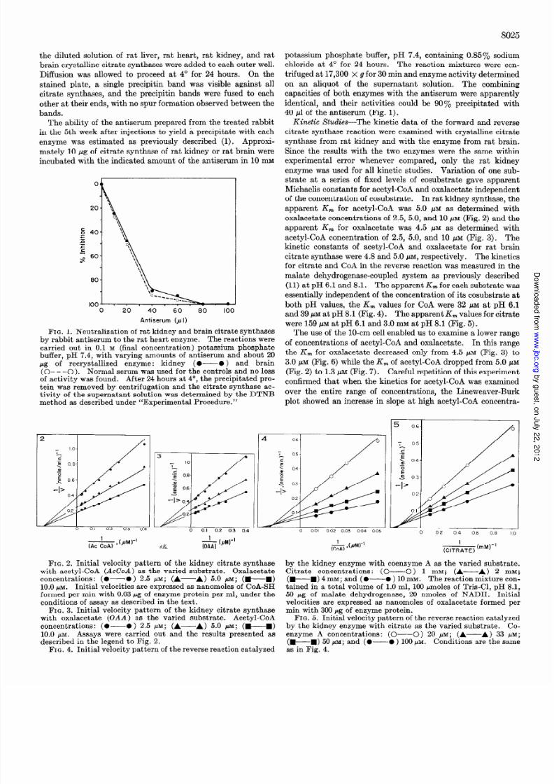
The ability of the antiserum prepared from the treated rabbit
in the 5th week after injections to yield a precipitate with each
enzyme was estimated as previous ly described (1). Approxi-
mately 10 pg of citrate synthase of rat kidney or rat brain were
incubated with the indicated amount of the antiserum in 10 mM
0 20 40 60 80 100
Antiserum (~1)
FIG. 1. Neutralization of rat kidney and brain citrate synthasesby rabbit antiserum to the rat heart enzyme. The reactions werecarried out in 0.1 M (final concentration) potassium phosphatebuffer, pH 7.4, with varying amounts of antiserum and about 20pg of recrystallized enzyme: kidney (O-O ) and brain(O- - -0). Normal serum was used for the controls and no lossof act ivi ty was found. After 24 hours at 4”, the precipitated pro-
tein was removed by centrifugation and the citrate synthase ac-tivi ty of the supernatant solution was determined by the DTNBmethod as described under “Experimental Procedure.”
0%
8025
potassium phosphate buf fer, pH 7.4, containing 0.85% sodium
chloride at 4” for 24 hours. The reaction mixtures were cen-
trifuged at 17,300 x g for 30 min and enzyme act ivi ty determined
on an aliquot of the supernatant solution. The combining
capacities of both enzymes with the antiserum were apparently
identical, and their activities could be 90% precipitated with
40 ~1 of the antiserum (Fig. 1).
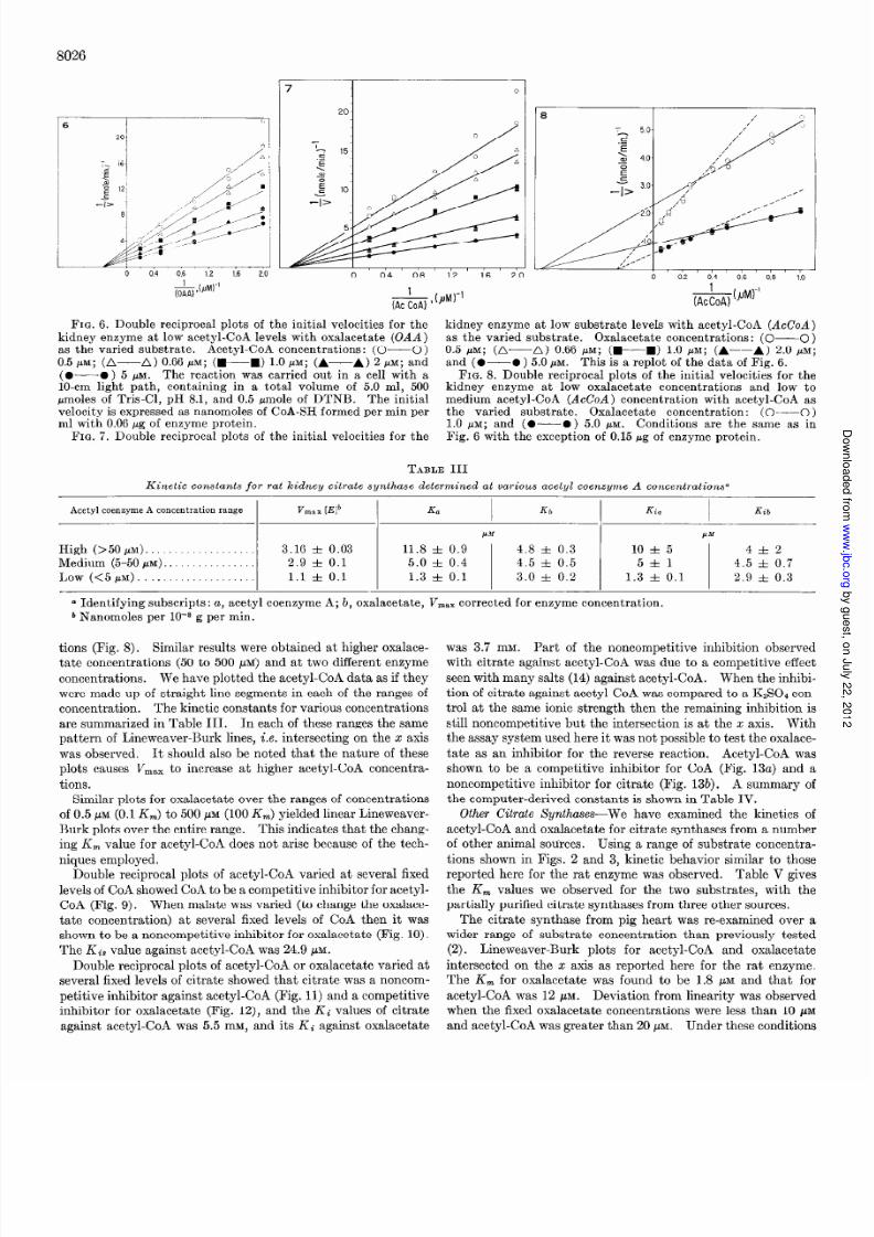
Kinetic Studies-The kinetic data of the forward and reverse
citrate synthase reaction were examined with crystalline citrate
synthase f rom rat kidney and with the enzyme from rat brain.
Since the results with the two enzymes were the same within
experimental error whenever compared, only the rat kidney
enzyme was used for all kinetic studies. Variation of one sub-
strate at a series of fixed levels o f cosubstrate gave apparent
Michaelis constants for acetyl-CoA and oxalacetate independent
of the concentration of cosubstrate. In rat kidney synthase, the
apparent K, for acetyl-CoA was 5.0 pM as determined with
oxalacetate concentrations of 2.5, 5.0, and 10 pM (Fig. 2) and the
apparent K, for oxalacetate was 4.5 pM as determined with
acetyl-CoA concentration of 2.5, 5.0, and 10 HAM (Fig. 3). The
kinetic constants of acetyl-CoA and oxalacetate for rat brain
citrate synthase were 4.8 and 5.0 PM, respectively. The kineticsfor citrate and CoA in the reverse reaction was measured in the
malate dehydrogenase-coupled system as previously described
(11) at pH 6.1 and 8.1. The apparent K, for each substrate was
essentially independent of the concentration of its cosubstrate at
both pH values, the K, values for CoA were 32 pM at pH 6.1
and 39 PM at pH 8.1 (Fig. 4). The apparent K, values for citrate
were 159 pM at pH 6.1 and 3.0 mM at pH 8.1 (Fig. 5).
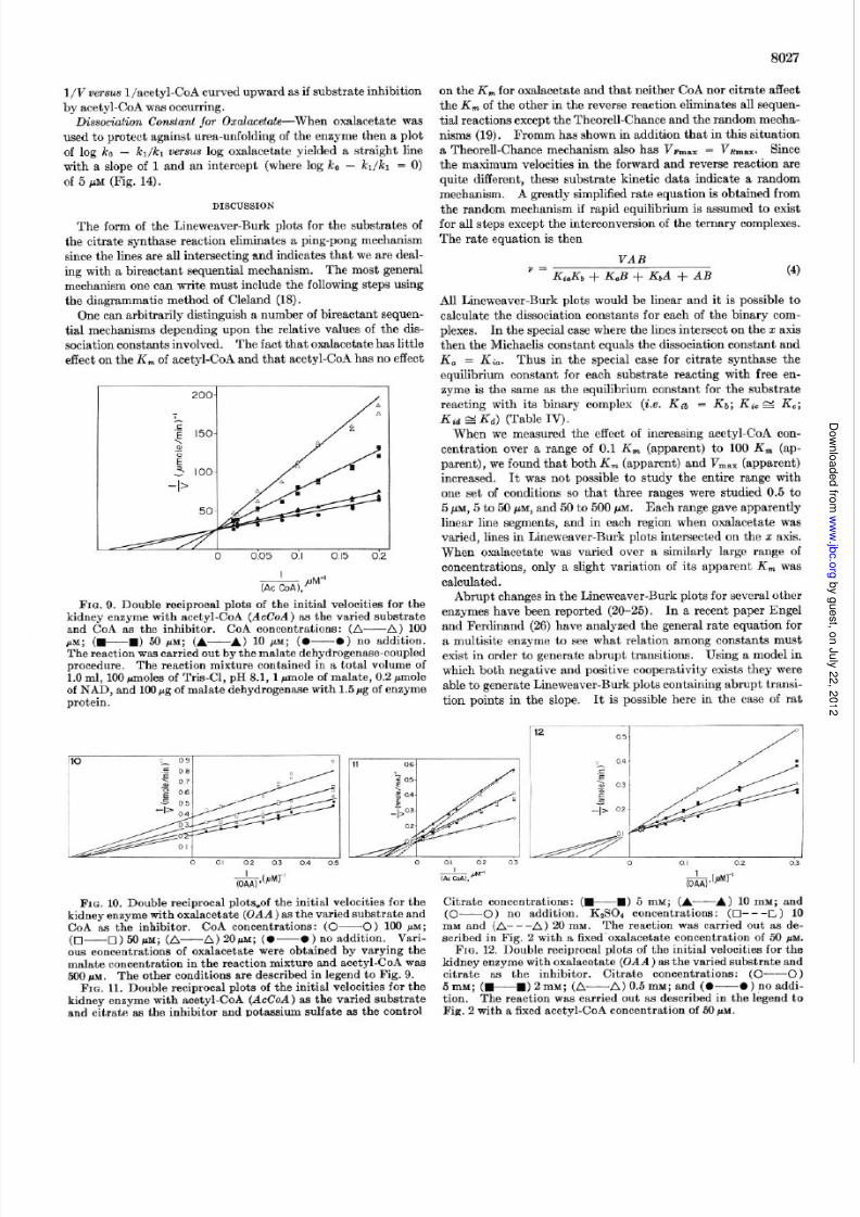
The use of the lo-cm cell enabled us to examine a lower range
of concentrations of acetyl-CoA and oxalacetate. In this range
the K, for oxalacetate decreased only from 4.5 pM (Fig. 3) to
3.0 PM (Fig. 6) while the K, of acetyl-CoA dropped from 5.0 pM
(Fig. 2) to 1.3 pM (Fig. 7). Careful repetition of this experiment
confirmed that when the kinetics for acetyl-CoA was examined
over the entire range of concentrations, the Lineweaver-Burk
plot showed an increase in slope at high acetyl-CoA concentra-
FIG. 2. Initial velocity pattern of the kidney citrate synthasewith acetyl-CoA (AcCoA) as the varied substrate. Oxalacetateconcentrations: (0-O) 2.5 PM; (A-A) 5.0 MM; 0-m)10.0 PM. Initial velocities are expressed as nanomoles of CoA-SH
formed per min with 0.03 pg of enzyme protein per ml, under theconditions of assay as described in the text.
FIG. 3. Initial velocity pattern of the kidney citrate synthasewith oxalacetate (OAA) as the varied substrate. Acetyl-CoA
concentrations: (0-O) 2.5 PM; (A--A) 5.0 MM; (W-----m)
10.0 PM. Assays were carried out and the results presented as
described in the legend to Fig. 2.FIG. 4. Initial velocity pattern of the reverse reaction catalyzed
(&) ,bJw’
E
by the kidney enzyme with coenzyme A as the varied substrate.Citrate concentrations: (C----C) 1 mM; (A-A) 2 mM;(W---W) 4 mm; and (O-O ) 10 mM. The reaction mixture con-tained in a total volume of 1.0 ml, 100 pmoles o f Tris-Cl, pH 8.1,50 pg of malate dehydrogenase, 20 nmoles of NADH. Initialvelocities are expressed as nanomoles of oxalacetate formed permin with 300 pg of enzyme protein.
FIG. 5. Initial velocity pattern of the reverse reaction catalyzedby the kidney enzyme with citrate as the varied substrate. Co-enzyme A concentrations: (O--O) 20 PM; (A-A) 33 PM;
(WM) 50 pM; and (0-O ) 100 pM. Conditions are the same
as in Fig. 4.
y g ,
y
,
j g
7/28/2019 Matsuoka Srere 1973
http://slidepdf.com/reader/full/matsuoka-srere-1973 5/9
8026
0 0.4 0.8 1.2 1.6 2.0
&@Jwl
L0 0.2 0.4 0.6 0.8 1.0
7iizPMK’
FIG. 6. Double reciprocal plots of the initial velocities for the kidney enzyme at low substrate levels with acetyl-CoA (AcCoA).-.._ . . _ ^ -kidney enzyme at low acetyl-CoA levels with oxalacetate (UAA)as the varied substrate. Acetyl-CoA concentrations: (O-O)0.5 M; (A-A) 0.66 PM; (+W) 1.0 PM; (A-A) 2 1~; and(O-O ) 5 MM. The reaction was carried out in a cell with alo-cm light path, containing in a total volume of 5.0 ml, 500rmoles of Tris-Cl, pH 8.1, and 0.5 pmole of DTNB. The initial
velocity is expressed as nanomoles of CoA-SH formed per min perml with 0.06 pg of enzyme protein.
FIG. 7. Double reciprocal plots of the initial velocities for the
as the varied substrate. Oxalacetate concentrations: (O-O)
0.5 j6M; (A-A) 0.66 PM; (+B) 1.0 PM; (A---A) 2.0 PM;
and (o-0 ) 5.0 PM . This is a replot of the data of Fig. 6.FIG. 8. Double reciprocal plots of the initial velocities for the
kidney enzyme at low oxalacetate concentrations and low tomedium acetyl-CoA (AcCoA) concentration with acetyl-CoA as
the varied substrate. Oxalacetate concentration: (O-O)1.0 PM; and (O--O) 5.0 MM. Conditions are the same as inFig. 6 with the exception of 0.15 pg of enzyme protein.
TABLE III
Kinetic constants for rat kidney citrate synthase determined at various acetyl coenzyme A concentrationsa
Acetyl coenzyme A concentration range V max [El*
High (>50 PM). .. . . . . . . . . . . . . . . . . 3.16 + 0.03
Medium (5-50 PM.). ... ... ... ... ... 2.9 f 0.1
LOW (<5pM) ................ .... 1.1 f 0.1
0 Identifying subscripts: a, acetyl coenzyme A; b, oxalacetate, V mBXcorrected for enzyme concentration.
b Nanomoles per low8 g per min.
tions (Fig. 8). Similar results were obtained at higher oxalace-
tate concentrations (50 to 500 pM) and at two different enzyme
concentrations. We have plotted the acetyl-CoA data as if they
were made up of straight line segments in each of the ranges of
concentration. The kinetic constants for various concentrations
are summarized in Table III . In each of these ranges the same
pattern of Lineweaver-Burk lines, i.e. intersecting on the II: axis
was observed. It should also be noted that the nature of these
plots causes V,,, to increase at higher acetyl-CoA concentra-
tions.
Similar plots for oxalacetate over the ranges of concentrations
of 0.5 PM (0.1 &) to 500 pM (100 Km) yielded linear Lineweaver-Burk plots over the entire range. This indicates that the chang-
ing K, value for acetyl-CoA does not arise because of the tech-
niques employed.
Double reciprocal plots of acetyl-CoA varied at several fixed
levels of CoA showed CoA to be a competitive inhibitor for acetyl-
CoA (Fig. 9). When malate was varied (to change the oxalace-
tate concentration) at several fixed levels of CoA then it was
shown to be a noncompetitive inhibitor for oxalacetate (Fig. 10).
The Ki , value against acetyl-CoA was 24.9 PM.
Double reciprocal plots of acetyl-CoA or oxalacetate varied at
several f ixed levels of citrate showed that citrate was a noncom-
petitive inhibitor against acetyl-CoA (Fig. 11) and a competitive
inhibitor for oxalacetate (Fig. la), and the Ki values of citrate
against acetyl-CoA was 5.5 mM, and its Ki against oxalacetate
was 3.7 mM. Part of the noncompetitive inhibition observed
with citrate against acetyl -CoA was due to a competitive effect
seen with many salts (14) against acetyl-CoA. When the inhibi-
tion of citrate against acetyl-CoA was compared to a K&O4 con-
trol at the same ionic strength then the remaining inhibition is
still noncompetitive but the intersection is at the z axis. With
the assay system used here it was not possible to test the oxalace-
tate as an inhibitor for the reverse reaction. Acetyl-CoA was
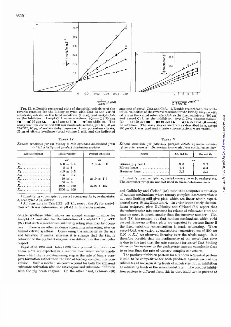
shown to be a competitive inhibitor for CoA (Fig. 13a) and a
noncompetitive inhibitor for citrate (Fig. 13b). A summary of
the computer-derived constants is shown in Table IV.
Other Citrate Xynthases-We have examined the kinetics o facetyl-CoA and oxalacetate for citrate synthases from a number
of other animal sources. Using a range of substrate concentra-
tions shown in Figs. 2 and 3, kinetic behavior similar to those
reported here for the rat enzyme was observed. Table V gives
the K, values we observed for the two substrates, with the
partially purified citrate synthases from three other sources.
The citrate synthase from pig heart was re-examined over a
wider range of substrate concentration than previous ly tested
(2). Lineweaver-Burk plots for acetyl-CoA and oxalacetate
intersected on the 2 axis as reported here for the rat enzyme.
The K, for oxalacetate was found to be 1.8 pM and that for
acetyl-CoA was 12 PM. Deviation from linearity was observed
when the fixed oxalacetate concentrations were less than 10 pM
and acety l-CoA was greater than 20 pM. Under these conditions
y g ,
y
,
j g
7/28/2019 Matsuoka Srere 1973
http://slidepdf.com/reader/full/matsuoka-srere-1973 6/9
8027
l/V versus l/acetyl-CoA curved upward as if substrate inhib ition
by acetyl-CoA was occurring.
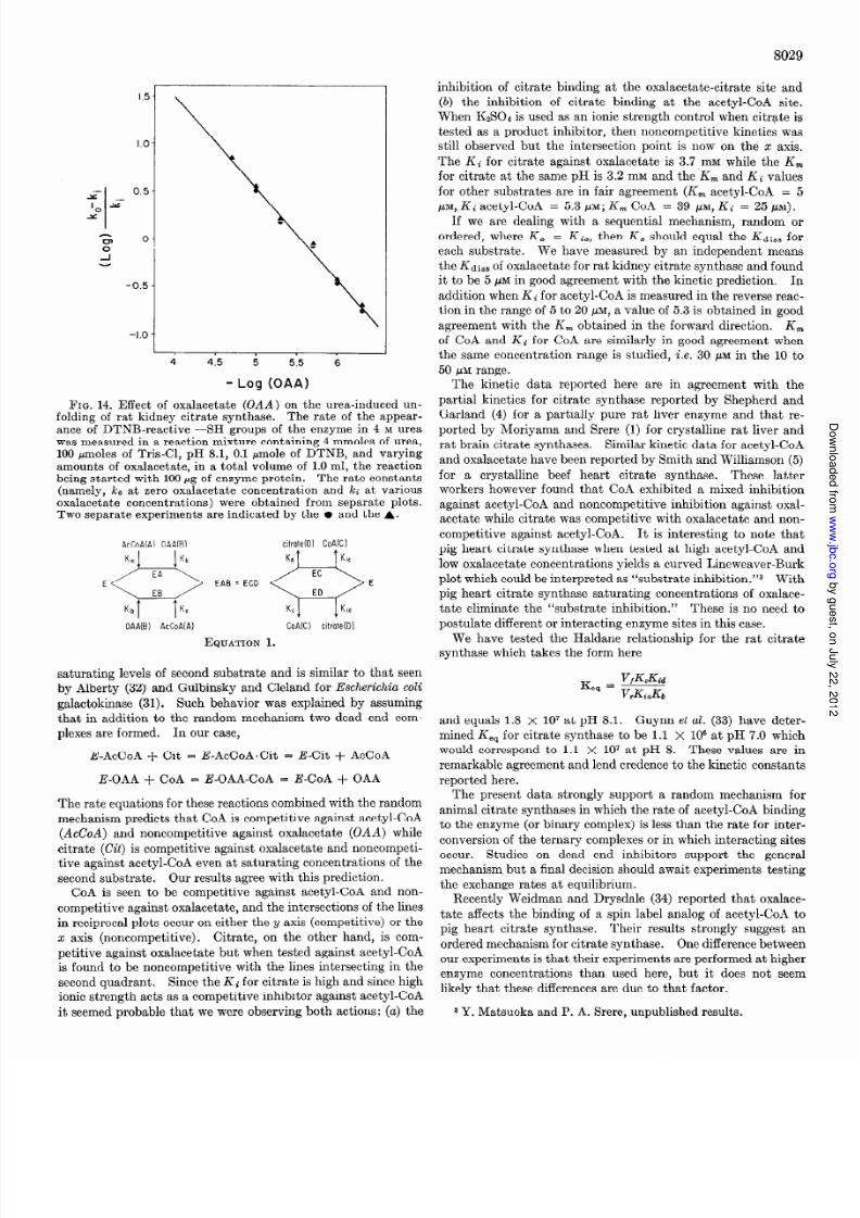
Dissociation Constant for Oxalacetate-When oxalacetate was
used to protect against urea-unfolding of the enzyme then a plot
of log k0 - kl/kl versus log oxalacetate yielded a straight line
with a slope of 1 and an intercept (where log ko - i&/k1 = 0)
of 5 pM (Fig. 14).
DISCUSSION
The form of the Lineweaver-Burk plots for the substratesofthe citrate synthase eaction eliminatesa ping-pongmechanismsince he linesare all intersectingand indicates hat we are deal-ing with a bireactant sequentialmechanism. The most generalmechanism ne can write must include the following steps usingthe diagrammaticmethodof Cleland (18).
One can arbitrarily distinguisha numberof bireactant sequen-tial mechanisms ependingupon the relative values of the dis-sociationconstantsnvolved. The fact that oxalacetatehas ittleeffect on the K, of acetyl-CoA and that acetyl-CoA hasno effect
&.fi”-’FIG. 9. Double reciprocal plots of the initial velocities for the
kidney enzymewith acetyl-CoA (AcCoA) as the varied substrateand CoA as the inhibitor. CoA concentrations: (A-A) 100PM; (wm) 50 PM; (A-----A) 10 PM; (0-O) no addition.The reaction was carried out by the malate dehydrogenase-coupledprocedure. The reaction mixture contained in a total volume o f1.0 ml, 100 moles of Tris-Cl, pH 8.1, 1 pmole of malate, 0.2 pmoleof NAD, and 1OOpg of malate dehydrogenase with 1.5 rg of enzyme
protein.
on theK,,, or oxalacetateand that neither CoA nor citrate affectthe K, of the other in the reverse eaction eliminatesall sequen-tial reactionsexcept the Theorell-Chance nd the randommecha-nisms 19). Fromm hasshown n addition that in this situationa Theorell-Chancemechanism lso has VFmax = VBmax. Sincethe maximum velocities n the forward and reverse eaction arequite different, these substrate kinetic data indicate a random
mechanism. A greatly simplified ate equation s obtained romthe random mechanismf rapid equilibrium is assumedo exist
for all stepsexcept the interconversionof the ternary complexes.The rate equation s then
VAB’ = Ki.Kb + KGB + KbA + AB (4)
All Lineweaver-Burk plots would be linear and it is possible ocalculate the dissociationconstants or each of the binary com-plexes. In the specialcasewhere he lines ntersect on the x axisthen the Michaelis constant equals he dissociation onstant andK, = Ki,. Thus in the special case or citrate synthase theequilibrium constant for each substrate reacting with free en-zyme is the sameas the equilibrium constant for the substratereacting with its binary complex (i.e. Kib = &; Ki, ~2 K,;Kid S Ka) (Table IV).
When we measured he effect of increasingacetyl-CoA con-centration over a range of 0.1 K, (apparent) to 100 K, (ap-
parent), we found that both K, (apparent) and Vmax apparent)increased. It was not possible o study the entire range withone set of conditions so that three rangeswere studied 0.5 to5 .&M, 5 to 50PM, and 50 to 500 M. Each rangegave apparentlylinear line segments, nd in each region when oxalacetate wasvaried, lines n Lineweaver-Burk plots intersectedon the x axis.When oxalacetate was varied over a similarly large range ofconcentrations,only a slight variation of its apparent Km wascalculated.
Abrupt changesn the Lineweaver-Burk plots for severalotherenzymeshave been reported (20-25). In a recent paper Engeland Ferdinand (26) have analyzed the general ate equation fora multisite enzyme to seewhat relation amongconstantsmustexist in order to generateabrupt transitions. Using a model nwhich both negative and positive cooperativity exists they wereable o generate ineweaver-Burk plots containingabrupt transi-tion points in the slope. It is possible here in the case of rat
1205
FIG. 10. Double reciprocal plothof the initial velocities for thekidney enzyme with oxalacetate (OAA ) as he varied substrateandCoA as the inhibitor. CoA concentrations: (O-O) 100 PM;
(m----O) 50NM; (A- A) 20~; (0-O) no addition. Vari-
ous concentrations of oxalacetate were obtained by varying themalate concentration in the reaction mixture and acetyl-CoA was500 PM. The other conditions are described in legend to Fig. 9.
FIG. 11. Double reciprocal plots of the initial velocities for the
kidney enzyme with acetyl-CoA (AcCoA) as the varied substrateand citrate as the inhibitor and potassium sulfate as the control
Citrate concentrations: (+M) 5 mM; (A-A) 10 mM; and(0-O) no addition. K&304 concentrations: (Cl---C) 10
mM and (A- --A) 20 mm. The reaction was carried out as de-scribed in Fig. 2 with a fixed’oxalacetate concentration of 50 PM.
FIG. 12. Double reciprocal plots of the initial velocities for thekidney enzyme with oxalacetate (OAA) ashe varied substrate andcitrate as the inhibitor. Citrate concentrations: (~~~0)5 mM; (wm) 2 mM; (A-A) 0.5 m&q and (0-O ) no addi-
tion. The reaction was carried out as described in the legend toFig. 2 with a fixed acetyl-CoA concentration of 50 PM.
y g ,
y
,
j g
7/28/2019 Matsuoka Srere 1973
http://slidepdf.com/reader/full/matsuoka-srere-1973 7/9
8028
0.6
7
.c: 0.5
\u
iz 0.4
-:> 03
FIG. 13. a, Double reciprocal plots of the initial velocities of thereverse reaction for the kidney enzyme with CoA as the varied
substrate , citrate as the fixed substrate (1 mM), and acetyl-CoA
as the inhibit or. Acetyl-CoA conce ntration s: (O-O) 20 PM;
(+W) 10 PM; (A-A) 5 PM; and (0-O ) no additio n. The
assay medium contained 100 mM imidazole acetate,.pH 6.1, 20 PM
NADH, 50 pg of malate dehydrogenase, 1 mM potassium citrate,
25 rg of citrate synthase (total volume 1 ml), and the indicated
TABLE IV
Kinetic constants for rat kidney citrate synthase determined frominitial velocity and product inhibition studies”
Kinetic constant Initial velocity Product inhibition
PM wf
K, 5.0 f 0.4 5.3 f O.lb
Ki,. . 5+1
Kb . . . . . 4.5 zk 0.5
Kib. . . 4.5 f 0.7
K,. . 39 l 4 24.9 f 1.6
Ki,. . 56 f 9
Kd.................. 3000 f 360 3738 AI 182
Kid. . . 4300 f 600
a Identifying subscripts: a, acetyl coenzyme A; b, oxalacetate;
c, coenzyme A; d, citrate.
b All constants in Tris-HCl, pH 8.1, except the Ki for acetyl-
CoA which was determined at pH 6.1 in imidazole acetate.
citrate synthase which shows an abrupt change in slope foracetyl-CoA and also for the inhibition of acetyl-CoA by ATP
(27) that such a mechanism with interacting sites may be opera-
tive . There is no other evidence concerning interacting sites on
animal citrate synthase. Considering the similarity in the size
and behavior of animal enzymes it is strange that the kinetic
behavior of the pig heart enzyme is so different in this particular
respect.
Segal et al. (28) and Dalziel (29) have pointed out that non-
linear plots are expected in a random mechanism under condi-
tions where the rate-determining step is the rate of binary com-
plex formation rather than the rate of ternary complex intercon-
version. Such a mechanism could account for both the apparent
substrate activation with the rat enzyme and substrate inhibition
with the pig heart enzyme. On the other hand, Schwert (30)
b
amounts of acetyl-CoA and CoA. b, Double reciprocal plots of theinit ial veloc ities of the reverse re action for the kidney enzyme with
citrate as the varied substrate , CoA as the fixed substra te (100 NM)
and acetyl-CoA as the inhibitor. Acetyl-CoA concentrations:
(O-O) 20 PM; (+m) 10 PM; (A---A) 5 PM; and (0-o)no addition. The assay was carried out as described in a, except100 PM CoA was used and citrate concentrations were varied.
TABLE V
Kinetic constants for partially purijied citrate synthase isolated
from other sources. Determinations made from initial velocities”
SOWCe Kio and Ka Kib and Kb
NM
Guinea pig heart. . 3.0 3.2
Mouse heart. . . 4.4 3.3
Hamster heart.................. 3.3 3.3
a Identifying subscripts: a, acetyl coenzyme A; b, oxalacetate.
The computer program was not used in these determinations.
and Gulbinsky and Cleland (31) state that computer simulation
of random mechanisms where ternary complex interconversion is
not rate-limiting still give plots which are linear within experi-
mental error, fitting Equation 4. In order to see clearly the non-
linear reciprocal plots Gulbinsky and Cleland (31) report that
the unimolecular rate constants for release of substrates from the
enzyme must be much smaller than the turnover number.Cle-land (18) has pointed out that random mechanisms which yield
curved Lineweaver-Burk plots are expected to become linear if
the fixed substrate concentration is made saturating. When
acetyl-CoA was varied at oxalacetate concentrations of 500 PM
(100 x Km) we observed linearity over the whole range. It is
therefore possible that the nonlinearity of the acetyl-CoA plots
is due to the fac t that the rate constant for acetyl-CoA binding
either to free enzyme or the oxalacetate enzyme complex is close
to or less than the rate of ternary complex conversion.
The product inhibition pattern for a random sequential pattern
is said to be competitive for both products against each of the
substrates at nonsaturating levels of substrates but no inhibition
at saturating levels of the second substrate. The product inhibi-
tion pattern is different from this in that inhibition is present at
y g ,
y
,
j g
7/28/2019 Matsuoka Srere 1973
http://slidepdf.com/reader/full/matsuoka-srere-1973 8/9
8029
1.5
1.0
r 0.5
I r2
E0
-I
-0.5
-1.0
4 4.5 5 5.5 6
- Log (OAA)
FIG. 14. Eff ect of oxalacetate (OAA) on the urea-induced un-folding of rat kidney citrate synthase. The rate of the appear-ance of DTNB-reactive -SH groups of the enzyme in 4 M ureawas measured in a reaction mixture containing 4 mmoles of urea,100 pmoles o f Tris-Cl, pH 8.1, 0.1 pmole o f DTNB, and varyingamounts of oxalacetate, in a total volume of 1.0 ml, the reactionbeing started with 100 Gg of enzyme protein. The rate constants
(namely, ko at zero oxalacetate concentration and kc at variousoxalacetate concentrations) were obtained from separate plots.Two separate experiments are indicated by the l and the A.
AcCoAlAl OAAIBI citrateD1 CoAKl
E<F> <$jZ'EAB=ECD
OAAIB I AcCoAIA) CoAKI citrate(D)
EQUATION 1.
saturating levels of second substrate and is similar to that seen
by Alberty (32) and Gulbinsky and Cleland for Escherichia coli
galactokinase (31). Such behavior was explained by assuming
that in addition to the random mechanism two dead end com-
plexes are formed. In our case,
E-AcCoA + Cit = E-AcCoA,Cit = E-Cit + AcCoA
E-OAA + CoA = E-OAA-CoA = E-CoA + OAA
The rate equations for these reactions combined with the randommechanism predicts that CoA is competitive against acetyl-CoA
(AcCoA) and noncompetitive against oxalacetate (OAA) while
citrate (Cit) is competitive against oxalacetate and noncompeti-
tive against acetyl-CoA even at saturating concentrations of the
second substrate. Our results agree with this prediction.
CoA is seen to be competitive against acetyl-CoA and non-
competitive against oxalacetate, and the intersections of the lines
in reciprocal plots occur on either the y axis (competitive) or the
z axis (noncompetitive). Citrate, on the other hand, is com-
petitive against oxalacetate but when tested against acetyl-CoA
is found to be noncompetitive with the lines intersecting in the
second quadrant. Since the Ki for citrate is high and since high
ionic strength acts as a competitive inhibitor against acetyl-CoA
it seemed probable that we were observing both actions: (a) the
inhibition of citrate binding at the oxalacetate-citrate site and
(b) the inhibition of citrate binding at the acetyl-CoA site.
When K&On is used as an ionic strength control when citrate is
tested as a product inhibitor, then noncompetitive kinetics was
still observed but the intersection point is now on the z axis.
The Ki for citrate against oxalacetate is 3.7 mM while the K,for citrate at the same pH is 3.2 mrvr and the K, and Kc values
for other substrates are in fair agreement (Kmacetyl-CoA = 5
PM, Ki acetyl-CoA = 5.3 PM; K, CoA = 39 PM, Ki = 25 PM).
I f we are dealing with a sequential mechanism, random or
ordered, where K, = Ki,, then K, should equal the Kdiss or
each substrate. We have measured by an independent means
the Kdiss f oxalacetate for rat kidney citrate synthase and found
it to be 5 pM in good agreement with the kinetic prediction. In
addition when Ki for acetyl-CoA is measured in the reverse reac-
tion in the range of 5 to 20 pM, a value of 5.3 is obtained in good
agreement with the K, obtained in the forward direction. K,of CoA and Ki for CoA are simi larly in good agreement when
the same concentration range is studied, i.e. 30 pM in the 10 to
50 piw range.
The kinet ic data reported here are in agreement with the
partial kinetics for citrate synthase reported by Shepherd andGarland (4) for a partially pure rat liver enzyme and that re-
ported by Moriyama and Srere (1) for crystalline rat liver and
rat brain citrate synthases. Similar kinetic data for acetyl-CoA
and oxalacetate have been reported by Smith and Williamson (5)
for a crystalline beef heart citrate synthase. These latter
workers however found that CoA exhibited a mixed inhibition
against acetyl-CoA and noncompetitive inhibition against oxal-
acetate while citrate was competitive with oxalacetate and non-
competitive against acetyl-CoA. It is interesting to note that
pig heart citrate synthase when tested at high acetyl-CoA and
low oxalacetate concentrations yields a curved Lineweaver-Burk
plot which could be interpreted as “substrate inhibition.“3 With
pig heart citrate synthase saturating concentrations of oxalace-
tate eliminate the “substrate inhibition.” These is no need to
postulate different or interacting enzyme sites in this case.
We have tested the Haldane relationship for the rat citrate
synthase which takes the form here
and equals 1.8 X lo7 at pH 8.1. Guynn et al. (33) have deter-
mined K,, for citrate synthase to be 1.1 x lo6 at pH 7.0 which
would correspond to 1.1 X lo7 at pH 8. These values are in
remarkable agreement and lend credence to the kinet ic constants
reported here.
The present data strongly support a random mechanism for
animal citrate synthases in which the rate of acetyl-CoA binding
to the enzyme (or binary complex) is less than the rate for inter-
conversion of the ternary complexes or in which interacting sites
occur. Studies on dead end inhibitors support the general
mechanism but a final decision should await experiments testing
the exchange rates at equilibrium.
Recently Weidman and Drysdale (34) reported that oxalace-
tate affects the binding of a spin label analog of acetyl-CoA to
pig heart citrate synthase. Their results strongly suggest an
ordered mechanism for citrate synthase. One difference between
our experiments is that their experiments are performed at higher
enzyme concentrations than used here, but it does not seem
likely that these differences are due to that factor.
8 Y. Matsuoka and P. A. Srere, unpublished results.
y g ,
y
,
j g

7/28/2019 Matsuoka Srere 1973
http://slidepdf.com/reader/full/matsuoka-srere-1973 9/9
8030
Acknowledgments-we would like to thank Dr. Olin Spivey 15. CEELAND, W. W. (1963) Nature 198, 463-465
for carrying out the computer analyses on the data reported in16. SRERE, P. A. (1966) J. Biol. Chem. 241, 2157-2165
” M ARCUS, D. M. & GROLLMAN, A. P. (1966) J. Imm unol. 97,
867-875his paper.- We are grateful to George Brooks for his assistance I’ ’
with data handling. Dr. W. W. Cleland helped considerably 18.
with his comments and we are indebted to Dr. Fromm for a pre- 19.
print of his paper.20.
CLELAND, W. W. (1963) Biochim. Biophys. Acta 67, 104-137
LTJECK , J. D. & FROMM, H. J. (1973) Fed. Eur. Biochem. Sot.
Lett. 32, 184-186
DALZIEL, K. & ENOEL, P. C. (1968) Fed. Eur. Biochem. Sot.
Lett. 1, 349-352
1.
2.
3.
4.
5.
6.
7.
8.
9.
REFERENCES
MORIYAMA, T. & SRERE, P. A. (1971) J. Biol. Chem. 246, 3217- 21.
3223
KOSICKI, G. W. & SRERE, P. A. (1961) J. Biol. Chem. 236,2557-22.
2559
SRERE, P. A., BRAZIL, H. & GONEN, L. (1963) Ac ta Chem.23.
Stand. 17, S129-S134
SHEPHERD, D. & GARLAND, P. B. (1969) Biochem. J. 114, 597-24.
610
SMITH, C. M. & WILLIAMSON, J. R. (1971) Fed. Eur. Biochem.25.26
SGC. Lett. 18, 35-38
LANOUE, K. F., BRYLA, J. & WILLIAMSON, J. R. (1972) J. 27.
Biol. Chem. 247, 667-679
JANGAARD, N. O., UNKELESS, J. & ATKINSON, D. E. (1968)
Biochim . Biophys. Acta 161, 225-235
28.
SIMON, E. J. & SHEMIN, D. (1953) J . Amer. Chem. Sot. 76,252O
RABINOWITZ J. C. & PRICER, W. E., JR. (1962) J. Biol. Chem.
2g
’~... 1--.
237 ) 2889-moz 30.10. BEISENHERZ, G., BOLTZE, H. J., BUTCHER, T., Czii~, R., 31
GARBADE, K. H., MEYER-ARENDT, E. & PFLEIDERER, G. .
(1953) z. Naturforsch. 8b, 576
11. WARBURG, 0. & CHRISTIAN, W. (1941) Biochem. 2.310,28432.
12.DAVIES, E. J. (1964) Ann. N. Y. Acad. Sci. 121, 404-427 33.
13. Oeno~, S. (1957) in Biochem ical Preparations (SHEMIN, D.,
ed) Vol. V, p. 19, John Wiley & Sons, Inc., New York 34.
14. SRERE, P. A. & KOSICKI, G. W. (1961) J. Biol. Chem. 236,2557-
2559
GODINOT, C. & GAUTHERON, D. (1971) Fed. Eur. Bioche m.Sot. Lett. 13. 235-240
FOURCADE, A.‘& VENARD, R. (1971) Biochim . Biophys. Acta
242, 331-344
PINTO, P. V. C., NEWTON, W. A., JR. & RICHARDSON, K. E.
(1966) J. CZin. Invest. 46, 8233831
ANDERSON, W. B., HORN;, R. N. & NORDLIE, R. C. (1968)
Biochemistry 7, 3997-4004
MUTO, S. & URITANI, I. (1970) J. Cell Physiol. 11, 767-776
ENGEL, P. C. & FERDINAND, W. (1973) Biochem . J. 131, 97-
105SRERE, P. A., MATSUOKA, Y. & MUKHERJEE, A. (1973) J.
Biol. Chem. 248, 8031-8035
SEGAL, H. L., KACHMAR, J. F. & BOYER, P. D. (1952) Enzy-
mologia 16, 187-198
DALZIEL, K. (1970) in Pyridoxine Nucleotide-Dependent De-
hydrogenuses (SUND, H., ed) p. 3, Springer-Verlag, Berlin
SCHWER T, G. W. (1954) Fed. Proc. 13,971
GULBINSKY, J. S. & CLELAND, W. W. (1968) Biochemistry 7,
566-575
ALBER TY, R. A. (1958) J. Amer. Chem. Sot. 80,1777-1782
GUYNN, R. W., GELBERG, H. & VEECH, R. L. (1973) J. BioZ.
Chem., 248, 6957-6965
WEIDMAN, S. W. & DRYSDALE, G. R. (1973) Abstracts 164th
American Chemical Society National Meeting, New York,
August 1972, Biol 91
y g ,
y
,
j g
Related Documents