Matrix-isolation FTIR, theoretical structural analysis and reactivity of amino-saccharins: N-(1,1-dioxo-1,2-benzisothiazol-3-yl)-N-methyl amine and -N,N-dimethyl amine R. Almeida a,b , A. Gómez-Zavaglia a,c , A. Kaczor a,d , A. Ismael b , M.L.S. Cristiano b , R. Fausto a, * a Department of Chemistry, University of Coimbra, P-3004-535 Coimbra, Portugal b CCMAR and Department of Chemistry and Pharmacy, F.C.T., University of Algarve, P-8005-039 Faro, Portugal c Center for Research and Development in Food Cryotechnology (Conicet – La Plata, UNLP), RA-1900, La Plata, Argentina d Faculty of Chemistry, Jagiellonian University, Ingardena 3, 30-060 Krakow, Poland article info Article history: Received 25 August 2009 Received in revised form 21 September 2009 Accepted 22 September 2009 Available online 26 September 2009 Keywords: Amino-substituted benzisothiazoles Molecular structure Quantum chemical calculations Matrix-isolation infrared spectroscopy C–N bond lengths Reactivity abstract In this work, two novel amino-substituted derivatives of saccharin, N-(1,1-dioxo-1,2-benzisothiazol-3- yl)-N-methyl amine (MBAD) and N-(1,1-dioxo-1,2-benzisothiazol-3-yl)-N,N-dimethyl amine (DMBAD), were synthesized and characterized, and their molecular structure and vibrational properties were inves- tigated by matrix-isolation FTIR spectroscopy and theoretical calculations undertaken using different lev- els of approximation. The calculations predicted the existence of two conformers of MBAD. The lowest energy form was predicted to be considerably more stable than the second conformer (DE > ca. 20 kJ mol 1 ) and was the sole form contributing to the infrared spectrum of the compound isolated in solid xenon. Both conformers have planar amine moieties. In the case of DMBAD, only one doubly-degen- erated-by-symmetry conformer exists, with the amine nitrogen atom considerably pyramidalized. The effect of the electron-withdrawing saccharyl ring on the C–N bond lengths is discussed. The different structural preferences around the amine nitrogen atom in the two molecules were explained in terms of repulsive interactions involving the additional methyl group of DMBAD. Observed structural features are correlated with the reactivity exhibited by the two compounds towards nucleophiles. The experimen- tally obtained spectra of the matrix-isolated monomers of MBAD and DMBAD were fully assigned by comparison with the corresponding calculated spectra. Ó 2009 Elsevier B.V. All rights reserved. 1. Introduction Saccharin (1,2-benzisothiazol-3(2H)-one-1,1-dioxide) is a commonly known substance as it is the oldest artificial sweetener and has been the subject of many studies. More recently, several saccharyl derivatives have also been attracting an increased attention, as they show herbicidal, antimicrobial and antifungal activities [1]. Benzisothiazoles are also important in synthesis. Due to the powerful electron-withdrawing properties of the saccharyl system, benzisothiazolyl ethers have been successfully used as intermedi- ate compounds for reductive cleavage of C–O bonds in phenols, benzyl- and naphthyl alcohols, catalysed by transition metals, using hydrogen donors [2–4]. The saccharyl system, together with the oxygen from the original phenol or alcohol, represents an effi- cient nucleofuge in heterogeneous catalytic transfer hydrogenoly- sis [2–4] or cross-coupling with organometallic reagents [5]. The electron-withdrawing abilities of the benzisothiazolyl system are also in keeping with the easy thermally induced sigmatropic isom- erisation of 3-allyloxy- and 3-alkoxy-derivatives of benzisothiazole to the corresponding N-allyl or N-alkyl isomers [6–10]. In recent studies on this family of compounds, we investigated the structure and spectroscopic properties of simple alkyloxy- and allyloxy-derivatives [specifically, 3-(methoxy)-1,2-benzisothiazole 1,1-dioxide and 3-(allyloxy)-1,2-benzisothiazole 1,1-dioxide] and the mechanisms involved in their thermal rearrangement to the corresponding N-alkyl or N-allyl isomers [9–14]. In those studies, the interpretation of the experimental data strongly relied on quantum chemical theoretical calculations, which had to be first duly calibrated in order to consider the specificities of the molecu- lar systems under analysis. One important result obtained from those studies was the conclusion that high-order polarization func- tions must be present in the basis sets used in the calculations for an appropriate description of the >SO 2 (or S@O) moiety [8]. In particular, the use of the DFT/B3LYP method with the 6-311++G(3df,3pd) basis set was shown to be appropriate to 0022-2860/$ - see front matter Ó 2009 Elsevier B.V. All rights reserved. doi:10.1016/j.molstruc.2009.09.027 * Corresponding author. E-mail address: [email protected] (R. Fausto). Journal of Molecular Structure 938 (2009) 198–206 Contents lists available at ScienceDirect Journal of Molecular Structure journal homepage: www.elsevier.com/locate/molstruc

Welcome message from author
This document is posted to help you gain knowledge. Please leave a comment to let me know what you think about it! Share it to your friends and learn new things together.
Transcript
Journal of Molecular Structure 938 (2009) 198–206
Contents lists available at ScienceDirect
Journal of Molecular Structure
journal homepage: www.elsevier .com/locate /molstruc
Matrix-isolation FTIR, theoretical structural analysis and reactivity ofamino-saccharins: N-(1,1-dioxo-1,2-benzisothiazol-3-yl)-N-methyl amineand -N,N-dimethyl amine
R. Almeida a,b, A. Gómez-Zavaglia a,c, A. Kaczor a,d, A. Ismael b, M.L.S. Cristiano b, R. Fausto a,*
a Department of Chemistry, University of Coimbra, P-3004-535 Coimbra, Portugalb CCMAR and Department of Chemistry and Pharmacy, F.C.T., University of Algarve, P-8005-039 Faro, Portugalc Center for Research and Development in Food Cryotechnology (Conicet – La Plata, UNLP), RA-1900, La Plata, Argentinad Faculty of Chemistry, Jagiellonian University, Ingardena 3, 30-060 Krakow, Poland
a r t i c l e i n f o a b s t r a c t
Article history:Received 25 August 2009Received in revised form 21 September2009Accepted 22 September 2009Available online 26 September 2009
Keywords:Amino-substituted benzisothiazolesMolecular structureQuantum chemical calculationsMatrix-isolation infrared spectroscopyC–N bond lengthsReactivity
0022-2860/$ - see front matter � 2009 Elsevier B.V. Adoi:10.1016/j.molstruc.2009.09.027
* Corresponding author.E-mail address: [email protected] (R. Fausto).
In this work, two novel amino-substituted derivatives of saccharin, N-(1,1-dioxo-1,2-benzisothiazol-3-yl)-N-methyl amine (MBAD) and N-(1,1-dioxo-1,2-benzisothiazol-3-yl)-N,N-dimethyl amine (DMBAD),were synthesized and characterized, and their molecular structure and vibrational properties were inves-tigated by matrix-isolation FTIR spectroscopy and theoretical calculations undertaken using different lev-els of approximation. The calculations predicted the existence of two conformers of MBAD. The lowestenergy form was predicted to be considerably more stable than the second conformer (DE > ca.20 kJ mol�1) and was the sole form contributing to the infrared spectrum of the compound isolated insolid xenon. Both conformers have planar amine moieties. In the case of DMBAD, only one doubly-degen-erated-by-symmetry conformer exists, with the amine nitrogen atom considerably pyramidalized. Theeffect of the electron-withdrawing saccharyl ring on the C–N bond lengths is discussed. The differentstructural preferences around the amine nitrogen atom in the two molecules were explained in termsof repulsive interactions involving the additional methyl group of DMBAD. Observed structural featuresare correlated with the reactivity exhibited by the two compounds towards nucleophiles. The experimen-tally obtained spectra of the matrix-isolated monomers of MBAD and DMBAD were fully assigned bycomparison with the corresponding calculated spectra.
� 2009 Elsevier B.V. All rights reserved.
1. Introduction
Saccharin (1,2-benzisothiazol-3(2H)-one-1,1-dioxide) is acommonly known substance as it is the oldest artificial sweetenerand has been the subject of many studies. More recently, severalsaccharyl derivatives have also been attracting an increasedattention, as they show herbicidal, antimicrobial and antifungalactivities [1].
Benzisothiazoles are also important in synthesis. Due to thepowerful electron-withdrawing properties of the saccharyl system,benzisothiazolyl ethers have been successfully used as intermedi-ate compounds for reductive cleavage of C–O bonds in phenols,benzyl- and naphthyl alcohols, catalysed by transition metals,using hydrogen donors [2–4]. The saccharyl system, together withthe oxygen from the original phenol or alcohol, represents an effi-cient nucleofuge in heterogeneous catalytic transfer hydrogenoly-
ll rights reserved.
sis [2–4] or cross-coupling with organometallic reagents [5]. Theelectron-withdrawing abilities of the benzisothiazolyl system arealso in keeping with the easy thermally induced sigmatropic isom-erisation of 3-allyloxy- and 3-alkoxy-derivatives of benzisothiazoleto the corresponding N-allyl or N-alkyl isomers [6–10].
In recent studies on this family of compounds, we investigatedthe structure and spectroscopic properties of simple alkyloxy- andallyloxy-derivatives [specifically, 3-(methoxy)-1,2-benzisothiazole1,1-dioxide and 3-(allyloxy)-1,2-benzisothiazole 1,1-dioxide] andthe mechanisms involved in their thermal rearrangement to thecorresponding N-alkyl or N-allyl isomers [9–14]. In those studies,the interpretation of the experimental data strongly relied onquantum chemical theoretical calculations, which had to be firstduly calibrated in order to consider the specificities of the molecu-lar systems under analysis. One important result obtained fromthose studies was the conclusion that high-order polarization func-tions must be present in the basis sets used in the calculations foran appropriate description of the >SO2 (or S@O) moiety [8]. Inparticular, the use of the DFT/B3LYP method with the6-311++G(3df,3pd) basis set was shown to be appropriate to

R. Almeida et al. / Journal of Molecular Structure 938 (2009) 198–206 199
attain reliable structural and spectroscopic results at moderatecomputational effort when compared to other theoretical ap-proaches suggested before [15–17].
In the present investigation we focused on two amino-substi-tuted benzisothiazoles: N-(1,1-dioxo-1,2-benzisothiazol-3-yl)-N-methyl amine (N-methyl-1,2-benzisothiazol-3-amine 1,1-dioxide;MBAD) and N-(1,1-dioxo-1,2-benzisothiazol-3-yl)-N,N-dimethylamine (N,N-dimethyl-1,2-benzisothiazol-3-amine 1,1-dioxide;DMBAD). From a structural point of view, C–N bonds in amineshave received much less attention than C–O bonds in ethers andalcohols [18]. In fact, due to the difference in electronegativity be-tween nitrogen and oxygen, cleavage of a C–N bond in aliphatic oraromatic amines, through hydrogenolysis or reaction with a nucle-ophile, is much more difficult than cleavage of a C–O bond in thecorresponding ethers or alcohols [19]. The saccharyl derivativesof methyl amine and dimethylamine, MBAD and DMBAD, were iso-lated in solid xenon and their conformational preferences andvibrational properties studied by infrared spectroscopy. Interpreta-tion of the experimental data received support from quantumchemical calculations. Structural analysis has shown that the di-methyl-compound has a pyramidal amino nitrogen group whereasthe mono-substituted compound has a planar nitrogen aminogroup; i.e., the C(ring)–N bond is considerably stronger in themono-substituted compound than in the di-substituted one. Thesestructural differences between the two saccharyl amines result intheir different reactivity towards nucleophilic displacement byphenoxide anion.
Fig. 1. Conformers of MBAD and DMBAD with atom numbering scheme. For MBAD thsymmetry; only one of the structures is shown in the picture, the second one being the
2. Experimental and computational methods
MBAD and DMBAD were synthesized from reaction ofpseudosaccharyl chloride (3-chloro-1,1-dioxo-1,2-benzisothiazole)with methylamine or dimethylamine, respectively:
2.1. N-(1,1-Dioxo-1,2-benzisothiazol-3-yl)-N-methyl amine (MBAD)
A solution of the methylamine (5 mmol) in THF (5 mL) wasadded to a stirring solution of pseudosaccharyl chloride (5 mmol)in THF (10 mL). The final mixture was stirred at room temperaturefor 6 h, then it was made alkaline (aq. NaOH). An excess of ice-water was added, and the formed precipitate filtered and recrystal-lised to give the required amine as colourless crystals (76% yield;m.p. 306–308 �C). 1H NMR (400 MHz, CD3OD): d 3.13 (3H, s,–CH3), 3.30 (s, N–H), 7.77–7.81 (2H, m, Ar-H), 7.93–7.95 (1H, d,Ar-H) MS (EI), m/z 197 [M+H]+.
2.2. N-(1,1-Dioxo-1,2-benzisothiazol-3-yl)-N,N-dimethyl amine(DMBAD)
A solution of the dimethylamine (5 mmol) in THF (5 mL) wasadded to a stirring solution of pseudosaccharyl chloride (5 mmol)in THF (10 mL). The final mixture was stirred at 40 �C for 8 h, thenit was made alkaline (aq. NaOH). An excess of ice-water was added,and the formed precipitate filtered and recrystallised to give therequired amine as colourless crystals (69% yield; m.p. 301–
e most stable conformer is form A. DMBAD conformer is doubly-degenerated bymirror image of that.
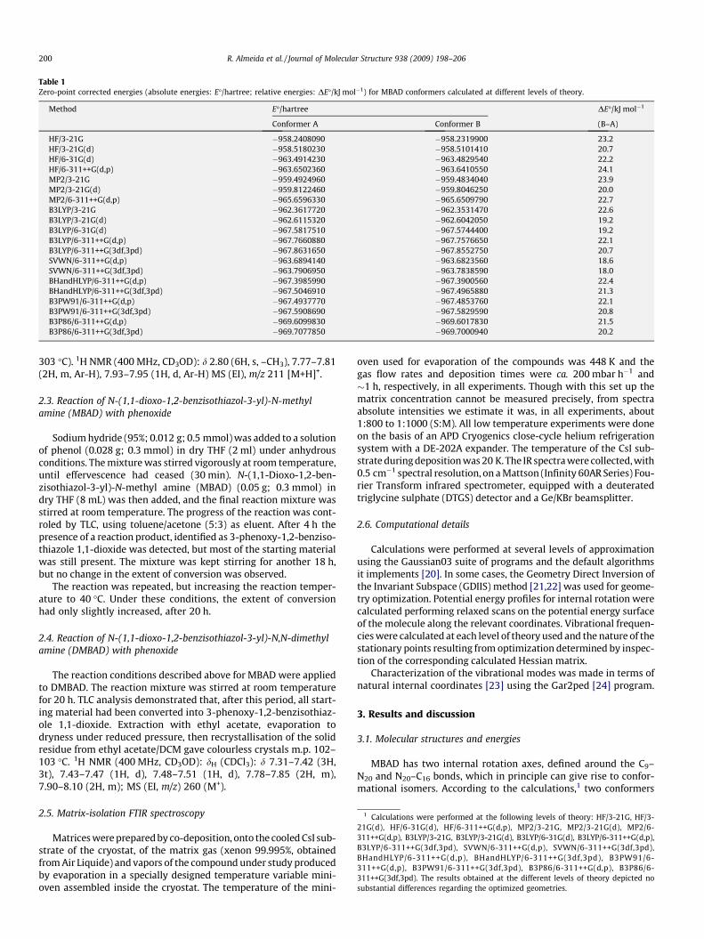
Table 1Zero-point corrected energies (absolute energies: E�/hartree; relative energies: DE�/kJ mol�1) for MBAD conformers calculated at different levels of theory.
Method E�/hartree DE�/kJ mol�1
Conformer A Conformer B (B–A)
HF/3-21G �958.2408090 �958.2319900 23.2HF/3-21G(d) �958.5180230 �958.5101410 20.7HF/6-31G(d) �963.4914230 �963.4829540 22.2HF/6-311++G(d,p) �963.6502360 �963.6410550 24.1MP2/3-21G �959.4924960 �959.4834040 23.9MP2/3-21G(d) �959.8122460 �959.8046250 20.0MP2/6-311++G(d,p) �965.6596330 �965.6509790 22.7B3LYP/3-21G �962.3617720 �962.3531470 22.6B3LYP/3-21G(d) �962.6115320 �962.6042050 19.2B3LYP/6-31G(d) �967.5817510 �967.5744400 19.2B3LYP/6-311++G(d,p) �967.7660880 �967.7576650 22.1B3LYP/6-311++G(3df,3pd) �967.8631650 �967.8552750 20.7SVWN/6-311++G(d,p) �963.6894140 �963.6823560 18.6SVWN/6-311++G(3df,3pd) �963.7906950 �963.7838590 18.0BHandHLYP/6-311++G(d,p) �967.3985990 �967.3900560 22.4BHandHLYP/6-311++G(3df,3pd) �967.5046910 �967.4965880 21.3B3PW91/6-311++G(d,p) �967.4937770 �967.4853760 22.1B3PW91/6-311++G(3df,3pd) �967.5908690 �967.5829590 20.8B3P86/6-311++G(d,p) �969.6099830 �969.6017830 21.5B3P86/6-311++G(3df,3pd) �969.7077850 �969.7000940 20.2
1 Calculations were performed at the following levels of theory: HF/3-21G, HF/3-1G(d), HF/6-31G(d), HF/6-311++G(d,p), MP2/3-21G, MP2/3-21G(d), MP2/6-11++G(d,p), B3LYP/3-21G, B3LYP/3-21G(d), B3LYP/6-31G(d), B3LYP/6-311++G(d,p),3LYP/6-311++G(3df,3pd), SVWN/6-311++G(d,p), SVWN/6-311++G(3df,3pd),HandHLYP/6-311++G(d,p), BHandHLYP/6-311++G(3df,3pd), B3PW91/6-11++G(d,p), B3PW91/6-311++G(3df,3pd), B3P86/6-311++G(d,p), B3P86/6-11++G(3df,3pd). The results obtained at the different levels of theory depicted nobstantial differences regarding the optimized geometries.
200 R. Almeida et al. / Journal of Molecular Structure 938 (2009) 198–206
303 �C). 1H NMR (400 MHz, CD3OD): d 2.80 (6H, s, –CH3), 7.77–7.81(2H, m, Ar-H), 7.93–7.95 (1H, d, Ar-H) MS (EI), m/z 211 [M+H]+.
2.3. Reaction of N-(1,1-dioxo-1,2-benzisothiazol-3-yl)-N-methylamine (MBAD) with phenoxide
Sodium hydride (95%; 0.012 g; 0.5 mmol) was added to a solutionof phenol (0.028 g; 0.3 mmol) in dry THF (2 ml) under anhydrousconditions. The mixture was stirred vigorously at room temperature,until effervescence had ceased (30 min). N-(1,1-Dioxo-1,2-ben-zisothiazol-3-yl)-N-methyl amine (MBAD) (0.05 g; 0.3 mmol) indry THF (8 mL) was then added, and the final reaction mixture wasstirred at room temperature. The progress of the reaction was cont-roled by TLC, using toluene/acetone (5:3) as eluent. After 4 h thepresence of a reaction product, identified as 3-phenoxy-1,2-benziso-thiazole 1,1-dioxide was detected, but most of the starting materialwas still present. The mixture was kept stirring for another 18 h,but no change in the extent of conversion was observed.
The reaction was repeated, but increasing the reaction temper-ature to 40 �C. Under these conditions, the extent of conversionhad only slightly increased, after 20 h.
2.4. Reaction of N-(1,1-dioxo-1,2-benzisothiazol-3-yl)-N,N-dimethylamine (DMBAD) with phenoxide
The reaction conditions described above for MBAD were appliedto DMBAD. The reaction mixture was stirred at room temperaturefor 20 h. TLC analysis demonstrated that, after this period, all start-ing material had been converted into 3-phenoxy-1,2-benzisothiaz-ole 1,1-dioxide. Extraction with ethyl acetate, evaporation todryness under reduced pressure, then recrystallisation of the solidresidue from ethyl acetate/DCM gave colourless crystals m.p. 102–103 �C. 1H NMR (400 MHz, CD3OD): dH (CDCl3): d 7.31–7.42 (3H,3t), 7.43–7.47 (1H, d), 7.48–7.51 (1H, d), 7.78–7.85 (2H, m),7.90–8.10 (2H, m); MS (EI, m/z) 260 (M+).
2.5. Matrix-isolation FTIR spectroscopy
Matrices were prepared by co-deposition, onto the cooled CsI sub-strate of the cryostat, of the matrix gas (xenon 99.995%, obtainedfrom Air Liquide) and vapors of the compound under study producedby evaporation in a specially designed temperature variable mini-oven assembled inside the cryostat. The temperature of the mini-
oven used for evaporation of the compounds was 448 K and thegas flow rates and deposition times were ca. 200 mbar h�1 and�1 h, respectively, in all experiments. Though with this set up thematrix concentration cannot be measured precisely, from spectraabsolute intensities we estimate it was, in all experiments, about1:800 to 1:1000 (S:M). All low temperature experiments were doneon the basis of an APD Cryogenics close-cycle helium refrigerationsystem with a DE-202A expander. The temperature of the CsI sub-strate during deposition was 20 K. The IR spectra were collected, with0.5 cm�1 spectral resolution, on a Mattson (Infinity 60AR Series) Fou-rier Transform infrared spectrometer, equipped with a deuteratedtriglycine sulphate (DTGS) detector and a Ge/KBr beamsplitter.
2.6. Computational details
Calculations were performed at several levels of approximationusing the Gaussian03 suite of programs and the default algorithmsit implements [20]. In some cases, the Geometry Direct Inversion ofthe Invariant Subspace (GDIIS) method [21,22] was used for geome-try optimization. Potential energy profiles for internal rotation werecalculated performing relaxed scans on the potential energy surfaceof the molecule along the relevant coordinates. Vibrational frequen-cies were calculated at each level of theory used and the nature of thestationary points resulting from optimization determined by inspec-tion of the corresponding calculated Hessian matrix.
Characterization of the vibrational modes was made in terms ofnatural internal coordinates [23] using the Gar2ped [24] program.
3. Results and discussion
3.1. Molecular structures and energies
MBAD has two internal rotation axes, defined around the C9–N20 and N20–C16 bonds, which in principle can give rise to confor-mational isomers. According to the calculations,1 two conformers
23BB33su

0
20
40
60
80
100 MBAD
ΔE (
kJ m
ol-1
)
N(8)=C(9)-N(20)-H(21) (degrees)
-180 -120 -60 0 60 120 180
0 60 120 180 240 300 360
0
10
20
30
40
50
60
70
Δ E (
kJ m
ol-1
)
N(8)=C(9)-N(20)-Lp (degrees)
DMBAD
A
B
Fig. 2. (A) Potential energy profile for internal rotation around the N(8)@C(9)–N(20)–H(21) dihedral angle for MBAD. Calculations were performed at the B3P86/6-311++G(3df,3pd) (black squares) and B3LYP/6-311++G(3df,3pd) (white circles)levels of theory. (B) Potential energy profile for internal rotation around theN(8)@C(9)–N(20)-Lp dihedral angle (Lp = lone pair, defined as localized in a planebisecting the N(8)@C(9)–N(20)–C(16) and N(8)@C(9)–N(20)–C(21) planes) forDMBAD, calculated at the B3P86/6-311++G(3df,3pd) level of theory.
R. Almeida et al. / Journal of Molecular Structure 938 (2009) 198–206 201
were found on the potential energy surface of the molecule, bothconformers belonging to the Cs symmetry point group and exhibitinga planar amine group, with one of the methyl hydrogens eclipsingthe amine hydrogen atom (Fig. 1). In the most stable conformer(A), the methyl group is oriented towards the nitrogen atom of theheterocycle (N8@C9–N20–C16: 0�), while in the less stable conformer(B) it is oriented towards the phenyl ring (N8@C9–N20–C16: 180�).The calculated structural parameters for conformer A, obtained atthe DFT(B3P86)/6-311++G(3df,3pd) level of theory are provided inTable S1 (Supplementary material).
The calculations predicted conformer A to be ca. 20 kJ mol�1
more stable than conformer B (Table 1). The greater stability ofconformer A can be ascribed essentially to the more favorableinteractions of the methyl and NH groups of the amine substituentoccurring in this conformer with the N(8) and H(10) atoms, respec-tively (see Fig. 1). Note that in conformer B the planarity of theamine moiety can still be retained in spite of the repulsive interac-tion between the methyl group and H(10), because the C(2)–C(9)–N(20) and C(9)–N(20)–C(16) angles can increase considerably atexpenses of approaching the amine hydrogen to N(8). As describedbelow, the situation is completely different for DMBAD, where theamine hydrogen is replaced by a more voluminous methyl group.
The potential energy profile for interconversion between thetwo MBAD conformers is presented in Fig. 2A. The energy barrierfor this process is very large (ca. 90 kJ mol�1 above the conforma-tional ground state), in agreement with the expected considerablepartial double bond character of the C(9)–N(20) bond, which is alsoreflected in the planarity assumed by the amine group in both min-ima. On the other hand, in the transition state the amine group isconsiderably pyramidalized (C–N(–H)–C dihedral: 148.3�), indicat-ing a strong reduction of delocalization and of the double bondcharacter of the C(9)–N(20) bond, with the consequent increaseof the energy of the molecule.
The presence in the DMBAD molecule of the additional methylgroup leads to a completely different picture. In this case, the con-formation equivalent to the minimum energy structures in MBADis destabilized due to the repulsive interaction between one of themethyl groups and H(10), which cannot be minimized by openingof the C(2)–C(9)–N(20) and C(9)–N(20)–C(21) angles as in con-former B of MBAD because in DMBAD this would lead to unfavor-able interactions between the second methyl group and N(8) (seeFig. 1). Hence, the amine group is forced to become pyramidal tominimize CH3. . .H(10) repulsions. Indeed, the potential energysurface of DMBAD exhibits two symmetry equivalent minima,corresponding to structures where the dimethylamine group isnon-planar (C–N(–C)–C dihedral: 167.3�), with one of the methylgroups nearly in the syn periplanar position relatively to N(8) andthe second methyl group out of the plane of the rings [C(21)–N(20)–C(9)–C(2) dihedral: 20.2�]. The calculated structuralparameters for the sole doubly-degenerated-by-symmetry con-former of DMBAD, obtained at the DFT(B3P86)/6–311++G(3df,3pd) level of theory, are provided in Table S2 (Supple-mentary material).
The potential energy profile for internal rotation around theC(9)–N(20) bond of DMBAD is given in Fig. 2B. All structures ob-tained after optimization but the transition states were of C1 sym-metry, with a pyramidalized amino group. The transition states areof Cs symmetry and correspond to the conformations where thelone electron pair of N(20) (defined as localized in a plane bisectingthe N(8)@C(9)–N(20)–C(16) and N(8)@C(9)–N(20)–C(21) planes)stays in the molecular plane. When N(8)@C(9)–N(20)-Lp is 180�,the energy barrier for internal rotation is of ca. 50 kJ mol�1,whereas when N(8)@C(9)–N(20)-Lp is 0� the barrier is consider-ably higher (ca. 70 kJ mol�1), since for this geometry the pairs ofelectrons of N(20) and N(8) are face-to-face and the two methylgroups in close contact to H(10).
It is important to note once again that the main structuralimplications of the presence of the second methyl substituent atthe amino group (in DMBAD) in relation to the mono-substitutedcompound (MBAD) are triggered by CH3. . .H(10) repulsions, whichlead to amine pyramidalization and reduction of the double bondcharacter of the C(9)–N(20) bond. Accordingly, the C(9)–N(20)bond length in DMBAD is predicted by the calculations to be0.9 pm longer than in MBAD (134.3 vs. 133.4 pm; see Tables S1and S2). This effect extends into the N(8)–C(9) bond, which is pre-dicted to be 0.6 pm longer in DMBAD than in MBAD (130.2 vs.129.6 pm), and also into the C(16)–N(20) bond, with predictedbond lengths of 145.1 and 144.5 pm, for DMBAD and MBAD,respectively.
The values predicted for bond lengths C(9)–N(20) and C(16)–N(20) in both benzisothiazolyl derivatives correspond to bond or-ders greater than unity. The powerful electron-withdrawing effectof the saccharyl ring results in an increase in electronegativity onthe amino nitrogen N(20), which is strongly conjugated with thesaccharyl system. These theoretical results are in keeping with
020406080
100
020406080
100
020406080
100
020406080
100
020406080
100
020406080
100
400 600 800 1000 1200 1400 1600
020406080
Wavenumbers (cm-1)
Abs
orba
nce
Rel
ativ
e In
tens
ity (
km m
ol-1
)
BHandHLYP/6-311++G(3df,3pd)
B3LYP/6-311++G(3df,3pd)
B3LYP/6-311++G(d,p)
SVWN/6-311++G(3df,3pd)
Xenon matrix (20 K)
B3P86/6-311++G(3df,3pd)
B3PW91/6-311++G(3df,3pd)ν1
ν1
ν1
ν1
ν2
ν2 ν1
ν2 ν1
A
B
ν2
ν2
ν2
ν2 ν1
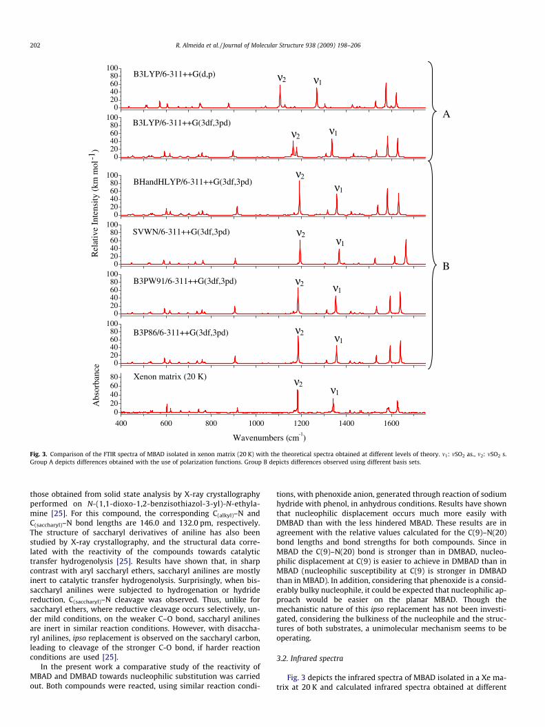
Fig. 3. Comparison of the FTIR spectra of MBAD isolated in xenon matrix (20 K) with the theoretical spectra obtained at different levels of theory. m1: mSO2 as., m2: mSO2 s.Group A depicts differences obtained with the use of polarization functions. Group B depicts differences observed using different basis sets.
202 R. Almeida et al. / Journal of Molecular Structure 938 (2009) 198–206
those obtained from solid state analysis by X-ray crystallographyperformed on N-(1,1-dioxo-1,2-benzisothiazol-3-yl)-N-ethyla-mine [25]. For this compound, the corresponding C(alkyl)–N andC(saccharyl)–N bond lengths are 146.0 and 132.0 pm, respectively.The structure of saccharyl derivatives of aniline has also beenstudied by X-ray crystallography, and the structural data corre-lated with the reactivity of the compounds towards catalytictransfer hydrogenolysis [25]. Results have shown that, in sharpcontrast with aryl saccharyl ethers, saccharyl anilines are mostlyinert to catalytic transfer hydrogenolysis. Surprisingly, when bis-saccharyl anilines were subjected to hydrogenation or hydridereduction, C(saccharyl)–N cleavage was observed. Thus, unlike forsaccharyl ethers, where reductive cleavage occurs selectively, un-der mild conditions, on the weaker C–O bond, saccharyl anilinesare inert in similar reaction conditions. However, with disaccha-ryl anilines, ipso replacement is observed on the saccharyl carbon,leading to cleavage of the stronger C-O bond, if harder reactionconditions are used [25].
In the present work a comparative study of the reactivity ofMBAD and DMBAD towards nucleophilic substitution was carriedout. Both compounds were reacted, using similar reaction condi-
tions, with phenoxide anion, generated through reaction of sodiumhydride with phenol, in anhydrous conditions. Results have shownthat nucleophilic displacement occurs much more easily withDMBAD than with the less hindered MBAD. These results are inagreement with the relative values calculated for the C(9)–N(20)bond lengths and bond strengths for both compounds. Since inMBAD the C(9)–N(20) bond is stronger than in DMBAD, nucleo-philic displacement at C(9) is easier to achieve in DMBAD than inMBAD (nucleophilic susceptibility at C(9) is stronger in DMBADthan in MBAD). In addition, considering that phenoxide is a consid-erably bulky nucleophile, it could be expected that nucleophilic ap-proach would be easier on the planar MBAD. Though themechanistic nature of this ipso replacement has not been investi-gated, considering the bulkiness of the nucleophile and the struc-tures of both substrates, a unimolecular mechanism seems to beoperating.
3.2. Infrared spectra
Fig. 3 depicts the infrared spectra of MBAD isolated in a Xe ma-trix at 20 K and calculated infrared spectra obtained at different

Table 2Observed wavenumbers (and qualitative intensities) for MBAD in Xe matrix and calculated infrared spectra, obtained at the B3P86/6-311++G(3df,3pd) level of approximation, forthe experimentally relevant conformer of the molecule (conformer A).
Approximate descriptiona Calculatedb B3P86/6-311++G(3df,3pd) Xenon matrixc (20 K)
m/cm�1 I/km mol�1 m/cm�1 I (qualitative)
dSO2 525 17.8 531 mcSO2, s6R 531 20.0 539 md5R, dC–C–C6R, 593 66.4 603 SdC–C–C6R , dSO2 615 38.8 620/623 S/ms6R, cNY, dN–C–N 657 15.9 664/666 m/mdC–C–C6R, mC–S 697 12.8 703 ms6R 737 29.5 744 mdC–C–C6R 759 50.3 771 wdN–C–N, s6R 771 17.1 775 ScC–H6R 867 0.0 909 vwmN-S, dN–C–N 906 87.1 917 ScC–H6R 947 0.4 943 vwcC–H6R 986 0.0 n.obs.mN–CM, dC–C–C6R 1010 1.8 n.obs.mC–C6R 1027 2.5 1025 wdC–C–C6R, mN–CM, mC–S 1054 5.1 1064 wcC–H6R 1122 0.2 n.obs.dC–C–C6R ,mC–C6R 1129 2.6 1137 vwcCH3 1153 1.9 1154 vwcCH3 1164 20.0 1168 mdC–C–C6R, mC–C6R 1173 6.2 1173 mmSO2 s 1186 328.9 1176/1184 m/VSdC–H6R 1266 5.6 1271 wdNH, mC–C5R 1312 42.7 1307 w/mdC–H6R 1349 3.8 1337 vwmSO2 as 1356 213.8 1341/1343 S/VSdCH3 s 1417 29.4 1416 mdCH3 as 1435 11.9 1437 mdC–H6R, mC@C6R 1450 0.8 n.obs. wdC–H6R, mC@C6R 1460 17.2 1461 wdCH3 as 1471 7.0 1467 vw/wmC–N, dN–H 1533 92.7 1527 wmC@C6R, mC@N5R 1594 214.9 1584/1588 SmC@C6R 1611 2.8 1604/1611 vw/vwmC@N5R 1640 269.8 1627/1631 VS/S
a m, bond stretching; d, bending; c, rocking; s, torsion; s, symmetric; as, anti-symmetric; subscripts 6R, 5R and M designate the six- and the five-membered rings and themethyl group, respectively.
b Wavenumbers were scaled by 0.9763.c S, strong; VS, very strong; m, medium; w, weak; vw, very weak; sh, shoulder; n. obs., non observed; n.a., non-analyzed.
R. Almeida et al. / Journal of Molecular Structure 938 (2009) 198–206 203
levels of approximation for the experimentally relevant conformer(form A). As expected, the overall agreement between theoreticaland observed spectra is generally improved by increasing the num-ber of polarization functions [e.g., from B3LYP/6–311++G(d,p) toB3LYP/6–311++G(3df,3pd); panel A in the figure], this being partic-ularly evident for vibrational modes of the SO2 group, such as themSO2 anti-symmetric and symmetric stretching modes (respec-tively, m1 and m2 in Fig. 3), but also for C@C stretching modes,around 1600 cm�1. Indeed, as mentioned in Section 1, the B3LYPfunctional used with the 6-311++G(3df,3pd) basis set could in factbe expected to yield a good reproduction of the experimental spec-trum. Nevertheless, in spite of the good performance of the B3LYPfunctional, the B3P86/6-311++G(3df,3pd) and B3PW91/6-311++G(3df,3pd) calculations were found to be those giving thebest overall agreement with the experiment (panel B in Fig. 3), thusappearing as good alternatives to B3LYP/6-311++G(3df,3pd) forcalculation of vibrational data on saccharin derivatives (and othermolecules containing the SO2 group). For MBAD, actually bothB3P86 and B3PW91 give indeed very similar results, the maximalpercentage error for the wavenumbers of the bands in the finger-print region (these taken into account in Table 2) being 0.18%and 0.23%, respectively. The average percentage error for thesewavenumbers is 0.02% and 0.01%, respectively. On the other hand,B3LYP with the same basis set (all the time we talk about 6-311++G(3df,3pd)) gives considerably less good results, the maxi-mal percentage error being 1.49% and the average percentage error
0.16%, i.e., one order of magnitude bigger than those obtained withboth the B3P86 and B3PW91 functionals.
Taking into account the excellent reproduction of the experi-mental spectra by these calculations, the assignment of the ob-served bands for the matrix isolated molecule of MBAD wasconsiderably simplified. The proposed assignments are providedin Table 2. Note that doublets of bands were assigned to most ofthe vibrations predicted to give rise to intense infrared bands sites(e.g., mSO2 as.: 1176/1184 cm�1; mSO2 as.: 1341/1343 cm�1). Thisobservation points to the existence of two main matrix sites forMBAD in xenon matrix. Annealing experiments revealed no spec-tral changes which could be ascribed to other phenomena thanaggregation, which started to be important above ca. 40 K.
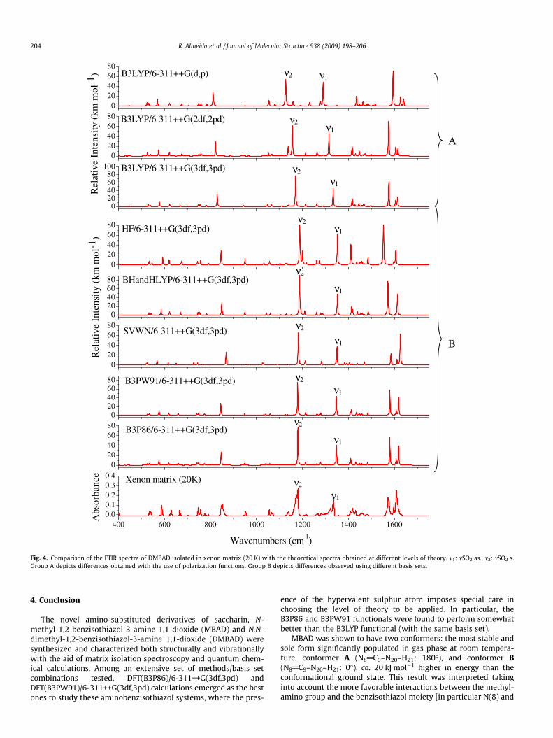
The infrared spectrum of DMBAD isolated in xenon matrixis shown in Fig. 4, together with calculated spectra for thiscompound. Like for MBAD, the correspondence between theB3P86/6-311++G(3df,3pd) theoretically predicted spectrum andthe experiment is very good and strongly supports the theoreticalpredictions regarding the structure of the stable conformation ofthis molecule. In particular, in consonance with the structural re-sults, the frequency of the stronger C(9)–N(20) bond in MBAD(1527 cm�1) is considerably larger than that observed for DMBAD(1432 cm�1). The complete assignments for DMBAD are given inTable 3. As for MBAD, matrix site splitting was also observed incase of DMBAD, with at least three main matrix sites appearingto be significantly populated.
020406080
020406080
020406080
020406080
020406080
400 600 800 1000 1200 1400 16000.00.10.20.30.4
020406080
020406080
020406080
100
SVWN/6-311++G(3df,3pd)
BHandHLYP/6-311++G(3df,3pd)
HF/6-311++G(3df,3pd)
B3PW91/6-311++G(3df,3pd)
Rel
ativ
e In
tens
ity
(km
mol
-1)
B3P86/6-311++G(3df,3pd)
Rel
ativ
e In
tens
ity
(km
mol
-1)
Xenon matrix (20K)
Abs
orba
nce
Wavenumbers (cm-1)
B3LYP/6-311++G(3df,3pd)
B3LYP/6-311++G(2df,2pd)
B3LYP/6-311++G(d,p)
ν2
ν1
ν2
ν1
ν2
ν1
ν2 ν1
ν2
ν1
ν2 ν1
A
B
ν2
ν1
ν2
ν1
ν2 ν1
Fig. 4. Comparison of the FTIR spectra of DMBAD isolated in xenon matrix (20 K) with the theoretical spectra obtained at different levels of theory. m1: mSO2 as., m2: mSO2 s.Group A depicts differences obtained with the use of polarization functions. Group B depicts differences observed using different basis sets.
204 R. Almeida et al. / Journal of Molecular Structure 938 (2009) 198–206
4. Conclusion
The novel amino-substituted derivatives of saccharin, N-methyl-1,2-benzisothiazol-3-amine 1,1-dioxide (MBAD) and N,N-dimethyl-1,2-benzisothiazol-3-amine 1,1-dioxide (DMBAD) weresynthesized and characterized both structurally and vibrationallywith the aid of matrix isolation spectroscopy and quantum chem-ical calculations. Among an extensive set of methods/basis setcombinations tested, DFT(B3P86)/6-311++G(3df,3pd) andDFT(B3PW91)/6-311++G(3df,3pd) calculations emerged as the bestones to study these aminobenzisothiazol systems, where the pres-
ence of the hypervalent sulphur atom imposes special care inchoosing the level of theory to be applied. In particular, theB3P86 and B3PW91 functionals were found to perform somewhatbetter than the B3LYP functional (with the same basis set).
MBAD was shown to have two conformers: the most stable andsole form significantly populated in gas phase at room tempera-ture, conformer A (N8@C9–N20–H21: 180�), and conformer B(N8@C9–N20–H21: 0�), ca. 20 kJ mol�1 higher in energy than theconformational ground state. This result was interpreted takinginto account the more favorable interactions between the methyl-amino group and the benzisothiazol moiety [in particular N(8) and
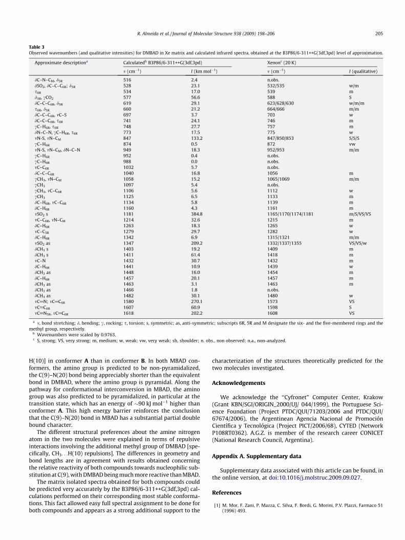
Table 3Observed wavenumbers (and qualitative intensities) for DMBAD in Xe matrix and calculated infrared spectra, obtained at the B3P86/6-311++G(3df,3pd) level of approximation.
Approximate descriptiona Calculatedb B3P86/6-311++G(3df,3pd) Xenonc (20 K)
m (cm�1) I (km mol�1) m (cm�1) I (qualitative)
dC–N–CM, d5R 516 2.4 n.obs.dSO2, dC–C–C6R; d5R 528 23.1 532/535 w/ms6R 534 17.0 539 md5R, cCO2 577 56.6 588 SdC–C–C6R, d5R 619 29.1 623/628/630 w/m/ms6R, d5R 660 21.2 664/666 m/mdC–C–C6R, mC–S 697 3.7 703 wdC–C–C6R, s6R 741 24.1 746 mcC–H6R, s6R 748 27.7 757 mdN–C–N, cC–H6R, s6R 773 17.5 775 wmN-S, mN–CM 847 133.2 847/850/853 S/S/ScC–H6R 874 0.5 872 vwmN-S, mN–CM, dN–C–N 949 18.3 952/953 m/mcC–H6R 952 0.4 n.obs.cC–H6R 988 0.0 n.obs.mC–C6R 1032 5.7 n.obs.dC–C–C6R 1040 16.8 1056 mcCH3, mN–CM 1058 15.2 1065/1069 m/mcCH3 1097 5.4 n.obs.cCH3, mC–C6R 1106 5.6 1112 wcCH3 1125 6.5 1133 mdC–H6R, mC–C6R 1134 5.8 1139 mdC–H6R 1160 4.3 1161 mmSO2 s 1181 384.8 1165/1170/1174/1181 m/S/VS/VSmC–C6R, mN–CM 1214 32.6 1215 mdC–H6R 1263 18.3 1265 wmC–C5R 1279 29.7 1282 wdC–H6R 1342 6.9 1315/1321 m/mmSO2 as 1347 209.2 1332/1337/1355 VS/VS/wdCH3 s 1403 19.2 1409 mdCH3 s 1411 61.4 1418 mmC–N 1432 30.7 1432 mdC–H6R 1441 10.9 1439 wdCH3 as 1448 16.0 1454 mdC–H6R 1457 20.1 1457 mdCH3 as 1463 3.1 1463 mdCH3 as 1466 1.8 n.obs.dCH3 as 1482 30.1 1480 wmC@N; mC@C6R 1580 270.1 1573 VSmC@C6R 1607 60.9 1598 SmC@N5R, mC@C6R 1618 202.2 1608 VS
a m, bond stretching; d, bending; c, rocking; s, torsion; s, symmetric; as, anti-symmetric; subscripts 6R, 5R and M designate the six- and the five-membered rings and themethyl group, respectively.
b Wavenumbers were scaled by 0.9763.c S, strong; VS, very strong; m, medium; w, weak; vw, very weak; sh, shoulder; n. obs., non observed; n.a., non-analyzed.
R. Almeida et al. / Journal of Molecular Structure 938 (2009) 198–206 205
H(10)] in conformer A than in conformer B. In both MBAD con-formers, the amino group is predicted to be non-pyramidalized,the C(9)–N(20) bond being appreciably shorter than the equivalentbond in DMBAD, where the amino group is pyramidal. Along thepathway for conformational interconversion in MBAD, the aminogroup was also predicted to be pyramidalized, in particular at thetransition state, which has an energy of �90 kJ mol�1 higher thanconformer A. This high energy barrier reinforces the conclusionthat the C(9)–N(20) bond in MBAD has a substantial partial doublebound character.
The different structural preferences about the amine nitrogenatom in the two molecules were explained in terms of repulsiveinteractions involving the additional methyl group of DMBAD [spe-cifically, CH3. . .H(10) repulsions]. The differences in geometry andbond lengths are in agreement with results obtained concerningthe relative reactivity of both compounds towards nucleophilic sub-stitution at C(9), with DMBAD being much more reactive than MBAD.
The matrix isolated spectra obtained for both compounds couldbe predicted very accurately by the B3P86/6-311++G(3df,3pd) cal-culations performed on their corresponding most stable conforma-tions. This fact allowed easy full spectral assignment to be done forboth compounds and appears as a strong additional support to the
characterization of the structures theoretically predicted for thetwo molecules investigated.
Acknowledgements
We acknowledge the ‘‘Cyfronet” Computer Center, Krakow(Grant KBN/SGI/ORIGIN_2000/UJ/ 044/1999), the Portuguese Sci-ence Foundation (Project PTDC/QUI/71203/2006 and PTDC/QUI/67674/2006), the Argentinean Agencia Nacional de PromociónCientífica y Tecnológica (Project PICT/2006/68), CYTED (NetworkP108RT0362). A.G.Z. is member of the research career CONICET(National Research Council, Argentina).
Appendix A. Supplementary data
Supplementary data associated with this article can be found, inthe online version, at doi:10.1016/j.molstruc.2009.09.027.
References
[1] M. Mor, F. Zani, P. Mazza, C. Silva, F. Bordi, G. Morini, P.V. Plazzi, Farmaco 51(1996) 493.

206 R. Almeida et al. / Journal of Molecular Structure 938 (2009) 198–206
[2] A.F. Brigas, R.A.W. Johnstone, Tetrahedron Lett. 31 (1990) 5789.[3] N.C.P. Araújo, A.F. Brigas, M.L.S. Cristiano, L.M.T. Frija, E.M.O. Guimarães, R.M.S.
Loureiro, J. Mol. Catal. A: Chem. 215 (2004) 113.[4] L.M.T. Frija, M.L.S. Cristiano, E.M.O. Guimarães, N.C. Martins, R.M.S. Loureiro,
J.F. Bickley, J. Mol. Catal. A: Chem. 242 (2005) 241 (and references therein).[5] A.F. Brigas, R.A.W. Johnstone, J. Chem. Soc. Chem. Commun. (1994) 1923.[6] J.V. Barkley, M.L.S. Cristiano, R.A.W. Johnstone, R.M.S. Loureiro, Acta
Crystallogr. C: Cryst. Struct. Commun. 53 (1997) 383.[7] M.L.S. Cristiano, A.F. Brigas, R.A.W. Johnstone, R.M.S. Loureiro, P.C.A. Pena, J.
Chem. Res. (S) (1999) 704.[8] N.C.P. Araújo, P.M.M. Barroca, J.F. Bickley, A.F. Brigas, M.L.S. Cristiano, R.A.W.
Johnstone, R.M.S. Loureiro, P.C.A. Pena, J. Chem. Soc. Perkin Trans. 1 (2002) 1213.[9] R. Almeida, A. Gómez-Zavaglia, A. Kaczor, M.L.S. Cristiano, M.E.S. Eusébio,
T.M.R. Maria, R. Fausto, Tetrahedron 64 (2008) 3296.[10] A. Gómez-Zavaglia, A. Kaczor, R. Almeida, M.L.S. Cristiano, M.E.S. Eusébio,
T.M.R. Maria, P. Mobili, R. Fausto, J. Phys. Chem. A 113 (2009) 3517.[11] A. Gómez-Zavaglia, A. Kaczor, D. Coelho, M.L.S. Cristiano, R. Fausto, J. Mol.
Struct. 919 (2009) 271.[12] A. Gómez-Zavaglia, A. Kaczor, R. Almeida, M.L.S. Cristiano, R. Fausto, J. Phys.
Chem. A 112 (2008) 1762.[13] A. Kaczor, R. Almeida, A. Gómez-Zavaglia, M.L.S. Cristiano, R. Fausto, J. Mol.
Struct. 876 (2008) 77.[14] A. Kaczor, L.M. Proniewicz, R. Almeida, A. Gómez-Zavaglia, M.L.S. Cristiano,
A.M. Matos Beja, M. Ramos Silva, R. Fausto, J. Mol. Struct. 892 (2008) 343.[15] A.H. Zeng, L. Yu, Y. Wang, Q.Y. Kong, Q. Xu, M.F. Zhou, J. Phys. Chem. A 108
(2004) 6656.[16] A. Borba, A. Gómez-Zavaglia, P.N.N.L. Simões, R. Fausto, Spectrochim. Acta A 61
(2005) 1461.
[17] A. Borba, A. Gómez-Zavaglia, P.N.N.L. Simões, R. Fausto, J. Phys. Chem. A 109(2005) 3578.
[18] D.R. Lide (Ed.), Handbook of Chemistry and Physics, 74th ed., CRC Press, BocaRaton, 1993. pp. 9–123.
[19] S.W. McCombie, in: B. Trost, I. Fleming (Eds.), Comprehensive OrganicSynthesis, vol. 8, Pergamon Press, Oxford, pp. 826–828 (Chapter 4.2).
[20] M.J. Frisch, G.W. Trucks, H.B. Schlegel, G.E. Scuseria, M.A. Robb, J.R. Cheeseman,J.A. Montgomery Jr., T. Vreven, K.N. Kudin, J.C. Burant, J.M. Millam, S.S. Iyengar,J. Tomasi, V. Barone, B. Mennucci, M. Cossi, G. Scalmani, N. Rega, G.A.Petersson, H. Nakatsuji, M. Hada, M. Ehara, K. Toyota, R. Fukuda, J. Hasegawa,M. Ishida, T. Nakajima, Y. Honda, O. Kitao, H. Nakai, M. Klene, X. Li, J.E. Knox,H.P. Hratchian, J.B. Cross, V. Bakken, C. Adamo, J. Jaramillo, R. Gomperts, R.E.Stratmann, O. Yazyev, A.J. Austin, R. Cammi, C. Pomelli, J.W. Ochterski, P.Y.Ayala, K. Morokuma, G.A. Voth, P. Salvador, J.J. Dannenberg, V.G. Zakrzewski, S.Dapprich, A.D. Daniels, M.C. Strain, O. Farkas, D.K. Malick, A.D. Rabuck, K.Raghavachari, J.B. Foresman, J.V. Ortiz, Q. Cui, A.G. Baboul, S. Clifford, J.Cioslowski, B.B. Stefanov, G. Liu, A. Liashenko, P. Piskorz, I. Komaromi, R.L.Martin, D.J. Fox, T. Keith, M.A. Al-Laham, C.Y. Peng, A. Nanayakkara, M.Challacombe, P.M.W. Gill, B. Johnson, W. Chen, M.W. Wong, C. González, J.A.Pople, GAUSSIAN 03, Revision C.02, Gaussian, Inc., Wallingford, CT, 2004.
[21] P. Csaszar, P. Pulay, J. Mol. Struct. 114 (1984) 31.[22] O. Farkas, H.B. Schlegel, J. Chem. Phys. 111 (1999) 10806.[23] P. Pulay, G. Fogarasi, F. Pang, J.E. Boggs, J. Am. Chem. Soc. 101 (1979)
2550.[24] J.M.L. Martin, C. Van Alsenoy, GAR2PED, University of Antwerp, 1995.[25] A.F. Brigas, W. Clegg, C.J. Dillon, C.F.C. Fonseca, R.A.W. Johnstone, J. Chem. Soc.
Perkin 2 (2001) 1315.
Related Documents


