Maternal Malaria Induces a Procoagulant and Antifibrinolytic State That Is Embryotoxic but Responsive to Anticoagulant Therapy John W. Avery 1¤a , Geoffrey M. Smith 1 , Simon O. Owino 1,2 , Demba Sarr 1 , Tamas Nagy 3 , Stephen Mwalimu 1,2 , James Matthias 4¤b , Lauren F. Kelly 1¤c , Jayakumar S. Poovassery 1¤d , Joab D. Middii 1,2 , Carlos Abramowsky 5 , Julie M. Moore 1 * 1 Department of Infectious Diseases and Center for Tropical and Emerging Global Diseases, University of Georgia, Athens, Georgia, United States of America, 2 Centre for Global Health Research, Kenya Medical Research Institute, Kisumu, Kenya, 3 Department of Veterinary Pathology, University of Georgia, Athens, Georgia, United States of America, 4 Department of Epidemiology and Biostatistics, University of Georgia, Athens, Georgia, United States of America, 5 Department of Pathology and Laboratory Medicine, Emory University, Atlanta, Georgia, United States of America Abstract Low birth weight and fetal loss are commonly attributed to malaria in endemic areas, but the cellular and molecular mechanisms that underlie these poor birth outcomes are incompletely understood. Increasing evidence suggests that dysregulated hemostasis is important in malaria pathogenesis, but its role in placental malaria (PM), characterized by intervillous sequestration of Plasmodium falciparum, proinflammatory responses, and excessive fibrin deposition is not known. To address this question, markers of coagulation and fibrinolysis were assessed in placentae from malaria-exposed primigravid women. PM was associated with significantly elevated placental monocyte and proinflammatory marker levels, enhanced perivillous fibrin deposition, and increased markers of activated coagulation and suppressed fibrinolysis in placental plasma. Submicroscopic PM was not proinflammatory but tended to be procoagulant and antifibrinolytic. Birth weight trended downward in association with placental parasitemia and high fibrin score. To directly assess the importance of coagulation in malaria-induced compromise of pregnancy, Plasmodium chabaudi AS-infected pregnant C57BL/6 mice were treated with the anticoagulant, low molecular weight heparin. Treatment rescued pregnancy at midgestation, with substantially decreased rates of active abortion and reduced placental and embryonic hemorrhage and necrosis relative to untreated animals. Together, the results suggest that dysregulated hemostasis may represent a novel therapeutic target in malaria-compromised pregnancies. Citation: Avery JW, Smith GM, Owino SO, Sarr D, Nagy T, et al. (2012) Maternal Malaria Induces a Procoagulant and Antifibrinolytic State That Is Embryotoxic but Responsive to Anticoagulant Therapy. PLoS ONE 7(2): e31090. doi:10.1371/journal.pone.0031090 Editor: Lars Hviid, University of Copenhagen, Denmark Received August 25, 2011; Accepted January 2, 2012; Published February 7, 2012 Copyright: ß 2012 Avery et al. This is an open-access article distributed under the terms of the Creative Commons Attribution License, which permits unrestricted use, distribution, and reproduction in any medium, provided the original author and source are credited. Funding: This work was supported by the National Institutes of Health grants R01 AI050240 and R01 HD046860 to JMM. The content is solely the responsibility of the authors and does not necessarily represent the official views of the National Institute of Allergy and Infectious Diseases (NIAID), the National Institute of Child Health and Human Development (NICHD) or the National Institutes of Health. The funders had no role in study design, data collection and analysis, decision to publish, or preparation of the manuscript. Competing Interests: The authors have declared that no competing interests exist. * E-mail: [email protected] ¤a Current address: Department of Biochemistry and Molecular Biology, University of Georgia, Athens, Georgia, United States of America ¤b Current address: Florida Department of Health, Tallahassee, Florida, United States of America ¤c Current address: Boston University School of Medicine, Boston, Massachusetts, United States of America ¤d Current address: Department of Microbiology, Carver College of Medicine, University of Iowa, Ames, Iowa, United States of America Introduction Recent estimates propose that nearly 55 million pregnant women are at high risk for Plasmodium falciparum infection annually [1]. Aside from significant maternal morbidity, a critical clinical feature of this infection is low infant birth weight (LBW; ,2500 g) secondary to intrauterine growth restriction and/or premature birth [2]. Each year, in Sub-Saharan Africa as many as 363,000 neonates die from malaria-associated LBW [2]. A large proportion of these cases are attributed to malaria-induced maternal anemia and placental, inflammatory pathology and resultant functional insufficiency [2–6]. In addition, among pregnant women living in low transmission conditions, who have little pre-existing immunity to malaria, this infection can result in abortion and stillbirth [2]. The major pathological features of malaria during pregnancy that are associated with poor birth outcomes are accumulation of infected red blood cells (iRBCs) in the maternal blood space of the placenta and the subsequent inappropriate maternal inflammatory response to these parasites, a syndrome referred to as placental malaria (PM). Although PM and its consequences for mother and fetus have been well studied, the precise mechanisms of pathology continue to elude investigators. Malarial pathogenesis is common- ly attributed to infiltration of immune effector cells and excessive proinflammatory cytokine release in response to sequestered parasites [4], but this proinflammatory immunopathology may not fully account for PM pathogenesis. A universally described histopathological feature of malarious placentae is excessive deposition of fibrin, the end-product of the coagulation cascade PLoS ONE | www.plosone.org 1 February 2012 | Volume 7 | Issue 2 | e31090

Welcome message from author
This document is posted to help you gain knowledge. Please leave a comment to let me know what you think about it! Share it to your friends and learn new things together.
Transcript

Maternal Malaria Induces a Procoagulant andAntifibrinolytic State That Is Embryotoxic but Responsiveto Anticoagulant TherapyJohn W. Avery1¤a, Geoffrey M. Smith1, Simon O. Owino1,2, Demba Sarr1, Tamas Nagy3, Stephen
Mwalimu1,2, James Matthias4¤b, Lauren F. Kelly1¤c, Jayakumar S. Poovassery1¤d, Joab D. Middii1,2, Carlos
Abramowsky5, Julie M. Moore1*
1 Department of Infectious Diseases and Center for Tropical and Emerging Global Diseases, University of Georgia, Athens, Georgia, United States of America, 2 Centre for
Global Health Research, Kenya Medical Research Institute, Kisumu, Kenya, 3 Department of Veterinary Pathology, University of Georgia, Athens, Georgia, United States of
America, 4 Department of Epidemiology and Biostatistics, University of Georgia, Athens, Georgia, United States of America, 5 Department of Pathology and Laboratory
Medicine, Emory University, Atlanta, Georgia, United States of America
Abstract
Low birth weight and fetal loss are commonly attributed to malaria in endemic areas, but the cellular and molecularmechanisms that underlie these poor birth outcomes are incompletely understood. Increasing evidence suggests thatdysregulated hemostasis is important in malaria pathogenesis, but its role in placental malaria (PM), characterized byintervillous sequestration of Plasmodium falciparum, proinflammatory responses, and excessive fibrin deposition is notknown. To address this question, markers of coagulation and fibrinolysis were assessed in placentae from malaria-exposedprimigravid women. PM was associated with significantly elevated placental monocyte and proinflammatory marker levels,enhanced perivillous fibrin deposition, and increased markers of activated coagulation and suppressed fibrinolysis inplacental plasma. Submicroscopic PM was not proinflammatory but tended to be procoagulant and antifibrinolytic. Birthweight trended downward in association with placental parasitemia and high fibrin score. To directly assess the importanceof coagulation in malaria-induced compromise of pregnancy, Plasmodium chabaudi AS-infected pregnant C57BL/6 micewere treated with the anticoagulant, low molecular weight heparin. Treatment rescued pregnancy at midgestation, withsubstantially decreased rates of active abortion and reduced placental and embryonic hemorrhage and necrosis relative tountreated animals. Together, the results suggest that dysregulated hemostasis may represent a novel therapeutic target inmalaria-compromised pregnancies.
Citation: Avery JW, Smith GM, Owino SO, Sarr D, Nagy T, et al. (2012) Maternal Malaria Induces a Procoagulant and Antifibrinolytic State That Is Embryotoxic butResponsive to Anticoagulant Therapy. PLoS ONE 7(2): e31090. doi:10.1371/journal.pone.0031090
Editor: Lars Hviid, University of Copenhagen, Denmark
Received August 25, 2011; Accepted January 2, 2012; Published February 7, 2012
Copyright: � 2012 Avery et al. This is an open-access article distributed under the terms of the Creative Commons Attribution License, which permitsunrestricted use, distribution, and reproduction in any medium, provided the original author and source are credited.
Funding: This work was supported by the National Institutes of Health grants R01 AI050240 and R01 HD046860 to JMM. The content is solely the responsibilityof the authors and does not necessarily represent the official views of the National Institute of Allergy and Infectious Diseases (NIAID), the National Institute ofChild Health and Human Development (NICHD) or the National Institutes of Health. The funders had no role in study design, data collection and analysis, decisionto publish, or preparation of the manuscript.
Competing Interests: The authors have declared that no competing interests exist.
* E-mail: [email protected]
¤a Current address: Department of Biochemistry and Molecular Biology, University of Georgia, Athens, Georgia, United States of America¤b Current address: Florida Department of Health, Tallahassee, Florida, United States of America¤c Current address: Boston University School of Medicine, Boston, Massachusetts, United States of America¤d Current address: Department of Microbiology, Carver College of Medicine, University of Iowa, Ames, Iowa, United States of America
Introduction
Recent estimates propose that nearly 55 million pregnant
women are at high risk for Plasmodium falciparum infection annually
[1]. Aside from significant maternal morbidity, a critical clinical
feature of this infection is low infant birth weight (LBW; ,2500 g)
secondary to intrauterine growth restriction and/or premature
birth [2]. Each year, in Sub-Saharan Africa as many as 363,000
neonates die from malaria-associated LBW [2]. A large proportion
of these cases are attributed to malaria-induced maternal anemia
and placental, inflammatory pathology and resultant functional
insufficiency [2–6]. In addition, among pregnant women living in
low transmission conditions, who have little pre-existing immunity
to malaria, this infection can result in abortion and stillbirth [2].
The major pathological features of malaria during pregnancy
that are associated with poor birth outcomes are accumulation of
infected red blood cells (iRBCs) in the maternal blood space of the
placenta and the subsequent inappropriate maternal inflammatory
response to these parasites, a syndrome referred to as placental
malaria (PM). Although PM and its consequences for mother and
fetus have been well studied, the precise mechanisms of pathology
continue to elude investigators. Malarial pathogenesis is common-
ly attributed to infiltration of immune effector cells and excessive
proinflammatory cytokine release in response to sequestered
parasites [4], but this proinflammatory immunopathology may
not fully account for PM pathogenesis. A universally described
histopathological feature of malarious placentae is excessive
deposition of fibrin, the end-product of the coagulation cascade
PLoS ONE | www.plosone.org 1 February 2012 | Volume 7 | Issue 2 | e31090
[5]. However, an independent role for fibrin in PM-induced
adverse birth outcomes has been directly examined in only two
studies. Menendez et al. found that malaria-infected placentae
with .30% of fetal villi engulfed in fibrin were significantly
associated with LBW due to preterm delivery [6]. Additionally,
Crocker et al. established an association between placental
parasitemia, LBW, and syncytiotrophoblast lesions associated with
fibrin-type fibrinoid deposition [3]. In general, abundant placental
fibrin deposition is a hallmark of pregnancies complicated by
intrauterine growth restriction and has been linked to physiolog-
ical states known to also occur in PM such as ischemia and
complement activation [7,8].
To date, assessment of indicators of coagulation other than
fibrin deposition in malaria-infected placentae has been limited.
Imamura et al. [9] showed that excessive fibrin deposition in the
infected placenta occurs in association with dramatic upregulation
of tissue factor (TF), the initiator of the extrinsic coagulation
cascade, on infiltrating monocytes. However, the complex
dynamics of inflammation, coagulation, and fibrinolysis in the
infected placenta, and how these phenomena converge to
compromise pregnancy, have not been investigated. To provide
evidence that PM induces dysregulated hemostasis, markers of
coagulation and fibrinolysis were assessed in placental plasma and
tissue derived from women exposed to holoendemic malaria.
Furthermore, to identify a potential therapeutic benefit of blocking
fibrin formation during pregnancy, P. chabaudi AS-infected
pregnant mice, which share important immunopathogenic
features with human PM [10–12] were treated with low molecular
weight heparin. The results suggest that dysregulated hemostasis is
an important feature of PM and anticoagulant treatment may
represent a novel therapeutic avenue for averting poor birth
outcomes associated with malaria during pregnancy.
Materials and Methods
Ethics statementAll study procedures and instruments involving human subjects,
data and sample collection, processing, and testing were approved
by the University of Georgia and Centers for Disease Control and
Prevention Institutional Review Boards and the Kenya Medical
Research Institute Ethical Review Board. All participants provided
informed, written consent under the auspices of these approved
protocols.
Mouse experiments were performed in accordance with the
guidelines and with the approval of the University of Georgia
Institutional Animal Care and Use Committee (AUP number
A2009 4-070).
Patient recruitment and sample collection andprocessing
Parturient women exposed to holoendemic malaria transmis-
sion in western Kenya were recruited into a cross-sectional study
designed to assess gravidity-dependent, T cell-mediated immune
responses to malaria. Recruitment was conducted at New Nyanza
Provincial General Hospital, a public referral hospital, in Kisumu
from November, 2002 to May, 2004, and subsequently at Siaya
District Hospital, a public secondary health facility in Siaya until
September, 2008, as described previously [13]. Women of all
gravidities with uncomplicated pregnancies and deliveries were
recruited randomly from patients admitted to the Delivery Wards
of these hospitals. Only women with no health issues other than
malaria or human immunodeficiency virus infections were eligible
for full participation in the study. In these areas, P. falciparum is the
predominant infective species. Maternal demographic and clinical
information was collected using a standard set of study forms.
Infant gestational age was estimated using the modified Dubowitz
score, and birth weight in grams was measured within eight hours
after delivery. Maternal placental (intervillous) blood (IVB) was
collected by the prick method within five minutes of placental
expulsion and by perfusion within eight hours of expulsion [14]. In
some cases, prick blood was submitted for a complete blood count
(COULTERH Ac.TTM 5diff CP, Beckman Coulter, Miami, FL).
EDTA and heparin-anticoagulated prick blood was centrifuged to
yield platelet-free plasma and blood cell pellets, both of which were
preserved in liquid nitrogen. PM was evaluated by thick and thin
IVB smear as described [13]. In addition, the percentage of
leukocytes that contained phagocytosed parasite hemozoin (Hz)
was enumerated among at least 300 total leukocytes on thick
smears. Active PM (PM+) was defined as detection of Plasmodium
falciparum on thick IVB smears by light microscopy. PM2 cases
were identified as having no evidence of parasites by IVB thick
smear unless otherwise indicated.
All recruited PM+ primigravidae for whom a placental plasma
sample and histology section were available were included in the
present study. An additional eight cases which lacked placental
tissue sections were also included (four with LBW and four with
normal birth weight infants). Age and season-matched PM2
primigravidae (one to two per PM+ case depending on availability
of samples) were selected as controls. Among PM2 cases, any
women bearing LBW infants were also further age and season-
matched with PM2 women with normal birth weight infants.
Human placental histologyTissue blocks (3 mm60.5–1 cm) representing the full thickness
of the placental disk were collected paracentrally from three areas
not subjected to perfusion and fixed with Streck Tissue Fixative
(Streck Inc., Omaha, NE). Fixed tissue was paraffin-embedded
and 5 mm sections stained with hematoxylin and eosin. All sections
were reviewed by one author (JMM) with additional independent
evaluation of a subset by a second author (CA). One block was
evaluated for 54 samples, two for 108, and three for 10, depending
on availability and tissue quality. All sections were approached in a
blinded manner and initially assessed at low power (1006magnification) for semi-quantitative scoring of placental fibrin on
a scale from 0 to 5. For the extent of fibrin deposition at the basal
and chorionic plates, the following scale was used to apply a score
to each: none (0), scant (1), minimal extension into intervillous
space, at chorionic plate with involvement of stem villi (2),
moderate (one observation of fibrin extension from basal plate to
involve a terminal villus/section; at least one episode of one stem
and surrounding villi involved in the same sub-chorionic fibrin
deposit/section; 3), heavy (.1 observation of fibrin extension from
basal plate to involve terminal villi/section; several episodes of
multiple stem and intermediate villi engulfed in fibrin, consuming
1/3 of low power field at chorionic plate/section; 4), or extensive
(.1 observation of fibrin extension from basal plate to involve
multiple terminal villi/section; several episodes of multiple stem
and intermediate villi engulfed in fibrin, consuming .1/2 of low
power field at chorionic plate/section; 5). In the remainder of each
section, intervillous and perivillous fibrin was also scored: none (0),
scant (small regions of free or perivillous fibrin, generally similar in
diameter to terminal villi, observed occasionally; 1), minimal (same
as scant except observed in multiple fields; 2), moderate (larger
regions of free or perivillous fibrin, with entire villi occasionally
completely engulfed, observed in multiple sections, with occasional
villi converted to fibrin-type fibrinoid; 3), heavy (very large regions
of free fibrin several times the diameter of terminal villi, groups of
villi completely engulfed, multiple villi converted to fibrin-type
Dysregulated Hemostasis in Placental Malaria
PLoS ONE | www.plosone.org 2 February 2012 | Volume 7 | Issue 2 | e31090
fibrinoid, all observed frequently; 4), or extensive (same as heavy,
but with at least one of these criteria in every low power section; 5).
The final fibrin score was calculated using the formula: (basal
score+chorion score+intervillous score610)/12, with the intervil-
lous score being heavily weighted to account for the relative
observed areas of each. Because some level of fibrin deposition is
normal, no placentae were scored 0, and only one scored 1.
Presence of Hz was similarly scored at 1006final magnification on
a scale of 0 (minimal) to 5 (extensive; very large accumulations
found throughout). Individual scores for Hz embedded in fibrin or
found within leukocytes were applied using essentially the same
criteria. Hz in syncytiotrophoblast was similarly scored from 0 to
5; the highest score was applied when multiple small crystals were
observed in multiple fields.
In addition, a subset of samples were selected for additional
screening by stereological analysis of histological sections modeled
after the method used by Crocker et al. [3]. Because this scoring
method and the semi-quantitative method yielded correlative data
(Figure S1), the latter was used for data analysis in this report.
Measurement of cytokines and indicators of coagulationand fibrinolysis in IVB plasma
Placental plasma was tested by ELISA for levels of tumor
necrosis factor (TNF), interleukin (IL)-10, plasminogen activator
inhibitor (PAI)-1, TF, and soluble intercellular adhesion molecule
(sICAM)-1 using matched antibody pairs and recombinant
standards in DuoSet ELISA Development Systems from R&D
Systems (Minneapolis, MN), or in the case of IL-6 as an OptEIA
set from Becton, Dickinson and Company (Franklin Lakes, NJ).
Lower limits of detection were 3.9 pg/mL, 1.95 pg/mL, 19.5 ng/
mL, 3.9 pg/mL, 1 pg/mL, and 1.95 pg/mL, respectively. Sam-
ples testing below the limits of detection were assigned half of these
amounts. Thrombin-antithrombin (TAT) complexes were mea-
sured according to manufacturer specifications using a Matched-
Pair Antibody Set and standards from Enzyme Research
Laboratories (South Bend, IN). TAT standards were generated
from purified antithrombin and thrombin as per manufacturer’s
specifications. The efficiency of TAT formation, routinely .92%,
was confirmed by measuring residual thrombin activity with the
colorimetric substrate S-2238 (Chromogenix; Bedford, MA). TF
Pathway Inhibitor (TFPI) was detected using mouse monoclonal
antibody (clone 374720; 1 mg/mL), biotinylated goat polyclonal
antibody (0.1 mg/mL), and recombinant human TFPI (residues
29–282) as standard (R&D Systems), with a limit of detection of
0.244 ng/mL. Soluble CD163 (sCD163) was detected using
monoclonal mouse antibody (clone 215927; 1 mg/mL), biotiny-
lated goat polyclonal antibody (25 ng/mL) and recombinant
extracellular domain of human CD163 (residues 41–1045) as
standard (R&D Systems), with a limit of detection of 1 ng/mL. D-
dimers were measured using mouse monoclonal antibody (clone
015-22-1; 0.5 mg/mL) from Santa Cruz Biotechnology (Santa
Cruz, CA), HRP-conjugated mouse monoclonal antibody (clone
DD4; 0.25 mg/mL) from Abcam (Cambridge, MA) and human D-
dimer standard from Lee Biosolutions (St. Louis, MO), with a limit
of detection of 2 ng/mL. TFPI, sCD163 and D-dimer assays were
optimized to maximize detection and minimize background.
SuperBlockH (Thermo Scientific) was used for blocking and
sample dilution in the D-dimer assay; TFPI and sCD163 used 1%
bovine serum albumin in Tris-buffered saline.
Flow cytometryIVB isolated by perfusion was processed, stained, and analyzed
by flow cytometry as previously described [15]. Monocyte levels
are presented as percent of CD45+ cells that are CD14+. To
obtain monocyte counts, the leukocyte count derived from a
complete blood count was multiplied by the percent of CD14+/
CD45+ cells.
Molecular malaria diagnosisTo identify sub-microscopic cases of human PM (PMsub), DNA
was isolated from 100–200 mL of frozen IVB pellets from all
samples found to be PM2 by light microscopy using the GE
Healthcare (Piscataway, NJ) Illustra Blood Genomic Prep Mini
Spin Kit according to manufacturer’s specifications. Isolated DNA
equivalent to 5 mL of cell pellet was introduced into a PCR
reaction targeting a recently described multi-copy sequence (220
basepair amplicon – Pfr364) unique to the P. falciparum genome
using Alt-Forward (59-CCG GAA ATT CGG GTT TTA GAC)
and Alt-Reverse (59-GCT TTG AAG TGC ATG TGA ATT
GTG CAC) primers as described by Demas et al [16]. Genomic
DNA similarly isolated from two non-malaria-exposed American
volunteers was included as a negative control and DNA from
placental blood of a microscopy-confirmed PM case was included
as a positive control in all reactions.
Mice, parasites, anticoagulant therapy, and clinicalassessment
C57BL/6J (B6) mice were bred and maintained at the
University of Georgia Animal Resources facility as previously
described [10,12]. Experimental breeding, parasite maintenance
and infections, and monitoring and sampling of experimental mice
were accomplished using a previously established protocol [10].
Briefly, the day on which a vaginal plug was observed in timed
mated eight- to twelve-week-old, female C57BL/6 mice was
considered day zero of pregnancy, experiment day (ED) 0.
Intravenous infections were initiated on this day with 103 infected
red blood cells. Mice were observed during ED 6 to 12;
parasitemia was monitored by counting 16103 erythrocytes in
four high-power fields on Giemsa-stained tail blood thin smears.
Hematocrit was used as a measure of anemia. Blood collected
from the tail vein into heparinized capillary tubes was centrifuged
in a microhematocrit centrifuge and percent hematocrit was
calculated according to the following: (volume of packed
erythrocytes)/(total blood volume)6100. Euthanasia was accom-
plished via CO2 asphyxiation followed by cardiac puncture. Blood
was collected into Microtainer K2EDTA tubes (Becton Dickson,
Franklin Lakes, NJ, USA).
Infected pregnant (IP) mice were administered 1000 IU/kg low
molecular weight heparin (LMWH; Calbiochem, San Diego, CA,
USA) or enoxaparin (Lovenox, Sanofi-Aventis, Bridgewater, NJ,
USA) subcutaneously, via the scruff of the neck, twice daily from
ED 6 through ED 12. The treatment protocol was confirmed to
induce no adverse effects in five uninfected pregnant (UP) mice
(data not shown). Initial low dose regimens (70 IU/Kg, 120 IU/
Kg, and 220 IU/Kg, given once every 24 hours or once every
12 hours via intraperitoneal or subcutaneous administration to IP
mice), guided by studies of spontaneous abortion in mice [17],
were abandoned due to lack of efficacy. Because two low
molecular weight heparins were used for anticoagulant treatment,
LMWH refers to the research grade sample, while enoxaparin
refers to the FDA approved drug. Infected non-pregnant (INP)
and uninfected pregnant (UP) control mice were given sham
subcutaneous injections of PBS.
Active abortion and embryo resorption were scored antemortem
and at necropsy as previously described [10]. In active abortion
cases, all embryos were scored as non-viable, regardless of the state
of the remaining embryos upon necropsy. If upon necropsy,
evidence of active expulsion was observed, all embryos were scored
Dysregulated Hemostasis in Placental Malaria
PLoS ONE | www.plosone.org 3 February 2012 | Volume 7 | Issue 2 | e31090
non-viable. Only embryos of females that did not demonstrate
active abortion were assessed for viability. Embryos exhibiting intra-
embryonic or placental hemorrhage were scored as non-viable.
Uteri from UP, IP, and IP LMWH-treated mice were harvested on
ED 12 and fixed in 4% paraformaldehyde overnight.
Mouse conceptus histologyTissues were processed and stained as above for human
placentae. Sections were evaluated independently by two authors
(JWA and TN), scoring for necrosis and loss of architecture of the
placental layers and embryo.
Cell Culture and parasite stimulationThe outbred Swiss Webster mouse trophoblast cell line, SM9-1,
was generously provided by Dr. Joan Hunt (University of Kansas
Medical Center, Kansas City, KS) and maintained in RPMI 1640
complete (10% fetal bovine serum, 2 mM L-glutamine, 100 U/ml
penicillin and 100 mg/ml streptomycin, 1 mM sodium pyruvate,
1.75 mM 2-mercaptoethanol) as described [18]. P. chabaudi AS-
infected erythrocytes were recovered from A/J mice and used to
stimulate SM9-1 cells as described previously [11]. Briefly, P.
chabaudi AS-iRBCs were recovered from A/J mice, washed and
loaded onto a 74% Percoll (Sigma-Aldrich, St. Louis, MO, USA)
density gradient. Following centrifugation at 15006 g for 20 min
at 4uC, the top interface, which routinely contained .85%
parasitized RBCs (mature trophozoite and schizont stages), was
harvested and washed. Uninfected RBCs were collected in the
same fashion from uninfected A/J mice and loaded onto a 90%
PercollH gradient, centrifuged at 15006 g for 20 min at 4uC, and
the top interface collected. Three million SM9-1 cells were plated
(106 cells/mL RPMI) in Corning CostarH 6 well culture plates
(Sigma-Aldrich, St. Louis, MO, USA) and at ,80% confluence
were co-cultured with either iRBCs or uninfected RBCs at a cell to
red blood cell ratio of 10:1 in a 37uC incubator with an
atmosphere of 5% CO2. Cells were harvested at times 0, 2, 4, 6,
and 8 hours post exposure via trypsinization for RNA isolation.
RNA Isolation, cDNA generation, and quantitativepolymerase chain reaction (qRT-PCR)
Uteri from ED 10 IP and UP mice were removed and
conceptuses isolated and homogenized in a TissueLyser II
(Qiagen, Valencia, CA, USA). Conceptus total RNA from
homogenates or SM9-1 cell RNA was reverse transcribed,
subjected to qRT-PCR, and analyzed as described [12]. Briefly,
RNA was reverse transcribed using High-Capacity cDNA Reverse
Transcription and DNA-free Kits (Applied Biosystems, Carlsbad,
CA, USA) or RNeasyH Plus Mini Kit (Qiagen, Valencia, CA,
USA) as described by the manufacturers’ protocols. Real-time
PCR was performed on and analyzed with an ABI 7500 Real-
Time PCR System (Applied Biosystems, Carlsbad, CA, USA)
using MaximaH SYBR Green with passive reference (Fermentas,
Glen Burnie, MD, USA). Target gene expression levels were
normalized to the internal 18S signal and represented as relative
expression calculated by the DDCT method. Specific PCR primer
pairs (Table 1) were used for the following mouse genes of interest:
coagulation factor III (F3); tissue factor pathway inhibitor (Tfpi);
thrombomodulin (Thbd); coagulation factor II (thrombin) receptor
(F2r); coagulation factor II (thrombin) receptor-like 1 (F2rl1);
protein C receptor, endothelial (Procr); serine peptidase inhibitor
member 1 (Serpine1); 18S ribosomal RNA (18S). Primers were
created using Primer Express (Applied Biosystems, Carlsbad, CA,
USA) and oligonucleotides were generated by (Eurofins MWG
Operon, Huntsville, AL, USA).
Western blottingPooled conceptuses (at least four) from two C57BL/6J mice at
ED 10 and 11 and their appropriate uninfected controls (two at
each time point) were homogenized and proteins isolated,
processed and detected by western blot as described [19].
Membranes were probed with mouse monoclonal antibody for
the fibrin beta chain (ADI 350; American Diagnostica, Stamford,
CT) and mouse monoclonal antibody for beta-actin (clone AC-15;
Sigma-Aldrich, St. Louis, MO) as a loading control. Proteins were
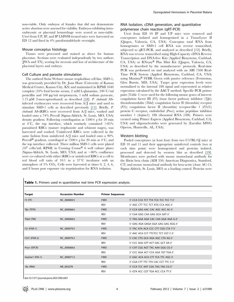
Table 1. Primers used in quantitative real time PCR expression analysis.
Target Accession Number Primer Sequences
F3 (TF) NC_000069.5 FWD 59-CCA CCA TCT TTA TCA TCC TCC T-39
REV 59-AGC CTT TCC TCT ATG CCA AGC-39
Tfpi (TFPI) NC_000068.6 FWD 59-CCA GAG AAC CAC AGC ACC AC-39
REV 59-CAA GGG CAA GAG GCA GAT-39
Thbd (TM) NC_000068.6 FWD 59-TAG GGA AGA CAC CAA GGA AGA G-39
REV 59-GAG AGA GAGA GGA GAG GAG AGG-39
F2r (PAR-1) NC_000079.5 FWD 59-TAC ATA ACA CCC CTT CGG CTA T-39
REV 59-AAC ACA CCT TTCTCC TCT CGT C-39
F2rl1 (PAR-2) NC_000079.5 FWD 59-CAC CTG GCA AGA AGC CTA AG-39
REV 59-CCC AGG GTT ACT GAC GCT AA-39
Procr (EPCR) NC_000068.6 FWD 59-CAT CGG AGT TAC AAA GGG CG-39
REV 59-CCC AGA ACT CCA GGA TGT TGA-39
Serpine1 (PAI-1) NC_000071.5 FWD 59-GGC ACA ACA CTT TCA TTC AGC-39
REV 59-CGA CTT TTC TTA CAC CCT TTC C-39
18s rRNA NR_003278 FWD 59-CCA TCC AAT CGG TAG TAG CG-39
REV 59-GTA ACC CGT TGA ACC CCA TT-39
doi:10.1371/journal.pone.0031090.t001
Dysregulated Hemostasis in Placental Malaria
PLoS ONE | www.plosone.org 4 February 2012 | Volume 7 | Issue 2 | e31090
detected with affinity purified horse anti-mouse horseradish
peroxidase (HRP) conjugate from Cell Signaling (Beverly, MA).
Statistical analysisData analyses were performed using GraphPad Prism 5 Software
(La Jolla, CA, USA) and SAS version 9.2 Software (Cary, NC,
USA). Correlation analysis was done using Spearman’s test and 262
contingency tables were used for testing differences between
proportions. The significance of difference of group means in the
case of normally distributed data were compared via t tests for
pairwaise comparisons or one-way ANOVA with Tukey’s Post-hoc
Multiple Comparison Test for multiple group comparisons. Non-
normally distributed data were analyzed by non-parametric, Mann
Whitney test for pairwise comparisons and Kruskal-Wallis test with
Dunn’s Multiple Comparison post-test for multiple group compar-
isons. Multiple linear regression analysis was performed including
interaction terms for dependent variables and selected parameters.
Statistical significance was not observed for select parameters,
notably low birth weight and interactions involving low birth
weight. Non-parametric human data are plotted using log10 scales
for ease of viewing but were not log-transformed. Proportional
analysis was accomplished via two-sided Fisher’s exact test. Values
of P#0.05 were considered to be significant.
Results
Study participant characteristicsThe investigation was restricted to primigravidae since they are
known to have the highest risk for PM, malaria-associated LBW and
prematurity, and PM-associated placental pathology, including
fibrin deposition [5,6]. Based on light microscopic evaluation of
intervillous blood thick and thin smears, samples from a total of 79
PM+ and 114 PM2 women were available for inclusion in the
study. Microscopic diagnosis of PM by blood smear has been shown
to have low accuracy; in a previous analysis of a subset of samples
from this study population, approximately half of all PM cases were
detectable only by PCR [13]. Targeting a newly described high-
copy target in the P. falciparum genome using PCR [16] revealed that
30 of 108 (28%) smear-negative samples were submicroscopically
infected (PMsub). Table 2 lists relevant clinical and sociological
attributes of all participants; for the purposes of these summary
characteristics, smear-negative participants for whom PCR testing
was not possible (n = 6) were included with the PM2 group.
Relative to PM2 women, a greater proportion of PM+ women bore
LBW infants, and mean birthweights among the latter were
significantly lower than among women with no active infection; to
some extent this was expected based on the sample selection
scheme. Most cases of LBW were due to fetal growth restriction
since #5% of infants in each group were born earlier than 36 weeks
gestation. PM+ women had lower hemoglobin levels than the other
groups and significant levels of hemozoin (Hz)-bearing phagocytes
on IVB thick smears. Although approximately half of all women
reported use of sulfadoxine/pyrimethamine during pregnancy,
more than 90% of all women had evidence of current or past PM
(malarial hemozoin in fibrin observed by histology).
Blood smear-positive PM is associated with inflammatoryresponses and dysregulated hemostasis in IVB
In initial analyses, participants were grouped according to
results of IVB blood smears. Thus, PMsub women were combined
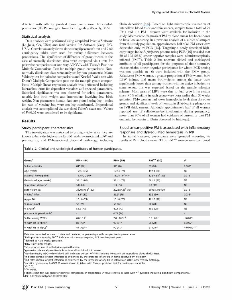
Table 2. Clinical and sociological attributes of human participants.
Groupa PM2 (84) PM+ (79) PMsub (30) P
% Luo ethnicity 84* (76) 97* (76) 89 (28) 0.005*
Age (years) 1963 (75) 1963 (77) 1963 (28) NS
Maternal hemoglobin 11.762.2 (49) 11.061.9{ (47) 12.562.4{ (22) 0.018
Gestational age (weeks) 3862 (80) 3861 (75) 3861 (30) NS
% preterm deliveryb 5.0 (80) 1.3 (75) 3.3 (30) NS
Birthweight (g) 31206458{ (80) 29226428{ (79) 30936379 (30) 0.013
% LBWc infant 13.8* (80) 26.6* (79) 13.3 (30) 0.033*
Apgar 10 1060 (75) 1060 (76) 1060 (28) NS
% male infant 58 (76) 53 (77) 50 (28) NS
% SPused 54.5 (77) 49.4 (77) 50.0 (28) NS
placental % parasitemiae - 0.72 (76) - -
% Hz-bearing WBCsf 0.060.1{ 7.8610.9{,1 0.060.01 ,0.0001
% with Hz in fibring 82 (79)* 99 (71)* 96 (28) 0.0007*
% with Hz in WBCsh 44 (79)*,# 90 (71)* 61 (28)# #0.0013*,#
Data are presented as mean 6 standard deviation or percentage with sample size in parentheses.aPM = placental malaria; PMsub indicates microscopy negative, PCR positive participants.bdefined as ,36 weeks gestation.cLBW = low birth weight.dSP = reported use of sulfadoxine-pyrimethamine.egeometric placental parasitemia from intervillous blood thin smear.fHz = hemozoin; WBC = white blood cell; indicates percent of WBCs bearing hemozoin on intervillous blood thick smear.gindicates chronic or past infection as evidenced by the presence of any Hz in fibrin observed by histology.hindicates chronic or past infection as evidenced by the presence of any Hz in intervillous WBCs observed by histology.Statistics by one-way ANOVA (P values shown in table) with Tukey’s post-hoc test for continuous variables:{P,0.05,{,1P,0.001.Fisher’s exact test was used for pairwise comparison of proportions (P values shown in table with *,# symbols indicating significant comparisons).doi:10.1371/journal.pone.0031090.t002
Dysregulated Hemostasis in Placental Malaria
PLoS ONE | www.plosone.org 5 February 2012 | Volume 7 | Issue 2 | e31090
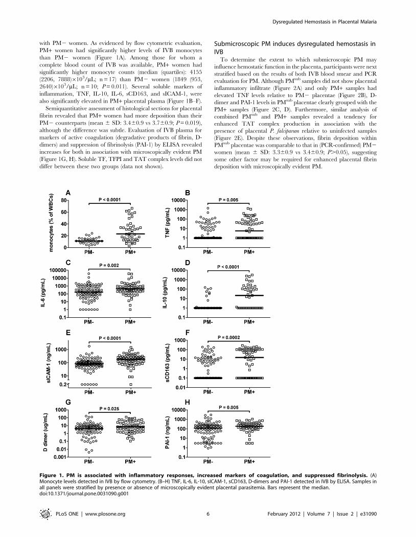
with PM2 women. As evidenced by flow cytometric evaluation,
PM+ women had significantly higher levels of IVB monocytes
than PM2 women (Figure 1A). Among those for whom a
complete blood count of IVB was available, PM+ women had
significantly higher monocyte counts (median (quartiles): 4155
(2206, 7888)6103/mL; n = 17) than PM2 women (1849 (953,
2640)6103/mL; n = 10; P = 0.011). Several soluble markers of
inflammation, TNF, IL-10, IL-6, sCD163, and sICAM-1, were
also significantly elevated in PM+ placental plasma (Figure 1B–F).
Semiquantitative assessment of histological sections for placental
fibrin revealed that PM+ women had more deposition than their
PM2 counterparts (mean 6 SD: 3.460.9 vs 3.760.9; P = 0.019),
although the difference was subtle. Evaluation of IVB plasma for
markers of active coagulation (degradative products of fibrin, D-
dimers) and suppression of fibrinolysis (PAI-1) by ELISA revealed
increases for both in association with microscopically evident PM
(Figure 1G, H). Soluble TF, TFPI and TAT complex levels did not
differ between these two groups (data not shown).
Submicroscopic PM induces dysregulated hemostasis inIVB
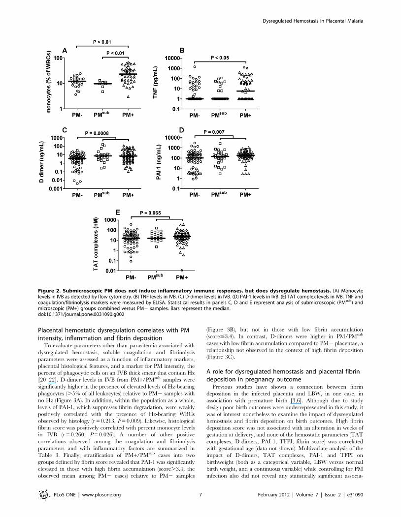
To determine the extent to which submicroscopic PM may
influence hemostatic function in the placenta, participants were next
stratified based on the results of both IVB blood smear and PCR
evaluation for PM. Although PMsub samples did not show placental
inflammatory infiltrate (Figure 2A) and only PM+ samples had
elevated TNF levels relative to PM2 placentae (Figure 2B), D-
dimer and PAI-1 levels in PMsub placentae clearly grouped with the
PM+ samples (Figure 2C, D). Furthermore, similar analysis of
combined PMsub and PM+ samples revealed a tendency for
enhanced TAT complex production in association with the
presence of placental P. falciparum relative to uninfected samples
(Figure 2E). Despite these observations, fibrin deposition within
PMsub placentae was comparable to that in (PCR-confirmed) PM2
women (mean 6 SD: 3.360.9 vs 3.460.9; P.0.05), suggesting
some other factor may be required for enhanced placental fibrin
deposition with microscopically evident PM.
Figure 1. PM is associated with inflammatory responses, increased markers of coagulation, and suppressed fibrinolysis. (A)Monocyte levels detected in IVB by flow cytometry. (B–H) TNF, IL-6, IL-10, sICAM-1, sCD163, D-dimers and PAI-1 detected in IVB by ELISA. Samples inall panels were stratified by presence or absence of microscopically evident placental parasitemia. Bars represent the median.doi:10.1371/journal.pone.0031090.g001
Dysregulated Hemostasis in Placental Malaria
PLoS ONE | www.plosone.org 6 February 2012 | Volume 7 | Issue 2 | e31090
Placental hemostatic dysregulation correlates with PMintensity, inflammation and fibrin deposition
To evaluate parameters other than parasitemia associated with
dysregulated hemostasis, soluble coagulation and fibrinolysis
parameters were assessed as a function of inflammatory markers,
placental histological features, and a marker for PM intensity, the
percent of phagocytic cells on an IVB thick smear that contain Hz
[20–22]. D-dimer levels in IVB from PM+/PMsub samples were
significantly higher in the presence of elevated levels of Hz-bearing
phagocytes (.5% of all leukocytes) relative to PM2 samples with
no Hz (Figure 3A). In addition, within the population as a whole,
levels of PAI-1, which suppresses fibrin degradation, were weakly
positively correlated with the presence of Hz-bearing WBCs
observed by histology (r = 0.213, P = 0.009). Likewise, histological
fibrin score was positively correlated with percent monocyte levels
in IVB (r = 0.260, P = 0.026). A number of other positive
correlations observed among the coagulation and fibrinolysis
parameters and with inflammatory factors are summarized in
Table 3. Finally, stratification of PM+/PMsub cases into two
groups defined by fibrin score revealed that PAI-1 was significantly
elevated in those with high fibrin accumulation (score.3.4, the
observed mean among PM2 cases) relative to PM2 samples
(Figure 3B), but not in those with low fibrin accumulation
(score#3.4). In contrast, D-dimers were higher in PM/PMsub
cases with low fibrin accumulation compared to PM2 placentae, a
relationship not observed in the context of high fibrin deposition
(Figure 3C).
A role for dysregulated hemostasis and placental fibrindeposition in pregnancy outcome
Previous studies have shown a connection between fibrin
deposition in the infected placenta and LBW, in one case, in
association with premature birth [3,6]. Although due to study
design poor birth outcomes were underrepresented in this study, it
was of interest nonetheless to examine the impact of dysregulated
hemostasis and fibrin deposition on birth outcomes. High fibrin
deposition score was not associated with an alteration in weeks of
gestation at delivery, and none of the hemostatic parameters (TAT
complexes, D-dimers, PAI-1, TFPI, fibrin score) was correlated
with gestational age (data not shown). Multivariate analysis of the
impact of D-dimers, TAT complexes, PAI-1 and TFPI on
birthweight (both as a categorical variable, LBW versus normal
birth weight, and a continuous variable) while controlling for PM
infection also did not reveal any statistically significant associa-
Figure 2. Submicroscopic PM does not induce inflammatory immune responses, but does dysregulate hemostasis. (A) Monocytelevels in IVB as detected by flow cytometry. (B) TNF levels in IVB. (C) D-dimer levels in IVB. (D) PAI-1 levels in IVB. (E) TAT complex levels in IVB. TNF andcoagulation/fibrinolysis markers were measured by ELISA. Statistical results in panels C, D and E represent analysis of submicroscopic (PMsub) andmicroscopic (PM+) groups combined versus PM2 samples. Bars represent the median.doi:10.1371/journal.pone.0031090.g002
Dysregulated Hemostasis in Placental Malaria
PLoS ONE | www.plosone.org 7 February 2012 | Volume 7 | Issue 2 | e31090
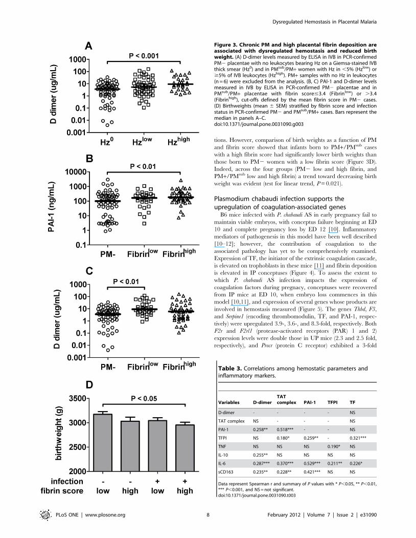
tions. However, comparison of birth weights as a function of PM
and fibrin score showed that infants born to PM+/PMsub cases
with a high fibrin score had significantly lower birth weights than
those born to PM2 women with a low fibrin score (Figure 3D).
Indeed, across the four groups (PM2 low and high fibrin, and
PM+/PMsub low and high fibrin) a trend toward decreasing birth
weight was evident (test for linear trend, P = 0.021).
Plasmodium chabaudi infection supports theupregulation of coagulation-associated genes
B6 mice infected with P. chabaudi AS in early pregnancy fail to
maintain viable embryos, with conceptus failure beginning at ED
10 and complete pregnancy loss by ED 12 [10]. Inflammatory
mediators of pathogenesis in this model have been well described
[10–12]; however, the contribution of coagulation to the
associated pathology has yet to be comprehensively examined.
Expression of TF, the initiator of the extrinsic coagulation cascade,
is elevated on trophoblasts in these mice [11] and fibrin deposition
is elevated in IP conceptuses (Figure 4). To assess the extent to
which P. chabaudi AS infection impacts the expression of
coagulation factors during pregnacy, conceptuses were recovered
from IP mice at ED 10, when embryo loss commences in this
model [10,11], and expression of several genes whose products are
involved in hemostasis measured (Figure 5). The genes Thbd, F3,
and Serpine1 (encoding thrombomodulin, TF, and PAI-1, respec-
tively) were upregulated 3.9-, 3.6-, and 8.3-fold, respectively. Both
F2r and F2rl1 (protease-activated receptors (PAR) 1 and 2)
expression levels were double those in UP mice (2.3 and 2.5 fold,
respectively), and Procr (protein C receptor) exhibited a 3-fold
Figure 3. Chronic PM and high placental fibrin deposition areassociated with dysregulated hemostasis and reduced birthweight. (A) D-dimer levels measured by ELISA in IVB in PCR-confirmedPM2 placentae with no leukocytes bearing Hz on a Giemsa-stained IVBthick smear (Hz0) and in PMsub/PM+ women with Hz in ,5% (Hzlow) or$5% of IVB leukocytes (Hzhigh). PM+ samples with no Hz in leukocytes(n = 6) were excluded from the analysis. (B, C) PAI-1 and D-dimer levelsmeasured in IVB by ELISA in PCR-confirmed PM2 placentae and inPMsub/PM+ placentae with fibrin score#3.4 (Fibrinlow) or .3.4(Fibrinhigh), cut-offs defined by the mean fibrin score in PM2 cases.(D) Birthweights (mean 6 SEM) stratified by fibrin score and infectionstatus in PCR-confirmed PM2 and PMsub/PM+ cases. Bars represent themedian in panels A–C.doi:10.1371/journal.pone.0031090.g003
Table 3. Correlations among hemostatic parameters andinflammatory markers.
Variables D-dimerTATcomplex PAI-1 TFPI TF
D-dimer - - - - NS
TAT complex NS - - - NS
PAI-1 0.258** 0.518*** - - NS
TFPI NS 0.180* 0.259** - 0.321***
TNF NS NS NS 0.190* NS
IL-10 0.255** NS NS NS NS
IL-6 0.287*** 0.370*** 0.529*** 0.211** 0.226*
sCD163 0.235** 0.228** 0.421*** NS NS
Data represent Spearman r and summary of P values with * P,0.05, ** P,0.01,*** P,0.001, and NS = not significant.doi:10.1371/journal.pone.0031090.t003
Dysregulated Hemostasis in Placental Malaria
PLoS ONE | www.plosone.org 8 February 2012 | Volume 7 | Issue 2 | e31090
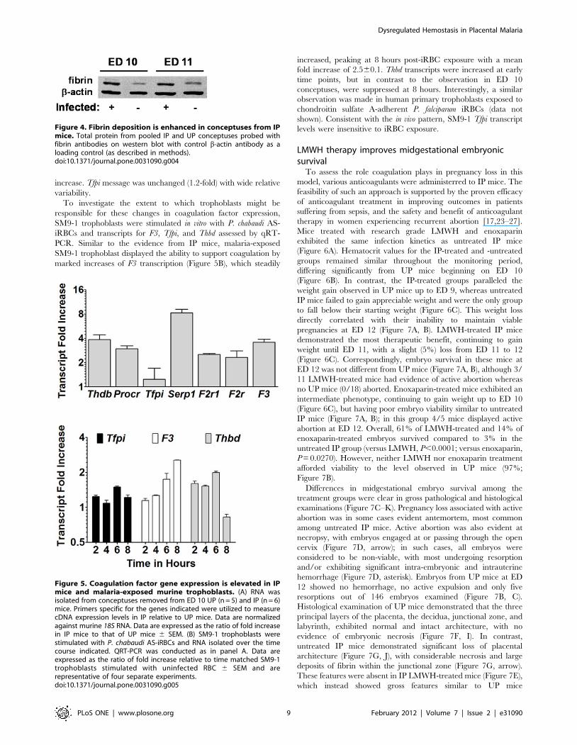
increase. Tfpi message was unchanged (1.2-fold) with wide relative
variability.
To investigate the extent to which trophoblasts might be
responsible for these changes in coagulation factor expression,
SM9-1 trophoblasts were stimulated in vitro with P. chabaudi AS-
iRBCs and transcripts for F3, Tfpi, and Thbd assessed by qRT-
PCR. Similar to the evidence from IP mice, malaria-exposed
SM9-1 trophoblast displayed the ability to support coagulation by
marked increases of F3 transcription (Figure 5B), which steadily
increased, peaking at 8 hours post-iRBC exposure with a mean
fold increase of 2.560.1. Thbd transcripts were increased at early
time points, but in contrast to the observation in ED 10
conceptuses, were suppressed at 8 hours. Interestingly, a similar
observation was made in human primary trophoblasts exposed to
chondroitin sulfate A-adherent P. falciparum iRBCs (data not
shown). Consistent with the in vivo pattern, SM9-1 Tfpi transcript
levels were insensitive to iRBC exposure.
LMWH therapy improves midgestational embryonicsurvival
To assess the role coagulation plays in pregnancy loss in this
model, various anticoagulants were administerred to IP mice. The
feasibility of such an approach is supported by the proven efficacy
of anticoagulant treatment in improving outcomes in patients
suffering from sepsis, and the safety and benefit of anticoagulant
therapy in women experiencing recurrent abortion [17,23–27].
Mice treated with research grade LMWH and enoxaparin
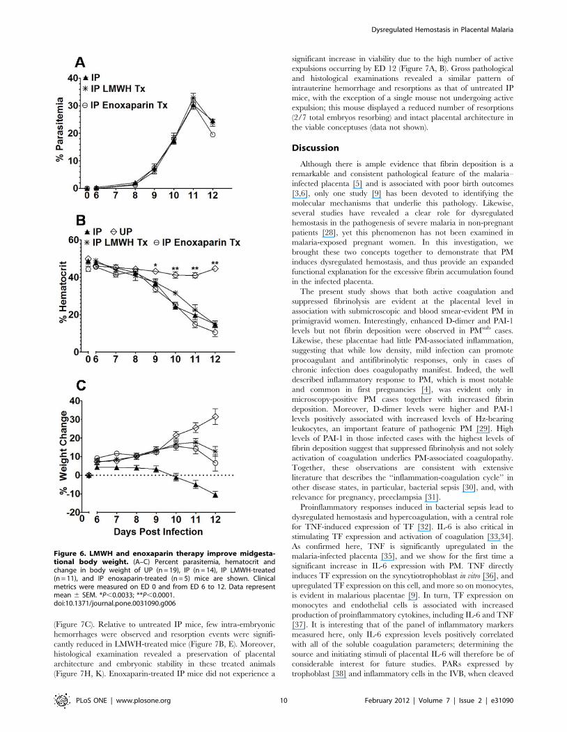
exhibited the same infection kinetics as untreated IP mice
(Figure 6A). Hematocrit values for the IP-treated and -untreated
groups remained similar throughout the monitoring period,
differing significantly from UP mice beginning on ED 10
(Figure 6B). In contrast, the IP-treated groups paralleled the
weight gain observed in UP mice up to ED 9, whereas untreated
IP mice failed to gain appreciable weight and were the only group
to fall below their starting weight (Figure 6C). This weight loss
directly correlated with their inability to maintain viable
pregnancies at ED 12 (Figure 7A, B). LMWH-treated IP mice
demonstrated the most therapeutic benefit, continuing to gain
weight until ED 11, with a slight (5%) loss from ED 11 to 12
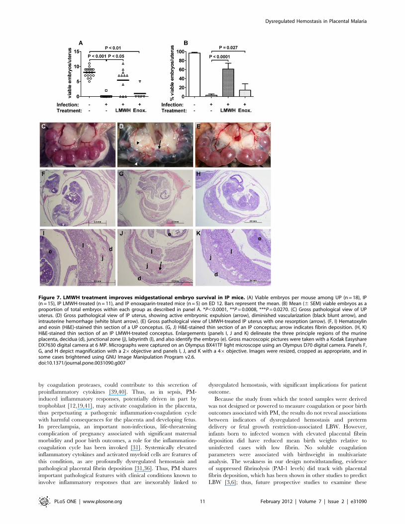
(Figure 6C). Correspondingly, embryo survival in these mice at
ED 12 was not different from UP mice (Figure 7A, B), although 3/
11 LMWH-treated mice had evidence of active abortion whereas
no UP mice (0/18) aborted. Enoxaparin-treated mice exhibited an
intermediate phenotype, continuing to gain weight up to ED 10
(Figure 6C), but having poor embryo viability similar to untreated
IP mice (Figure 7A, B); in this group 4/5 mice displayed active
abortion at ED 12. Overall, 61% of LMWH-treated and 14% of
enoxaparin-treated embryos survived compared to 3% in the
untreated IP group (versus LMWH, P,0.0001; versus enoxaparin,
P = 0.0270). However, neither LMWH nor enoxaparin treatment
afforded viability to the level observed in UP mice (97%;
Figure 7B).
Differences in midgestational embryo survival among the
treatment groups were clear in gross pathological and histological
examinations (Figure 7C–K). Pregnancy loss associated with active
abortion was in some cases evident antemortem, most common
among untreated IP mice. Active abortion was also evident at
necropsy, with embryos engaged at or passing through the open
cervix (Figure 7D, arrow); in such cases, all embryos were
considered to be non-viable, with most undergoing resorption
and/or exhibiting significant intra-embryonic and intrauterine
hemorrhage (Figure 7D, asterisk). Embryos from UP mice at ED
12 showed no hemorrhage, no active expulsion and only five
resorptions out of 146 embryos examined (Figure 7B, C).
Histological examination of UP mice demonstrated that the three
principal layers of the placenta, the decidua, junctional zone, and
labyrinth, exhibited normal and intact architecture, with no
evidence of embryonic necrosis (Figure 7F, I). In contrast,
untreated IP mice demonstrated significant loss of placental
architecture (Figure 7G, J), with considerable necrosis and large
deposits of fibrin within the junctional zone (Figure 7G, arrow).
These features were absent in IP LMWH-treated mice (Figure 7E),
which instead showed gross features similar to UP mice
Figure 5. Coagulation factor gene expression is elevated in IPmice and malaria-exposed murine trophoblasts. (A) RNA wasisolated from conceptuses removed from ED 10 UP (n = 5) and IP (n = 6)mice. Primers specific for the genes indicated were utilized to measurecDNA expression levels in IP relative to UP mice. Data are normalizedagainst murine 18S RNA. Data are expressed as the ratio of fold increasein IP mice to that of UP mice 6 SEM. (B) SM9-1 trophoblasts werestimulated with P. chabaudi AS-iRBCs and RNA isolated over the timecourse indicated. QRT-PCR was conducted as in panel A. Data areexpressed as the ratio of fold increase relative to time matched SM9-1trophoblasts stimulated with uninfected RBC 6 SEM and arerepresentative of four separate experiments.doi:10.1371/journal.pone.0031090.g005
Figure 4. Fibrin deposition is enhanced in conceptuses from IPmice. Total protein from pooled IP and UP conceptuses probed withfibrin antibodies on western blot with control b-actin antibody as aloading control (as described in methods).doi:10.1371/journal.pone.0031090.g004
Dysregulated Hemostasis in Placental Malaria
PLoS ONE | www.plosone.org 9 February 2012 | Volume 7 | Issue 2 | e31090
(Figure 7C). Relative to untreated IP mice, few intra-embryonic
hemorrhages were observed and resorption events were signifi-
cantly reduced in LMWH-treated mice (Figure 7B, E). Moreover,
histological examination revealed a preservation of placental
architecture and embryonic stability in these treated animals
(Figure 7H, K). Enoxaparin-treated IP mice did not experience a
significant increase in viability due to the high number of active
expulsions occurring by ED 12 (Figure 7A, B). Gross pathological
and histological examinations revealed a similar pattern of
intrauterine hemorrhage and resorptions as that of untreated IP
mice, with the exception of a single mouse not undergoing active
expulsion; this mouse displayed a reduced number of resorptions
(2/7 total embryos resorbing) and intact placental architecture in
the viable conceptuses (data not shown).
Discussion
Although there is ample evidence that fibrin deposition is a
remarkable and consistent pathological feature of the malaria–
infected placenta [5] and is associated with poor birth outcomes
[3,6], only one study [9] has been devoted to identifying the
molecular mechanisms that underlie this pathology. Likewise,
several studies have revealed a clear role for dysregulated
hemostasis in the pathogenesis of severe malaria in non-pregnant
patients [28], yet this phenomenon has not been examined in
malaria-exposed pregnant women. In this investigation, we
brought these two concepts together to demonstrate that PM
induces dysregulated hemostasis, and thus provide an expanded
functional explanation for the excessive fibrin accumulation found
in the infected placenta.
The present study shows that both active coagulation and
suppressed fibrinolysis are evident at the placental level in
association with submicroscopic and blood smear-evident PM in
primigravid women. Interestingly, enhanced D-dimer and PAI-1
levels but not fibrin deposition were observed in PMsub cases.
Likewise, these placentae had little PM-associated inflammation,
suggesting that while low density, mild infection can promote
procoagulant and antifibrinolytic responses, only in cases of
chronic infection does coagulopathy manifest. Indeed, the well
described inflammatory response to PM, which is most notable
and common in first pregnancies [4], was evident only in
microscopy-positive PM cases together with increased fibrin
deposition. Moreover, D-dimer levels were higher and PAI-1
levels positively associated with increased levels of Hz-bearing
leukocytes, an important feature of pathogenic PM [29]. High
levels of PAI-1 in those infected cases with the highest levels of
fibrin deposition suggest that suppressed fibrinolysis and not solely
activation of coagulation underlies PM-associated coagulopathy.
Together, these observations are consistent with extensive
literature that describes the ‘‘inflammation-coagulation cycle’’ in
other disease states, in particular, bacterial sepsis [30], and, with
relevance for pregnancy, preeclampsia [31].
Proinflammatory responses induced in bacterial sepsis lead to
dysregulated hemostasis and hypercoagulation, with a central role
for TNF-induced expression of TF [32]. IL-6 is also critical in
stimulating TF expression and activation of coagulation [33,34].
As confirmed here, TNF is significantly upregulated in the
malaria-infected placenta [35], and we show for the first time a
significant increase in IL-6 expression with PM. TNF directly
induces TF expression on the syncytiotrophoblast in vitro [36], and
upregulated TF expression on this cell, and more so on monocytes,
is evident in malarious placentae [9]. In turn, TF expression on
monocytes and endothelial cells is associated with increased
production of proinflammatory cytokines, including IL-6 and TNF
[37]. It is interesting that of the panel of inflammatory markers
measured here, only IL-6 expression levels positively correlated
with all of the soluble coagulation parameters; determining the
source and initiating stimuli of placental IL-6 will therefore be of
considerable interest for future studies. PARs expressed by
trophoblast [38] and inflammatory cells in the IVB, when cleaved
Figure 6. LMWH and enoxaparin therapy improve midgesta-tional body weight. (A–C) Percent parasitemia, hematocrit andchange in body weight of UP (n = 19), IP (n = 14), IP LMWH-treated(n = 11), and IP enoxaparin-treated (n = 5) mice are shown. Clinicalmetrics were measured on ED 0 and from ED 6 to 12. Data representmean 6 SEM. *P,0.0033; **P,0.0001.doi:10.1371/journal.pone.0031090.g006
Dysregulated Hemostasis in Placental Malaria
PLoS ONE | www.plosone.org 10 February 2012 | Volume 7 | Issue 2 | e31090
by coagulation proteases, could contribute to this secretion of
proinflammatory cytokines [39,40]. Thus, as in sepsis, PM-
induced inflammatory responses, potentially driven in part by
trophoblast [12,19,41], may activate coagulation in the placenta,
thus perpetuating a pathogenic inflammation-coagulation cycle
with harmful consequences for the placenta and developing fetus.
In preeclampsia, an important non-infectious, life-threatening
complication of pregnancy associated with significant maternal
morbidity and poor birth outcomes, a role for the inflammation-
coagulation cycle has been invoked [31]. Systemically elevated
inflammatory cytokines and activated myeloid cells are features of
this condition, as are profoundly dysregulated hemostasis and
pathological placental fibrin deposition [31,36]. Thus, PM shares
important pathological features with clinical conditions known to
involve inflammatory responses that are inexorably linked to
dysregulated hemostasis, with significant implications for patient
outcome.
Because the study from which the tested samples were derived
was not designed or powered to measure coagulation or poor birth
outcomes associated with PM, the results do not reveal associations
between indicators of dysregulated hemostasis and preterm
delivery or fetal growth restriction-associated LBW. However,
infants born to infected women with elevated placental fibrin
deposition did have reduced mean birth weights relative to
uninfected cases with low fibrin. No soluble coagulation
parameters were associated with birthweight in multivariate
analysis. The weakness in our design notwithstanding, evidence
of suppressed fibrinolysis (PAI-1 levels) did track with placental
fibrin deposition, which has been shown in other studies to predict
LBW [3,6]; thus, future prospective studies to examine these
Figure 7. LMWH treatment improves midgestational embryo survival in IP mice. (A) Viable embryos per mouse among UP (n = 18), IP(n = 15), IP LMWH-treated (n = 11), and IP enoxaparin-treated mice (n = 5) on ED 12. Bars represent the mean. (B) Mean (6 SEM) viable embryos as aproportion of total embryos within each group as described in panel A. *P,0.0001, **P = 0.0008, ***P = 0.0270. (C) Gross pathological view of UPuterus. (D) Gross pathological view of IP uterus, showing active embryonic expulsion (arrow), diminished vascularization (black blunt arrow), andintrauterine hemorrhage (white blunt arrow). (E) Gross pathological view of LMWH-treated IP uterus with one resorption (arrow). (F, I) Hematoxylinand eosin (H&E)-stained thin section of a UP conceptus. (G, J) H&E-stained thin section of an IP conceptus; arrow indicates fibrin deposition. (H, K)H&E-stained thin section of an IP LMWH-treated conceptus. Enlargements (panels I, J and K) delineate the three principle regions of the murineplacenta, decidua (d), junctional zone (j), labyrinth (l), and also identify the embryo (e). Gross macroscopic pictures were taken with a Kodak EasyshareDX7630 digital camera at 6 MP. Micrographs were captured on an Olympus BX41TF light microscope using an Olympus D70 digital camera. Panels F,G, and H depict magnification with a 26objective and panels I, J, and K with a 46objective. Images were resized, cropped as appropriate, and insome cases brightened using GNU Image Manipulation Program v2.6.doi:10.1371/journal.pone.0031090.g007
Dysregulated Hemostasis in Placental Malaria
PLoS ONE | www.plosone.org 11 February 2012 | Volume 7 | Issue 2 | e31090
associations in more detail are justified. In this context, testing of
hemostatic parameters, including functional measures of coagula-
tion and regulatory function, in the peripheral blood of malaria-
exposed pregnant women will be critical, since detection of
dysregulated hemostasis, if it is to be a potential therapeutic target,
must be possible antenatally in venous blood. Because coagulation
and fibrinolysis are significantly perturbed in severe P. falciparum
malaria [42–47], providing compelling evidence for a pathogenic
role of dysregulated hemostasis in PM, as in cerebral malaria
[28,48], will require coupling of functional coagulation metrics
with identification of specific pathological outcomes, such as
placental fibrin deposition, associated placental damage, prema-
ture birth and/or fetal growth restriction.
Investigation of profound pregnancy complications due to
malaria, such as fetal loss, are difficult to conduct with human
subjects due to ethical considerations, making the availability of
mouse models very important. We have shown that P. chabaudi AS
infection of B6 mice results in complete loss of pregnancy
midgestationally [10,11]; this corresponds to the time during
which risk for malaria is highest in pregnant women (second
trimester) and failure to protect against infection is associated with
fetal loss [2,49]. Importantly, antibody neutralization of TNF in IP
B6 mice both restored midgestational pregnancy success and
facilitated maintenance of placental TF expression at normal (low)
levels [12]. The results herein confirm dysregulated hemostasis at
the level of the conceptus in malaria-infected mice, with several
indicators of activation and control of coagulation, as well as
suppressors of coagulation and fibrinolysis, being increased at the
level of gene expression. Likewise, mRNA for PARs 1 and 2 are
upregulated. The results also provide evidence that in addition to
maternal monocytic promotion of placental coagulation through
upregulation of TF [9,12], the fetal trophoblast in contact with
maternal blood may also help to tip the balance toward net
increased production of fibrin with sustained, enhanced expression
of TF [12] while TFPI and thrombomodulin decline. Cumula-
tively, the results suggest that, as in human PM, malarial infection
in mice promotes a pathogenic inflammation-coagulation cycle
with significant negative consequences for pregnancy.
The striking efficacy of LMWH treatment in restoring
midgestation embryo viability in P. chabaudi AS-infected mice
provides indirect evidence that coagulation plays a pivotal role in
malaria-associated pregnancy loss. However, inflammatory re-
sponses are also operational in pregnancy loss in this model [12].
Thus, the independent pathogenic effects of inflammation and
coagulation on placental and embryonic viability should be
assessed, although the interconnectedness of the inflammation-
coagulation cycle may make such dissection difficult. Ultimately,
confirmation of a critical role for either pathway in malaria-
induced compromise of pregnancy will still leave the molecular
mechanisms that drive embryo loss in mice and fetoplacental
damage in humans to be elucidated. Of particular interest for
malaria-associated placental fibrin formation and fibrinolysis, it
was recently shown that fibrin degradation products directly
damage placental architecture via trophoblast cell death [50,51].
Additionally, the role of PARs in the inflammation-coagulation
cycle also should be considered. In a mouse model of bacterial
sepsis and in human endotoxin challenge studies, interruption of
coagulation in the absence of uncoupling of the inflammation-
coagulation cycle, in which PARs are central, did not abrogate
disease [52,53]. Relevant to pregnancy specifically, recent
evidence suggests that both PAR1 and 2 are important players
in the pathogenesis of preeclampsia [54,55].
A second more intriguing implication of LMWH-mediated
rescue of pregnancy in malaria-infected mice is the potential for a
novel therapeutic intervention based on anticoagulant treatment
for pregnancies at risk for malaria-associated poor birth outcomes.
Because even a submicroscopic level of placental infection is
associated with dysregulated hemostasis and a short-lived, rapidly
treated infection during pregnancy can still have an adverse
impact on birth outcome [56,57], it is plausible that the
coagulation-inflammation cycle continues to cause coagulopathy
in the placenta even after curative antimalarial treatment is
delivered. Therefore, simultaneous disruption of coagulation (and
therefore coagulation protease-driven inflammatory responses
through PARs) and removal of inflammation-inducing iRBCs
with combination antimalarial/anticoagulant drug treatments may
be more effective in preventing PM-induced pre-term labor and
LBW. There is historic precedent for this approach in treatment of
severe malaria in non-pregnant patients. Two early studies showed
that treatment of pediatric cerebral malaria with unfractionated
heparin and antimalarial drug reduced morbidity and mortality
[58,59]. Despite this success, however, failure of heparin therapy
in two of three trials in rhesus macaques [60–62], and concerns
about severe bleeding precipitating patient death in association
with unfractionated heparin use [63–65] have compelled the
World Health Organization to concur with concerned scientists
that anticoagulant treatment should not be used in malaria
therapy [60,66–68]. It is becoming increasingly clear, however,
that the low molecular weight fractions of heparin (LMWH) retain
excellent anticoagulant function but with greatly minimized
bleeding-associated complications in treated patients [69]. Even
within LMWHs, different manufacturing processes yield different
structural fractions, yielding drugs with distinct activities and
specificities that cannot be used interchangeably [70]. This may
explain why a disparity in pregnancy success between mice treated
with different LMWHs was observed; enoxaparin is generated by
benzylation followed by alkaline hydrolysis, whereas the research
grade LMWH used in this study was generated by oxidative
depolymerization with Cu2+ and hydrogen peroxide, which is the
method used to create the LMWH, parnaparin [71].
Aside from inhibition of coagulation, glycoconjugates, including
fractions of heparin, have potential adjunctive therapeutic value
for severe malaria syndromes due to activity in iRBC rosette
disruption, blockage of merozoite invasion and inhibition of iRBC
sequestration [72–76]. Such glycoconjugates have little to no
anticoagulant activity, yet in at least one case, some clinical benefit
was observed following administration of curdlan sulfate in severe/
cerebral malaria patients [77]. The paucity of contemporary
efforts to test the efficacy of anticoagulant treatment in severe
malaria syndromes, such as PM, might therefore remain given
fears of bleeding and the promise of other glycoconjugate-based
adjuncts which act directly on the parasite and/or iRBC.
Nonetheless, the data presented herein demonstrate that humans
express markers for malaria-induced dysregulated hemostasis
during PM and a rodent model of PM exhibits enhanced
midgestational embryonic survival upon treatment with LMWH.
Importantly, P. chabaudi is known to form rosettes, but these
rosettes, unlike those of P. falciparum, are insensitive to glycocon-
jugate treatment [78]. As reported here, infection kinetics were not
different in LMWH-treated and -untreated IP mice. Thus, the
improvement in midgestational status of treated mice suggests that
dysregulated hemostasis leading to a procoagulant environment is
at least partially responsible for malaria-induced embryo loss, and
suppression of coagulation protects against this outcome.
Further work to demonstrate the efficacy of anticoagulant
therapy to allow murine pregnancies to proceed to term, reverse
coagulopathy already established in the placenta, and improve
outcomes in concert with anti-malarial treatment remains to be
Dysregulated Hemostasis in Placental Malaria
PLoS ONE | www.plosone.org 12 February 2012 | Volume 7 | Issue 2 | e31090
achieved. In the meantime, however, the present results warrant
prospective, longitudinal investigations in malaria-exposed women
to establish the presence, antenatally, of dysregulated hemostasis in
association with infection, and identify the extent to which this
hemostatic disruption predicts placental coagulopathy and poor
birth outcomes. Should clear associations be found and confir-
mations in rodent models be achieved, then evaluation of the
safety and efficacy of anticoagulants as an adjunctive treatment to
antenatal, curative anti-malarial treatment may be considered.
Importantly, hemostatic disorders in pregnancy are currently
safely and successfully treated with such therapies [79–82].
Although first generation anticoagulant treatment for malaria
met with clinical failure due to bleeding complications [63–65],
this risk is much lower with the new generation drugs. The latest
generation is available in oral formulations, which although not
currently indicated for use in pregnant women, may with further
development and safety testing make delivery and patient
compliance more facile. Overall, while a small risk of bleeding
complications remains with any anticoagulant treatment, and is
especially relevant for parturient women who are at risk for peri-
and post-partum hemorrhage, the potential benefits of limited,
monitored inclusion of drugs like LMWH in treatment for malaria
during pregnancy deserves careful consideration.
Recognition that pathogenesis in both PM [12] and cerebral
malaria [28,48,83,84] is mediated by the inflammation-coagula-
tion cycle is likely to become increasingly relevant, particularly in
the critical search for much-needed novel therapies. In our mouse
model for PM, targeting either inflammation [12] or coagulation
provides significant clinical benefit. A recent study by Francischetti
and colleagues [83] showed that defibrotide, a nucleotide-based
drug [85–87], has multipotent effects against malaria-induced
cellular activation, inflammatory responses and dysregulated
hemostasis, and delayed disease development in a murine model
for CM. Interestingly, although defibrotide has low intrinsic
anticoagulant activity, it effectively interferes with TF function,
thrombin generation, and platelet activation [83,85–87]. Common
among all of these treatment strategies is interruption of the
inflammation-coagulation cycle. Thus, further study of the
molecular events at the intersection of this pathogenic cycle in
model systems and affected human populations has the potential
to reveal critical, novel targets in the host response to malaria that
contribute substantially to pathogenesis.
Supporting Information
Figure S1 Stereological assessment of fibrin in placen-tal sections correlates with semi-quantitative scoringmethod. Photomicrographs of tissue sections at 2006 final
magnification were captured. One image each from the basal and
chorionic plates and eleven randomly selected intervillous regions
spanning the full thickness of the placental disk, each representing
an area measuring 615 mm6460 mm (2.836105 mm2), were
assessed. Using GNU Image Manipulation Program (v2.6), a grid
of 30 mm630 mm was superimposed over the images, and at each
intersection on the grid (300 total per image) the structural
component present was scored. Components scored were basal
plate, chorionic plate, villus (stroma and trophoblast), fetal blood
vessel, syncytial knot, intervillous space, and intervillous or
perivillous fibrin deposition. Villi converted to fibrinoid-type fibrin
were counted as fibrin deposition. Fibrin score, represented as a
percentage of intervillous space occupied, was calculated using the
following formula: (total number of grid intersections scored as
fibrin/(fibrin intersections+intervillous space intersections))6100.
(TIF)
Acknowledgments
We thank the parturient women and Labour Ward staff at New Nyanza
Provincial General Hospital, Kisumu and Siaya District Hospital, Siaya,
Kenya, and the Kenya-based UGA/KEMRI team, without whose
participation, active support and dedication this study would not have
been possible. We also appreciate the fruitful discussions and enthusiastic
encouragement of Drs. Alexander Duncan and Ivo Francischetti. This
work is published with the permission of the Director, Kenya Medical
Research Institute.
Author Contributions
Conceived and designed the experiments: JWA GMS JMM. Performed the
experiments: JWA GMS SOO DS SM LFK JDM JMM. Analyzed the
data: JWA GMS TN JM CA JMM. Wrote the paper: JWA JMM.
Coordinated and conducted human recruitment and sample collection and
processing: SOO SM JDM JMM. Conceptualized use of mouse model to
study malaria-induced coagulopathy: JSP.
References
1. Dellicour S, Tatem AJ, Guerra CA, Snow RW, ter Kuile FO (2010) Quantifying
the number of pregnancies at risk of malaria in 2007: a demographic study.
PLoS Med 7: e1000221.
2. Desai M, Ter Kuile FO, Nosten F, McGready R, Asamoa K, et al. (2007)
Epidemiology and burden of malaria in pregnancy. Lancet Infect Dis 7: 93–104.
3. Crocker IP, Tanner OM, Myers JE, Bulmer JN, Walraven G, et al. (2004)
Syncytiotrophoblast degradation and the pathophysiology of the malaria-
infected placenta. Placenta 25: 273–282.
4. Rogerson SJ, Hviid L, Duffy PE, Leke RF, Taylor DW (2007) Malaria in
pregnancy: pathogenesis and immunity. Lancet Infect Dis 7: 105–117.
5. Brabin BJ, Romagosa C, Abdelgalil S, Menendez C, Verhoeff FH, et al. (2004)
The sick placenta-the role of malaria. Placenta 25: 359–378.
6. Menendez C, Ordi J, Ismail MR, Ventura PJ, Aponte JJ, et al. (2000) The
impact of placental malaria on gestational age and birth weight. J Infect Dis 181:
1740–1745.
7. Scifres CM, Nelson DM (2009) Intrauterine growth restriction, human placental
development and trophoblast cell death. J Physiol 587: 3453–3458.
8. Mackintosh CL, Beeson JG, Marsh K (2004) Clinical features and pathogenesis
of severe malaria. Trends Parasitol 20: 597–603.
9. Imamura T, Sugiyama T, Cuevas LE, Makunde R, Nakamura S (2002)
Expression of tissue factor, the clotting initiator, on macrophages in Plasmodium
falciparum-infected placentas. J Infect Dis 186: 436–440.
10. Poovassery J, Moore JM (2006) Murine malaria infection induces fetal loss
associated with accumulation of Plasmodium chabaudi AS-infected erythrocytes
in the placenta. Infect Immun 74: 2839–2848.
11. Poovassery J, Moore JM (2009) Association of malaria-induced murine
pregnancy failure with robust peripheral and placental cytokine responses.
Infect Immun 77: 4998–5006.
12. Poovassery JS, Sarr D, Smith G, Nagy T, Moore JM (2009) Malaria-induced
murine pregnancy failure: distinct roles for IFN-gamma and TNF. J Immunol
183: 5342–5349.
13. Perrault SD, Hajek J, Zhong K, Owino SO, Sichangi M, et al. (2009) Human
immunodeficiency virus co-infection increases placental parasite density and
transplacental malaria transmission in Western Kenya. Am J Trop Med Hyg 80:
119–125.
14. Othoro C, Moore JM, Wannemuehler K, Nahlen BL, Otieno J, et al. (2006)
Evaluation of various methods of maternal placental blood collection for
immunology studies. Clin Vaccine Immunol 13: 568–574.
15. Moore JM, Shi YP, Othoro C, Nahlen BL, Lal AA, et al. (2003) Comparative
flow cytometric analysis of term placental intervillous and peripheral blood from
immediate postpartum women in Western kenya. Placenta 24: 779–785.
16. Demas A, Oberstaller J, Debarry J, Lucchi NW, Srinivasamoorthy G, et al. (2011)
Applied Genomics: Data Mining Reveals Species-Specific Malaria Diagnostic
Targets More Sensitive than 18S rRNA. J Clin Microbiol 49: 2411–2418.
17. Gutierrez G, Sarto A, Berod L, Canellada A, Gentile T, et al. (2004) Regulation
of interleukin-6 fetoplacental levels could be involved in the protective effect of
low-molecular weight heparin treatment on murine spontaneous abortion.
Am J Reprod Immunol 51: 160–165.
18. Bowen JA, Hunt JS (1999) Expression of cell adhesion molecules in murine
placentas and a placental cell line. Biol Reprod 60: 428–434.
Dysregulated Hemostasis in Placental Malaria
PLoS ONE | www.plosone.org 13 February 2012 | Volume 7 | Issue 2 | e31090
19. Lucchi NW, Peterson DS, Moore JM (2008) Immunologic activation of human
syncytiotrophoblast by Plasmodium falciparum. Malar J 7: 42.
20. van Eijk AM, Ayisi JG, ter Kuile FO, Misore AO, Otieno JA, et al. (2003) HIV
increases the risk of malaria in women of all gravidities in Kisumu, Kenya. Aids
17: 595–603.
21. Chaisavaneeyakorn S, Moore JM, Mirel L, Othoro C, Otieno J, et al. (2003)
Levels of macrophage inflammatory protein 1 alpha (MIP-1 alpha) and MIP-1
beta in intervillous blood plasma samples from women with placental malaria
and human immunodeficiency virus infection. Clin Diagn Lab Immunol 10:
631–636.
22. Moore JM, Chaisavaneeyakorn S, Perkins DJ, Othoro C, Otieno J, et al. (2004)
Hemozoin differentially regulates proinflammatory cytokine production in
human immunodeficiency virus-seropositive and -seronegative women with
placental malaria. Infect Immun 72: 7022–7029.
23. Freeman BD, Zehnbauer BA, Buchman TG (2003) A meta-analysis of
controlled trials of anticoagulant therapies in patients with sepsis. Shock 20: 5–9.
24. Levi M, de Jonge E, van der Poll T (2001) Rationale for restoration of
physiological anticoagulant pathways in patients with sepsis and disseminated
intravascular coagulation. Crit Care Med 29: S90–94.
25. Matthay MA (2001) Severe sepsis–a new treatment with both anticoagulant and
antiinflammatory properties. N Engl J Med 344: 759–762.
26. Laskin CA, Spitzer KA, Clark CA, Crowther MR, Ginsberg JS, et al. (2009)
Low molecular weight heparin and aspirin for recurrent pregnancy loss: results
from the randomized, controlled HepASA Trial. J Rheumatol 36: 279–287.
27. Levi M, Lowenberg E, Meijers JC (2010) Recombinant anticoagulant factors for
adjunctive treatment of sepsis. Semin Thromb Hemost 36: 550–557.
28. Francischetti IM, Seydel KB, Monteiro RQ (2008) Blood coagulation,
inflammation, and malaria. Microcirculation 15: 81–107.
29. Rogerson SJ, Pollina E, Getachew A, Tadesse E, Lema VM, et al. (2003)
Placental monocyte infiltrates in response to Plasmodium falciparum malaria
infection and their association with adverse pregnancy outcomes. Am J Trop
Med Hyg 68: 115–119.
30. Levi M, van der Poll T (2010) Inflammation and coagulation. Crit Care Med 38:
S26–34.
31. Dusse LM, Rios DR, Pinheiro MB, Cooper AJ, Lwaleed BA (2011) Pre-
eclampsia: relationship between coagulation, fibrinolysis and inflammation. Clin
Chim Acta 412: 17–21.
32. Camerer E, Kolsto AB, Prydz H (1996) Cell biology of tissue factor, the principal
initiator of blood coagulation. Thromb Res 81: 1–41.
33. van der Poll T, Levi M, Hack CE, ten Cate H, van Deventer SJ, et al. (1994)
Elimination of interleukin 6 attenuates coagulation activation in experimental
endotoxemia in chimpanzees. J Exp Med 179: 1253–1259.
34. Levi M, van der Poll T, ten Cate H, van Deventer SJ (1997) The cytokine-
mediated imbalance between coagulant and anticoagulant mechanisms in sepsis
and endotoxaemia. Eur J Clin Invest 27: 3–9.
35. Fried M, Muga RO, Misore AO, Duffy PE (1998) Malaria elicits type 1
cytokines in the human placenta: IFN-gamma and TNF-alpha associated with
pregnancy outcomes. J Immunol 160: 2523–2530.
36. Teng YC, Lin QD, Lin JH, Ding CW, Zuo Y (2009) Coagulation and
fibrinolysis related cytokine imbalance in preeclampsia: the role of placental
trophoblasts. J Perinat Med 37: 343–348.
37. Levi M (2010) The coagulant response in sepsis and inflammation. Hamosta-
seologie 30: 10–12, 14–16.
38. Isermann B, Hendrickson SB, Hutley K, Wing M, Weiler H (2001) Tissue-
restricted expression of thrombomodulin in the placenta rescues thrombomo-
dulin-deficient mice from early lethality and reveals a secondary developmental
block. Development 128: 827–838.
39. Mackman N (2009) The many faces of tissue factor. J Thromb Haemost 7 Suppl
1: 136–139.
40. Coughlin SR (2000) Thrombin signalling and protease-activated receptors.
Nature 407: 258–264.
41. Lucchi NW, Sarr D, Owino SO, Mwalimu SM, Peterson DS, et al. (2011)
Natural hemozoin stimulates syncytiotrophoblast to secrete chemokines and
recruit peripheral blood mononuclear cells. Placenta 32: 579–585.
42. Rojanasthien S, Surakamolleart V, Boonpucknavig S, Isarangkura P (1992)
Hematological and coagulation studies in malaria. J Med Assoc Thai 75 Suppl 1:
190–194.
43. Rojanasthien S, Surakamollert V, Isarangkura P, Boonpucknavig S (1993) A
new method for factor VII deficient substrate preparation and coagulation
studies in malaria. Southeast Asian J Trop Med Public Health 24 Suppl 1:
225–228.
44. Prasad R, Das BK, Pengoria R, Mishra OP, Shukla J, et al. (2009) Coagulation
Status and Platelet Functions in Children with Severe Falciparum Malaria and
their Correlation of Outcome. J Trop Pediatr.
45. Dennis LH, Eichelberger JW, Inman MM, Conrad ME (1967) Depletion of
coagulation factors in drug-resistant Plasmodium falciparum malaria. Blood 29:
713–721.
46. Vogetseder A, Ospelt C, Reindl M, Schober M, Schmutzhard E (2004) Time
course of coagulation parameters, cytokines and adhesion molecules in
Plasmodium falciparum malaria. Trop Med Int Health 9: 767–773.
47. Clemens R, Pramoolsinsap C, Lorenz R, Pukrittayakamee S, Bock HL, et al.
(1994) Activation of the coagulation cascade in severe falciparum malaria
through the intrinsic pathway. Br J Haematol 87: 100–105.
48. Francischetti IM (2008) Does activation of the blood coagulation cascade have arole in malaria pathogenesis? Trends Parasitol 24: 258–263.
49. Aziken ME, Akubuo KK, Gharoro EP (2011) Efficacy of intermittent preventivetreatment with sulfadoxine-pyrimethamine on placental parasitemia in pregnant
women in midwestern Nigeria. Int J Gynaecol Obstet 112: 30–33.
50. Isermann B, Sood R, Pawlinski R, Zogg M, Kalloway S, et al. (2003) The
thrombomodulin-protein C system is essential for the maintenance of pregnancy.Nat Med 9: 331–337.
51. Guo YH, Hernandez I, Isermann B, Kang TB, Medved L, et al. (2009)Caveolin-1-dependent apoptosis induced by fibrin degradation products. Blood
113: 4431–4439.
52. Petaja J (2011) Inflammation and coagulation. An overview. Thromb Res 127
Suppl 2: S34–37.
53. de Jonge E, Dekkers PE, Creasey AA, Hack CE, Paulson SK, et al. (2000) Tissue
factor pathway inhibitor dose-dependently inhibits coagulation activationwithout influencing the fibrinolytic and cytokine response during human
endotoxemia. Blood 95: 1124–1129.
54. Al-Ani B, Hewett PW, Cudmore MJ, Fujisawa T, Saifeddine M, et al. (2010)
Activation of proteinase-activated receptor 2 stimulates soluble vascularendothelial growth factor receptor 1 release via epidermal growth factor
receptor transactivation in endothelial cells. Hypertension 55: 689–697.
55. Estrada-Gutierrez G, Cappello RE, Mishra N, Romero R, Strauss JF, 3rd, et al.
(2011) Increased expression of matrix metalloproteinase-1 in systemic vessels of
preeclamptic women: a critical mediator of vascular dysfunction. Am J Pathol178: 451–460.
56. Landis SH, Lokomba V, Ananth CV, Atibu J, Ryder RW, et al. (2009) Impact ofmaternal malaria and under-nutrition on intrauterine growth restriction: a
prospective ultrasound study in Democratic Republic of Congo. EpidemiolInfect 137: 294–304.
57. Luxemburger C, McGready R, Kham A, Morison L, Cho T, et al. (2001) Effectsof malaria during pregnancy on infant mortality in an area of low malaria
transmission. Am J Epidemiol 154: 459–465.
58. Munir M, Tjandra H, Rampengan TH, Mustadjab I, Wulur FH (1980) Heparin
in the treatment of cerebral malaria. Paediatr Indones 20: 47–50.
59. Rampengan TH (1991) Cerebral malaria in children. Comparative study
between heparin, dexamethasone and placebo. Paediatr Indones 31: 59–66.
60. Reid HA, Sucharit P (1972) Ancrod, heparin, and -aminocaproic acid in simian
Knowlesi malaria. Lancet 2: 1110–1112.
61. Howard WA, Collins WE (1972) Heparin therapy in simian Plasmodium
knowlesi malaria. Lancet 2: 738–739.
62. Dennis LH, Conrad ME (1968) Anticoagulant and antimalarial action of
heparin in simian malaria. Lancet 1: 769–771.
63. Stone WJ, Hanchett JE, Knepshield JH (1972) Acute renal insufficiency due tofalciparum malaria. Review of 42 cases. Arch Intern Med 129: 620–628.
64. Smitskamp H, Wolthuis FH (1971) New concepts in treatment of malignanttertian malaria with cerebral involvement. Br Med J 1: 714–716.
65. Borochovitz D, Crosley AL, Metz J (1970) Disseminated intravascularcoagulation with fatal haemorrhage in cerebral malaria. Br Med J 2: 710.
66. Hall AP (1976) The treatment of malaria. Br Med J 1: 323–328.
67. Reid HA (1975) Letter: Adjuvant treatment of severe falciparum malaria,
intravascular coagulation, and heparin. Lancet 1: 167–168.
68. Reyburn H (2010) New WHO guidelines for the treatment of malaria. BMJ 340:
c2637.
69. Greer IA, Nelson-Piercy C (2005) Low-molecular-weight heparins for thrombo-
prophylaxis and treatment of venous thromboembolism in pregnancy: asystematic review of safety and efficacy. Blood 106: 401–407.
70. Linhardt RJ, Gunay NS (1999) Production and chemical processing of lowmolecular weight heparins. Semin Thromb Hemost 25 Suppl 3: 5–16.
71. Merli GJ, Groce JB (2010) Pharmacological and clinical differences betweenlow-molecular-weight heparins: implications for prescribing practice and
therapeutic interchange. P T 35: 95–105.
72. Carlson J, Ekre HP, Helmby H, Gysin J, Greenwood BM, et al. (1992)
Disruption of Plasmodium falciparum erythrocyte rosettes by standard heparinand heparin devoid of anticoagulant activity. Am J Trop Med Hyg 46: 595–602.
73. Rowe A, Berendt AR, Marsh K, Newbold CI (1994) Plasmodium falciparum: afamily of sulphated glycoconjugates disrupts erythrocyte rosettes. Exp Parasitol
79: 506–516.
74. Vogt AM, Pettersson F, Moll K, Jonsson C, Normark J, et al. (2006) Release of
sequestered malaria parasites upon injection of a glycosaminoglycan. PLoSPathog 2: e100.
75. Kyriacou HM, Steen KE, Raza A, Arman M, Warimwe G, et al. (2007) In vitroinhibition of Plasmodium falciparum rosette formation by Curdlan sulfate.
Antimicrob Agents Chemother 51: 1321–1326.
76. Leitgeb AM, Blomqvist K, Cho-Ngwa F, Samje M, Nde P, et al. (2011) Low
anticoagulant heparin disrupts Plasmodium falciparum rosettes in fresh clinicalisolates. Am J Trop Med Hyg 84: 390–396.
77. Havlik I, Looareesuwan S, Vannaphan S, Wilairatana P, Krudsood S, et al.(2005) Curdlan sulphate in human severe/cerebral Plasmodium falciparum
malaria. Trans R Soc Trop Med Hyg 99: 333–340.
78. Mackinnon MJ, Walker PR, Rowe JA (2002) Plasmodium chabaudi: rosetting in
a rodent malaria model. Exp Parasitol 101: 121–128.
79. Badawy AM, Khiary M, Sherif LS, Hassan M, Ragab A, et al. (2008) Low-
molecular weight heparin in patients with recurrent early miscarriages ofunknown aetiology. J Obstet Gynaecol 28: 280–284.
Dysregulated Hemostasis in Placental Malaria
PLoS ONE | www.plosone.org 14 February 2012 | Volume 7 | Issue 2 | e31090
80. Rowan JA, McLintock C, Taylor RS, North RA (2003) Prophylactic and
therapeutic enoxaparin during pregnancy: indications, outcomes and monitor-ing. Aust N Z J Obstet Gynaecol 43: 123–128.
81. Brenner B (2005) Low-molecular-weight heparin in pregnancy: state of the art.
Womens Health (Lond Engl) 1: 153–156.82. Brenner B, Hoffman R, Blumenfeld Z, Weiner Z, Younis JS (2000) Gestational
outcome in thrombophilic women with recurrent pregnancy loss treated byenoxaparin. Thromb Haemost 83: 693–697.
83. Francischetti IM, Oliveira CJ, Ostera GR, Yager SB, Debierre-Grockiego F,
et al. (2011) Defibrotide Interferes With Several Steps of the Coagulation-Inflammation Cycle and Exhibits Therapeutic Potential to Treat Severe
Malaria. Arterioscler Thromb Vasc Biol.84. Francischetti IM, Seydel KB, Monteiro RQ, Whitten RO, Erexson CR, et al.
(2007) Plasmodium falciparum-infected erythrocytes induce tissue factor
expression in endothelial cells and support the assembly of multimolecular
coagulation complexes. J Thromb Haemost 5: 155–165.
85. Kornblum N, Ayyanar K, Benimetskaya L, Richardson P, Iacobelli M, et al.
(2006) Defibrotide, a polydisperse mixture of single-stranded phosphodiester
oligonucleotides with lifesaving activity in severe hepatic veno-occlusive disease:
clinical outcomes and potential mechanisms of action. Oligonucleotides 16:
105–114.
86. Larocca A, Cavallo F, Magarotto V, Rossi D, Patriarca F, et al. (2008)
Defibrotide: a review on clinical use and future development. Expert Opin Biol
Ther 8: 1201–1212.
87. Richardson P, Linden E, Revta C, Ho V (2009) Use of defibrotide in the
treatment and prevention of veno-occlusive disease. Expert Rev Hematol 2:
365–376.
Dysregulated Hemostasis in Placental Malaria
PLoS ONE | www.plosone.org 15 February 2012 | Volume 7 | Issue 2 | e31090
Related Documents