Chapter - 4 Materials & Methods

Welcome message from author
This document is posted to help you gain knowledge. Please leave a comment to let me know what you think about it! Share it to your friends and learn new things together.
Transcript

Chapter - 4
Materials & Methods
Materials & Methods
53
The present study “A study on Mycobacterial species causing
lymph node tuberculosis” was carried out in the Department of
Microbiology, Blue Peter Research Center (BPRC) - LEPRA Society,
Hyderabad. The clinical specimens were obtained from the Institute of
Chest Diseases, Irrumnuma, Hyderabad. The period of the study was
from January 2004 to January 2006.
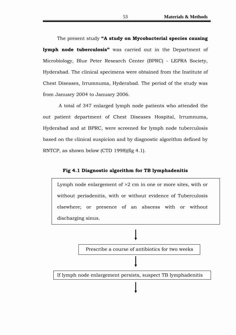
A total of 347 enlarged lymph node patients who attended the
out patient department of Chest Diseases Hospital, Irrumnuma,
Hyderabad and at BPRC, were screened for lymph node tuberculosis
based on the clinical suspicion and by diagnostic algorithm defined by
RNTCP, as shown below (CTD 1998)(fig 4.1).
Fig 4.1 Diagnostic algorithm for TB lymphadenitis
Lymph node enlargement of >2 cm in one or more sites, with or
without periadenitis, with or without evidence of Tuberculosis
elsewhere; or presence of an abscess with or without
discharging sinus.
Prescribe a course of antibiotics for two weeks
If lymph node enlargement persists, suspect TB lymphadenitis

Materials & Methods
54
Pus from discharging sinus / aspirate from lymph node
using Fine Needle Aspiration Cytology (FNAC)
Smear examination for AFB (using pus/aspirate) by Ziehl
Nielsen’s method
Mantoux test for children <14 years
Diagnosis confirmed if the pus / aspirate from FNAC
show:
ZN stain positive for AFB, or
Granulomatous changes (where facilities
available)
If FNAC results are inconclusive, excision biopsy is
advisable for smear histo-pathological examination
Start Category III
Treatment

Materials & Methods
55
Based on the above algorithm, a total of 200 lymph node
tuberculosis patients formed the study material and the rest of the
patients was excluded from the study.
Patient information, detailed clinical examination, history of
previous episodes of TB and history of previous anti TB treatment
were carefully obtained (annexure 1). All the patients were subjected
to voluntary and confidential HIV testing. (The institutional ethical
committee of Blue Peter Research Center approved the study protocol.)
(Fig 4.2).

Materials & Methods
56
Enlarged lymph node Fine needle aspirate
ZN-staining H & E staining Culture
LJ/BacT
2- LJ & 1 BacT Bottle
at 370 c
• Tuberculous
Lymphadenitis.
• Granulomatous
lymphadenitis
• Non Specific
lymphadenitis.
• Presence of AFB
• Without AFB.
AFB Growth No AFB Growth
(after incubation of 8 wks)
Drug Susceptibility
testing. ( Isoniazid,
Rifampicin, Streptomycin,
Ethambutol)
(1% proportion method)
Rate of Growth
Slow Grower
( > 5 days onwards)
Rapid Growers
( 1-4 days)
Pigmentation
Photochromogen
Sctochromogen
Non Pigmentation
Growth on PNB
Bio chemical Tests NRT, Niacin,
Tween-80,
Arylsulfatase etc.,
Non Tuberculous Mycobacteria/
NTM
Mycobacterium
tuberculosis- M.Tb
Fig 4.2. Study Protocol.

Materials & Methods
57
Annexure 1
Patient Details Code No: Date of Sample Collection :
Name :
Age : Sex:
Address :
Weight :
Complaints
History
Present
Duration of Swelling :
Presence of Similar Swelling any where
Other than the presenting site :
Swelling associated with Fever / Cough :
Periodicity of Fever (if any) :
Cough (duration in days) :
Associated Hemoptysis :
Loss of Appetite :
Loss of Weight :
Past
Had any similar Swelling / Complaints :
Had any past history of Tuberculosis :

Materials & Methods
58
Had any other diseases (Please Specify)
Allergy: Tonsillitis: Diabetes: Hepatitis: HIV:
Thyroid: Alcohol: Smoking Habit: Steroid Use:
Treatment History
History of Anti-TB treatment
Medicine Duration
1.
2.
3.
4.
5.
6.
History of any other treatment :
Family History / History of Contact
Any member of family / neighbors who are
either suffering with pulmonary TB / has taken
treatment for tuberculosis (Yes /NO) :

Materials & Methods
59
If YES – Details :
Clinical Examination
Features of the Lymph node enlargement
Anatomical site of swelling :
Shape :
Size :
Number (single /multiple) :
Discrete / matted :
Attached with the under lying tissues/ skin :
Consistency :
Discharge/ sinuses :
Any abrasions or injury in the draining area
Of lymph node :
General Examination

Materials & Methods
60
Investigations
Total WBC count :
Differential WBC count :
ESR :
Hemoglobin :
Mantoux Test :
Chest X ray :
Sputum AFB :
Sputum Culture :
Any Other (Specify) :
H I V sero-positivity :

Materials & Methods
61
4.2. Specimen Collection
Fine Needle aspiration of lymph node was done using 22-23gauge
needle attached to a 10ml disposable plastic syringe, after fixing the node
with the help of left forefinger and thumb, 2-3 passes were made into the
lymph node (Witht.Vielh & Orell et al 1999). Aspirates from the involved
lymph node were divided into two portions, one portion of the material
expressed on to a clean grease free microscope glass slides. Two slides
were fixed in 90% Isopropyl alcohol for routine haematoxylin & eosin
staining. Two slides were air dried for Ziehl-Neelsen staining. The other
aliquot meant for mycobacterial culture was rinsed with 0.5 ml of sterile
distilled water, and processed immediately or preserved at 4 0C for not
more than 24 hours. In the laboratory, the aliquot for culture was
inoculated on two Lowenstein-Jensen slopes and on rapid culture system
(MB BacT).
4.3Laboratory procedures
4.3.1.Procedure for Haematoxylin and Eosin.
Fixes the smears in 90% ethanol for 15 minutes.
Immersed the slides in Harris Haematoxylin for 10-15 minutes.
Washed the slides under running tap water (gentle) for 10 minutes.
Diped the smears in 0.5% eosin twice.
Dehydrated the slides in ascending grade of alcohol (60%, 70%,
and 90%).

Materials & Methods
62
Blot dried and mounted the slides with DPX (Diestrene Plasticene
Xylene).
Observed the smear under scanner(4x), low power (10x), and
Highpower (40x). (Bancroft JD 4th edn)
(All the lab procedures were as per the lab manual of Tuberculosis
Research Center, Chennai)
4.3.2.Procedure for Ziehl-Neelsen staining.
Air dried the aspirate on a clean, grease free glass slide.
Heat fixed the smear.
Flooded the slide with 1% carbol fuschin,
o Heated the slide intermittently with stain until vaporization
(without boiling).
o Waited for 5 minutes and washed the slides with running
tap water gently.
Flooded the slide with 25 % sulphuric acid for 2- 3 minutes.
Washed the slide under running tap water gently.
Counter stained with 0.1% methylene blue solution for 30 seconds.
Washed the slide under running tap water gently and air dried.
Observed the smear under 100x oil immersion. (RNTCP 2005)

Materials & Methods
63
4.3.3. Culture of the Aspirate
The LJ Media cultures were examined everyday for one week and
thereafter once a week for 8 weeks. Once the growth appeared, it was
confirmed by ZN staining and screened for NTM by testing tolerance of
the isolates to para-nitrobenozic acid (PNB) in a concentration of 500
mg/L incorporated in the LJ medium. Species identification of all the
NTM was achieved by performing the battery of biochemical tests: i)
assessment of photo reactivity of mycobacteria, 2) growth at 250 C, 370 C,
and 440C, 3) aryl sulphatase test, 4) tween -80 hydrolysis, 5) catalase
test, 6) tellurite reduction test, 7) growth on MC Conkey agar and 8)
resistance to thiophen-2-carbonic acid hydrazide as described below. All
the Isolated Mycobacterium tuberculosis were tested for their antibiotic
susceptibility testing for the first line drugs (isoniazid, rifampicin,
streptomycin and ethambutol) using 1% proportion method.
4.4. Biochemical testing for speciation of Mycobacteria.
4.4.1Preparation of Standard suspension for biochemical
testing
Two thirds loopful of culture taken in 24 SWG nichrome loop with
3mm with internal diameter (to approximately coincide with a moist
weight of 4 mg of the organism) was added to 0.2 ml of sterile distilled
water in a 7 ml bijou bottle containing 6 glass beads. The bottle was
mechanically shaken for one minute at a speed which just lifts the beads

Materials & Methods
64
from the bottom of bottle to produce a uniform suspension. Then 0.8 ml
of sterile distilled water was added and the bottle was shaken in a vortex
and the resultant suspension contained 4mg/ml of the organism. Using
24 SWG nichrome wire loop with a 3 mm external diameter. One loopful
of the suspension was taken for performance of the individual tests or for
sub culturing onto each slope of the medium. Positive and negative
controls were included with each test.
4.4.2Assessment of Photo reactivity of Mycobacterium
species
Principle
The appearance of yellow pigment in the colonies of
photochromogenic mycobacteria is the result of yellowish orange
carotene crystals that are produced by active metabolism of the
microorganisms on exposure to bright light. Scotochromogenic species
have the capability of producing yellow pigment without exposure to
light; however, the type of pigment is unknown. The pigment of young
colonies of mycobacteria after growth in the dark or following exposure to
light can be an important aid in the identification of certain
Mycobacterium species.

Materials & Methods
65
Specimen
Primary broth culture of the test organism, diluted sufficiently to
produce isolated colonies when inoculated on LJ Media and Middlebrook
7H10 agar plates.
Materials
A. Equipment
1. Biologic safety cabinet.
2. 37 C incubator.
B. Supplies.
1. Sterile screw cap test tubes, 20 x110 or 20 x 125 mm
2. Sterile Pasteur pipettes
3. Inoculating wires and loops.
C. Medium.
1. Three slants of Lowenstein Jensen medium.
2. Three Middlebrook 7H10 agar plates.
Standards and Controls
1. Positive photochromogen: M kansasii.
2. Positive scotochromogen: Stock strains of M scrofulaceum or M
gordonae.
3. Negative chromogen: M tuberculosis.
(Standards & Controls were obtained from JALMA, institute for
mycobacterial diseases, Agra)

Materials & Methods
66
Procedure
1. Inoculated the surfaces of three Lowenstein-Jensen slant media or
three Middle book 7H11 agar plates with fluid from a diluted broth
culture of the organism to be tested. Wraped two of the tubes or
plates with aluminum foil; leave the third exposed to the ambient
light in the incubator.
2. Incubated one of the wrapped tubes or plates at 250-300c; the
other wrapped tubes or plates at 370c.
3. Light exposed control tube or plate is observed daily for any
growth. Once a growth is seen on the light exposed tubes or plates,
the wrapped ones are opened to check for growth.
4. If early growth is detected in the wrapped tubes or plates, expose
one of each pair to strong light for approximately 5 hours. A 100 W
tungsten bulb or fluorescent equivalent is adequate. Loosen the
cap of the culture tube during this period of light exposure.
5. Following exposure to light, the tube or plate is returned to the
incubator and inspected after 24 to 48 hours for the appearance of
yellow pigment.
Interpretation
Mycobacteria that are scotochromogenic produce an equal amount
of pigment whether light exposed or left in the dark. M scrofulaceum, M
gordonae, M flavescens, M xenopi and M szulgai (the latter is

Materials & Methods
67
scotochromogenic only when incubated at 370 C) compose the
scotochromogenic group.
Mycobacteria that are photochromogenic produce yellow pigment
only after exposure to light. The more commonly encountered
photochromogens include M kansasii, M marinum, M simiae and M
asiaticum.
Nonchromogenic mycobacteria are incapable of producing pigment
either in the dark or after exposure to light. M tuberculosis, M bovis, M
ulcerans, M fortuitum, M chelonae, and classic strains or M avium are the
more commonly encountered nonchromogens.
(The procedures and their interpretation were strictly as per the
laboratory manual of tuberculosis research center, Chennai)

Materials & Methods
68
4.4.3. Niacin Accumulation/ Niacin Test.
Principle
All the Mycobacterial species produce niacin ribonucleotide;
however, virtually all strains of M tuberculosis, M simiae and some strains
of M chelonae lack the enzyme to further convert niacin to nicotinamide
adenine dinucleotide (NAD). Comparative studies have shown that,
M.tuberculosis accumulates the largest amount of nicotinic acid and its
detection is useful for its definitive diagnosis. Niacin negative
M.tuberculosis strains are very rare, while very few other mycobacterial
species yield positive niacin tests. Cultures grown on egg medium
containing asparagines yield the most consistent results in the niacin
test and LJ medium is therefore recommended. A culture must be at
least three to four weeks old and must have sufficient growth of at least
100 colonies.
Materials
Equipments:
1. Biological Safety Cabinet
2. 37 C incubator.
Reagents
O-toludine, 1.5%
O-toludine 1.5 g
Ethanol 100ml
Materials & Methods
69
Prepared fresh weekly an amber bottle and store in the dark in the
refrigerator.
Cyanogen bromide solution, approx. 10%.: A saturated aqueous solution
of cyanogen bromide is approx 10%. Stored at 4oC in the refrigerator.
Quality control
Positive control: M tuberculosis
Negative control: M intracellulare
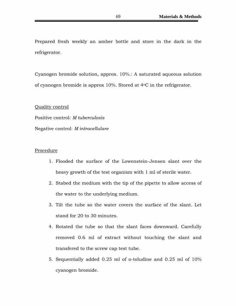
Procedure
1. Flooded the surface of the Lowenstein-Jensen slant over the
heavy growth of the test organism with 1 ml of sterile water.
2. Stabed the medium with the tip of the pipette to allow access of
the water to the underlying medium.
3. Tilt the tube so the water covers the surface of the slant. Let
stand for 20 to 30 minutes.
4. Rotated the tube so that the slant faces downward. Carefully
removed 0.6 ml of extract without touching the slant and
transfered to the screw cap test tube.
5. Sequentially added 0.25 ml of o-toludine and 0.25 ml of 10%
cyanogen bromide.

Materials & Methods
70
6. Closed the tube and observed the solution for the formation of a
pink color (=positive) within 5 minutes.
7. Added 2-3 ml of 4% NaOH to each tube to neutralize cynogen
bromide before discarding.
Interpretation
The development of pink color indicates a positive test.
Precautions
Cyanogen bromide is a severe lacrimator and toxic, if inhaled.
Work in a well-ventilated fume hood when preparing the solution and in
a biological safety cabinet while testing cultures. In acid solutions,
cyanogen bromide hydrolyses to hydrocyanic acid, which is extremely
toxic. Discard all reaction tubes into a disinfectant solution made
alkaline by addition of sodium hydroxide.

Materials & Methods
71
4.4.4. Nitrate Reduction test for Mycobacteria
Principle
Mycobacteria producing nitroreductase are capable of catalyzing
the reduction of nitrate to nitrite. In reaction, oxygen is extracted from
nitrate according to the following formula
NO3+2 e- + 2H ------ NO2 + H2O
The nitrate produced is detected by the addition of
naphthalamime and sulfanilic acid, forming the red diazonium dye, p-
sulfobenzene-azonaphthalamine.
Specimen
Three to four week old culture of the test organism growing on
Lowenstein Jensen medium.
Materials
Biological safety cabinet.
370C water bath or heating block
Reagents
(1). Nitrate test substrate
0.067M (M/15) phosphate buffer solution, pH 7.0
Na2HPO4, anhydrous 9.47 g
Distilled water 1 liter
Materials & Methods
72
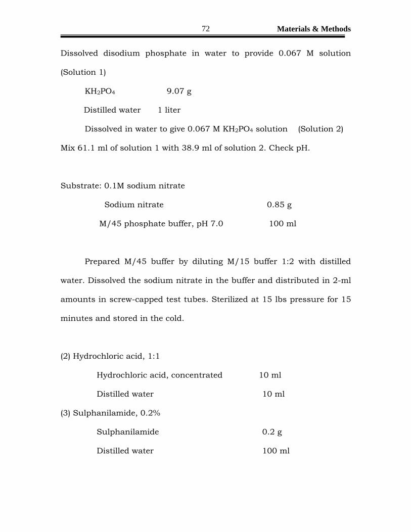
Dissolved disodium phosphate in water to provide 0.067 M solution
(Solution 1)
KH2PO4 9.07 g
Distilled water 1 liter
Dissolved in water to give 0.067 M KH2PO4 solution (Solution 2)
Mix 61.1 ml of solution 1 with 38.9 ml of solution 2. Check pH.
Substrate: 0.1M sodium nitrate
Sodium nitrate 0.85 g
M/45 phosphate buffer, pH 7.0 100 ml
Prepared M/45 buffer by diluting M/15 buffer 1:2 with distilled
water. Dissolved the sodium nitrate in the buffer and distributed in 2-ml
amounts in screw-capped test tubes. Sterilized at 15 lbs pressure for 15
minutes and stored in the cold.
(2) Hydrochloric acid, 1:1
Hydrochloric acid, concentrated 10 ml
Distilled water 10 ml
(3) Sulphanilamide, 0.2%
Sulphanilamide 0.2 g
Distilled water 100 ml

Materials & Methods
73
(4) Coupling agent
N- (1-naphthyl)-ethylene diamine di-HCl 0.1 g
Distilled water 100 ml
(5) Zinc dust
Quality control
Positive control: M tuberculosis.
Negative control: M avium complex
Procedure
1. Emulsified one loopful of colonies from solid medium into 2 ml of
nitrate substrate.
2. Shaken by hand to mix and incubated at 370C for two hours.
3. Added reagents to mixture in the following order:
a. One drop of concentrated HCL
b. Two drops of 0.2% sulfanilamide.
c. Two drops of 0.1% N-(1-naphthyl) ethylenediamine
dihydrochloride.
4. Allowed mixture to sit at room temperature for 5 minutes.
5. Read for the development of a pink to red color.

Materials & Methods
74
Interpretation
The appearance of a red or pink color is indicative of a positive
test. A quantitative reading can be made by comparing with nitrate
reduction standards.
Confirmed all negative reaction by adding a pinch of zinc dust.
Development of a pink color at this stage indicates that the initial
negative reaction was genuine. If no color change occurs after adding the
zinc dust, the reaction has proceeded beyond nitrite into other
components. Repeat the entire test.
4.4.5. Arylsulfatase Test
Principle
Arylsulfatase is an enzyme that splits free phenolphthalein from
the tripotassium salts of phenolphthalein disulfite. The test for the
identification of Mycobacterium species is performed in a tube containing
a substrate of phenolphthalein in oleic acid agar (Wayner). After 3 (or 14)
days of incubation of subculture of the unknown species, the appearance
of a pink color after addition of sodium carbonate indicates a positive
reaction.
Specimen
Mature colony of the unknown Mycobacterium species recovered
from clinical material grown on an LJ slants or on Middlebrook 7H-10

Materials & Methods
75
agar. Prepare a suspension of the organism in sterile water and incubate
for 3 days (or 14 days).
Materials
A. Equipment.
1. Biologic safety cabinet
B. Media.
1. Arylsulfatase stock Substrate:
Dissolved 2.6 g of phenolphthalein disulfate tripotassium salt in 50 ml of
sterile deionized water. Sterilized by membrane filtration (0.22µm-pore
size filter).
a. Stored in the refrigerator at 20- 80c.
b. Shelf life is indefinite if stored properly. Discard if
solution becomes cloudy.
2. Arylsulfatase broth: 3 days test.
a. Aseptically added 2.5 ml of stock substrate to 200 ml
of sterile Dubo’s Tween broth.
b. Aseptically dispensed 2.0 ml amounts into screw cap
test tubes (16 x 125 mm).
c. Store at 20-80 C.
3. Arylsulfatase broth: 2 week test.
a. Aseptically added 7.5 ml of stock substrate to 200 ml
of sterile Dubo’s Tween broth.

Materials & Methods
76
b. Aseptically dispensed 2.0 ml amounts into screw cap
test tubes (16x25mm).
c. Store at 20-80 C.
C. Reagents
2N Sodium carbonate (Na2CO3): Dissolved 10.6 g of
anhydrous sodium carbonate in 100 ml of distilled
water.
Quality Control
A. Three Day test
1. Positive Control: Mycobacterium fortuitum (if positive control
is negative, repeat tests with a fresh subculture of the
positive organism. If the negative controls are positive, repeat
the tests with a new lot of media).
2. Negative Control: Mycobacterium intracellulare.
3. Uninoculated medium and reagent only: No color.
B. Fourteen Day test.
1. Positive control: Mycobacterium fortuitum
2. Negative control: Mycobacterium intracellulare.
3. Uninoculated medium and reagent only: No color.
Materials & Methods
77
Procedure
1. Inoculated each tube of substrate with a lightly turbid suspension
of the test organism in sterile water. Thoroughly emulsified the
culture in the broth.
2. Incubated them for 3 days or 14 days (2 weeks) at 350 C in the non
CO2 incubator.
3. Following incubation, added 1 ml of the 2N sodium carbonate
reagent, mixed and observed for color change.
Procedure Note:
Included an actively growing control organism with each batch of
test. For slow growers used a 3 to 5 week subculture, for rapid growers, a
1- 3 week subculture. If the positive control is negative, repeated the
tests with a fresh subculture of the positive organism. If the negative
controls are positive, repeated the tests with a new lot of media.

Materials & Methods
78
4.4.6. Catalase 680C Test:
Catalase is an enzyme that decomposes hydrogen peroxide (H2O2)
into water and oxygen. Chemically, catalase is a hemoprotein, similar in
structure to hemoglobin, except that the four iron atoms in the molecule
are in the oxidized (Fe3+), rather than the reduced (Fe2+), state.
Principle
Catalase splits hydrogen peroxide into water and oxygen. The
evolution of oxygen appears as bubbles. Some forms of catalase are
inactivated by heating at 680C for 20 minutes, a valuable identifying
feature for certain Mycobacterium species. The hydrogen peroxide used
for the identification of Mycobacterium species differs from that used to
detect catalase in other types of bacteria, by using a 30% concentration
(Superoxal) in a strong detergent solution (10% tween-80). The detergent
helps disperse the hydrophobic tightly clumped mycobacteria from large
aggregates to individual bacilli, maximizing the detection of catalase.
Specimen
Mature colony of the unknown Mycobacterium species recovered
from clinical material, grown on an LJ slant or on Middlebrook7H10
agar.

Materials & Methods
79
Materials
1. 680 C water bath or heat block
2. Lowenstein Jensen deeps in 25x150 mm screw cap test tubes.
3. 30% hydrogen peroxide (commercially available as Superoxol)
4. M/15 phosphate buffer (0.067M).
Quality controls
Negative control: M tuberculosis; bubbles at 22-250 C, but not at 680C.
Positive control: M fortuitum, bubbles at 22-250C and at 680C.
Procedure
1. Set up one screw cap tube per specimen to be tested.
2. Labeled each tube with the specimen number.
3. Added 0.5ml of sterile 0.067M phosphate buffer to each tube.
4. Inoculated the buffer with a spadeful of growth from an actively
growing subculture of the organism to be tested (2-4 weeks old).
5. Thoroughly emulsified the culture in the buffer.
6. Incubated the tubes in a 680C water bath for exactly 20 min.
7. Removed the tubes from the water bath. Cooled to room
temperature.
8. Added 0.5ml of freshly prepared Tween 80 hydrogen peroxide
reagent.
Materials & Methods
80
9. All the tubes to sit at room temperature for 20 minutes. Do not
shake the tubes.
10. Visually observe for the evolution of bubbles.
Interpretation
The appearance of bubbles indicates a positive test; lack of bubbles is a
negative reaction. M tuberculosis and other mycobacteria lose their
catalase activity when heated to 680C.
4.4.7. Growth on MacConkey Agar.
Principle
The ability to grow on special MacConkey agar, formulated without
crystal violet, differentiates M fortuitum and M chelonian, which can grow
within 5 days, from other rapidly growing mycobacteria that show only
slight growth after 11 days.
Specimen
A mature colony of the unknown Mycobacterium species recovered
from clinical material, growth on an LJ slant or on Middlebrook 7H-10
agar is tested.
Materials
MacConkey agar without crystal violet.
Quality Control

Materials & Methods
81
Positive control: M fortuitum
Negative control: M phlei.
Procedure
1. Inoculated a fresh MacConkey’s agar plate with 3 drops of the
organism grown for 7 to 10 days in 7H9broth and streaked for
isolation.
2. Incubated at 280C to 300C for 11 days without CO2.
3. At 5 and 11 days, visually observed the surface of the agar plate
for the presence of growth.
Interpretation
A positive fest for M fortuitum will show growth along the entire
streak area and possibly a color change in the medium in 5 days. The
absence of growth indicates a negative test.
4.4.8. Tween-80 hydrolysis
Principle
Tween 80 is the trade name of the polyethylene derivative of
sorbitan monooleate. Some Mycobacterium species possess an enzyme
that releases oleic acid from the tween 80. The color change from orange
to pink is due to hydrolysis of tween 80, which modifies the optical
rotation of light passing through the substrate.

Materials & Methods
82
Specimen
Mature colony of the unknown Mycobacterium species recovered
from clinical material growth on an LJ slant.
Materials & Reagents
1. Biological safety cabinet
2. Screw cap test tubes.
3. M/15 phosphate buffer, pH 7
0.067M (M/15) phosphate buffer solution, pH 7.0
Na2HPO4, anhydrous 9.47 g
Distilled water 1 liter
Dissolved disodium phosphate in water to provide 0.067 M
solution (Solution 1)
KH2PO4 9.07 g
Distilled water 1 liter
Dissolved in water to give 0.067 M KH2PO4 solution (Solution 2)
Mixed 61.1 ml of solution 1 with 38.9 ml of solution 2. Check pH.
4. Tween-80.
5. Neutral red solution: Dissolved 0.1 gm of neutral red powder in
100 ml of distilled water.

Materials & Methods
83
6. Mixed 0.5 ml of Tween-80 with 100 ml of phosphate buffer. Added
2 ml neutral red solution (substrate). Dispensed in 2 ml substrate
in screw-capped test tubes and sterilized at 15 lbs pressure for 15
minutes. Stored in amber color tubes in the cold.
Quality control
Positive control: M kansasii
Negative control: M intracellulare / Uninoculated tubes of
substrate.
Procedure
1. Inoculated a loopful of the organism to be tested to the substrate.
2. Incubated at 370C in the dark with tight caps.
3. Visually read tubes at 24 hours. If negative, read again at 5 and 10
days.
4. Compared the color of the liquid with that in the control tubes.
Interpretation
A positive result is recorded when the liquid, not the cells, turn
from light orange to pink or red. M kansasii usually turns positive within
24 hours. Read again at 3, 5 and 10 to 12 days. Record results and
discard positives. Discard all tubes at 12 day.
Materials & Methods
84
4.4.9.Tellurite reduction
Reagents:
1) Middlebrook’s 7H9 liquid medium:
Omit glycerol and supplement with 0.05% Tween-80 by adding 5 ml of
10% solution per liter of medium. Dispense aseptically in 5 ml amounts
in sterile universal containers.
Aqueous 0.2% potassium tellurite solution. Distribute in several
tubes in 2 ml amounts. Sterilize and refrigerate.
Procedure:
Inoculated the 7H9 medium with one loopful of culture from L-J
medium.
Incubated at 37oC for 7 days.
Removed from incubator, added aseptically 2 drops of the tellurite
solution.
Reincubated at 37oC for 72 hours.
Removed from incubator and read without shaking the medium.
A black metallic precipitate of tellurium indicates a positive reaction.
Otherwise, report as negative.
Controls:
Uninoculated medium with reagent as negative control and M.
avium complex as positive control.
Materials & Methods
85
4.4.10. Sodium chloride tolerance
Principle
Of the slowly growing mycobacteria, only M triviale grows in media
containing 5% sodium chloride; of the medically significant rapidly
growing mycobacteria, only M chelnoae subsp chelonae fail to grow on
such media.
Specimen
A 3 to 4 weeks old culture of the test organism growing on LJ
medium.
Medium
To 100 ml of L-J medium add 5 g of sodium chloride and mixed
well. Distributed in 5-6 ml amounts in universal containers and
inspissated once at 85oC for 50 minutes. For controls use plain L-J
medium without NaCl.
Procedure
Inoculated one slope each of control and NaCl medium with one
loopful of a standard suspension.
Incubated at 37oC for 4 weeks.
Read at weekly intervals.
Materials & Methods
86
A positive test is indicated by growth on both the control and on
the NaCl medium. If growth is seen only on the control slope,
report as negative.
Control
Use M. smegmatis as positive control.
4.4.11. Resistance to Thiophen-2-carbonic acid hydrazide,
Hydroxylamine and p-nitro benzoic acid (PNB).
Media
Stock solutions of inhibitors:
1. Thiophen-2-carbonic acid hydrazide (TCH): 1 mg/ml in 50%
ethanol.
2. Hydroxylamine hydrochloride (HA): 25 mg/ml in distilled water.
3. p-nitro benzoic acid (PNB): 50 mg/ml in propylene glycol.
Complete medium
Prepared 400 ml of L-J fluid. Distributed in 100 ml amounts in
flasks. To each 100 ml portion added
TCH stock solution, 0.1 ml (Final concentration 1 mg/l)
HA stock solution, 1.0 ml (Final concentration 250 mg/l)
PNB stock solution, 1.0 ml (Final concentration 500 mg/l)
Dispensed in universal containers and inspissated once.
Materials & Methods
87
Procedure
1. Inoculated each slope with one loopful of a standard suspension
2. Also inoculated one slope of plain L-J medium
3. Incubated at 37oC for 3-4 weeks
4. Recorded results as for standard sensitivity tests
Controls
M.chelonei is resistant to all four compounds while all the four
compounds inhibit M.tuberculosis.
4.4.12. Pyrazinamidase
Principle
The deamidation of pyrazinamide to pyrazinoic acid in 4 days is a
useful phenotypic characteristics by which M marinum (positive) can be
differentiated from M kansasii (Negative) and by which weakly niacin-
positive strains of M bovis (negative) can be distinguished for M
tuberculosis complex (positive).
Media/Reagents
Dubos Broth base (Difco) 6.5 g
Distilled water 1 liter
Dissolve the base in water. Then add,
Materials & Methods
88
Pyrazinamide 0.1 g
Sodium pyruvate 2.0 g
Agar 15 g
Heated to melt the agar. Dispensed in 5 ml amounts in screw-capped
tubes. Autoclaved at 15 lbs/15 minutes. Allow to solidify upright.
1% Ferrous ammonium sulphate: Place 0.1 g of ferrous ammonium
sulfate in sterile screw cap test tubes. Add 10 ml of sterile deionized
water immediately before use, and allow the crystals to dissolve.
Quality control
Positive control: M intracellulare
Negative control: M kansasii
Procedure
1. Inoculated 2 tubes of the medium with a few drops of a heavy
bacterial suspension.
2. Incubated at 37oC.
3. After 4 days, removed one tube and added 1 ml of freshly prepared
ferrous ammonium sulphate solution.
4. Placed the tubes in the refrigerator for 4 hours.
5. Examined the tubes for a pink band in the agar. Used a white
background and incident light.

Materials & Methods
89
Note: If the 4-day test is positive, it is not necessary to do the 7-day test.
Interpretation
After 4 hours, examine the tubes for a pink band in the reagent
layer on the surface of the agar (positive reaction), using incident room
light against a white background.
4.5.Antibiotic susceptibility testing
4.5.1. 1% PROPORTION METHOD
BACTERIAL SUSPENSION
A suspension is prepared by adding approximately 4 mg moist
weight of a representative sample of the bacterial mass visualized as 2/3
loopful of 3mm internal diameter 24 SWG wire loop into 0.2 ml of sterile
distilled water in a 7 ml Bijou bottle containing 10-12 glass beads (3 mm
diameter). This is vortexed for 30 seconds to produce a uniform
suspension. To this 3.8 ml of sterile distilled water is added to give a
suspension containing approximately 1mg/ml (S1). This suspension is
kept on the bench for 15-20 minutes to allow the coarser particles to
settle down. From this suspension a 10-fold dilution is made by carefully
adding 0.2 ml to 1.8 ml sterile distilled water (S2, 10-1). Two further serial
dilutions 10-2 (S3) and 10-3 (S4) are prepared in a similar manner. One
standard loopful (3 mm diameter, 27 SWG) is inoculated on to drug-free
as well as drug-containing LJ slopes as indicated below:
Materials & Methods
90
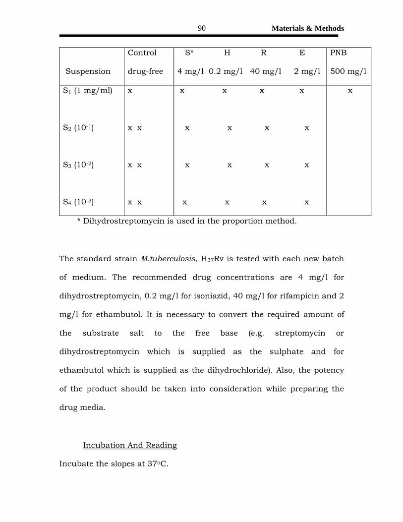
Suspension
Control
drug-free
S* H R E
4 mg/l 0.2 mg/l 40 mg/l 2 mg/l
PNB
500 mg/l
S1 (1 mg/ml)
S2 (10-1)
S3 (10-2)
S4 (10-3)
x
x x
x x
x x
x x x x
x x x x
x x x x
x x x x
x
* Dihydrostreptomycin is used in the proportion method.
The standard strain M.tuberculosis, H37Rv is tested with each new batch
of medium. The recommended drug concentrations are 4 mg/l for
dihydrostreptomycin, 0.2 mg/l for isoniazid, 40 mg/l for rifampicin and 2
mg/l for ethambutol. It is necessary to convert the required amount of
the substrate salt to the free base (e.g. streptomycin or
dihydrostreptomycin which is supplied as the sulphate and for
ethambutol which is supplied as the dihydrochloride). Also, the potency
of the product should be taken into consideration while preparing the
drug media.
Incubation And Reading
Incubate the slopes at 37oC.

Materials & Methods
91
Read the growth at 28 days and again at 42 days.
Record growth as
+ + + Confluent growth
+ + More than 100 colonies
1-100 cols. The actual number of colonies
When the number of colonies on a given dilution is less than 5, count the
number of colonies with the next larger inoculum, or estimate if more
than 100. (Make no attempt to estimate the number of colonies if the
growth is + + +)
Interpretation of Test.
Interpretation of all tests is based on the 42-day readings.
However, if a strain shows clear-cut resistance based on the 28-day
reading, no further reading is needed and the report may be sent as
such. Strains that are susceptible at 28 days must be read again at 42
days and the report is based on the later reading only. For each strain,
express the number of organisms resistant to each drug concentration as
a percentage of the number of organisms growing on the drug -free slope.
Make the selection of slopes for estimating the growth on the drug-free
and drug containing media in the following order of preference:
Drug-free slope:
20-70 colonies
Materials & Methods
92
5–19 colonies
More than 70 colonies
Drug-containing slope:
5-100 colonies in the same row or the row nearest to the control
slope
1-4 colonies in the same row or the row nearest to the control slope
No colonies in the farthest row
More than 100 colonies, if there are no other acceptable counts.
Related Documents