The ovarian follicular wave pattern and induction of ovulation in the mated and non-mated one-humped camel (Camelus dromedarius) J. A. Skidmore, M. Billah and W. R. Allen 1The Camel Reproduction Laboratory, Nakhlee, PO Box 11808, Dubai, UAE; and TBA Equ Fertility Unit, Mertoun Paddocks, Woodditton Road, Newmarket, Suffolk CB8 9BH, UK Ovarian follicular wave patterns were studied ultrasonographically in three groups of dromedary. Group 1 camels (n = 20) were teased daily with a vasectomized male but mating was prevented; group 2 camels (n = 8) ran freely with a vasectomized male camel for 10 h each day and group 3 camels (n = 8) were kept completely separate from any males. In a second experiment (n = 63), when the diameter of the dominant follicle reached 0.5\p=n-\0.9cm, 1.0\p=n-\1.9cm, 2.0\p=n-\2.9cm or > 3 cm, the camel was given one of three treatments to induce ovulation: (i) natural mating; (ii) 20 \g=m\g of the GnRH analogue, buserelin; or (iii) 3000 iu hCG. The ovaries were re-scanned regularly to monitor ovulation, and daily blood samples were assayed for progesterone and oestradiol concentrations. The follicular cycle was divisible into a growth phase (10.5 \m=+-\0.5 days), a mature phase (7.6 \m=+-\0.8 days) and a regression phase (11.9 \m=+-\0.8 days). The dominant follicle reached a mean \m=+-\SEM maximum diameter of 2.0 \m=+-\0.1 cm (range 1.5\p=n-\2.5 cm) in 34 cycles (52%) before it began to regress. In the other 32 cycles (48%), however, the dominant follicle continued to grow to 4.2 \m=+-\0.2 cm (range 4.0\p=n-\6.0 cm) before regression commenced. Group 2 camels were mated when their follicles reached 1.3 \m=+-\ 0.1cm in diameter and the mean interval between successive matings was 13.8 \m=+-\1.0 days. Mean \m=+-\sem serum concentrations of oestradiol reached peak values at 39.0 \m=+-\1.8 pg ml \m=-\1, when the dominant follicle measured 1.7 \m=+-\ 0.1 cm and, after ovulation, mean serum concentrations of progesterone reached peak values at 2.6 \m=+-\0.3 ng ml \m=-\1on day 8, before decreasing to < 1 ng ml\m=-\1 by day 10 or 11. When the dominant follicle measured 0.5\p=n-\0.9cm in diameter, 70%, 60% and 60% of them ovulated in response to mating, or treatment with buserelin or hCG, respectively. These ovulation rates increased to 85% (mating), 81% (buserelin) and 67% (hCG) when the follicle measured 1\p=n-\1.9 cm, but they decreased again to 12.5% (mating), 29% (buserelin) and 13% (hCG) when the diameter had increased to 2.0\p=n-\2.9cm at the time of treatment. No follicles measuring > 3.0 cm ovulated in response to any of the treatments. These results indicated that the optimum time to mate or attempt to induce ovulation in the female dromedary is when the growing follicle measures 0.9\p=n-\1.9cm in diameter. Introduction Camelids of the New World (e.g. llama and alpaca) and the Old World (dromedary and bactrian camel) are seasonal breeders and induced ovulators (Fernandez Baca et al, 1970; Musa and Abusineina, 1978; Adams et al, 1990; Bravo et al, 1990). Original studies on the follicular wave pattern in dromedaries were based on ovaries obtained from an abattoir and on serial palpations of the ovaries per rectum in small numbers of camels. From these surveys the duration of the follicular wave was reported to range from 17.2 to 23.4 days in India (Joshi et al, 1978), and to have a mean duration of 24.2 days in Egypt (Wilson, 1984) and 28 days in Sudan (Musa and Abusineina, 1978). In bactrian camels, Chen and Yuen (1979) reported a mean follicular wave duration of 19 days, with a range of 14—21 days. More recently, realtime ultrasonography has been used to monitor day-to-day ovarian follicular changes in large animals such as cows (Savio et al, 1988; Sirois and Fortune, 1988), sheep (Ravindra et al, 1994), horses (Pierson and Ginther, 1987; Sirois et al, 1989) and llamas (Adams et al, 1990; Bravo et al, 1990). In cows, large antral follicles grow in two (Ginther et al, 1989) or three (Savio et al, 1988; Sirois and Fortune, 1988) waves, each of which is characterized by the simultaneous emergence of a cohort of follicles from a pool of small follicles and the establishment of a dominant follicle that continues to develop while apparently suppressing the growth of the other follicles (Fortune et al, 1988; Pierson and Ginther, 1988; Savio et al, 1988). The factors that control ovulation in the camelids are not well understood. In bactrian camels, ovulation can be induced by deep intravaginal deposition of whole semen or sperm-free *Correspondence. Revised manuscript received 1 November 1995. Downloaded from Bioscientifica.com at 02/01/2022 02:05:00PM via free access

Welcome message from author
This document is posted to help you gain knowledge. Please leave a comment to let me know what you think about it! Share it to your friends and learn new things together.
Transcript
The ovarian follicular wave pattern and induction of ovulation in themated and non-mated one-humped camel (Camelus dromedarius)
J. A. Skidmore, M. Billah and W. R. Allen1The Camel Reproduction Laboratory, Nakhlee, PO Box 11808, Dubai, UAE; and TBA Equ
Fertility Unit, Mertoun Paddocks, Woodditton Road, Newmarket, Suffolk CB8 9BH, UK
Ovarian follicular wave patterns were studied ultrasonographically in three groups ofdromedary. Group 1 camels (n = 20) were teased daily with a vasectomized male but matingwas prevented; group 2 camels (n = 8) ran freely with a vasectomized male camel for 10 heach day and group 3 camels (n = 8) were kept completely separate from any males. In a
second experiment (n = 63), when the diameter of the dominant follicle reached 0.5\p=n-\0.9cm,1.0\p=n-\1.9cm, 2.0\p=n-\2.9cm or > 3 cm, the camel was given one of three treatments to induceovulation: (i) natural mating; (ii) 20 \g=m\gof the GnRH analogue, buserelin; or (iii) 3000 iu hCG.The ovaries were re-scanned regularly to monitor ovulation, and daily blood samples were
assayed for progesterone and oestradiol concentrations. The follicular cycle was divisibleinto a growth phase (10.5 \m=+-\0.5 days), a mature phase (7.6 \m=+-\0.8 days) and a regressionphase (11.9 \m=+-\0.8 days). The dominant follicle reached a mean \m=+-\SEM maximum diameter of2.0 \m=+-\0.1 cm (range 1.5\p=n-\2.5cm) in 34 cycles (52%) before it began to regress. In the other32 cycles (48%), however, the dominant follicle continued to grow to 4.2 \m=+-\0.2 cm (range4.0\p=n-\6.0cm) before regression commenced. Group 2 camels were mated when their folliclesreached 1.3 \m=+-\0.1cm in diameter and the mean interval between successive matings was
13.8 \m=+-\1.0 days. Mean \m=+-\sem serum concentrations of oestradiol reached peak values at39.0 \m=+-\1.8 pg ml \m=-\1, when the dominant follicle measured 1.7 \m=+-\0.1 cm and, after ovulation,mean serum concentrations of progesterone reached peak values at 2.6 \m=+-\0.3 ng ml \m=-\1on
day 8, before decreasing to < 1 ng ml\m=-\1by day 10 or 11. When the dominant folliclemeasured 0.5\p=n-\0.9cm in diameter, 70%, 60% and 60% of them ovulated in response tomating, or treatment with buserelin or hCG, respectively. These ovulation rates increased to85% (mating), 81% (buserelin) and 67% (hCG) when the follicle measured 1\p=n-\1.9cm, but theydecreased again to 12.5% (mating), 29% (buserelin) and 13% (hCG) when the diameter hadincreased to 2.0\p=n-\2.9cm at the time of treatment. No follicles measuring > 3.0 cm ovulatedin response to any of the treatments. These results indicated that the optimum time to mateor attempt to induce ovulation in the female dromedary is when the growing folliclemeasures 0.9\p=n-\1.9cm in diameter.
Introduction
Camelids of the New World (e.g. llama and alpaca) and the OldWorld (dromedary and bactrian camel) are seasonal breedersand induced ovulators (Fernandez Baca et al, 1970; Musa andAbusineina, 1978; Adams et al, 1990; Bravo et al, 1990).Original studies on the follicular wave pattern in dromedarieswere based on ovaries obtained from an abattoir and on serialpalpations of the ovaries per rectum in small numbers of camels.From these surveys the duration of the follicular wave was
reported to range from 17.2 to 23.4 days in India (Joshiet al, 1978), and to have a mean duration of 24.2 days in Egypt(Wilson, 1984) and 28 days in Sudan (Musa and Abusineina,1978). In bactrian camels, Chen and Yuen (1979) reported a
mean follicular wave duration of 19 days, with a range of14—21 days. More recently, realtime ultrasonography has beenused to monitor day-to-day ovarian follicular changes in largeanimals such as cows (Savio et al, 1988; Sirois and Fortune,1988), sheep (Ravindra et al, 1994), horses (Pierson andGinther, 1987; Sirois et al, 1989) and llamas (Adams et al,1990; Bravo et al, 1990). In cows, large antral follicles grow intwo (Ginther et al, 1989) or three (Savio et al, 1988; Sirois andFortune, 1988) waves, each of which is characterized by thesimultaneous emergence of a cohort of follicles from a pool ofsmall follicles and the establishment of a dominant follicle thatcontinues to develop while apparently suppressing the growthof the other follicles (Fortune et al, 1988; Pierson and Ginther,1988; Savio et al, 1988).
The factors that control ovulation in the camelids are notwell understood. In bactrian camels, ovulation can be inducedby deep intravaginal deposition of whole semen or sperm-free*Correspondence.
Revised manuscript received 1 November 1995.Downloaded from Bioscientifica.com at 02/01/2022 02:05:00PM
via free access
seminal plasma (Chen et al, 1985), as well as by i.m. injectionof semen or seminal fluid (Zhao et al, 1990). In dromedaries,ovulation is induced by mating with an intact or vasectomizedmale (Marie and Anouassi, 1987), but manual stimulation of thecervix or intrauterine injection of whole semen, seminal plasma,water or the prostaglandin F analogue, cloprostenol, does notstimulate the release of sufficient LH from the pituitary to causeovulation (Musa and Abusineina, 1978; Sheldrick et al, 1992).Efficient methods to induce and synchronize ovulation indromedaries are now required as interest grows in the potentialapplication of artificial insemination and embryo transfer to thisspecies (Cooper et al, 1992). However, mating to a vasect¬omized male is not acceptable because of the potential forspreading venereal diseases. This leaves treatment with an
LH-like gonadotrophic hormone at the optimal time in thefollicular growth cycle as the most practical alternative.
The purpose of the present study was twofold. First, todefine accurately the patterns of follicular growth and regres¬sion in dromedaries by means of repeated realtime ultrasono¬graphic examinations of the ovaries, and to relate thesemorphological events to changes in behaviour and steroidhormone concentrations in peripheral blood. Second, to com¬
pare the efficacy of two forms of gonadotrophin therapy toinduce ovulation in non-mated camels and to determine theoptimal time in the follicular wave cycle for their application.
Materials and Methods
AnimalsA total of 73 mature female dromedaries aged 5—14 years
was used in two experiments. They were maintained in groupsof 8-30 animals in fenced pens, each of 0.002-0.004 km2 area.
They were fed a diet of mixed concentrates and lucerne hayonce a day and were given water ad libitum. Thirty-six camelswere used in Expt 1 (1 December—31 January, 1993—1994) and60 were used in Expt 2 (12 November-30 March, 1993-1994).
Ovarian examinations
The ovaries of every camel were examined on alternate daysthroughout the defined experimental periods by transrectalultrasonography as described by Skidmore et al (1992). AnAloka Model 500 realtime scanner with a 5 MHz linear arraytransducer (Al Carmal Ltd, Dubai, UAE) was used and allfollicles > 0.5 cm in diameter and corpora lutea present in bothovaries were counted, measured using the internal electroniccalipers and photographed using a Sony Model UP 860CEvideoprinter (Al Carmal Ltd) interfaced with the scanner.
Blood sampling and hormone assays
Samples of jugular vein blood (8 ml) were recovered dailyfrom all camels throughout the experimental periods. Theywere kept at room temperature (19—23°C) for 1-2 h beforebeing centrifuged at 1500 g for 5 min. The serum was decantedand stored at
—
20°C until subsequent assay for progesteroneand oestradiol. Owing to a limited supply of antibody and
90
coo 50
20
10m
0.1 1.0 10.0 100Unlabelled progesterone (ng) (log scale)
Fig. 1. Typical standard curves for the progesterone assay:(i) standards made up in human serum supplied in the assay kit;(ii) standards made up in charcoal-stripped male camel serum; (iii) and(iv) serial dilutions of a sample of camel serum containing a highconcentration of progesterone diluted in either (iii) human serum or
(iv) charcoal-stripped male camel serum. Results are plotted as
log—logit linear transformations.
other materials for the oestradiol assay, the sera from onlyeight camels could be assayed for oestradiol concentration.These animals were selected randomly from Group 1 of Expt I.
Progesterone assay. Progesterone concentrations were
measured directly in 25 µ aliquots of sample or standard in a
commercially available assay kit (Amerlite Progesterone Assay:Kodak Clinical Diagnostics, Amersham) which used a competi¬tive immunoassay technique based on enhanced luminescence(Whitehead et al, 1983). The suitability of the assay for use
with camel serum was tested by comparing the parallelism ofthe standard curves produced by standards made up incharcoal-stripped male camel serum and the human serum
provided in the assay kit. In addition, a sample of pregnantfemale camel serum with a high progesterone concentrationwas diluted serially in male camel serum and human serum andrun against the standard curves in the assay. All the curves
were parallel with the kit standard curve (Fig. 1), indicating thatcamel serum did not interfere with binding in the assay. Theminimum detection limit of the assay was 0.11 ng progesteroneml- serum and the intra- and interassay coefficients ofvariation, calculated by the method described by Dighe andHunter (1976), were found to be 7.5% and 8%, respectively.The crossreactivities of the anti-progesterone serum used in theassay were stated by the manufacturers to be 5.2% with5a-pregnane-3,20-dione, 4.17% with 5ß-pregnane-3,20-dioneand 1.1% with 20a-dihydroprogesterone.
Oestradiol assay. Concentrations of free oestradiol were
measured in selected sera recovered in Expt 1, using a radio¬immunoassay based on the method described by Heap andHamon (1979). The anti-oestradiol serum was raised in a rabbitimmunized with oestradiol-6-(CMO) conjugated to bovineserum albumin (E002; Steranti Research Ltd, Herts) and itshowed stated crossreactivities of 0.55% with oestrone and0.19% with oestriol. Aliquots (1 ml) of serum were extractedwith 5 ml diethyl ether before being assayed in duplicate. Tomeasure procedural losses during the extraction process, 0.1 ml
Downloaded from Bioscientifica.com at 02/01/2022 02:05:00PMvia free access
90 r.
_CDCO
a so
Sa 20CO
DÛ 10DÛ<S)
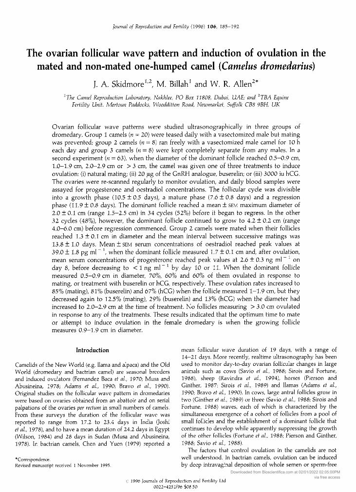
10 100 1000Unlabelled oestradiol (pg) (log scale)
Fig. 2. (i) A typical standard curve for oestradiol and, (ii) a serialdilution curve of a sample of camel serum known to contain a highconcentration of oestradiol. Results are plotted as log—logit lineartransformations. NSB: nonspecific binding.
of the tritiated oestradiol ([2,4,6,7-H]oestradiol; specific activity3.15 TBq µ " ; Amersham Life Sciences, Bucks) was addedto 1 ml of charcoal-stripped male camel serum and extracted asabove. This gave a mean recovery rate of 82.0+ 1.7%.
The assay was validated for camel serum by measuring therecovery of oestradiol added to a sample of male camel serum
to give concentrations of 25, 100 and 250 pg oestradiol ml- ;the mean recovery of this cold oestradiol was 109 ± 6.9%. Inaddition, a sample of camel serum containing a known highconcentration of oestradiol was diluted serially and assayedagainst the standard curve. This gave a parallel curve, therebyindicating that camel serum did not interfere with binding inthe assay (Fig. 2). The intra- and interassay coefficients ofvariation were 6.2% and 10.1%, respectively, and the meanminimum detection limit was 8.3 pg ml
~
\
Experimental designExperiment 1. The 36 camels in this experiment were
divided into three groups. Group 1 animals (n = 20) were keptin one pen and were teased daily with a vasectomized malecamel for at least 30 min in the early morning. Signs of oestrus(following the male, stradling the hind legs, lifting the tail andurinating when being examined by the male) were recorded butmating was not allowed. Group 2 (n = 8) camels were kept inanother pen in which a vasectomized male was allowed to run
freely for 10 h each day. They were observed constantly forsigns of oestrus and any matings that occurred were recordedand timed. Group 3 (n = 8) camels were kept together in an
isolated pen that was out of the sight, sound and smell of anymale camels so that their follicular wave patterns could bemonitored in the complete absence of any male influence.
On the basis of the alternate day scanning results, a growthand regression profile of the dominant follicle was plotted for
each camel in the three groups. This was defined as a folliclethat grew to at least 1.5 cm and exceeded the diameter of allother follicles. These individual dominant follicle profiles werestarted when the follicle was first detected at a diameter of0.5 cm. The mean number of follicles present in both ovaries,from 20 days before to 20 days after a reference point, wastabulated in relation to the diameter of the largest follicle, toconfirm that follicular growth occurred in waves. Becauseovulation did not occur in group 1 and group 3 camels, thiscommon reference point (day 0) was taken as the day on whichthe dominant follicle first reached its maximum diameter.
Experiment 2. The ovaries of all 60 camels included in theexperiment were examined ultrasonographically on alternatedays throughout the defined experimental periods. All follicles> 0.5 cm in diameter were counted and measured and whenthe dominant follicle reached a diameter of 0.5-0.9 cm (n = 30),1.0-1.9 cm (ii = 51), 2.0-2.9 cm (« = 48) or > 3 cm (n = 45),the camel was assigned to one of three treatment groups toinduce ovulation. These treatments were: (i) natural mating(n = 61); (ii) 20 pg of the GnRH analogue, buserelin (Receptal:Hoechst Animal Health, Milton Keynes) given i.v. (n = 58); or
(iii) 3000 iu hCG (Chorulon: Intervet Laboratories, Cambridge),also given i.v. (n = 55). The ovaries were rescanned at 36, 48,72 and 96 h after treatment to monitor ovulation, which was
confirmed subsequently by measuring a rise in peripheral serum
progesterone concentrations. Some camels were used more
than once in the experiment but, in each case, at least one
complete follicular cycle was allowed to occur betweensuccessive treatments.
Statistical analysesTo investigate possible associations between the number of
follicles and the diameter of the largest follicle, the productmoment correlation coefficient (r) was calculated. The resultwas confirmed by carrying out a periodic regression analysisusing sine and cosine terms, as well as calculating the equiva¬lent rank coefficient (Kendall's Tau). Student's t test was usedto compare cycles in which follicles grew to 1.5-2.5 cm indiameter with those in which they grew to 2.6—6.4 cm indiameter, and differences were considered to be significant if < 0.05. Fisher's exact test was used to compare the ovulationresponses of follicles of different sizes. The relationshipbetween oestradiol concentrations and follicle growth was
emphasized by calculating the magnitude of the mean ( ± sem)differences between the time periods days
-
8—0, anddays 0- + 8.
Results
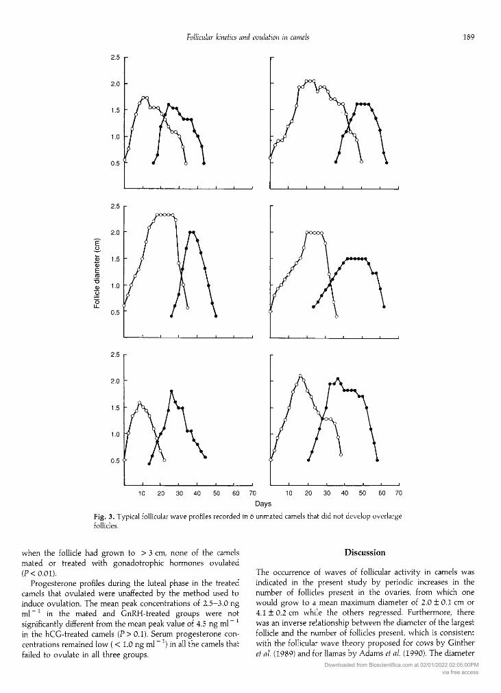
Experiment 1: follicular wave dynamicsAlthough the follicular wave pattern varied considerably
between individual camels in both group 1 and group 3, it was
none the less clearly divisible into three phases in all animals:a growth phase, a mature phase and a regression phase(see Table 1; Fig. 3). New follicles became visible and started togrow before the mature follicle had fully regressed, to give a
Downloaded from Bioscientifica.com at 02/01/2022 02:05:00PMvia free access
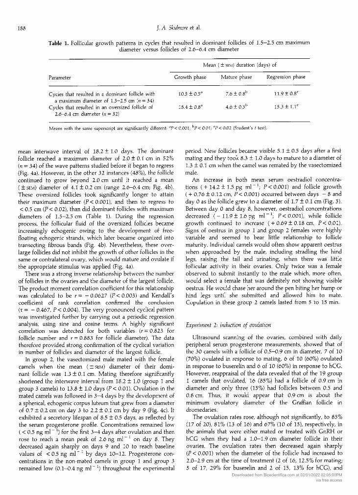
Table 1. Follicular growth patterns in cycles that resulted in dominant follicles of 1.5—2.5 cm maximumdiameter versus follicles of 2.6—6.4 cm diameter
Mean ( ± sem) duration (days) of
Parameter Growth phase Mature phase Regression phase
Cycles that resulted in a dominant follicle with 10.5 ± 0.5'' 7.6 ± 0.8b 11.9 ± 0.8Ca maximum diameter of 1.5-2.5 cm (n = 34)
Cycles that resulted in an oversized follicle of 18.4 ± 0.8a 4.6 ± 0.5b 15.3 + 1.Ie2.6-6.4 cm diameter (n = 32)
Means with the same superscript are significantly different: 'P < 0.001: hP < 0.01; CP < 0.02 (Student's I test).
mean interwave interval of 18.2 ± 1.0 days. The dominantfollicle reached a maximum diameter of 2.0 ± 0.1 cm in 52%(n
-
34) of the wave patterns studied before it began to regress(Fig. 4a). However, in the other 32 instances (48%), the folliclecontinued to grow beyond 2.0 cm until it reached a mean
( ± sem) diameter of 4.1 ± 0.2 cm (range 2.6-6.4 cm; Fig. 4b).These oversized follicles took significantly longer to attaintheir maximum diameter (P< 0.001), and then to regress to< 0.5 cm (P < 0.02), than did dominant follicles with maximum
diameters of 1.5-2.5 cm (Table 1). During the regressionprocess, the follicular fluid of the oversized follicles becameincreasingly echogenic owing to the development of free-floating echogenic strands, which later became organized intotransecting fibrous bands (Fig. 4b). Nevertheless, these over-
large follicles did not inhibit the growth of other follicles in thesame or contralateral ovary, which would mature and ovulate ifthe appropriate stimulus was applied (Fig. 4a).
There was a strong inverse relationship between the numberof follicles in the ovaries and the diameter of the largest follicle.The product moment correlation coefficient for this relationshipwas calculated to be r= -0.0627 (P< 0.003) and Kendall'scoefficient of rank correlation confirmed the conclusion( =
—
0.467, < 0.004). The very pronounced cyclical patternwas investigated further by carrying out a periodic regressionanalysis, using sine and cosine terms. A highly significantcorrelation was detected for both variables (r = 0.823 forfollicle number and r = 0.883 for follicle diameter). The datatherefore provided strong confirmation of the cyclical variationin number of follicles and diameter of the largest follicle.
In group 2, the vasectomized male mated with the femalecamels when the mean ( ± sem) diameter of their domi¬nant follicle was 1.3 ± 0.1 cm. Mating therefore significantlyshortened the interwave interval from 18.2 ± 1.0 (group 1 andgroup 3 camels) to 13.8 ± 1.0 days (P < 0.01). Ovulation in themated camels was followed in 3—4 days by the development ofa spherical, echogenic corpus luteum that grew from a diameterof 0.7 ± 0.2 cm on day 3 to 2.2 ± 0.1 cm by day 9 (Fig. 4c). Itexhibited a secretory lifespan of 8.5 ± 0.5 days, as reflected bythe serum progesterone profile. Concentrations remained low( < 0.5 ng ml
~ ) for the first 3—4 days after ovulation and thenrose to reach a mean peak of 2.6 ng ml" on day 8. Theydecreased again sharply on days 9 and 10 to reach baselinevalues of < 0.5 ng ml
~
' by days 10—12. Progesterone con¬
centrations in the non-mated camels in group 1 and group 3remained low (0.1—0.4 ng ml~ ) throughout the experimental
period. New follicles became visible 5.1 ±0.5 days after a firstmating and they took 8.3 + 1.0 days to mature to a diameter of1.3 ± 0.1 cm when the camel was remated by the vasectomizedmale.
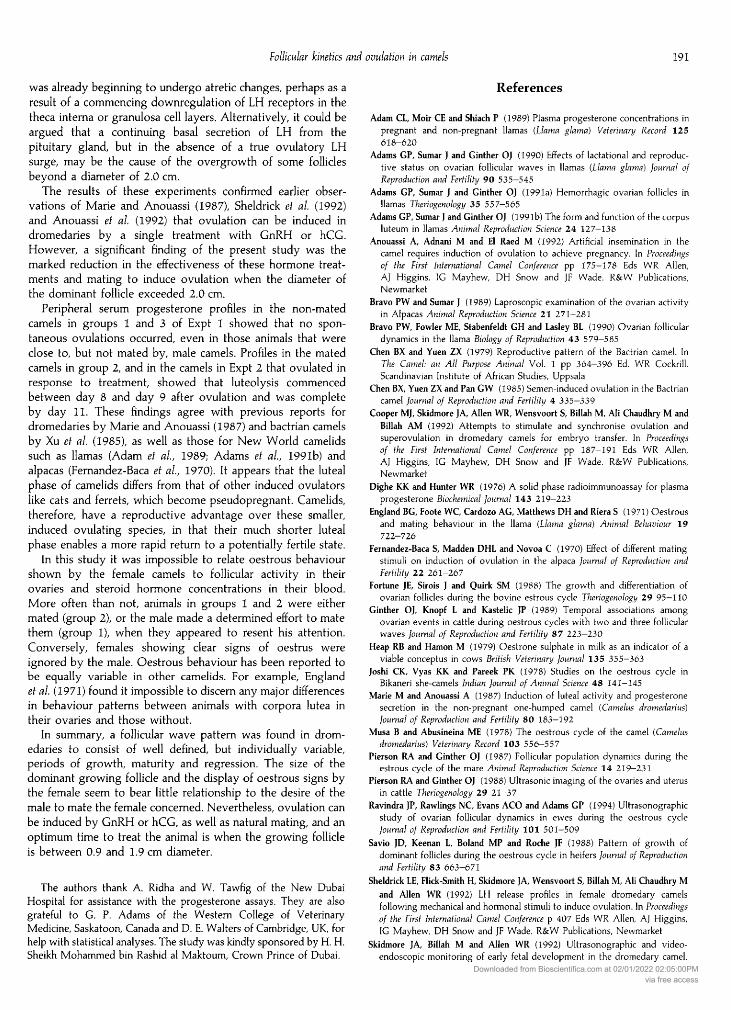
An increase in both mean serum oestradiol concentra¬tions (+14.2 +1.5 pg ml-1; < 0.001) and follicle growth( + 0.76 ± 0.12 cm, < 0.001) occurred between days
-
8 andday 0 as the follicle grew to a diameter of 1.7 ± 0.1 cm (Fig. 5).Between day 0 and day 8, however, oestradiol concentrationsdecreased (-11.9 ± 1.6 pg ml"1; < 0.001), while folliclegrowth continued to increase ( +0.69 ± 0.18 cm, < 0.01).Signs of oestrus in group 1 and group 2 females were highlyvariable and seemed to bear little relationship to folliclematurity. Individual camels would often show apparent oestruswhen approached by the male, including stradling the hindlegs, raising the tail and urinating, when there was littlefollicular activity in their ovaries. Only twice was a femaleobserved to submit instantly to the male which, more often,would select a female that was definitely not showing visibleoestrus. He would chase her around the pen biting her hump or
hind legs until she submitted and allowed him to mate.Copulation in these group 2 camels lasted from 5 to 15 min.
Experiment 2: induction of ovulationUltrasound scanning of the ovaries, combined with daily
peripheral serum progesterone measurements, showed that ofthe 30 camels with a follicle of 0.5-0.9 cm in diameter, 7 of 10(70%) ovulated in response to mating, 6 of 10 (60%) ovulatedin response to buserelin and 6 of 10 (60%) in response to hCG.However, reappraisal of the data revealed that of the 19 group1 camels that ovulated, 16 (85%) had a follicle of 0.9 cm indiameter and only three (15%) had follicles between 0.5 and0.8 cm. Thus, it would appear that 0.9 cm is about theminimum ovulatory diameter of the Graffian follicle indromedaries.
The ovulation rates rose, although not significantly, to 85%(17 of 20), 81% (13 of 16) and 67% (10 of 15), respectively, inthe animals that were either mated or treated with GnRH or
hCG when they had a 1.0-1.9 cm diameter follicle in theirovaries. The ovulation rates then decreased again sharply(P < 0.001) when the diameter of the follicle had increased to2.0-2.9 cm at the time of treatment (2 of 16, 12.5% for mating;5 of 17, 29% for buserelin and 2 of 15, 13% for hCG), and
Downloaded from Bioscientifica.com at 02/01/2022 02:05:00PMvia free access
2.5
2.0
0.5 f
_1_I_L
2.5
-J_I-L_
2.5
2.0
0.5 -
J_I_I_I_I_l_
10 20 30 40 50 60 70 10 20 30 40 50 60 70
DaysFig. 3. Typical follicular wave profiles recorded in 6 unmated camels that did not develop overlargefollicles.
when the follicle had grown to > 3 cm, none of the camelsmated or treated with gonadotrophic hormones ovulated(P<0.01).
Progesterone profiles during the luteal phase in the treatedcamels that ovulated were unaffected by the method used toinduce ovulation. The mean peak concentrations of 2.5-3.0 ngml ~l in the mated and GnRH-treated groups were notsignificantly different from the mean peak value of 4.5 ng ml
~
in the hCG-treated camels (P>0.1). Serum progesterone con¬
centrations remained low ( < 1.0 ng ml~
) in all the camels thatfailed to ovulate in all three groups.
Discussion
The occurrence of waves of follicular activity in camels was
indicated in the present study by periodic increases in thenumber of follicles present in the ovaries, from which one
would grow to a mean maximum diameter of 2.0 ±0.1 cm or
4.1 ± 0.2 cm while the others regressed. Furthermore, therewas an inverse relationship between the diameter of the largestfollicle and the number of follicles present, which is consistentwith the follicular wave theory proposed for cows by Gintheret al (1989) and for llamas by Adams et al (1990). The diameter
Downloaded from Bioscientifica.com at 02/01/2022 02:05:00PMvia free access
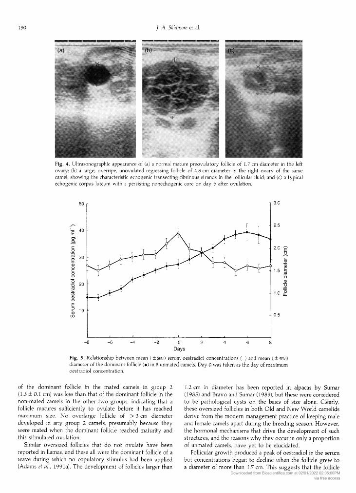
Fig. 4. Ultrasonographic appearance of (a) a normal mature preovulatory follicle of 1.7 cm diameter in the leftovary; (b) a large, overripe, unovulated regressing follicle of 4.8 cm diameter in the right ovary of the same
camel, showing the characteristic echogenic transecting fibrinous strands in the follicular fluid; and (c) a typicalechogenic corpus luteum with a persisting nonechogenic core on day 6 after ovulation.
Fig. 5. Relationship between mean ( + sem) serum oestradiol concentrations (: ) and mean ( ± sem)diameter of the dominant follicle (·) in 8 unmated camels. Day 0 was taken as the day of maximumoestradiol concentration.
of the dominant follicle in the mated camels in group 2(1.3 ±0.1 cm) was less than that of the dominant follicle in thenon-mated camels in the other two groups, indicating that a
follicle matures sufficiently to ovulate before it has reachedmaximum size. No overlarge follicle of > 3 cm diameterdeveloped in any group 2 camels, presumably because theywere mated when the dominant follicle reached maturity andthis stimulated ovulation.
Similar oversized follicles that do not ovulate have beenreported in llamas, and these all were the dominant follicle of a
wave during which no copulatory stimulus had been applied(Adams et al, 1991a). The development of follicles larger than
1.2 cm in diameter has been reported in alpacas by Sumar(1985) and Bravo and Sumar (1989), but these were consideredto be pathological cysts on the basis of size alone. Clearly,these oversized follicles in both Old and New World camelidsderive from the modern management practice of keeping maleand female camels apart during the breeding season. However,the hormonal mechanisms that drive the development of suchstructures, and the reasons why they occur in only a proportionof unmated camels, have yet to be elucidated.
Follicular growth produced a peak of oestradiol in the serumbut concentrations began to decline when the follicle grew toa diameter of more than 1.7 cm. This suggests that the follicle
Downloaded from Bioscientifica.com at 02/01/2022 02:05:00PMvia free access
was already beginning to undergo atretic changes, perhaps as a
result of a commencing downregulation of LH receptors in thetheca interna or granulosa cell layers. Alternatively, it could beargued that a continuing basal secretion of LH from thepituitary gland, but in the absence of a true ovulatory LHsurge, may be the cause of the overgrowth of some folliclesbeyond a diameter of 2.0 cm.
The results of these experiments confirmed earlier obser¬vations of Marie and Anouassi (1987), Sheldrick et al (1992)and Anouassi et al (1992) that ovulation can be induced indromedaries by a single treatment with GnRH or hCG.However, a significant finding of the present study was themarked reduction in the effectiveness of these hormone treat¬ments and mating to induce ovulation when the diameter ofthe dominant follicle exceeded 2.0 cm.
Peripheral serum progesterone profiles in the non-matedcamels in groups 1 and 3 of Expt 1 showed that no spon¬taneous ovulations occurred, even in those animals that wereclose to, but not mated by, male camels. Profiles in the matedcamels in group 2, and in the camels in Expt 2 that ovulated inresponse to treatment, showed that luteolysis commencedbetween day 8 and day 9 after ovulation and was completeby day 11. These findings agree with previous reports fordromedaries by Marie and Anouassi (1987) and bactrian camelsby Xu el al (1985), as well as those for New World camelidssuch as llamas (Adam et al, 1989; Adams et al, 1991b) andalpacas (Fernandez-Baca et al, 1970). It appears that the lutealphase of camelids differs from that of other induced ovulatorslike cats and ferrets, which become pseudopregnant. Camelids,therefore, have a reproductive advantage over these smaller,induced ovulating species, in that their much shorter lutealphase enables a more rapid return to a potentially fertile state.
In this study it was impossible to relate oestrous behaviourshown by the female camels to follicular activity in theirovaries and steroid hormone concentrations in their blood.More often than not, animals in groups 1 and 2 were eithermated (group 2), or the male made a determined effort to matethem (group 1), when they appeared to resent his attention.Conversely, females showing clear signs of oestrus were
ignored by the male. Oestrous behaviour has been reported tobe equally variable in other camelids. For example, Englandet al (1971) found it impossible to discern any major differencesin behaviour patterns between animals with corpora lutea intheir ovaries and those without.
In summary, a follicular wave pattern was found in drom¬edaries to consist of well defined, but individually variable,periods of growth, maturity and regression. The size of thedominant growing follicle and the display of oestrous signs bythe female seem to bear little relationship to the desire of themale to mate the female concerned. Nevertheless, ovulation can
be induced by GnRH or hCG, as well as natural mating, and an
optimum time to treat the animal is when the growing follicleis between 0.9 and 1.9 cm diameter.
The authors thank A. Ridha and W. Tawfig of the New DubaiHospital for assistance with the progesterone assays. They are alsograteful to G. P. Adams of the Western College of VeterinaryMedicine, Saskatoon, Canada and D. E. Walters of Cambridge, UK, forhelp with statistical analyses. The study was kindly sponsored by H. H.Sheikh Mohammed bin Rashid al Maktoum, Crown Prince of Dubai.
References
Adam CL, Moir CE and Shiach P (1989) Plasma progesterone concentrations inpregnant and non-pregnant llamas (Llama glama) Veterinary Record 125ó 18-620
Adams GP, Sumar J and Ginther OJ (1990) Effects of lactational and reproduc¬tive status on ovarian follicular waves in llamas (Llama glama) Journal ofReproduction and Fertility 90 535—545
Adams GP, Sumar J and Ginther OJ (1991a) Hemorrhagic ovarian follicles inllamas Theriogenology 35 557-565
Adams GP, Sumar J and Ginther OJ (1991b) The form and function of the corpusluteum in llamas Animal Reproduction Science 24 127—138
Anouassi A, Adnani M and El Raed M (1992) Artificial insemination in thecamel requires induction of ovulation to achieve pregnancy. In Proceedingsof the First International Camel Conference pp 175—178 Eds WR Allen,AJ Higgins, IG Mayhew, DH Snow and JF Wade. R&W Publications,Newmarket
Bravo PW and Sumar J (1989) Laproscopic examination of the ovarian activityin Alpacas Animal Reproduction Science 21 271—281
Bravo PW, Fowler ME, Stabenfeldt GH and Lasley BL (1990) Ovarian folliculardynamics in the llama Biology of Reproduction 43 579—585
Chen BX and Yuen ZX (1979) Reproductive pattern of the Bactrian camel. InThe Camel: an All Purpose Animal Vol. 1 pp 364-396 Ed. WR Cockrill.Scandinavian Institute of African Studies, Uppsala
Chen BX, Yuen ZX and Pan GW (1985) Semen-induced ovulation in the Bactriancamel Journal of Reproduction and Fertility 4 335—339
Cooper MJ, Skidmore JA, Allen WR, Wensvoort S, Billah M, Ali Chaudhry M andBillah AM (1992) Attempts to stimulate and synchronise ovulation andsuperovulation in dromedary camels for embryo transfer. In Proceedingsof the First International Camel Conference pp 187—191 Eds WR Allen,AJ Higgins, IG Mayhew, DH Snow and JF Wade. R&W Publications,Newmarket
Dighe KK and Hunter WR (1976) A solid phase radioimmunoassay for plasmaprogesterone Biochemical Journal 143 219—223
England BG, Foote WC, Cardozo AG, Matthews DH and Riera S (1971) Oestrousand mating behaviour in the llama {Llama glama) Animal Behaviour 19722-726
Fernandez-Baca S, Madden DHL and Novoa C (1970) Effect of different matingstimuli on induction of ovulation in the alpaca Journal of Reproduction andFertility 22 261-267
Fortune JE, Sirois J and Quirk SM (1988) The growth and differentiation ofovarian follicles during the bovine estrous cycle Theriogenology 29 95—110
Ginther OJ, Knopf L and Kastelic JP (1989) Temporal associations amongovarian events in cattle during oestrous cycles with two and three follicularwaves Journal of Reproduction and Fertility 87 223—230
Heap RB and Hamon M (1979) Oestrone sulphate in milk as an indicator of a
viable conceptus in cows British Veterinary Journal 135 355—363Joshi CK, Vyas KK and Pareek PK (1978) Studies on the oestrous cycle in
Bikaneri she-camels Indian Journal of Animal Science 48 141—145Marie M and Anouassi A (1987) Induction of luteal activity and progesterone
secretion in the non-pregnant one-humped camel (Camelus dromedarius)Journal of Reproduction and Fertility 80 183—192
Musa and Abusineina ME (1978) The oestrous cycle of the camel (Camelusdromedarius) Veterinary Record 103 556—557
Pierson RA and Ginther OJ (1987) Follicular population dynamics during theestrous cycle of the mare Animal Reproduction Science 14 219—231
Pierson RA and Ginther OJ (1988) Ultrasonic imaging of the ovaries and uterusin cattle Theriogenology 29 21—37
Ravindra JP, Rawlings NC, Evans ACO and Adams GP (1994) Ultrasonographicstudy of ovarian follicular dynamics in ewes during the oestrous cycleJournal of Reproduction and Fertility 101 501—509
Savio JD, Keenan L, Boland MP and Roche JF (1988) Pattern of growth ofdominant follicles during the oestrous cycle in heifers Journal of Reproductionand Fertility 83 663-671
Sheldrick LE, Flick-Smith H, Skidmore JA, Wensvoort S, Billah M, Ali Chaudhry Mand Allen WR (1992) LH release profiles in female dromedary camelsfollowing mechanical and hormonal stimuli to induce ovulation. In Proceedingsof the First International Camel Conference 407 Eds WR Allen, AJ Higgins,IG Mayhew, DH Snow and JF Wade. R&W Publications, Newmarket
Skidmore JA, Billah M and Allen WR (1992) Ultrasonographic and video-endoscopic monitoring of early fetal development in the dromedary camel.
Downloaded from Bioscientifica.com at 02/01/2022 02:05:00PMvia free access
In Proceedings of the First International Camel Conference pp 193—201Eds WR Allen, AJ Higgins, IG Mayhew, DH Snow and JF Wade.R&W Publications, Newmarket
Sirois J and Fortune JE (1988) Ovarian follicular dynamics during theestrous cycle in heifers monitored by real-time ultrasonography Biology ofReproduction 39 308-317
Sirois J, Ball BA and Fortune JE (1989) Patterns of growth and regression ofovarian follicles during the oestrous cycle and after hemiovariectomy inmares Equine Veterinary Journal Supplement 8 43—48
Sumar J (1985) Reproductive physiology in South American camelids. InGenetics of Reproduction in Sheep pp 81—95 Eds RB Land and DW Robinson.Butterworths, London
Whitehead TP, Thorpe EHG, Carter JJN, Groucutt C and Kricka L] (1983)Enhanced luminescence procedure for sensitive determination of peroxidase-labelled conjugates in immunoassay Nature 305 158—159
Wilson RT (1984) The Camel pp 53—102. Longmans, LondonXu YS, Jian LG, Wang HY, Ge SL, Zeng GQ, Jiang GT and Gao YH (1985)
Hormone concentrations before and after induced ovulation in theBactrian camel (Camelus bactrianus) Journal of Reproduction and Fertility 74341-346
Zhao XX, Pan GW, Huang YM and Chen BX (1990) Studies on the ovulationinducing factor in the seminal plasma of the Bactrian camel. In Proceedings ofthe Workshop "Is it Possible to Improve the Reproductive Performance of theCamel?" pp 197-210 Ed. G Saint-Martin. CIRAO-EMVT, Paris
Downloaded from Bioscientifica.com at 02/01/2022 02:05:00PMvia free access
Related Documents