Mastocytosis Suparat Sirivimonpan 3/5/13

Welcome message from author
This document is posted to help you gain knowledge. Please leave a comment to let me know what you think about it! Share it to your friends and learn new things together.
Transcript
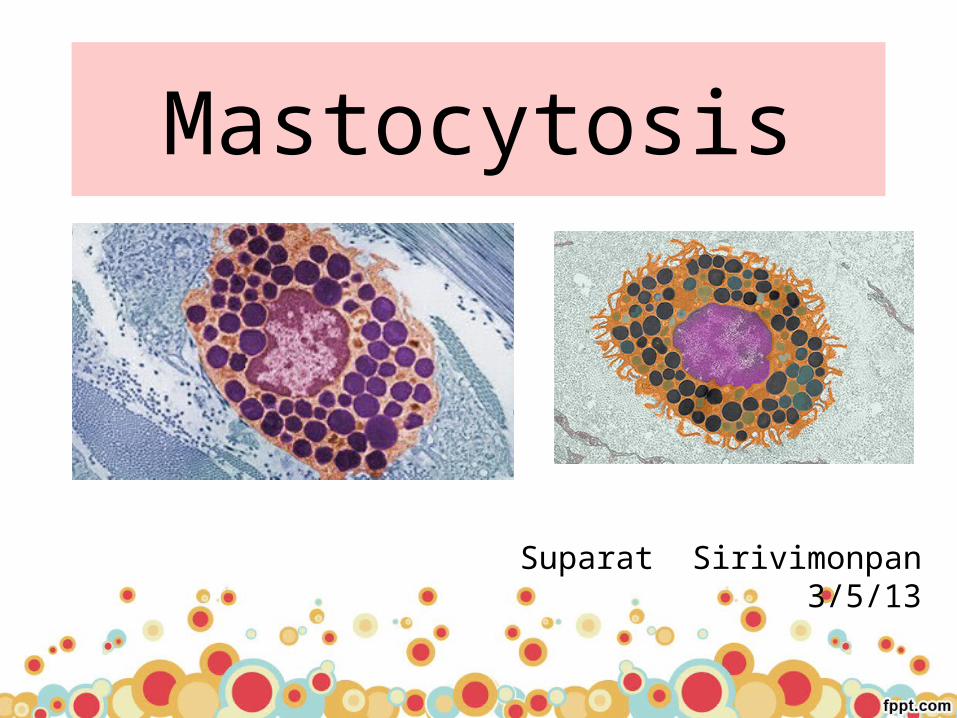
Mastocytosis
Suparat Sirivimonpan3/5/13
Outline
• Introduction• Etiology and pathogenesis• Clinical features and classification• Diagnosis and evaluation• Management• Prognosis

Introduction
• Mast cell disease, or mastocytosis : variety of disorders that are characterized by clonal, neoplastic proliferations of mast cells in one or multiple organs
• The most remarkable pathologic features– mast cell hyperplasia in the skin, GI tract, bone
marrow, liver, spleen, and lymph nodes– frequent association of mast cell hyperplasia with
hematologic disorders
Dean D. Metcalfe .Middleton’s Allergy 7’th edition ,1051-1062.Hematol Oncol Clin N Am 25 (2011) 1067–1083
Introduction
• Clinical features : pruritus, flushing, nausea, vomiting, diarrhea, abdominal pain, and vascular instability
• The prevalence of the disease is unknown• Mastocytosis occurs in all ethnic groups
• may present at any age• Cutaneous mastocytosis : children• Systemic mastocytosis : adults
Dean D. Metcalfe .Middleton’s Allergy 7’th edition ,1051-1062.
Hematol Oncol Clin N Am 26 (2012) 1143–1168
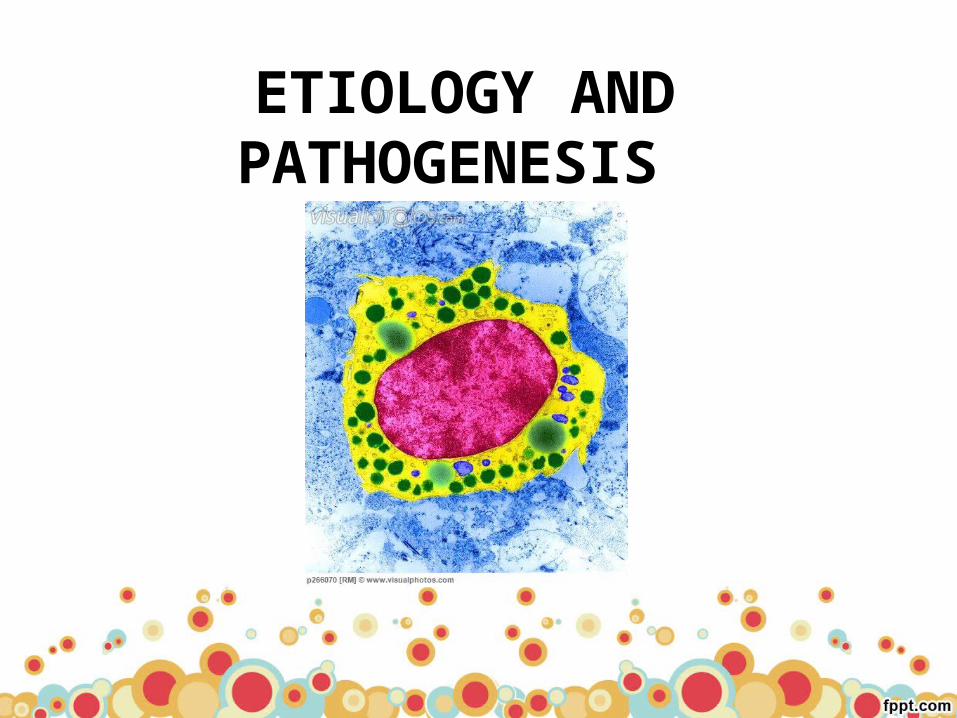
ETIOLOGY AND PATHOGENESIS

Mast cell• Human mast cells develop from a bone marrow-derived
hematopoietic pluripotential precursor cell (CD34+, Kit [CD117]+)
• complete maturation in vascularized peripheral tissues• During this maturation : downregulate CD34 but remain
CD117+
• Mature mast cells have – prominent cytoplasmic granules that contain histamine, and
other chemical mediators, and – surface receptors that bind the Fc portion of IgE with high affinity
Dean D. Metcalfe .Middleton’s Allergy 7’th edition ,1051-1062.

Mast cell
• Mast cells within tissues are often found adjacent to blood vessels and under epithelial surfaces– prominent in GI, respiratory tracts, lymphoid tissues, skin
• Mature mast cells normally do not circulate, are long-lived, and appear to retain a limited capacity to proliferate
Dean D. Metcalfe .Middleton’s Allergy 7’th edition ,1051-1062.

Mast cell• SCF-Kit system plays a role in the development of mast cells• Stem cell factor (SCF) : mast cell growth • c-kit (protooncogene) encodes Kit (CD117) : transmembrane
tyrosine kinase receptor for SCF
Dean D. Metcalfe .Middleton’s Allergy 7’th edition ,1051-1062.
Hematol Oncol Clin N Am 26 (2012) 1143–1168
Etiology : c-kit mutation
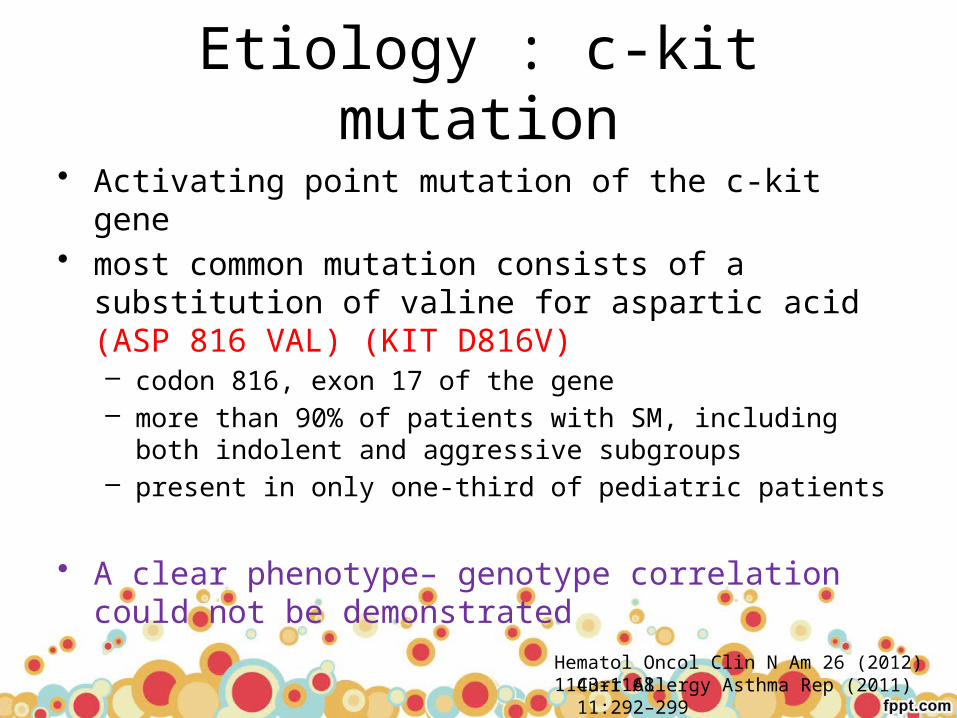
• Activating point mutation of the c-kit gene• most common mutation consists of a substitution of
valine for aspartic acid (ASP 816 VAL) (KIT D816V) – codon 816, exon 17 of the gene– more than 90% of patients with SM, including both indolent and
aggressive subgroups– present in only one-third of pediatric patients
• A clear phenotype– genotype correlation could not be demonstrated
Hematol Oncol Clin N Am 26 (2012) 1143–1168Curr Allergy Asthma Rep (2011) 11:292–299

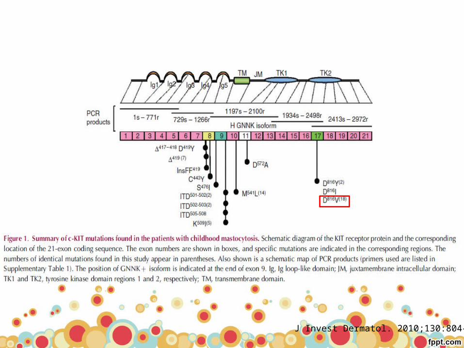
• cutaneous biopsies of 50 children with mastocytosis
• mutations in c-kit in codon 816 : 42%
• outside exon 17: 44%
J Invest Dermatol. 2010;130:804–15
J Invest Dermatol. 2010;130:804–15

Expert Rev. Hematol. 5(3), 261–274 (2012)

Expert Rev. Hematol. 5(3), 261–274 (2012)

Etiology• The downstream signal transduction pathways
responsible for oncogenesis by these point mutations are not fully understood
• play a role in ligand-independent growth and suppression of apoptosis
Dean D. Metcalfe .Middleton’s Allergy 7’th edition ,1051-1062.

Etiology : inhibition of MCs apoptosis
• A subset of patients with – increased mast cells and peripheral eosinophilia and – increase in serum tryptase levels
has been described that carry the Fip1-like-1-platelet-derived growth factor receptor (FIP1L1-PDGFRA) fusion oncogene in pluripotential hematopoietic progenitor cells,
which results from an approximately 800-kb interstitial deletion of chromosome 4q12
Dean D. Metcalfe .Middleton’s Allergy 7’th edition ,1051-1062.

Etiology• Disease associated with mutations in c-kit may be
modified by the genetic composition of the affected individual– a polymorphism in the gene for the IL-4 receptor α-chain : less
extensive mast cell involvement, with disease usually localized to the skin
– the bone marrow cells of patients with mastocytosis have been found to constitutively express the antiapoptotic proteins Bcl-XL and Bcl-2
• This may explain the long survival of these cells and perhaps their resistance to chemotherapy-induced apoptosis
Dean D. Metcalfe .Middleton’s Allergy 7’th edition ,1051-1062.
PATHOLOGIC EFFECTS OF INCREASED MAST CELLS
• The pathologic changes observed in mastocytosis are the result of the increased number of mast cells residing within tissues, and the release of mast cell-dependent mediators within tissues
• Mast cell-derived mediators also circulate through the bloodstream and lymphatic system to produce biologic effects
Dean D. Metcalfe .Middleton’s Allergy 7’th edition ,1051-1062.
Dean D. Metcalfe .Middleton’s Allergy 7’th edition ,1051-1062.

CLINICAL FEATURES
Clinical feature• Skin, GI tract, lymph nodes, liver, spleen, bone marrow,
and skeletal system : common• RS, Endocrine, Renal systems : seldom
• Patients in every category of mastocytosis sometimes experience flushing and/or episodic hypotension
• Occasionally, hypotension may be provoked by alcohol, aspirin, insect stings, infection, or exposure to iodinated contrast materials
• Patients with mastocytosis do not suffer from an increase in bacterial, fungal, or viral infection
Dean D. Metcalfe .Middleton’s Allergy 7’th edition ,1051-1062.
Mastocytosis
2 main categories:
• Cutaneous mastocytosis (CM)– MC infiltrate is confined to one or more lesions on the skin
• Systemic mastocytosis (SM)– by MC infiltration of at least one extracutaneous organ with or
without evidence of skin involvement
Hematol Oncol Clin N Am 26 (2012) 1143–1168

Dean D. Metcalfe .Middleton’s Allergy 7’th edition ,1051-1062.

• The symptoms of SM are usually grouped into 4 categories:
(1) constitutional symptoms : fatigue, weight loss, sweats, and fever
(2) skin symptoms
(3) MC mediator-related symptoms
(4) musculoskeletal symptoms, which include bone, muscle, and joint pain
Hematol Oncol Clin N Am 26 (2012) 1143–1168

Am J Med Sci 2011;342(5):409–415
Skin
• Urticaria pigmentosa (UP)/maculopapular cutaneous mastocytosis (MPCM)
• Diffuse cutaneous mastocytosis (DCM)• Solitary mastocytoma of the skin
Urticaria pigmentosa(UP)
• The most common skin manifestation of mastocytosis in both children and adults
• It is the most common pattern of skin involvement in CM • UP is also observed in
– > 90% of ISM– 50% of SM-AHNMD or ASM
Dean D. Metcalfe .Middleton’s Allergy 7’th edition ,1051-1062.

Dean D. Metcalfe .Middleton’s Allergy 7’th edition ,1051-1062.
UP: small yellowish-tan to reddish-brown macules or slightly raised papules
raised nodules Plaque like lesions
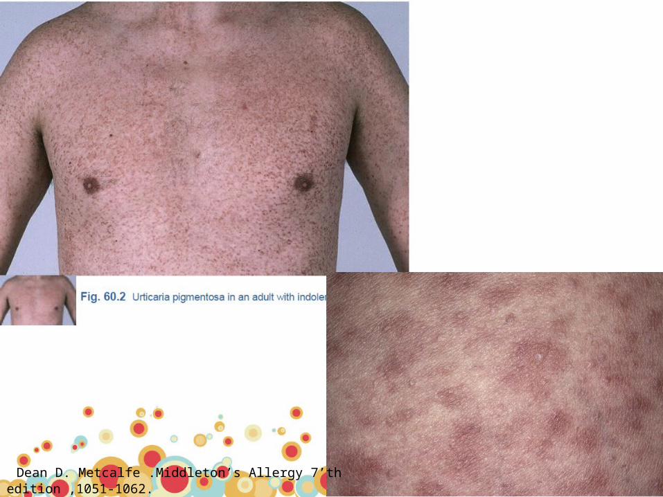
Dean D. Metcalfe .Middleton’s Allergy 7’th edition ,1051-1062.
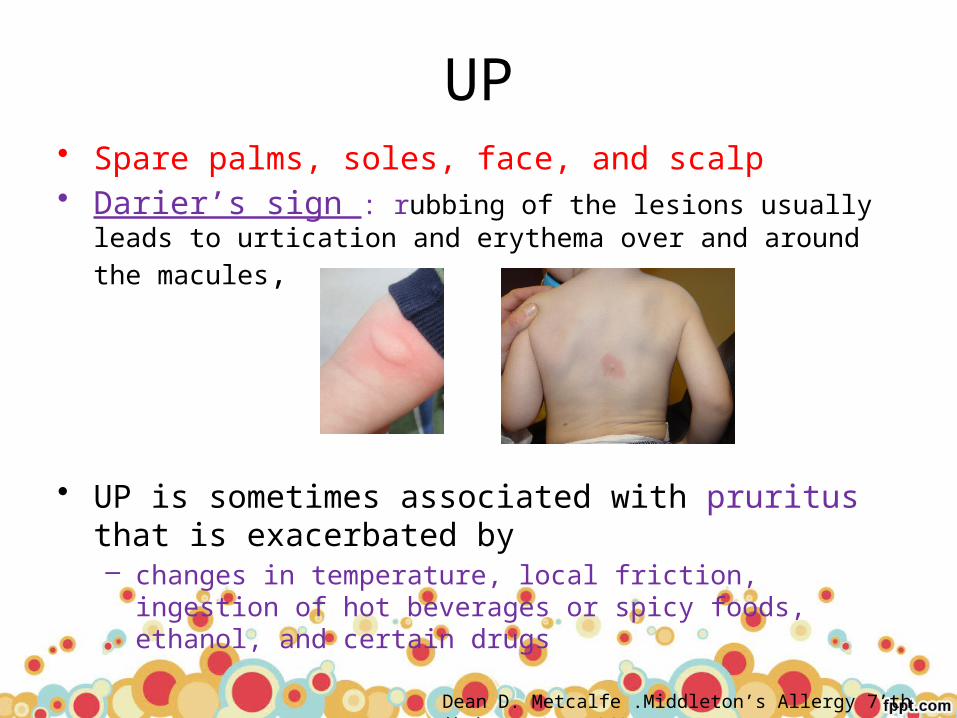
UP• Spare palms, soles, face, and scalp• Darier’s sign : rubbing of the lesions usually leads to urtication
and erythema over and around the macules,
• UP is sometimes associated with pruritus that is exacerbated by – changes in temperature, local friction, ingestion of hot beverages
or spicy foods, ethanol, and certain drugs
Dean D. Metcalfe .Middleton’s Allergy 7’th edition ,1051-1062.

Diffuse cutaneous mastocytosis (DCM)
• extremely rare form of CM• diffuse mast cell infiltration in the dermis• no discrete lesions • entire cutaneous integument is involved• Onset < age of 3 years• The skin is normal to yellowish-brown
and is thickened • may exhibit discoloration with a peau
d’orange appearance• spontaneous resolution has usually
occurred before 5 years of age
Dean D. Metcalfe .Middleton’s Allergy 7’th edition ,1051-1062.
Hematol Oncol Clin N Am 26 (2012) 1143–1168

• Young children with UP or DCM may have bullous eruptions with hemorrhage
• Blisters may erupt spontaneously or in association with infection or immunization
• Blisters may also occur at birth• CM is thus included in the differential diagnosis of neonatal disorders
with blisters Dean D. Metcalfe .Middleton’s Allergy 7’th edition ,1051-1062.
Extensive diffuse skin involvement Bullous eruption
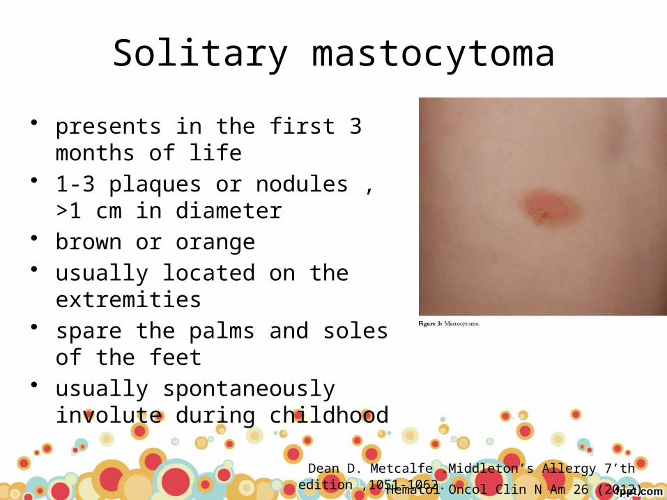
Solitary mastocytoma
• presents in the first 3 months of life • 1-3 plaques or nodules , >1 cm in
diameter• brown or orange • usually located on the extremities • spare the palms and soles of the
feet• usually spontaneously involute
during childhood
Dean D. Metcalfe .Middleton’s Allergy 7’th edition ,1051-1062.
Hematol Oncol Clin N Am 26 (2012) 1143–1168
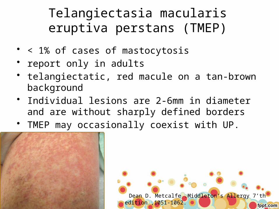
Telangiectasia macularis eruptiva perstans (TMEP)
• < 1% of cases of mastocytosis• report only in adults• telangiectatic, red macule on a tan-brown background• Individual lesions are 2-6mm in diameter and are without
sharply defined borders• TMEP may occasionally coexist with UP.
Dean D. Metcalfe .Middleton’s Allergy 7’th edition ,1051-1062.

GI• Common(80%) : as frequent as pruritus(88%) or flushing (43%)
• Abdominal pain is the most common GI symptom, followed by diarrhea, nausea, and vomiting
• GI bleeding is uncommon• Peptic ulcer disease is relatively infrequent (4-44%)
despite hyperhistaminemia
• The pathogenesis of abdominal symptoms appears multifactorial
Dean D. Metcalfe .Middleton’s Allergy 7’th edition ,1051-1062.

Musculoskeletal
• Musculoskeletal pain• associated with osteopenia or osteoporosis pathologic
fractures• osteoporosis or pathologic fractures, or both may be the
initial manifestation of mastocytosis
Dean D. Metcalfe .Middleton’s Allergy 7’th edition ,1051-1062.
Bone marrow• The bone marrow is the most common site of pathologic
mast cell infiltrates– BM>spleen>liver>LN
• Initial diagnosis– palpable splenomegaly 48%– Hepatomegaly 41%– lymphadenopathy 26%
Dean D. Metcalfe .Middleton’s Allergy 7’th edition ,1051-1062.
Bone marrow• BM biopsy
– most useful biopsy site for diagnosis of systemic mastocytosis– important prognostic information– Immunohistochemical staining with antitryptase : visualize mast
cells
• The majority of infiltrates in the bone marrow are focal,
• Focal mastocytosis lesions are most commonly situated – Paratrabecular > Perivascular > Parafollicular
Dean D. Metcalfe .Middleton’s Allergy 7’th edition ,1051-1062.

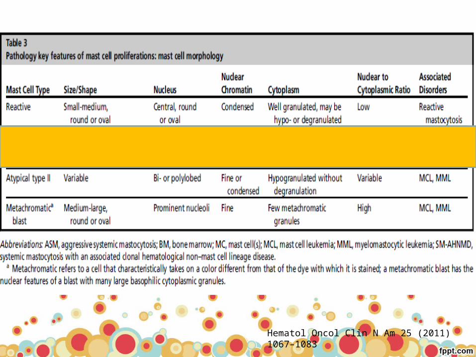
Fig. 60.8 (A) Paratrabecular aggregate of spindle-shaped mast cells. The hematopoietic marrow is hypercellular, and the bone trabeculae are slightly thickened. This patient has an aggressive form of systemic mastocytosis. (Hematoxylin-eosin stain; ×20.)
(B) Higher power demonstrates the spindle shape of the mast cells and the faint granularity of the cytoplasm. (Giemsa stain; plastic imbedded; ×250).

Bone marrow• Early stage : MCs infiltrate(cellular)• Late stage : mast cells number may decrease and the
lesions may become fibrotic• osteosclerotic or osteolytic changes in the bone
trabeculae
• DDX: – granulomas, myelofibrosis, Hodgkin’s disease, metastatic
carcinoma, Kaposi’s sarcoma, and histiocytosis X– because these cells resemble fibroblasts and histiocytes.
Dean D. Metcalfe .Middleton’s Allergy 7’th edition ,1051-1062.
Bone marrow• Most sensitive and specific method to support the
diagnosis of SM in BM– flow cytometry of bone marrow aspirates or by
immunohistochemical analysis of bone marrow biopsies
– The co-expression of CD2 and/or CD25 in CD117 (Kit)-positive mast cells
Dean D. Metcalfe .Middleton’s Allergy 7’th edition ,1051-1062.
HEPATIC AND SPLENIC INVOLVEMENT
Spleen• The most common finding is trabecular fibrotic thickening• found in a paratrabecular, parafollicular, follicular, or diffuse
red pulp distribution
Lymph node• The most common location is the paracortical region
– Parafollicular and follicular replacement, medullary cord and sinus infiltration: less frequent
• Mastocytosis infiltrates in the spleen and lymph nodes : DDx– follicular and T cell lymphomas, monocytoid B cell hyperplasia
and lymphoma, Kaposi’s sarcoma, hairy cell leukemia, and histiocytosis X
Dean D. Metcalfe .Middleton’s Allergy 7’th edition ,1051-1062.
HEPATIC AND SPLENIC INVOLVEMENT
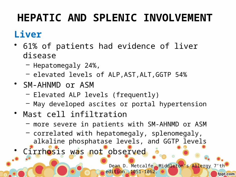
Liver• 61% of patients had evidence of liver disease
– Hepatomegaly 24%, – elevated levels of ALP,AST,ALT,GGTP 54%
• SM-AHNMD or ASM – Elevated ALP levels (frequently)– May developed ascites or portal hypertension
• Mast cell infiltration – more severe in patients with SM-AHNMD or ASM– correlated with hepatomegaly, splenomegaly, alkaline
phosphatase levels, and GGTP levels
• Cirrhosis was not observed
Dean D. Metcalfe .Middleton’s Allergy 7’th edition ,1051-1062.

NEUROPSYCHIATRIC ABNORMALITIES
• Headache, dizziness• Seizures• Decreased attention span, memory impairment, and irritability• Depression
Dean D. Metcalfe .Middleton’s Allergy 7’th edition ,1051-1062.
DIAGNOSIS AND EVALUATION
Dean D. Metcalfe .Middleton’s Allergy 7’th edition ,1051-1062.
Dean D. Metcalfe .Middleton’s Allergy 7’th edition ,1051-1062.
Hematol Oncol Clin N Am 25 (2011) 1067–1083

Dean D. Metcalfe .Middleton’s Allergy 7’th edition ,1051-1062.

Hematol Oncol Clin N Am 25 (2011) 1067–1083
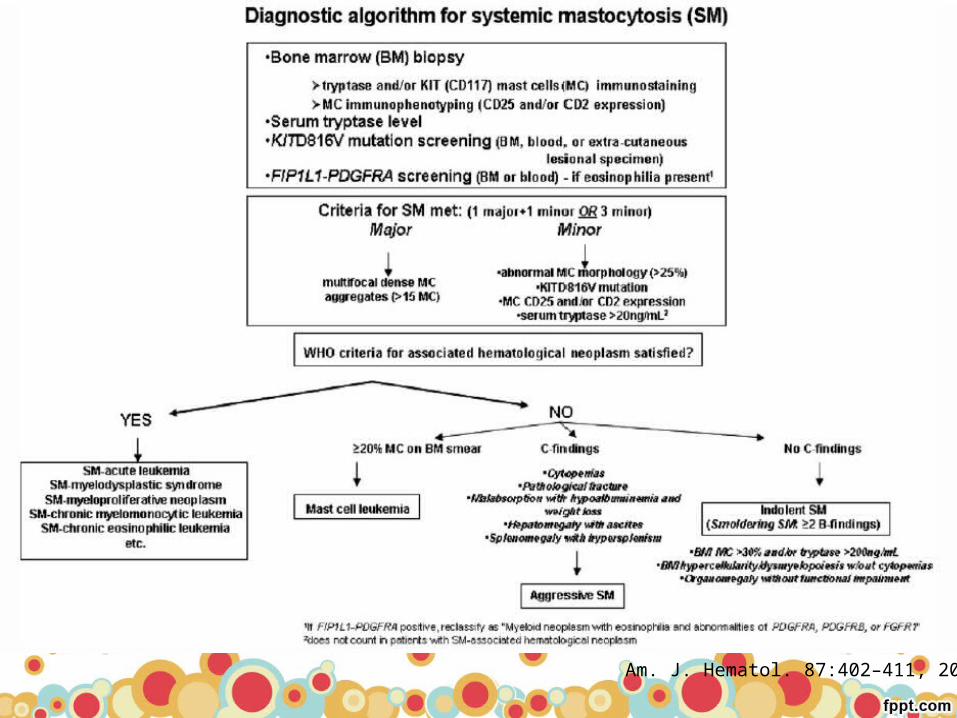
impaired organ function
≥ 2 B findings, no C finding smoldering mastocytosis
≥ 1 C aggressive SM (ASM)
Systemic mastocytosis
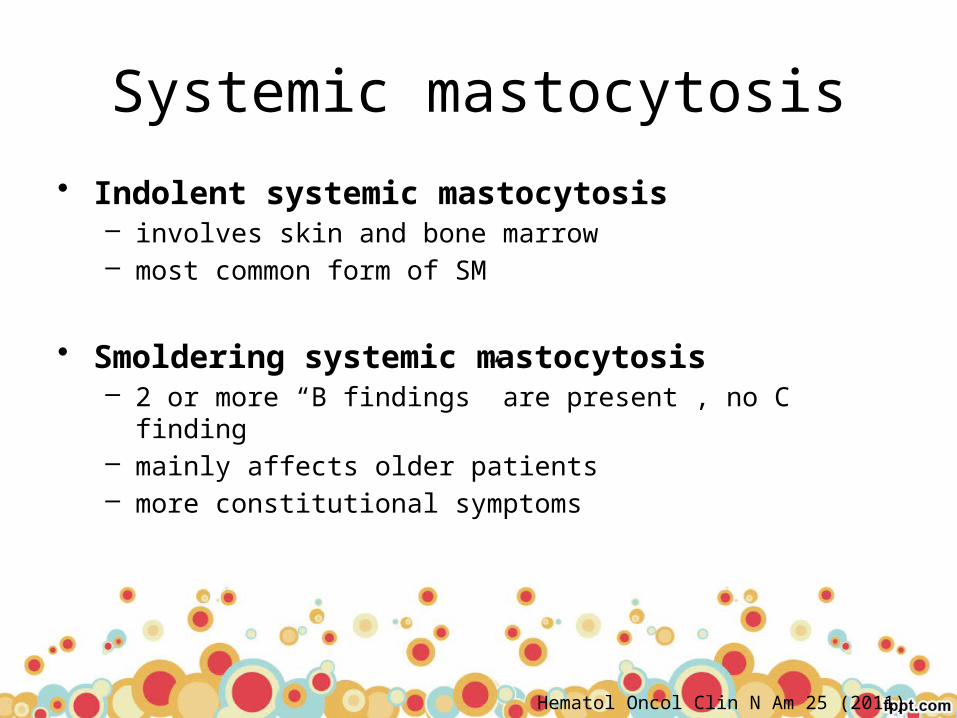
• Indolent systemic mastocytosis – involves skin and bone marrow – most common form of SM
• Smoldering systemic mastocytosis – 2 or more “B findings” are present , no C finding– mainly affects older patients– more constitutional symptoms
Hematol Oncol Clin N Am 25 (2011) 1067–1083
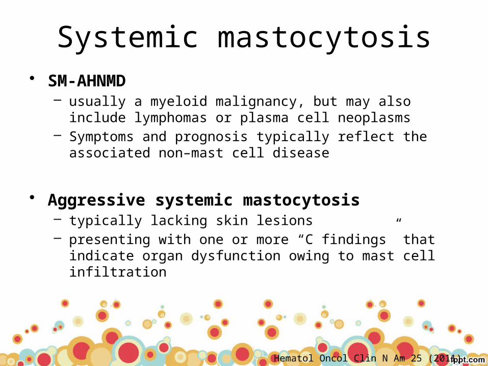
Systemic mastocytosis• SM-AHNMD
– usually a myeloid malignancy, but may also include lymphomas or plasma cell neoplasms
– Symptoms and prognosis typically reflect the associated non–mast cell disease
• Aggressive systemic mastocytosis – typically lacking skin lesions – presenting with one or more “C findings” that indicate organ
dysfunction owing to mast cell infiltration
Hematol Oncol Clin N Am 25 (2011) 1067–1083

MCL
• rare • characterized by circulating MCs and 20% or greater
MCs on the bone marrow aspirate smear• Most patients are adults• Cutaneous lesions are typically absent• present with
– episodes of mediator-related symptoms – later develop constitutional symptoms, including weight loss and
bone pain, and symptoms and signs of organomegaly
Hematol Oncol Clin N Am 26 (2012) 1143–1168
Expert Rev. Hematol. 5(3), 261–274 (2012)
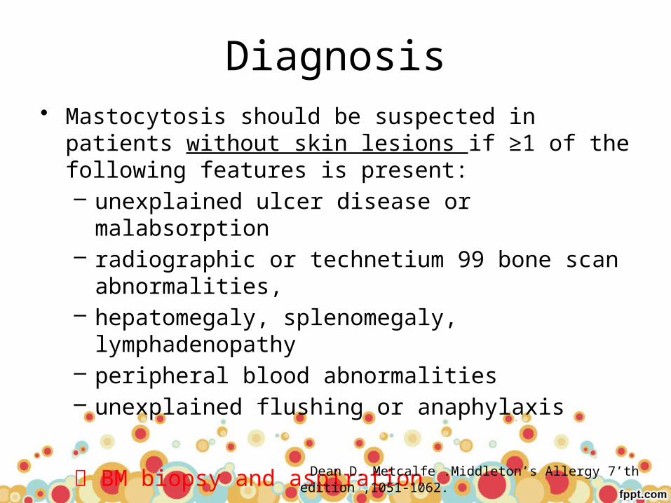
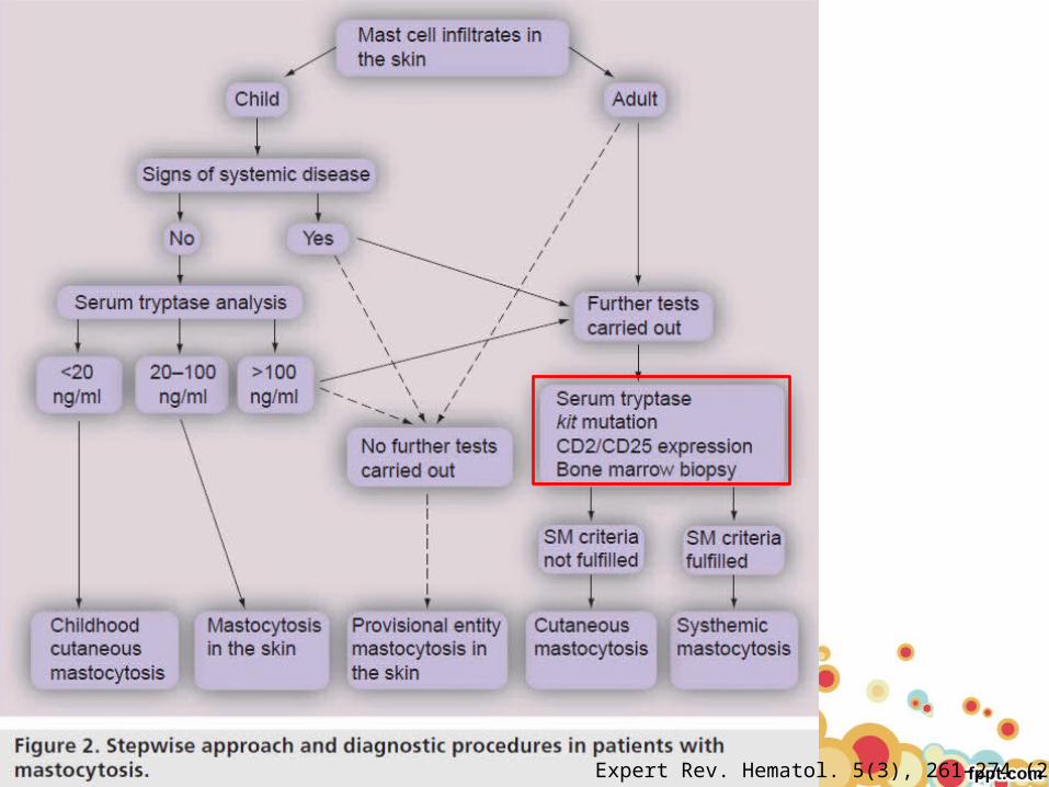
Diagnosis• Mastocytosis should be suspected in patients without
skin lesions if ≥1 of the following features is present: – unexplained ulcer disease or malabsorption– radiographic or technetium 99 bone scan
abnormalities, – hepatomegaly, splenomegaly, lymphadenopathy – peripheral blood abnormalities– unexplained flushing or anaphylaxis
BM biopsy and aspiration
Dean D. Metcalfe .Middleton’s Allergy 7’th edition ,1051-1062.
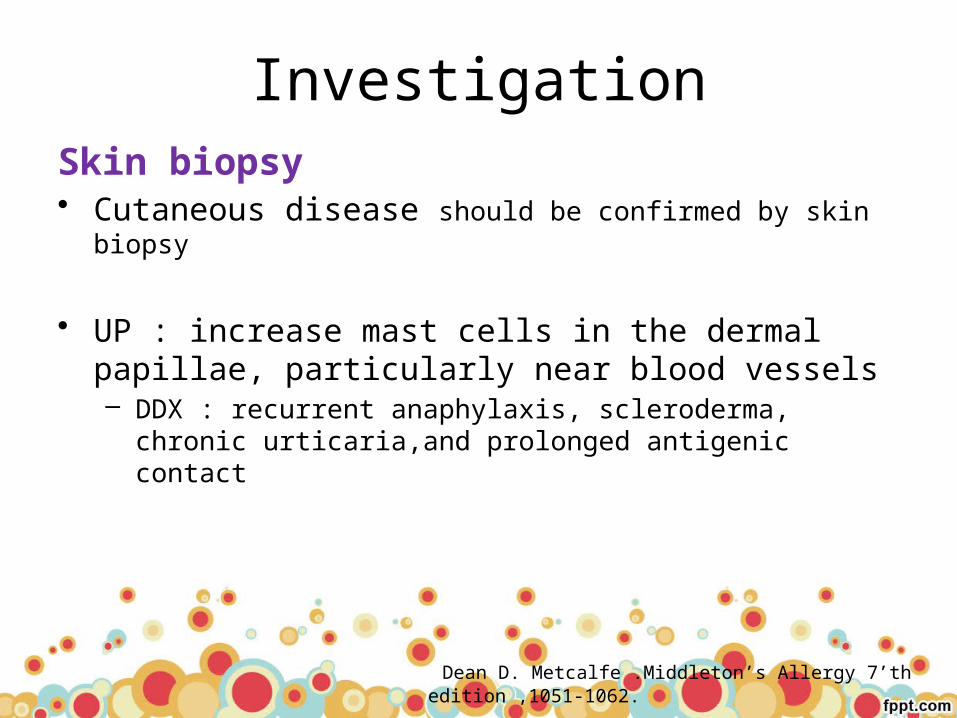
InvestigationSkin biopsy• Cutaneous disease should be confirmed by skin biopsy
• UP : increase mast cells in the dermal papillae, particularly near blood vessels– DDX : recurrent anaphylaxis, scleroderma, chronic urticaria,and
prolonged antigenic contact
Dean D. Metcalfe .Middleton’s Allergy 7’th edition ,1051-1062.
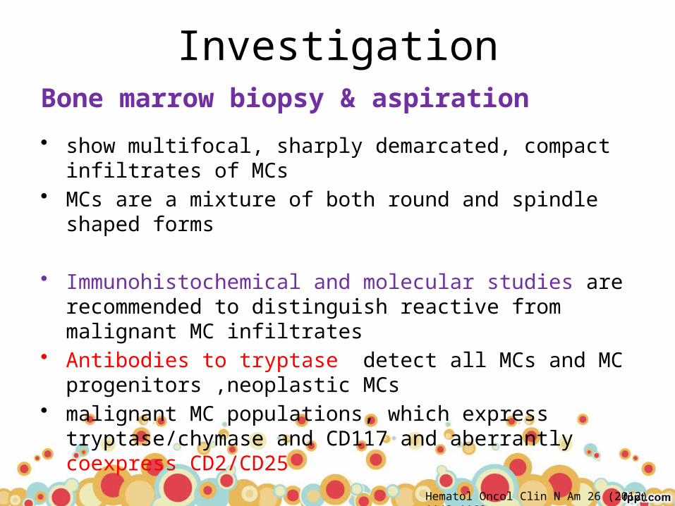
InvestigationBone marrow biopsy & aspiration
• show multifocal, sharply demarcated, compact infiltrates of MCs• MCs are a mixture of both round and spindle shaped forms
• Immunohistochemical and molecular studies are recommended to distinguish reactive from malignant MC infiltrates
• Antibodies to tryptase detect all MCs and MC progenitors ,neoplastic MCs
• malignant MC populations, which express tryptase/chymase and CD117 and aberrantly coexpress CD2/CD25
Hematol Oncol Clin N Am 26 (2012) 1143–1168
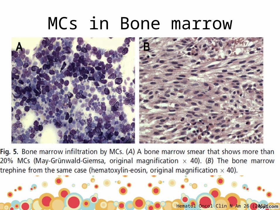
MCs in Bone marrow
Hematol Oncol Clin N Am 26 (2012) 1143–1168
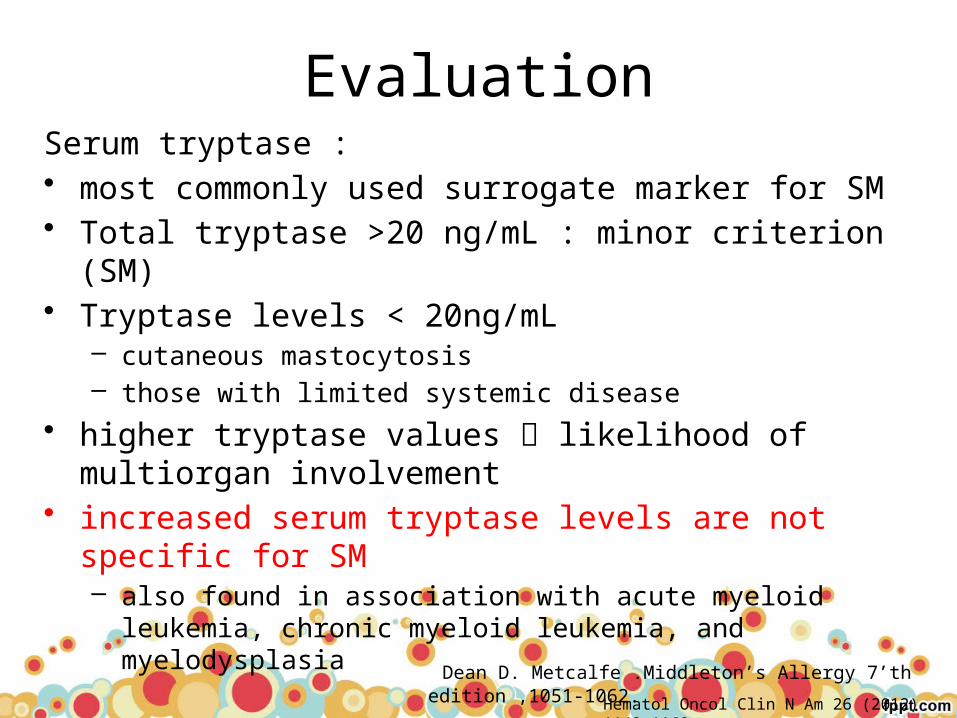
EvaluationSerum tryptase : • most commonly used surrogate marker for SM• Total tryptase >20 ng/mL : minor criterion (SM)• Tryptase levels < 20ng/mL
– cutaneous mastocytosis – those with limited systemic disease
• higher tryptase values likelihood of multiorgan involvement
• increased serum tryptase levels are not specific for SM – also found in association with acute myeloid leukemia, chronic
myeloid leukemia, and myelodysplasia
Dean D. Metcalfe .Middleton’s Allergy 7’th edition ,1051-1062.
Hematol Oncol Clin N Am 26 (2012) 1143–1168
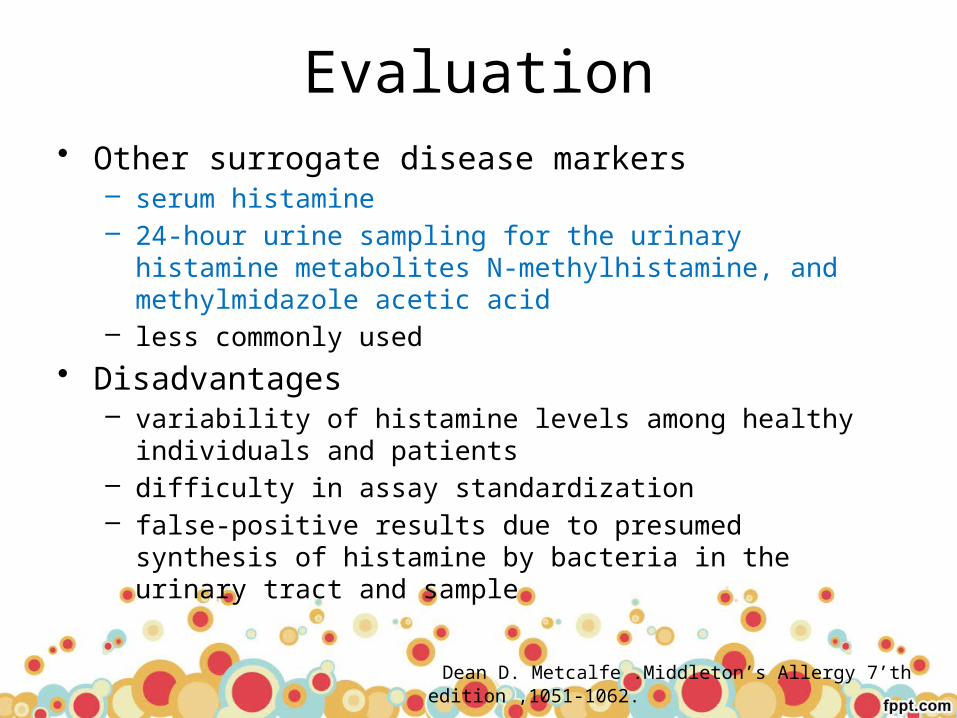
Evaluation• Other surrogate disease markers
– serum histamine – 24-hour urine sampling for the urinary histamine metabolites N-
methylhistamine, and methylmidazole acetic acid– less commonly used
• Disadvantages – variability of histamine levels among healthy individuals and
patients– difficulty in assay standardization– false-positive results due to presumed synthesis of histamine by
bacteria in the urinary tract and sample
Dean D. Metcalfe .Middleton’s Allergy 7’th edition ,1051-1062.

Evaluation• Examination of other tissue specimens can help define
the extent of mast cell involvement– lymph nodes, spleen, liver, and GI mucosa– performed only when necessary
• Identification of genetic markers– point mutations of c-kit, help support the diagnosis of
mastocytosis– In patients with coexisting eosinophilia, peripheral blood should
be examined for the presence of the FIP1L1/PDGFRA fusion gene
Dean D. Metcalfe .Middleton’s Allergy 7’th edition ,1051-1062.

Evaluation
• Additional diagnostic studies – bone scans or skeletal surveys– ultrasound or computed tomography scan of the abdomen– upper GI series– small bowel radiography– endoscopy
Dean D. Metcalfe .Middleton’s Allergy 7’th edition ,1051-1062.
Am. J. Hematol. 87:402–411, 2012
Treatment

Treatment
• Counseling and education• Management of MC mediator-release symptoms• Cytoreductive treatement
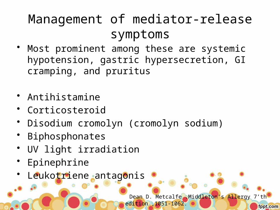
Management of mediator-release symptoms
• Most prominent among these are systemic hypotension, gastric hypersecretion, GI cramping, and pruritus
• Antihistamine• Corticosteroid• Disodium cromolyn (cromolyn sodium)• Biphosphonates• UV light irradiation• Epinephrine• Leukotriene antagonis
Dean D. Metcalfe .Middleton’s Allergy 7’th edition ,1051-1062.

Antihistamine• H1-receptor antagonists
– classic or non-sedating antihistamines – reduce pruritus and flushing
• H2 antagonist– If H1 is insufficient– ranitidine, cimetidine or famotidine may be beneficial
• Many patients continue to complain of musculoskeletal pain, headaches, and flushing– inability of histamine antagonists to block the effects of high
levels of histamine , presence of other mast cell mediators– adding a leukotriene-modifying agent.
Dean D. Metcalfe .Middleton’s Allergy 7’th edition ,1051-1062.

CorticosteroidsOral steroids• control malabsorption,abdominal pain, nausea and
vomiting• prevention or treatment of anaphylaxis• should only be used for short periods as a second- or
third-line therapy osteopenia or osteoporosis
Topical steroids• treat UP or DCM• Lesions recur after discontinuation of therapy
Hematol Oncol Clin N Am 26 (2012) 1143–1168
Dean D. Metcalfe .Middleton’s Allergy 7’th edition ,1051-1062.

Disodium cromoglycate (cromolyn sodium)
• inhibits degranulation of mast cells and • relief of GI complaints
Ketotifen• antihistamine with mast cell stabilizing properties• relieving the pruritus and whealing • no advantage over hydroxyzine
Dean D. Metcalfe .Middleton’s Allergy 7’th edition ,1051-1062.

Epinephrine
• treat episodes of systemic hypotension• Self-administer IM epinephrine
Dean D. Metcalfe .Middleton’s Allergy 7’th edition ,1051-1062.

UV light irradiation
• Oral methoxypsoralen with UVA (PUVA) • relieve pruritus and whealing after 1-2 months of
treatment• Relapse occurs within 3-6 months after discontinuation
of therapy
• Photochemotherapy should be used only in instances of extensive cutaneous disease unresponsive to other forms of therapy
Dean D. Metcalfe .Middleton’s Allergy 7’th edition ,1051-1062.

Cytoreductive Therapy• Use in aggressive SM, SM-AHNMD,MCL
• interferon-α2b and 2-chloro-2-deoxyadenosine (cladribine, 2-CdA) are potential first- and second-line therapeutic options
• In highly aggressive or relapsed cases : combination chemotherapy followed by a hematopoietic stem cell transplant should be considered– cytarabine, fludarabine, and hydroxyurea
Hematol Oncol Clin N Am 26 (2012) 1143–1168
Dean D. Metcalfe .Middleton’s Allergy 7’th edition ,1051-1062.

Cytoreductive Therapy
• Specific tyrosine kinase inhibitors – patients who are negative for D816V but have non–
codon 816 mutations or wild-type KIT – such as imatinib, or other tyrosine kinase inhibitors
Hematol Oncol Clin N Am 26 (2012) 1143–1168
Dean D. Metcalfe .Middleton’s Allergy 7’th edition ,1051-1062.
Bone marrow transplantation
• treatment option for patients with advanced categories of mastocytosis associated with poor survival in only a few reported instances
• may yield a better prognosis if mast cell suppression is attempted prior to the transplantation
Hematol Oncol Clin N Am 26 (2012) 1143–1168
Dean D. Metcalfe .Middleton’s Allergy 7’th edition ,1051-1062.
Curr Allergy Asthma Rep (2011) 11:292–299

PROGNOSIS
Hematol Oncol Clin N Am 26 (2012) 1143–1168

Prognosis
• Patients with CM only have the best prognosis• For children with isolated UP, at least 50% of cases are
reported to resolve by adulthood• UP in adulthood may evolve into systemic disease• Occasionally, ISM converts to SM-AHNMD
Dean D. Metcalfe .Middleton’s Allergy 7’th edition ,1051-1062.
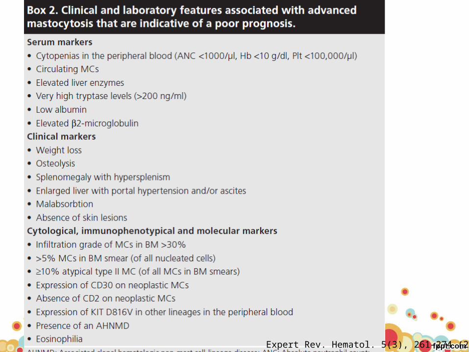
Expert Rev. Hematol. 5(3), 261–274 (2012)

Prognosis
Expert Rev. Hematol. 5(3), 261–274 (2012)
Take home messages
• Mastocytosis is associated with a pathologic increase in mast cells in one or more organ systems
• Most adult patients have an activating mutation in Kit • Serum tryptase is usually elevated• The signs and symptoms are due to release of mast cell
mediators, the increase in mast cell burden, and, in some patients, an associated hematologic disorder
• Treatment is largely symptomatic, with specific treatment of any associated hematologic disorder

Thank you
Related Documents


