Received 06/04/2017 Review began 06/14/2017 Review ended 06/16/2017 Published 06/22/2017 © Copyright 2017 Gupta et al. This is an open access article distributed under the terms of the Creative Commons Attribution License CC-BY 3.0., which permits unrestricted use, distribution, and reproduction in any medium, provided the original author and source are credited. Unusual Case of Bilateral Tubercular Mastitis Archit Gupta , Mudita Gupta , Jagdish Gupta 1. General Surgery, Indira Gandhi Medical College, Shimla 2. Dermatology, Indira Gandhi Medical College, Shimla Corresponding author: Archit Gupta, [email protected] Disclosures can be found in Additional Information at the end of the article Abstract Bilateral involvement of the breast with tuberculosis is extremely rare. It most commonly affects young lactating multiparous females, although rarely it may be reported in prepubescent males also. We present a case of a 27-year-old nulliparous female who presented with a history of multiple pus discharging sinuses around both areolae and was diagnosed as a case of bilateral tubercular mastitis. Tubercular mastitis being a paucibacillary disease, diagnosis is often difficult. Treatment consists of antitubercular therapy with or without surgery. Categories: Dermatology, General Surgery, Infectious Disease Keywords: tuberculosis, tb, breast, mastitis Introduction Breast tuberculosis is a rare form of tuberculosis with an incidence of <0.1% in Western countries [1]. Even though the disease is more common in India as compared to the West, it is often misdiagnosed as carcinoma of the breast or breast abscess [2]. It most commonly affects young lactating multiparous females, although rarely it may be reported in prepubescent males also. Bilateral breast involvement is even more rare and very few cases have been reported till date [3]. We report a case of bilateral tubercular mastitis presenting in a young nulliparous female. We report this case due to its rarity and clinical significance. Case Presentation A 27-year-old nulliparous female presented to our outpatient department with a history of pus discharge from multiple sites around the nipple-areola complex of both the breasts for four years. The patient had undergone incision and drainage twice, with a suspicion of breast abscess at a peripheral hospital. There was no history of any palpable lump in the breasts, fever, night sweats, weight loss, or any respiratory symptoms. There was no family history of breast cancer or tuberculosis. There was no history of diabetes, immunosuppression, previous treatment for tuberculosis or history of contact with a known case of tuberculosis. The general physical examination and systemic examination were within normal limits. On local examination, nipple retraction with multiple scars, indurated granulomatous erythematous plaques, and discharging sinuses around the nipple-areola complex were found in both the breasts (Figures 1-2). 1 2 1 Open Access Case Report DOI: 10.7759/cureus.1383 How to cite this article Gupta A, Gupta M, Gupta J (June 22, 2017) Unusual Case of Bilateral Tubercular Mastitis. Cureus 9(6): e1383. DOI 10.7759/cureus.1383

Welcome message from author
This document is posted to help you gain knowledge. Please leave a comment to let me know what you think about it! Share it to your friends and learn new things together.
Transcript

Received 06/04/2017 Review began 06/14/2017 Review ended 06/16/2017 Published 06/22/2017
© Copyright 2017Gupta et al. This is an open accessarticle distributed under the terms ofthe Creative Commons AttributionLicense CC-BY 3.0., which permitsunrestricted use, distribution, andreproduction in any medium,provided the original author andsource are credited.
Unusual Case of Bilateral TubercularMastitisArchit Gupta , Mudita Gupta , Jagdish Gupta
1. General Surgery, Indira Gandhi Medical College, Shimla 2. Dermatology, Indira Gandhi MedicalCollege, Shimla
Corresponding author: Archit Gupta, [email protected] Disclosures can be found in Additional Information at the end of the article
AbstractBilateral involvement of the breast with tuberculosis is extremely rare. It most commonlyaffects young lactating multiparous females, although rarely it may be reported inprepubescent males also. We present a case of a 27-year-old nulliparous female who presentedwith a history of multiple pus discharging sinuses around both areolae and was diagnosed as acase of bilateral tubercular mastitis. Tubercular mastitis being a paucibacillary disease,diagnosis is often difficult. Treatment consists of antitubercular therapy with or withoutsurgery.
Categories: Dermatology, General Surgery, Infectious DiseaseKeywords: tuberculosis, tb, breast, mastitis
IntroductionBreast tuberculosis is a rare form of tuberculosis with an incidence of <0.1% in Westerncountries [1]. Even though the disease is more common in India as compared to the West, it isoften misdiagnosed as carcinoma of the breast or breast abscess [2]. It most commonly affectsyoung lactating multiparous females, although rarely it may be reported in prepubescent malesalso. Bilateral breast involvement is even more rare and very few cases have been reported tilldate [3]. We report a case of bilateral tubercular mastitis presenting in a young nulliparousfemale. We report this case due to its rarity and clinical significance.
Case PresentationA 27-year-old nulliparous female presented to our outpatient department with a history of pusdischarge from multiple sites around the nipple-areola complex of both the breasts for fouryears. The patient had undergone incision and drainage twice, with a suspicion of breastabscess at a peripheral hospital. There was no history of any palpable lump in the breasts, fever,night sweats, weight loss, or any respiratory symptoms. There was no family history of breastcancer or tuberculosis. There was no history of diabetes, immunosuppression, previoustreatment for tuberculosis or history of contact with a known case of tuberculosis.
The general physical examination and systemic examination were within normal limits. Onlocal examination, nipple retraction with multiple scars, indurated granulomatouserythematous plaques, and discharging sinuses around the nipple-areola complex were foundin both the breasts (Figures 1-2).
1 2 1
Open Access CaseReport DOI: 10.7759/cureus.1383
How to cite this articleGupta A, Gupta M, Gupta J (June 22, 2017) Unusual Case of Bilateral Tubercular Mastitis. Cureus 9(6):e1383. DOI 10.7759/cureus.1383

FIGURE 1: Right breast of the patient showing periareolarplaques (white arrow) and sinus (red arrow)
2017 Gupta et al. Cureus 9(6): e1383. DOI 10.7759/cureus.1383 2 of 5
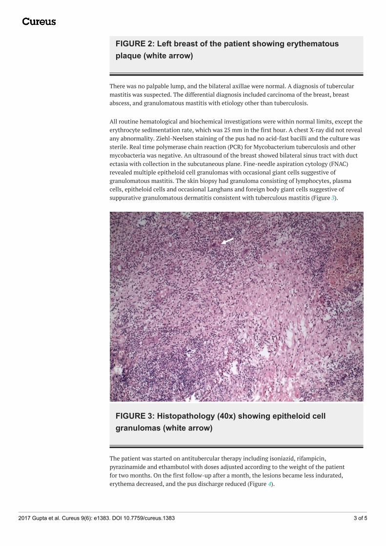
FIGURE 2: Left breast of the patient showing erythematousplaque (white arrow)
There was no palpable lump, and the bilateral axillae were normal. A diagnosis of tubercularmastitis was suspected. The differential diagnosis included carcinoma of the breast, breastabscess, and granulomatous mastitis with etiology other than tuberculosis.
All routine hematological and biochemical investigations were within normal limits, except theerythrocyte sedimentation rate, which was 25 mm in the first hour. A chest X-ray did not revealany abnormality. Ziehl-Neelsen staining of the pus had no acid-fast bacilli and the culture wassterile. Real time polymerase chain reaction (PCR) for Mycobacterium tuberculosis and othermycobacteria was negative. An ultrasound of the breast showed bilateral sinus tract with ductectasia with collection in the subcutaneous plane. Fine-needle aspiration cytology (FNAC)revealed multiple epitheloid cell granulomas with occasional giant cells suggestive ofgranulomatous mastitis. The skin biopsy had granuloma consisting of lymphocytes, plasmacells, epitheloid cells and occasional Langhans and foreign body giant cells suggestive ofsuppurative granulomatous dermatitis consistent with tuberculous mastitis (Figure 3).
FIGURE 3: Histopathology (40x) showing epitheloid cellgranulomas (white arrow)
The patient was started on antitubercular therapy including isoniazid, rifampicin,pyrazinamide and ethambutol with doses adjusted according to the weight of the patientfor two months. On the first follow-up after a month, the lesions became less indurated,erythema decreased, and the pus discharge reduced (Figure 4).
2017 Gupta et al. Cureus 9(6): e1383. DOI 10.7759/cureus.1383 3 of 5

FIGURE 4: Bilateral breasts showing disappearance oferythematous plaque (white arrow) and sinus (red arrow) aftertreatment with antitubercular therapy for one month
The patient had satisfactory response to antitubercular treatment and was called for follow-upand readjustment of drugs after a month.
DiscussionBreast tuberculosis was first recorded by Sir Astley Cooper in 1829, who called it ‘scrofulousswelling of the bosom’. Breast tuberculosis is rare because of the high resistance offered by thebreast tissue to the survival and multiplication of tubercle bacilli [4]. It commonly affectsmultiparous females usually in the age group of 21–40 years [5]. Our patient was a 27-year-oldnulliparous female.
Breast tuberculosis can be primary or secondary. Primary is when the tuberculous infection isconfined to the breast and is extremely uncommon. It is secondary when there is a coexistingfocus of tuberculosis somewhere else in the body, most commonly, pulmonary tuberculosis.Our patient was a case of primary tuberculosis of the breast [5].
Diagnosis of breast tuberculosis is a challenge and the patient is usually subjected to numerousinvestigations before a diagnosis is made. Mantoux test is usually positive in endemic areas andstands obsolete. Mammogram is rarely used for diagnosis as it usually does notdistinguish from carcinoma of the breast [6]. Ultrasonography is cheap, easily accessible, andhelps in characterizing the lesion better without exposure to radiation [7]. Diagnosis is ideallyby bacteriological confirmation from breast tissue by Ziehl-Neelsen stain or culture. But as thedisease is usually paucibacillary, bacteria are isolated only in 25% of the cases [8].
Fine needle aspiration cytology (FNAC) is an important tool for diagnosis of breast tuberculosis.Approximately 73% of the cases can be diagnosed by demonstrating epitheloid cells withcaseous necrosis on FNAC [8]. In our case, FNAC showed granulomas with epitheloid cells;however, there was no necrosis, which can be explained because only a small amount of tissuewas harvested and examined in FNAC. Histopathological findings usually include epitheloid
2017 Gupta et al. Cureus 9(6): e1383. DOI 10.7759/cureus.1383 4 of 5
cell granulomas, but this picture can also be present in other diseases which includesarcoidosis, other fungal infections, and idiopathic granulomatous mastitis [7-8].
The treatment primarily comprises of antitubercular therapy with or without surgery. Residuallump following surgery may require surgical removal. A simple mastectomy can also be done.
Our patient had granulomas consisting of epitheloid cells in FNAC and biopsy and respondedwell to treatment by antitubercular therapy. Hence, a final diagnosis of tubercular mastitis wasmade after one month of treatment.
ConclusionsBreast tuberculosis is a rare form of tuberculosis that is often misdiagnosed as carcinoma of thebreast or breast abscess. Bilateral breast involvement is even more rare and very few cases havebeen reported till date. A high index of clinical suspicion is required to make the diagnosis. Wereport a case of bilateral breast tuberculosis where diagnosis of tuberculosis was confirmed onlyafter the patient showed response to antitubercular treatment.
Additional InformationDisclosuresHuman subjects: Consent was obtained by all participants in this study. Conflicts of interest:In compliance with the ICMJE uniform disclosure form, all authors declare the following:Payment/services info: All authors have declared that no financial support was received fromany organization for the submitted work. Financial relationships: All authors have declaredthat they have no financial relationships at present or within the previous three years with anyorganizations that might have an interest in the submitted work. Other relationships: Allauthors have declared that there are no other relationships or activities that could appear tohave influenced the submitted work.
References1. Kalac N, Ozkan B, Bayiz H, et al.: Breast tuberculosis. Breast. 2002, 11:346–9.
10.1054/brst.2002.04202. Green RM, Ormerod LP: Mammary tuberculosis: rare but still present in the United Kingdom .
Int J Tuberc. 2000, 4:788-90.3. Sreeramulu PN, Venkatachalapathy TS, Prathima S: A case report of bilateral tuberculosis of
breast. J Clin Case Rep. 2012, 2:222. 10.4172/2165-7920.10002224. Khanna R, Prasanna GV, Gupta P, et al.: Mammary tuberculosis: report on 52 cases . Postgrad
Med J. 2002, 78:422–4. 10.1136/pmj.78.921.4225. Shinde SR, Chandawarkar RY, Deshmukh SP: Tuberculosis of the breast masquerading as
carcinoma: a study of 100 patients. World J Surg. 1995, 19:379-81.6. Tewari M, Shukla HS: Breast tuberculosis: diagnosis, clinical features & management . Indian J
Med Res. 2005, 122:103–110.7. Popli MB: Pictorial essay: tuberculosis of the breast . Indian J Radiol Imag. 1999, 9:127-132.8. Kakkar S, Kapila K, Singh MK, et al.: Tuberculosis of the breast. A cytomorphologic study .
Acta Cytol. 2000, 44:292–6.
2017 Gupta et al. Cureus 9(6): e1383. DOI 10.7759/cureus.1383 5 of 5
Related Documents


