Submitted 1 August 2013 Accepted 23 August 2013 Published 12 September 2013 Corresponding author Philip G. Cox, [email protected] Academic editor William Jungers Additional Information and Declarations can be found on page 14 DOI 10.7717/peerj.160 Copyright 2013 Cox et al. Distributed under Creative Commons CC-BY 3.0 OPEN ACCESS Masticatory biomechanics of the Laotian rock rat, Laonastes aenigmamus, and the function of the zygomaticomandibularis muscle Philip G. Cox 1 , Joanna Kirkham 2,3 and Anthony Herrel 4 1 Centre for Anatomical and Human Sciences, Hull York Medical School, University of Hull, Hull, UK 2 Centre for Anatomical and Human Sciences, Hull York Medical School, University of York, York, UK 3 College of Medical and Dental Sciences, University of Birmingham, Birmingham, UK 4 Ecologie et Gestion de la Biodiversite, CNRS/MNHN, Paris, France ABSTRACT The Laotian rock rat, Laonastes aenigmamus, is one of the most recently discovered species of rodent, and displays a cranial morphology that is highly specialised. The rostrum of L. aenigmamus is exceptionally elongate and bears a large attachment site for the infraorbital portion of the zygomaticomandibularis muscle (IOZM), which is particularly well-developed in this species. In this study, we used finite element anal- ysis to investigate the biomechanical performance of the Laotian rock rat cranium and to elucidate the function of the IOZM. A finite element model of the skull of L. aenigmamus was constructed and solved for biting on each of the teeth (incisors, premolar and molars). Further load cases were created and solved in which the origin of the IOZM had been moved anteriorly and posteriorly along the rostrum. Finally, a set of load cases were produced in which the IOZM was removed entirely, and its force was redistributed between the remaining masticatory muscles. The analysis showed that, during biting, the most stressed areas of the skull were the zygomatic and orbital regions. Compared to other rodents, L. aenigmamus is highly efficient at incisor gnawing, but less efficient at molar chewing. However, a relatively constant bite force across the molar tooth row may be an adaptation to folivory. Movement of the origin of the IOZM had little on the patterns of von Mises stresses, or the overall stress experienced by the cranium. However, removal of the IOZM had a substantial effect on the total deformation experienced by the skull. In addition, the positioning and presence of the IOZM had large impact on bite force. Moving the IOZM origin to the anterior tip of the rostrum led to a substantially reduced bite force at all teeth. This was hypothesised to be a result of the increasing horizontal component to the pull of this muscle as it is moved anteriorly along the rostrum. Removal of the IOZM also resulted in reduced bite force, even when the total input muscle force was maintained at the same level. It was thus concluded that the function of the IOZM in L. aenigmamus is to increase bite force whilst reducing cranial deformation. If the IOZM can be shown to have this function in other rodent groups, this may help explain the evolution of this muscle, and may also provide an understanding of why it has evolved independently several times within rodents. How to cite this article Cox et al. (2013), Masticatory biomechanics of the Laotian rock rat, Laonastes aenigmamus, and the function of the zygomaticomandibularis muscle. PeerJ 1:e160; DOI 10.7717/peerj.160

Welcome message from author
This document is posted to help you gain knowledge. Please leave a comment to let me know what you think about it! Share it to your friends and learn new things together.
Transcript

Submitted 1 August 2013Accepted 23 August 2013Published 12 September 2013
Corresponding authorPhilip G. Cox,[email protected]
Academic editorWilliam Jungers
Additional Information andDeclarations can be found onpage 14
DOI 10.7717/peerj.160
Copyright2013 Cox et al.
Distributed underCreative Commons CC-BY 3.0
OPEN ACCESS
Masticatory biomechanics of the Laotianrock rat, Laonastes aenigmamus, and thefunction of the zygomaticomandibularismusclePhilip G. Cox1, Joanna Kirkham2,3 and Anthony Herrel4
1 Centre for Anatomical and Human Sciences, Hull York Medical School, University of Hull, Hull,UK
2 Centre for Anatomical and Human Sciences, Hull York Medical School, University of York, York,UK
3 College of Medical and Dental Sciences, University of Birmingham, Birmingham, UK4 Ecologie et Gestion de la Biodiversite, CNRS/MNHN, Paris, France
ABSTRACTThe Laotian rock rat, Laonastes aenigmamus, is one of the most recently discoveredspecies of rodent, and displays a cranial morphology that is highly specialised. Therostrum of L. aenigmamus is exceptionally elongate and bears a large attachment sitefor the infraorbital portion of the zygomaticomandibularis muscle (IOZM), which isparticularly well-developed in this species. In this study, we used finite element anal-ysis to investigate the biomechanical performance of the Laotian rock rat craniumand to elucidate the function of the IOZM. A finite element model of the skull ofL. aenigmamus was constructed and solved for biting on each of the teeth (incisors,premolar and molars). Further load cases were created and solved in which the originof the IOZM had been moved anteriorly and posteriorly along the rostrum. Finally,a set of load cases were produced in which the IOZM was removed entirely, and itsforce was redistributed between the remaining masticatory muscles. The analysisshowed that, during biting, the most stressed areas of the skull were the zygomaticand orbital regions. Compared to other rodents, L. aenigmamus is highly efficient atincisor gnawing, but less efficient at molar chewing. However, a relatively constantbite force across the molar tooth row may be an adaptation to folivory. Movement ofthe origin of the IOZM had little on the patterns of von Mises stresses, or the overallstress experienced by the cranium. However, removal of the IOZM had a substantialeffect on the total deformation experienced by the skull. In addition, the positioningand presence of the IOZM had large impact on bite force. Moving the IOZM originto the anterior tip of the rostrum led to a substantially reduced bite force at all teeth.This was hypothesised to be a result of the increasing horizontal component tothe pull of this muscle as it is moved anteriorly along the rostrum. Removal of theIOZM also resulted in reduced bite force, even when the total input muscle force wasmaintained at the same level. It was thus concluded that the function of the IOZMin L. aenigmamus is to increase bite force whilst reducing cranial deformation. Ifthe IOZM can be shown to have this function in other rodent groups, this may helpexplain the evolution of this muscle, and may also provide an understanding of why ithas evolved independently several times within rodents.
How to cite this article Cox et al. (2013), Masticatory biomechanics of the Laotian rock rat, Laonastes aenigmamus, and the function ofthe zygomaticomandibularis muscle. PeerJ 1:e160; DOI 10.7717/peerj.160
Subjects Bioengineering, Evolutionary Studies, ZoologyKeywords Masticatory muscles, Feeding, Rodentia, Skull, Biomechanics, Hystricomorphy,Finite element analysis
INTRODUCTIONThe Rodentia is the most speciose of all mammalian orders, with over 2,200 extant
species (Wilson & Reeder, 2005) in addition to a large number of fossil forms. Despite
such taxonomic diversity, morphological variation within the order is relatively limited,
particularly with regard to the skull and mandible (Wood, 1965; Hautier et al., 2011).
Hence, determining relationships between rodent species based on morphology is
difficult, and rodent taxonomy has been historically controversial, with two competing
classifications arising in the second half of the 19th century. First, Brandt (1855) split
the rodents into three suborders (Sciuromorpha, Hystricomorpha and Myomorpha)
based on the morphology of the masseter muscle and its attachment to the rostrum
(see Cox & Jeffery, 2011 for details). Later, Tullberg (1899) divided the rodents into two
groups (Sciurognathi and Hystricognathi) based on the position of the angular process
of the mandible relative to the incisor. Recent molecular phylogenies (Blanga-Kanfi
et al., 2009; Churakov et al., 2010; Fabre et al., 2012) have shown that neither of these
classifications accurately resolves the evolutionary relationships between the rodents. The
three suborders of Brandt (1855), also used by Simpson (1945), have now been discarded,
as they are thought to represent polyphyletic groupings of rodents, although the names
have been retained in their adjectival form (sciuromorphous, etc.) to describe the three
morphotypes of the skull and masseter (Wood, 1965; Cox & Jeffery, 2011). The classification
of Tullberg (1899) has fared slightly better, with the Hystricognathi still recognised as a
monophyletic clade (the Sciurognathi is paraphyletic with respect to the Hystricognathi).
However, recent work (Hautier et al., 2011) has questioned the usefulness of sciurognathy
and hystricognathy as morphological terms, noting that there exists a continuum of
mandibular forms rather than two discrete morphologies, and that some members of
the Hystricognathi actually have mandibles that appear to be almost sciurognathous in
form.
The Laotian rock rat, Laonastes aenigmamus (Jenkins et al., 2005), is a recently
discovered species of rodent from Southeast Asia. It shows an unusual mixture of cranial,
mandibular and muscular morphologies, combining a large part of the zygomatico-
mandibularis muscle that extends through the enlarged infraorbital foramen to attach
to the rostrum (Hautier & Saksiri, 2009) with a weak lateral displacement of the angular
process of the mandible (Hautier et al., 2011). Thus, the Laotian rock rat brings together
a hystricomorphous skull and masseter with a lower jaw that is intermediate between
sciurognathous and hystricognathous. This combination of characters has made its
phylogenetic relationships difficult to ascertain. When first described, a new family, the
Laonastidae, was created to house L. aenigmamus (Jenkins et al., 2005). This family was
placed within the Hystricognathi as the sister-group to African mole-rats (Bathyergidae)
or the dassie rat (Petromuridae). A subsequent analysis (Dawson et al., 2006) showed
Cox et al. (2013), PeerJ, DOI 10.7717/peerj.160 2/16

that L. aenigmamus was in fact a member of the Diatomyidae, a family of rodents
previously thought to have gone extinct in the Miocene. Further work then showed the
Laotian rock rat to be the sister-taxon to the Ctenodactylidae (Huchon et al., 2007), a
family of rodents that also display the combination of a hystricomorphous skull with a
sciurognathous mandible (Hautier, 2010). The Ctenodactylidae and Diatomyidae together
form the sister-group to Hystricognathi, within the more inclusive clade Ctenohystrica
(Fabre et al., 2012).
One particularly notable characteristic of the Laotian rock rat is the morphology of
the zygomaticomandibularis muscle. This muscle is the innermost layer of the masseter,
and in the hystricomorph condition extends anteriorly through the orbit and the grossly
enlarged infraorbital foramen to take an attachment on the rostrum (Wood, 1965; Cox &
Jeffery, 2011). In L. aenigmamus, the infraorbital portion of the zygomaticomandibularis
(IOZM) is especially well-developed, extending over halfway along the rostrum (Hautier &
Saksiri, 2009), which is itself exceptionally elongated compared to other rodents (Herrel et
al., 2012). Various functions have been proposed for the IOZM. Becht (1953) noted that its
origin on the rostrum between the incisors and cheek teeth would enable it to function as a
second class lever during chewing at the molars, but as a third class lever during gnawing at
the incisors. Thus, it can produce both fine control at the incisors and strong pressure at the
molars. Using electromyography, Weijs & Dantuma (1975) found that the IOZM was firing
at low intensities during jaw opening in rats, and suggested that it may have a role in the
fine control of the opening phase of mastication. With regard to L. aenigmamus specifically,
Herrel et al. (2012) proposed that the strong development of the IOZM and its anterior
origin on the rostrum would produce a strong horizontal force component to the bite. This
would result in optimal functioning at low gape angles and the generation of uniform force
along the tooth row, both of which would be beneficial for a folivorous diet, which has been
suggested for the Laotian rock rat (Scopin et al., 2011).
The aim of this study is to investigate the biomechanics of feeding in Laonastes
aenigmamus. Finite element analysis (FEA) will be used to examine the response of
the cranium during gnawing at the incisors and chewing at the molars. FEA is a
computer-based engineering technique that enables the prediction of stress, strain and
deformation in a complex geometric object subjected to a load (Rayfield, 2007). It has been
successfully used to study the biomechanics of feeding in a number of mammalian groups
(e.g. Dumont, Piccirillo & Grosse, 2005; Kupczik et al., 2007; Wroe, 2010; Bright & Rayfield,
2011) including rodents (Cox et al., 2011; Cox et al., 2012). The major advantage of FEA
in the study of biological systems is that elements such as muscle or bone can be modified
or removed at will, without the practical and ethical concerns that would arise with in
vivo work. In this study, the contribution of the IOZM to feeding will be investigated by
changing its attachment site and removing it altogether. In this way, we aim to understand
the function of the IOZM, which is so highly developed in the Laotian rock rat, and to
elucidate why such an unusual morphology has evolved in this species.
Cox et al. (2013), PeerJ, DOI 10.7717/peerj.160 3/16

MATERIALS & METHODSSample and model creationMicroCT scans of an adult female Laonastes aenigmamus (specimen number KY213),
previously obtained for an earlier research project (Herrel et al., 2012), were provided by
AH. Voxels were isometric and the voxel resolution was 0.137 mm. Further details of the
scanning protocol can be found in Herrel et al. (2012).
Avizo 7.0 (Visualization Sciences Group, Burlington, MA, USA) was used to create a 3D
volume reconstruction of the cranium of the Laotian rock rat. The bone and teeth were
segmented separately so that different elastic properties could be assigned to them. Within
the incisors, the enamel, dentine and pulp were not differentiated, as the scan resolution
was not sufficient to distinguish these materials from one another. In addition, varying the
material properties of these components has been shown to make little difference to the
overall deformation of the skull (Cox & Jeffery, 2011). For similar reasons, the periodontal
ligament was not included (Wood et al., 2011). The model was converted to an eight-noded
FE mesh by direct voxel conversion, using VOX-FE, custom-built FE software (Liu et al.,
2012), resulting in a model of 1734787 elements.
Material properties, constraints and loadsBone was assigned a Young’s modulus of 17 GPa, and the teeth were given a Young’s
modulus of 30 GPa. Both materials were modelled to be linearly elastic and isotropic
with a Poisson’s ratio of 0.3. These values were based on previous nano-indentation work
on rodents (Cox et al., 2012) and FE studies on other mammals (Kupczik et al., 2007).
The model was constrained at three locations: the left and right temporo-mandibular
joints (TMJ) and the biting tooth. The TMJs were constrained on the ventral surface of the
zygomatic process of the squamosal in all three dimensions. However, the biting tooth was
only constrained in the dorsoventral axis (i.e., perpendicular to the occlusal plane). The
number of nodes constrained at each location varied between 191 and 221.
Loads were added to the model to represent the forces generated by the following
muscles: superficial masseter; deep masseter; zygomaticomandibularis (anterior, posterior
and infraorbital parts); temporalis (main, orbital and posterior parts); internal pterygoid;
and external pterygoid. Muscle attachment sites and directions of pull were assigned
based on the detailed dissections presented in Hautier & Saksiri (2009) and are shown in
Fig. 1. Table 1 gives the muscle mass and mean fibre length of each masticatory muscle,
measured from the dissection of specimen KY213. Muscle masses were converted to
volumes, assuming a muscle density of 1.0564 g cm−3 (Murphy & Beardsley, 1974), and the
physiological cross-sectional area (PCSA) of each muscle (given in Table 1) was calculated
by dividing the muscle volume by mean fibre length. Muscle forces (Table 2) were
calculated by multiplying PCSAs by an intrinsic muscle stress value of 0.3 N mm−2 (van
Spronsen et al., 1989). In order to investigate the function of the IOZM, alternate versions
of the models were created with the origin of this muscle moved anteriorly and posteriorly,
and also with the IOZM omitted completely on each side. In the models with a posteriorly
shifted IOZM, the insertion point of the IOZM was kept the same, resulting in a more
Cox et al. (2013), PeerJ, DOI 10.7717/peerj.160 4/16

Figure 1 Attachment sites and orientations of muscle loads applied to FE model. Skull of Laonastesaenigmamus shown in (A) lateral and (B) ventral view. AZM, anterior zygomaticomandibularis; DM,deep masseter; EP, external pterygoid; IOZM, infraorbital part of the zygomaticomandibularis; IOZMa,anterior placement of IOZM origin; IOZMp, posterior placement of IOZM origin; IP, internal pterygoid;MT, main part of termporalis; OT, orbital part of temporalis; PT, posterior part of temporalis; SM,superficial masseter. Posterior zygomaticomandibularis not shown.
vertically directed muscle vector. However, in the models with an anteriorly shifted IOZM,
the insertion point was moved to reflect the wrapping of the muscle around the zygomatic
process of the maxilla, resulting in a highly horizontally directed muscle vector (see Fig. 1).
To facilitate comparisons between models with and without the IOZM whilst retaining
the same overall input muscle load, where it was omitted, the force of the IOZM was
redistributed across the remaining masticatory muscles. This was done in such a way as to
preserve their relative proportions. The muscle forces applied in the absence of the IOZM
are given in Table 2.
Model solution and analysisThe finite element model of Laonastes aenigmamus was solved for biting at each tooth
along the dental arcade, using VOX-FE. Gnawing was assumed always to be bilateral, as
a result of the close apposition of the incisors, whereas chewing at the premolars and
molars was modelled unilaterally on the left side. Von Mises stress patterns across the
skull and bite forces at the teeth were recorded from the solved models. Following Cox &
Jeffery (2011) and O’Higgins et al. (2011), geometric morphometrics was used to study
the deformation patterns across the cranium. Thus, a set of landmarks (3D co-ordinate
data) was recorded from each loaded skull, as well as from the original unsolved model.
The landmark set was partially based on that used in Cox & Jeffery (2011) and is shown
Cox et al. (2013), PeerJ, DOI 10.7717/peerj.160 5/16

Table 1 Muscle mass (g), mean fibre length (mm) and PCSA (cm2) of masticatory muscles of Laonastesaenigmamus, specimen KY213.
Mass (g) Fibre length (mm) PCSA (cm2)
Superficial masseter 0.348 8.73 0.376
Deep masseter 0.235 7.71 0.287
Anterior ZM 0.285 9.16 0.293
Posterior ZM 0.033 3.70 0.084
Infraorbital ZM 0.152 9.71 0.148
Main temporalis 0.047 8.58 0.052
Orbital temporalis 0.019 4.67 0.037
Posterior temporalis 0.017 3.70 0.042
Internal pterygoid 0.188 6.34 0.280
External pterygoid 0.118 4.67 0.239
Notes.ZM, zygomaticomandibularis.
Table 2 Muscle loads (N) applied to each side of each model in the presence and absence of the IOZM.
With IOZM Without IOZM
Superficial masseter 11.31 12.30
Deep masseter 8.62 9.37
Anterior ZM 8.80 9.57
Posterior ZM 2.50 2.72
Infraorbital ZM 4.44 —
Main temporalis 1.56 1.70
Orbital temporalis 1.12 1.21
Posterior temporalis 1.27 1.38
Internal pterygoid 8.40 9.13
External pterygoid 7.15 7.78
Total 55.17 55.17
Notes.ZM, zygomaticomandibularis.
in Fig. 2. Landmarks were concentrated in the rostral, orbital and zygomatic regions, as
these were the areas experiencing the highest strains. The landmarks were then subjected
to a geometric morphometric analysis using MorphoJ software (Klingenberg, 2011). This
consisted of co-registering the landmarks via Procrustes superimposition followed by a
principal components analysis (PCA). Cranial deformations along PC axes were visualised
using the EVAN toolbox (http://www.evan-society.org).
The results generated by the Laotian rock rat model were compared to those found in
other rodents, such as squirrels, guinea pigs and rats (Cox et al., 2012). As it’s difficult to
compare absolute bite force between models of different sizes, the mechanical efficiency
of biting was calculated. Mechanical efficiency of biting is the ratio of output bite force to
input muscle force, and represents the proportion of muscle force that is translated into
bite force, i.e., is not lost to deformation of the mandible or generation of joint reaction
Cox et al. (2013), PeerJ, DOI 10.7717/peerj.160 6/16

Figure 2 Landmarks used in GMM analysis of skull deformations. Reconstruction of skull of Laonastesaenigmamus in (A) dorsal, (B) ventral and (C) left lateral view. 1, anteriormost point on internasal suture;2, midpoint on cranium between anterior roots of zygomatic arch; 3, midpoint between medialmostpoints on orbital margins; 4, midpoint on skull roof between zygomatic processes of the squamosal;5, posteriormost point in dorsal midline; 6, midpoint between ventral margins of incisal alveoli; 7,midpoint between anteriormost points of premolars; 8, posteriormost midline point on palate; 9,midpoint between posterior margins of pterygoid flanges; 10, ventralmost point on margin of foramenmagnum; 11, anteriormost point on naso-frontal suture; 12, dorsalmost point on incisal alveolar margin;13, rostralmost point of infraorbital fossa; 14, midpoint between incisor and premolar on ventrolateralrostral margin; 15, midpoint of dorsal margin of infraorbital fossa; 16, midpoint between 15 and 17; 17,anteriormost attachment of zygomatic arch to rostrum; 18; posteriormost point of infraorbital margin;19, point on ventrolateral margin of zygomatic arch in same coronal plane as midpoint of M1; 20, apexof tubercle on anterior orbital margin; 21, dorsalmost point on orbital margin; 22, ventralmost point onorbital margin; 23, midpoint between 21 and 24; 24, posteriormost point on orbital margin. Landmarks11-24 recorded on both sides.
Cox et al. (2013), PeerJ, DOI 10.7717/peerj.160 7/16
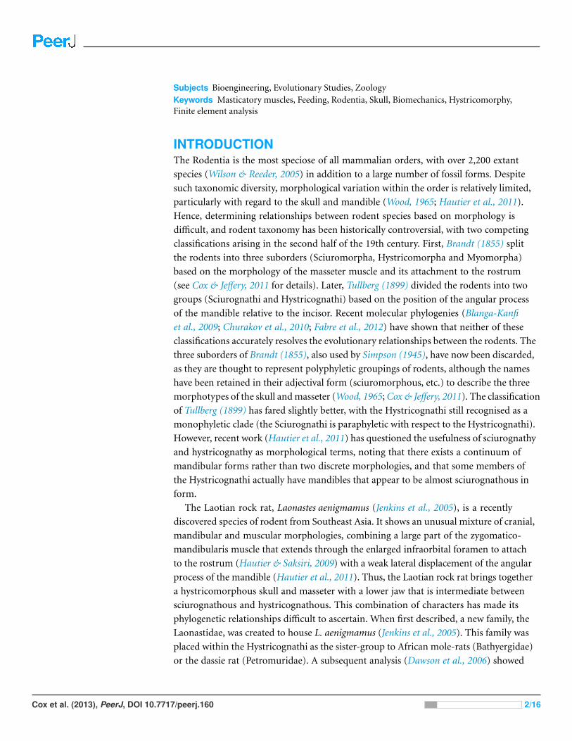
Figure 3 Predicted distribution of von Mises stresses across the skull of Laonastes aenigmamus. Arrows indicate the biting tooth. First column,incisor bites; second column, M1 bites; third column, M3 bites. First line, original models; second line, origin of IOZM moved anteriorly to front ofrostrum; third line, origin of IOZM moved posteriorly to back of rostrum; fourth line, IOZM muscle force removed and redistributed proportionallybetween the remaining masticatory muscles.
force at the condyles (Dumont et al., 2011). As it is a proportion, mechanical efficiency is
size-independent and facilitates clearer comparisons between skulls of varying sizes.
RESULTSFigure 3 shows the von Mises stress patterns generated across the skull of L. aenigmamus
during biting at the incisor, first molar and third molar. It can be seen that, aside from
the biting tooth, the zygomatic arch is the most stressed region of the skull, followed by
the orbital region. The rostrum experiences a moderate degree of stress during incisor
gnawing, but is unstressed during molar chewing, and the occipital region is unstressed
during all bites. From visual inspection of the stress distribution figures it is difficult to
determine a great deal of variation between bites at different points along the tooth row,
even between incisors and molars. However, by studying the mean von Mises stresses
across the skull (Table 3) it can be seen that there are indeed subtle differences between
bites on different teeth. In general, overall stress increases as the bite point moves closer to
the jaw articulation from the premolar to the third molar. However, the incisor bite does
Cox et al. (2013), PeerJ, DOI 10.7717/peerj.160 8/16
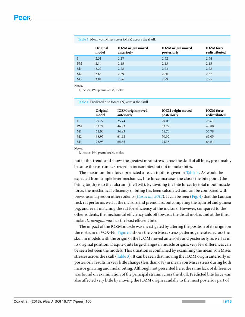
Table 3 Mean von Mises stress (MPa) across the skull.
Originalmodel
IOZM origin movedanteriorly
IOZM origin movedposteriorly
IOZM forceredistributed
I 2.31 2.27 2.32 2.34
PM 2.14 2.15 2.13 2.15
M1 2.29 2.28 2.23 2.28
M2 2.66 2.59 2.60 2.57
M3 3.04 2.86 2.99 2.95
Notes.I, incisor; PM, premolar; M, molar.
Table 4 Predicted bite forces (N) across the skull.
Originalmodel
IOZM origin movedanteriorly
IOZM origin movedposteriorly
IOZM forceredistributed
I 29.27 25.74 29.05 26.61
PM 53.74 46.93 53.72 48.80
M1 61.00 54.93 61.70 55.78
M2 68.97 61.92 70.32 62.05
M3 73.93 65.35 74.38 66.61
Notes.I, incisor; PM, premolar; M, molar.
not fit this trend, and shows the greatest mean stress across the skull of all bites, presumably
because the rostrum is stressed in incisor bites but not in molar bites.
The maximum bite force predicted at each tooth is given in Table 4. As would be
expected from simple lever mechanics, bite force increases the closer the bite point (the
biting tooth) is to the fulcrum (the TMJ). By dividing the bite forces by total input muscle
force, the mechanical efficiency of biting has been calculated and can be compared with
previous analyses on other rodents (Cox et al., 2012). It can be seen (Fig. 4) that the Laotian
rock rat performs well at the incisors and premolars, outcompeting the squirrel and guinea
pig, and even matching the rat for efficiency at the incisors. However, compared to the
other rodents, the mechanical efficiency tails off towards the distal molars and at the third
molar, L. aenigmamus has the least efficient bite.
The impact of the IOZM muscle was investigated by altering the position of its origin on
the rostrum in VOX-FE. Figure 3 shows the von Mises stress patterns generated across the
skull in models with the origin of the IOZM moved anteriorly and posteriorly, as well as in
its original position. Despite quite large changes in muscle origins, very few differences can
be seen between the models. This situation is confirmed by examining the mean von Mises
stresses across the skull (Table 3). It can be seen that moving the IOZM origin anteriorly or
posteriorly results in very little change (less than 6%) in mean von Mises stress during both
incisor gnawing and molar biting. Although not presented here, the same lack of difference
was found on examination of the principal strains across the skull. Predicted bite force was
also affected very little by moving the IOZM origin caudally to the most posterior part of
Cox et al. (2013), PeerJ, DOI 10.7717/peerj.160 9/16
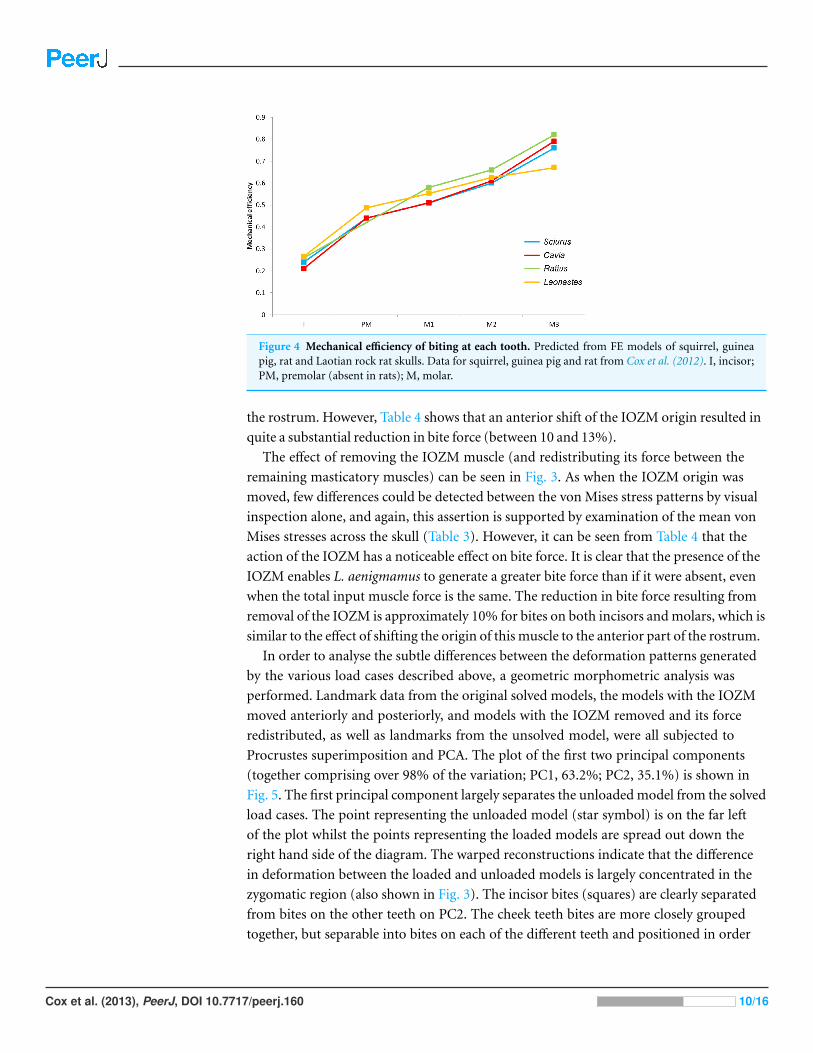
Figure 4 Mechanical efficiency of biting at each tooth. Predicted from FE models of squirrel, guineapig, rat and Laotian rock rat skulls. Data for squirrel, guinea pig and rat from Cox et al. (2012). I, incisor;PM, premolar (absent in rats); M, molar.
the rostrum. However, Table 4 shows that an anterior shift of the IOZM origin resulted in
quite a substantial reduction in bite force (between 10 and 13%).
The effect of removing the IOZM muscle (and redistributing its force between the
remaining masticatory muscles) can be seen in Fig. 3. As when the IOZM origin was
moved, few differences could be detected between the von Mises stress patterns by visual
inspection alone, and again, this assertion is supported by examination of the mean von
Mises stresses across the skull (Table 3). However, it can be seen from Table 4 that the
action of the IOZM has a noticeable effect on bite force. It is clear that the presence of the
IOZM enables L. aenigmamus to generate a greater bite force than if it were absent, even
when the total input muscle force is the same. The reduction in bite force resulting from
removal of the IOZM is approximately 10% for bites on both incisors and molars, which is
similar to the effect of shifting the origin of this muscle to the anterior part of the rostrum.
In order to analyse the subtle differences between the deformation patterns generated
by the various load cases described above, a geometric morphometric analysis was
performed. Landmark data from the original solved models, the models with the IOZM
moved anteriorly and posteriorly, and models with the IOZM removed and its force
redistributed, as well as landmarks from the unsolved model, were all subjected to
Procrustes superimposition and PCA. The plot of the first two principal components
(together comprising over 98% of the variation; PC1, 63.2%; PC2, 35.1%) is shown in
Fig. 5. The first principal component largely separates the unloaded model from the solved
load cases. The point representing the unloaded model (star symbol) is on the far left
of the plot whilst the points representing the loaded models are spread out down the
right hand side of the diagram. The warped reconstructions indicate that the difference
in deformation between the loaded and unloaded models is largely concentrated in the
zygomatic region (also shown in Fig. 3). The incisor bites (squares) are clearly separated
from bites on the other teeth on PC2. The cheek teeth bites are more closely grouped
together, but separable into bites on each of the different teeth and positioned in order
Cox et al. (2013), PeerJ, DOI 10.7717/peerj.160 10/16
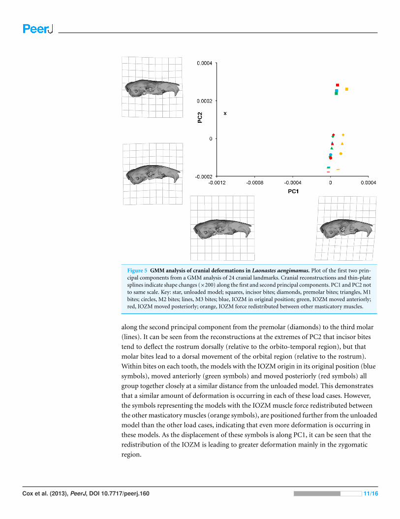
Figure 5 GMM analysis of cranial deformations in Laonastes aengimamus. Plot of the first two prin-cipal components from a GMM analysis of 24 cranial landmarks. Cranial reconstructions and thin-platesplines indicate shape changes (×200) along the first and second principal components. PC1 and PC2 notto same scale. Key: star, unloaded model; squares, incisor bites; diamonds, premolar bites; triangles, M1bites; circles, M2 bites; lines, M3 bites; blue, IOZM in original position; green, IOZM moved anteriorly;red, IOZM moved posteriorly; orange, IOZM force redistributed between other masticatory muscles.
along the second principal component from the premolar (diamonds) to the third molar
(lines). It can be seen from the reconstructions at the extremes of PC2 that incisor bites
tend to deflect the rostrum dorsally (relative to the orbito-temporal region), but that
molar bites lead to a dorsal movement of the orbital region (relative to the rostrum).
Within bites on each tooth, the models with the IOZM origin in its original position (blue
symbols), moved anteriorly (green symbols) and moved posteriorly (red symbols) all
group together closely at a similar distance from the unloaded model. This demonstrates
that a similar amount of deformation is occurring in each of these load cases. However,
the symbols representing the models with the IOZM muscle force redistributed between
the other masticatory muscles (orange symbols), are positioned further from the unloaded
model than the other load cases, indicating that even more deformation is occurring in
these models. As the displacement of these symbols is along PC1, it can be seen that the
redistribution of the IOZM is leading to greater deformation mainly in the zygomatic
region.
Cox et al. (2013), PeerJ, DOI 10.7717/peerj.160 11/16

DISCUSSIONA finite element model of the skull of the Laotian rock rat, Laonastes aenigmamus,
was created, loaded, constrained and solved. It was shown that the area of the skull
experiencing the highest levels of stress was the zygomatic arch, including the zygomatic
processes of the maxillary and frontal bones, which is likely to be a result of the large
amount of masticatory musculature that attaches directly to this area. Similarly high
zygomatic stresses have been noted in other rodents (Cox et al., 2012) as well as in other
mammalian groups (Dumont, Piccirillo & Grosse, 2005; Bright & Rayfield, 2011; Dumont
et al., 2011). It has been suggested that, in primates, the downward pull of the masseter
muscle on the zygomatic arch may be counterbalanced to some degree by the upward pull
of a soft tissue structure, namely the temporal fascia (Curtis et al., 2011). Despite many
careful dissections (e.g., Baverstock, Jeffery & Cobb, 2013), no temporal fascia has been
found in rodents, and this is also true of L. aenigmamus (Hautier & Saksiri, 2009). Thus,
for the time being, it must be assumed that, although the zygomatic stresses are high in
L. aegnimamus, they are not so high as to pose a danger of bone fracture.
The bite forces predicted in this study demonstrate that the skull of L. aenigmamus can
generate bites of 29 N during gnawing and between 53 and 74 N during chewing. By divid-
ing these values by the total input muscle force, the mechanical efficiency of biting was cal-
culated. It was shown that the Laotian rock rat is particularly efficient at incisor biting, hav-
ing a mechanical efficiency greater than squirrels or guinea pigs, and similar to that of rats
(Cox et al., 2012), but is less efficient at molar bites compared to these three rodent species.
This would seem to indicate that L. aenigmamus is well-adapted for gnawing and less so
for chewing, a conclusion that at first glance appears somewhat at odds with the suggested
diet of this species, which is thought to be largely folivorous (Scopin et al., 2011). However,
Onoda et al. (2011) have proposed that the generation of a uniform bite force across the
tooth row may be beneficial in the processing of fibrous plant material such as leaves.
Viewed in this light, L. aenigmamus is well-adapted to folivory with a difference of just 20 N
between its premolar and M3 bites, compared to 30–35 N in squirrels and guinea pigs.
One of the more intriguing results of this study is that the position of the IOZM muscle
has little effect on the overall stress experienced by the skull of L. aenigmamus. It can be
seen from both Fig. 3 and Table 3 that neither the pattern of von Mises stresses nor the
mean stress across the skull are greatly affected by moving the origin of the IOZM forwards
or backwards. This may be because the IOZM is contributing a relatively small proportion
of the total muscle force (around 8%). Although the IOZM appears to be quite a large
muscle in the lateral view (see Hautier & Saksiri, 2009), it is relatively small compared to
the superficial and deep masseters. Moreover, it also possesses the longest muscle fibres
of all masticatory muscles, and so its PCSA (and hence muscle force) is comparatively
reduced (Table 1). Thus, its ability to impact the overall stress patterns across the skull may
be fairly limited and, furthermore, these patterns may be more strongly influenced by skull
morphology than by muscle geometry.
In contrast to the position of its attachment, the presence or absence of the IOZM has
a much stronger effect on the cranial biomechanics of L. aenigmamus. Figure 5 shows
Cox et al. (2013), PeerJ, DOI 10.7717/peerj.160 12/16

that the symbols representing the models without the IOZM are positioned further from
the unloaded model that the other load cases, indicating that the removal of the IOZM
leads to greater deformation (and therefore strain) across the skull. Thus, one of the major
functions of the IOZM, at least in L. aenigmamus, appears to be to minimise strain during
feeding. This conclusion holds true at all bites, both gnawing and chewing.
In addition to its effect on cranial deformation, it was also found that the IOZM has
a strong impact on the bite force produced by L. aenigmamus (Table 4). Specifically,
removing the IOZM altogether (and redistributing its force between the remaining
masticatory muscles) reduces the bite force generated at all teeth. Similarly, moving the
origin of the IOZM to the anterior tip of the rostrum reduces bite force to a similar degree.
It is likely that this effect is a result of the wrapping of the IOZM around the zygomatic
process of the maxilla, which means that as the IOZM is moved forward, its vector of pull
becomes more horizontal (Fig. 1), and so less able to generate bite force. On the other
hand, moving the origin of the IOZM posteriorly appears to have little effect on bite
force. Interestingly, the reduction in bite force resulting from the anterior movement or
removal of the IOZM is largely constant along the tooth row – around 10%. Therefore, it
would seem that the extension of the zygomaticomandibularis on to the rostrum evolved
in order to increase bite force (as proposed by Becht, 1953). Given that there is minimal
difference in bite force between the models with the IOZM in its original position and
those with the origin moved posteriorly, the position of the IOZM origin halfway along the
rostrum may simply have coevolved with the lengthening of the rostrum in this species.
Alternatively, Herrel et al. (2012) have suggested that the anterior insertion of the IOZM
in L. aenigmamus increases the horizontal component of biting, which leads to anterior
displacement of the mandible during jaw closing, which may be advantageous for the
processing of leaves. Whatever the driving force behind the anterior extension of the
IOZM, the results here indicate that the reason that the IOZM does not extend any further
along the rostrum, as is seen in some other hystricomorph rodents such as the springhare
(Offermans & De Vree, 1989) and capybara (Muller, 1933), is because this would lead to a
reduction in bite force at both the incisors and cheek teeth (Table 4).
The results of this study have provided some important insights into the role of the
IOZM muscle in the feeding behaviour of L. aenigmamus. Further investigations into
other rodents, particularly other hystricomorphs, will enable us to understand whether
the ability of the IOZM to increase bite force is unique to L. aenigmamus or common to all
rodents that possess it. If the latter scenario is true, this could provide a selective advantage
that may have driven the evolution of the IOZM, and could explain why it has evolved
independently in several rodent groups (Ctenohystrica, Dipodidae, Anomaluroidea and
Gliridae). However, finite element models can only shed light on static loading, and
therefore cannot inform about the dynamic processes to which the IOZM may contribute,
such as propalineal (antero-posterior) movements of the lower jaw, jaw opening or the fine
control of gnawing. These activities can be addressed with dynamic modelling techniques,
such as multibody dynamics analysis (e.g., Jones et al., 2012), and may well provide a
fruitful avenue of research in the future.
Cox et al. (2013), PeerJ, DOI 10.7717/peerj.160 13/16

ACKNOWLEDGEMENTSWe thank Jean-Pierre Hugot for providing access to the specimens and Dominique
Adriaens, Loes Brabant and Luc Van Hoorebeke for scanning the specimens at the
University of Ghent CT facility (UGCT). We are grateful to Paul O’Higgins and Michael
Fagan for access to VOX-FE finite element software. Thanks are due to Laura Fitton for
help with model construction, Peter Bazira for technical support with high-performance
computing, and Andrew McIntosh and Thomas Puschel for assistance with GMM
software.
ADDITIONAL INFORMATION AND DECLARATIONS
FundingThe funding for Joanna Kirkham’s intercalated BSc was provided by the UK National
Health Service. The funders had no role in study design, data collection and analysis,
decision to publish, or preparation of the manuscript.
Grant DisclosuresThe following grant information was disclosed by the authors:
UK National Health Service.
Competing InterestsThe authors declare that they have no competing interests.
Author Contributions• Philip G. Cox conceived and designed the experiments, analyzed the data, wrote the
paper.
• Joanna Kirkham and Anthony Herrel performed the experiments, analyzed the data,
wrote the paper.
REFERENCESBaverstock H, Jeffery NS, Cobb SN. 2013. The morphology of the mouse masticatory
musculature. Journal of Anatomy 223:46–60 DOI 10.1111/joa.12059.
Becht G. 1953. Comparative biologic-anatomical researcher on mastication in some mammals.Proceedings of the Koninklijke Nederlandse Akademie van Wetenschappen. Series C 56:508–526.
Blanga-Kanfi S, Miranda H, Penn O, Pupko T, DeBry RW, Huchon D. 2009. Rodent phylogenyrevised: analysis of six nuclear genes from all major rodent clades. BMC Evolutionary Biology9:71 DOI 10.1186/1471-2148-9-71.
Brandt JF. 1855. Beitrage zur nahern Kenntniss der Saugethiere Russlands. Memoires de l’AcademieImperiale des Sciences de St Petersbourg, Sixieme Serie 9:1–375.
Bright JA, Rayfield EJ. 2011. Sensitivity and ex vivo validation of finite element models of thedomestic pig cranium. Journal of Anatomy 219:456–471 DOI 10.1111/j.1469-7580.2011.01408.x.
Churakov G, Sadasivuni M, Rosebloom K, Huchon D, Brosius J, Schmitz J. 2010.Rodent evolution: back to the root. Molecular Biology and Evolution 27:1315–1327DOI 10.1093/molbev/msq019.
Cox et al. (2013), PeerJ, DOI 10.7717/peerj.160 14/16
Cox PG, Fagan MJ, Rayfield EJ, Jeffery N. 2011. Finite element modelling of squirrel, guineapig and rat skulls: using geometric morphometrics to assess sensitivity. Journal of Anatomy219:696–709 DOI 10.1111/j.1469-7580.2011.01436.x.
Cox PG, Jeffery N. 2011. Reviewing the jaw-closing musculature in squirrels, rats and guinea pigswith contrast-enhanced microCT. Anatomical Record 294:915–928 DOI 10.1002/ar.21381.
Cox PG, Rayfield EJ, Fagan MJ, Herrel A, Pataky TC, Jeffery N. 2012. Functional evolution of thefeeding system in rodents. PLoS ONE 7(4):e36299 DOI 10.1371/journal.pone.0036299.
Curtis N, Witzel U, Fitton L, O’Higgins P, Fagan M. 2011. The mechanical significance ofthe temporal fasciae in Macaca fascicularis: an investigation using finite element analysis.Anatomical Record 294:1178–1190 DOI 10.1002/ar.21415.
Dawson MR, Marivaux L, Li C, Beard KC, Metais G. 2006. Laonastes aenigmamus and the‘Lazarus effect’ in recent mammals. Science 311:1456–1458 DOI 10.1126/science.1124187.
Dumont ER, Davis JL, Grosse IR, Burrows AM. 2011. Finite element analysis ofperformance in the skulls of marmosets and tamarins. Journal of Anatomy 218:151–162DOI 10.1111/j.1469-7580.2010.01247.x.
Dumont ER, Piccirillo J, Grosse IR. 2005. Finite-element analysis of biting behaviour and bonestress in the facial skeletons of bats. The Anatomical Record Part A: Discoveries in Molecular,Cellular, and Evolutionary Biology 283:319–330 DOI 10.1002/ar.a.20165.
Fabre P-H, Hautier L, Dimitrov D, Douzery EJP. 2012. A glimpse on the pattern of rodentdiversification: a phylogenetic approach. BMC Evolutionary Biology 12:88 DOI 10.1186/1471-2148-12-88.
Hautier L. 2010. Masticatory muscle architecture in the gundi, Ctenodactylus vali (Mammalia:Rodentia). Mammalia 74:153–162 DOI 10.1515/mamm.2010.025.
Hautier L, Rebrun R, Saksiri S, Michaux J, Vianey-Liaud M, Marivaux L. 2011. Hystricognathyvs sciurognathy in the rodent jaw: a new morphometric assessment of hystricognathyapplied to the living fossil Laonastes (Diatomyidae). PLoS ONE 6(4):e18698DOI 10.1371/journal.pone.0018698.
Hautier L, Saksiri S. 2009. Masticatory muscle architecture in the Laotian rock rat Laonastesaenigmamus (Mammalia, Rodentia): new insights into the evolution of hystricognathy. Journalof Anatomy 215:401–410 DOI 10.1111/j.1469-7580.2009.01130.x.
Herrel A, Fabre A-C, Hugot J-P, Keovichit K, Adriaens D, Brabant L, Van Hoorebeke L,Cornette R. 2012. Ontogeny of the cranial system in Laonastes aenigmamus. Journal of Anatomy221:128–137 DOI 10.1111/j.1469-7580.2012.01519.x.
Huchon D, Chevret P, Jordan U, Kilpatrick CW, Ranwez V, Jenkins PD, Brosius J, Schmitz J.2007. Multiple molecular evidences for a living mammalian fossil. Proceedings of the NationalAcademy of Sciences of the United States of America 104:7495–7499DOI 10.1073/pnas.0701289104.
Jenkins PD, Kilpatrick CW, Robinson MF, Timmins RJ. 2005. Morphological and molecularinvestigations of a new family, genus and species of rodent (Mammalia: Rodentia:Hystricognatha) from Lao PDR. Systematics and Biodiversity 2:419–454DOI 10.1017/S1477200004001549.
Jones MEH, O’Higgins P, Fagan MJ, Evans SE, Curtis N. 2012. Shearing mechanics and theinfluence of a flexible symphysis during oral food processing in Sphenodon (Lepidosauria:Rhynchocephalia). Anatomical Record 295:1075–1091 DOI 10.1002/ar.22487.
Klingenberg CP. 2011. MorphoJ: an integrated software package for geometric morphometrics.Molecular Ecology Resources 11:353–357 DOI 10.1111/j.1755-0998.2010.02924.x.
Cox et al. (2013), PeerJ, DOI 10.7717/peerj.160 15/16
Kupczik K, Dobson CA, Fagan MJ, Crompton RH, Oxnard CE, O’Higgins P. 2007. Assessingmechanical function of the zygomatic region in macaques: validation and sensitivity testing offinite element models. Journal of Anatomy 210:41–53 DOI 10.1111/j.1469-7580.2006.00662.x.
Liu J, Shi L, Fitton LC, Phillips R, O’Higgins P, Fagan MJ. 2012. The application of musclewrapping to voxel-based finite element models of skeletal structures. Biomechanics and Modelingin Mechanobiology 11:35–47 DOI 10.1007/s10237-011-0291-5.
Muller A. 1933. Die Kaumuskulatur des Hydrochoeruscapybara und ihre Bedeutung fur dieFormgestaltung des Schadels. Morphologisches Jahrbuch 72:1–59.
Murphy RA, Beardsley AC. 1974. Mechanical properties of the cat soleus muscle in situ. AmericanJournal of Physiology 227:1008–1013.
Offermans M, De Vree F. 1989. Morphology of the masticatory apparatus in the springhare,Pedetes capensis. Journal of Mammalogy 70:701–711 DOI 10.2307/1381705.
O’Higgins P, Cobb SN, Fitton LC, Groning F, Phillips R, Liu J, Fagan MJ. 2011. Combininggeometric morphometrics and functional simulation: an emerging toolkit for virtual functionalanalyses. Journal of Anatomy 218:3–15 DOI 10.1111/j.1469-7580.2010.01301.x.
Onoda Y, Westoby M, Adler PB, Choong AMF, Clissold FJ, Cornelisson JHC, Dıaz S,Dominy NJ, Elgart A, Enrico L, Fine PVA, Howard JJ, Jalili A, Kitajima K, Kurokawa H,McArthur C, Lucas PW, Markesteijn L, Perez-Harguindeguy N, Poorter L, Richards L,Santiago LS, Sosinski EE, Van Bael SA, Warton DI, Wright IJ, Wright SJ, Yamashita N.2011. Global patterns of leaf mechanical properties. Ecology Letters 14:301–312DOI 10.1111/j.1461-0248.2010.01582.x.
Rayfield EJ. 2007. Finite element analysis and understanding the biomechanics and evolutionof living and fossil organisms. Annual Review of Earth and Planetary Science 35:541–576DOI 10.1146/annurev.earth.35.031306.140104.
Scopin AE, Saveljev AP, Suntsova NA, Gnophanxay S, Tikhonov AN, Abramov AV. 2011.Digestive system of the Laotian rock rat Laonastes aenigmamus (Rodentia: Diatomyidae) fromthe evolutionary viewpoint. Proceedings of the Zoological Institute RAS 315:3–18.
Simpson GG. 1945. The principles of classification and a classification of mammals. Bulletin of theAmerican Museum of Natural History 85:1–350.
Tullberg T. 1899. Uber das System der Nagethiere, eine phylogenetische Studie. Nova Acta RegiaeSocietatis Scientarium Upsaliensis Series 3 18:1–514.
van Spronsen PH, Weijs WA, Valk J, Prahl-Andersen B, van Ginkel FC. 1989. Comparisonof jaw-muscle bite-force cross-sections obtained by means of magnetic resonanceimaging and high-resolution CT scanning. Journal of Dental Research 68:1765–1770DOI 10.1177/00220345890680120901.
Weijs WA, Dantuma R. 1975. Electromyography and mechanics of mastication in the albino rat.Journal of Morphology 146:1–34 DOI 10.1002/jmor.1051460102.
Wilson DE, Reeder DM. 2005. Mammal species of the world. Baltimore: Johns Hopkins Press.
Wood AE. 1965. Grades and clades among rodents. Evolution 19:115–130 DOI 10.2307/2406300.
Wood SA, Strait DS, Dumont ER, Ross CF, Grosse IR. 2011. The effects of modeling simplifi-cations on craniofacial finite element analyses: the alveoli (tooth sockets) and periodontalligaments. Journal of Biomechanics 44:1831–1838 DOI 10.1016/j.jbiomech.2011.03.022.
Wroe S. 2010. Cranial mechanics of mammalian carnivores: recent advances using a finite elementapproach. In: Goswami A, Friscia A, eds. Carnivoran evolution. New views on phylogeny, formand function. Cambridge: Cambridge University Press, 466–485.
Cox et al. (2013), PeerJ, DOI 10.7717/peerj.160 16/16
Related Documents