Advances in Surgical Sciences 2021; 9(1): 6-9 http://www.sciencepublishinggroup.com/j/ass doi: 10.11648/j.ass.20210901.12 ISSN: 2376-6174 (Print); ISSN: 2376-6182 (Online) Massive Splenic Infarction Secondary to Distal Pancreatectomy for a Blunt Abdominal Trauma Iosvany Rivero Hernández 1 , Antonio Calvo Durán 1 , Ana Diez Núñez 1 , Juan Alija Castro 2 , Vicente Vega Ruiz 1 1 Department of General and Digestive Surgery, Universitary Hospital of Puerto Real, Puerto Real, Spain 2 Department of Radiology, Universitary Hospital of Puerto Real, Puerto Real, Spain Email address: To cite this article: Iosvany Rivero Hernández, Antonio Calvo Durán, Ana Diez Núñez, Juan Alija Castro,Vicente Vega Ruiz. Massive Splenic Infarction Secondary to Distal Pancreatectomy for a Blunt Abdominal Trauma. Advances in Surgical Sciences. Vol. 9, No. 1, 2021, pp. 6-9. doi: 10.11648/j.ass.20210901.12 Received: January 29, 2021; Accepted: February 6, 2021; Published: February 23, 2021 Abstract: Massive splenic infarction is an uncommon diagnosis that is often overlooked. It is whether asymptomatic or symptomatic with abdominal pain, fever and splenomegaly. There is no consensus on which the best treatment option is, in most cases it is about deciding on conservative or surgical management according to individual patient characteristics. Case presentation: We present the case of a 33- year- old patient with a blunt abdominal trauma while riding a horse, who is taken to the hospital emergency department. A contrast-enhanced abdominal computed tomography is carried out with the diagnosis of a Grade III pancreatic lesion involving the distal portion of the body and tail of the pancreas with associated active bleeding. An urgent laparotomy is performed with a spleen-preserving distal pancreatectomy procedure. During the immediate postoperative phase a massive splenic infarction is diagnosed after a follow-up contrast-enhanced abdominal computed tomography is carried out and successfully treated with conservative measures. Conclusions: It is important to highlight the feasibility of the non-surgical treatment in this kind of spleen lesion, because the spleen preservation proves to be necessary, whenever possible, due to the importance of its immunological role for the high risk of sepsis associated with death and neoplastic processes on those individuals with splenectomy. Keywords: Massive Splenic Infarct, Distal Pancreatectomy, Blunt Abdominal Trauma 1. Introduction Massive splenic infarction is an uncommon diagnosis and is characterized by vessel occlusion, parenchymal ischemia, and subsequent tissue necrosis involving more than half of the spleen. However, finding a patient with abdominal pain and fever is a common situation with a broad range of differential diagnosis among which splenomegaly is included. This could be considered an isolated and common sign in some ailments that manifests with a distended or painful abdomen and could be caused by chronic haemolysis, haematological and non- haematological neoplastic diseases [1]. The splenomegaly related to a massive splenic infarction is an uncommon finding. It is generally associated with hematologic, oncologic and infectious diseases, thus it is not considered a first- line diagnostic option and is overlooked in over a 25% of cases [2]. There is very little evidence in the literature regarding this issue, as a result, diagnosis is less likely to be established. It has an acute progression and could be mistaken for an acute abdomen, except that when the diagnosis is made, it could be managed conservatively with serial follow-up imaging tests. We describe the case of a patient with a massive splenic infarct secondary to a distal pancreatectomy for blunt abdominal trauma with a grade III pancreatic lesion. 2. Case Presentation A 33-year-old woman was brought by emergency medical services to our emergency department with a blunt abdominal and cranial trauma while riding a horse without losing

Welcome message from author
This document is posted to help you gain knowledge. Please leave a comment to let me know what you think about it! Share it to your friends and learn new things together.
Transcript

Advances in Surgical Sciences 2021; 9(1): 6-9
http://www.sciencepublishinggroup.com/j/ass
doi: 10.11648/j.ass.20210901.12
ISSN: 2376-6174 (Print); ISSN: 2376-6182 (Online)
Massive Splenic Infarction Secondary to Distal Pancreatectomy for a Blunt Abdominal Trauma
Iosvany Rivero Hernández1, Antonio Calvo Durán
1, Ana Diez Núñez
1, Juan Alija Castro
2,
Vicente Vega Ruiz1
1Department of General and Digestive Surgery, Universitary Hospital of Puerto Real, Puerto Real, Spain 2Department of Radiology, Universitary Hospital of Puerto Real, Puerto Real, Spain
Email address:
To cite this article: Iosvany Rivero Hernández, Antonio Calvo Durán, Ana Diez Núñez, Juan Alija Castro,Vicente Vega Ruiz. Massive Splenic Infarction
Secondary to Distal Pancreatectomy for a Blunt Abdominal Trauma. Advances in Surgical Sciences. Vol. 9, No. 1, 2021, pp. 6-9.
doi: 10.11648/j.ass.20210901.12
Received: January 29, 2021; Accepted: February 6, 2021; Published: February 23, 2021
Abstract: Massive splenic infarction is an uncommon diagnosis that is often overlooked. It is whether asymptomatic or
symptomatic with abdominal pain, fever and splenomegaly. There is no consensus on which the best treatment option is, in
most cases it is about deciding on conservative or surgical management according to individual patient characteristics. Case
presentation: We present the case of a 33- year- old patient with a blunt abdominal trauma while riding a horse, who is taken to
the hospital emergency department. A contrast-enhanced abdominal computed tomography is carried out with the diagnosis of
a Grade III pancreatic lesion involving the distal portion of the body and tail of the pancreas with associated active bleeding.
An urgent laparotomy is performed with a spleen-preserving distal pancreatectomy procedure. During the immediate
postoperative phase a massive splenic infarction is diagnosed after a follow-up contrast-enhanced abdominal computed
tomography is carried out and successfully treated with conservative measures. Conclusions: It is important to highlight the
feasibility of the non-surgical treatment in this kind of spleen lesion, because the spleen preservation proves to be necessary,
whenever possible, due to the importance of its immunological role for the high risk of sepsis associated with death and
neoplastic processes on those individuals with splenectomy.
Keywords: Massive Splenic Infarct, Distal Pancreatectomy, Blunt Abdominal Trauma
1. Introduction
Massive splenic infarction is an uncommon diagnosis and is
characterized by vessel occlusion, parenchymal ischemia, and
subsequent tissue necrosis involving more than half of the
spleen. However, finding a patient with abdominal pain and
fever is a common situation with a broad range of differential
diagnosis among which splenomegaly is included. This could
be considered an isolated and common sign in some ailments
that manifests with a distended or painful abdomen and could
be caused by chronic haemolysis, haematological and non-
haematological neoplastic diseases [1].
The splenomegaly related to a massive splenic infarction is
an uncommon finding. It is generally associated with
hematologic, oncologic and infectious diseases, thus it is not
considered a first- line diagnostic option and is overlooked in
over a 25% of cases [2].
There is very little evidence in the literature regarding this
issue, as a result, diagnosis is less likely to be established. It
has an acute progression and could be mistaken for an acute
abdomen, except that when the diagnosis is made, it could be
managed conservatively with serial follow-up imaging tests.
We describe the case of a patient with a massive splenic
infarct secondary to a distal pancreatectomy for blunt
abdominal trauma with a grade III pancreatic lesion.
2. Case Presentation
A 33-year-old woman was brought by emergency medical
services to our emergency department with a blunt abdominal
and cranial trauma while riding a horse without losing

Advances in Surgical Sciences 2021; 9(1): 6-9 7
consciousness. Hemodynamically stable at the time of arrival
with BP 90/60mmHg, Pulse 81, GCS 15, Haemoglobin 13.9
g/dl, small wound over the left side of the forehead without
active bleeding and abdominal pain over the left upper
quadrant without abdominal tenderness.
(a) (b)
(c) (d)
Figure 1. Diagnosis and evolution of lesions through abdominal sonography and Computed tomography Scan. Complete pancreatic laceration- fracture over
the pancreatic body and tail union with associated active bleeding (white arrow) (a); Massive splenic infarction on the 10th post-operative day (white arrow)
(b); Follow-up sonography, in the first month after the surgical procedure, of an 8.5 cm diameter spleen (dash line), and a large hypoechoic area occupying
two thirds of the spleen, concerning a necrotic-ischaemic area previously known with preserved areas of normal parenchyma (c); Follow-up sonography over
the ninth month of a 7 cm interpolar diameter spleen (dash line). A one-centimetre small fluid collection/haematoma is identified, having remarkably shrunken
concerning previous exams (d).
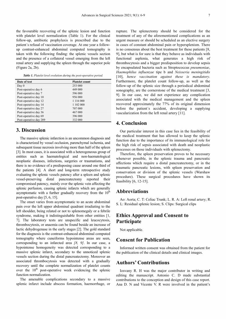
(a) (b)
Figure 2. One year follow-up Contrast-enhanced abdominal computed tomography showing the spleen vascularization. Collateral vessel emerging from the
left renal artery, coronal view (a); Collateral vessel supplying the spleen through the superior pole, axial view (b).
A contrast-enhanced abdominal computed tomography is
taken and a total laceration of the distal pancreas with a
complete transection of the wirsun duct was detected,
associated with active bleeding (Figure 1a) and a
retroperitoneal peripancreatic hematoma with extension into
the intraperitoneal cavity. An urgent spleen and splenic vessel-
preserving distal pancreatectomy is performed without
incidences. The post-operative period proceeded normally
although a gradual increase in platelets count was noticed on a
follow-up blood test (Table 1). A contrast-enhanced abdominal
computed tomography is taken (Figure 1b) and a massive
splenic infarction is detected with an adjacent small associated
fluid collection. The treatment consists in percutaneous
drainage of the fluid collection with conservative management
of the splenic infarction. After a remarkable clinical
improvement, the patient was discharged with periodical
follow-up, abdominal sonography (Figures 1c, 1d) and blood
test on a weekly basis, gradually getting better over time due to
Advances in Surgical Sciences 2021; 9(1): 6-9 7
the favourable recovering of the splenic lesion and function
with platelet level normalization (Table 1). For the clinical
follow-up, antibiotic prophylaxis is prescribed due to the
patient´s refusal of vaccination coverage. At one year a follow-
up contrast-enhanced abdominal computed tomography is
taken with the following finding: the splenic vessels section
and the presence of a collateral vessel emerging from the left
renal artery and supplying the spleen through the superior pole
(Figure 2a, 2b).
Table 1. Platelet level evolution during the post-operative period.
Date of test Platelet count
Day 0 253 000
Post-operative day 6 449 000
Post-operative day 7 596 000
Post-operative day 10 956 000
Post-operative day 12 1 116 000
Post-operative day 14 1 102 000
Post-operative day 27 707 000
Post-operative day 36 467 000
Post-operative day 69 396 000
Post-operative day 289 332 000
3. Discussion
The massive splenic infarction is an uncommon diagnosis and
is characterized by vessel occlusion, parenchymal ischemia, and
subsequent tissue necrosis involving more than half of the spleen
[3]. In most cases, it is associated with a heterogeneous group of
entities such as haematological and non-haematological
neoplastic diseases, infections, surgeries or traumatisms, and
there is no evidence of a predisposing cause around one third of
the patients [4]. A short and long-term retrospective study
evaluating the splenic vessels patency after a spleen and splenic
vessel-preserving distal pancreatectomy reported their
compromised patency, mainly over the splenic vein affecting the
splenic perfusion, causing splenic infarcts which are generally
asymptomatic with a further gradually recovery from the 10th
post-operative day [5, 6, 15].
The onset varies from asymptomatic to an acute abdominal
pain over the left upper abdominal quadrant irradiating to the
left shoulder, being related or not to splenomegaly or a febrile
syndrome, making it indistinguishable from other entities [1,
7]. The laboratory tests are unspecific and leucocytosis,
thrombocytosis, or anaemia can be found beside an increase of
lactic dehydrogenase in the early stages [2]. The gold standard
for the diagnosis is the contrast-enhanced abdominal computed
tomography where cuneiforms hypointense areas are seen,
corresponding to an infarcted area [8, 9]. In our case, a
hypointense homogeneity was detected corresponding to a
massive splenic infarct, secondary to the unnoticed splenic
vessels section during the distal pancreatectomy. Moreover an
associated thrombocytosis was detected with a gradually
recovery until the complete normalization of platelet counts
over the 10th post-operative week evidencing the splenic
function normalization.
The amenable complications secondary to a massive
splenic infarct include abscess formation, haemorrhage, or
rupture. The splenectomy should be considered for the
treatment of any of the aforementioned complications as an
urgent measure or should be scheduled as an elective surgery
in cases of constant abdominal pain or hypersplenism. There
is no consensus about the best treatment for these patients [8,
9], but what is for sure is that they behave as individuals with
functional asplenia, what generates a high risk of
thrombocytosis and a bigger predisposition to develop sepsis
by encapsulated bacteria such as Streptococcus pneumoniae,
Haemophilus influenzae tipe b and Neisseria meningitidis
[10], hence vaccination against these is mandatory.
Furthermore, the platelet count follow-up, as well as the
follow-up of the splenic size through a periodical abdominal
sonography, are the cornerstone of the medical treatment [3,
9]. In our case, we did not experience any complication
associated with the medical management and the spleen
recovered approximately the 77% of its original dimension
before the patient´s accident, developing a supplying
vascularization from the left renal artery [11].
4. Conclusion
Our particular interest in this case lies in the feasibility of
the medical treatment that has allowed to keep the splenic
function due to the importance of its immunological role for
the high risk of sepsis associated with death and neoplastic
processes on those individuals with splenectomy.
Therefore, the spleen preservation proves to be necessary
whenever possible, in the splenic trauma and pancreatic
affections which require a distal pancreatectomy, or in the
traumatic pancreatic lesions, with spleen preservation and
conservation or division of the splenic vessels (Warshaw
procedure). These surgical procedures have shown its
feasibility [6, 12-15].
Abbreviations
Ao: Aorta; C. T: Celiac Trunk; L. R. A: Left renal artery; R.
S. L: Residual splenic lesion; S. Clips: Surgical clips
Ethics Approval and Consent to
Participate
Not applicable.
Consent for Publication
Informed written consent was obtained from the patient for
the publication of the clinical details and clinical images.
Authors’ Contributions
Iosvany R. H was the major contributor in writing and
editing the manuscript. Antonio C. D made substantial
contributions to the conception and design of this case report.
Ana D. N and Vicente V. R were involved in the patient’s

8 Iosvany Rivero Hernández et al.: Massive Splenic Infarction Secondary to Distal Pancreatectomy for a
Blunt Abdominal Trauma
treatment. Juan A. C contributed with the patient radiological
follow-up and the images selection. All authors read and
approved the final manuscript.
Funding
None.
Availability of Data and Materials
All relevant data are provided in the manuscript.
Competing Interests
The authors declare that they have no competing interests.
Acknowledgements
Not applicable.
References
[1] Friedman-Klabanoff D, Ball A, Rutare S, McCall N, et al. Three Rwandan Children with Massive Splenomegaly and Epstein-Barr Virus-associated Lymphoproliferative Disorders: Case Presentations and the Literature Review. J Pediatr Hematol Oncol. 2016; 38 (5): e158-61.
[2] Kranidiotis G, Efstratiadis E, Kapsalakis G, Loizos G, et al. Splenic infarcts as a rare manifestation of parvovirus B19 infection. IDCases. 2016; 4: 62-4.
[3] Bokman Christine L.; Sfeir Maroun; Chahwala Veer; Ginzburg Enrique. Spontaneous massive splenic infarction in the setting of renal transplant and septic shock: a case report and review of the literature. Case Rep Med. 2014.
[4] Arenal Vera, J. J. et al. Splenic infarction secondary to acute pancreatitis. Rev. Esp. Enferm. Dig. 2008; 100 (5): 300-303.
[5] Y.-S. Yoon, K. H. Lee, H.-S. Han, J. Y. Cho and K. S. Ahn. Patency of splenic vessels after laparoscopic spleen and splenic vessel-preserving distal pancreatectomy. British Journal of Surgery 2009; 96: 633–640.
[6] Sun et al. Clinical efficacy of spleen-preserving distal pancreatectomy with or without splenic vessel preservation. A Meta-analysis. Medicine (2017) 96: 48.
[7] Antopolsky M, Hiller N, Salameh S, Goldshtein B, et al. Splenic infarction: 10 years of experience. Am J Emerg Med. 2009; 27 (3): 262-5.
[8] F. Fatjó, J. Ramos, A. Culla y J. M. Grau. Splenic infarctions: an uncommon cause of febril syndrome. Med Clin (Barc) 2002; 119 (9): 356-9.
[9] D’Angelo G, Marseglia L, Russo T, Gitto E, et al. Long term follow up of a massive, conservatively treated, splenic infarction in a young adolescent. Pediatr Int. 2017; 59 (11): 1210-2.
[10] Lamsfus-Prieto JA et al. Prevention of sepsis in asplenic patients. Cir Esp. 2007; 81 (5): 247-51.
[11] Skandalakis JE, Colborn GL, Pemberton LB, Skandalakis TN, Skandalakis LJ, Gray SW. The surgical anatomy of the spleen. Prob Gen Surg 7 (1): 1-17, 1990.
[12] Carrére et al.: Spleen-preserving distal pancreatectomy with excision of splenic artery and vein: A case matched comparison with conventional distal pancreatectomy with splenectomy. World J Surg (2007) 31: 375–382.
[13] A. L. Warshaw, “Conservation of the spleen with distal pancreatectomy,” JAMA Surgery, vol. 123, no. 5, pp. 550–553, 1988.
[14] Ferrone et al, “Twenty-Three Years of the Warshaw Operation for Distal Pancreatectomy With Preservation of the Spleen”. Ann Surg 2011; 253: 1136–1139.
[15] Kimura et al, “A case of complete splenic infarction after laparoscopic spleen-preserving distal pancreatectomy. BMC Surgery (2018) 18: 22.
Related Documents