Mass Spectrometric Characterization of Glycated p-Lactoglobulin Peptides Derived from Galacto-oligosaccharides Surviving the In Vitro Gastrointestinal Digestion F. Javier Moreno,a Jesus Eduardo Quintanilla-L6pez,b,c Rosa Lebr6n-Aguilar,c Agustin Olano,a and Maria Luz Sanz d a Instituto de Fermentaciones Industriales (CSIC), Madrid, Spain b Departamento de lngenieria Quirnica Industrial y del Medio Ambiente, ETS Ingenieros Industriales (V. P.M.) Madrid, Spain c lnstituto de Quimica-Fisica "Rocasolano" (CSIC), Madrid, Spain d lnstituto de Quimica Organica General (CSIC), Madrid, Spain A mass spectrometric study has been carried out to elucidate the structures of glycated pep tides obtained after in vitro gastrointestinal digestion of bovine {3-lactoglobulin ({3-LG) glycated with prebiotic galacto-oligosaccharides (GOS). The digests of both native and glycated {3-LG were analyzed by MALDI-MS, LC-ESI-MS, and LC-ESI-MS/MS. MALDI-MS profiles showed marked differences mainly related to the lower intensity of ions correspond- ing to the digest of glycated {3-LG. Overall, 58 and 23 unglycated peptides covering 97% and 63% of the mature {3-LG sequence could be identified in the digests of native and glycated samples, respectively. The LC-ESI-MS analyses corroborated the MALDI-MS results regarding the unglycated pep tides but they also enabled an extensive investigation into the digest of glycated {3-LG. Thus, a total of 19 pep tides glycated with GOS from two to seven hexose units could be identified. The tandem mass spectra of glycated pep tides were mostly characterized by two neutral losses of 1026/1056, 864/894, 702/732, 540/570, 378/408, and 216/246 u, corresponding to the formation of the furylium ion and its subsequent "CHOH" loss, indicative of the peptide glycation with hepta-, hexa-, penta-, tetra-, tri-, and disaccharides, respectively. Also, other minor ionic species containing the furylium ring linked to different galactose units could be also detected, showing the diversity of the fragmentation pattern of peptides glycated with larger size carbohydrates. Finally, the putative GOS glycation sites could be determined at the NHz-terminal Leu residue and at Lys residues located in positions 14, 47, 75, 77, 83, 91, 100, 135, and 138. (J Am Soc Mass Spectrom 2008, 19, 927-937) © 2008 American Society for Mass Spectrometry T he Maillard reaction is one of the most important and complex reactions between reducing sugars . and amino acids, pep tides, or proteins that may spontaneously occur in living organisms or during food processing. Controversially, either beneficial or detri- mental effects have been attributed to the Maillard reaction products (MRP). Thus, some compounds such as reductones and melanoidins have shown antioxidant activity [I, 2], whereas the so-called "advanced glyca- tion end products" (AGEs) include a variety of protein adducts implicated in inflammatory reactions and tis- sue damage [3,4]. The resistance of some MRP to the gastrointestinal digestion environment has been proven [5-7], indicat- ing that they could be available for gut microflora Address reprint requests to Dr. M. L. Sanz, Instituto de Quimica Orgamca General (CSIC), Analisis Instrumental y Quimica Ambiental, C/ Juan de la Cierva, 3 28006 Madrid, Spain. E-mail: [email protected] fermentation. However, little is known about their effects on the growth of these micro-organisms and consequently on chronic gut disorders. Some studies have revealed that the number of anaerobic species increased following fermentation of melanoidins produced from glucose and lysine [8]. Similarly, Dell'Aquila et al. [9] demonstrated that melanoidins derived from gluten glycated with glucose affect the growth of the colonic microbiota. Tuohy et al. [10] showed an increase of the more detrimental micro- organisms against a diminution of the beneficial ones (bifidobacteria and lactobacilli) following fermentation of BSA glycated with glucose on fecal bacteria. How- ever, to the best of our knowledge, no gut fermentation studies have so far been performed using proteins glycated with prebiotic carbohydrates (nondigestible compounds, mainly carbohydrates, which selectively stimulate the growth of certain intestinal bacteria ben- eficially affecting host health [11]). © 2008 American Society for Mass Spectrometry. Published by Elsevier Inc. 1044-0305/08/$32.00 doi:1 0.101 fi /Liasms.2oo8.04.016 Published online April 10, 2008 Received January 8, 2008 Revised March 5, 2008 Accepted April 1, 2008

Welcome message from author
This document is posted to help you gain knowledge. Please leave a comment to let me know what you think about it! Share it to your friends and learn new things together.
Transcript
Mass Spectrometric Characterization ofGlycated p-Lactoglobulin Peptides Derivedfrom Galacto-oligosaccharides Surviving theIn Vitro Gastrointestinal Digestion
F. Javier Moreno,a Jesus Eduardo Quintanilla-L6pez,b,cRosa Lebr6n-Aguilar,c Agustin Olano,a and Maria Luz Sanzd
a Instituto de Fermentaciones Industriales (CSIC), Madrid, Spainb Departamento de lngenieria Quirnica Industrial y del Medio Ambiente, ETS Ingenieros Industriales(V. P.M.) Madrid, Spainc lnstituto de Quimica-Fisica "Rocasolano" (CSIC), Madrid, Spaind lnstituto de Quimica Organica General (CSIC), Madrid, Spain
A mass spectrometric study has been carried out to elucidate the structures of glycatedpeptides obtained after in vitro gastrointestinal digestion of bovine {3-lactoglobulin ({3-LG)glycated with prebiotic galacto-oligosaccharides (GOS). The digests of both native andglycated {3-LG were analyzed by MALDI-MS, LC-ESI-MS, and LC-ESI-MS/MS. MALDI-MSprofiles showed marked differences mainly related to the lower intensity of ions corresponding to the digest of glycated {3-LG. Overall, 58 and 23 unglycated peptides covering 97% and63% of the mature {3-LG sequence could be identified in the digests of native and glycatedsamples, respectively. The LC-ESI-MS analyses corroborated the MALDI-MS results regardingthe unglycated peptides but they also enabled an extensive investigation into the digest ofglycated {3-LG. Thus, a total of 19 peptides glycated with GOS from two to seven hexose unitscould be identified. The tandem mass spectra of glycated peptides were mostly characterizedby two neutral losses of 1026/1056, 864/894, 702/732, 540/570, 378/408, and 216/246 u,corresponding to the formation of the furylium ion and its subsequent "CHOH" loss,indicative of the peptide glycation with hepta-, hexa-, penta-, tetra-, tri-, and disaccharides,respectively. Also, other minor ionic species containing the furylium ring linked to differentgalactose units could be also detected, showing the diversity of the fragmentation pattern ofpeptides glycated with larger size carbohydrates. Finally, the putative GOS glycation sitescould be determined at the NHz-terminal Leu residue and at Lys residues located in positions14, 47, 75, 77, 83, 91, 100, 135, and 138. (J Am Soc Mass Spectrom 2008, 19, 927-937) © 2008American Society for Mass Spectrometry
The Maillard reaction is one of the most importantand complex reactions between reducing sugars
. and amino acids, peptides, or proteins that mayspontaneously occur in living organisms or during foodprocessing. Controversially, either beneficial or detrimental effects have been attributed to the Maillardreaction products (MRP). Thus, some compounds suchas reductones and melanoidins have shown antioxidantactivity [I, 2], whereas the so-called "advanced glycation end products" (AGEs) include a variety of proteinadducts implicated in inflammatory reactions and tissue damage [3,4].
The resistance of some MRP to the gastrointestinaldigestion environment has been proven [5-7], indicating that they could be available for gut microflora
Address reprint requests to Dr. M. L. Sanz, Instituto de Quimica OrgamcaGeneral (CSIC), Analisis Instrumental y Quimica Ambiental, C/ Juan de laCierva, 3 28006 Madrid, Spain. E-mail: [email protected]
fermentation. However, little is known about theireffects on the growth of these micro-organisms andconsequently on chronic gut disorders. Some studieshave revealed that the number of anaerobic speciesincreased following fermentation of melanoidinsproduced from glucose and lysine [8]. Similarly,Dell'Aquila et al. [9] demonstrated that melanoidinsderived from gluten glycated with glucose affect thegrowth of the colonic microbiota. Tuohy et al. [10]showed an increase of the more detrimental microorganisms against a diminution of the beneficial ones(bifidobacteria and lactobacilli) following fermentationof BSA glycated with glucose on fecal bacteria. However, to the best of our knowledge, no gut fermentationstudies have so far been performed using proteinsglycated with prebiotic carbohydrates (nondigestiblecompounds, mainly carbohydrates, which selectivelystimulate the growth of certain intestinal bacteria beneficially affecting host health [11]).
© 2008 American Society for Mass Spectrometry. Published by Elsevier Inc.1044-0305/08/$32.00doi:1 0.101 fi /Liasms.2oo8.04.016
Published online April 10, 2008Received January 8, 2008
Revised March 5, 2008Accepted April 1, 2008
Recent researches in the production of prebiotics areleading to the development of persistent compounds,which could achieve the hind gut where most of theintestinal affections take place [12]. Considering thatAmadori compounds, the relatively stable productsformed during the early stages of the Maillard reaction,can reach the distal regions of the gut [5–7], the conju-gation between a prebiotic carbohydrate and a proteincould potentially allow carbohydrate to reach the distalparts of the colon and be fermented slowly.In a recent study, we reported the glycation of
bovine �-lactoglobulin (�-LG) with prebiotic carbohy-drates, galacto-oligosaccharides (GOS), and its gastro-intestinal digestion using a physiologically relevant invitro model system [13]. The results showed that �-LGcan be efficiently glycated without substantial confor-mational changes and that the �-LG-GOS conjugatescould form stable glycated peptides surviving the invitro gastrointestinal digestion. Nevertheless, no fur-ther identification of these potentially prebiotic pep-tides was carried out.The power of mass spectrometric techniques for the
study of glycated and nonglycated peptides has beenwidely demonstrated [14, 15]. Matrix-assisted laser de-sorption ionization (MALDI) and electrospray ioniza-tion (ESI) have been used to determine the structure ofproteins and peptides and their glycation products withmonosaccharides [14, 16, 17]. In the case of �-LG,several articles describing the identification of lactoseand galactose glycation sites using different mass spec-trometric strategies have been published in the lastdecade [18–24]. However, none of these studies ad-dressed the mass spectrometric characterization of thepeptide mapping resulting from the action of an in vitrogastrointestinal digestion model which mimics the pas-sage of the �-LG into the stomach, and then, into thegut. In this work, MS techniques (MALDI-MS, LC-ESI-MS and LC-ESI-MS/MS) have been used to obtainstructural information of the glycated and unglycatedpeptides derived from the gastric (pepsin) and duode-nal (trypsin and chymotrypsin) digestion of �-LG-GOSconjugates and native �-LG. The characterization of the
glycated peptides is a requirement for understandingtheir effects on the gut microbiota.
Experimental
Glycation of �-LG with GOS
Preparation of galacto-oligosaccharides (GOS) and invitro glycation of �-LG with GOS was carried out asindicated by Sanz et al. [13]. Briefly, aliquots of asolution consisting of 2.0 mg/mL �-LG (mixture of Aand B variants) (Sigma, St. Louis, MO) and 2.0 mg/mLGOS in 0.1 M sodium phosphate buffer, pH 7 (Merck,Darmstadt, Germany), were lyophilized. These werekept under vacuum in a desiccator at 40 °C for 16 daysand at water activity of 0.44, achieved with a saturatedK2CO3 solution (Merck). The original GOS source em-ployed was a commercial product Vivinal-GOS, kindlygifted by Borculo Domo (Hanzeplein, The Netherlands).
In Vitro Gastrointestinal Digestion
The native and glycated �-LG were digested in vitro byfollowing the simplified procedure described byMoreno et al. [25]. A 3 mg amount of protein wasdissolved in 1 mL of simulated gastric fluid (SGF; 0.15M NaCl, pH 2.5) and the pH was adjusted to 2.5 with 1M HCl. A solution of 0.32% (wt:vol) porcine pepsin (EC3.4.23.1) in SGF, pH 2.5 (activity: 3300 U per mg ofprotein; Sigma), was added at an approximately phys-
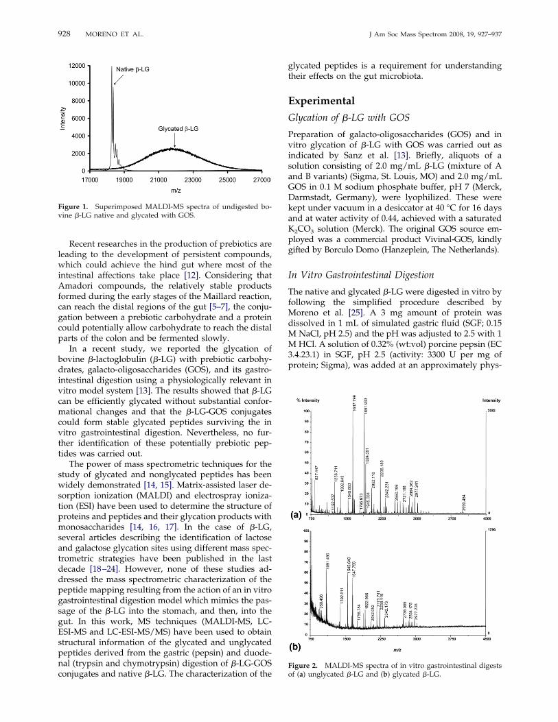
Figure 1. Superimposed MALDI-MS spectra of undigested bo-vine �-LG native and glycated with GOS.
Figure 2. MALDI-MS spectra of in vitro gastrointestinal digestsof (a) unglycated �-LG and (b) glycated �-LG.
928 MORENO ET AL. J Am Soc Mass Spectrom 2008, 19, 927–937
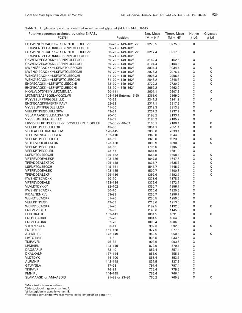
Table 1. Unglycated peptides identified in native and glycated �-LG by MALDI-MS
Putative sequence assigned by using ExPASyP02754 Position
Exp. Mass[M � H]a
Theor. Mass[M � H]a
Native�-LG
Glycated�-LG
LQKWENDbECAQKKLSFNPTQLEEQCHI orQKWENDbECAQKKILSFNPTQLEEQCHI
58–70 149–162d or59–71 149–162d
3275.5 3275.6 X
LQKWENGcECAQKKLSFNPTQLEEQCHI orQKWENGcECAQKKILSFNPTQLEEQCHI
58–70 149–162d or59–71 149–162d
3217.4 3217.6 X
QKWENDbECAQKKLSFNPTQLEEQCHI 59–70 149–162d 3162.4 3162.5 XQKWENGcECAQKKLSFNPTQLEEQCHI 59–70 149–162d 3104.4 3104.5 XKWENDbECAQKKLSFNPTQLEEQCHI 60–70 149–162d 3034.4 3034.4 X XKWENGcECAQKKLSFNPTQLEEQCHI 60–70 149–162d 2976.3 2976.4 X XWENDbECAQKKLSFNPTQLEEQCHI 61–70 149–162d 2906.3 2906.3 X XWENGcECAQKKLSFNPTQLEEQCHI 61–70 149–162d 2848.2 2848.3 X XENDbECAQKKLSFNPTQLEEQCHI 62–70 149–162d 2720.2 2720.2 X XENGcECAQKKLSFNPTQLEEQCHI 62–70 149–162d 2662.2 2662.2 XNKVLVLDTDYKKYLLFCMENSA 90–111 2607.1 2607.3 X XLFCMENSAEPEQSLAcCQCLVR 104–124 (Internal S-S) 2369.0 2369.0 XRVYVEELKPTPEGDLEILLQ 40–59 2341.2 2341.3 X XENGcECAQKKIIAEKTKIPAVF 62–82 2317.1 2317.3 XVYVEELKPTPEGDLEILLQK 41–60 2313.3 2313.3 XVEELKPTPEGDLEILLQKW 43–61 2237.2 2237.2 X XYSLAMAASDISLLDAQSAPLR 20–40 2193.2 2193.1 XVYVEELKPTPEGDLEILLQ 41–59 2185.2 2185.2 XLRVYVEELKPTPEGDLEI or RVYVEELKPTPEGDLEIL 39–56 or 40–57 2100.1 2100.1 X XVEELKPTPEGDLEILLQK 43–60 2051.1 2051.1 X XVDDEALEKFDKALKALPM 128–145 2033.0 2033.1 XYLLFCMENSAEPEQSLAc 102–118 1945.0 1944.9 XVEELKPTPEGDLEILLQ 43–59 1923.0 1923.0 X XVRTPEVDDEALEKFDK 123–138 1890.9 1890.9 X XVEELKPTPEGDLEILL 43–58 1795.0 1795.0 XVEELKPTPEGDLEIL 43–57 1681.9 1681.9 XLSFNPTQLEEQCHI 149–162 1658.8 1658.8 X XVRTPEVDDEALEKF 123–136 1647.8 1647.8 X XTPEVDDEALEKFDK 125–138 1635.7 1635.8 X XLSFNPTQLEEQCH 149–161 1545.7 1545.7 X XVRTPEVDDEALEK 123–135 1500.7 1500.8 X XTPEVDDEALEKF 125–136 1392.6 1392.7 X XKWENDbECAQKK 60–70 1378.6 1378.6 XVRTPEVDDEALE 123–134 1372.6 1372.7 XVLVLDTDYKKY 92–102 1356.7 1356.7 XKWENGcECAQKK 60–70 1320.6 1320.6 XKIDALNENKVL 83–93 1256.7 1256.7 XWENDbECAQKK 61–70 1250.5 1250.5 XVEELKPTPEGD 43–53 1213.6 1213.6 XWENGcECAQKK 61–70 1192.5 1192.5 X XENKVLVLDTD 89–98 1145.6 1145.6 XLEKFDKALK 133–141 1091.5 1091.6 X XENDbECAQKK 62–70 1064.5 1064.5 XENGcECAQKK 62–70 1006.4 1006.5 XVTQTMKGLD 3–11 992.3 992.5 X XFNPTQLEE 151–158 977.5 977.5 XALPMHIRL 142–149 950.5 950.6 X XLIVTQTMK 1–8 933.5 933.5 XTKIPAVFK 76–83 903.5 903.6 XLPMHIRL 143–149 879.5 879.5 XDAQSAPLR 33–40 857.4 857.4 XDKALKALP 137–144 855.0 855.5 XVLDTDYK 94–100 853.4 853.5 XALPMHIR 142–148 837.5 837.5 XGTWYSLA 17–23 797.4 797.4 XTKIPAVF 76–82 775.4 775.5 XPMHIRL 144–149 766.4 766.4 XSLAMAASD or AMAASDIS 21–28 or 23–30 765.2 765.3 X X
aMonoisotopic mass values.b�-lactoglobulin genetic variant A.c�-lactoglobulin genetic variant B.dPeptides containing two fragments linked by disulfide bond ().
929J Am Soc Mass Spectrom 2008, 19, 927–937 MS CHARACTERIZATION OF GLYCATED �-LG PEPTIDES
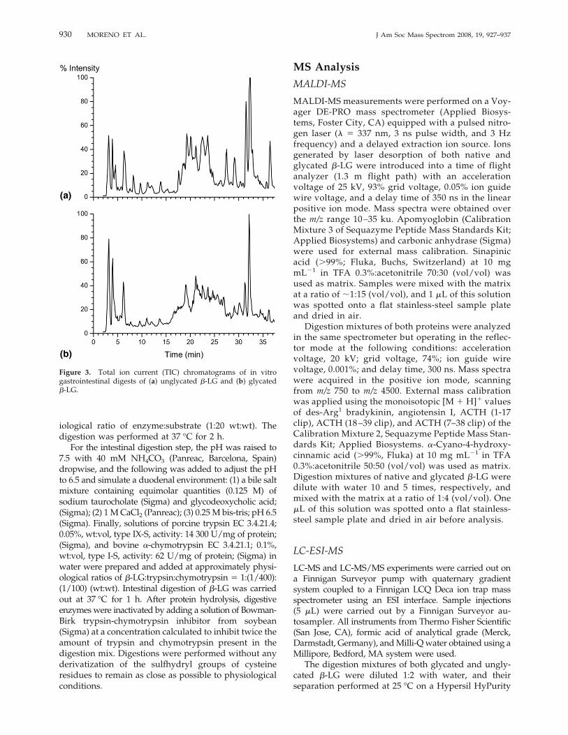
iological ratio of enzyme:substrate (1:20 wt:wt). Thedigestion was performed at 37 °C for 2 h.For the intestinal digestion step, the pH was raised to
7.5 with 40 mM NH4CO3 (Panreac, Barcelona, Spain)dropwise, and the following was added to adjust the pHto 6.5 and simulate a duodenal environment: (1) a bile saltmixture containing equimolar quantities (0.125 M) ofsodium taurocholate (Sigma) and glycodeoxycholic acid;(Sigma); (2) 1MCaCl2 (Panreac); (3) 0.25M bis-tris; pH 6.5(Sigma). Finally, solutions of porcine trypsin EC 3.4.21.4;0.05%, wt:vol, type IX-S, activity: 14 300 U/mg of protein;(Sigma), and bovine �-chymotrypsin EC 3.4.21.1; 0.1%,wt:vol, type I-S, activity: 62 U/mg of protein; (Sigma) inwater were prepared and added at approximately physi-ological ratios of �-LG:trypsin:chymotrypsin 1:(1/400):(1/100) (wt:wt). Intestinal digestion of �-LG was carriedout at 37 °C for 1 h. After protein hydrolysis, digestiveenzymes were inactivated by adding a solution of Bowman-Birk trypsin-chymotrypsin inhibitor from soybean(Sigma) at a concentration calculated to inhibit twice theamount of trypsin and chymotrypsin present in thedigestion mix. Digestions were performed without anyderivatization of the sulfhydryl groups of cysteineresidues to remain as close as possible to physiologicalconditions.
MS Analysis
MALDI-MS
MALDI-MS measurements were performed on a Voy-ager DE-PRO mass spectrometer (Applied Biosys-tems, Foster City, CA) equipped with a pulsed nitro-gen laser (� 337 nm, 3 ns pulse width, and 3 Hzfrequency) and a delayed extraction ion source. Ionsgenerated by laser desorption of both native andglycated �-LG were introduced into a time of flightanalyzer (1.3 m flight path) with an accelerationvoltage of 25 kV, 93% grid voltage, 0.05% ion guidewire voltage, and a delay time of 350 ns in the linearpositive ion mode. Mass spectra were obtained overthe m/z range 10–35 ku. Apomyoglobin (CalibrationMixture 3 of Sequazyme Peptide Mass Standards Kit;Applied Biosystems) and carbonic anhydrase (Sigma)were used for external mass calibration. Sinapinicacid (�99%; Fluka, Buchs, Switzerland) at 10 mgmL�1 in TFA 0.3%:acetonitrile 70:30 (vol/vol) wasused as matrix. Samples were mixed with the matrixat a ratio of �1:15 (vol/vol), and 1 �L of this solutionwas spotted onto a flat stainless-steel sample plateand dried in air.Digestion mixtures of both proteins were analyzed
in the same spectrometer but operating in the reflec-tor mode at the following conditions: accelerationvoltage, 20 kV; grid voltage, 74%; ion guide wirevoltage, 0.001%; and delay time, 300 ns. Mass spectrawere acquired in the positive ion mode, scanningfrom m/z 750 to m/z 4500. External mass calibrationwas applied using the monoisotopic [M � H]� valuesof des-Arg1 bradykinin, angiotensin I, ACTH (1-17clip), ACTH (18–39 clip), and ACTH (7–38 clip) of theCalibration Mixture 2, Sequazyme Peptide Mass Stan-dards Kit; Applied Biosystems. �-Cyano-4-hydroxy-cinnamic acid (�99%, Fluka) at 10 mg mL�1 in TFA0.3%:acetonitrile 50:50 (vol/vol) was used as matrix.Digestion mixtures of native and glycated �-LG weredilute with water 10 and 5 times, respectively, andmixed with the matrix at a ratio of 1:4 (vol/vol). One�L of this solution was spotted onto a flat stainless-steel sample plate and dried in air before analysis.
LC-ESI-MS
LC-MS and LC-MS/MS experiments were carried out ona Finnigan Surveyor pump with quaternary gradientsystem coupled to a Finnigan LCQ Deca ion trap massspectrometer using an ESI interface. Sample injections(5 �L) were carried out by a Finnigan Surveyor au-tosampler. All instruments from Thermo Fisher Scientific(San Jose, CA), formic acid of analytical grade (Merck,Darmstadt, Germany), andMilli-Qwater obtained using aMillipore, Bedford, MA system were used.The digestion mixtures of both glycated and ungly-
cated �-LG were diluted 1:2 with water, and theirseparation performed at 25 °C on a Hypersil HyPurity
Figure 3. Total ion current (TIC) chromatograms of in vitrogastrointestinal digests of (a) unglycated �-LG and (b) glycated�-LG.
930 MORENO ET AL. J Am Soc Mass Spectrom 2008, 19, 927–937
C18 (100 mm � 2.1 mm, 3 �m) column (Thermo FisherScientific) at a flow rate of 100 �L min�1. A gradient oftwo eluents was used: eluent A consisted in 0.1%(vol/vol) of formic acid (analytical grade; Merck, Darm-stadt, Germany) in water, and eluent B in acetonitrile(LC-MS Chromasolv grade; Riedel-de Haën, Seelz, Ger-many) containing 0.1% of formic acid (vol/vol). Theelution program was applied as follows: at the start 5%B; after 5 min the percentage of B was linearly increased
to 50% in 50 min; 50% to 90% B linear from 55 to 57 min;90% B isocratic from 57 to 70 min; ramped to originalcomposition in 1 min, then equilibrated for 15 min.The mass spectrometer spray voltage was set at 4.5
kV, heated capillary temperature at 200 °C, nitrogen(99.5% purity) was used as sheath (0.6 L min�1) andauxiliary (6 L min�1) gas, and helium (99.999% purity)as the collision gas. Mass spectra were recorded in thepositive ion mode. MS/MS data were acquired in the
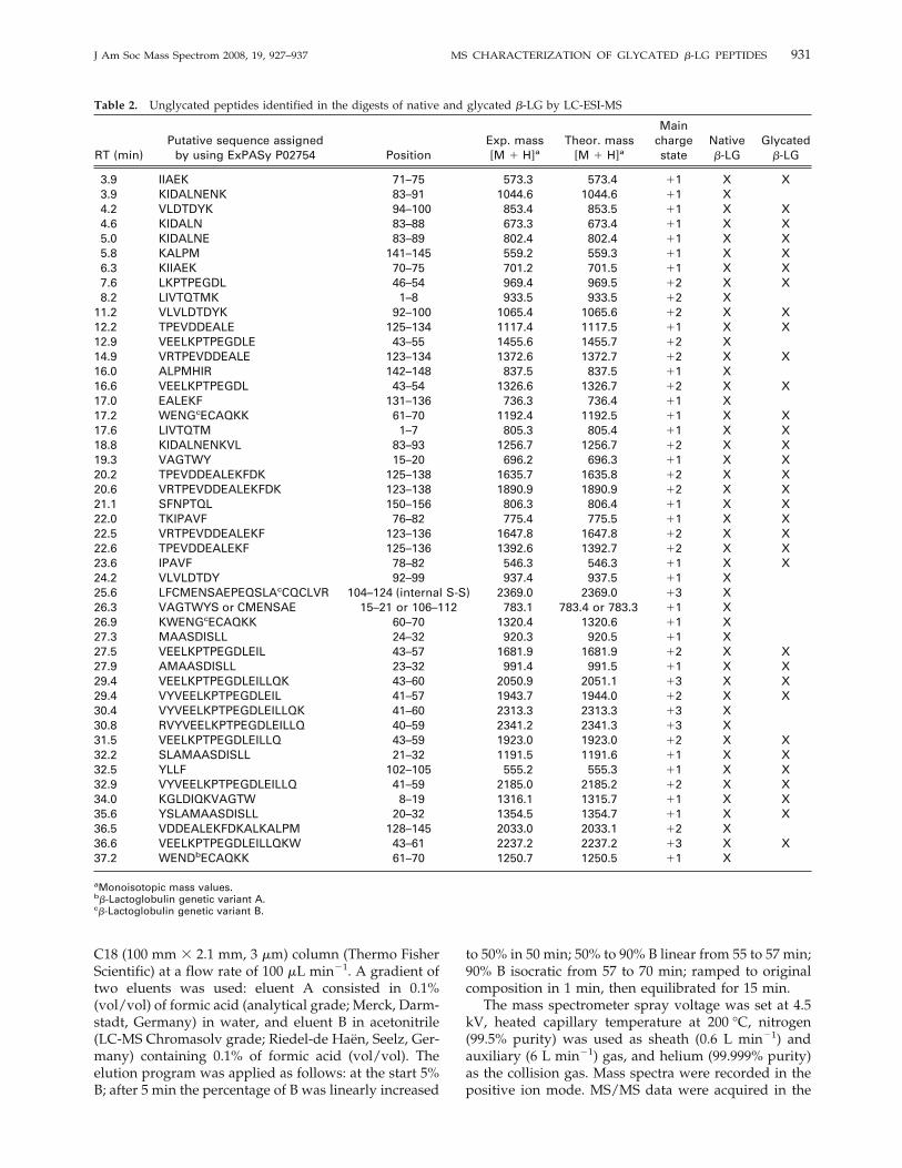
Table 2. Unglycated peptides identified in the digests of native and glycated �-LG by LC-ESI-MS
RT (min)Putative sequence assigned
by using ExPASy P02754 PositionExp. mass[M � H]a
Theor. mass[M � H]a
Mainchargestate
Native�-LG
Glycated�-LG
3.9 IIAEK 71–75 573.3 573.4 �1 X X3.9 KIDALNENK 83–91 1044.6 1044.6 �1 X4.2 VLDTDYK 94–100 853.4 853.5 �1 X X4.6 KIDALN 83–88 673.3 673.4 �1 X X5.0 KIDALNE 83–89 802.4 802.4 �1 X X5.8 KALPM 141–145 559.2 559.3 �1 X X6.3 KIIAEK 70–75 701.2 701.5 �1 X X7.6 LKPTPEGDL 46–54 969.4 969.5 �2 X X8.2 LIVTQTMK 1–8 933.5 933.5 �2 X
11.2 VLVLDTDYK 92–100 1065.4 1065.6 �2 X X12.2 TPEVDDEALE 125–134 1117.4 1117.5 �1 X X12.9 VEELKPTPEGDLE 43–55 1455.6 1455.7 �2 X14.9 VRTPEVDDEALE 123–134 1372.6 1372.7 �2 X X16.0 ALPMHIR 142–148 837.5 837.5 �1 X16.6 VEELKPTPEGDL 43–54 1326.6 1326.7 �2 X X17.0 EALEKF 131–136 736.3 736.4 �1 X17.2 WENGcECAQKK 61–70 1192.4 1192.5 �1 X X17.6 LIVTQTM 1–7 805.3 805.4 �1 X X18.8 KIDALNENKVL 83–93 1256.7 1256.7 �2 X X19.3 VAGTWY 15–20 696.2 696.3 �1 X X20.2 TPEVDDEALEKFDK 125–138 1635.7 1635.8 �2 X X20.6 VRTPEVDDEALEKFDK 123–138 1890.9 1890.9 �2 X X21.1 SFNPTQL 150–156 806.3 806.4 �1 X X22.0 TKIPAVF 76–82 775.4 775.5 �1 X X22.5 VRTPEVDDEALEKF 123–136 1647.8 1647.8 �2 X X22.6 TPEVDDEALEKF 125–136 1392.6 1392.7 �2 X X23.6 IPAVF 78–82 546.3 546.3 �1 X X24.2 VLVLDTDY 92–99 937.4 937.5 �1 X25.6 LFCMENSAEPEQSLAcCQCLVR 104–124 (internal S-S) 2369.0 2369.0 �3 X26.3 VAGTWYS or CMENSAE 15–21 or 106–112 783.1 783.4 or 783.3 �1 X26.9 KWENGcECAQKK 60–70 1320.4 1320.6 �1 X27.3 MAASDISLL 24–32 920.3 920.5 �1 X27.5 VEELKPTPEGDLEIL 43–57 1681.9 1681.9 �2 X X27.9 AMAASDISLL 23–32 991.4 991.5 �1 X X29.4 VEELKPTPEGDLEILLQK 43–60 2050.9 2051.1 �3 X X29.4 VYVEELKPTPEGDLEIL 41–57 1943.7 1944.0 �2 X X30.4 VYVEELKPTPEGDLEILLQK 41–60 2313.3 2313.3 �3 X30.8 RVYVEELKPTPEGDLEILLQ 40–59 2341.2 2341.3 �3 X31.5 VEELKPTPEGDLEILLQ 43–59 1923.0 1923.0 �2 X X32.2 SLAMAASDISLL 21–32 1191.5 1191.6 �1 X X32.5 YLLF 102–105 555.2 555.3 �1 X X32.9 VYVEELKPTPEGDLEILLQ 41–59 2185.0 2185.2 �2 X X34.0 KGLDIQKVAGTW 8–19 1316.1 1315.7 �1 X X35.6 YSLAMAASDISLL 20–32 1354.5 1354.7 �1 X X36.5 VDDEALEKFDKALKALPM 128–145 2033.0 2033.1 �2 X36.6 VEELKPTPEGDLEILLQKW 43–61 2237.2 2237.2 �3 X X37.2 WENDbECAQKK 61–70 1250.7 1250.5 �1 X
aMonoisotopic mass values.b�-Lactoglobulin genetic variant A.c�-Lactoglobulin genetic variant B.
931J Am Soc Mass Spectrom 2008, 19, 927–937 MS CHARACTERIZATION OF GLYCATED �-LG PEPTIDES
automatic data-dependent mode with relative collisionenergy of 35%.
Data Treatment
MALDI-MS data were evaluated by the Data Explorerprogram (Voyager 5.1 Software with Data Explorer;Applied Biosystems). The LC-MS system, data acquisi-tion and processing were managed by Xcalibur soft-ware, (1.2 version; Thermo Fisher Scientific).The assignment of observed ions to the correspond-
ing amino acid sequences was based on the knownsequence of �-LG [26] by using the protein databaseSwiss-Prot and TrEMBL and the tools Peptide Mass andFindPept available at www.expasy.org. Parameters forthe search were the following: (1) Monoisotopic peptidemasses were indicated as [M � H]� with cysteinestreated with nothing; (2) as enzymes, pepsin, andtrypsin/chymotrypsin were chosen; (3) four missedcleavages were allowed; (4) peptides with a mass largerthan 500 u were displayed; (5) the mass tolerance waskept at 0.5 u.All full scan MS/MS spectral data were searched
using the Bioworks version 3.1 suite of programs(Thermo Fisher Scientific). A precursor mass tolerance
of 1.4 u was used to search the resulting DTA filesagainst computer-predicted fragments. All Bioworks3.1 output files were further filtered according to cross-correlation (Xcorr) scores as a function of charge statesto increase confidence limits. Specifically, the Xcorrcutoff values used to determine acceptable peptidematches were 1.5 for digest fragments originating fromprecursor ions with a charge state of one, 2.0 for digestfragments originating from doubly charged precursorions, and 2.5 for triply charged ions.
Results and Discussion
Determination of the Extent of Glycation
The MALDI-MS analysis of undigested �-LG storedwith GOS for 16 days demonstrated that this proteincan be glycated through the Maillard reaction to aconsiderable and variable degree (Figure 1). Either thepresence of two �-LG genetic variants with multipleglycation sites (15 Lys, 3 Arg, and the NH2-terminal Leuresidues) or the use of GOS with different degrees ofpolymerization (from 2 to 7) explain the great variety ofindividual glycated protein species obtained after stor-age. Thus, the mass spectrum of the native �-LG was
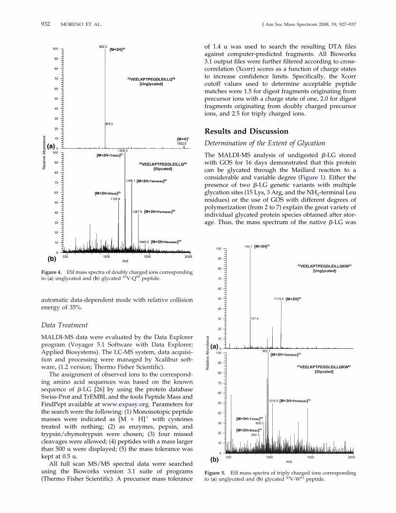
Figure 4. ESI mass spectra of doubly charged ions correspondingto (a) unglycated and (b) glycated 43V-Q59 peptide.
000200510001005
m/z
Rel
ativ
e A
bund
ance
746.7
1119.5
747.4
962.7
1016.9
908.5
854.3
0
10
20
30
40
50
60
70
80
90
1000
10
20
30
40
50
60
70
80
90
100[M+3H]3+
[M+2H]2+
[M+3H+Disac]3+
[M+3H+Trisac]3+
[M+3H+Tetrasac]3+
[M+3H+Pentasac]3+
43VEELKPTPEGDLEILLQKW61
[Unglycated]
43VEELKPTPEGDLEILLQKW61
[Glycated]
(a)
(b)
Figure 5. ESI mass spectra of triply charged ions correspondingto (a) unglycated and (b) glycated 43V-W61 peptide.
932 MORENO ET AL. J Am Soc Mass Spectrom 2008, 19, 927–937

Table 3. Glycated peptides of �-LG identified by LC-ESI-MS and LC-ESI-MS/MS. The glycated amino acid is indicated by anasterisk.
RT (min)Putative sequence assigned
by using ExPASy P02754 PositionExp. mass[M � H]a
Theor. mass[M � H]a
Main chargestate D.P. GOS
4.0–4.1 IIAEK* 71–75 1059.6 1059.4 �1 31221.5 1221.4 4
4.1 ENK*VLVLDTD 89–98 1955.5 1955.6 �2 54.2 K*IDALNE 83–89 1450.4 1450.4 �1 44.5–4.6 FDK*ALK 136–141 1207.5 1207.4 �2 3
1369.8 1369.4 48.2–9.2 LK*PTPEGDL 46–54 1293.9 1293.5 �2 2
1455.9 1455.5 31617.9 1617.5 41779.7 1779.5 5
12.7–13.3 TK*IPAVFK 76–83 1389.4 1389.6 �1 31551.5 1551.6 41713.9 1713.6 51875.5 1875.6 62037.9 2037.6 7
14.4–16.2 VEELK*PTPEGDL 43–54 1650.5 1650.7 �2 21813.1 1812.7 31974.7 1974.7 42136.9 2136.7 5
15.4 L*IVTQTM 1–7 1129.3 1129.4 �1 217.0–18.0 K*IDALNENK*VL 83–93 1581.4 1580.7 �2 2
1743.4 1742.7 31905.2 1904.7 42067.4 2066.7 52229.4 2228.7 6
19.0–20.0 VLVLDTDYK*K 92–101 1841.5 1841.7 �2 42003.5 2003.7 52165.5 2165.7 6
20.7–21.5 TK*IPAVF 76–82 1261.5 1261.5 �2 31423.8 1423.5 41585.9 1585.5 5
20.6–21.4 TPEVDDEALEK*F 125–136 1879.0 1878.7 �2 32041.4 2040.7 42203.0 2202.7 5
25.1–25.9 VEELK*PTPEGDLEIL 43–57 2168.0 2167.9 �2 32330.0 2329.9 42492.0 2491.9 52654.0 2653.9 6
25.3 VRTPEVDDEALEK*FDK*AL 123–140 2723.4 2723.1 �3 425.1–26.0 TPEVDDEALEK*FDK*AL 125–140 2306.9 2305.9 �3 3
2468.4 2467.9 42630.4 2629.9 5
25.0–26.6 GLDIQK*VAGTWY 9–20 1836.8 1836.7 �2 31998.8 1998.7 42160.5 2160.7 52323.0 2322.7 6
28.0–28.9 VEELK*PTPEGDLEILLQK 43–60 2375.6 2375.1 �3 22537.5 2537.1 32699.3 2699.1 42861.4 2861.1 5
30.0–30.9 VEELK*PTPEGDLEILLQ 43–59 2247.0 2247.0 �2 22410.0 2409.0 32571.2 2571.0 42732.8 2733.0 52895.6 2895.0 6
34.9–35.9 VEELK*PTPEGDLEILLQK*W 43–61 2561.5 2561.2 �3 22723.5 2723.2 32885.6 2885.2 43047.2 3047.2 5
aMonoisotopic mass values.
933J Am Soc Mass Spectrom 2008, 19, 927–937 MS CHARACTERIZATION OF GLYCATED �-LG PEPTIDES
characterized by the molecular mass values of 18,278and 18,363 u corresponding to the theoretical masses ofthe variants B and A, respectively. Other minor massesattributed mainly to some matrix-adducts were alsodetected in the native �-LG mass spectrum (Figure 1).However, a Gaussian distribution of masses over therange of approximately m/z 19,000 to 25,000 u wasdetected for the glycated �-LG. The Gaussian peakshowed a maximum of intensity at �21,700 u, indicat-ing an increase of �19% in the molecular mass of the�-LG sequence (Figure 1).
Identification of �-LG Peptides Resulting from theIn Vitro Gastrointestinal Digestion
Both native and glycated �-LG were subjected to apepsin digestion; then they were hydrolyzed by trypsin
and chymotrypsin simulating a duodenal environment.As expected, both proteins were very resistant to pep-sinolysis (data not shown) because �-LG is a poorsubstrate for pepsin [27–30]. Nevertheless, the duodenaldigestion (trypsin and chymotrypsin) of the native andglycated �-LG gave rise to a very complex mixture ofpeptides after 1 h of incubation as was previouslyobserved by reversed-phase LC-UV [13]. The differencein amino acid specificity of these two enzymes maycontribute to the release of a great number of peptides.Trypsin cleaves on the C-side of basic residues (Arg andLys) whilst chymotrypsin requires an aromatic or bulkynonpolar side-chain (Phe, Tyr, Trp, Leu, Met) on the
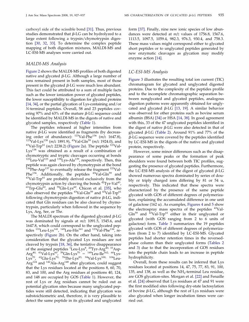
Figure 6. MS/MS spectra of single charged ion corresponding to(a) glycated 83K-E89 peptide, and doubly charged ions correspond-ing to (b) glycated 89E-D98 peptide, and (c) glycated 136F-K141
peptide.
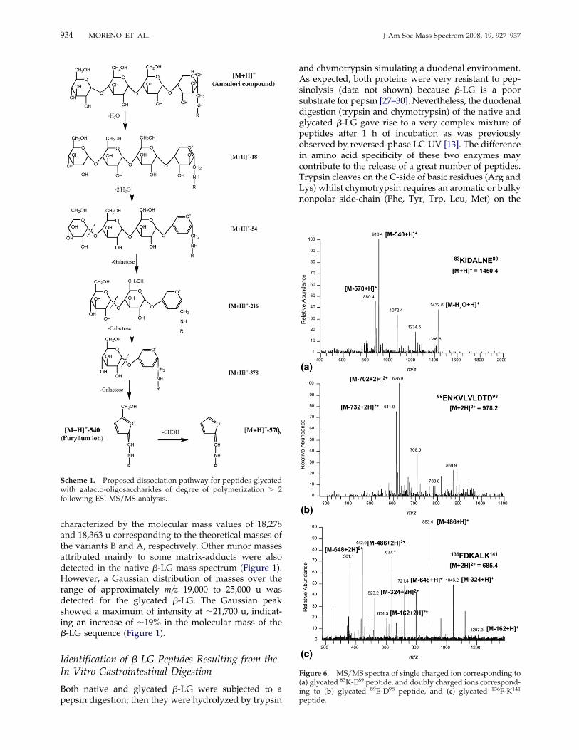
Scheme 1. Proposed dissociation pathway for peptides glycatedwith galacto-oligosaccharides of degree of polymerization � 2following ESI-MS/MS analysis.
934 MORENO ET AL. J Am Soc Mass Spectrom 2008, 19, 927–937
carboxyl side of the scissible bond [31]. Thus, previousstudies demonstrated that �-LG can be hydrolyzed to alarge extent following a trypsin/chymotrypsin diges-tion [30, 32, 33]. To determine the complex peptidemapping of both digestion mixtures, MALDI-MS andLC-ESI-MS analyses were carried out.
MALDI-MS Analysis
Figure 2 shows the MALDI-MS profiles of both digestednative and glycated �-LG. Although a large number ofions remained present in both samples, most of thosepresent in the glycated �-LG were much less abundant.This fact could be attributed to a sum of multiple factssuch as the lower ionization power of glycated peptides,the lower susceptibility to digestion for glycated proteins[14, 34], or the partial glycation of Lys-containing and/orN-terminal peptides. Overall, 58 and 23 peptides cov-ering 97% and 63% of the mature �-LG sequence couldbe identified by MALDI-MS in the digests of native andglycated samples, respectively (Table 1).The peptides released at higher intensities from
native �-LG were identified as fragments (by decreas-ing order of abundance): 123Val-Phe136 (m/z 1647.8),123Val-Lys138 (m/z 1891.9), 43Val-Gln59 (m/z 1924.0), and43Val-Trp61 (m/z 2238.2) (Figure 2a). The peptide 123Val-Lys138 was obtained as a result of a combination ofchymotryptic and tryptic cleavages occurring at bonds122Leu-Val123 and 138Lys-Ala139, respectively. Then, thispeptide was again cleaved by chymotrypsin at the bond136Phe-Asp137 to eventually release the fragment 123Val-Phe136. Additionally, the peptides 43Val-Gln59 and43Val-Trp61 are probably derived exclusively from thechymotrypsin action by cleaving the bonds 42Tyr-Val43,61Trp-Glu62, and 59Gln-Lys60. Chicon et al. [35], whoalso observed the peptides 43Val-Gln59 and 43Val-Trp61
following chymotrypsin digestion of native �-LG, indi-cated that Gln residues can be also cleaved by chymo-trypsin, particularly when followed in the sequence byLys, Arg, Ser, or Thr.The MALDI spectrum of the digested glycated �-LG
was dominated by signals at m/z 1091.5, 1545.6, and1647.8, which could correspond to the unglycated pep-tides 133Leu-Lys141, 149Leu-His161 and 123Val-Phe136, re-spectively (Figure 2b). On the other hand, taking intoconsideration that the glycated Lys residues are notcleaved by trypsin [18, 36], the tentative disappearanceof the assigned peptides 1Leu-Lys8, 20Tyr-Arg40, 33Asp-Arg40, 41Val-Lys60, 59Gln-Lys70 149Leu-Ile162, 60Lys-Lys70, 62Glu-Lys70, 76Thr-Lys83, 94Val-Lys100, 104Leu-Arg124 and 142Ala-Arg148 after glycation, could suggestthat the Lys residues located at the positions 8, 60, 70,83, and 100, and the Arg residues at positions 40, 124,and 148 are occupied by GOS (Table 1). However, therest of Lys or Arg residues cannot be ruled out aspotential glycation sites because many unglycated pep-tides were still detected, indicating that glycation wassubstoichiometric and, therefore, it is very plausible todetect the same peptide in its glycated and unglycated
form [37]. Finally, nine new ionic species of low abun-dances were detected at m/z values of 1756.8, 1567.6,1113.5, 1097.5, 1059.4, 982.3, 976.3, 930.4, and 758.3.These mass values might correspond either to glycatedshort peptides or to unglycated peptides generated bynew enzymatic cleavages as glycation may modifyenzyme action [14].
LC-ESI-MS Analysis
Figure 3 illustrates the resulting total ion current (TIC)chromatogram for glycated and unglycated digestedproteins. Due to the complexity of the peptides profileand to the incomplete chromatographic separation be-tween nonglycated and glycated peptides, analogousdigestion patterns were apparently obtained for ungly-cated and glycated �-LG [13, 19]. A similar behaviorwas observed for other proteins such as bovine serumalbumin (BSA) [34] or HSA [14, 38]. In good agreementwith this, 33 of the 47 unglycated peptides identified inthe digest of native �-LG were also detected in that ofglycated �-LG (Table 2). Around 91% and 77% of the�-LG sequence were covered by the peptides identifiedby LC-ESI-MS in the digests of the native and glycatedprotein, respectively.However, some minor differences such as the disap-
pearance of some peaks or the formation of peakshoulders were found between both TIC profiles, sug-gesting the presence of glycated peptides. Furthermore,the LC-ESI-MS analysis of the digest of glycated �-LGshowed numerous spectra dominated by series of dou-bly or triply charged ions differing by 81 or 54 u,respectively. This indicated that these spectra werecharacterized by the presence of the same peptideglycated with GOS of different degrees of polymeriza-tion, explaining the accumulated difference in one unitof galactose (162 u). As examples, Figures 4 and 5 showthe electrospray mass spectra of the peptides 43Val-Gln59 and 43Val-Trp61 either in their unglycated orglycated (with GOS ranging from 2 to 6 units ofgalactose) form. Table 3 summarizes the 19 peptidesglycated with GOS of different degrees of polymeriza-tion (from 2 to 7) identified by LC-ESI-MS. Glycatedpeptides had shorter retention times in the reversed-phase column than their unglycated forms (Tables 2and 3) due to that the incorporation of GOS residuesinto the peptide chain leads to an increase in peptidehydrophilicity.Overall, from these results can be inferred that Lys
residues located at positions 14, 47, 75, 77, 83, 91, 100,135, and 138, as well as the NH2-terminal Leu residue,are GOS glycation-sites. Morgan et al. [22] and Fenailleet al. [24] observed that Lys residues at 47 and 91 werethe first modified sites following dry-state lactosylationof bovine �-LG, although the rest of Lys residues werealso glycated when longer incubation times were car-ried out.
935J Am Soc Mass Spectrom 2008, 19, 927–937 MS CHARACTERIZATION OF GLYCATED �-LG PEPTIDES
LC-ESI-MS/MS Analysis
LC-ESI-MS/MS analyses were performed to confirmthe structure of those peptides which showed the high-est relative abundances by MALDI-MS and LC-ESI-MS.Marked differences in the fragmentation behavior be-tween glycated and unglycated peptides were found.Thus, the unglycated peptides produced a MS/MSspectrum in which the typical y- and b-series predom-inate (data not shown). The 22 unglycated peptidesidentified by tandem MS were located at the NH2terminus and at the regions spanning the residues20–32, 40–61, 71–100, and 123–138. Without reduction,no peptides containing cysteine residues were assigned,suggesting that all cysteines in �-LG could be partici-pating in disulfide bridges.The tandem mass spectra of the glycated peptides
identified by LC-ESI-MS (Table 3) were mainly charac-terized by the formation of the furylium ion and itssubsequent CHOH loss. Other minor ionic speciescontaining the furylium ring linked to galactose unitscould be also detected, pointing out the diversity of thefragmentation pattern of peptides glycated with largersize carbohydrates. As an example, Scheme 1 illustratesa possible fragmentation pathway of a peptide glycatedwith a tetrasaccharide compatible with the main ionsdetected by MS/MS. Mollé et al. [21] and Jeric et al. [39]described the pathways for the formation of the fury-lium ion and its CHOH loss from Amadori rearrange-ment products involving di- and monosaccharideslinked to a peptidic chain, respectively. The data re-ported in our study demonstrate that these ionic speciesare also formed following ESI-MS/MS analysis of pep-tides glycated with carbohydrates of higher molecularweight. Thus, the losses of 1026/1056, 864/894, 702/732, 540/570, 378/408, and 216/246 u were indicative ofthe peptide glycation with hepta-, hexa-, penta-, tetra-,tri-, and disaccharides, respectively. Considering thepresence of doubly and triply charged glycated pep-tides, these mass values were also divided by 2 and 3 toscan for constant formal neutral losses. Figure 6a and bshow the MS/MS spectra of the peptides 83Lys-Glu89
and 89Glu-Asp98 glycated with GOS of 4 and 5 hexoseunits, respectively. Strikingly, MS/MS spectra of twoglycated peptides, 136Phe-Lys141 (Figure 6c) and 43Val-Glyn59 (spectrum not shown), were dominated by sin-gly and doubly charged ions derived exclusively fromlosses of galactose units (Figure 6c).
Conclusions
In general terms, a good correlation between the mo-lecular masses obtained for the unglycated peptides byESI-MS and MALDI-MS was obtained, although pep-tides with low masses (�800 u) were mainly identifiedby LC-ESI-MS, whereas those with mass values be-tween 2300 and 3000 u were detected by MALDI-MS.On the contrary, substantial differences between bothMS techniques were observed for the detection of the
glycated peptides, where LC-ESI-MS showed to be amore effective technique than MALDI-MS.Further ESI-MS/MS analyses corroborated the LC-
ESI-MS results and also enabled an extensive investiga-tion into the glycated peptides produced by the in vitrogastrointestinal digestion. Thus, the formation of thecharacteristic furylium ion and its CHOH loss allowedthe detection of a number of peptides glycated withGOS of different degrees of polymerization (from 2 to7). Consequently, the putative GOS glycation sites werelocated at the NH2-terminal Leu residue and at the Lysresidues 14, 47, 75, 77, 83, 91, 100, 135, and 138. Thestructural characterization of the glycated peptides sur-viving the in vitro gastrointestinal digestion of �-LGdescribed in this work is important for future studiesinvestigating the effect of these peptides on the gutmicrobiota.
AcknowledgmentsThe authors gratefully acknowledge supported of this work byprojects 200670M027, ALIBIRD S-0505/AGR/000153, andANALYSIC S-505/AGR-0312 financed by the Comunidad deMadrid. The authors thank to Dr. G. Klarenbeek and Dr. J. deSlegte from Borculo Domo for providing them with Vivinal-GOS.
References1. Borrelli, R. C.; Visconti, A.; Mennella, C.; Anese, M.; Fogliano, V.Chemical Characterization and Antioxidant Properties of Coffee Mela-noidins. J. Agric. Food Chem. 2002, 50, 6527–6533.
2. Xu, Q. P.; Tao, W. Y.; Ao, Z. H. Antioxidant Activity of VinegarMelanoidins. Food Chem. 2007, 102, 841–849.
3. Brownlee, M.; Vlassara, H.; Cerami, A. Nonenzymatic Glycosylationand the Pathogenesis of Diabetic Complications. Ann. Intern. Med. 1984,101, 527–537.
4. Horie, K.; Miyata, T.; Maeda, K.; Miyata, S.; Sugiyama, S.; Sakai, H.; deStrihou, C. V.; Monnier, V. M.; Witztum, J. L.; Kurokawa, K. Immuno-histochemical Colocalization of Glycoxidation Products and Lipid Per-oxidation Products in Diabetic Renal Glomerular Lesions—Implicationfor Glyco-oxidative Stress in the Pathogenesis of Diabetic Nephropathy.J. Clin. Invest. 1997, 100, 2995–3004.
5. Erbersdobler, H. F.; Faist, V. Metabolic Transit of Amadori Products.Nahrung 2001, 45, 177–181.
6. Faist, V.; Erbersdobler, H. F. Metabolic Transit and in Vivo Effects ofMelanoidins and Precursor Compounds Deriving from the MaillardReaction. Ann. Nutr. Metab. 2001, 45, 1–12.
7. Finot, P. A. The Absorption and Metabolism of Modified Amino Acidsin Processed Foods. J. AOAC Int. 2005, 88, 894–903.
8. Ames, J. M.; Wynne, A.; Hofmann, A.; Plos, S.; Gibson, G. R. The Effectof a Model Melanoidin Mixture on Fecal Bacterial Populations in Vitro.Br. J. Nutr. 1999, 82, 489–495.
9. Dell’Aquila, C.; Ames, J. M.; Gibson, G. R.; Wynne, A. G. Fermentationof Heated Gluten Systems by Gut Microflora. Eur. Food Res. Technol.2003, 217, 382–386.
10. Tuohy, K. M.; Hinton, D. J. S.; Davies, S. J.; Crabbe, M. J. C.; Gibson,G. R.; Ames, J. M. Metabolism of Maillard Reaction Products by theHuman Gut Microbiota—Implications for Health. Mol. Nutr. Food Res.2006, 50, 847–857.
11. Gibson, G. R.; Roberfroid, M. B. Dietary Modulation of the HumanColonic Microbiota: Introducing the Concept of Prebiotics. J. Nutr. 1995,125, 1401–1412.
12. Rastall, R. A.; Maitin, V. Prebiotics and Synbiotics: Towards the NextGeneration. Curr. Opin. Biotechnol. 2002, 13, 490–496.
13. Sanz, M. L.; Corzo-Martínez, M.; Rastall, R. A.; Olano, A.; Moreno, F. J.Characterization and in Vitro Digestibility of Bovine �-LactoglobulinGlycated with Galacto-oligosaccharides. J. Agric. Food Chem. 2007, 55,7916–7925.
14. Lapolla, A.; Fedele, D.; Reitano, R.; Arico, N. C.; Seraglia, R.; Traldi, P.;Marotta, E.; Tonani, R. Enzymatic Digestion and Mass Spectrometry inthe Study of Advanced Glycation End Products/Peptides. J. Am. Soc.Mass Spectrom. 2004, 15, 496–509.
15. Meltretter, J.; Seeber, S.; Humeny, A.; Becker, C. M.; Pischetsrieder, M.Site-Specific Formation of Maillard, Oxidation, and Condensation Prod-ucts from Whey Proteins During Reaction with Lactose. J. Agric. FoodChem. 2007, 55, 6096–6103.
936 MORENO ET AL. J Am Soc Mass Spectrom 2008, 19, 927–937
16. Brancia, F. L.; Bereszczak, J. Z.; Lapolla, A.; Fedele, D.; Baccarin, L.;Seraglia, R.; Traldi, P. Comprehensive Analysis of Glycated HumanSerum Albumin Tryptic Peptides by Off-Line Liquid ChromatographyFollowed by MALDI Analysis on a Time-of-Flight/Curved Field Re-flectron Tandem Mass Spectrometer. J. Mass Spectrom. 2006, 41, 1179–1185.
17. Frolov, A.; Hoffmann, P.; Hoffmann, R. Fragmentation Behavior ofGlycated Peptides Derived from D-Glucose, D-Fructose, and D-Ribosein Tandem Mass Spectrometry. J. Mass Spectrom. 2006, 41, 1459–1469.
18. Morgan, F.; Leonil, J.; Molle, D.; Bouhallab, S. Nonenzymatic Lactosy-lation of Bovine �-Lactoglobulin Under Mild Heat Treatment Leads toStructural Heterogeneity of the Glycoforms. Biochem. Biophys. Res.Commun. 1997, 236, 413–417.
19. Leonil, J.; Molle, D.; Fauquant, J.; Maubois, J. L.; Pearce, R. J.; Bouhallab,S. Characterization by Ionization Mass Spectrometry of Lactosyl �-Lactoglobulin Conjugates Formed During Heat Treatment of Milk andWhey, and Identification of One Lactose-Binding Site. J. Dairy Sci. 1997,80, 2270–2281.
20. Fogliano, V.; Monti, S. M.; Visconti, A.; Randazzo, G.; Facchiano, A. M.;Colonna, G.; Ritieni, A. Identification of a �-Lactoglobulin Lactosylationsite. Biochim. Biophys. Acta 1998, 1388, 295–304.
21. Mollé, D.; Morgan, F.; Bouhallab, S.; Leonil, J. Selective Detection ofLactolated Peptides in Hydrolysates by Liquid Chromatography Elec-trospray Tandem Mass Spectrometry. Anal. Biochem. 1998, 259, 152–161.
22. Morgan, F.; Bouhallab, S.; Molle, D.; Henry, G.; Maubois, J. L.; Leonil, J.Lactolation of �-Lactoglobulin Monitored by electrospray IonizationMass Spectrometry. Int. Dairy J. 1998, 8, 95–98.
23. Fenaille, F.; Morgan, F.; Parisod, V.; Tabet, J. C.; Guy, P. A. Solid-StateGlycation of �-Lactoglobulin Monitored by Electrospray IonizationMass Spectrometry and Gel Electrophoresis Techniques. Rapid Commun.Mass Spectrom. 2003, 17, 1483–1492.
24. Fenaille, F.; Morgan, F.; Parisod, V.; Tabet, J. C.; Guy, P. A. Solid-StateGlycation of �-Lactoglobulin by Lactose and Galactose: Localization ofthe Modified Amino Acids Using Mass Spectrometric Techniques. J.Mass Spectrom. 2004, 39, 16–28.
25. Moreno, F. J.; Mellon, F. A.; Wickham, M. S. J.; Bottrill, A. R.; Mills,E. N. C. Stability of the Major Allergen Brazil Nut 2S Albumin (Ber e 1)to Physiologically-Relevant in Vitro Gastrointestinal Digestion. FEBS J.2005, 272, 341–352.
26. Eigel, W. N.; Butler, J. E.; Ernstrom, C. A.; Farrell, H. M.; Harwalkar,V. R.; Jenness, R.; Whitney, R. M. Nomenclature of Proteins of Cow’sMilk, Fifth Revision. J. Dairy Sci. 1984, 67, 1599–1631.
27. Reddy, I. M.; Kella, N. K. D.; Kinsella, J. E. Structural and Conforma-tional Basis of the Resistance of �-Lactoglobulin to Peptic and Chymot-ryptic Digestion. J. Agric. Food Chem. 1988, 36, 737–741.
28. Dalgalarrondo, M.; Dufour, E.; Chobert, J.-M.; Bertrand-Harb, C.;Haertle, T. Proteolysis of �-Lactoglobulin and �-Casein by Pepsin inEthanolic Media. Int. Dairy J. 1995, 5, 1–14.
29. Astwood, J. D.; Leach, J. N.; Fuchs, R. L. Stability of Food Allergens toDigestion in Vitro. Nat. Biotechnol. 1996, 14, 1269–1273.
30. Peyron, S.; Mouecoucou, J.; Fremont, S.; Sanchez, C.; Gontard, N. Effectsof Heat Treatment and Pectin Addition on �-Lactoglobulin Allergenic-ity. J. Agric. Food Chem. 2006, 54, 5643–5650.
31. Gauthier, S. F.; Pouliot, Y. Functional and Biological Properties ofPeptides Obtained by Enzymatic Hydrolysis of Whey Proteins. J. DairySci. 2003, 86, E78–87.
32. Van Willige, R. W. G.; Fitzgerald, R. J.Tryptic and Chymotryptic Hydro-lysis of �-Lactoglobulin-A, �-Lactoglobulin-B, and �-Lactoglobulin-AB atAmbient and High-Pressure. Milchwissenschaft 1995, 50, 183–186.
33. Galvao, C. M. A.; Silva, A. F. S.; Custodio, M. F.; Monti, R.; Giordano,R. D. C. Controlled Hydrolysis of Cheese Whey Proteins UsingTrypsin and �-Chymotrypsin. Appl. Biochem. Biotechnol. 2001, 91–93,761–776.
34. Lapolla, A.; Fedele, D.; Martano, L.; Arico, N. C.; Garbeglio, M.; Traldi,P.; Seraglia, R.; Favretto, D. Advanced Glycation End Products: AHighly Complex Set of Biologically Relevant Compounds Detected byMass Spectrometry. J. Mass Spectrom. 2001, 36, 370–378.
35. Chicon, R.; Lopez-Fandino, R.; Quiros, A.; Belloque, J. Changes inChymotrypsin Hydrolysis of �-Lactoglobulin A Induced by HighHydrostatic Pressure. J. Agric. Food Chem. 2006, 54, 2333–2341.
36. Henle, T.; Klostermeyer, H. The Reactivity of the Individual Protein-Bound Lysine Residues of �-Casein a1 During the Initial Stages of theMaillard Reaction. In International Dairy Federation Special Issue 9303.Protein and Fat Globule Modifications by Heat Treatment, Homogenization,and Other Technological Means for High Quality Dairy Products; Interna-tional Dairy Federation: Brussels, Belgium, 1993; p. 183–189.
37. Lapolla, A.; Brancia, F. L.; Bereszczak, J.; Fedele, D.; Baccarin, L.;Seraglia, R.; Traldi, P. Off-Line Liquid Chromatography-MALDI bywith Various Matrices and Tandem Mass Spectrometry for Analysis ofGlycated Human Serum Albumin Tryptic Peptides. Mol. Nutr. Food Res.2007, 51, 456–461.
38. Lapolla, A.; Fedele, D.; Senesi, A.; Arico’, N. C.; Reitano, R.; Favretto, D.;Seraglia, R.; Astner, H.; Traldi, P. Advanced Glycation Endproducts/Peptides: A Preliminary Investigation by LC and LC/MS. Farmaco 2002,57, 845–852.
39. Jeric, I.; Versluis, C.; Horvat, S.; Heck, A. J. R. Tracing GlycoproteinStructures: Electron Ionization Tandem Mass Spectrometric Analysis ofSugar-Peptide Adducts. J. Mass Spectrom. 2002, 37, 803–811.
937J Am Soc Mass Spectrom 2008, 19, 927–937 MS CHARACTERIZATION OF GLYCATED �-LG PEPTIDES
Related Documents