Mapping the functional connectome traits of levels of consciousness. Enrico Amico a,b , Daniele Marinazzo b , Carol Di Perri a,c , Lizette Heine a,c , Jitka Annen a,c , Charlotte Martial a,c , Mario Dzemidzic d , Murielle Kirsch c , Vincent Bonhomme c , Steven Laureys a,c, * and Joaquín Goñi e,f,g, * a Coma Science Group, GIGA Research Center, University of Liège, Liège, Belgium b Department of Data-analysis, University of Ghent, B9000 Ghent, Belgium c University Hospital of Liège, Liège, Belgium d Department of Neurology and Radiology and Imaging Sciences, Indiana University School of Medicine, Indianapolis, IN, USA e School of Industrial Engineering, Purdue University, West-Lafayette, IN, USA f Weldon School of Biomedical Engineering, Purdue University, West-Lafayette, IN, USA g Purdue Institute for Integrative Neuroscience, Purdue University, West-Lafayette, IN, USA *Authors contributed equally. [email protected] [email protected] Classification: Computational Modeling and Analysis Keywords: fMRI, Brain Connectivity, Network Science, Consciousness Short title: Mapping the functional traits of consciousness.

Welcome message from author
This document is posted to help you gain knowledge. Please leave a comment to let me know what you think about it! Share it to your friends and learn new things together.
Transcript

Mapping the functional connectome traits of levels of consciousness.
Enrico Amicoa,b
, Daniele Marinazzob, Carol Di Perri
a,c, Lizette Heine
a,c, Jitka Annen
a,c, Charlotte Martial
a,c, Mario
Dzemidzicd, Murielle Kirsch
c, Vincent Bonhomme
c, Steven Laureys
a,c,* and Joaquín Goñi
e,f,g,*
aComa Science Group, GIGA Research Center, University of Liège, Liège, Belgium
bDepartment of Data-analysis, University of Ghent, B9000 Ghent, Belgium
cUniversity Hospital of Liège, Liège, Belgium
dDepartment of Neurology and Radiology and Imaging Sciences, Indiana University School of Medicine,
Indianapolis, IN, USA
eSchool of Industrial Engineering, Purdue University, West-Lafayette, IN, USA
fWeldon School of Biomedical Engineering, Purdue University, West-Lafayette, IN, USA
gPurdue Institute for Integrative Neuroscience, Purdue University, West-Lafayette, IN, USA
*Authors contributed equally.
Classification: Computational Modeling and Analysis
Keywords: fMRI, Brain Connectivity, Network Science, Consciousness
Short title: Mapping the functional traits of consciousness.

Abstract
Examining task-free functional connectivity (FC) in the human brain offers insights on how
spontaneous integration and segregation of information relate to human cognition, and how
this organization may be altered in different conditions, and neurological disorders. This is
particularly relevant for patients in disorders of consciousness (DOC) following severe
acquired brain damage and coma, one of the most devastating conditions in modern medical
care.
We present a novel data-driven methodology, connICA, which implements Independent
Component Analysis (ICA) for the extraction of robust independent FC patterns (FC-traits)
from a set of individual functional connectomes, without imposing any a priori data
stratification into groups.
We here apply connICA to investigate associations between network traits derived from task-
free FC and cognitive/clinical features that define levels of consciousness. Three main
independent FC-traits were identified and linked to consciousness-related clinical features.
The first one represents the functional configuration it is
associated to a sedative (sevoflurane), the overall effect of the pathology and the level of
arousal. The second FC-trait reflects the disconnection of the visual and sensory-motor
connectivity patterns. It also relates to the time since the insult and to the ability of
communicating with the external environment. The third FC-trait isolates the connectivity
pattern encompassing the fronto-parietal and the default-mode network areas as well as the
interaction between left and right hemispheres, which are also associated to the awareness of
the self and its surroundings.
Each FC-trait represents a distinct functional process with a role in the degradation of
conscious states of functional brain networks, shedding further light on the functional sub-
circuits that get disrupted in severe brain-damage.

Introduction
Disorders of consciousness (DOC) remain among the most challenging and poorly
understood conditions in modern medical care. The term spreads over several pathological
states qualified by dissociation between awareness and arousal (Bernat, 2009; Laureys,
2005). Among these, patients in coma show no signs of awareness nor arousal; patients with
unresponsive wakefulness syndrome/vegetative state (UWS) show no signs of awareness but
do have an altered sleep and wake cycle; patients in a minimally conscious state (MCS)
retain minimal non-reflexive and highly fluctuating signs of awareness. When patients regain
functional object use and/or reliable communication they are referred to as emerging from
MCS (EMCS) (Giacino et al., 2014; Laureys et al., 2004). A particular outcome is represented
by patients with a locked-in syndrome (LIS), who have no means of producing speech, limb or
facial movements (except mostly for eye movement and/or blinking) but are still awake and
fully conscious (Giacino et al., 1995; Laureys et al., 2005). To date, the most validated
diagnosis of these patients is based on the behavioral presentation of the patient. The
distinction between these pathological levels of consciousness can be very challenging, as
the boundaries between these states are often uncertain and ambiguous (Giacino et al.,
2014).
In the last decade, advances in neuroimaging techniques have allowed the medical
community to gain important insights into the pathophysiology of DOC and to observe that
altered states of consciousness are related to complex disruptions in the functional and
structural organization of the brain (Boly et al., 2012; Di Perri et al., 2014; Fernández Espejo
et al., 2012; Koch et al., 2016; Owen et al., 2009).
At the same time, quantitative analysis based on complex networks have become more
commonly used to study the brain as a network (Bullmore and Sporns, 2009), giving rise to
the area of research so called Brain Connectomics (Fornito et al., 2016; Sporns, 2011). In
brain network models, nodes correspond to grey-matter regions (based on brain atlases or
parcellations) while links or edges correspond to connections. Structural connections are
modeled using white matter fiber-tracts and functional connections represent coupling
between brain regions while subjects are either at rest or performing a task (van den Heuvel
and Hulshoff Pol, 2010). Recent advances in functional neuroimaging have provided new
tools to measure and examine in vivo whole-brain temporal dependence of the dynamics of
anatomically separated brain regions, defined as functional connectivity (FC) (Fox and
Raichle, 2007; Fox et al., 2005; Friston et al., 1993).
In parallel to the development of methods and network features in Brain Connectomics,
analyses of functional magnetic resonance imaging (fMRI) data based on independent
component analysis (ICA) have become an increasingly popular voxel-level approach
(Calhoun et al., 2009). ICA, by relying upon a general assumption of the independence of the
mixed signals, is a powerful and versatile data-driven approach for studying the brain, at both
temporal and spatial scales (Erhardt et al., 2011).
Examining functional connectivity in the human brain offers unique insights on how integration
and segregation of information relates to human behavior and how this organization may be
altered in diseases (Boly et al., 2012; Greicius, 2008). In the case of disorders of
consciousness, voxel-level ICA-based fMRI studies of levels of consciousness in DOC
patients have mainly shown alterations in the functional connectivity of the default mode
network (DMN) (Heine et al., 2012; Soddu et al., 2012; Vanhaudenhuyse et al., 2010). Recent
studies have also shown disrupted functional connectivity in resting state networks other than
DMN (Demertzi et al., 2014) and possibility to correctly classify patients based on the level of
connectivity of the auditory network (Demertzi et al., 2015). Furthermore, analyses of the
functional networks of comatose brains have also evidenced a radical reorganization of high
(Achard et al., 2012) and also showing that most of the affected regions
in patients belonged to highly interconnected central nodes (Crone et al., 2014; Koch et al.,
2016).
The potential of functional connectivity (FC) in particular and of Brain Connectomics in
general in exploring the diseased human brain as a network going through systemic changes
is undisputed. However, there is still no clear way to accomplish two critical steps of great
clinical importance. First, to separate underlying FC patterns representing different functional
mechanisms and, second, to relate those FC patterns or subsequent network features to
individual cognitive performance or clinical evaluations. This is specially the case when
studying a continuum of states, where the stratification of the cohort-subjects into categories
or groups is inappropriate and/or poorly defined. Furthermore, standard FC techniques are
not able to model and disentangle common underlying forces or competing processes arising

from different functional patterns of healthy and diseased human brains in a data-driven
fashion, as for instance ICA does in the case of fMRI voxel time series (Calhoun et al., 2009;
Erhardt et al., 2011). This was indeed our motivation for the approach presented here.
In this study we bridge this gap by presenting a novel data-driven methodology, connICA,
which consists of the extraction of robust independent patterns (traits) from a set of individual
functional connectomes (see scheme in Figure 1). In this sense, connICA is a multiplex
network framework both in the input (i.e., layers are individual FC connectomes) and in the
output (i.e., layers are independent patterns or FC-traits). Here we apply connICA to
investigate the link between cognitive/clinical features that define states of consciousness and
resting-state functional connectivity (FC) data. This method allows the assessment of
individual FC patterns (or FC layers) in a joint data-driven fashion providing as outputs
multivariate independent FC-traits, which model independent sources or phenomena present
in the input (i.e. the aforementioned individual FC patterns). In a final step, we assess the
predictability of the weights (fingerprints) of each FC-trait on each subject from demographic
and consciousness related variables, allowing for a continuous mapping of levels of
consciousness within functional connectomes.
Materials and methods
Subjects
The cohort studied here consists of 88 subjects with different levels of consciousness. From
those, 31 were healthy controls (mean age 44 years ± 15 years, 20 males, 11 females). We
included 57 patients from an initial cohort of 216 patients in different levels of consciousness.
Exclusion criteria were: 1) neuroimaging examination in an acute state, i.e. <28 days from
brain insult, 2) large focal brain damage, i.e. >2/3 of one hemisphere, as stated by a certified
neuroradiologist, 3) suboptimal segmentation, normalization and/or parcellation of the brain
volumes after visual inspection. Out of the selected 57, 39 were patients with disorders of
consciousness (2 coma, 17 UWS, 21 MCS), 13 EMCS and 4 LIS. Also, 28 out of 57 patients
had traumatic brain injury (TBI), and 30 were sedated during the fMRI acquisition (please see
Demographics section for details).

Healthy volunteers were free of psychiatric or neurological history. The study was approved
by the Ethics Committee of the Medical School of the University of Liège. Written informed
consent to participate in the study was obtained from the healthy subjects and from the legal
surrogates of the patients.
Demographics
Nuisance variables included age, gender, etiology (1 for TBI, 0 otherwise), sedation and the
inverse of the time (in days) since onset (i.e. insult)
since onset to be infinite and hence corresponding to zero in our codification.
The presence of sedated and non-sedated patients in the sample has to be taken into
account as a major confound, since the "sedation" effect in resting state FC has been shown
to depend on the specific sedative, including differences between sedation and natural sleep
(Fiset et al., 2005; Laureys, 2005; Palanca et al., 2015; Tagliazucchi et al., 2012).
Furthermore, recent studies have established that the depth of sedation has an effect on FC
measured using task-free fMRI (Monti et al., 2013; Stamatakis et al., 2010).
In the study presented here, patients were scanned on clinical demand, and the magnetic
resonance imaging (MRI) acquisition was performed without sedation whenever possible. In
case of excessive movements of the patient in the scanner, the senior anesthesiologist
determined, based on the clinical history, to administer light sedation to the patient by using
the minimum necessary dose. The type of sedative chosen by the senior anesthesiologist, for
the sedated patients included in this cohort, was either propofol (N=18) or sevoflurane (N=9),
or the combination of the two (N=3). Propofol was always administered intravenously (1-2
µg/mL), using a target-controlled infusion system allowing targeting a precise plasma
concentration, and based on a pharmacokinetic model. Sevoflurane was given through
inhalation (1-2% concentration), in spontaneously breathing subjects. The choice of the
sedative agent was left at the discretion of the anesthesiologist in charge, taking into account
the presence of devices for controlling the airway (endotracheal tube or tracheostomy) and
allowing the easy administration of inhaled agents. Since the concentrations of the sedatives
were comparable across subjects, we decided to factor the confound effects as two
independent binary variables, one for sevoflurane (sevof), and one for propofol (propo).
To assess the level of consciousness, we used the scores obtained from the JFK Coma
Recovery Scale-Revised (CRS-R) (Giacino and Kalmar, 2006; Kalmar and Giacino, 2005;
Schnakers et al., 2008) assessment for each DOC patient. The CRS-R is the most sensitive
and validated (Seel et al., 2010) scale to fully characterize and monitor DOC patients and
provide a global quantification of their levels of consciousness. In particular, CRS-R integrates
25 arranged items that comprise 6 sub-scales addressing auditory, visual, motor, oromotor,
communication, and arousal processes. Each item assesses the presence or absence of a
specific physical sign that represents the integrity of brain function at one of four levels:
generalized, localized, emergent, or cognitively mediated responsiveness. Scoring is based
on the presence or absence of specific behavioral responses to sensory stimuli administered
in a standardized manner. The reader can refer to (Giacino and Kalmar, 2006; Giacino et al.,
1991; Schnakers et al., 2008) for a detailed description of the scale.
Image acquisition
Each subject underwent structural MRI and a 10 minute fMRI resting-state (task-free)
session. Whole-brain structural MRI T1 data (T1-weighted 3D MP-RAGE, 120 transversal
slices, repetition time = 2300 ms, voxel size = 1.0 x 1.0 x 1.2 mm3, flip angle = 9 , field of view
= 256 x 256 mm2 ) and resting state Blood-oxygenation-level dependent (BOLD) fMRI data
(Echo Planar Imaging sequence, gradient echo, volumes = 300, repetition time = 2000 ms,
echo time = 30 ms, flip angle = 78°, voxel size = 3 x 3 x 3 mm3, field of view = 192×192 mm2,
32 transversal slices) were acquired on a Siemens 3T Trio scanner. Healthy subjects were
instructed to keep eyes open during the fMRI acquisition.
Data processing and Functional Connectivity modeling
Data processing was performed by combining functions from FSL (Jenkinson et al., 2012)
and in-house developed Matlab (MATLAB 6.1, The MathWorks Inc., Natick, MA, 2000) code.
The individual functional connectomes were modeled in the native BOLD fMRI space of each
subject.
Processing steps were based on state of the art fMRI processing guidelines (Power et al.,
2012; Power et al., 2014). Structural images were first denoised to improve the signal-to-
noise ratio (Coupé et al., 2012), bias-field corrected, and then segmented (FSL FAST) to

extract white matter, grey matter and cerebrospinal fluid (CSF) tissue masks. These masks
non-linear registrations (FSL flirt 6dof, FSL flirt 12dof and fnirt).
BOLD fMRI functional volumes were processed according to the steps recommended by
(Power et al., 2014). These steps included: slice timing correction, motion correction,
normalization to mode 1000, demeaning and linear detrending, inclusion of 18 regressors
consisting of 3 translations [x,y,z], 3 rotations [pitch, yaw, roll], and 3 tissue regressors (mean
signal of whole-brain, white matter (WM) and cerebrospinal fluid (CSF)), and the 9
corresponding derivatives (backwards difference, see Figure S1). A scrubbing procedure
censoring high head motion volumes was based on Frame Displacement (FD), DVARS and
SD metrics. FD measures the movement of the head from one volume to the next, and is
calculated as the sum of the absolute values of the differentiated realignment estimates (by
backwards differences) at every time-point (Power et al., 2014); DVARS (D referring to
temporal derivative of BOLD time courses, VARS referring to root mean square variance over
voxels) measures the change in signal intensity from one volume to the next, and is
calculated as the root mean square value of the differentiated BOLD time-series (by
backwards differences) within a spatial mask at every time-point (Smyser et al., 2011); SD
stands for the standard deviation of the BOLD signal within brain voxels at every time-point
(outlier volumes higher than 75 percentile +1.5 of the interquartile range were discarded, see
Fig. S1). There were no significant differences in the number of volumes censored between
controls and patients (Wilcoxon ranksum test, p=0.18).
A bandpass first-order Butterworth filter in forward and reverse directions [0.001 Hz, 0.08 Hz]
was then applied. After that, the 3 principal components of the BOLD signal in the WM and
CSF tissue were regressed out of the gray matter (GM) signal.
A whole-brain data-driven functional parcellation based on 278 regions, as obtained by Shen
and colleagues (Shen et al., 2013), was first
6dof, FSL flirt 12dof and finally FSL
improve the registration of the structural masks and the parcellation to the functional volumes
FSL boundary-based-registration (Greve and Fischl, 2009) was also applied. Individual
functional connectivity matrices (FC) were then estimated by means of pairwise Pearson

correlations between the averaged signals of the regions of the parcellation, excluding the
censored volumes as determined by the above-mentioned scrubbing procedure. We did not
perform voxel-level spatial smoothing prior to averaging of the voxel time-series per region
because spatial smoothing with a Full-Width Half-Maximum of 4mm isotropic Gaussian kernel
produced almost unnoticeable differences (results not shown).
Finally, the resulting individual FC matrices were ordered according to 7 resting-state cortical
sub-networks (RSNs) as proposed by Yeo and colleagues (Yeo et al., 2011) (see insert of Fig
2B). For completeness, we added two more sub-networks: an 8th sub-network comprised of
the subcortical regions and a 9th sub-network including the cerebellar regions.
ConnICA: Independent component analyses of sets of individual functional
connectomes
The input of ConnICA consists of all the individual FC profiles embedded into a dataset matrix
where each row contains all the entries of the upper triangular part of the FC matrix for each
subject (given the symmetry of FC) and hence provides an individual FC pattern. Note that
this includes all FC matrices from all subjects, without any a priori information or any
stratification of the data into groups (see scheme at Fig. 1). With this input, ICA
decomposition of the FC patterns was applied by running FastICA algorithm (Hyvarinen,
1999) and setting the number of independent components to 15.
The output of connICA consists of two vectors per component. The first output vector will be
referred to as FC-trait, which represents an independent pattern of functional connectivity.
Interestingly, this vector can be represented back to its spatial form, i.e. a square symmetric
matrix with brain regions in rows and columns. While the values here express connectivity
units, they are not Pearson correlation coefficients and hence not restricted to the [-1,1]
range. The second output vector is the weight of the FC-trait on each subject, which
quantifies the prominence or presence of the trait in each individual FC matrix (note that this
value can be positive or negative). In that sense, connICA is maximizing the individual
variance explained by the multi-linear regression of the obtained ensemble of FC-traits and
subsequent subject weights.

Figure 1. Workflow scheme of the Connectivity Independent Component Analysis (connICA). The upper
triangular of each individual functional connectivity (FC) matrix (left) is added to a matrix where rows are the
subjects and columns are their vectorized functional connectivity patterns. The ICA algorithm extracts the M
independent components (i.e. functional traits) associated to the whole population and their relative weights
correlation coefficient values in the case of individual FC matrices (left side of scheme), and unitless connectivity
weights in the case of FC-traits (right side of the scheme).
Given the non-deterministic nature of the FastICA decomposition into components
(Hyvarinen, 1999), it was very important to run it several times and only select the most
robust ones (from now on simply denominated FC-traits).Therefore, rather than analyzing the
connICA components from a single FastICA run, we evaluated their similarity over 100 runs.
For an FC-trait to be robust, it has to appear (correlation of 0.75 or higher across runs) in at
least 75% of the runs. This procedure was divided in two steps: first, traits from each runs
were compared all-to-
that did not appear in at least 75% of the runs were discarded. The similar single-run traits
that survived to this threshold (i.e., the most frequent ones), were then averaged together, in
order to obtain mean robust traits across all runs. This criterion resulted in 5 robust FC-traits.
(see Figure S2).
Each robust FC-trait was characterized by the mean and standard deviation of explained
variance with respect to the individual FC matrices. The subject weights associated to each
assessed FC-traits were then used as response in an incremental multi-linear regression
model. Predictors included the Coma Recovery Scale Revised (CRS-R) (30) clinical
subscores of each patient (Arousal, Auditory, Communication, Motor, Oromotor, Visual), and

the sum of these scores. The control population was assigned with the highest scores for
each of the coma recovery subscales. As aforementioned, the following variables were also
included: age, gender, etiology (traumatic/non traumatic), sedation level (a binary variable for
sevoflurane, a binary variable for propofol, see Demographics section for details) and the
inverse of the time (days) since onset. In order to increase the validity of our binary
codification of both sedatives and, overall, be able to better account for sedation effects, six
patients with higher or different dose regimes were excluded from the multi-linear modeling.
We then identified the FC-traits whose presence (weights) in individual FCs was significantly
explained by a cognitive predictor (statistical significance set at p-
3I). The aim was to extract the connectivity patterns or traits associated to consciousness-
related clinical features.
Modularity analyses
Modularity is a measure of the strength of division of a network into modules or communities.
Networks with high modularity have dense connections between the nodes within modules
but sparse connections between nodes in different modules. One of the network modularity
metrics is the Newman-Girvan quality function Q, defined as the fraction of edges that fall
within modules minus the expected number of edges for a random graph with the same node
degree distribution as the given network (Newman and Girvan, 2004). Particularly, we here
use the extension of Q for signed undirected networks proposed by Mucha et al. (Mucha et
al., 2010), and inspired by others (Gómez et al., 2009; Traag and Bruggeman, 2009).
To investigate the functional organization properties of the FC-traits extracted with connICA,
we first identified to what extent the organization into communities (RSNs) presented by Yeo
et al. is reflected in each FC-trait. We first assessed the similarity of each trait with Yeo's
partitions. To do so, we used Newman-Girvan modularity function Q for signed undirected
networks (Mucha et al., 2010) as a fitness function of the RSNs partition into each FC-trait.
We then assessed the community structure of each FC-trait by using the Louvain method for
identifying communities in large networks (Blondel et al., 2008). In order to improve the
stability of the community detection procedure, we performed consensus clustering
(Lancichinetti and Fortunato, 2012) out of a set of 100 partitions obtained by the Louvain
method. The consensus clustering technique performs a search for a consensus partition,

i.e. the partition that is most similar, on average, to all the input partitions. The similarity
can be measured in several ways, for instance co-occurrence of the nodes in the clusters
of the input partitions (Lancichinetti and Fortunato, 2012) ition was
finally selected for being the most robust one.
Results
Following individual subject BOLD fMRI data processing (see Figure S1 for examples of four
individual sessions) and subsequent modeling of the individual task-free functional
connectomes, connICA (see scheme at Figure 1) was applied to the cohort of 88 subjects (31
conscious controls and 57 severely brain-damaged patients at different levels of
consciousness; see Methods for details) without imposing any a priori information or
stratification into groups. The procedure including 100 runs of connICA generated five robust
FC-traits present with high frequency and reproducibility across runs (see Figure S2 and
methods for details). Each FC-trait consists of two elements: 1) an FC map of the unitless
connectivity weights with the same dimensions as an individual FC matrix, and 2) a vector
indicating the amount of the FC-trait present on each individual functional connectome (i.e.
the weight of the trait on each subject). Importantly, this latter connICA outcome allows us to
associate individual cognitive and clinical features to each trait. Each of these 5 components
(FC-traits) was then evaluated in terms of explained FC variance and Newman's modularity
quality function Q (Newman and Girvan, 2004) generalized for signed networks (Mucha et al.,
2010) with respect to the partition into RSNs proposed by Yeo and colleagues (Yeo et al.,
2011). The highest explained variance components were 1, 2 and 4, where a dominant FC-
trait 1 explained 18% of variance on average (Figure 2). It is important to note that the
explained variance of a given FC-trait with respect to the
variables of interest (i.e. those related to levels of consciousness in this case), but only the
average prominence of that trait in the set of the FC connectivity matrices extracted from the
population of subjects.
Of the 5 extracted traits, both FC-traits 1 and 2 had a high modularity ratio Q score (see
Figure 2A), which denotes their strong fingerprint on the underlying RSNs organization (see
insert in Figure 2B) in functional communities. In subsequent analysis we focused on FC traits

1, 2 and 4, which had highest R2 and at the same time captured different aspects of the RSNs
modular architecture.
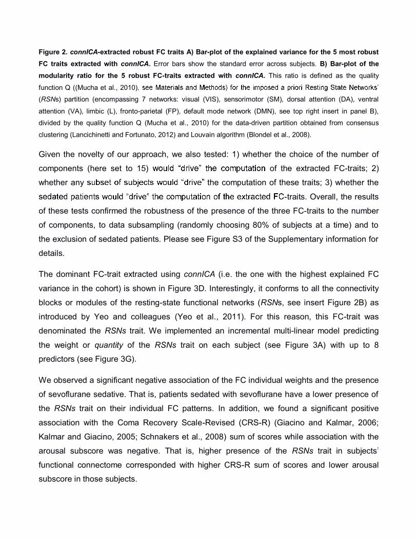
Figure 2. connICA-extracted robust FC traits A) Bar-plot of the explained variance for the 5 most robust
FC traits extracted with connICA. Error bars show the standard error across subjects. B) Bar-plot of the
modularity ratio for the 5 robust FC-traits extracted with connICA. This ratio is defined as the quality
function Q ((Mucha et al., 2010)
(RSNs) partition (encompassing 7 networks: visual (VIS), sensorimotor (SM), dorsal attention (DA), ventral
attention (VA), limbic (L), fronto-parietal (FP), default mode network (DMN), see top right insert in panel B),
divided by the quality function Q (Mucha et al., 2010) for the data-driven partition obtained from consensus
clustering (Lancichinetti and Fortunato, 2012) and Louvain algorithm (Blondel et al., 2008).
Given the novelty of our approach, we also tested: 1) whether the choice of the number of
components (here set to 15) of the extracted FC-traits; 2)
whether any the computation of these traits; 3) whether the
-traits. Overall, the results
of these tests confirmed the robustness of the presence of the three FC-traits to the number
of components, to data subsampling (randomly choosing 80% of subjects at a time) and to
the exclusion of sedated patients. Please see Figure S3 of the Supplementary information for
details.
The dominant FC-trait extracted using connICA (i.e. the one with the highest explained FC
variance in the cohort) is shown in Figure 3D. Interestingly, it conforms to all the connectivity
blocks or modules of the resting-state functional networks (RSNs, see insert Figure 2B) as
introduced by Yeo and colleagues (Yeo et al., 2011). For this reason, this FC-trait was
denominated the RSNs trait. We implemented an incremental multi-linear model predicting
the weight or quantity of the RSNs trait on each subject (see Figure 3A) with up to 8
predictors (see Figure 3G).
We observed a significant negative association of the FC individual weights and the presence
of sevoflurane sedative. That is, patients sedated with sevoflurane have a lower presence of
the RSNs trait on their individual FC patterns. In addition, we found a significant positive
association with the Coma Recovery Scale-Revised (CRS-R) (Giacino and Kalmar, 2006;
Kalmar and Giacino, 2005; Schnakers et al., 2008) sum of scores while association with the
arousal subscore was negative. That is, higher presence of the RSNs trait in subjects
functional connectome corresponded with higher CRS-R sum of scores and lower arousal
subscore in those subjects.
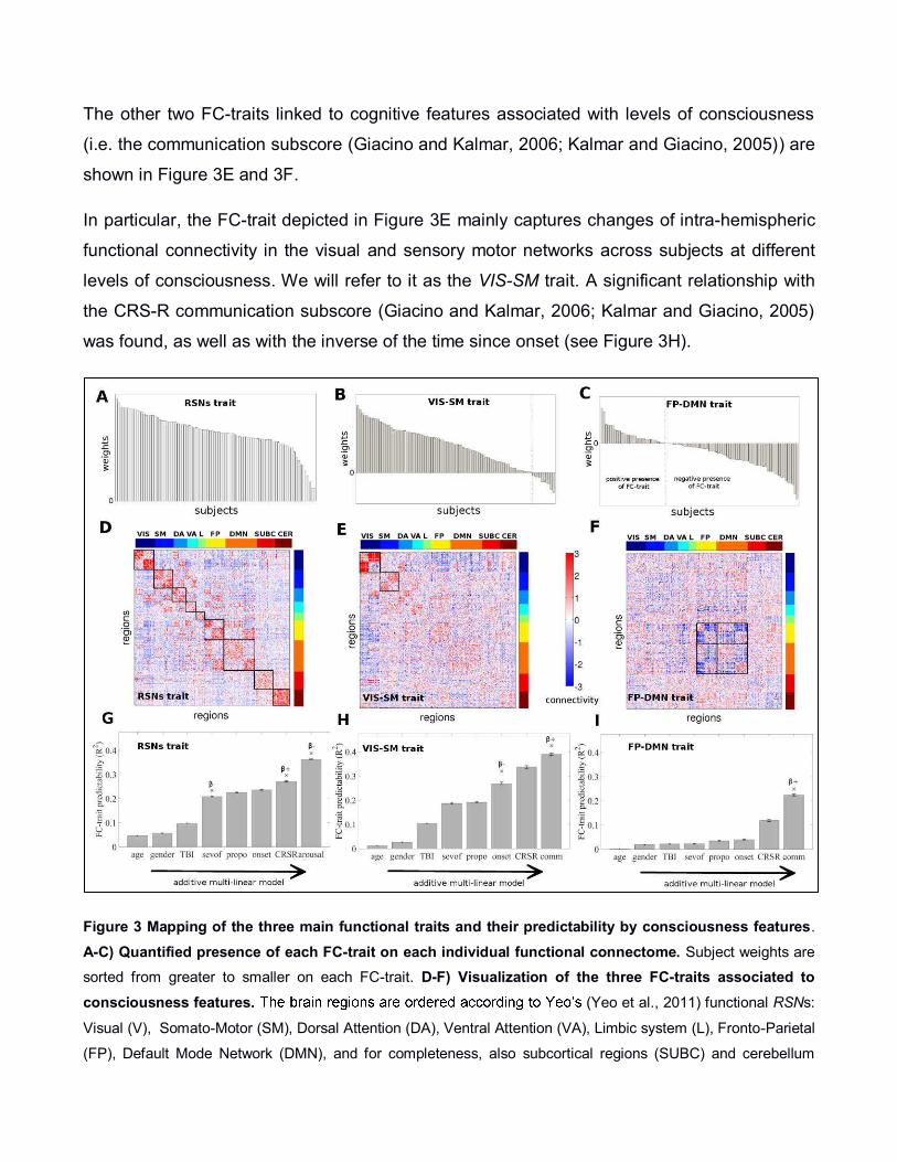
The other two FC-traits linked to cognitive features associated with levels of consciousness
(i.e. the communication subscore (Giacino and Kalmar, 2006; Kalmar and Giacino, 2005)) are
shown in Figure 3E and 3F.
In particular, the FC-trait depicted in Figure 3E mainly captures changes of intra-hemispheric
functional connectivity in the visual and sensory motor networks across subjects at different
levels of consciousness. We will refer to it as the VIS-SM trait. A significant relationship with
the CRS-R communication subscore (Giacino and Kalmar, 2006; Kalmar and Giacino, 2005)
was found, as well as with the inverse of the time since onset (see Figure 3H).
Figure 3 Mapping of the three main functional traits and their predictability by consciousness features.
A-C) Quantified presence of each FC-trait on each individual functional connectome. Subject weights are
sorted from greater to smaller on each FC-trait. D-F) Visualization of the three FC-traits associated to
consciousness features. (Yeo et al., 2011) functional RSNs:
Visual (V), Somato-Motor (SM), Dorsal Attention (DA), Ventral Attention (VA), Limbic system (L), Fronto-Parietal
(FP), Default Mode Network (DMN), and for completeness, also subcortical regions (SUBC) and cerebellum

(CER). G-I) Bar-plots of the FC-traits predictability. They show additive multi-linear regression models with
predictors sequentially introduced in the following order: age, gender, trauma, sedative sevof (i.e., sevoflurane),
sedative propo (i.e., propofol), inverse of the time since onset, Coma Recovery Scale - Revised (CRS-R) total
scores and the CRS-R arousal (for the RSNs trait) or communication (for the VIS-SM and FP-DMN traits)
subscore. Error bars show the standard error across the 100 ICA runs. Crosses on the top of a bar indicate that
the inclusion of the correspondent predictor significantly increased the predictability of the model. The sign of the
beta coefficient associated to each significant variable is shown below each asterisk, indicating whether there is
a negative or positive trend with respect to the weights of the FC traits.
The positive sign of the beta coefficient associated to the communication subscore indicates
that a subject with higher communication subscore has higher contribution or presence of the
VIS-SM trait in his/her functional connectome (Figure 3B). Interestingly, when adding time
since onset (quantified here as the inverse of the days since the insult, see Methods), the
explained variance of the model significantly increased. The negative sign of the associated
beta coefficients for the onset indicates a negative slope in the fit with the FC individual
weights. That is, the more recent the insult, the lower the prominence of the VIS-SM trait on
the individual FC of the patient.
The trait shown in Figure 3F mainly captures modifications in the connectivity between DMN
and fronto-parietal networks (hence denominated FP-DMN trait). Interestingly, the FP-DMN
trait is related to the CRS-R communication subscore, even when the sum of scores is
already added to the multi-linear model (Figure 3I). The positive sign of the beta coefficient
associated to this predictor indicates that a subject with higher CRS-R sum of scores
(communication subscore) has higher presence of the FP-DMN trait on his functional
connectome. Notably, as one goes lower in the levels of consciousness, the contribution of
the FP-DMN trait on the FC of a subject changes sign (see the sorted individual weights
associated to FP-DMN trait, Figure 3C, 3F), with this sign change also evident in a few
subjects for the VIS-SM trait (Figure 3B, 3E).
Notably, adding the number of censored volumes as an additional predictor was not
significant for any of the three multi-linear models discussed above (RSN-trait p=0.65, VIS-
SM trait p=0.43, FP-DMN trait p=0.61). Finally, in order to test the appropriateness of the
multi-linear models, scatter plots of actual vs predicted responses and of standardized
residuals vs predicted responses were also evaluated (see Figure S4 in the Supplementary
information).
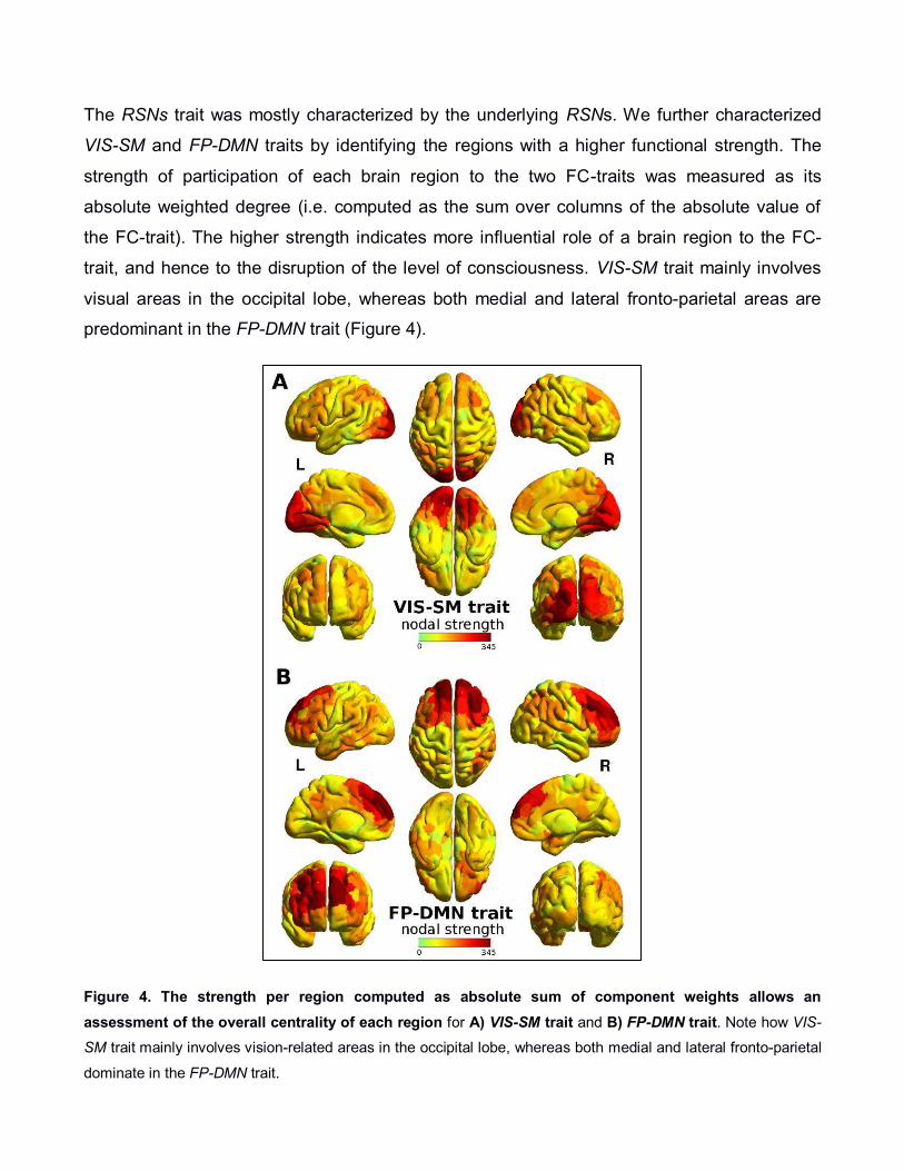
The RSNs trait was mostly characterized by the underlying RSNs. We further characterized
VIS-SM and FP-DMN traits by identifying the regions with a higher functional strength. The
strength of participation of each brain region to the two FC-traits was measured as its
absolute weighted degree (i.e. computed as the sum over columns of the absolute value of
the FC-trait). The higher strength indicates more influential role of a brain region to the FC-
trait, and hence to the disruption of the level of consciousness. VIS-SM trait mainly involves
visual areas in the occipital lobe, whereas both medial and lateral fronto-parietal areas are
predominant in the FP-DMN trait (Figure 4).
Figure 4. The strength per region computed as absolute sum of component weights allows an
assessment of the overall centrality of each region for A) VIS-SM trait and B) FP-DMN trait. Note how VIS-
SM trait mainly involves vision-related areas in the occipital lobe, whereas both medial and lateral fronto-parietal
dominate in the FP-DMN trait.

Further analyses were performed on VIS-SM and FP-DMN traits to assess the presence of
communities (Figure 5) by using consensus clustering (Lancichinetti and Fortunato, 2012)
over 100 modularity solutions computed using the Louvain algorithm (Blondel et al., 2008)
with quality function Q extended to signed networks (Mucha et al., 2010) (see Methods). Note
that the obtained modular configuration gives insights on the data-driven organization of the
functional cores common to the whole cohort. When going back to the individual space, the
depending on the sign (i.e. it changes the FC-trait signed network, see Figure 3). Hence,
performing consensus clustering on the FC-traits allowed us conscious
(positive weights) configuration that gets disrupted towards lower (negative weights)
levels of consciousness (Fig. 3A, 3B, 3C).
We looked at the prominence of each community by averaging the correspondent connectivity
values within each module. Interactions between every two communities in the FC-traits were
then evaluated by averaging the connectivity values connecting them, hence providing a
representation of the coupling between communities.
For both traits, the highest modularity was associated to partitions consisting of three
modules. In line with results of Figure 4, the most influential module (the highest within-
module average) for the VIS-SM trait appears to be the one comprising the occipital cortex
and higher order visual areas. This module is, notably, strongly decoupled from the DMN
module (highest between-modules negative connectivity, Figure 5B), suggesting that in a
healthy brain these two modules are negatively correlated. This modular configuration is then
altered depending on the levels of consciousness.
The modular organization of FP-DMN trait revealed a substantial division of the brain in two
hemispheres. The between-modules average weight shows that the most
communities encompass the two different hemispheres (Figure 5D), indicating that in normal
consciousness the hemispheres are also anti-correlated. This or negative inter-
module connectivity might change (i.e. it turns to positive, Figure 3C, 3F) following loss of
consciousness.

Figure 5. A) Brain rendering of the modules obtained for the VIS-SM trait (see Materials and Methods).
B) Left: bar plot of the average weight within each
module in VIS-SM trait. Right: bar plot of the average between-module weight for the VIS-SM trait. C) Brain
rendering of the modules obtained for the FP-DMN trait. D) Left: bar plot of the average weight within each
module in FP-DMN trait. Right: bar plot of the average between-module weight for the FP-DMN trait.
Discussion
In this work we applied a novel data-based methodology, connICA, to the field of Brain
Connectomics. Our approach is based on extracting independent connectivity traits from a set
of individual functional connectomes to extract and map robust independent mechanisms or
processes that explain the FC patterns of an entire cohort of subjects, without setting any a
priori stratification into groups. We used the connICA framework to assess task-free FC in 88
subjects with different levels of consciousness: 31 conscious controls and 57 severely
damaged patients (2 coma, 17 UWS, 20 MCS, 143 EMCS, 4 LIS) of different etiology and
duration, 31 of whom were acquired while receiving sedative drugs to control for head

movement artifacts. We investigated the functional connectivity traits underlining specific
sensorimotor/cognitive capacities related to consciousness.
We showed how these traits separate the FC data into network subsystems with significant
associations to levels of consciousness. Notably, this methodology allowed us to map and
match the most meaningful functional traits to consciousness-related predictors taken at the
the alteration of levels of
consciousness and the connectivity core associated to it.
The connICA framework provides a multiplex data-driven way to extract and compact
(dimensionality reduction) the most meaningful multivariate information contained in the
functional connectomes in a relatively small set of connectivity traits. In this work we showed
how the modification of levels of consciousness is associated to specific connectivity
disruptions using as reference seven widely accepted RSNs (i.e., visual, somatomotor, dorsal
attention, ventral attention, limbic system, fronto-parietal, default mode network (Yeo et al.,
2011), and for completeness, also subcortical regions (SUBC) and cerebellum (CER), see
insert in Figure 2B).
One additional advantage of this approach is that the dimensionality of the output is
significantly reduced, both in the number of the robust components extracted with respect to
the initial population size (in the study analyzed here, 5 FC-traits starting from 88 FC
matrices) and in the number of variables to be encoded in the multi-linear models, hence
notably decreasing number of multiple comparisons. As opposed to univariate approaches
mapping up to n(n-1)/2 functional connections and their subsequent multi-linear models (n
being the number of brain regions), the multi-layered output of connICA results in a small
subset of robust FC-traits (by definition, a subset smaller or equal to the number of
components set). This dimensionality reduction does not compromise but rather considerably
facilitates the interpretability of the results, by compressing the individual variability into the
most meaningful independent functional cores. It is noteworthy that most if not all the traits
would have been missed with a standard group-average analysis of the functional
connectomes.

By using connICA, we extracted three independent functional connectivity traits linked to
cognitive features of levels of consciousness. Below is a characterization of each FC-trait and
its association to different aspects of consciousness.
The RSNs trait (Figure 3D) is also the one which explains the most of the FC variance (Figure
2A) and is the closest RSNs organization (Yeo et al., 2011). It seems mainly
associated to a global reduction of the functional connectivity within each of the RSNs, and
relates to three significant predictors. It was significantly associated to the effect of the
pathology (i.e. CRS-R sum of scores and arousal) and to effect of the sedation, specifically to
the sevoflurane sedative. Notably, this is in line with several studies reporting the effect of
sevoflurane mainly on the functional connectivity of sensory and motor cortices (Deshpande
et al., 2010; Hudetz, 2012; Peltier et al., 2005), even at light dosage (i.e. 1-2% concentration,
(Peltier et al., 2005), as well as attentional networks (Palanca et al., 2015), frontal and
thalamo-cortical networks (Ranft et al., 2016). On the other hand, propofol light sedation at
concentrations around 1-2 µg/mL seems to affect mainly the connectivity of fronto-parietal
and posterior cingulate cortex (Stamatakis et al., 2010), although in a limited manner as
compared to deep sedation (Amico et al., 2014; Boveroux et al., 2010). This may explain why
propofol did not show significant associations to any FC-trait in the additive multi-linear
models.
The RSNs-trait also captures the functional connectivity core organization of a functioning
human brain, which gets disrupted during pathological loss of consciousness as evidenced by
its strong correlation with the CRS-R sum of scores. This finding is in an agreement with a
vast literature showing widespread connectivity breakdowns in several RSNs of DOC patients
(Boly et al., 2012; Demertzi et al., 2015; Demertzi et al., 2014; Di Perri et al., 2014; Heine et
al., 2012; Soddu et al., 2012; Vanhaudenhuyse et al., 2010). One can hypothesize that this
general disruption of functional connectivity might be connected and/or partially driven by the
widespread underlying structural damage present in DOC and/or TBI patients (Fernández
Espejo et al., 2012; Kraus et al., 2007; Perlbarg et al., 2009; Sidaros et al., 2008). Arousal
was negatively associated with the RSNs trait after controlling for the sum of CRS-R scores
as well as other covariates. The assessment of arousal might easily lead to false positive
evaluations. For this finding in particular, it is not possible to rule out that the estimation in the
level of arousal of a DOC patient may greatly fluctuate and t can thus influence the frequency
and complexity of neurobehavioral responses (Schnakers et al., 2008; Seel et al., 2010).
The VIS-SM trait seems also associated with the pathology (i.e. time since onset) (Figure 3H).
It shows a more prominent disruption of the occipital and sensorimotor areas as the level of
consciousness decreases (Figure 3E), and it also correlates with functional communication
(Figure 3H). Interestingly, the modularity analysis suggests that visual areas and DMN are
anti-correlated in normal wakefulness (Figure 4A, 4B), stressing the importance of the
interaction between the so called slave regions (Crick and Koch, 1995) and higher
order cognitive regions as the DMN, for consciousness and functional communication (Koch
et al., 2016). This corroborates the hypothesis that loss of consciousness might correlate with
the disruption of primary sensory areas and higher-order associative cortices, which are
thought to be required for conscious perception (i.e. global workspace, (Dehaene and
Changeux, 2011; Demertzi et al., 2015)).
However, the recovery of this connectivity pattern does not necessarily imply the restoration
of levels of consciousness. Another independent functional trait appears to be linked to
behavioral assessment of levels of consciousness, particularly to both the CRS-R total score
and the communication subscore (FP-DMN trait, Figure 3F).
The FP-DMN trait captures changes in the anti-correlation between the FP-DMN networks.
Notably, as one goes towards the deepest unconsciousness, the FP-DMN anti-correlation
decreases, until the point where it (see Figure 3C, 3F). This is in
line with previous studies showing decreasing anti-correlation in anesthesia (Amico et al.,
2014; Boveroux et al., 2010), sleep (Sämann et al., 2011) and UWS patients (Boly et al.,
2009). Particularly, a recent study (Di Perri et al., 2016) showed that negative connectivity
between DMN and FP networks was significantly different between patients and healthy
controls. Indeed, UWS and MCS patients showed a pathological positive connectivity
between these two networks, whereas patients who emerged from MCS and recovered a
level of consciousness sufficient for functional communication and/or object use, exhibited
partial preserved between-network negative connectivity (Di Perri et al., 2016). In this respect,
the fact that the FP-DMN trait is strongly correlated to the communication subscore

corroborates the idea that recovery of the FP-DMN between-network negative connectivity is
prerequisite in order to regain functional communication.
Notably, the modularity analysis on FP-DMN trait reveals that the decoupling between the two
hemispheres (Figure 5C) represents a ion between left and right
brain hemispheres. The anti-correlation between hemispheres tends to disappear (i.e. trends
toward zero or even becomes positive correlation, see the individual weights of FP-DMN trait
in Figure 3C) as levels of consciousness decrease.
Indeed, there is evidence suggesting that coordination between the two hemispheres is
essential for a correct communication between them (Gazzaniga, 2005). It has been reported
that transection of corpus callosum in refractory epileptic patients (i.e. split brain patients)
caused each hemisphere to have its own separate perception, concepts, and impulses to act
(Gazzaniga, 2014). The conscious abilities of the two hemispheres are strongly differentiated
in specialized cognitive modules (Marinsek et al., 2014), modulated by the thalamo-cortical
system (subcortical regions are also split in left and right modules in FP-DMN trait, see Figure
5C). In this study we show that the interaction between specialized modules, as the VIS-SM
interaction with DMN or the FP-DMN between-network negative connectivity, is crucial for the
emergence of consciousness. Perhaps this laterality enhances the complexity of ongoing
brain processes and facilitates demanding cognitive processes such as consciousness of the
self and the surrounding.
Taken together, these findings suggest that the connectivity core which differentiates across
levels of consciousness is a combination of positive and negative interactions between
functional sub-networks. This evidence stresses the importance of a whole-brain network
modulation between coherent and non-coherent functional states. The disruption of the
equilibrium between these two might lead to changes in levels of consciousness and,
ultimately, to reduced levels of consciousness.
In fact, the connICA results presented in this study depict a very challenging reality. Within the
set of individual functional connectomes analyzed here, there is not just one but at least three
independent mechanisms, namely FC-traits, whose predictability by consciousness related
features is present but different for each one, and hence is most likely capturing different
phenomena or mechanisms. The first RSNs trait predicted by the CRS-R sum of scores,

isolates the functional connectivity blocks of typical RSNs present in a human brain (Figure
3D). The second VIS-SM trait, with predominant influence of visual and sensory regions,
relates disruption of sensory networks to the CRS-R functional communication subscore
(Figure 3E). The third FP-DMN trait, significantly associated to CRS-R sum of scores and
communication, stresses the key role of the negative connectivity between FP and DMN
networks (Figure 3F) and inter-hemispheric communication (Figure 5C,D) in the alteration of
levels of consciousness.
It is worth mentioning here that the traits found by connICA and the sensitivity of those to
demographical and cognitive features is highly dependent on the population analyzed.
Indeed, when considering the RSNs trait, demographics such as age appear to have a strong
fingerprint on it when looking only to the healthy cohort (without considering DOC patients,
see supplementary Figure S5). This suggests that age has a fingerprint in FC-traits obtained
from a healthy population, but its relative effect is blurred when assessing subjects at different
.
The study presented here adds to recent studies from Iraji et al. (Iraji et al., 2016) assessing
ICA components of voxel-based functional connectivity, and from Misic et al. (Misic et al.,
2016), where levels of integration of joint structural-functional connectivity patterns are
assessed from sets of individual connectomes by means of a single-value decomposition
(Misic et al., 2016). Together with the methodology presented here, these recent efforts
suggest that the area of Brain Connectomics is evolving into new data-driven ways of
analyzing connectivity data at different spatial scales without stratifying subjects into a priori
groups and hence, also without performing group-averages of individual connectivity
matrices.
Our study has several limitations. The optimal size of the cohort for the extraction of the
connICA components needs to be further investigated. Similarly, the best choice of the
starting number of ICA components (here set to 15) and the threshold for the final selection of
the most frequent components over multiple ICA runs (here set to 75%) need to be
characterized in more detail. In this work we used the Shen brain parcellation (Shen et al.,
2013) because of the uniformity of the size of brain regions and its functional data-driven
approach. We also used the well-assessed RSNs decomposition provided by Yeo as

obtained in a large cohort (n=1000) of healthy volunteers (Yeo et al., 2011). However, other
parcellations (Desikan et al., 2006; Gordon et al., 2016) or finer decompositions (Demertzi et
al., 2015; Demertzi et al., 2014) might be beneficial in the connICA framework, depending on
the research problem at hand and the desired level of spatial resolution.
Future work can be extended to the use of connICA for structural connectivity patterns, hence
identifying SC-traits within a population of subjects. This approach is not limited to assessing
consciousness, but it has the potential of studying other progressive diseases and disorders,
drug-induced effects, and also differences based on aging or gender. We have here
addressed the effect of the sedation as a binary confound (see Materials and Methods). An
interesting future avenue would be to apply connICA for disentangling differences between
FC-traits at different concentrations of the anesthetic agent at hand, e.g. in a population of
healthy subjects.
When associating traits with cognitive/clinical features, multi-linear models employed here can
be expanded by allowing for non-linear terms and interactions, which could capture more
complex associations between connectivity patterns and cognition.
In conclusion, we here proposed a novel data-driven approach, connICA, to extract the most
influential connectivity patterns in the alteration of levels of consciousness. Our results shed
light on isolating key functional core changes involved in the degradation of conscious states
and establish links between isolated clinical/cognitive features and specific FC-traits.
Acknowledgements
We thank Marie-Aurelie Bruno, Athena Demertzi and Audrey Vanhaudenhuyse for help in
acquiring the data. We thank Prof. Jaroslaw Harezlak and Prof. Thomas Talavage for useful
comments. This research was supported by the This research was supported by the Belgian
Funds for Scientific Research (FRS), European Commission, James McDonnell Foundation,
European Space Agency, Belgian Science Policy (CEREBNET, BELSPO), Wallonia-Brussels
Federation Concerted Research Action, Mind Science Foundation, Public Utility Foundation
"Université Européenne du Travail" and "Fondazione Europea di Ricerca Biomedica",
University and University Hospital of Liège. LH is a research fellow and SL a research director
at FNRS. JG was supported by the National Institute of Health (1R01 MH108467-01).

References
Achard, S., Delon-Martin, C., Vertes, P.E., Renard, F., Schenck, M., Schneider, F., Heinrich, C., Kremer, S., Bullmore, E.T., 2012. Hubs of brain functional networks are radically reorganized in comatose patients. Proc Natl Acad Sci U S A 109, 20608-20613. Amico, E., Gomez, F., Di Perri, C., Vanhaudenhuyse, A., Lesenfants, D., Boveroux, P., Bonhomme, V., Brichant, J.F., Marinazzo, D., Laureys, S., 2014. Posterior cingulate cortex-related co-activation patterns: a resting state FMRI study in propofol-induced loss of consciousness. PLoS One 9, e100012. Bernat, J.L., 2009. Chronic consciousness disorders. Annu Rev Med 60, 381-392. Blondel, V.D., Guillaume, J.-L., Lambiotte, R., Lefebvre, E., 2008. Fast unfolding of communities in large networks. Journal of statistical mechanics: theory and experiment 2008, P10008. Boly, M., Massimini, M., Garrido, M.I., Gosseries, O., Noirhomme, Q., Laureys, S., Soddu, A., 2012. Brain connectivity in disorders of consciousness. Brain Connect 2, 1-10. Boly, M., Tshibanda, L., Vanhaudenhuyse, A., Noirhomme, Q., Schnakers, C., Ledoux, D., Boveroux, P., Garweg, C., Lambermont, B., Phillips, C., Luxen, A., Moonen, G., Bassetti, C., Maquet, P., Laureys, S., 2009. Functional connectivity in the default network during resting state is preserved in a vegetative but not in a brain dead patient. Hum Brain Mapp 30, 2393-2400. Boveroux, P., Vanhaudenhuyse, A., Bruno, M.A., Noirhomme, Q., Lauwick, S., Luxen, A., Degueldre, C., Plenevaux, A., Schnakers, C., Phillips, C., Brichant, J.F., Bonhomme, V., Maquet, P., Greicius, M.D., Laureys, S., Boly, M., 2010. Breakdown of within- and between-network resting state functional magnetic resonance imaging connectivity during propofol-induced loss of consciousness. Anesthesiology 113, 1038-1053. Bullmore, E., Sporns, O., 2009. Complex brain networks: graph theoretical analysis of structural and functional systems. Nat Rev Neurosci 10, 186-198. Calhoun, V.D., Liu, J., Adali, T., 2009. A review of group ICA for fMRI data and ICA for joint inference of imaging, genetic, and ERP data. Neuroimage 45, S163-172. Coupé, P., Manjón, J.V., Robles, M., Collins, D.L., 2012. Adaptive multiresolution non-local means filter for three-dimensional magnetic resonance image denoising. Image Processing, IET 6, 558-568. Crick, F., Koch, C., 1995. Are we aware of neural activity in primary visual cortex? Nature 375, 121-123. Crone, J.S., Soddu, A., Holler, Y., Vanhaudenhuyse, A., Schurz, M., Bergmann, J., Schmid, E., Trinka, E., Laureys, S., Kronbichler, M., 2014. Altered network properties of the fronto-parietal network and the thalamus in impaired consciousness. Neuroimage Clin 4, 240-248. Dehaene, S., Changeux, J.P., 2011. Experimental and theoretical approaches to conscious processing. Neuron 70, 200-227. Demertzi, A., Antonopoulos, G., Heine, L., Voss, H.U., Crone, J.S., de Los Angeles, C., Bahri, M.A., Di Perri, C., Vanhaudenhuyse, A., Charland-Verville, V., Kronbichler, M., Trinka, E., Phillips, C., Gomez, F., Tshibanda, L., Soddu, A., Schiff, N.D., Whitfield-Gabrieli, S., Laureys, S., 2015. Intrinsic functional connectivity differentiates minimally conscious from unresponsive patients. Brain 138, 2619-2631. Demertzi, A., Gomez, F., Crone, J.S., Vanhaudenhuyse, A., Tshibanda, L., Noirhomme, Q., Thonnard, M., Charland-Verville, V., Kirsch, M., Laureys, S., 2014. Multiple fMRI system-level baseline connectivity is disrupted in patients with consciousness alterations. Cortex 52, 35-46.

Deshpande, G., Kerssens, C., Sebel, P.S., Hu, X., 2010. Altered local coherence in the default mode network due to sevoflurane anesthesia. Brain research 1318, 110-121. Desikan, R.S., Ségonne, F., Fischl, B., Quinn, B.T., Dickerson, B.C., Blacker, D., Buckner, R.L., Dale, A.M., Maguire, R.P., Hyman, B.T., 2006. An automated labeling system for subdividing the human cerebral cortex on MRI scans into gyral based regions of interest. Neuroimage 31, 968-980. Di Perri, C., Bahri, M.A., Amico, E., Thibaut, A., Heine, L., Antonopoulos, G., Charland-Verville, V., Wannez, S., Gomez, F., Hustinx, R., Tshibanda, L., Demertzi, A., Soddu, A., Laureys, S., 2016. Neural correlates of consciousness in patients who have emerged from a minimally conscious state: a cross-sectional multimodal imaging study. Lancet Neurol. Di Perri, C., Stender, J., Laureys, S., Gosseries, O., 2014. Functional neuroanatomy of disorders of consciousness. Epilepsy Behav 30, 28-32. Erhardt, E.B., Rachakonda, S., Bedrick, E.J., Allen, E.A., Adali, T., Calhoun, V.D., 2011. Comparison of multi-subject ICA methods for analysis of fMRI data. Hum Brain Mapp 32, 2075-2095.
Fernández Espejo, D., Soddu, A., Cruse, D., Palacios, E.M., Junque, C., Vanhaudenhuyse,
A., Rivas, E., Newcombe, V., Menon, D.K., Pickard, J.D., 2012. A role for the default mode network in the bases of disorders of consciousness. Annals of neurology 72, 335-343. Fiset, P., Plourde, G., Backman, S.B., 2005. Brain imaging in research on anesthetic mechanisms: studies with propofol. Progress in brain research 150, 245-598. Fornito, A., Zalesky, A., Bullmore, E.T., 2016. Fundamentals of Brain Network Analysis. Academic Press. Fox, M.D., Raichle, M.E., 2007. Spontaneous fluctuations in brain activity observed with functional magnetic resonance imaging. Nature Reviews Neuroscience 8, 700-711. Fox, M.D., Snyder, A.Z., Vincent, J.L., Corbetta, M., Van Essen, D.C., Raichle, M.E., 2005. The human brain is intrinsically organized into dynamic, anticorrelated functional networks. Proc Natl Acad Sci U S A 102, 9673-9678. Friston, K.J., Frith, C.D., Liddle, P.F., Frackowiak, R.S., 1993. Functional connectivity: the principal-component analysis of large (PET) data sets. J Cereb Blood Flow Metab 13, 5-14. Gazzaniga, M.S., 2005. Forty-five years of split-brain research and still going strong. Nature Reviews Neuroscience 6, 653-659. Gazzaniga, M.S., 2014. The split-brain: rooting consciousness in biology. Proc Natl Acad Sci U S A 111, 18093-18094. Giacino, J., Kalmar, K., 2006. Coma recovery scale-revised. The Center for Outcome Measurement in Brain Injury, 36-51. Giacino, J.T., Fins, J.J., Laureys, S., Schiff, N.D., 2014. Disorders of consciousness after acquired brain injury: the state of the science. Nat Rev Neurol 10, 99-114. Giacino, J.T., Kezmarsky, M.A., DeLuca, J., Cicerone, K.D., 1991. Monitoring rate of recovery to predict outcome in minimally responsive patients. Archives of Physical Medicine and Rehabilitation 72, 897-901. Giacino, J.T., Zasler, N.D., Whyte, J., Katz, D.I., Glen, M., Andary, M., 1995. Recommendations for use of uniform nomenclature pertinent to patients with severe alterations in consciousness. Archives of Physical Medicine and Rehabilitation 76, 205-209. Gordon, E.M., Laumann, T.O., Adeyemo, B., Huckins, J.F., Kelley, W.M., Petersen, S.E., 2016. Generation and evaluation of a cortical area parcellation from resting-state correlations. Cerebral cortex 26, 288-303.

Greicius, M., 2008. Resting-state functional connectivity in neuropsychiatric disorders. Curr Opin Neurol 21, 424-430. Greve, D.N., Fischl, B., 2009. Accurate and robust brain image alignment using boundary-based registration. Neuroimage 48, 63-72. Gómez, S., Jensen, P., Arenas, A., 2009. Analysis of community structure in networks of correlated data. Physical Review E 80, 016114. Heine, L., Soddu, A., Gomez, F., Vanhaudenhuyse, A., Tshibanda, L., Thonnard, M., Charland-Verville, V., Kirsch, M., Laureys, S., Demertzi, A., 2012. Resting state networks and consciousness: alterations of multiple resting state network connectivity in physiological, pharmacological, and pathological consciousness States. Front Psychol 3, 295. Hudetz, A.G., 2012. General anesthesia and human brain connectivity. Brain connectivity 2, 291-302. Hyvarinen, A., 1999. Fast and robust fixed-point algorithms for independent component analysis. IEEE Trans Neural Netw 10, 626-634. Iraji, A., Calhoun, V.D., Wiseman, N., Davoodi-Bojd, E., Avanaki, M.R., Haacke, E.M., Kou, Z., 2016. The connectivity domain: Analyzing resting state fMRI data using feature-based data-driven and model-based methods. Neuroimage. Jenkinson, M., Beckmann, C.F., Behrens, T.E.J., Woolrich, M.W., Smith, S.M., 2012. Fsl. Neuroimage 62, 782-790. Kalmar, K., Giacino, J.T., 2005. The JFK Coma Recovery Scale--Revised. Neuropsychol Rehabil 15, 454-460. Koch, C., Massimini, M., Boly, M., Tononi, G., 2016. Neural correlates of consciousness: progress and problems. Nature Reviews Neuroscience 17, 307-321. Kraus, M.F., Susmaras, T., Caughlin, B.P., Walker, C.J., Sweeney, J.A., Little, D.M., 2007. White matter integrity and cognition in chronic traumatic brain injury: a diffusion tensor imaging study. Brain 130, 2508-2519. Lancichinetti, A., Fortunato, S., 2012. Consensus clustering in complex networks. Scientific reports 2. Laureys, S., 2005. The neural correlate of (un)awareness: lessons from the vegetative state. Trends Cogn Sci 9, 556-559. Laureys, S., Owen, A.M., Schiff, N.D., 2004. Brain function in coma, vegetative state, and related disorders. Lancet Neurol 3, 537-546. Laureys, S., Pellas, F., Van Eeckhout, P., Ghorbel, S., Schnakers, C., Perrin, F., Berre, J., Faymonville, M.-E., Pantke, K.-H., Damas, F., 2005. The locked-in syndrome: what is it like to be conscious but paralyzed and voiceless? Progress in brain research 150, 495-611. Marinsek, N., Turner, B.O., Gazzaniga, M., Miller, M.B., 2014. Divergent hemispheric reasoning strategies: reducing uncertainty versus resolving inconsistency. Front Hum Neurosci, Switzerland, p. 839. Misic, B., Betzel, R.F., de Reus, M.A., van den Heuvel, M.P., Berman, M.G., McIntosh, A.R., Sporns, O., 2016. Network-Level Structure-Function Relationships in Human Neocortex. Cereb Cortex. Monti, M.M., Lutkenhoff, E.S., Rubinov, M., Boveroux, P., Vanhaudenhuyse, A., Gosseries, O., Bruno, M.-A., Noirhomme, Q., Boly, M., Laureys, S., 2013. Dynamic change of global and local information processing in propofol-induced loss and recovery of consciousness. PLoS Comput Biol 9, e1003271. Mucha, P.J., Richardson, T., Macon, K., Porter, M.A., Onnela, J.-P., 2010. Community structure in time-dependent, multiscale, and multiplex networks. science 328, 876-878.

Newman, M.E.J., Girvan, M., 2004. Finding and evaluating community structure in networks. Physical review E 69, 026113. Owen, A.M., Schiff, N.D., Laureys, S., 2009. A new era of coma and consciousness science. Progress in brain research 177, 399-411. Palanca, B.J.A., Mitra, A., Larson-Prior, L., Snyder, A.Z., Avidan, M.S., Raichle, M.E., 2015. Resting-state functional magnetic resonance imaging correlates of sevoflurane-induced unconsciousness. The Journal of the American Society of Anesthesiologists 123, 346-356. Peltier, S.J., Kerssens, C., Hamann, S.B., Sebel, P.S., Byas-Smith, M., Hu, X., 2005. Functional connectivity changes with concentration of sevoflurane anesthesia. Neuroreport 16, 285-288. Perlbarg, V., Puybasset, L., Tollard, E., Lehericy, S., Benali, H., Galanaud, D., 2009. Relation between brain lesion location and clinical outcome in patients with severe traumatic brain
injury: A diffusion tensor imaging study using voxel based approaches. Human brain mapping
30, 3924-3933. Power, J.D., Barnes, K.A., Snyder, A.Z., Schlaggar, B.L., Petersen, S.E., 2012. Spurious but systematic correlations in functional connectivity MRI networks arise from subject motion. Neuroimage 59, 2142-2154. Power, J.D., Mitra, A., Laumann, T.O., Snyder, A.Z., Schlaggar, B.L., Petersen, S.E., 2014. Methods to detect, characterize, and remove motion artifact in resting state fMRI. Neuroimage 84, 320-341. Ranft, A., Golkowski, D., Kiel, T., Riedl, V., Kohl, P., Rohrer, G., Pientka, J., Berger, S., Thul, A., Maurer, M., Preibisch, C., Zimmer, C., Mashour, G.A., Kochs, E.F., Jordan, D., Ilg, R., 2016. Neural Correlates of Sevoflurane-induced Unconsciousness Identified by Simultaneous Functional Magnetic Resonance Imaging and Electroencephalography. Anesthesiology 125, 861-872. Schnakers, C., Majerus, S., Giacino, J., Vanhaudenhuyse, A., Bruno, M.-A., Boly, M., Moonen, G., Damas, P., Lambermont, B., Lamy, M., 2008. A french validation study of the Coma Recovery Scale-Revised (CRS-R). Brain Injury 22, 786-792. Seel, R.T., Sherer, M., Whyte, J., Katz, D.I., Giacino, J.T., Rosenbaum, A.M., Hammond, F.M., Kalmar, K., Pape, T.L.-B., Zafonte, R., 2010. Assessment scales for disorders of consciousness: evidence-based recommendations for clinical practice and research. Archives of physical medicine and rehabilitation 91, 1795-1813. Shen, X., Tokoglu, F., Papademetris, X., Constable, R.T., 2013. Groupwise whole-brain parcellation from resting-state fMRI data for network node identification. Neuroimage 82, 403-415. Sidaros, A., Engberg, A.W., Sidaros, K., Liptrot, M.G., Herning, M., Petersen, P., Paulson, O.B., Jernigan, T.L., Rostrup, E., 2008. Diffusion tensor imaging during recovery from severe traumatic brain injury and relation to clinical outcome: a longitudinal study. Brain 131, 559-572. Smyser, C.D., Snyder, A.Z., Neil, J.J., 2011. Functional connectivity MRI in infants: exploration of the functional organization of the developing brain. Neuroimage 56, 1437-1452. Soddu, A., Vanhaudenhuyse, A., Bahri, M.A., Bruno, M.A., Boly, M., Demertzi, A., Tshibanda, J.F., Phillips, C., Stanziano, M., Ovadia-Caro, S., Nir, Y., Maquet, P., Papa, M., Malach, R., Laureys, S., Noirhomme, Q., 2012. Identifying the default-mode component in spatial IC analyses of patients with disorders of consciousness. Hum Brain Mapp 33, 778-796. Sporns, O., 2011. Networks of the Brain. MIT press.

Stamatakis, E.A., Adapa, R.M., Absalom, A.R., Menon, D.K., 2010. Changes in resting neural connectivity during propofol sedation. PloS one 5, e14224. Sämann, P.G., Wehrle, R., Hoehn, D., Spoormaker, V.I., Peters, H., Tully, C., Holsboer, F., Czisch, M., 2011. Development of the brain's default mode network from wakefulness to slow wave sleep. Cerebral cortex, bhq295. Tagliazucchi, E., von Wegner, F., Morzelewski, A., Borisov, S., Jahnke, K., Laufs, H., 2012. Automatic sleep staging using fMRI functional connectivity data. Neuroimage 63, 63-72. Traag, V.A., Bruggeman, J., 2009. Community detection in networks with positive and negative links. Physical Review E 80, 036115. van den Heuvel, M.P., Hulshoff Pol, H.E., 2010. Exploring the brain network: a review on resting-state fMRI functional connectivity. Eur Neuropsychopharmacol 20, 519-534. Vanhaudenhuyse, A., Noirhomme, Q., Tshibanda, L.J., Bruno, M.A., Boveroux, P., Schnakers, C., Soddu, A., Perlbarg, V., Ledoux, D., Brichant, J.F., Moonen, G., Maquet, P., Greicius, M.D., Laureys, S., Boly, M., 2010. Default network connectivity reflects the level of consciousness in non-communicative brain-damaged patients. Brain 133, 161-171. Yeo, B.T., Krienen, F.M., Sepulcre, J., Sabuncu, M.R., Lashkari, D., Hollinshead, M., Roffman, J.L., Smoller, J.W., Zollei, L., Polimeni, J.R., Fischl, B., Liu, H., Buckner, R.L., 2011. The organization of the human cerebral cortex estimated by intrinsic functional connectivity. J Neurophysiol 106, 1125-1165.

Figure S1: Illustration of the major fMRI preprocessing steps described in the Materials and Methods
section of the task-free session for four individual subjects (A-D). For each subject, the 4 plots from top to
bottom: 1) The fMRI time courses in all GM voxels after slice timing and motion correction, normalization to
mode 1000, demeaning and detrending; 2) The 18 motion and physiological noise regressors; [x, y , z, pitch,
yaw, roll], the tissue mean signal of whole-brain, WM and CSF and their corresponding nine derivatives
(backwards difference); 3) Visual representation of the scrubbing procedure using the Frame Displacement (FD),
DVARS and SD metrics to drop (censor) BOLD volumes with head motion (indicated by the dark vertical bars)
from the computation of the pairwise correlations. Note that, as explained in Materials and Methods, first and last
7 volumes in each session were always excluded; 4) Residuals of the BOLD time courses of GM voxels after
regressing out the 18 regressors. Subjects A and B had no censored volumes, with C and D having 14% and
11% censored volumes, respectively.

Figure S2: A) Plot of the correspondent weights per subjects for each single connICA run. B) Plot of the five
robust connectivity traits extracted using connICA based on 100 runs. Only three of the FC-traits were
associated with cognitive scores linked to levels of consciousness.

Figure S3: Robustness analysis of FC-traits. A-B-C) Robustness of FC-traits to number of components.
Histograms of the correlations between each FC-trait and its best matching component for each of the 1,100
connICA runs performed. Those runs consisted of running connICA while varying the number of components
between 10 and 20 (100 runs for each). This test was meant to check whether the choice on the number of
-traits found. D-E-F) Robustness of FC-traits to
data sampling. Histograms show the correlations between the components that best match each of the
presented 3 FC-traits. Here, the number of components is fixed to 15, but random subsamples of the 80% of the
initial dataset are included for every run (500 runs) to extract the components. This allowed us to evaluate
whether the generation of the observed FC-traits . G-H-I) Robustness of
FC-traits to exclusion of sedated subjects. Histograms show the correlations between the components that

best match each of the presented 3 FC-traits. Here, the number of component is fixed to 15, but sedated
patients were excluded from the dataset used to extract the components (500 runs). This allowed us to evaluate
if the generation of the observed FC-traits .
Figure S4: Evaluation of the multi-linear models. A-B-C) Predicted vs actual values. Scatter plots of the
subject weights (Y-axis) associated to each of the 3 extracted FC-traits (i.e., RSNs, VIS-SM and FP-DMN)
versus the subject weights predicted by the final multi-linear regression models when including all 8 predictors
(see Material and Methods and Figure 3 for details and description of predictors). Colors denote diagnosis as
follows: dark blue refers to Healthy Controls (HC); blue to Locked-in Syndrome patients (LIS); light blue to
patients emerging for minimally conscious state (EMCS); green to minimally conscious state patients (MCS);
orange to unresponsive wakefulness syndrome patients (UWS); yellow to coma patients (Coma). Shape denotes
sedatives administered to subjects: circles indicate non-sedated, upward-pointing triangles indicate sevoflurane,
downward-pointing triangles indicate propofol, and hence stars indicate both sevoflurane and propofol. Note how
the linear trend between actual-predicted values is not driven by any specific group or a sedative, providing
evidence on the reliability of the employed model. D-E-F) Predicted vs residuals. Scatter plots of the
standardized residuals (Y-axis) versus the predicted subject weights associated to each of the 3 extracted FC-
traits (i.e., RSNs, VIS-SM and FP-DMN) for the multi-linear model described above (see also Material and
Methods for details). The residuals are symmetrically distributed, tending to cluster around 0, and within 3
standard deviations of zero.

Figure S5. Scatter plot of age vs RSNs-trait subject weights in HC group. When looking at HC subjects only
(N=31), we found a significant negative correlation (r=-0.55, p<0.05) between age and the subject-weight for
RSNs-trait (top right insert). In other words, the contribution of the RSNs-trait in the resting-state FC is reduced
in older subjects.
Trait/Predictor Age Gender TBI Sevoflurane Propofol Onset CRS-R Subscore
RSNs N.S. N.S. N.S. -0.0079 N.S. N.S. 0.0156 -0.0143
VIS-SM N.S. N.S. N.S. N.S. N.S. -0.0074 N.S. 0.0188
FP-DMN N.S. N.S. N.S. N.S. N.S. N.S. N.S. 0.0206
Table S1. Standardized beta coefficients of the final models for each FC trait and including all predictors (see
Materials and Methods for details on each predictor). N.S. denotes non-significant values. Statistical significance
set at p < 0.05.
Related Documents