Mapping of nucleocapsid residues important for HIV-1 genomic RNA dimerization and packaging Jafar Kafaie a,b , Rujun Song a,b , Levon Abrahamyan a , Andrew J. Mouland a,b , Michael Laughrea a,b, ⁎ a McGill AIDS Center, Lady Davis Institute for Medical Research, Jewish General Hospital, Montreal, Québec, Canada H3T 1E2 b Department of Medicine, McGill University, Montreal, Québec, Canada H3A 2B4 Received 24 December 2007; returned to author for revision 14 January 2008; accepted 1 February 2008 Available online 17 March 2008 Abstract Retroviral genomic RNA (gRNA) dimerization appears essential for viral infectivity, and the nucleocapsid protein (NC) of human immunodeficiency virus type 1 (HIV-1) facilitates HIV-1 gRNA dimerization. To identify the relevant and dispensable positions of NC, 34 of its 55 residues were mutated, individually or in small groups, in a panel of 40 HIV-1 mutants prepared by site-directed mutagenesis. It was found that the amino-terminus, the proximal zinc finger, the linker, and the distal zinc finger of NC each contributed roughly equally to efficient HIV-1 gRNA dimerization. The N-terminal and linker segments appeared to play predominantly electrostatic and steric roles, respectively. Mutating the hydrophobic patch of either zinc finger, or substituting alanines for their glycine doublet, was as disabling as deleting the corresponding finger. Replacing the CysX 2 CysX 4 HisX 4 Cys motif of either finger by CysX 2 CysX 4 CysX 4 Cys or CysX 2 CysX 4 HisX 4 His, interchanging the zinc fingers or, replacing one zinc finger by a copy of the other one, had generally intermediate effects; among these mutations, the His23→Cys substitution in the N-terminal zinc finger had the mildest effect. The charge of NC could be increased or decreased by up to 18%, that of the linker could be reduced by 75% or increased by 50%, and one or two electric charges could be added or subtracted from either zinc finger, without affecting gRNA dimerization. Shortening, lengthening, or making hydrophobic the linker was as disabling as deleting the N-terminal or the C-terminal zinc finger, but a neutral and polar linker was innocuous. The present work multiplies by 4 and by 33 the number of retroviral and lentiviral NC mutations known to inhibit gRNA dimerization, respectively. It shows the first evidence that gRNA dimerization can be inhibited by: 1) mutations in the N-terminus or the linker of retroviral NC; 2) mutations in the proximal zinc finger of lentiviral NC; 3) mutations in the hydrophobic patch or the conserved glycines of the proximal or the distal retroviral zinc finger. Some NC mutations impaired gRNA dimerization more than mutations inactivating the viral protease, indicating that gRNA dimerization may be stimulated by the NC component of the Gag polyprotein. Most, but not all, mutations inhibited gRNA packaging; some had a strong effect on virus assembly or stability. © 2008 Elsevier Inc. All rights reserved. Keywords: HIV-1; RNA dimerization; Nucleocapsid protein; Zinc finger; Linker; RNA packaging Introduction The genome of HIV-1, a member of the Retroviridae family, is a single-stranded RNA. Two copies are packaged and they can dimerize. This dimeric structure appears essential for viral infectivity via, among others, facilitating viral RNA strand exchange during reverse transcription (Chin et al., 2005, 2007). It is not clear that protein-free formation of viral genomic RNA (gRNA) dimers can occur in the infected cell, the test tube, or even the virus. No gRNA dimers are detectable in newly released protease inactive (PR - ) HIV-1, but thermolabile and partially dimeric viral RNA dimers slowly accumulate with time Available online at www.sciencedirect.com Virology 375 (2008) 592 – 610 www.elsevier.com/locate/yviro Abbreviations: C, cysteine, except in “C-terminal”, where it means “carboxyl-terminal”; CA, capsid; DIS, dimerization initiation site; Gag, group specific antigen; gRNA, genomic RNA; HIV-1, human immunodeficiency virus type 1; MoMuLV, Moloney murine leukemia virus; N, aspargine except in “N-terminal”, where it means “amino-terminal”; NC, nucleocapsid protein; Pol, polymerase; PR − , protease inactive; Pr55gag, the Gag precursor polyprotein; Pr160gag-pol, the Gag-Pol precursor polyprotein; RT, reverse transcriptase; SIV, simian immunodeficiency virus; SL1, stem-loop 1; WT, wild type. ⁎ Corresponding author. Lady Davis Institute for Medical Research, 3755 Cote Ste Catherine, Montreal, Québec, Canada H3T 1E2. Fax: +1 514 340 7502. E-mail address: [email protected] (M. Laughrea). 0042-6822/$ - see front matter © 2008 Elsevier Inc. All rights reserved. doi:10.1016/j.virol.2008.02.001

Welcome message from author
This document is posted to help you gain knowledge. Please leave a comment to let me know what you think about it! Share it to your friends and learn new things together.
Transcript
Available online at www.sciencedirect.com
8) 592–610www.elsevier.com/locate/yviro
Virology 375 (200
Mapping of nucleocapsid residues important for HIV-1 genomicRNA dimerization and packaging
Jafar Kafaie a,b, Rujun Song a,b, Levon Abrahamyan a,Andrew J. Mouland a,b, Michael Laughrea a,b,⁎
a McGill AIDS Center, Lady Davis Institute for Medical Research, Jewish General Hospital, Montreal, Québec, Canada H3T 1E2b Department of Medicine, McGill University, Montreal, Québec, Canada H3A 2B4
Received 24 December 2007; returned to author for revision 14 January 2008; accepted 1 February 2008Available online 17 March 2008
Abstract
Retroviral genomic RNA (gRNA) dimerization appears essential for viral infectivity, and the nucleocapsid protein (NC) of humanimmunodeficiency virus type 1 (HIV-1) facilitates HIV-1 gRNA dimerization. To identify the relevant and dispensable positions of NC, 34 of its55 residues were mutated, individually or in small groups, in a panel of 40 HIV-1 mutants prepared by site-directed mutagenesis. It was found thatthe amino-terminus, the proximal zinc finger, the linker, and the distal zinc finger of NC each contributed roughly equally to efficient HIV-1gRNA dimerization. The N-terminal and linker segments appeared to play predominantly electrostatic and steric roles, respectively. Mutating thehydrophobic patch of either zinc finger, or substituting alanines for their glycine doublet, was as disabling as deleting the corresponding finger.Replacing the CysX2CysX4HisX4Cys motif of either finger by CysX2CysX4CysX4Cys or CysX2CysX4HisX4His, interchanging the zinc fingersor, replacing one zinc finger by a copy of the other one, had generally intermediate effects; among these mutations, the His23→Cys substitution inthe N-terminal zinc finger had the mildest effect. The charge of NC could be increased or decreased by up to 18%, that of the linker could bereduced by 75% or increased by 50%, and one or two electric charges could be added or subtracted from either zinc finger, without affectinggRNA dimerization. Shortening, lengthening, or making hydrophobic the linker was as disabling as deleting the N-terminal or the C-terminal zincfinger, but a neutral and polar linker was innocuous. The present work multiplies by 4 and by 33 the number of retroviral and lentiviral NCmutations known to inhibit gRNA dimerization, respectively. It shows the first evidence that gRNA dimerization can be inhibited by: 1) mutationsin the N-terminus or the linker of retroviral NC; 2) mutations in the proximal zinc finger of lentiviral NC; 3) mutations in the hydrophobic patch orthe conserved glycines of the proximal or the distal retroviral zinc finger. Some NC mutations impaired gRNA dimerization more than mutationsinactivating the viral protease, indicating that gRNA dimerization may be stimulated by the NC component of the Gag polyprotein. Most, but notall, mutations inhibited gRNA packaging; some had a strong effect on virus assembly or stability.© 2008 Elsevier Inc. All rights reserved.
Keywords: HIV-1; RNA dimerization; Nucleocapsid protein; Zinc finger; Linker; RNA packaging
Abbreviations: C, cysteine, except in “C-terminal”, where it means“carboxyl-terminal”; CA, capsid; DIS, dimerization initiation site; Gag, groupspecific antigen; gRNA, genomic RNA; HIV-1, human immunodeficiency virustype 1; MoMuLV, Moloney murine leukemia virus; N, aspargine except in“N-terminal”, where it means “amino-terminal”; NC, nucleocapsid protein; Pol,polymerase; PR−, protease inactive; Pr55gag, the Gag precursor polyprotein;Pr160gag-pol, the Gag-Pol precursor polyprotein; RT, reverse transcriptase; SIV,simian immunodeficiency virus; SL1, stem-loop 1; WT, wild type.⁎ Corresponding author. Lady Davis Institute for Medical Research, 3755
Cote Ste Catherine, Montreal, Québec, Canada H3T 1E2. Fax: +1 514 340 7502.E-mail address: [email protected] (M. Laughrea).
0042-6822/$ - see front matter © 2008 Elsevier Inc. All rights reserved.doi:10.1016/j.virol.2008.02.001
Introduction
The genome of HIV-1, a member of the Retroviridae family,is a single-stranded RNA. Two copies are packaged and they candimerize. This dimeric structure appears essential for viralinfectivity via, among others, facilitating viral RNA strandexchange during reverse transcription (Chin et al., 2005, 2007).It is not clear that protein-free formation of viral genomic RNA(gRNA) dimers can occur in the infected cell, the test tube,or even the virus. No gRNA dimers are detectable in newlyreleased protease inactive (PR-) HIV-1, but thermolabile andpartially dimeric viral RNA dimers slowly accumulate with time
593J. Kafaie et al. / Virology 375 (2008) 592–610
(Song et al., 2007). One or more maturation products of the viralpolyproteins is required for fast or complete formation ofthermolabile viral RNA dimers, and for refolding of thermolabiledimers into mature, thermostable dimers (Song et al., 2007).Among these maturation products, a central role is attributed tothe HIV-1 nucleocapsid protein (NC) for at least four reasons:1) avian and murine NC are implicated in the dimerization ofavian andmurine gRNA (Bowles et al., 1993; Dupraz et al., 1990;Fu and Rein, 1993; Méric et al., 1988; Méric and Spahr, 1986). 2)NC stimulates the in vitro dimerization of partial HIV-1 RNAtranscripts containing the gRNA dimerization initiation site (Fenget al., 1999; Morellet et al., 1994; Muriaux et al., 1996). 3) theHIV-1 NC stabilizes HIV-1 RNA dimers (Muriaux et al., 1996)and murine retroviral RNA dimers (Feng et al., 1996); 4) amutation in NC can decrease HIV-1 gRNA dimerization as muchas disruption of the dimerization initiation site (Laughrea et al.,2001). Disruption of the dimerization initiation site (DIS) reducesby≥50% the proportion of viral RNA dimers in isolated viruses(Clever and Parslow, 1997; Haddrick et al., 1996; Laughrea et al.,1997, 2001; Shen et al., 2000, 2001; Song et al., 2007). Unlessotherwise noted, NC designates the HIV-1 NC, and RNA desig-nates HIV-1 RNA.
The DIS is located in the apical loop of SL1, a 35 nucleotide-long stem-loop belonging to the 5′ untranslated region of gRNA.This has stimulated studies on the role of NC in the dimerizationof SL1 or molecules similar to SL1, with emphasis on: 1) therearrangement of these molecules from a metastable kissingdimer to a stable extended duplex (Hagan and Fabris, 2007;Mihailescu and Marino, 2004; Mujeeb et al., 2007; Rist andMarino, 2002; Takahashi et al., 2000; Turner et al., 2007); and2) the effect of NC mutations on the dimerization of a 39merRNA similar to wild type (WT) SL1 but lengthened by twoadditional base-pairs for technical reasons (Baba et al., 2003;Takahashi et al., 2001). A role for the zinc fingers and the basicregions of NC was identified, but there were two major ambi-guities: 1) substituting completely scrambled N-terminal orlinker sequences for the WT sequence did not inhibit dimeriza-tion, as if nonspecific effects were studied under the in vitroconditions (Takahashi et al., 2001); 2) the two zinc fingers werejointly mutated, preventing an identification of the implicatedfinger(s) (Baba et al., 2003).
The NC protein is a sequence of 55 amino acid residues, 15 ofwhich are highly basic and only four highly acidic. Like alllentiviral NC, it possesses two zinc containing motifs (oftencalled zinc fingers) of the form CysX2CysX4HisX4Cys, whereX=variable or conservatively substituted amino acid residue(Berg and Shi, 1996; Bess et al., 1992; Green and Berg, 1989;Leitner et al., 2005). Zinc finger 1 (residues 14 to 28) and zincfinger 2 (residues 36 to 49) are preceded by a N-terminal seg-ment (residues 1 to 13, containing four arginine and lysineresidues and no acidic residues), separated by a linker peptide(residues 29 to 35, containing four arginine and lysine residuesand no acidic residues), and followed by a short C-terminalportion (Table 1). NC is initially part of the precursor poly-proteins Pr55gag and Pr160gag-pol. As proteolytic maturationof Pr55gag progresses under the direction of the viral pro-tease, NC becomes part of progressively smaller proteins called
NCp15 (NCp7-p1-p6), NCp9 (NCp7-p1) and NCp7, thenumbers indicating the approximate molecular weights in thou-sands of daltons. These four proteins account for 95% of NCs.The other NCs are part of Pr160gag-pol and maturation productsthereof, which are NCp120 (NCp7 linked to the Pol poly-protein), NCp8 (NCp7 + an 8 residue Pol coded peptide), andpossibly some NCp7 (Chen et al., 2001; Pettit et al., 2003). Inmature virions, almost all NC is in the form of NCp7.
In vitro studies of the dimerization of partial HIV-1 RNAtranscripts can be prone to artefacts (Laughrea and Jetté, 1994).It is thus crucial to also investigate the effect of mutations on theproperties of WT gRNA produced by viruses. Regarding theeffect of NC mutations, gRNA dimerization was unaffected by a75% reduction of the number of basic residues in the N-terminusor the linker; and inactivating the C-terminal zinc fingerinhibited gRNA dimerization as much as inactivating the DIS,but clearly less than inactivating the viral protease (Laughreaet al., 2001). Since mature NC is believed to be solely respon-sible for the increased dimerization seen in WT virions relativeto PR− virions, this differential effect suggests that NC segmentsother than the C-terminal zinc finger can modulate gRNA di-merization. Thus many issues were left unresolved, such as therole of the N-terminal zinc finger and its components (e.g. itshydrophobic patch (or cleft), its highly basic residues, its highlyconserved histidine and glycines), the role of the correspondingcomponents in the distal zinc finger, a deeper inquiry intowhetherthe N-terminal and linker segments can modulate gRNA dimer-ization. Moreover, the NC mutations were studied in the NL4-3isolate (Laughrea et al., 2001). HIV-1NL4-3 is less impaired by theneutralisation of basic NC residues than the HXB2-BH10 isolate(Cimarelli and Luban, 2001; Krogstad et al., 2002), even though itis also a subtype B strain (HIV-1NL4-3 has a NY5 5′ half and aHXB2-Lai 3′ half) (Adachi et al., 1986).
Here, the impact of 40 NC mutations on gRNA dimerizationwas investigated. Thirty-eight were investigated for the firsttime; the remaining two were previously studied in HIV-1NL4-3(Laughrea et al., 2001). The 40 mutations were engineered in thefirst four segments of NC from HIV-1HXB2. They collectivelyinvolve 34 of their 49 residues (the six-residue C-terminal seg-ment was not mutated). In each investigated segment, relevantand dispensable residues were identified. The results reveal thatthe amino-terminus, the proximal zinc finger, the linker, and thedistal zinc finger of NC each contributed roughly equally toefficient gRNA dimerization, the N-terminal and linkersegments playing predominantly electrostatic and steric roles,respectively. Mutating the hydrophobic patch or the conservedglycines of either zinc finger was as disabling as deleting thecorresponding finger; some mutations involving as little as threeNC residues impaired gRNA dimerization more than inactivat-ing the viral protease; the charge of NC could be increased ordecreased by up to 18% without affecting gRNA dimerization.
NCmutations can also impair other late infection events suchas gRNA packaging (Aldovini and Young, 1990; Déméné et al.,1994; Dorfman et al., 1993; Gorelick et al., 1990, 1993; Ottmannet al., 1995; Poon et al., 1996; Zhang and Barklis, 1995), virusassembly or stability (Cimarelli et al., 2000; Dawson and Yu,1998; Dorfman et al., 1993; Khorchid et al., 2002; Ott et al.,
Table 1Mutations introduced into the NC protein of HIV-1HXB2
The wild-type sequence is indicated at the top and bottom lines; using a ruler joining the two HXB2s, mutated residues can be easily identified. The sequence is dividedinto segments and the residue number corresponding to the first residue of each segment is given. One letter abbreviations are used for each amino acid residue. Linesindicate amino acid residues that are the same as wild type. Deletions are shown by blank spaces in the mutant sequences; blank spaces within the wild-type sequenceare present only to simplify presentation of the mutants. Insertions are indicated by letters where there is a blank in the wild-type sequence.
594 J. Kafaie et al. / Virology 375 (2008) 592–610
2003; Sandefur et al., 2000; Wang and Aldovini, 2002; Wanget al., 2004; Zabransky et al., 2002), as well as packaging ofPr160gag-pol or reverse transcriptase activity (Cen et al., 2004;Ottman et al., 1995). We investigated the effect of our NCmutations on these functions, as well as on viral replication.
Results
HeLa cells were transfected in parallel with equal amountsof pSVC21.BH10 or mutant proviral vectors. Proviral vector
pSVC21.BH10 encodes an infectious HIV-1HXB2 molecularclone derived from the IIIB strain of HIV-1 (Laughrea et al.,1997). After 48 h, viruses were isolated from the culture super-natant, their capsid protein (CA) and reverse transcriptase (RT)content was measured, and their gRNA was extracted, electro-phoresed on a non-denaturing agarose gel and visualized byNorthern blotting with a 35S-labeled HIV-1 riboprobe, followedby autoradiography. Prior to virus purification, a small volumeof culture supernatant was kept to measure its CA content anddetermine viral replication per unit of supernatant CA (Materials
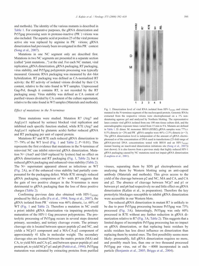
Fig. 1. Dimerization level of viral RNA isolated from HIV-1HXB2 and virionsmutated in the N-terminus segment of the nucleocapsid protein. Genomic RNAsextracted from the respective virions were electrophoresed on a 1% non-denaturing agarose gel and analyzed by Northern blotting. The representativelanes contain viral gRNA isolated from one 100 mm tissue culture dish, and theautoradiographic exposure times varied from 15 min to 9 h. Mutants are definedin Table 1. D: dimer. M: monomer. BH10 (HXB2) gRNA samples were 77%±0.5% dimeric (n=29) and PR− gRNA samples were 46%±2.5% dimeric (n=3).The gRNA dimerization level is independent of the amount of gRNA electro-phoresed or of the concentration of DNA used in transfections (25-fold range ofgRNA/proviral DNA concentrations tested with BH10 and an HIV-1HXB2mutant bearing an inactivated dimerization initiations site (Song et al., 2007))(not shown). It is also known from a previous study that highly reduced HIV-1gRNA packaging caused by NC mutations need not impair gRNA dimerization(Laughrea et al., 2001).
595J. Kafaie et al. / Virology 375 (2008) 592–610
and methods). The identity of the various mutants is described inTable 1. For comparative purposes, the gRNA dimerization andPr55gag processing seen in protease-inactive (PR−) virions wasalso included. The aspartic acid at position 25 of the viral proteaseactive site was replaced by arginine in PR− virions; gRNAdimerization had previously been investigated in this PR− context(Song et al., 2007).
Mutations in one NC segment only are described first.Mutations in two NC segments are presented in a separate section(called “joint mutations…”) at the end. For each NC mutant, viralreplication, gRNAdimerization, gRNApackaging, RT packaging,virus stability, and Pr55gag polyprotein processing was typicallymeasured. Genomic RNA packaging was measured by dot–blothybridization. RT packaging was defined as CA-normalized RTactivity: the RT activity of isolated virions divided by their CAcontent, relative to the ratio found in WT samples. UnprocessedGag-Pol, though it contains RT, is not recorded by the RTpackaging assay. Virus stability was defined as CA content ofpurified viruses divided by CA content of the culture supernatant,relative to the ratio found inWTsamples (Materials andmethods).
Effect of mutations in the N-terminus
Three mutations were studied. Mutation R7 (Arg7 andArgLys11 replaced by serines) blocked viral replication andinhibited each specific function studied, while R7E (Arg7 andArgLys11 replaced by glutamic acids) further reduced gRNAand RT packaging per unit of capsid protein.
Mutations R7 and R7E each reduced gRNA dimerization to77–79% of the WT level (Fig. 1 and Table 2; Pb0.01). Thisrepresents the first evidence that mutations in the N-terminus ofretroviral NC can inhibit retroviral gRNA dimerization. Muta-tion N+ (Asn5 and Gln9 replaced by lysines) had no effect ongRNA dimerization and RT packaging (Fig. 1, Table 2); but itreduced gRNA packaging and enhanced virus stability (Table 2).The N+ supernatant appeared almost as infectious as WT(Fig. 2A), as if the enhanced virus stability had partially com-pensated for the packaging defect. While R7E strongly reducedgRNA packaging, comparison of N+ with R7 suggests thatthe gain of two positive charges in the N-terminus is moredetrimental to gRNA packaging than the loss of three positivecharges (Table 2).
Confirming previous data also obtained with HIV-1HXB2produced by HeLa cells (Fu et al., 1994; Song et al., 2007), thegRNA isolated from PR− virions was 46% dimeric, i.e. 60% ofWT (Fig. 1 and Table 2). Therefore a mutation in NC coulda priori inhibit gRNA dimerization via impairing proteolyticmaturation of the HIV-1 Gag precursor polyproteins. The pro-teolytic processing of Pr55gag occurs in several steps denotedprimary, secondary, and tertiary cleavage events. The primarycleavage site is located between spacer peptide p2 and NC, andyields a NCp15 component and a MA-CA-p2 component ofapproximately 41 kDa in molecular weight. The secondarycleavage sites are located between the matrix protein (MA) andCA, to yield MA and CA-p2, and between spacer peptide p1 andprotein p6, to yield NCp7-p1 and p6 (Pettit et al., 1994). Pr55gagmaturation was estimated by extracting proteins from purified
viruses, separating them by SDS gel electrophoresis andanalysing them by Western blotting using an anti-capsidantibody (Materials and methods). This gives access to theyield of the cleavage between p2 and NC, MA and CA, and CAand p2. The absence of cleavage between NCp7 and p1 orbetween p1 and p6 had respectively no and little effect on gRNAdimerization (Kafaie et al., in preparation). Therefore the keyproteolytic blockages susceptible to inhibit gRNA dimerizationwere accessible in our Western blots.
The reduced gRNA dimerization in mutant R7 is unlikely tobe due to poor Pr55gag processing because Pr55gag was 73%processed (Fig. 3A). Interestingly, Pr55gag was only 60%processed in R7E without any further reduction in gRNA di-merization relative to R7 (Fig. 3A; Table 2). This suggests that alimited degree of incomplete Pr55gag processing has no impacton gRNA dimerization, or that replacing basic residues byacidic residues has less direct influence on dimerization thanreplacing them by neutral ones. The first possibility seems morelikely: presumably, full gRNA dimerization requires no more,and possibly much less, than one or two thousand processedPr55gag per virus, out of the ∼4000 incorporated in eachparticle (Benjamin et al., 2005; Briggs et al., 2004).
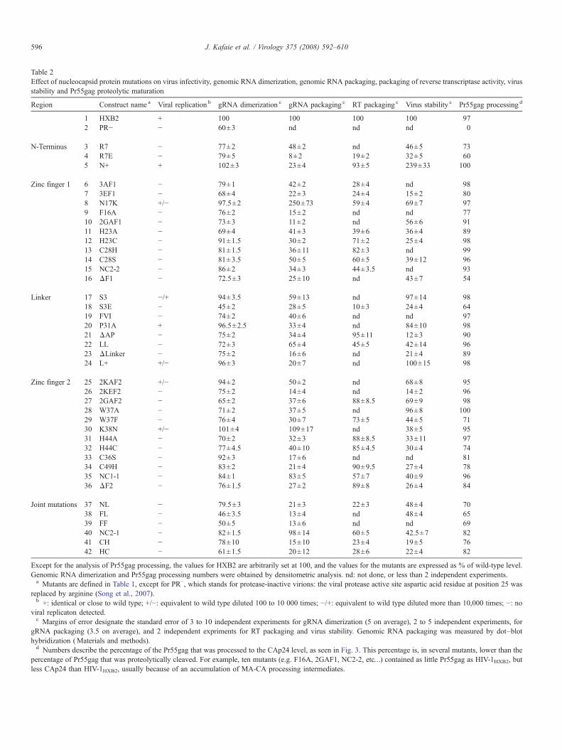
Table 2Effect of nucleocapsid protein mutations on virus infectivity, genomic RNA dimerization, genomic RNA packaging, packaging of reverse transcriptase activity, virusstability and Pr55gag proteolytic maturation
Region Construct name a Viral replication b gRNA dimerization c gRNA packaging c RT packaging c Virus stability c Pr55gag processing d
1 HXB2 + 100 100 100 100 972 PR− − 60±3 nd nd nd 0
N-Terminus 3 R7 − 77±2 48±2 nd 46±5 734 R7E − 79±5 8±2 19±2 32±5 605 N+ + 102±3 23±4 93±5 239±33 100
Zinc finger 1 6 3AF1 − 79±1 42±2 28±4 nd 987 3EF1 − 68±4 22±3 24±4 15±2 808 N17K +/− 97.5±2 250±73 59±4 69±7 979 F16A − 76±2 15±2 nd nd 7710 2GAF1 − 73±3 11±2 nd 56±6 9111 H23A − 69±4 41±3 39±6 36±4 8912 H23C − 91±1.5 30±2 71±2 25±4 9813 C28H − 81±1.5 36±11 82±3 nd 9914 C28S − 81±3.5 50±5 60±5 39±12 9615 NC2-2 − 86±2 34±3 44±3.5 nd 9316 ΔF1 − 72.5±3 25±10 nd 43±7 54
Linker 17 S3 −/+ 94±3.5 59±13 nd 97±14 9818 S3E − 45±2 28±5 10±3 24±4 6419 FVI − 74±2 40±6 nd nd 9720 P31A + 96.5±2.5 33±4 nd 84±10 9821 ΔAP − 75±2 34±4 95±11 12±3 9022 LL − 72±3 65±4 45±5 42±14 9623 ΔLinker − 75±2 16±6 nd 21±4 8924 L+ +/− 96±3 20±7 nd 100±15 98
Zinc finger 2 25 2KAF2 +/− 94±2 50±2 nd 68±8 9526 2KEF2 − 75±2 14±4 nd 14±2 9627 2GAF2 − 65±2 37±6 88±8.5 69±9 9828 W37A − 71±2 37±5 nd 96±8 10029 W37F − 76±4 30±7 73±5 44±5 7130 K38N +/− 101±4 109±17 nd 38±5 9531 H44A − 70±2 32±3 88±8.5 33±11 9732 H44C − 77±4.5 40±10 85±4.5 30±4 7433 C36S − 92±3 17±6 nd nd 8134 C49H − 83±2 21±4 90±9.5 27±4 7835 NC1-1 − 84±1 83±5 57±7 40±9 9636 ΔF2 − 76±1.5 27±2 89±8 26±4 84
Joint mutations 37 NL − 79.5±3 21±3 22±3 48±4 7038 FL − 46±3.5 13±4 nd 48±4 6539 FF − 50±5 13±6 nd nd 6940 NC2-1 − 82±1.5 98±14 60±5 42.5±7 8241 CH − 78±10 15±10 23±4 19±5 7642 HC − 61±1.5 20±12 28±6 22±4 82
Except for the analysis of Pr55gag processing, the values for HXB2 are arbitrarily set at 100, and the values for the mutants are expressed as % of wild-type level.Genomic RNA dimerization and Pr55gag processing numbers were obtained by densitometric analysis. nd: not done, or less than 2 independent experiments.a Mutants are defined in Table 1, except for PR−, which stands for protease-inactive virions: the viral protease active site aspartic acid residue at position 25 was
replaced by arginine (Song et al., 2007).b +: identical or close to wild type; +/−: equivalent to wild type diluted 100 to 10 000 times; −/+: equivalent to wild type diluted more than 10,000 times; −: no
viral replicaton detected.c Margins of error designate the standard error of 3 to 10 independent experiments for gRNA dimerization (5 on average), 2 to 5 independent experiments, for
gRNA packaging (3.5 on average), and 2 independent expriments for RT packaging and virus stability. Genomic RNA packaging was measured by dot–blothybridization (Materials and methods).d Numbers describe the percentage of the Pr55gag that was processed to the CAp24 level, as seen in Fig. 3. This percentage is, in several mutants, lower than the
percentage of Pr55gag that was proteolytically cleaved. For example, ten mutants (e.g. F16A, 2GAF1, NC2-2, etc…) contained as little Pr55gag as HIV-1HXB2, butless CAp24 than HIV-1HXB2, usually because of an accumulation of MA-CA processing intermediates.
596 J. Kafaie et al. / Virology 375 (2008) 592–610
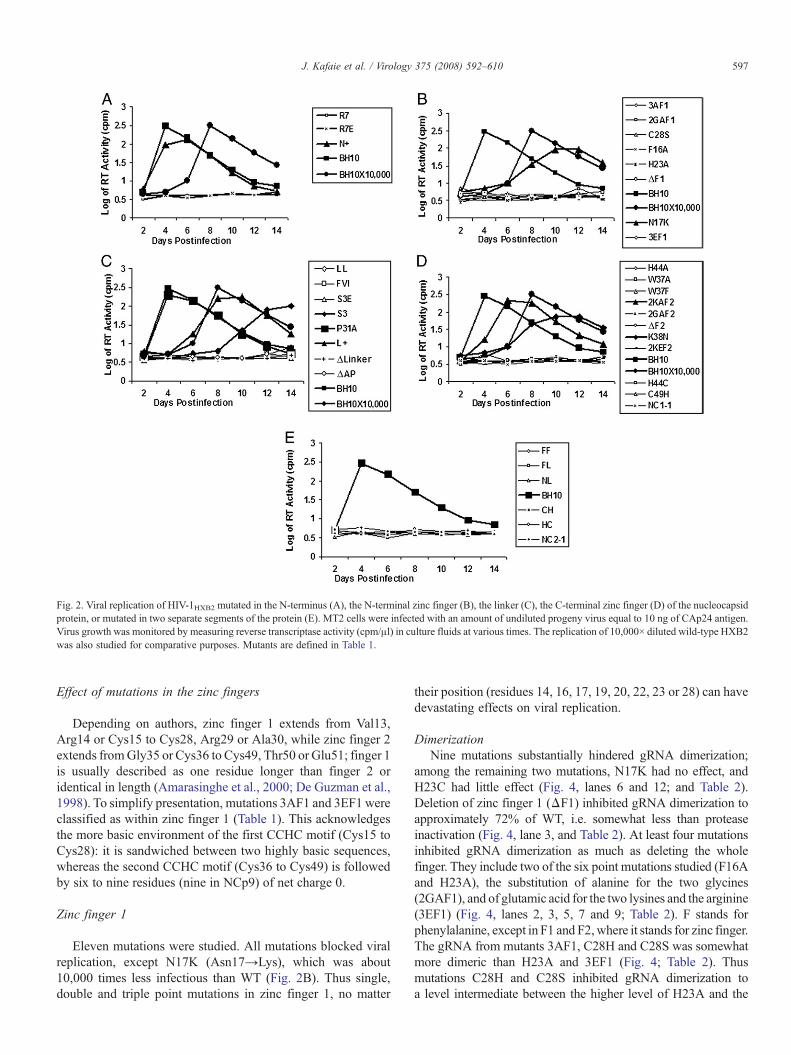
Fig. 2. Viral replication of HIV-1HXB2 mutated in the N-terminus (A), the N-terminal zinc finger (B), the linker (C), the C-terminal zinc finger (D) of the nucleocapsidprotein, or mutated in two separate segments of the protein (E). MT2 cells were infected with an amount of undiluted progeny virus equal to 10 ng of CAp24 antigen.Virus growth was monitored by measuring reverse transcriptase activity (cpm/μl) in culture fluids at various times. The replication of 10,000× diluted wild-type HXB2was also studied for comparative purposes. Mutants are defined in Table 1.
597J. Kafaie et al. / Virology 375 (2008) 592–610
Effect of mutations in the zinc fingers
Depending on authors, zinc finger 1 extends from Val13,Arg14 or Cys15 to Cys28, Arg29 or Ala30, while zinc finger 2extends fromGly35 or Cys36 to Cys49, Thr50 or Glu51; finger 1is usually described as one residue longer than finger 2 oridentical in length (Amarasinghe et al., 2000; De Guzman et al.,1998). To simplify presentation, mutations 3AF1 and 3EF1 wereclassified as within zinc finger 1 (Table 1). This acknowledgesthe more basic environment of the first CCHC motif (Cys15 toCys28): it is sandwiched between two highly basic sequences,whereas the second CCHC motif (Cys36 to Cys49) is followedby six to nine residues (nine in NCp9) of net charge 0.
Zinc finger 1
Eleven mutations were studied. All mutations blocked viralreplication, except N17K (Asn17→Lys), which was about10,000 times less infectious than WT (Fig. 2B). Thus single,double and triple point mutations in zinc finger 1, no matter
their position (residues 14, 16, 17, 19, 20, 22, 23 or 28) can havedevastating effects on viral replication.
DimerizationNine mutations substantially hindered gRNA dimerization;
among the remaining two mutations, N17K had no effect, andH23C had little effect (Fig. 4, lanes 6 and 12; and Table 2).Deletion of zinc finger 1 (ΔF1) inhibited gRNA dimerization toapproximately 72% of WT, i.e. somewhat less than proteaseinactivation (Fig. 4, lane 3, and Table 2). At least four mutationsinhibited gRNA dimerization as much as deleting the wholefinger. They include two of the six point mutations studied (F16Aand H23A), the substitution of alanine for the two glycines(2GAF1), and of glutamic acid for the two lysines and the arginine(3EF1) (Fig. 4, lanes 2, 3, 5, 7 and 9; Table 2). F stands forphenylalanine, except in F1 and F2,where it stands for zinc finger.The gRNA from mutants 3AF1, C28H and C28S was somewhatmore dimeric than H23A and 3EF1 (Fig. 4; Table 2). Thusmutations C28H and C28S inhibited gRNA dimerization toa level intermediate between the higher level of H23A and the
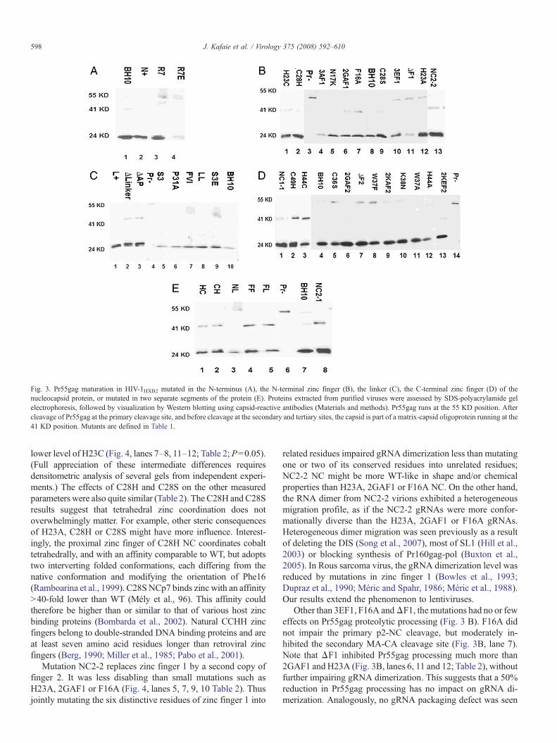
Fig. 3. Pr55gag maturation in HIV-1HXB2 mutated in the N-terminus (A), the N-terminal zinc finger (B), the linker (C), the C-terminal zinc finger (D) of thenucleocapsid protein, or mutated in two separate segments of the protein (E). Proteins extracted from purified viruses were assessed by SDS-polyacrylamide gelelectrophoresis, followed by visualization by Western blotting using capsid-reactive antibodies (Materials and methods). Pr55gag runs at the 55 KD position. Aftercleavage of Pr55gag at the primary cleavage site, and before cleavage at the secondary and tertiary sites, the capsid is part of a matrix-capsid oligoprotein running at the41 KD position. Mutants are defined in Table 1.
598 J. Kafaie et al. / Virology 375 (2008) 592–610
lower level of H23C (Fig. 4, lanes 7–8, 11–12; Table 2; P=0.05).(Full appreciation of these intermediate differences requiresdensitometric analysis of several gels from independent experi-ments.) The effects of C28H and C28S on the other measuredparameters were also quite similar (Table 2). The C28H and C28Sresults suggest that tetrahedral zinc coordination does notoverwhelmingly matter. For example, other steric consequencesof H23A, C28H or C28S might have more influence. Interest-ingly, the proximal zinc finger of C28H NC coordinates cobalttetrahedrally, and with an affinity comparable to WT, but adoptstwo interverting folded conformations, each differing from thenative conformation and modifying the orientation of Phe16(Ramboarina et al., 1999). C28SNCp7 binds zinc with an affinityN40-fold lower than WT (Mély et al., 96). This affinity couldtherefore be higher than or similar to that of various host zincbinding proteins (Bombarda et al., 2002). Natural CCHH zincfingers belong to double-stranded DNA binding proteins and areat least seven amino acid residues longer than retroviral zincfingers (Berg, 1990; Miller et al., 1985; Pabo et al., 2001).
Mutation NC2-2 replaces zinc finger 1 by a second copy offinger 2. It was less disabling than small mutations such asH23A, 2GAF1 or F16A (Fig. 4, lanes 5, 7, 9, 10 Table 2). Thusjointly mutating the six distinctive residues of zinc finger 1 into
related residues impaired gRNA dimerization less than mutatingone or two of its conserved residues into unrelated residues;NC2-2 NC might be more WT-like in shape and/or chemicalproperties than H23A, 2GAF1 or F16A NC. On the other hand,the RNA dimer from NC2-2 virions exhibited a heterogeneousmigration profile, as if the NC2-2 gRNAs were more confor-mationally diverse than the H23A, 2GAF1 or F16A gRNAs.Heterogeneous dimer migration was seen previously as a resultof deleting the DIS (Song et al., 2007), most of SL1 (Hill et al.,2003) or blocking synthesis of Pr160gag-pol (Buxton et al.,2005). In Rous sarcoma virus, the gRNA dimerization level wasreduced by mutations in zinc finger 1 (Bowles et al., 1993;Dupraz et al., 1990; Méric and Spahr, 1986; Méric et al., 1988).Our results extend the phenomenon to lentiviruses.
Other than 3EF1, F16A andΔF1, the mutations had no or feweffects on Pr55gag proteolytic processing (Fig. 3 B). F16A didnot impair the primary p2-NC cleavage, but moderately in-hibited the secondary MA-CA cleavage site (Fig. 3B, lane 7).Note that ΔF1 inhibited Pr55gag processing much more than2GAF1 and H23A (Fig. 3B, lanes 6, 11 and 12; Table 2), withoutfurther impairing gRNA dimerization. This suggests that a 50%reduction in Pr55gag processing has no impact on gRNA di-merization. Analogously, no gRNA packaging defect was seen
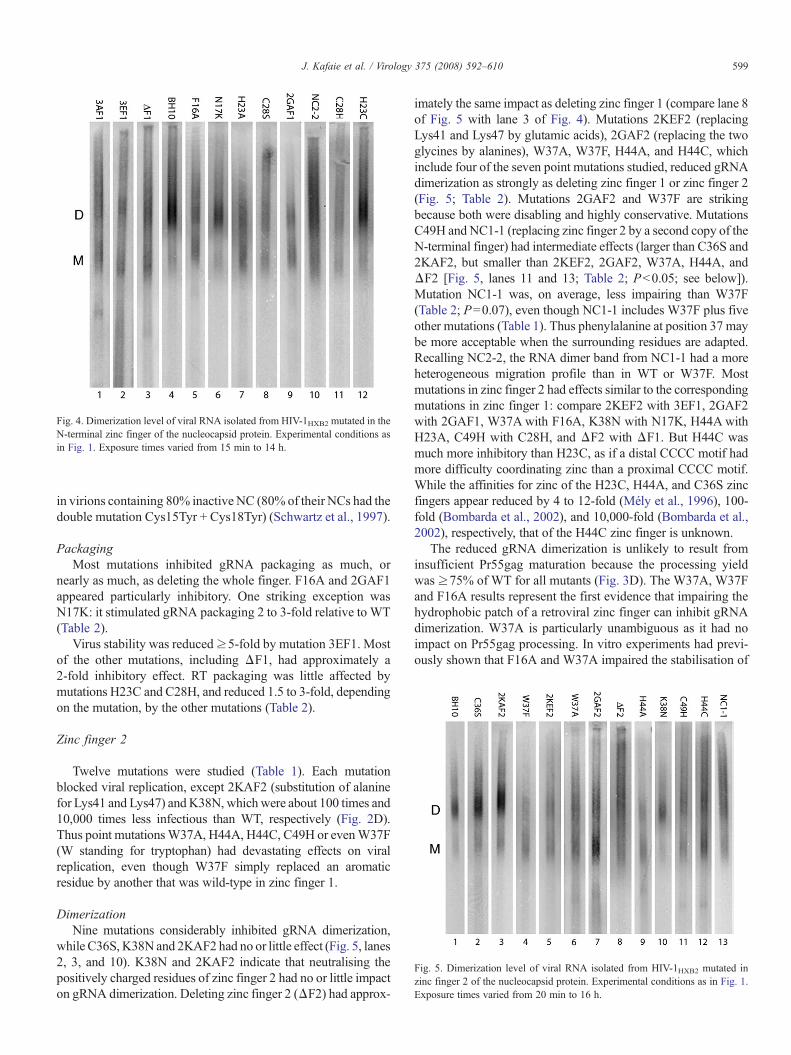
Fig. 4. Dimerization level of viral RNA isolated from HIV-1HXB2 mutated in theN-terminal zinc finger of the nucleocapsid protein. Experimental conditions asin Fig. 1. Exposure times varied from 15 min to 14 h.
Fig. 5. Dimerization level of viral RNA isolated from HIV-1HXB2 mutated inzinc finger 2 of the nucleocapsid protein. Experimental conditions as in Fig. 1.Exposure times varied from 20 min to 16 h.
599J. Kafaie et al. / Virology 375 (2008) 592–610
in virions containing 80% inactive NC (80% of their NCs had thedouble mutation Cys15Tyr + Cys18Tyr) (Schwartz et al., 1997).
PackagingMost mutations inhibited gRNA packaging as much, or
nearly as much, as deleting the whole finger. F16A and 2GAF1appeared particularly inhibitory. One striking exception wasN17K: it stimulated gRNA packaging 2 to 3-fold relative to WT(Table 2).
Virus stability was reduced≥5-fold by mutation 3EF1. Mostof the other mutations, including ΔF1, had approximately a2-fold inhibitory effect. RT packaging was little affected bymutations H23C and C28H, and reduced 1.5 to 3-fold, dependingon the mutation, by the other mutations (Table 2).
Zinc finger 2
Twelve mutations were studied (Table 1). Each mutationblocked viral replication, except 2KAF2 (substitution of alaninefor Lys41 and Lys47) andK38N, which were about 100 times and10,000 times less infectious than WT, respectively (Fig. 2D).Thus point mutationsW37A, H44A, H44C, C49H or evenW37F(W standing for tryptophan) had devastating effects on viralreplication, even though W37F simply replaced an aromaticresidue by another that was wild-type in zinc finger 1.
DimerizationNine mutations considerably inhibited gRNA dimerization,
while C36S, K38N and 2KAF2 had no or little effect (Fig. 5, lanes2, 3, and 10). K38N and 2KAF2 indicate that neutralising thepositively charged residues of zinc finger 2 had no or little impacton gRNA dimerization. Deleting zinc finger 2 (ΔF2) had approx-
imately the same impact as deleting zinc finger 1 (compare lane 8of Fig. 5 with lane 3 of Fig. 4). Mutations 2KEF2 (replacingLys41 and Lys47 by glutamic acids), 2GAF2 (replacing the twoglycines by alanines), W37A, W37F, H44A, and H44C, whichinclude four of the seven point mutations studied, reduced gRNAdimerization as strongly as deleting zinc finger 1 or zinc finger 2(Fig. 5; Table 2). Mutations 2GAF2 and W37F are strikingbecause both were disabling and highly conservative. MutationsC49H and NC1-1 (replacing zinc finger 2 by a second copy of theN-terminal finger) had intermediate effects (larger than C36S and2KAF2, but smaller than 2KEF2, 2GAF2, W37A, H44A, andΔF2 [Fig. 5, lanes 11 and 13; Table 2; Pb0.05; see below]).Mutation NC1-1 was, on average, less impairing than W37F(Table 2; P=0.07), even though NC1-1 includes W37F plus fiveother mutations (Table 1). Thus phenylalanine at position 37 maybe more acceptable when the surrounding residues are adapted.Recalling NC2-2, the RNA dimer band from NC1-1 had a moreheterogeneous migration profile than in WT or W37F. Mostmutations in zinc finger 2 had effects similar to the correspondingmutations in zinc finger 1: compare 2KEF2 with 3EF1, 2GAF2with 2GAF1, W37Awith F16A, K38N with N17K, H44AwithH23A, C49H with C28H, and ΔF2 with ΔF1. But H44C wasmuch more inhibitory than H23C, as if a distal CCCC motif hadmore difficulty coordinating zinc than a proximal CCCC motif.While the affinities for zinc of the H23C, H44A, and C36S zincfingers appear reduced by 4 to 12-fold (Mély et al., 1996), 100-fold (Bombarda et al., 2002), and 10,000-fold (Bombarda et al.,2002), respectively, that of the H44C zinc finger is unknown.
The reduced gRNA dimerization is unlikely to result frominsufficient Pr55gag maturation because the processing yieldwas ≥75% of WT for all mutants (Fig. 3D). The W37A, W37Fand F16A results represent the first evidence that impairing thehydrophobic patch of a retroviral zinc finger can inhibit gRNAdimerization. W37A is particularly unambiguous as it had noimpact on Pr55gag processing. In vitro experiments had previ-ously shown that F16A and W37A impaired the stabilisation of
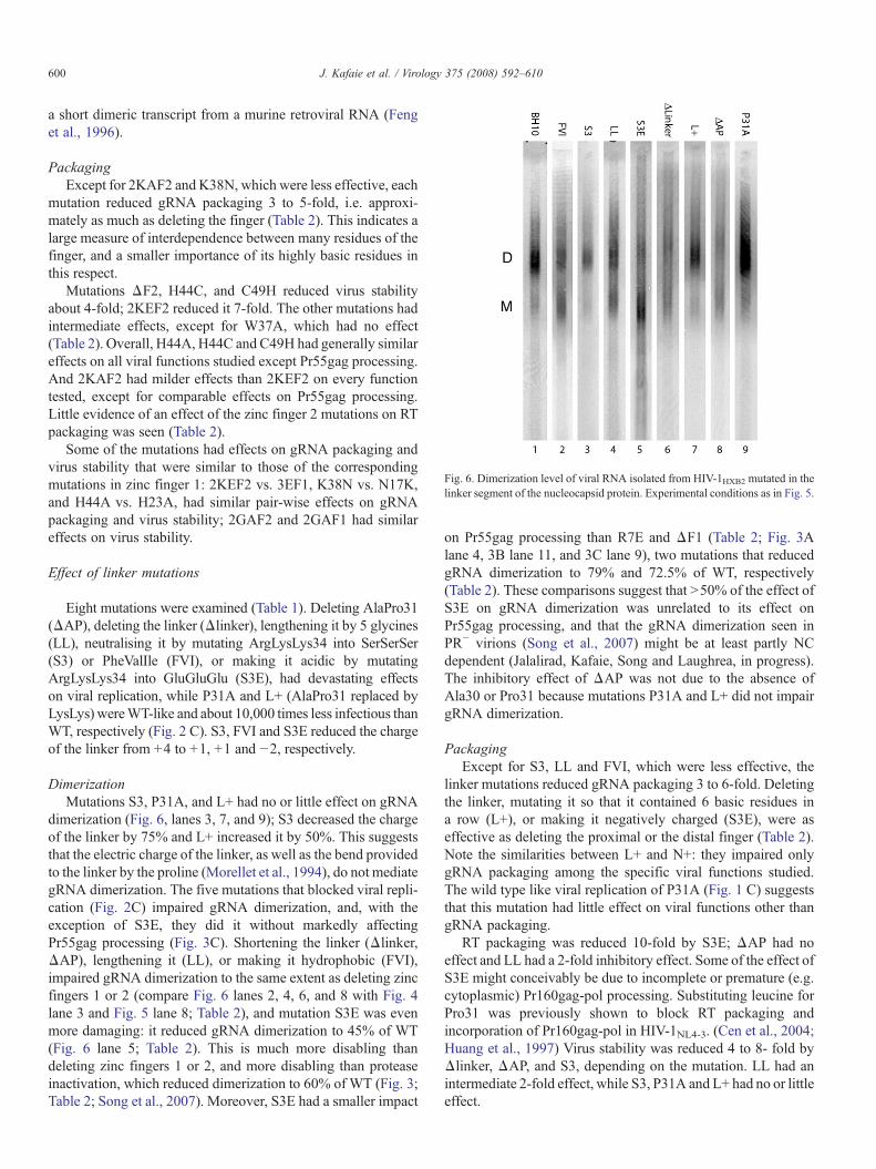
Fig. 6. Dimerization level of viral RNA isolated from HIV-1HXB2 mutated in thelinker segment of the nucleocapsid protein. Experimental conditions as in Fig. 5.
600 J. Kafaie et al. / Virology 375 (2008) 592–610
a short dimeric transcript from a murine retroviral RNA (Fenget al., 1996).
PackagingExcept for 2KAF2 and K38N, which were less effective, each
mutation reduced gRNA packaging 3 to 5-fold, i.e. approxi-mately as much as deleting the finger (Table 2). This indicates alarge measure of interdependence between many residues of thefinger, and a smaller importance of its highly basic residues inthis respect.
Mutations ΔF2, H44C, and C49H reduced virus stabilityabout 4-fold; 2KEF2 reduced it 7-fold. The other mutations hadintermediate effects, except for W37A, which had no effect(Table 2). Overall, H44A, H44C and C49H had generally similareffects on all viral functions studied except Pr55gag processing.And 2KAF2 had milder effects than 2KEF2 on every functiontested, except for comparable effects on Pr55gag processing.Little evidence of an effect of the zinc finger 2 mutations on RTpackaging was seen (Table 2).
Some of the mutations had effects on gRNA packaging andvirus stability that were similar to those of the correspondingmutations in zinc finger 1: 2KEF2 vs. 3EF1, K38N vs. N17K,and H44A vs. H23A, had similar pair-wise effects on gRNApackaging and virus stability; 2GAF2 and 2GAF1 had similareffects on virus stability.
Effect of linker mutations
Eight mutations were examined (Table 1). Deleting AlaPro31(ΔAP), deleting the linker (Δlinker), lengthening it by 5 glycines(LL), neutralising it by mutating ArgLysLys34 into SerSerSer(S3) or PheValIle (FVI), or making it acidic by mutatingArgLysLys34 into GluGluGlu (S3E), had devastating effectson viral replication, while P31A and L+ (AlaPro31 replaced byLysLys) wereWT-like and about 10,000 times less infectious thanWT, respectively (Fig. 2 C). S3, FVI and S3E reduced the chargeof the linker from +4 to +1, +1 and −2, respectively.
DimerizationMutations S3, P31A, and L+ had no or little effect on gRNA
dimerization (Fig. 6, lanes 3, 7, and 9); S3 decreased the chargeof the linker by 75% and L+ increased it by 50%. This suggeststhat the electric charge of the linker, as well as the bend providedto the linker by the proline (Morellet et al., 1994), do not mediategRNA dimerization. The five mutations that blocked viral repli-cation (Fig. 2C) impaired gRNA dimerization, and, with theexception of S3E, they did it without markedly affectingPr55gag processing (Fig. 3C). Shortening the linker (Δlinker,ΔAP), lengthening it (LL), or making it hydrophobic (FVI),impaired gRNA dimerization to the same extent as deleting zincfingers 1 or 2 (compare Fig. 6 lanes 2, 4, 6, and 8 with Fig. 4lane 3 and Fig. 5 lane 8; Table 2), and mutation S3E was evenmore damaging: it reduced gRNA dimerization to 45% of WT(Fig. 6 lane 5; Table 2). This is much more disabling thandeleting zinc fingers 1 or 2, and more disabling than proteaseinactivation, which reduced dimerization to 60% of WT (Fig. 3;Table 2; Song et al., 2007). Moreover, S3E had a smaller impact
on Pr55gag processing than R7E and ΔF1 (Table 2; Fig. 3Alane 4, 3B lane 11, and 3C lane 9), two mutations that reducedgRNA dimerization to 79% and 72.5% of WT, respectively(Table 2). These comparisons suggest that N50% of the effect ofS3E on gRNA dimerization was unrelated to its effect onPr55gag processing, and that the gRNA dimerization seen inPR− virions (Song et al., 2007) might be at least partly NCdependent (Jalalirad, Kafaie, Song and Laughrea, in progress).The inhibitory effect of ΔAP was not due to the absence ofAla30 or Pro31 because mutations P31A and L+ did not impairgRNA dimerization.
PackagingExcept for S3, LL and FVI, which were less effective, the
linker mutations reduced gRNA packaging 3 to 6-fold. Deletingthe linker, mutating it so that it contained 6 basic residues ina row (L+), or making it negatively charged (S3E), were aseffective as deleting the proximal or the distal finger (Table 2).Note the similarities between L+ and N+: they impaired onlygRNA packaging among the specific viral functions studied.The wild type like viral replication of P31A (Fig. 1 C) suggeststhat this mutation had little effect on viral functions other thangRNA packaging.
RT packaging was reduced 10-fold by S3E; ΔAP had noeffect and LL had a 2-fold inhibitory effect. Some of the effect ofS3E might conceivably be due to incomplete or premature (e.g.cytoplasmic) Pr160gag-pol processing. Substituting leucine forPro31 was previously shown to block RT packaging andincorporation of Pr160gag-pol in HIV-1NL4-3. (Cen et al., 2004;Huang et al., 1997) Virus stability was reduced 4 to 8- fold byΔlinker, ΔAP, and S3, depending on the mutation. LL had anintermediate 2-fold effect, while S3, P31A and L+ had no or littleeffect.
601J. Kafaie et al. / Virology 375 (2008) 592–610
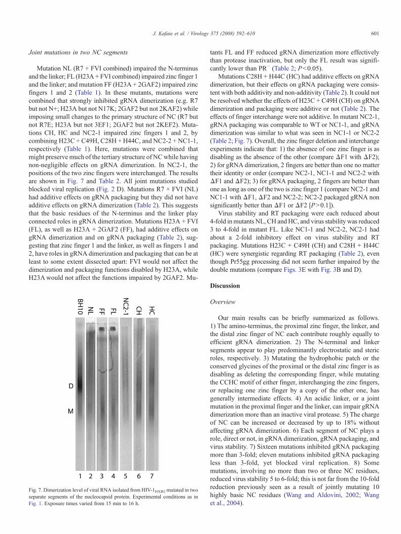
Joint mutations in two NC segments
Mutation NL (R7 + FVI combined) impaired the N-terminusand the linker; FL (H23A+ FVI combined) impaired zinc finger 1and the linker; and mutation FF (H23A + 2GAF2) impaired zincfingers 1 and 2 (Table 1). In these mutants, mutations werecombined that strongly inhibited gRNA dimerization (e.g. R7but not N+; H23A but not N17K; 2GAF2 but not 2KAF2) whileimposing small changes to the primary structure of NC (R7 butnot R7E; H23A but not 3EF1; 2GAF2 but not 2KEF2). Muta-tions CH, HC and NC2-1 impaired zinc fingers 1 and 2, bycombining H23C + C49H, C28H + H44C, and NC2-2 + NC1-1,respectively (Table 1). Here, mutations were combined thatmight preservemuch of the tertiary structure of NCwhile havingnon-negligible effects on gRNA dimerization. In NC2-1, thepositions of the two zinc fingers were interchanged. The resultsare shown in Fig. 7 and Table 2. All joint mutations studiedblocked viral replication (Fig. 2 D). Mutations R7 + FVI (NL)had additive effects on gRNA packaging but they did not haveadditive effects on gRNA dimerization (Table 2). This suggeststhat the basic residues of the N-terminus and the linker playconnected roles in gRNA dimerization. Mutations H23A + FVI(FL), as well as H23A + 2GAF2 (FF), had additive effects ongRNA dimerization and on gRNA packaging (Table 2), sug-gesting that zinc finger 1 and the linker, as well as fingers 1 and2, have roles in gRNA dimerization and packaging that can be atleast to some extent dissected apart: FVI would not affect thedimerization and packaging functions disabled by H23A, whileH23Awould not affect the functions impaired by 2GAF2. Mu-
Fig. 7. Dimerization level of viral RNA isolated from HIV-1HXB2 mutated in twoseparate segments of the nucleocapsid protein. Experimental conditions as inFig. 1. Exposure times varied from 15 min to 16 h.
tants FL and FF reduced gRNA dimerization more effectivelythan protease inactivation, but only the FL result was signifi-cantly lower than PR− (Table 2; Pb0.05).
Mutations C28H + H44C (HC) had additive effects on gRNAdimerization, but their effects on gRNA packaging were consis-tent with both additivity and non-additivity (Table 2). It could notbe resolved whether the effects of H23C + C49H (CH) on gRNAdimerization and packaging were additive or not (Table 2). Theeffects of finger interchange were not additive. In mutant NC2-1,gRNA packaging was comparable to WT or NC1-1, and gRNAdimerization was similar to what was seen in NC1-1 or NC2-2(Table 2; Fig. 7). Overall, the zinc finger deletion and interchangeexperiments indicate that: 1) the absence of one zinc finger is asdisabling as the absence of the other (compare ΔF1 with ΔF2);2) for gRNA dimerization, 2 fingers are better than one no mattertheir identity or order (compare NC2-1, NC1-1 and NC2-2 withΔF1 and ΔF2); 3) for gRNA packaging, 2 fingers are better thanone as long as one of the two is zinc finger 1 (compare NC2-1 andNC1-1 withΔF1,ΔF2 and NC2-2; NC2-2 packaged gRNA nonsignificantly better than ΔF1 or ΔF2 [PN0.1]).
Virus stability and RT packaging were each reduced about4-fold inmutants NL, CH andHC, and virus stability was reduced3 to 4-fold in mutant FL. Like NC1-1 and NC2-2, NC2-1 hadabout a 2-fold inhibitory effect on virus stability and RTpackaging. Mutations H23C + C49H (CH) and C28H + H44C(HC) were synergistic regarding RT packaging (Table 2), eventhough Pr55gg processing did not seem further impaired by thedouble mutations (compare Figs. 3E with Fig. 3B and D).
Discussion
Overview
Our main results can be briefly summarized as follows.1) The amino-terminus, the proximal zinc finger, the linker, andthe distal zinc finger of NC each contribute roughly equally toefficient gRNA dimerization. 2) The N-terminal and linkersegments appear to play predominantly electrostatic and stericroles, respectively. 3) Mutating the hydrophobic patch or theconserved glycines of the proximal or the distal zinc finger is asdisabling as deleting the corresponding finger, while mutatingthe CCHC motif of either finger, interchanging the zinc fingers,or replacing one zinc finger by a copy of the other one, hasgenerally intermediate effects. 4) An acidic linker, or a jointmutation in the proximal finger and the linker, can impair gRNAdimerization more than an inactive viral protease. 5) The chargeof NC can be increased or decreased by up to 18% withoutaffecting gRNA dimerization. 6) Each segment of NC plays arole, direct or not, in gRNA dimerization, gRNA packaging, andvirus stability. 7) Sixteen mutations inhibited gRNA packagingmore than 3-fold; eleven mutations inhibited gRNA packagingless than 3-fold, yet blocked viral replication. 8) Somemutations, involving no more than two or three NC residues,reduced virus stability 5 to 6-fold; this is not far from the 10-foldreduction previously seen as a result of jointly mutating 10highly basic NC residues (Wang and Aldovini, 2002; Wanget al., 2004).
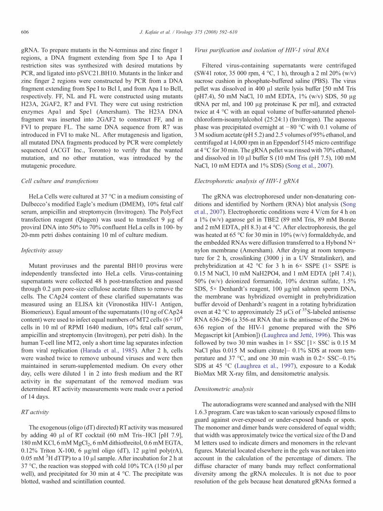
Table 3Comparative effect of NC mutations on gRNA packaging in HIV-1HXB2, HIV-1NL4-3, SIV and Rous sarcoma virus a
Mutation b Retroviruses
HIV-1HXB2 HIV-1NL4-3 SIV Rous sarcomavirus
1 R7 48 50 c; 22 d
2 3AF1 42; 13 e
3 N17K 250; 1300 f
4 F16A 15; 15 g
5 H23A 41; 20 g
6 H23C 30 15 h; 10 i; 110 j 4 k
7 C28H 36 32 j 20 k
8 C28S 50; 15 g 85 l
9 NC2-2 34; 15 g b15m 70 n
10 ΔF1 25 3 o; 10 i 99 l; 85 p ≤20 q
11 S3 59 20 d
12 S3E 28; 12 r
13 H44A 32; 15 g
14 H44C 40 167 j 2 k
15 C36S 17 6 o
16 C49H 21 151 j 98 k
17 NC1-1 83 70m 10–15 n
18 ΔF2 27 45 l; 65 p ≤20 q
19 NC2-1 98 b15m 50 n
20 CH 15 78 j
21 HC 20 5 j
a gRNA packaging is expressed as percentage of wild-type level. Unlessotherwise stated, HIV-1HXB2 and HIV-1NL4-3 were produced by HeLa cells,SIVs by 293T cells, and Rous sarcoma virus by chicken embryo fibroblasts.Unless otherwise stated, packaging per unit of capsid protein is indicated.b For each mutation, the first HIV-1HXB2 number quoted comes from Table 2.
Presented are mutations whose effect (on packaging) was studied by us andother laboratories. If a similar Table had been created for gRNA dimerization, 7numbers would have come from outside this paper: gRNA dimerizationpercentages for Rous sarcoma virus (Méric et al., 1988; Bowles et al., 1993) andfor R7 and S3 in HIV-1NL4-3 (Laughrea et al., 2001).c From Berthoux et al. (1997).d From Cen et al. (1999). Viruses produced by Cos7 cells.e From Poon et al. (1996). Viruses produced by Cos-1 cells.f FromMark-Danieli et al. (2005). Viruses produced by 293T cells. Packaging
measured per unit of reverse transcriptase activity.g From Dorfman et al. (1993).h From Déméné et al. (1994). Viruses produced by Cos7 cells.i From Tanchou et al. (1998).j From Gorelick et al. (1999a). Viruses produced by 293T cells.k From Fu et al. (2007). SIVagm studied. The exact residue numbers differ
from HIV-1.l From Gorelick et al. (1999b). SIVmne studied. The first 4 residues of zinc
fingers 1 or 2 were deleted, respectively. Mutation C25S, corresponding to C28Sin HIV-1, was studied.m From Gorelick et al. (1993).n From Bowles et al. (1993).o From Gorelick et al. (1990). Packaging measured per unit of reverse
transcriptase activity.p From Yovandich et al. (2001). SIVmne studied. The first 4 residues of zinc
fingers 1 or 2 were deleted, respectively. Packaging measured per unit of reversetranscriptase activity.q From Méric et al. (1988).r From Sheng et al. (1997).
602 J. Kafaie et al. / Virology 375 (2008) 592–610
Regarding gRNA dimerization, the effects of 38 and 40 ofthe NC mutations were studied for the first time in HIV-1 and inHIV-1HXB2, respectively. Regarding virus stability and RT pack-aging, the effects of all mutations were novel. Regarding gRNApackaging, the effects of 20 and 32 of the mutations were studiedfor the first time in HIV-1 and in HIV-1HXB2, respectively. Theeffect on packaging of 8 mutations, though previously studied inHIV-1HXB2 (Table 3), was examined for comparative purposes.The effect on gRNA packaging of 13 of the mutations hadpreviously been studied in HIV-1NL4-3. (Table 3). Knowing theeffect of these mutations in both HIV-1NL4-3 and HIV-1HXB2 canclarify the influence of genetic context on phenotype. Each NCsegment appeared similarly important for gRNA packaging.
NC is implicated in late infection events (e.g. gRNAdimerization, gRNA packaging, Pr55gag processing and virusstability, all studied here) and in early infection events such asreverse transcription of gRNA and proviral DNA integrationinto the host genome (e.g. Buckman et al., 2003; Levin et al.,2005). Some NC mutations might accordingly have no effect onlate infection events, yet impair or block viral replication. Thiswas not seen here. Each of the 40 mutations impaired one orseveral of the late functions studied. However, mutations P31Aand N+ had mild effects on late infection events and little impacton viral replication: they are thus unlikely to inhibit earlyinfection events in HIV-1HXB2. With the exception of borderline2KAF2, the 38 other mutations probably inhibit both early andlate infection events because their impact on viral replicationwas inordinately large relative to their effect on late infectionevents. For example, mutation NC2-1 blocked viral replicationand had relatively mild effects on late infection events (Table 2),suggesting that it impairs early infection events, which it does(Guo et al., 2002; Williams et al., 2002).
Breaking the protease inactivation “barrier”
Mutations FF and HC impaired gRNA dimerization asstrongly as viral protease inactivation (PR−), while S3E and FLwere significantly more disabling. S3E and FL are the onlyretroviral NC mutations known to inhibit gRNA dimerizationmore strongly than protease inactivation. This suggests thatthe gRNA dimerization seen in PR− virions is stimulated byunprocessed viral proteins. For example, the NC component ofPr55gag may stimulate viral RNA dimerization. Alternatively,S3E and FL may obstruct a gRNA dimerization site in cis (at theRNA level), or in trans, e.g. via aberrant binding of maturemutant NC to gRNA. The cis option is unlikely because the NC-coding sequence can be deleted (as long as the missing proteinsare provided in trans) without reducing gRNA dimerization ormaking the dimers thermolabile (Sakuragi and Panganiban,1997); other experiments indicate that, though there may begRNA interactions sites downstream of the 5′ untranslated re-gion (Song et al., in press), the NC region of gRNA is not one ofthem (Sakuragi et al., 2002).
None of the mutants reduced gRNA dimerization close tozero. It cannot be ruled out that up to 40% of the dimeric RNAsseen in the isolated virions may have accumulated sponta-neously (i.e. in a protein-independent manner). Inactivating the
DIS or the viral protease impairs gRNA dimerization at leasttwo and five times more, respectively, in newly released virionsthan in grown-up, i.e. predominantly ≥10 h old, virions (Song
603J. Kafaie et al. / Virology 375 (2008) 592–610
et al., 2007). It is possible that mutations in NC may also provemore impairing in freshly released viruses.
Losing or gaining one or two electric charges has no impact ongRNA dimerization but a generally large impact on gRNApackaging
Genomic RNA dimerization was unaffected by altering thecharge of NC by up to 18% (removing or adding one or twohighly basic residues); removing three positive charges wasgenerally disabling (R7 and 3AF1, but not S3); removing≥ fourwas always disabling. Out of seven mutations that had no effecton gRNA dimerization, six involved a change in electric charge:mutations N+ and L+ added two positive charges each to NC;2KAF2 and S3 subtracted two and three charges, respectively,while N17K and K38N added and subtracted one charge,respectively (the seventh mutation was P31A). The implicatedresidues (Asn5 + Gln9, Asn17, AlaPro31, ArgLysLys34, Lys38,and Lys41 + Lys47) represent 20% of NC.
A more basic NC had disparate residue-dependent effects ongRNA packaging. Mutations N+ and L+ reduced gRNApackaging 4 to 5-fold, while N17K increased gRNA packaging2 to 3-fold (Table 2). Subtracting two to three, or four to six,positive charges from NC reduced gRNA packaging an averageof 2-fold (see R7, 3AF1, S3, FVI and 2KAF2) and 6.5-fold (seeR7E, 3EF1, S3E, 2KEF2, and NL), respectively. Subtracting onepositive charge was harmless in the single example studied(K38N). The three mutations that preserved or enhanced gRNApackaging, namely N17K, K38N and NC2-1, made one zincfinger more similar in sequence to the other. Mutation N+ im-proved virus stability.
Proline-independent steric role for the linker in gRNAdimerization
Regarding gRNA dimerization, the role of the linker appearsmore steric than electrostatic: a neutral and polar linker (S3) wasinnocuous, but a shortened (ΔAP, Δlinker), lengthened (LL),hydrophobic (FVI) or negatively charged (S3E) linker was atleast as disabling as deleting zinc fingers 1 or 2. The five residuesat the centre of the linker are dispensable because AlaPro31,Pro31 and ArgLysLys34 could be replaced without inhibitinggRNA dimerization. Shortening or lengthening the distancebetween the two zinc fingers (ΔAP, Δlinker, or LL), ordisturbing their interaction via a hydrophobic linker (FVI),was half as disabling as inactivating both zinc fingers (mutationFF [Table 2]). Mutating zinc finger 1 and the linker had additiveeffects identical to those of mutating both zinc fingers (compareFL with FF), as if the role of the linker was to properly orient thedistal zinc finger. A spatial proximity of the two zinc fingers,notably Phe16 and Trp37, has been observed (Lee et al., 1998;Morellet et al., 1994); this spatial proximity influences thebinding constant for zinc of each finger (Mély et al., 1996).Substituting Leu for Pro31 did not inhibit HIV-1NL4-3 gRNAdimerization (Laughrea et al., 2001).
Regarding gRNA packaging, a long linker (LL) was two tofour times less disabling than a short linker (ΔAP, Δlinker).
This suggests that specific finger–finger interactions are lesscrucial for gRNA packaging than for gRNA dimerization.
An electrostatic role for the N-terminus in gRNA dimerization?
The role of the N-terminus appears to be more electrostaticthan that of the linker, since reducing the charge of the N-terminus from +5 to +2 (R7) impaired gRNA dimerization,contrary to the null impact of reducing the charge of the linkerfrom +4 to +1 (S3). Since mutations in the N-terminus and thelinker did not have additive effects (compare NL with R7 andFVI), they may inactivate a common effector. In Moloneymurine leukemia virus (MoMuLV), the replacement of threearginines by leucine and serines in the N-terminus of NC did notimpair gRNA dimerization but strongly inhibited gRNApackaging (Housset et al., 1993).
Powerful glycine pairs
In human cellular nucleic acid-binding proteins and mostlentiviral NCs, GlyX2Gly immediately precedes the histidine ofeach zinc finger (Green and Berg, 1989; Leitner et al., 2005;McGrath et al., 2003). Consistent with this high degree ofconservation, we found that substituting alanines for the glycines,in any one of the zinc fingers, inhibited gRNA dimerization andpackaging as strongly as deleting the finger (Table 2). There isextensive hydrogen bonding within the N-terminal (Summerset al., 1990) and the C-terminal (South et al., 1991) zinc fingers.The presence of sterically nondemanding glycines at these posi-tions might be essential for a stable finger structure. In Roussarcoma virus, the distal glycine of the N-terminal zinc fingercould be replaced by alanine without affecting gRNA packagingand dimerization; replacement by a bulkier valine reduced pack-aging by 90% without affecting dimerization (Dupraz et al.,1990). In simian immunodeficiency virus strain mne (SIVmne;isolated from Macaca nemestrina), the distal glycine of zincfingers 1 or 2 could be replaced by aspartic acid without affectinggRNA packaging (Gorelick et al., 1999b). In MoMuLV, re-placement of the same glycine by valine strongly inhibited gRNApackaging (Méric and Goff, 1989).
Powerful aromatic residues
In each zinc finger of lentiviral NCs and human cellular nucleicacid-binding proteins, an aromatic residue adjoins the firstcysteine (Green and Berg, 1989; Leitner et al., 2005; McGrathet al., 2003). When NCp7 interacts with the SL2 or SL3 stem-loops of gRNA, Phe16 and Trp37 form hydrogen bonds withguanosines from each apical loop (Amarasinghe et al., 2000; DeGuzman et al., 1998). The point mutations F16A, W37A andW37F inhibited gRNA dimerization and packaging as strongly asdeleting the fingers to which they belonged (Table 2).
In MoMuLV NC, replacing an aromatic residue of the zincfinger by glycine, serine or leucine strongly reduced gRNApackaging (Gorelick et al., 1988; Méric and Goff, 1989) withoutimpairing gRNA dimerization (Méric and Goff, 1989). However,protease inactivation does not reduce the percentage of gRNA
604 J. Kafaie et al. / Virology 375 (2008) 592–610
dimerization in MoMuLV (Fu and Rein, 1993; Fu et al., 2006;Hibbert et al., 2004; Oshima et al., 2004) but strongly reduces it inHIV-1, be it HIV-1HXB2 produced by Cos-7 (Laughrea et al.,2001), HeLa (Song et al., 2007; Fu et al., 1994; Table 2), or293T cells (Song et al., 2007; Shehu-Xhilaga et al., 2001, 2002),or HIV-1NL4-3 (Buxton et al., 2005; Fu et al., 2006; Sakuragi et al.,2002). Thus mutating the aromatic residues of the zinc fingers ofHIV-1 and MoMuLV NC disabled gRNA dimerization compar-ably, i.e. no more than inactivating the protease (Table 2). Sincevery few NC mutations can impair gRNA dimerization morestrongly than protease inactivation (Table 2 and Laughrea et al.,2001), it is not surprising that none of 10MoMuLVNCmutationsreduced the gRNA dimerization yield (Fu and Rein, 1993;Gorelick et al., 1999c; Housset et al., 1993; Méric and Goff,1989).
Zinc finger deletion
Deleting the N-terminal or the C-terminal zinc finger reducedgRNA dimerization similarly (Table 2). Analogous results wereobtained in Rous sarcoma virus (Méric et al., 1988). This isconsistent with experiments indicating that the proximal and thedistal zinc fingers of NC each play an important role in the invitro stabilization of a short murine retroviral RNA dimer (Fenget al., 1996).
Deleting zinc finger 1 reduced gRNA packaging by dissimilaramounts in HIV-1HXB2, HIV-1NL4-3, and SIVmne (Table 3).Deleting zinc finger 2 reduced gRNA packaging by approxi-mately 75% in HIV-1HXB2 and 35 to 55% in SIVmne (Table 3).Interstrain differences were also seen when a functional homo-logue of finger deletion, i.e. the replacement of 2 or 3 cysteinesof one finger by serines, was studied. When in finger 1, suchmutations reduced gRNA packaging by 90 to 98% in HIV-1NL4-3(Gorelick et al., 1990; Mizuno et al., 1996; Schwartz et al., 1997),and by 5, 74, or 95% in various SIV strains (Akahata et al., 2003;Fu et al., 2007; Yovandich et al., 2001). When in finger 2,they reduced gRNA packaging by 0 (Mizuno et al., 1996),27 (Schwartz et al., 1997), or 80% (Gorelick et al., 1990) inHIV-1NL4-3, and by 20 to 97% in various SIV strains (Akahataet al., 2003; Fu et al., 2007; Yovandich et al., 2001).When the firsttwo cysteines of both zinc fingers were replaced by tyrosines,HIV-1HXB2 gRNA packaging was reduced by 75–80% (Zhangand Barklis, 1995) to N99% (Aldovini and Young, 1990), andHIV-1NL4-3 gRNA packaging was reduced by 93% (Schwartzet al., 1997). The origin of these variabilities is unknown butmay be partly related to variant quantities of proviral DNA pertransfected cell.
Zinc finger interchange or replacement of one finger by a copyof the other
These three mutations inhibited gRNA dimerization approxi-mately 60% as well as zinc finger deletion (Table 2). Inter-changing the zinc fingers or replacing finger 1 by a copy offinger 2 was studied in Rous sarcoma virus, with an effectcomparable to that of deleting a zinc finger (Bowles et al.,1993).
Interchanging the zinc fingers or replacing finger 2 by acopy of finger 1 had no or little impact on gRNA packagingin HIV-1HXB2, i.e. generally less than in HIV-1NL4-3 or Roussarcoma virus (Table 3). Replacing zinc finger 1 by a copy offinger 2 reduced gRNA packaging by 30 to 85% in HIV-1HXB2,HIV-1NL4-3 and Rous sarcoma virus (Table 3). The qualitativeretroviral consensus is: replacing zinc finger 1 by a copy of finger2 inhibits gRNApackaging; interchanging the fingers or replacinga finger by a copy of the other inhibits gRNA dimerization.
Dysfunctional alternative zinc coordination motifs
Retroviral type zinc fingers that have been mutated into aCCCC or a CCHH motif are suspected of retaining the abilityto coordinate zinc (Berg and Shi, 1996; Dannull et al., 1994;Déméné et al., 1994; Krizek et al., 1993; Mély et al., 1996; Valléeand Auld, 1990).We have shown that a CCHHmotif in any of thetwo zinc fingers, or a CCCC motif in the distal finger, inhibitedgRNA dimerization and packaging≥70% as well as deleting thecorresponding finger (Table 2). While mutation H23C inhibitedgRNA packaging as strongly as deleting the N-terminal zincfinger, it had modest inhibitory effects on gRNA dimerization(∼30% of the effect of deleting the finger). Thus lentiviral gRNAdimerization requires a zinc coordinationmotif that clearly cannotbe replaced by CCHH or CCCC at the distal position and byCCHHat the proximal position. One possible interpretation is thatthe affinity of the CCHH and CCCC motifs for zinc is generallyinsufficient, particularly in zinc finger 2. A CCHH motif in thesole zinc finger of MoMuLV NC did not impair gRNA dimer-ization and packaging (Gorelick et al., 1999c); a CCCCmotif didnot impair gRNA packaging (Gorelick et al., 1996).
Point mutations H23A andH44A reduced gRNA dimerizationas much as F16A, W37A, or deleting the fingers to which theybelonged (Table 2). Interestingly, H44A causes an improperorientation of the residues forming the hydrophobic cleft that iscritical for interaction of NCp7 with nucleic acids (Stote et al.,2004). For example, in NCp7 complexed with d(ACGCC), Trp37inserts between C2 and G3 and stacks on the latter, while Phe16stacks on the C2 ring (Morellet et al., 1998). Thus, H44A andW37A have similar effects on gRNA dimerization and packagingplausibly because both displace Trp37.
Mutations C28S and C36S inhibited gRNA dimerization lessefficiently than H23A and H44A, but reduced gRNA packagingas strongly as these mutations (Table 2). Though C28S and C36Sconsiderably reduce the zinc-binding ability of the N-terminal(Mély et al., 1996) and the C-terminal (Bombarda et al., 2002)zinc fingers, respectively, they may in some respect be lesssterically damaging than H23A and H44A, because cysteine andserine only differ by the substitution of one sulfur for one oxygenatom. Overall, the effect of C28S and C36S on gRNA dimer-ization and packaging was surprisingly comparable to the effectof transforming the CCHC motif into CCHH or CCCC.
Context-dependent phenotype of some mutations
We have compared the effect of 10 mutations on gRNAdimerization in HXB2 and NL4-3 virions produced by HeLa
605J. Kafaie et al. / Virology 375 (2008) 592–610
cells. We have found that mutations FVI, C49H and HC weremore disabling in HIV-1HXB2 than in HIV-1NL4-3, and that H23C,C28H, H44C, CH, NC1-1, NC2-2 and NC2-1 had comparableeffects in the two isolates (data not shown). Except for FVI, theseNL4-3 proviral clones were gifts from Robert Gorelick. R7 wasmore disabling in HIV-1HXB2 produced by HeLa cells (Table 2)than in HIV-1NL4-3 produced by Cos-7 cells (Laughrea et al.,2001). Overall, the results indicate that HIV-1HXB2 may be moresensitive than HIV-1NL4-3 regarding the effect on gRNA dimer-ization of 25% of the mutations investigated. It is also noteworthythat HIV-1HXB2 gRNAswere 8±2% less dimeric thanHIV-1NL4-3gRNAs (data not shown).
HIV-1HXB2 gRNA packaging seemed more impaired by somemutations and less by others. Mutations H44C, C49H, and CHwere more disabling in HXB2 than in HIV-1NL4-3;ΔF1, S3, HC,and NC2-1 were less disabling in HIV-1HXB2 than in HIV-1NL4-3;and R7, C28H, C36S, NC2-2 and NC1-1 had comparable effectsin both isolates (Table 3). Substituting AlaAla for ArgLys11 wasmore disabling in HIV-1HXB2 than in HIV-1NL4-3 (Cimarelli and
Table 4Primers used to introduce intended mutations in HIV-1HXB2
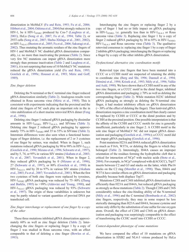
Construct name Primer (all primers are sense except NC2-2)
1 R7 5′TGATGCAGAGAGGCAATTTTAGCAACCAA2 R7E 5′CACAATTGAAACACTTAACAATCTCCTCT3 N+ 5′CCATAATGATGCAGAGAGGCAAATTTAGG
4 3AF1 5′CCAAAGAAAGATTGTTGCGTGTTTGAATT5 3EF1 5′CCAAAGAAAGATTGTTGAGTGTTTCAATT6 N17K 5′CCAAAGAAAGATTGTTAAGTGTTTCAAAT7 F16A 5′CCAAAGAAAGATTGTTAAGTGTGCCAATT8 2GAF1 5′GATTGTTAAGTGTTTCAATTGTGCCAAAG9 H23A 5′GTGTTTCAATTGTGGCAAAGAAGGGGCC10 H23C 5′GTGTTTCAATTGTGGCAAAGAAGGGTGCA11 C28H 5′GCAAAGAAGGGCACACAGCCAGAAATCA12 C28S 5′GCAAAGAAGGGCACACAGCCAGAAATTC13 NC2-2 5′CTTTTTCCTAGGGGCCCTGCAGTCCTTCA14 ΔF1 5′GAACCAAAGAAAGATTGTTAAGGGAGGA
15 S3 5′CCAGAAATTGCAGGGCCCCTTCTTCTTCT16 S3E 5′GCCAGAAATTGCAGGGCCCCTGAGGAAG17 FVI 5′CCAGAAATTGCAGGGCCCCTTTTGTTATT18 P31A 5′GCACACAGCCAGAAATTGCAGGGCCGCT19 ΔAP 5′GCACACAGCCAGAAATTGCAGGAGGAAA20 LL 5′GGAAAAAGGGCGGCGGCGGCGGCGGCT21 ΔLinker 5′GGGCACACAGCCAGAAATTGCGGCGGCT22 L+ 5′CACACAGCCAGAAATTGCAGGAAGAAGA
23 2KAF2 5′GGGCTGTTGGAAATGTGGAGCGGAAGGA24 2KEF2 5′GGGCTGTTGGAAATGTGGAGAGGAAGGA25 2GAF2 5′GGAAAAAGGGCTGTTGGAAATGTGCAAA26 W37A 5′GCCCCTAGGAAAAAGGGCTGTGCCAAAT27 W37F 5′GCCCCTAGGAAAAAGGGCTGTATCAAATG28 K38N 5′CCCCTAGGAAAAAGGGCTGTTGGAATTG29 H44A 5′GCTGTTGGAAATGTGGAAAGGAAGGAGC30 H44C 5′GCTGTTGGAAATGTGGAAAGGAAGGATG31 C36S 5′AGGAAAAAGGGCTCATGGAAATGTGGAA32 C49H 5′GGAAGGACACCAAATGAAAGATCACACT33 NC1-1 5′CTAGGAAAAAGGGCTGTTTTAATTGTGGA34 ΔF2 5′GCAGGGCCCCTAGGAAAAAGGGCGGAGG
All primers were synthesized by ACGT Inc. (Toronto).
Luban, 2001) (H23C could not be compared because of dis-cordant effects yielding NL4-3 results on either side of the HXB2data [Table 3]). Thus 5mutations out of 13 showed no evidence ofcontext-dependent gRNA packaging; the context-dependence ofthe remaining mutations favored neither HXB2 nor NL4-3 interms of phenotypic robustness. We conclude that 25 to 50% ofNC mutations may display a context dependent gRNA dimeriza-tion or packaging yield; gRNA dimerization, but not gRNApackaging, appears to be somewhat more labile in HIV-1HXB2than in HIV-1NL4-3.
Materials and methods
Plasmid construction
Proviral vector pSVC21.BH10 encodes a HIV-1HXB2 cDNAclone. Mutant proviral vectors were constructed from pSVC21.BH10 by PCR mutagenesis, using primers described in Table 4.The nucleotides positions are based on the sequence of HIV-1
AGCTCGATTGTTAAGTGTTTCAATTGTGGTTCTCAAAATTGCCTCTCTGCATCATTATGAACAAAAGAAAGATTGTTAAGTGTTTC
GTGGCGCAGAAGGGCACACAGCCGCAAATTGCAGGGCCCCGTGGCGAAGAAGGGCACACAGCCGAAAATTGCAGGGCCCCGTGGCAAAGAAGGGCACACGTGGCAAAGAAGGGCACACAAGCGCACACAGCCAGAAATTGCAGGACAGCCAGAAATTGCAGGGCCCCAGCCAGAAATTGCAGGGCCCCAGGGCCCCTAGGAAAAAGGGCCAGGGCCCCTAGGAAAAAGGGCTCTGGTGCCCTTCTTTGCCACATTTCCAACACTTAACAATCTTTCTTTGAGGGCCCCGAGGAAAAAAGGGC
GGCTGTTGGAAATGTGGAAAGGAGGGCTGTTGGAAATGTGGAAAGGGCTGTTGGAAATGTGGAAAGGAGGAAAAAGGGCTGTTGGAAATGTGGAAGGGCTGTTGGAAATGTGGGTTGGAAATGTGGAAAGGAAGGACACCAAATGAAAGGTTGGAAATGTGGAAAGGAAGGGAAAAAGGGCTGTTGG
CACCAAATGGCAGATTGTACTGAGAGACAGGCCACCAAATGGAAGATTGTACTGAGAGACAGGCGGAAGCACACCAAATGAAAGATTGTACGTGGAAAGGAAGGACACCTGGAAAGGAAGGACACC
TGGAAAGGAAGGACACCCCAAATGAAAGATTCTACTGAGAGCCAAATGAAAGATTGTACTGAGAGAGGAAGGACACCAAATGGAGAGACAGGCTAATTTTTTAGGAAGGAAGGACACACAGCGAGAAATTGTACTGAGAGACAGGCAACTGAGAGACAGGCTAATTTTTTAGG
606 J. Kafaie et al. / Virology 375 (2008) 592–610
gRNA. To prepare mutants in the N-terminus and zinc finger 1regions, a DNA fragment extending from Spe І to Apa Іrestriction sites was synthesized with desired mutations byPCR, and ligated into pSVC21.BH10. Mutants in the linker andzinc finger 2 regions were constructed by PCR from a DNAfragment extending from Spe I to Bcl I, and from Apa I to BclI,respectively. FF, NL and FL were constructed using mutantsH23A, 2GAF2, R7 and FVI. They were cut using restrictionenzymes Apa1 and Spe1 (Amersham). The H23A DNAfragment was inserted into 2GAF2 to construct FF, and inFVI to prepare FL. The same DNA sequence from R7 wasintroduced in FVI to make NL. After mutagenesis and ligation,all mutated DNA fragments produced by PCR were completelysequenced (ACGT Inc., Toronto) to verify that the wantedmutation, and no other mutation, was introduced by themutagenic procedure.
Cell culture and transfections
HeLa Cells were cultured at 37 °C in a medium consisting ofDulbecco's modified Eagle's medium (DMEM), 10% fetal calfserum, ampicillin and streptomycin (Invitrogen). The PolyFecttransfection reagent (Qiagen) was used to transfect 9 μg ofproviral DNA into 50% to 70% confluent HeLa cells in 100- by20-mm petri dishes containing 10 ml of culture medium.
Infectivity assay
Mutant proviruses and the parental BH10 provirus wereindependently transfected into HeLa cells. Virus-containingsupernatants were collected 48 h post-transfection and passedthrough 0.2 μm pore-size cellulose acetate filters to remove thecells. The CAp24 content of these clarified supernatants wasmeasured using an ELISA kit (Vironostika HIV-1 Antigen,Biomerieux). Equal amount of the supernatants (10 ng of CAp24content) were used to infect equal numbers of MT2 cells (6×10⁶
cells in 10 ml of RPMI 1640 medium, 10% fetal calf serum,ampicillin and streptomycin (Invitrogen), per petri dish). In thehuman T-cell line MT2, only a short time lag separates infectionfrom viral replication (Harada et al., 1985). After 2 h, cellswere washed twice to remove unbound viruses and were thenmaintained in serum-supplemented medium. On every otherday, cells were diluted 1 in 2 into fresh medium and the RTactivity in the supernatant of the removed medium wasdetermined. RT activity measurements were made over a periodof 14 days.
RT activity
The exogenous (oligo (dT) directed) RTactivity was measuredby adding 40 μl of RT cocktail (60 mM Tris–HCl [pH 7.9],180mMKCl, 6 mMMgCl2, 6 mMdithiothreitol, 0.6 mMEGTA,0.12% Triton X-100, 6 μg/ml oligo (dT), 12 μg/ml poly(rA),0.05 mM 3H dTTP) to a 10 μl sample. After incubation for 2 h at37 °C, the reaction was stopped with cold 10% TCA (150 μl perwell), and precipitated for 30 min at 4 °C. The precipitate wasblotted, washed and scintillation counted.
Virus purification and isolation of HIV-1 viral RNA
Filtered virus-containing supernatants were centrifuged(SW41 rotor, 35 000 rpm, 4 °C, 1 h), through a 2 ml 20% (w/v)sucrose cushion in phosphate-buffered saline (PBS). The viruspellet was dissolved in 400 μl sterile lysis buffer [50 mM Tris(pH7.4), 50 mM NaCl, 10 mM EDTA, 1% (w/v) SDS, 50 μgtRNA per ml, and 100 μg proteinase K per ml], and extractedtwice at 4 °C with an equal volume of buffer-saturated phenol-chloroform-isoamylalcohol (25:24:1) (Invitrogen). The aqueousphase was precipitated overnight at −80 °C with 0.1 volume of3M sodium acetate (pH 5.2) and 2.5 volumes of 95% ethanol, andcentrifuged at 14,000 rpm in an Eppendorf 5145 micro centrifugeat 4 °C for 30min. The gRNApellet was rinsedwith 70% ethanol,and dissolved in 10 μl buffer S (10 mM Tris (pH 7.5), 100 mMNaCl, 10 mM EDTA and 1% SDS) (Song et al., 2007).
Electrophoretic analysis of HIV-1 gRNA
The gRNA was electrophoresed under non-denaturing con-ditions and identified by Northern (RNA) blot analysis (Songet al., 2007). Electrophoretic conditions were 4 V/cm for 4 h ona 1% (w/v) agarose gel in TBE2 (89 mM Tris, 89 mM Borateand 2 mM EDTA, pH 8.3) at 4 °C. After electrophoresis, the gelwas heated at 65 °C for 30 min in 10% (w/v) formaldehyde, andthe embedded RNAs were diffusion transferred to a Hybond N+nylon membrane (Amersham). After drying at room tempera-ture for 2 h, crosslinking (3000 j in a UV Stratalinker), andprehybridization at 42 °C for 3 h in 6× SSPE (1× SSPE is0.15 M NaCl, 10 mM NaH2PO4, and 1 mM EDTA {pH 7.4}),50% (w/v) deionized formamide, 10% dextran sulfate, 1.5%SDS, 5× Denhardt's reagent, 100 μg/ml salmon sperm DNA,the membrane was hybridized overnight in prehybridizationbuffer devoid of Denhardt's reagent in a rotating hybridizationoven at 42 °C to approximately 25 μCi of 35S-labeled antisenseRNA 636-296 (a 356-nt RNA that is the antisense of the 296 to636 region of the HIV-1 genome prepared with the SP6Megascript kit [Ambion]) (Laughrea and Jetté, 1996). This wasfollowed by two 30 min washes in 1× SSC [1× SSC is 0.15 MNaCl plus 0.015 M sodium citrate]– 0.1% SDS at room tem-perature and 37 °C, and one 30 min wash in 0.2× SSC–0.1%SDS at 45 °C (Laughrea et al., 1997), exposure to a KodakBioMax MR X-ray film, and densitometric analysis.
Densitometric analysis
The autoradiograms were scanned and analysed with the NIH1.6.3 program. Care was taken to scan variously exposed films toguard against over-exposed or under-exposed bands or spots.The monomer and dimer bands were considered of equal width;that width was approximately twice the vertical size of the D andM letters used to indicate dimers and monomers in the relevantfigures. Material located elsewhere in the gels was not taken intoaccount in the calculation of the percentage of dimers. Thediffuse character of many bands may reflect conformationaldiversity among the gRNA molecules. It is not due to poorresolution of the gels because heat denatured gRNAs formed a
607J. Kafaie et al. / Virology 375 (2008) 592–610
sharp band at the monomer position (not shown). The RNAbands seen here are not more diffuse, and sometimes sharper,than what was seen in earlier studies by us and others (Clever andParslow, 1997; Hill et al., 2003; Laughrea et al., 1997; Sakuragiand Panganiban, 1997; Sakuragi et al., 2001, 2003; Shen et al.,2000).
Genomic RNA packaging
The amount of gRNA per unit CAp24 of virus was quan-titated by hybridization with antisense RNA 636–296 using adot–blot assay. Virus pellets were resuspended in 400 μl ofTrizol LS reagent (Invitrogen), and incubated at 30 °C for 5 min.100 μl of chloroform was added, followed by shaking for15 seconds and incubation at room temperature for 15 min. Aftercentrifugation (12,000 ×g, 15 min, 4 °C), the colorless aqueousupper phase was mixed with 250 μl of isopropyl alcohol,incubated at room temperature for 10 min and centrifuged again.The precipitated RNA was washed once with 500 μl of 70%ethanol, pelleted (7500 ×g, 5 min, 4 °C), air-dried, dissolved in10 μl RNase-free water and stored at −20 °C. Serial 10-folddilutions of wild type RNA samples, normalized for input virionCAp24, were used to construct a standard curve. 29 μl of bufferF (100% deionized formamide, 20 μl; 20× SSC, 2 μl; 37%formaldehyde, 7 μl) was added to each sample, followed byincubation (68 °C, 15 min) and chilling on ice. After adding78 μl of 20× SSC buffer, samples were vacuum-suction trans-ferred to a Hybond N+ nylon membrane (Amersham) sand-wiched within a Hybri-Dot filtration manifold (BethesdaResearch Laboratories). The wells were washed twice with1 ml of 10× SSC, and suction continued for a further 5 min to drythe membrane. Themembrane was removed, dried for 4 h, cross-linked, pre-hybridized, hybridized, autoradiographed andscanned as for Northern blot analysis (above). To confirm thescans, each individual spot of the nylon membrane was excisedand scintillation counted.
Virus stability and RT packaging
1.2 ml of filtered virus-containing supernatant was pelletedthrough a 0.3 ml 20% sucrose cushion in the TL-100 Beckmanultracentrifuge (TLA 55 rotor, 45,000 rpm, 1 h, 4 °C). The viruspellet was dissolved in 10 μl of PBS and its CAp24 content wasmeasured using an ELISA kit (Vironostika HIV-1 Antigen,Biomerieux). The CA content of the purified viruses divided bythe CA content of the 48 h culture supernatant, relative to the ratiofound in WT, was taken as a measure of virus stability (Wanget al., 2002; 2004). The ratio pellet/supernatant was 0.43±0.07 inWT (this was taken to mean 100 in Table 2).
The exogenous RT activity of the pelleted viruses divided bytheir CA content, relative to the ratio found in WT, wasinterpreted as RT packaging.
Pr55gag proteolytic maturation
At 48 h post-transfection, cells were lysed in ice-cold NP-40containing buffer (100 mM NaCl, 10 mM Tris, 1 mM EDTA,
0.5% NP-40, and protease inhibitor cocktail [Roche]). Super-natants were cleared by centrifugation at 3000 ×g and filtered(0.22 μm). Viruses were then concentrated through a sucrosecushion by ultracentrifugation. Equal amounts of viruses (judgedby CAp24-ELISA) were lysed in the above buffer and subjectedto SDS-polyacrylamide gel electrophoresis. Viral proteins weredetected by Western blotting using a rabbit anti-capsid antibody(ABT-Trinity Biotechnology, CA, USA) that recognizes Pr55gag,CAp24, andCA-containing partially cleavedGag proteins, and anenhanced chemiluminescence Western blot detection kit (Amer-sham). The signals for CA-containing proteins were quantitatedby densitometric scanning and analysed with the NIH 1.6.3program. The signal obtained from the CAp24 band was dividedby the total signal obtained from all CA-containing proteins, tocalculate Pr55gag processing level.
Statistics
P values were measured using the Student's t test.
Acknowledgments
We thank Mark A. Wainberg for access to the HIV growthfacility, and Robert J. Gorelick for the gift of NL4-3 proviralDNA clones. This work was supported by CIHR group grant13918, by CIHR grant #MOP-12312 to M. L., and by CIHRgrant #MOP-38111 to A. J. M.
References
Adachi, A., Gendelman, H.E., Koenig, S., Folks, T., Willey, R., Rabson, A.,Martin, M.A., 1986. Production of acquired immunodeficiency syndrome-associated retrovirus in human and non human cells transfected with aninfectious molecular clone. J. Virol. 59, 284–291.
Akahata, W., Ido, E., Hayami, M., 2003. Mutational analysis of two zinc-fingermotifs in the nucleocapsid protein of simian immunodeficiency virusmac239. J. Gen. Virol. 84, 1641–1648.
Aldovini, A., Young, R.A., 1990. Mutations of RNA and protein sequencesinvolved in human immunodeficiency virus type 1 packaging result inproduction of noninfectious virus. J. Virol. 64, 1920–1926.
Amarasinghe, G.K., De Guzman, R.N., Turner, R.B., Chancellor, K.J., Wu,Z.R., Summers, M.F., 2000. NMR structure of the HIV-1 nucleocapsidprotein bound to stem-loop SL2 of the psi-RNA packaging signal.Implications for genome recognition. J. Mol. Biol. 301, 491–511.
Baba, S., Takahashi, K., Koyanagi, Y., Yamamoto, N., Takaku, H., Gorelick,R.J., Kawai, G., 2003. Role of the zinc fingers of HIV-1 nucleocapsid proteinin maturation of genomic RNA. J. Biochem. (Tokyo) 134, 637–639.
Benjamin, J., Ganser-Pornillos, B.K., Tivol, W.F., Sundquist, W.I., Jensen, G.J.,2005. Three-dimensional structure of HIV-1 virus-like particles by electroncryotomography. J. Mol. Biol. 346, 577–588.
Berg, J.M., 1990. Zinc finger domains: hypotheses and current knowledge.Annu. Rev. Biophys. Biophys. Chem. 19, 405–421.
Berg, J.M., Shi, Y., 1996. The galvanization of biology: a growing appreciationfor the roles of zinc. Science 271, 1081–1085.
Berthoux, L., Péchoux, C., Ottmann, M., Morel, G., Darlix, J.L., 1997.Mutations in the N-terminal domain of human immunodeficiency virustype 1 nucleocapsid protein affect virion core structure and proviral DNAsynthesis. J. Virol. 71, 6973–6981.
Bess Jr., J.W., Powell, P.J., Issaq, H.J., Schumack, L.J., Grimes, M.K.,Henderson, L.E., Arthur, L.O., 1992. Tightly bound zinc in humanimmunodeficiency virus type 1, human T-cell leukemia virus type 1, andother retroviruses. J. Virol. 66, 840–847.
608 J. Kafaie et al. / Virology 375 (2008) 592–610
Bombarda, E., Cherradi, H., Morellet, N., Roques, B.P., Mély, Y., 2002. Zn2+
binding properties of single-point mutants of the C-terminal zinc finger ofthe HIV-1 nucleocapsid protein: evidence of a critical role of cysteine 49 inZn2+ dissociation. Biochemistry 41, 4312–4320.
Bowles, N.E., Damay, P., Spahr, P.-F., 1993. Effect of rearrangements andduplications of the Cys–His motifs of Rous sarcoma virus nucleocapsidprotein. J. Virol. 67, 623–631.
Briggs, J.A.G., Simon, M.N., Gross, I., Krausslich, H.-G., Fuller, S.D., Vogt,V.M., Johnson, M.C., 2004. The stoichiometry of Gag protein in HIV-1.Nat. Struct. Mol. Biol. 11, 672–675.
Buckman, J.S., Bosche, W.J., Gorelick, R.J., 2003. Human immunodeficiencyvirus type 1 nucleocapsid Zn2+ fingers are required for efficient reverse tran-scription, initial integration processes, and protection of newly synthesizedviral DNA. J. Virol. 77, 1469–1480.
Buxton, P., Tachedjian, G., Mak, J., 2005. Analysis of the contribution of reversetranscriptase and integrase proteins to retroviral RNA dimer conformation.J. Virol. 79, 6338–6348.
Cen, S., Huang, Y., Khorchid, A., Darlix, J.L., Wainberg, M.A., Kleiman, L.,1999. The role of Pr55gag in the annealing of tRNA3Lys3 to humanimmunodeficiency virus type 1 genomic RNA. J. Virol. 73, 4485–4488.
Cen, S., Niu, M., Saadatmand, J., Guo, F., Huang, Y., Nabel, G.J., Kleiman, L.,2004. Incorporation of Pol into human immunodeficiency virus type 1 Gagvirus-like particles occurs independently of the upstream Gag domain inGag-Pol. J. Virol. 78, 1042–1049.
Chen, N., Morag, A., Almog, N., Blumenzweig, I., Dreazin, O., Kotler, M.,2001. Extended nucleocapsid protein is cleaved from the Gag-Pol precursorof human immunodeficiency virus type 1. J. Gen. Virol. 82, 581–590.
Chin, M.P.S., Chen, J., Nikolaitchik, O.A., Hu, W.-S., 2007. Molecular determi-nants of HIV-1 intersubtype recombination potential. Virology 363, 437–446.
Chin, M.P.S., Rhodes, T.D., Chen, J., Fu,W., Hu,W.-S., 2005. Identification of amajor restriction in HIV-1 intersubtype recombination. Proc. Natl. Acad.Sci. U. S. A. 102, 9002–9007.
Cimarelli, A., Luban, J., 2001. Context-dependent phenotype of a human immuno-deficiency virus type 1 nucleocapsid mutation. J. Virol. 75, 7193–7197.
Cimarelli, A., Sandin, S., Hoglund, S., Luban, J., 2000. Basic residues in humanimmunodeficiency virus type 1 nucleocapsid promote virion assembly viainteraction with RNA. J. Virol. 74, 3046–3057.
Clever, J.L., Parslow, T.G., 1997. Mutant human immunodeficiency virus type 1genomes with defects in RNA dimerization or encapsidation. J. Virol. 71,3407–3414.
Dannull, J., Surovoy, A., Jung, G., Moelling, K., 1994. Specific binding of HIV-1 nucleocapsid protein to PSI RNA in vitro requires N-terminal zinc fingerand flanking basic amino acid residues. EMBO J. 13, 1525–1533.
Dawson, L., Yu, X.F., 1998. The role of nucleocapsid of HIV-1 in virusassembly. Virology, 251, 141–157.
De Guzman, R.N., Wu, Z.R., Stalling, C.C., Pappalardo, L., Borer, P.N.,Summers, M.F., 1998. Structure of the HIV-1 nucleocapsid protein bound tothe SL3 psi-RNA recognition element. Science 279, 384–388.
Déméné, H., Dong, C.Z., Ottmann, M., Rouyez, M.C., Jullian, N., Morellet, N.,Mély, Y., Darlix, J.L., Fournié-Zaluski, M.C., Saragosti, S., Roques, B.P.,1994. 1H NMR structure and biological studies of the His23 -N Cys mutantnucleocapsid protein of HIV-1 indicate that the conformation of the first zincfinger is critical for virus infectivity. Biochemistry 33, 11707–11716.
Dorfman, T., Luban, J., Goff, S.P., Haseltine, W.A., Gottlinger, H.G., 1993.Mapping of functionally important residues of a cysteine–histidine box inthe human immunodeficiency virus type 1 nucleocapsid protein. J. Virol. 67,6159–6169.
Dupraz, P., Oertle, S., Méric, C., Damay, P., Spahr, P.-F., 1990. Point mutationsin the proximal Cys–His box of Rous sarcoma virus nucleocapsid protein.J. Virol. 64, 4978–4987.
Feng, Y.-X., Copeland, T.D., Henderson, L.E., Gorelick, R.J., Bosche, W.J.,Levin, J.G., Rein, A., 1996. HIV-1 nucleocapsid protein induces “matura-tion" of dimeric retroviral RNA in vitro. Proc. Natl. Acad. Sci. U. S. A. 93,7577–7581.
Feng, Y.-X., Campbell, S., Harvin, D., Ehresmann, B., Ehresmann, C., Rein, A.,1999. The human immunodeficiency virus type 1 Gag polyprotein has nucleicacid chaperone activity: possible role in dimerization of genomic RNA andplacement of tRNA on the primer binding site. J. Virol. 73, 4251–4256.
Fu, W., Rein, A., 1993. Maturation of dimeric viral RNA of Moloney murineleukemia virus. J. Virol. 67, 5443–5449.
Fu, W., Gorelick, R.J., Rein, A., 1994. Characterization of human immunode-ficiency virus type 1 dimeric RNA from wild-type and protease-defectivevirions. J. Virol. 68, 5013–5018.
Fu, W., Dang, Q., Nagashima, K., Freed, E.O., Pathak, V.K., Hu, W.-S., 2006.Effects of Gagmutation and processing on retroviral dimeric RNAmaturation.J. Virol. 80, 1242–1249.
Fu, W., Prasad, V.V.S.P., Chen, J., Nikolaitchik, O., Hu, W.-S., 2007. Molecularmechanisms of simian immunodeficiency virus SIVagm RNA encapsidation.Virology 363, 210–219.
Gorelick, R.J., Henderson, L.E., Hanser, J.P., Rein, A., 1988. Point mutants ofMoloney murine leukemia virus that fail to package viral RNA: evidence forspecific RNA recognition by a “zinc finger-like” protein sequence. Proc.Natl. Acad. Sci. U. S. A. 85, 8420–8424.
Gorelick, R.J., Nigida Jr., S.M., Bess Jr., J.W., Arthur, L.O., Henderson, L.E.,Rein, A., 1990. Noninfectious human immunodeficiency virus type 1mutants deficient in genomic RNA. J. Virol. 64, 3207–3211.
Gorelick, R.J., Chabot, D.J., Rein, A., Henderson, L.E., Arthur, L.O., 1993. Thetwo zinc fingers in the human immunodeficiency virus type 1 nucleocapsidprotein are not functionally equivalent. J. Virol. 67, 4027–4036.
Gorelick, R.J., Chabot, D.J., Ott, D.E., Gagliardi, T.D., Rein, A., Henderson, L.E.,Arthur, L.O., 1996. Genetic analysis of the zinc finger in the Moloney murineleukemia virus nucleocapsid domain: replacement of zinc-coordinatingresidues with other zinc-coordinating residues yields noninfectious particlescontaining genomic RNA. J. Virol. 70, 2593–2597.
Gorelick, R.J., Gagliardi, T.D., Bosche, W.J., Wiltrout, T.A., Coren, L.V.,Chabot, D.J., Lifson, J.D., Henderson, L.E., Arthur, L.O., 1999a. Strictconservation of the retroviral nucleocapsid protein zinc finger is stronglyinfluenced by its role in viral infection processes: characterization of HIV-1particles containing mutant nucleocapsid zinc-coordinating sequences.Virology 256, 92–104.
Gorelick, R.J., Benveniste, R.E., Gagliardi, T.D., Wiltrout, T.A., Busch, L.K.,Bosche, W.J., Coren, L.V., Lifson, J.D., Bradley, P.J., Henderson, L.E.,Arthur, L.O., 1999b. Nucleocapsid protein zinc-finger mutants of simianimmunodeficiency virus strain mne produce virions that are replicationdefective in vitro and in vivo. Virology 253, 259–270.
Gorelick, R.J., Fu, W., Gagliardi, T.D., Bosche, W.J., Rein, A., Henderson, L.E.,Arthur, L.O., 1999c. Characterization of the block in replication ofnucleocapsid protein zinc finger mutants from Moloney murine leukemiavirus. J. Virol. 73, 8185–8195.
Green, L.M., Berg, J.M., 1989. A retroviral Cys-Xaa2-Cys-Xaa4-His-Xaa4-Cyspeptide binds metal ions: spectroscopic studies and a proposed three-dimensional structure. Proc. Natl. Acad. Sci. U. S. A. 86, 4047–4051.
Guo, J., Wu, T., Kane, B.F., Johnson, D.G., Henderson, L.E., Gorelick, R.J.,Levin, J.G., 2002. Subtle alterations of the native zinc finger structure havedramatic effects on The nucleic acid chaperone activity of human immuno-deficiency virus type 1 nucleocapsid protein. J. Virol. 76, 4370–4378.
Haddrick, M., Lear, A.L., Cann, A.J., Heaphy, S., 1996. Evidence that a kissingloop structure facilitates genomic RNA dimerisation in HIV-1. J. Mol. Biol.259, 58–68.
Hagan, N.A., Fabris, D., 2007. Dissecting the protein-RNA and RNA-RNAinteractions in the nucleocapsid-mediated dimerization and isomerization ofHIV-1 stemloop 1. J. Mol. Biol. 365, 396–410.
Harada, S., Koyanagi, Y., Yamamoto, N., 1985. Infection of HTLV III/LAV inHTLV-I- carrying cells MT-2 and MT-4 and application in a plaque assay.Science 229, 563–566.
Hibbert, C.S., Mirro, J., Rein, A., 2004. mRNA molecules containing murineleukemia virus packaging signals are encapsidated as dimers. J. Virol. 78,10927–10938.
Hill, M.K., Shehu-Xhilaga, M., Campbell, S.M., Poumbourios, P., Crowe, S.M.,Mak, J., 2003. The dimer initiation sequence stem-loop of humanimmunodeficiency virus type 1 is dispensable for viral replication inperipheral blood mononuclear cells. J. Virol. 77, 8329–8335.
Housset, V., De Rocquigny, H., Roques, B.P., Darlix, J.L., 1993. Basic aminoacids flanking the zinc finger of Moloney murine leukemia virusnucleocapsid protein NCp10 are critical for virus infectivity. J. Virol. 67,2537–2545.

609J. Kafaie et al. / Virology 375 (2008) 592–610
Huang, Y., Khorchid, A., Wang, J., Parniak, M.A., Darlix, J.L., Wainberg, M.A.,Kleiman, L., 1997. Effect of mutations in the nucleocapsid protein (NCp7)upon Pr160gag-pol and tRNALys incorporation into human immunodeficiencyvirus type 1. J. Virol. 71, 4378–4384.
Krogstad, P., Geng, Y.-Z., Rey, O., Canon, J., Ibarrondo, F.J., Ackerson, B.,Patel, J., Aldovini, A., 2002. Human immunodeficiency virus nucleocapsidprotein polymorphisms modulate the infectivity of RNA packaging mutants.Virology 294, 282–288.
Khorchid, A., Halwani, R., Wainberg, M.A., Kleiman, L., 2002. Role of RNA infacilitating Gag/Gag–Pol interaction. J. Virol. 76, 4131–4137.
Krizek, B.A., Merkle, D.L., Berg, J.M., 1993. Ligand variation and metal ionbinding specificity in zinc finger peptides. Inorg. Chem. 32, 937–940.
Laughrea, M., Jetté, L., 1994. A nineteen nucleotide sequence upstream of the5′ major splice donor is part of the dimerization domain of the HIV-1genome. Biochemistry 33, 13464–13475.
Laughrea, M., Jetté, L., 1996. Kissing-loop model of HIV-1 genomedimerization: HIV-1 RNAs can assume alternative dimeric forms and allsequences upstream or downstream of hairpin 248–271 are dispensable fordimer formation. Biochemistry 35, 1589–1598.
Laughrea, M., Jetté, L., Mak, J., Kleiman, L., Liang, C., Wainberg, M.A., 1997.Mutations in the kissing-loop hairpin of human immunodeficiency virustype 1 reduce viral infectivity as well as genomic RNA packaging anddimerization. J. Virol. 71, 3397–3406.
Laughrea, M., Shen, N., Jetté, L., Darlix, J.L., Kleiman, L., Wainberg, M.A.,2001. Role of distal zinc finger of nucleocapsid protein in genomic RNAdimerization of human immunodeficiency virus type 1; no role for thepalindrome crowning the R-U5 hairpin. Virology 281, 109–116.
Lee, B.M., De Guzman, R.N., Turner, B.G., Tjandra, N., Summers, M.F., 1998.Dynamical behavior of the HIV-1 nucleocapsid protein. J. Mol. Biol. 279,633–649.
Leitner, T., Foley, B., Hahn, B., Marx, P., McCutchan, F., Mellors, J.W.,Wolinksky, S., Korber, B., 2005. HIV sequence compendium 2005. LosAlamos National Laboratory, Los Alamos, N. Mex.
Levin, J.G., Guo, J., Rouzina, I., Musier-Forsyth, K., 2005. Nucleic acidchaperone activity of HIV-1 nucleocapsid protein: critical role in reversetranscription and molecular mechanism. Prog. Nucleic Acid Res. Mol. Biol.80, 217–286.
Mark-Danieli, M., Laham, N., Kenan-Eichler, M., Castiel, A., Melamed, D.,Landau, M., Bouvier, N.M., Evans, M.J., Bacharach, E., 2005. Single pointmutations in the zinc finger motifs of the human immunodeficiency virustype 1 nucleocapsid alter RNA binding specificities of the Gag protein andenhance packaging and infectivity. J. Virol. 79, 7756–7767.
McGrath, C.F., Buckman, J.S., Gagliardi, T.D., Bosche, W.J., Coren, L.V.,Gorelick, R.J., 2003. Human cellular nucleic acid-binding protein Zn2+
fingers support replication of human immunodeficiency virus type 1 whenthey are substituted in the nucleocapsid protein. J. Virol. 77, 8524–8531.
Mély, Y., De Rocquigny, H., Morellet, N., Roques, B.P., Gérard, D., 1996. Zincbinding to the HIV-1 nucleocapsid protein: a thermodynamic investigationby fluorescence spectroscopy. Biochemistry 35, 5175–5182.
Méric, C., Goff, S.P., 1989. Characterization of Moloney murine leukemia virusmutants with single-amino-acid substitutions in the Cys–His box of thenucleocapsid protein. J. Virol. 63, 1558–1568.
Méric, C., Spahr, P.-F., 1986. Rous sarcoma virus nucleic acid-binding proteinp12 is necessary for viral 70S RNA dimer formation and packaging. J. Virol.60, 450–459.
Méric, C., Gouilloud, E., Spahr, P.-F., 1988. Mutations in Rous sarcoma virusnucleocapsid protein p12 (NC): deletions of Cys–His boxes. J. Virol. 62,3328–3333.
Mihailescu, M.-R., Marino, J.P., 2004. A proton-coupled dynamic conforma-tional switch in the HIV-1 dimerization initiation site kissing complex. Proc.Natl. Acad. Sci. U. S. A. 101, 1189–1194.
Miller, J., McLachlan, A.D., Klug, A., 1985. Repetitive zinc-binding domainsin the protein transcription factor IIIA from Xenopus oocytes. EMBO J. 4,1609–1614.
Mizuno, A., Ido, E., Goto, T., Kuwata, T., Nakai, M., Hayami, M., 1996.Mutational analysis of two zinc finger motifs in HIV type 1 nucleocapsidproteins: effects on proteolytic processing of Gag precursors and particleformation. AIDS Res. Hum. Retrovir. 10, 793–800.
Morellet, N., De Rocquigny, H., Mély, Y., Jullian, N., Déméné, H., Ottmann, M.,Gérard, D., Darlix, J.L., Fournie-Zaluski, M.C., Roques, B.P., 1994.Conformational behaviour of the active and inactive forms of the nucleo-capsid NCp7 of HIV-1 studied by 1H NMR. J. Mol. Biol. 235, 287–301.
Morellet, N., Déméné, H., Teilleux, V., Huynh-Dinh, T., De Rocquigny, H.,Fournié-Zaluski, M.-C., Roques, B.P., 1998. Structure of the complexbetween the HIV-1 nucleocapsid protein NCp7 and the single-strandedpentanucleotide d(ACGCC). J. Mol. Biol. 283, 419–434.
Mujeeb, A., Ulyanov, N.B., Georgantis, S., Smirnov, I., Chung, J., Parslow, T.G.,James, T.L., 2007. Nucleocapsid protein-mediated maturation of dimer ini-tiation complex of full-length SL1 stemloop of HIV-1: sequence effects andmechanism of RNA refolding. Nucleic Acids Res. 35, 2026–2034.
Muriaux, D., De Rocquigny, H., Roques, B.P., Paoletti, J., 1996. NCp7 activatesHIV-1Lai RNA dimerization by converting a transient loop–loop complexinto a stable dimer. J. Biol. Chem. 271, 33686–33692.
Ott, D.E., Coren, L.V., Chertova, E.N., Gagliardi, T.D., Nagashima, K., SowderII, R.C., Poon, D.T.K., Gorelick, R.J., 2003. Elimination of protease activityrestores efficient virion production to a human immunodeficiency virus type1 nucleocapsid deletion mutant. J. Virol. 77, 5547–5556.
Ottmann, M., Gabus, C., Darlix, J.L., 1995. The central globular domain of thenucleocapsid protein of human immunodeficiency virus type 1 is critical forvirion structure and infectivity. J. Virol. 69, 1778–1784.
Oshima, M., Muriaux, D., Mirro, J., Nagashima, K., Dryden, K., Yeager, M.,Rein, A., 2004. Effects of blocking individual maturation cleavages inmurine leukemia virus Gag. J. Virol. 78, 1411–1420.
Pabo, C.O., Peisach, E., Grant, R.A., 2001. Design and selection of novelCys2His2 zinc finger proteins. Annu. Rev. Biochem. 70, 313–340.
Pettit, S.C., Moody, M.D., Wehbie, R.S., Kaplan, A.H., Nantermet, P.V., Klein,C.A., Swanstrom, R., 1994. The p2 domain of human immunodeficiencyvirus type 1 Gag regulates sequential proteolytic processing and is requiredto produce fully infectious virions. J. Virol. 68, 8017–8027.
Pettit, S.C., Gulnik, S., Everitt, L., Kaplan, A.H., 2003. The dimer interfaces ofprotease and extra-protease domains influence the activation of protease andthe specificity of GagPol cleavage. J. Virol. 77, 366–374.
Poon, D.T.K., Wu, J., Aldovini, A., 1996. Charged amino acid residues ofhuman immunodeficiency virus type 1 nucleocapsid p7 protein involved inRNA packaging and infectivity. J. Virol. 70, 6607–6616.
Ramboarina, S., Moreller, N., Fournié-Zaluski, M.-C., Roques, B.P., 1999.Structural investigation on the requirement of CCHH zinc finger type innucleocapsid protein of human immunodeficiency virus 1. Biochemistry 38,9600–9607.
Rist, M.J., Marino, J.P., 2002. Mechanism of nucleocapsid protein catalyzedstructural isomerization of the dimerization initiation site of HIV-1.Biochemistry 41, 14762–14770.
Sakuragi, J., Panganiban, A.T., 1997. Human immunodeficiency virus type 1RNA outside the primary encapsidation and dimer linkage region affectsRNA dimer stability in vivo. J. Virol. 71, 3250–3254.
Sakuragi, J., Shioda, T., Panganiban, A.T., 2001. Duplication of the primaryencapsidation and dimer linkage region of human immunodeficiency virustype 1 RNA results in the appearance of monomeric RNA in virions. J. Virol.75, 2557–2565.
Sakuragi, J., Iwamoto, A., Shioda, T., 2002. Dissociation of genome dimer-ization from packaging functions and virion maturation of human immuno-deficiency virus type 1. J. Virol. 76, 959–967.
Sakuragi, J.I., Ueda, S., Iwamoto, A., Shioda, T., 2003. Possible role of dimer-ization in human imunodeficiency virus type 1 genome RNA packaging. J.Virol. 77, 4060–4069.
Sandefur, S., Smith, R.M., Varthakavi, V., Spearman, P., 2000. Mapping andcharacterization of the N-terminal I domain of human immunodeficiencyvirus type 1 Pr55 Gag. J. Virol. 74, 7238–7249.
Schwartz, M.D., Fiore, D., Panganiban, A.T., 1997. Distinct functions andrequirements for the Cys–His boxes of the human immunodeficiency virustype 1 nucleocapsid protein during RNA encapsidation and replication.J. Virol. 71, 9295–9305.
Shehu-Xhilaga, M., Hill, M., Marshall, J.A., Kappes, J., Crowe, S.M., Mak, J.,2002. The conformation of the mature dimeric human immunodeficiencyvirus type 1 RNA genome requires packaging of pol protein. J. Virol. 76,4331–4340.
610 J. Kafaie et al. / Virology 375 (2008) 592–610
Shehu-Xhilaga, M., Crowe, S.M., Mak, J., 2001. Maintenance of the Gag/Gag–Pol ratio is important for human immunodeficiency virus type 1 RNAdimerization and viral infectivity. J. Virol. 75, 1834–1841.
Shen, N., Jetté, L., Wainberg, M.A., Laughrea, M., 2001. Role of stem B, loopB, and nucleotides next to the primer binding site and the kissing-loopdomain in human immunodeficiency virus type 1 replication and genomic-RNA dimerization. J. Virol. 75, 10543–10549.
Shen, N., Jetté, L., Liang, C., Wainberg, M.A., Laughrea, M., 2000. Impact ofhuman immunodeficiency virus type 1 RNA dimerization on viral infectivityand of stem-loop B on RNA dimerization and reverse transcription anddissociation of dimerization from packaging. J. Virol. 74, 5729–5735.
Sheng, N., Pettit, S.C., Tritch, R.J., Ozturk, D.H., Rayner, M.M., Swanstrom, R.,Erickson-Viitanen, S., 1997. Determinants of the human immunodeficiencyvirus type 1 p15NC-RNA interaction that affect enhanced cleavage by theviral protease. J. Virol. 71, 5723–5732.
Song, R., Kafaie, J., Yang, L., Laughrea, M., 2007. HIV-1 Viral RNA is selectedin the form of monomers that dimerize in a three-step protease-dependentprocess; the DIS of stem-loop 1 initiates viral RNA dimerization. J. Mol.Biol. 371, 1084–1098.
Song, R., Kafaie, J., Laughrea, M., in press. Role of the 5′ TAR stem-loop andthe U5-AUG duplex in dimerization of HIV-1 genomic RNA. Biochemistry.doi:10.1021/bi7023173.
South, T.L., Blake, P.R., Hare, D.R., Summers, M.F., 1991. C-terminalretroviral-type zinc finger domain from the HIV-1 nucleocapsid protein isstructurally similar to the N-terminal zinc finger domain. Biochemistry 30,6342–6349.
Stote, R.H., Kellenberger, E., Muller, H., Bombarda, E., Roques, B.P., Kieffer,B., Mély, Y., 2004. Structure of the His44 -N Ala single point mutant of thedistal finger motif of HIV-1 nucleocapsid protein: a combined NMR,molecular dynamics simulation, and fluorescence study. Biochemistry 43,7687–7697.
Summers, M.F., South, T.L., Kim, B., Hare, D.R., 1990. High-resolutionstructure of an HIV zinc fingerlike domain via a new NMR-based distancegeometry approach. Biochemistry 29, 329–340.
Takahashi, K., Baba, S., Koyanagi, Y., Yamamoto, N., Takaku, H., Kawai, G.,2001. Two basic regions of NCp7 are sufficient for conformational con-version of HIV-1 dimerization initiation site from kissing-loop dimer toextended-duplex dimer. J. Biol. Chem. 276, 31274–31278.
Takahashi, K.I., Baba, S., Chattopadhyay, P., Koyanagi, Y., Yamamoto, N.,Takaku, H., Kawai, G., 2000. Structural requirement for the two-step dimer-ization of human immunodeficiency virus type 1 genome. RNA 6, 96–102.
Tanchou, V., Decimo, D., Péchoux, C., Lener, D., Rogemond, V., Berthoux, L.,Ottmann, M., Darlix, J.L., 1998. Role of the N-terminal zinc finger of humanimmunodeficiency virus type 1 nucleocapsid protein in virus structure andreplication. J. Virol. 72, 4442–4447.
Turner, K.B., Hagan, N.A., Fabris, D., 2007. Understanding the isomerizationof the HIV-1 dimerization initiation domain by the nucleocapsid protein.J. Mol. Biol. 369, 812–828.
Vallée, B.L., Auld, D.S., 1990. Zinc coordination, function, and structure of zincenzymes and other proteins. Biochemistry 29, 5647–5659.
Wang, S.W., Aldovini, A., 2002. RNA incorporation is critical for retroviralparticle integrity after cell membrane assembly of Gag complexes. J. Virol.76, 11853–11865.
Wang, S.-W., Noonan, K., Aldovini, A., 2004. Nucleocapsid–RNA interactionsare esssential to structural stability but not to assembly of retroviruses.J. Virol. 78, 716–723.
Williams, M.C., Gorelick, R.J., Musier-Forsyth, K., 2002. Specific zinc-fingerarchitecture required for HIV-1 nucleocapsid protein's nucleic acidchaperone function. Proc. Natl. Acad. Sci. U. S. A. 99, 8614–8619.
Yovandich, J.L., Chertova, E.N., Kane, B.P., Gagliardi, T.D., Bess Jr., J.W.,Sowder II, R.C.., Henderson, L.E., Gorelick, R.J., 2001. Alteration of zinc-binding residues of simian immunodeficiency virus p8NC results in subtledifferences in Gag processing and virion maturation associated withdegradative loss of mutant NC. J. Virol. 75, 115–124.
Zabransky, A., Hunter, E., Sakalian, M., 2002. Identification of a minimal HIV-1Gag domain sufficient for self-association. Virology 294, 141–150.
Zhang, Y., Barklis, E., 1995. Nucleocapsid protein effects on the specificity ofretrovirus RNA encapsidation. J. Virol. 69, 5716–5722.
Related Documents