Mapping key interactions in the dimerization process of HBHA from Mycobacterium tuberculosis, insights into bacterial agglutination Carla Esposito a,1 , Marco Cantisani b,1 , Gabriella D’Auria c , Lucia Falcigno c , Emilia Pedone a , Stefania Galdiero a,b , Rita Berisio a,⇑ a Institute of Biostructures and Bioimaging, CNR, Via Mezzocannone 16, I-80134 Napoli, Italy b Department of Biological Sciences, Via Mezzocannone 16, I-80134 Napoli, Italy c Department of Chemistry, University of Naples ‘‘Federico II’’, I-80126, Via Cinthia 45, Napoli, Italy article info Article history: Received 2 December 2011 Revised 28 December 2011 Accepted 18 January 2012 Available online 31 January 2012 Edited by Renee Tsolis Keywords: Coiled coil Tuberculosis Agglutination Dimerization abstract HBHA is a cell-surface protein implicated in the dissemination of Mycobacterium tuberculosis (Mtb) from the site of primary infection. Its N-terminal coiled-coil region is also involved in bacterial agglutination. However, despite the importance of HBHA dimerization in agglutination, protein regions involved in dimerization are hitherto not known. Here, we mapped these regions by cou- pling peptide synthesis, biochemical and computational analyses, and identified structural determi- nants for HBHA monomer–monomer recognition. Importantly, we obtained the first molecule able to induce HBHA dimer disaggregation at 37 °C, the typical growth temperature of Mtb. This result provides new opportunities towards the development of Mtb anti-aggregation molecules with ther- apeutic interest. Structured summary of protein interactions: HBHA and HBHA bind by molecular sieving (View interaction) HBHA and H1 peptide bind by competition binding (View Interaction) HBHA and H1ext peptide bind by competition binding (View Interaction) HBHA and H2ext peptide bind by competition binding (View Interaction) HBHA and H2 peptide bind by competition binding (View Interaction) HBHA and H2ext peptide bind by competition binding (View Interaction) HBHA and HBHA bind by blue native page (View interaction) Ó 2012 Federation of European Biochemical Societies. Published by Elsevier B.V. All rights reserved. 1. Introduction Mycobacterium tuberculosis (Mtb), the etiologic agent of Tuber- culosis (TB), is one of the deadliest human pathogens, infecting more than one third of the human population. Peculiar of Mtb is its ability to survive in harsh conditions, replicate in host cells and disseminate from the site of primary infection to potentially any organ, where it can persist for decades and eventually reacti- vate to cause active TB [1–3]. Dissemination from the site of pri- mary infection involves interactions of Mtb with epithelial cells through a surface protein called heparin-binding hemagglutinin, HBHA [4–6]. Indeed, HBHA mediates binding of mycobacteria to epithelial cells and to extracellular matrix components. HBHA contains three functional domains: a transmembrane domain of 15–20 amino acids located near the N-terminus of the protein; an a-helical coiled coil region which may be involved in protein oligomerization and a C-terminal region containing meth- ylated lys-pro-ala-rich motifs. Interactions of HBHA with host components such as cell surface sulfated glycoconjugates and pro- teoglycans, which are also abundant in interstitial lung tissue, is mediated via the HBHA C-terminal domain [5,7,8]. Similar to other adhesins, HBHA is also capable to promote bacterial agglutination, a function due to HBHA N-terminal part (residues 1–160) [8]. Using single-molecule atomic-force microscopy, it was shown that HBHA coiled coil domain is responsible for protein multimerization [9]. Small-angle X-ray scattering studies and other biophysical tech- niques have further confirmed the role of the coiled coil domain in protein oligomerization. More precisely, these studies showed that HBHA has a dimeric coiled coil structure with an elongated 0014-5793/$36.00 Ó 2012 Federation of European Biochemical Societies. Published by Elsevier B.V. All rights reserved. doi:10.1016/j.febslet.2012.01.047 Abbreviations: HBHA, heparin binding hemagglutinin A; Mtb, Mycobacterium tuberculosis; CD, circular dichroism; PDB, protein data bank; HBHADC, HBHA deprived of its C-terminal arm (residues 161–198) ⇑ Corresponding author. Fax: +39 0812536642. E-mail address: [email protected] (R. Berisio). 1 These authors equally contributed to this work. FEBS Letters 586 (2012) 659–667 journal homepage: www.FEBSLetters.org

Welcome message from author
This document is posted to help you gain knowledge. Please leave a comment to let me know what you think about it! Share it to your friends and learn new things together.
Transcript

FEBS Letters 586 (2012) 659–667
journal homepage: www.FEBSLetters .org
Mapping key interactions in the dimerization process of HBHAfrom Mycobacterium tuberculosis, insights into bacterial agglutination
Carla Esposito a,1, Marco Cantisani b,1, Gabriella D’Auria c, Lucia Falcigno c, Emilia Pedone a,Stefania Galdiero a,b, Rita Berisio a,⇑a Institute of Biostructures and Bioimaging, CNR, Via Mezzocannone 16, I-80134 Napoli, Italyb Department of Biological Sciences, Via Mezzocannone 16, I-80134 Napoli, Italyc Department of Chemistry, University of Naples ‘‘Federico II’’, I-80126, Via Cinthia 45, Napoli, Italy
a r t i c l e i n f o
Article history:Received 2 December 2011Revised 28 December 2011Accepted 18 January 2012Available online 31 January 2012
Edited by Renee Tsolis
Keywords:Coiled coilTuberculosisAgglutinationDimerization
0014-5793/$36.00 � 2012 Federation of European Biodoi:10.1016/j.febslet.2012.01.047
Abbreviations: HBHA, heparin binding hemagglutuberculosis; CD, circular dichroism; PDB, proteindeprived of its C-terminal arm (residues 161–198)⇑ Corresponding author. Fax: +39 0812536642.
E-mail address: [email protected] (R. Berisio).1 These authors equally contributed to this work.
a b s t r a c t
HBHA is a cell-surface protein implicated in the dissemination of Mycobacterium tuberculosis (Mtb)from the site of primary infection. Its N-terminal coiled-coil region is also involved in bacterialagglutination. However, despite the importance of HBHA dimerization in agglutination, proteinregions involved in dimerization are hitherto not known. Here, we mapped these regions by cou-pling peptide synthesis, biochemical and computational analyses, and identified structural determi-nants for HBHA monomer–monomer recognition. Importantly, we obtained the first molecule ableto induce HBHA dimer disaggregation at 37 �C, the typical growth temperature of Mtb. This resultprovides new opportunities towards the development of Mtb anti-aggregation molecules with ther-apeutic interest.
Structured summary of protein interactions:HBHA and HBHA bind by molecular sieving (View interaction)HBHA and H1 peptide bind by competition binding (View Interaction)HBHA and H1ext peptide bind by competition binding (View Interaction)HBHA and H2ext peptide bind by competition binding (View Interaction)HBHA and H2 peptide bind by competition binding (View Interaction)HBHA and H2ext peptide bind by competition binding (View Interaction)HBHA and HBHA bind by blue native page (View interaction)
� 2012 Federation of European Biochemical Societies. Published by Elsevier B.V. All rights reserved.
1. Introduction
Mycobacterium tuberculosis (Mtb), the etiologic agent of Tuber-culosis (TB), is one of the deadliest human pathogens, infectingmore than one third of the human population. Peculiar of Mtb isits ability to survive in harsh conditions, replicate in host cellsand disseminate from the site of primary infection to potentiallyany organ, where it can persist for decades and eventually reacti-vate to cause active TB [1–3]. Dissemination from the site of pri-mary infection involves interactions of Mtb with epithelial cellsthrough a surface protein called heparin-binding hemagglutinin,
chemical Societies. Published by E
tinin A; Mtb, Mycobacteriumdata bank; HBHADC, HBHA
HBHA [4–6]. Indeed, HBHA mediates binding of mycobacteria toepithelial cells and to extracellular matrix components.
HBHA contains three functional domains: a transmembranedomain of 15–20 amino acids located near the N-terminus of theprotein; an a-helical coiled coil region which may be involved inprotein oligomerization and a C-terminal region containing meth-ylated lys-pro-ala-rich motifs. Interactions of HBHA with hostcomponents such as cell surface sulfated glycoconjugates and pro-teoglycans, which are also abundant in interstitial lung tissue, ismediated via the HBHA C-terminal domain [5,7,8]. Similar to otheradhesins, HBHA is also capable to promote bacterial agglutination,a function due to HBHA N-terminal part (residues 1–160) [8]. Usingsingle-molecule atomic-force microscopy, it was shown that HBHAcoiled coil domain is responsible for protein multimerization [9].Small-angle X-ray scattering studies and other biophysical tech-niques have further confirmed the role of the coiled coil domainin protein oligomerization. More precisely, these studies showedthat HBHA has a dimeric coiled coil structure with an elongated
lsevier B.V. All rights reserved.

660 C. Esposito et al. / FEBS Letters 586 (2012) 659–667
shape and that HBHA dimerization is key to its structural integrity[10,11]. Coiled coil proteins are notably capable of dynamic switch-ing of monomer subunits [12]. In this scenario, HBHA is likely toform reversible bridge-like structures connecting bacteria throughthe N-terminal coiled coil domain [11]. This hypothesis is sup-ported by atomic force microscopy studies, which measured hom-ophilic HBHA–HBHA interacting forces occurring at the bacterialsurface of live mycobacteria [9].
HBHA-mediated aggregation of bacteria leads to clumping ofbacilli, an extremely simple, yet effective, defense mechanism.Indeed, clustering by aggregation enables bacteria to generate a ro-bust spatial structure with a high local cell density. These multicel-lular aggregates can easily resist a toxic onslaught from chemicalssuch as antibiotics [13]. The existing correlation between Mtbclumping and HBHA oligomerization has prompted us to investi-gate the interaction mode between HBHA subunits to form dimers.This feature of HBHA, so for not understood, is of great relevance tothe understanding of Mtb aggregation phenomena. Here, we suc-cessfully identified these regions by combining peptide synthesiswith biochemical experiments and molecular modeling analyses.Together with mapping critical regions for protein dimerization,we discovered a peptide molecule able to disaggregate HBHA di-mers, with the production of a well-structured monomeric pro-tein–peptide hybrid. Our findings provide the first molecularentities able to interfere with HBHA dimerization and, likely, withMtb agglutination. The ability of our best working peptide, herenamed as H2ext, to disaggregate HBHA dimers at 37 �C will likelyprovide a strong contribution to the formulation of anti-agglutina-tion molecular entities of therapeutic interest.
2. Methods
2.1. Cloning, expression and purification
HBHADC (lacking residues 161–199) was cloned, expressed andpurified as previously reported [10,11].
2.2. Peptide design
Peptides of the first generation (H1, H2, H3) were designedbased on bioinformatics predictions of helix boundaries and coiledcoil dimerization interface. These peptides were acetylated andamidated at their N- and C-terminal ends, respectively. Peptidesof the second generation were designed by either extending themat their N- and C-terminal ends (H2ext, H1ext, H2shift) or by mod-ifying the peptide sequence (H4, H5) in order to stabilize their helixpropensities.
2.3. Peptide synthesis
Peptides were synthesized by solid-phase 9-fluorenylmethoxy-carbonyl (Fmoc) method. Peptides were fully deprotected andcleaved from the resin with trifluoroacetic acid (TFA) with 5% thio-anisole, 3% ethandithiol, and 2% anisole. The peptides were purifiedto homogeneity by preparative reverse-phase high-pressure liquidchromatography (RP-HPLC). Identity of the purified peptides wasconfirmed by Thermo Electron Surveyor MSQ RP-HPLC-electrospray ionization-mass spectrometer.
2.4. Analysis of HBHA dimer disaggregation—unfolding/refoldingprotocol
HBHA was incubated with peptide and slowly denatured byincreasing the temperature up to 90 �C. After denaturation, theprotein:peptide mixture was slowly cooled down to 20 �C. For
analytical preparations, the refolded mixture was loaded on aSuperdex 75 pc 3.2/30 size exclusion chromatography column.The extent of HBHA dimer disaggregation was evaluated as thepercentage of decrease of the peak area corresponding to themolecular weight of HBHA dimers. For preparative scale purifica-tion, the refolded mixture was analyzed using a Superdex 75 10/30 column.
2.5. Analysis of HBHA dimer disaggregation at 37 �C
HBHA dimer disaggregation was also evaluated at 37 �C byincubating the HBHA protein with peptides at increasing timeintervals (from 2 to 16 h) and with protein:peptide ratios rangingfrom 1:6 to 1:12. Incubates were analyzed using native gel electro-phoresis using 8% non-denaturating polyacrylamide gel in Tris–glycine buffer (pH 8.8). Electrophoresis was performed at 10 mAfor 2 h.
2.6. HPLC analysis
Analysis of hybrid protein–peptide complex collected fractionwas performed with analytical RP-HPLC ESI-MS, using a linear gra-dient from 15% to 80% of B solvent (as reported previously) over15 min at a flow rate of 1 ml/min.
2.7. Molecular modeling
Modeling of the coiled coil embedding helix H2 was per-formed using the structure of the basic coiled coil protein fromEubacterium eligens (PDB 3HNW) as a template. The program‘‘O’’ was used to model the proline induced wobble of helices.Energy minimization of the generated 3D-model was done throughGROMACS [14] by using Steepest Descent and Conjugate GradientAlgorithms. The coiled coil model was validated using the softwareSOCKET [15].
3. Results
3.1. Peptides as mimics of coiled coil regions of HBHA
Coiled-coil motifs represent a natural mechanism for guidingand cementing protein–protein interactions. Several evidenceshave suggested that dimerization between HBHA monomers, anevent that dictates bacterial agglutination, proceeds through coiledcoil recognition [9–11]. Using coiled coil predicting softwares, likePCOILS [16] and MULTICOIL [17], we identified potential interactingcoiled coil regions (Fig. 1A). HBHA coiled coil domain (HBHADC)contains three main coiled coil helices (Fig. 1A), here denoted asH1 (residues 24–44), H2 (residues 49–69) and H3 (residues 87–107). Starting from this information, we synthesized three peptidescorresponding to sequences H1, H2 and H3 (Fig. 1 and Table 1). Tocheck the potential ability of the peptides to disrupt HBHADC di-mers by mimicking interaction sites, we took advantage of the fullyreversible unfolding of HBHADC, as we previously evidenced bothusing CD spectroscopy and differential scanning calorimetry[10,11]. Therefore, we adopted an unfolding–refolding protocol inthe presence of each peptide and followed species formation usingsize exclusion chromatography. Unfolding–refolding experimentsof mixtures of HBHA with either H1, H2 or H3 provided different re-sults for each peptide species (Fig. 1). Indeed, chromatographic pro-files after protein unfolding–refolding in the presence of thepeptide H1 indicate some level of interactions between HBHADCand the peptide H1, although peaks are not resolved (Fig. 1B). Con-sistent with bioinformatics analyses, predicting that the helix H3does not form dimeric coiled coils (Fig. 1A), the presence of H3
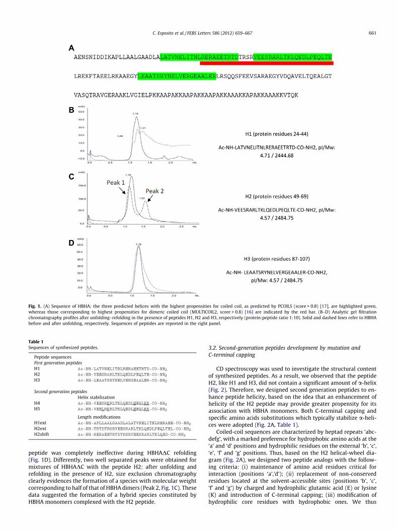
Fig. 1. (A) Sequence of HBHA; the three predicted helices with the highest propensities for coiled coil, as predicted by PCOILS (score > 0.8) [17], are highlighted green,whereas those corresponding to highest propensities for dimeric coiled coil (MULTICOIL2, score > 0.8) [16] are indicated by the red bar. (B–D) Analytic gel filtrationchromatography profiles after unfolding–refolding in the presence of peptides H1, H2 and H3, respectively (protein:peptide ratio 1:10). Solid and dashed lines refer to HBHAbefore and after unfolding, respectively. Sequences of peptides are reported in the right panel.
Table 1Sequences of synthesized peptides.
Peptide sequencesFirst generation peptidesH1 Ac-NH-LATVNELITNLRERAEETRTD-CO-NH2
H2 Ac-NH-VEESRARLTKLQEDLPEQLTE-CO-NH2
H3 Ac-NH-LEAATSRYNELVERGEAALER-CO-NH2
Second generation peptidesHelix stabilization
H4 Ac-NH-VEESRKRLTKLQEDLEEQLKK-CO-NH2
H5 Ac-NH-VEELRKRLTKLQEDLEEQLKK-CO-NH2
Length modificationsH1ext Ac-NH-APLLAALGAADLALATVNELITNLRERAEE-CO-NH2
H2ext Ac-NH-TRTDTRSRVEESRARLTKLQEDLPEQLTEL-CO-NH2
H2shift Ac-NH-RERAEETRTDTRSRVEESRARLTKLQED-CO-NH2
C. Esposito et al. / FEBS Letters 586 (2012) 659–667 661
peptide was completely ineffective during HBHADC refolding(Fig. 1D). Differently, two well separated peaks were obtained formixtures of HBHADC with the peptide H2: after unfolding andrefolding in the presence of H2, size exclusion chromatographyclearly evidences the formation of a species with molecular weightcorresponding to half of that of HBHA dimers (Peak 2, Fig. 1C). Thesedata suggested the formation of a hybrid species constituted byHBHA monomers complexed with the H2 peptide.
3.2. Second-generation peptides development by mutation andC-terminal capping
CD spectroscopy was used to investigate the structural contentof synthesized peptides. As a result, we observed that the peptideH2, like H1 and H3, did not contain a significant amount of a-helix(Fig. 2). Therefore, we designed second generation peptides to en-hance peptide helicity, based on the idea that an enhancement ofhelicity of the H2 peptide may provide greater propensity for itsassociation with HBHA monomers. Both C-terminal capping andspecific amino acids substitutions which typically stabilize a-heli-ces were adopted (Fig. 2A, Table 1).
Coiled-coil sequences are characterized by heptad repeats ‘abc-defg’, with a marked preference for hydrophobic amino acids at the‘a’ and ‘d’ positions and hydrophilic residues on the external ‘b’, ‘c’,‘e’, ‘f’ and ‘g’ positions. Thus, based on the H2 helical-wheel dia-gram (Fig. 2A), we designed two peptide analogs with the follow-ing criteria: (i) maintenance of amino acid residues critical forinteraction (positions ‘a’,’d’); (ii) replacement of non-conservedresidues located at the solvent-accessible sites (positions ‘b’, ‘c’,‘f’ and ‘g’) by charged and hydrophilic glutamic acid (E) or lysine(K) and introduction of C-terminal capping; (iii) modification ofhydrophilic core residues with hydrophobic ones. We thus
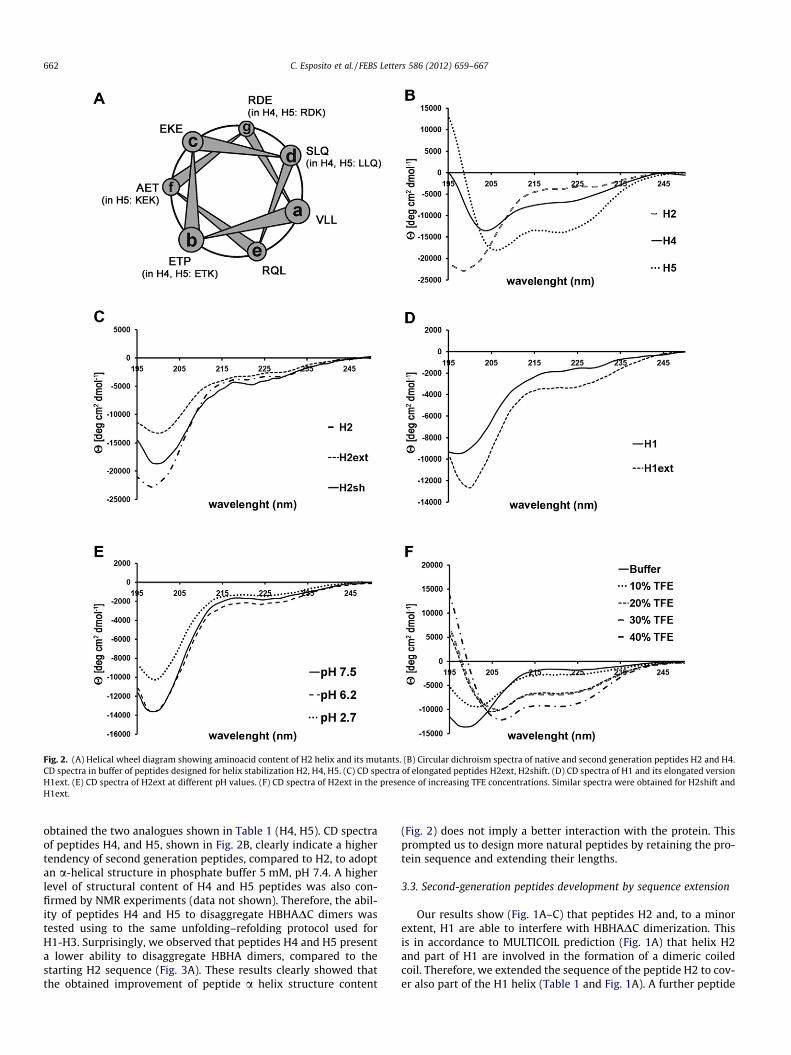
Fig. 2. (A) Helical wheel diagram showing aminoacid content of H2 helix and its mutants. (B) Circular dichroism spectra of native and second generation peptides H2 and H4.CD spectra in buffer of peptides designed for helix stabilization H2, H4, H5. (C) CD spectra of elongated peptides H2ext, H2shift. (D) CD spectra of H1 and its elongated versionH1ext. (E) CD spectra of H2ext at different pH values. (F) CD spectra of H2ext in the presence of increasing TFE concentrations. Similar spectra were obtained for H2shift andH1ext.
662 C. Esposito et al. / FEBS Letters 586 (2012) 659–667
obtained the two analogues shown in Table 1 (H4, H5). CD spectraof peptides H4, and H5, shown in Fig. 2B, clearly indicate a highertendency of second generation peptides, compared to H2, to adoptan a-helical structure in phosphate buffer 5 mM, pH 7.4. A higherlevel of structural content of H4 and H5 peptides was also con-firmed by NMR experiments (data not shown). Therefore, the abil-ity of peptides H4 and H5 to disaggregate HBHADC dimers wastested using to the same unfolding–refolding protocol used forH1-H3. Surprisingly, we observed that peptides H4 and H5 presenta lower ability to disaggregate HBHA dimers, compared to thestarting H2 sequence (Fig. 3A). These results clearly showed thatthe obtained improvement of peptide a helix structure content
(Fig. 2) does not imply a better interaction with the protein. Thisprompted us to design more natural peptides by retaining the pro-tein sequence and extending their lengths.
3.3. Second-generation peptides development by sequence extension
Our results show (Fig. 1A–C) that peptides H2 and, to a minorextent, H1 are able to interfere with HBHADC dimerization. Thisis in accordance to MULTICOIL prediction (Fig. 1A) that helix H2and part of H1 are involved in the formation of a dimeric coiledcoil. Therefore, we extended the sequence of the peptide H2 to cov-er also part of the H1 helix (Table 1 and Fig. 1A). A further peptide

C. Esposito et al. / FEBS Letters 586 (2012) 659–667 663
was synthesized by shortening the C-terminus and further elongat-ing the N-terminus, to eliminate the proline residue (P64 in HBHAsequence) in the peptide sequence, since prolines are known helixbreaker residues (H2shift, Table 1). We also analyzed the effectsproduced after peptide elongation of H1. The peptide H1ext wassignificantly elongated at the N-terminus (Table 1). CD spectra ofpeptides H2ext, H2shift, H1ext, shown in Fig. 2, clearly show thatsequence extension did not improve peptide structuration. Indeed,CD spectra recorded at different pH values evidenced a randomconformation for all peptides (Fig. 2E). When we used increasingpercentages of TFE, a helical enhancer, CD spectra show the typicalfeatures of a-helix conformations, this indicating a tendency ofpeptides to form a-helix conformations (Fig. 2F).
All synthesized peptides were checked for their ability to disag-gregate HBHADC dimers. Interestingly, elongation of H1 (peptideH1ext) provided only a small improvement in HBHA disaggrega-tion (Fig. 3B), whereas the elongation of H2 to obtain H2shift pro-duced no effect at all (Fig. 3D). Differently, elongation of H2 to
Fig. 3. Analytic gel filtration chromatography profiles after unfolding–refolding in thepeptide ratio of 1:10. Solid and dashed lines refer to HBHA before and after unfolding, rrefolding protocol, using protein:H2ext ratios 1:6 (lane 2), 1:8 (lane 3), 1:12 (lane 4). Thprofiles after unfolding–refolding in the presence of peptides H2ext on a preparative Su
obtain H2ext resulted in a striking enhancement of ratios betweendisaggregated and dimeric HBHA forms (Fig. 3C). Indeed, H2extwas able to disrupt more than 80% of HBHA dimers, to formHBHA–peptide hybrids.
HBHA dimer disaggregation after unfolding–refolding ofHBHADC:H2ext mixture was also analyzed using native gel electro-phoresis, with protein:peptide ratios ranging from 1:6 to 1:12(Fig. 3E). As a result, native gels show two extra bands, comparedto the native gel of the sole protein (Fig. 3E). The upper band corre-sponds to a monomeric state of HBHA whereas the intermediateband likely corresponds to a different conformational state of HBHAdimers, induced by H2ext (Fig. 3E). Preparative scale purification ofthe protein–peptide hybrid was carried out using the unfolding–refolding protocol described above (See Section 2). The size exclu-sion chromatography experiments, performed using a Superdex75 10/30 column, provided a profile with two well separated peaks(1 and 2 in Fig. 3F), which were collected and used for both HPLCchromatography and far-ultraviolet (UV) CD analysis. Using HPLC
presence of peptides H4 (A), H1ext (B) H2ext (C) and H2shift (D) using a protein:espectively. (E) Native gel electrophoresis of HBHADC mixture after the unfolding–e reference lane (lane 1) refers to HBHADC alone. (F) Gel filtration chromatographyperdex 75 10/30 column.
664 C. Esposito et al. / FEBS Letters 586 (2012) 659–667
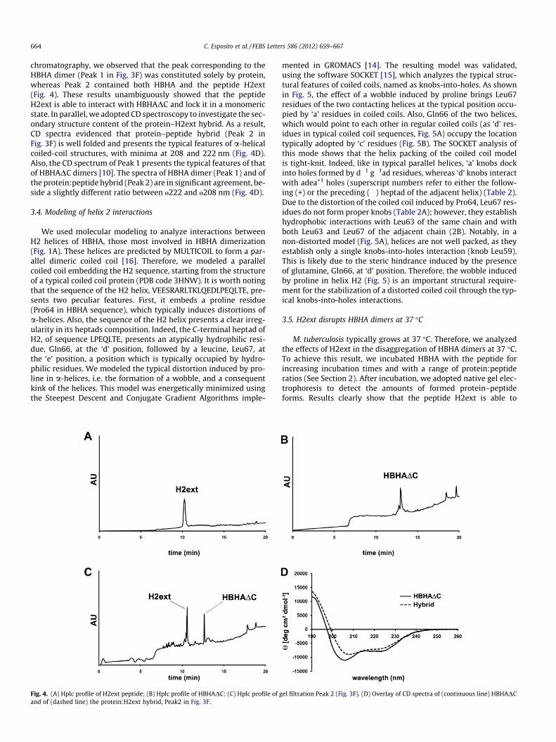
chromatography, we observed that the peak corresponding to theHBHA dimer (Peak 1 in Fig. 3F) was constituted solely by protein,whereas Peak 2 contained both HBHA and the peptide H2ext(Fig. 4). These results unambiguously showed that the peptideH2ext is able to interact with HBHADC and lock it in a monomericstate. In parallel, we adopted CD spectroscopy to investigate the sec-ondary structure content of the protein–H2ext hybrid. As a result,CD spectra evidenced that protein–peptide hybrid (Peak 2 inFig. 3F) is well folded and presents the typical features of a-helicalcoiled-coil structures, with minima at 208 and 222 nm (Fig. 4D).Also, the CD spectrum of Peak 1 presents the typical features of thatof HBHADC dimers [10]. The spectra of HBHA dimer (Peak 1) and ofthe protein:peptide hybrid (Peak 2) are in significant agreement, be-side a slightly different ratio between „222 and „208 nm (Fig. 4D).
3.4. Modeling of helix 2 interactions
We used molecular modeling to analyze interactions betweenH2 helices of HBHA, those most involved in HBHA dimerization(Fig. 1A). These helices are predicted by MULTICOIL to form a par-allel dimeric coiled coil [16]. Therefore, we modeled a parallelcoiled coil embedding the H2 sequence, starting from the structureof a typical coiled coil protein (PDB code 3HNW). It is worth notingthat the sequence of the H2 helix, VEESRARLTKLQEDLPEQLTE, pre-sents two peculiar features. First, it embeds a proline residue(Pro64 in HBHA sequence), which typically induces distortions ofa-helices. Also, the sequence of the H2 helix presents a clear irreg-ularity in its heptads composition. Indeed, the C-terminal heptad ofH2, of sequence LPEQLTE, presents an atypically hydrophilic resi-due, Gln66, at the ‘d’ position, followed by a leucine, Leu67, atthe ‘e’ position, a position which is typically occupied by hydro-philic residues. We modeled the typical distortion induced by pro-line in a-helices, i.e. the formation of a wobble, and a consequentkink of the helices. This model was energetically minimized usingthe Steepest Descent and Conjugate Gradient Algorithms imple-
Fig. 4. (A) Hplc profile of H2ext peptide; (B) Hplc profile of HBHADC; (C) Hplc profile of gand of (dashed line) the protein:H2ext hybrid, Peak2 in Fig. 3F.
mented in GROMACS [14]. The resulting model was validated,using the software SOCKET [15], which analyzes the typical struc-tural features of coiled coils, named as knobs-into-holes. As shownin Fig. 5, the effect of a wobble induced by proline brings Leu67residues of the two contacting helices at the typical position occu-pied by ‘a’ residues in coiled coils. Also, Gln66 of the two helices,which would point to each other in regular coiled coils (as ‘d’ res-idues in typical coiled coil sequences, Fig. 5A) occupy the locationtypically adopted by ‘c’ residues (Fig. 5B). The SOCKET analysis ofthis mode shows that the helix packing of the coiled coil modelis tight-knit. Indeed, like in typical parallel helices, ‘a’ knobs dockinto holes formed by d�1 g�1ad residues, whereas ‘d’ knobs interactwith adea+1 holes (superscript numbers refer to either the follow-ing (+) or the preceding (�) heptad of the adjacent helix) (Table 2).Due to the distortion of the coiled coil induced by Pro64, Leu67 res-idues do not form proper knobs (Table 2A); however, they establishhydrophobic interactions with Leu63 of the same chain and withboth Leu63 and Leu67 of the adjacent chain (2B). Notably, in anon-distorted model (Fig. 5A), helices are not well packed, as theyestablish only a single knobs-into-holes interaction (knob Leu59).This is likely due to the steric hindrance induced by the presenceof glutamine, Gln66, at ‘d’ position. Therefore, the wobble inducedby proline in helix H2 (Fig. 5) is an important structural require-ment for the stabilization of a distorted coiled coil through the typ-ical knobs-into-holes interactions.
3.5. H2ext disrupts HBHA dimers at 37 �C
M. tuberculosis typically grows at 37 �C. Therefore, we analyzedthe effects of H2ext in the disaggregation of HBHA dimers at 37 �C.To achieve this result, we incubated HBHA with the peptide forincreasing incubation times and with a range of protein:peptideratios (See Section 2). After incubation, we adopted native gel elec-trophoresis to detect the amounts of formed protein–peptideforms. Results clearly show that the peptide H2ext is able to
el filtration Peak 2 (Fig. 3F). (D) Overlay of CD spectra of (continuous line) HBHADC

Fig. 5. (A) Side view (top) and top view (bottom) of a hypothetical non-distorted coiled coil. (B) Side view (top) and top view (bottom) of the wobbled coiled coil. Red circles inbottom panels indicate typical positions occupied by ‘a’ and ‘d’ residues in coiled coils.
Table 2(A) Knobs-into-hole interactions in the coiled coil model; (B) Hydrophobic coreinteractions.
(A)Knobs (chain A) Holes (chain B)Leu56 (a) Ser52 (a), Arg55 (g), Leu56 (a), Leu59 (d)Leu59 (d) Leu56 (a), Leu59 (d), Gln60 (e), Leu63 (a)
Knobs (chain B) Holes (chain A)Leu56 (a) Ser52 (a), Arg55 (g), Leu56 (a), Leu59 (d)Leu59 (d) Leu56 (a), Leu59 (d), Gln60 (e), Leu63 (a)
(B)Leu56 (chain A) Leu56 (chain B), Leu59 (chain B)Leu59 (chain A) Leu59 (chain B), Leu63 (chain B)Leu63 (chain A) Leu59 (chain B), Leu63 (chain B), Leu67 (chain A)Leu67 (chain A) Leu63 (chain B), Leu63 (chain A), Leu67 (chain B)
C. Esposito et al. / FEBS Letters 586 (2012) 659–667 665
disrupt HBHA dimers also at 37 �C (Fig. 6A) and significantamounts of hybrid species are evident also after short incubationtimes (about 2 h). Parallel experiments carried out with peptidesH4 and H5 did not produce detectable amounts of protein peptide
hybrids (Fig. 6B). Notably, the native gel obtained after incubationof the protein with H2ext at 37 �C (Fig. 6A) presents the same fea-tures observed after unfolding–refolding (Fig. 3E). This suggeststhat the unfolding–refolding procedure well reproduces the con-formational states generated at 37 �C by the presence of H2ext.
4. Discussion
Bacterial aggregation and cell adhesion mediated by HBHA aretwo important steps of TB pathogenesis. Mtb is able to escape fromthe phagosome and translocate into the cytosol, where it can befound in tightly packed clumps [18] . Whether aggregation occursduring the intracellular life or once inside the cytosol is hithertonot clear. However, HBHA-mediated aggregation is a strong instru-ment for the formation of clumps of bacilli, which more effectivelyadhere and invade epithelial cells than single bacilli. It has beensuggested that HBHA may interact with cytosolic components ofthe host cells such as actin [19]. Also in this case, bacterialaggregation likely ensures a more efficient binding of bacterialcells to their host targets.

Fig. 6. Native gel electrophoresis at 37 �C (A) 60 lg of HBHADC (lane 1) wereincubated for two hours with protein: H2ext ratios 1:6 (lane 2), 1:8 (lane 3), 1:12(lane 4). (B) 60 lg of HBHADC (lane 1) were incubated overnight with peptides H4(lane 2), H5 (lane 3) and H2shift (lane 4) with 1:8 protein:peptide ratio.
666 C. Esposito et al. / FEBS Letters 586 (2012) 659–667
It was previously shown that homophilic HBHA–HBHA interact-ing forces occur at the bacterial surface of live mycobacteria [9].Several studies have suggested that these interactions are mediatedby the coiled coil regions of HBHA [10,11], which likely stabilizeHBHA dimer formation. Therefore, a strong correlation exists be-tween HBHA dimerization and its ability to induce bacterial aggre-gation. In this framework, we here mapped molecular determinantsof HBHA dimerization by combining peptide synthesis with bio-chemical and computational studies. We designed and synthesizedseveral peptides which mimicked the most critical predicted re-gions in HBHA dimerization. As a result, we identified a peptide,covering a specific region of HBHA sequence (residues 41–70, pep-tide H2ext), which strongly competes with protein dimerization bymimicking the protein dimerization interface. This peptide is ableto disaggregate HBHA dimers during the protein refolding process,with the formation of species with a lower molecular weight (Figs.3C,E,F). We proved that this species is a hybrid protein–peptideform (HPLC analysis, Fig. 4A–C) with a well-preserved secondarystructure (CD spectroscopy, Fig. 4D). Notably, the ability of H2extto disaggregate HBHA dimers and form a hybrid form is preservedat 37 �C, the growth temperature of Mtb.
To rationalize the different behavior of our synthesized peptidesand to identify critical regions of HBHA monomer–monomer recog-nition, we performed molecular modeling of the helix which is mostinvolved in HBHA dimerization (helix H2). These studies clearlyidentified a peculiar structural feature of H2, which embeds a Proresidue at its C-terminus. The proline residue, traditionally consid-ered as a ‘helix breaker’, is well represented in helices of transmem-brane proteins [20], where it plays the crucial role of conferringflexibility in the local conformational dynamics of transmembranesegments. We modeled a coiled coil formed by kinked helices, inwhich a wobble angle exists between the postproline helix seg-ment, with respect to the preproline segment. Energy minimizationof this model provided a well-packed coiled coil, which explains theanomalous presence of Gln66 at the ‘d’ position and of Leu67 at the‘e’ of the ‘abcdef’ typical coiled coil heptad (Fig. 5). A helical kink in-duced by proline was previously observed in the coiled coil portionof Dynein, a motor protein that transports cellular cargo along themicrotubules within the cytoplasm [21]. Notably, as observed forDynein, the proline residue responsible for the helical kink, Pro64,is strictly conserved in bacteria (data not shown). These consider-ations strongly suggest that the kink of H2 helices is an important
structural determinant for HBHA monomer–monomer recognition.To enforce this observation, peptides H4 and H5, which contain aproline mutation, are barely able to disaggregate HBHA dimers(Fig. 6), despite their higher helical content, compared to H2 andH2ext (Fig. 2). Similar considerations apply to the peptide H2shift,which embeds the protein sequence immediately preceding Pro64(Table 1 and Fig. 3A, C, and D). In conclusion, we identified the firstmolecule (H2ext) able to disaggregate HBHA dimers, and likely toseverely interfere with Mtb agglutination. The ability of the peptideH2ext (embedding Pro64) to disaggregate HBHA dimers at thegrowing temperature of Mtb has a strong implication for the devel-opment of anti-agglutinating molecules with therapeutic interest.These molecules will likely weaken Mtb agglutination-driven de-fense mechanisms and have a strong impact on the Mtb adhesionto epithelial cells.
Acknowledgements
This work was funded by MIUR (PRIN 2009–prot. 200993WWF9). R.B. also acknowledges the COST action BM1003 ‘‘Microbialcell surface determinants of virulence as targets for new therapeu-tics in Cystic Fibrosis’’.
Appendix A. Supplementary data
Supplementary data (Supplementary Materials and Methods)associated with this article can be found, in the online version, atdoi:10.1016/j.febslet.2012.01.047.
References
[1] Russell, D.G., Barry 3rd, C.E. and Flynn, J.L. (2010) Tuberculosis: what we don’tknow can, and does, hurt us. Science 328, 852–856.
[2] Kana, B.D. et al. (2008) The resuscitation-promoting factors of Mycobacteriumtuberculosis are required for virulence and resuscitation from dormancy butare collectively dispensable for growth in vitro. Mol Microbiol 67, 672–684.
[3] Biketov, S., Potapov, V., Ganina, E., Downing, K., Kana, B.D. and Kaprelyants, A.(2007) The role of resuscitation promoting factors in pathogenesis andreactivation of Mycobacterium tuberculosis during intra-peritoneal infectionin mice. BMC Infect Dis 7, 146.
[4] Menozzi, F.D., Reddy, V.M., Cayet, D., Raze, D., Debrie, A.S., Dehouck, M.P.,Cecchelli, R. and Locht, C. (2006) Mycobacterium tuberculosis heparin-bindinghaemagglutinin adhesin (HBHA) triggers receptor-mediated transcytosiswithout altering the integrity of tight junctions. Microbes Infect 8, 1–9.
[5] Menozzi, F.D., Bischoff, R., Fort, E., Brennan, M.J. and Locht, C. (1998) Molecularcharacterization of the mycobacterial heparin-binding hemagglutinin, amycobacterial adhesin. Proc Natl Acad Sci USA 95, 12625–12630.
[6] Pethe, K., Alonso, S., Biet, F., Delogu, G., Brennan, M.J., Locht, C. and Menozzi,F.D. (2001) The heparin-binding haemagglutinin of M. tuberculosis is requiredfor extrapulmonary dissemination. Nature 412, 190–194.
[7] Pethe, K., Aumercier, M., Fort, E., Gatot, C., Locht, C. and Menozzi, F.D. (2000)Characterization of the heparin-binding site of the mycobacterial heparin-binding hemagglutinin adhesin. J Biol Chem 275, 14273–14280.
[8] Delogu, G. and Brennan, M.J. (1999) Functional domains present in themycobacterial hemagglutinin, HBHA. J Bacteriol 181, 7464–7469.
[9] Verbelen, C., Raze, D., Dewitte, F., Locht, C. and Dufrene, Y.F. (2007) Single-molecule force spectroscopy of mycobacterial adhesin–adhesin interactions. JBacteriol 189, 8801–8806.
[10] Esposito, C., Pethoukov, M.V., Svergun, D.I., Ruggiero, A., Pedone, C., Pedone, E.and Berisio, R. (2008) Evidence for an elongated dimeric structure of heparin-binding hemagglutinin from Mycobacterium tuberculosis. J Bacteriol 190,4749–4753.
[11] Esposito, C., Carullo, P., Pedone, E., Graziano, G., Del Vecchio, P. and Berisio, R.(2010) Dimerisation and structural integrity of Heparin BindingHemagglutinin A from Mycobacterium tuberculosis: implications for bacterialagglutination. FEBS Lett 584, 1091–1096.
[12] Soba, P. et al. (2005) Homo- and heterodimerization of APP family memberspromotes intercellular adhesion. Embo J 24, 3624–3634.
[13] Mittal, N., Budrene, E.O., Brenner, M.P. and Van Oudenaarden, A. (2003)Motility of Escherichia coli cells in clusters formed by chemotactic aggregation.Proc Natl Acad Sci USA 100, 13259–13263.
[14] Van Der Spoel, D., Lindahl, E., Hess, B., Groenhof, G., Mark, A.E. and Berendsen,H.J. (2005) GROMACS: fast, flexible, and free. J Comput Chem 26, 1701–1718.
[15] Walshaw, J. and Woolfson, D.N. (2001) Socket: a program for identifying andanalysing coiled-coil motifs within protein structures. J Mol Biol 307, 1427–1450.

C. Esposito et al. / FEBS Letters 586 (2012) 659–667 667
[16] Trigg, J., Gutwin, K., Keating, A.E. and Berger, B. (2011) Multicoil2: predictingcoiled coils and their oligomerization states from sequence in the twilightzone. PLoS ONE 6, e23519.
[17] Gruber, M., Soding, J. and Lupas, A.N. (2006) Comparative analysis of coiled-coil prediction methods. J Struct Biol 155, 140–145.
[18] van der Wel, N., Hava, D., Houben, D., Fluitsma, D., van Zon, M., Pierson, J.,Brenner, M. and Peters, P.J. (2007) M. tuberculosis and M. leprae translocatefrom the phagolysosome to the cytosol in myeloid cells. Cell 129, 1287–1298.
[19] Esposito, C., Marasco, D., Delogu, G., Pedone, E. and Berisio, R. (2011) Heparin-binding hemagglutinin HBHA from Mycobacterium tuberculosis affects actinpolymerisation. Biochem Biophys Res Commun 410, 339–344.
[20] Cordes, F.S., Bright, J.N. and Sansom, M.S. (2002) Proline-induced distortions oftransmembrane helices. J Mol Biol 323, 951–960.
[21] Carter, A.P., Garbarino, J.E., Wilson-Kubalek, E.M., Shipley, W.E., Cho, C.,Milligan, R.A., Vale, R.D. and Gibbons, I.R. (2008) Structure and functional roleof dynein’s microtubule-binding domain. Science 322, 1691–1695.
Related Documents