RESEARCH ARTICLE Open Access Molecular organization and chromosomal localization of 5S rDNA in Amazonian Engystomops (Anura, Leiuperidae) Débora Silva Rodrigues 1* , Miryan Rivera 2 and Luciana Bolsoni Lourenço 1 Abstract Background: For anurans, knowledge of 5S rDNA is scarce. For Engystomops species, chromosomal homeologies are difficult to recognize due to the high level of inter- and intraspecific cytogenetic variation. In an attempt to better compare the karyotypes of the Amazonian species Engystomops freibergi and Engystomops petersi, and to extend the knowledge of 5S rDNA organization in anurans, the 5S rDNA sequences of Amazonian Engystomops species were isolated, characterized, and mapped. Results: Two types of 5S rDNA, which were readily differentiated by their NTS (non-transcribed spacer) sizes and compositions, were isolated from specimens of E. freibergi from Brazil and E. petersi from two Ecuadorian localities (Puyo and Yasuní). In the E. freibergi karyotypes, the entire type I 5S rDNA repeating unit hybridized to the pericentromeric region of 3p, whereas the entire type II 5S rDNA repeating unit mapped to the distal region of 6q, suggesting a differential localization of these sequences. The type I NTS probe clearly detected the 3p pericentromeric region in the karyotypes of E. freibergi and E. petersi from Puyo and the 5p pericentromeric region in the karyotype of E. petersi from Yasuní, but no distal or interstitial signals were observed. Interestingly, this probe also detected many centromeric regions in the three karyotypes, suggesting the presence of a satellite DNA family derived from 5S rDNA. The type II NTS probe detected only distal 6q regions in the three karyotypes, corroborating the differential distribution of the two types of 5S rDNA. Conclusions: Because the 5S rDNA types found in Engystomops are related to those of Physalaemus with respect to their nucleotide sequences and chromosomal locations, their origin likely preceded the evolutionary divergence of these genera. In addition, our data indicated homeology between Chromosome 5 in E. petersi from Yasuní and Chromosomes 3 in E. freibergi and E. petersi from Puyo. In addition, the chromosomal location of the type II 5S rDNA corroborates the hypothesis that the Chromosomes 6 of E. petersi and E. freibergi are homeologous despite the great differences observed between the karyotypes of the Yasuní specimens and the others. Keywords: 5S rDNA, non-transcribed spacer, chromosome, Engystomops Background Ribosomal RNAs (rRNA) are molecules that combine to form the basic structures of the small and large riboso- mal subunits. rRNAs are transcribed by two distinct multigene families. The 45S rDNA family synthesizes the 18S, 5.8S and 28S rRNAs, and the 5S rDNA family transcribes the 5S rRNA (as reviewed in references [1-3]). The 5S ribosomal genes are found as conserved copies of 120-bp sequences arranged in tandem and interspersed with variable non-transcribed spacers (NTSs) that differ in length and nucleotide composition (see reviews in [3] and [4]). Within the conserved 120- bp sequence lies an internal control region (ICR) con- sisting of 3 characteristic regions: the A box, an inter- mediate element (IE), and the C box [5]. These regions act as promoters for transcription; the A box is a gen- eral binding sequence for RNA polymerase III, and the intermediate element and the C box are interaction sites * Correspondence: [email protected] 1 Department of Structural and Functional Biology, Institute of Biology, University of Campinas (UNICAMP), Campinas, SP 13083-863, Brazil Full list of author information is available at the end of the article Rodrigues et al. BMC Genetics 2012, 13:17 http://www.biomedcentral.com/1471-2156/13/17 © 2012 Rodrigues et al; licensee BioMed Central Ltd. This is an Open Access article distributed under the terms of the Creative Commons Attribution License (http://creativecommons.org/licenses/by/2.0), which permits unrestricted use, distribution, and reproduction in any medium, provided the original work is properly cited.

Welcome message from author
This document is posted to help you gain knowledge. Please leave a comment to let me know what you think about it! Share it to your friends and learn new things together.
Transcript

RESEARCH ARTICLE Open Access
Molecular organization and chromosomallocalization of 5S rDNA in AmazonianEngystomops (Anura, Leiuperidae)Débora Silva Rodrigues1*, Miryan Rivera2 and Luciana Bolsoni Lourenço1
Abstract
Background: For anurans, knowledge of 5S rDNA is scarce. For Engystomops species, chromosomal homeologiesare difficult to recognize due to the high level of inter- and intraspecific cytogenetic variation. In an attempt tobetter compare the karyotypes of the Amazonian species Engystomops freibergi and Engystomops petersi, and toextend the knowledge of 5S rDNA organization in anurans, the 5S rDNA sequences of Amazonian Engystomopsspecies were isolated, characterized, and mapped.
Results: Two types of 5S rDNA, which were readily differentiated by their NTS (non-transcribed spacer) sizes andcompositions, were isolated from specimens of E. freibergi from Brazil and E. petersi from two Ecuadorian localities(Puyo and Yasuní). In the E. freibergi karyotypes, the entire type I 5S rDNA repeating unit hybridized to thepericentromeric region of 3p, whereas the entire type II 5S rDNA repeating unit mapped to the distal region of 6q,suggesting a differential localization of these sequences. The type I NTS probe clearly detected the 3ppericentromeric region in the karyotypes of E. freibergi and E. petersi from Puyo and the 5p pericentromeric regionin the karyotype of E. petersi from Yasuní, but no distal or interstitial signals were observed. Interestingly, this probealso detected many centromeric regions in the three karyotypes, suggesting the presence of a satellite DNA familyderived from 5S rDNA. The type II NTS probe detected only distal 6q regions in the three karyotypes, corroboratingthe differential distribution of the two types of 5S rDNA.
Conclusions: Because the 5S rDNA types found in Engystomops are related to those of Physalaemus with respectto their nucleotide sequences and chromosomal locations, their origin likely preceded the evolutionary divergenceof these genera. In addition, our data indicated homeology between Chromosome 5 in E. petersi from Yasuní andChromosomes 3 in E. freibergi and E. petersi from Puyo. In addition, the chromosomal location of the type II 5SrDNA corroborates the hypothesis that the Chromosomes 6 of E. petersi and E. freibergi are homeologous despitethe great differences observed between the karyotypes of the Yasuní specimens and the others.
Keywords: 5S rDNA, non-transcribed spacer, chromosome, Engystomops
BackgroundRibosomal RNAs (rRNA) are molecules that combine toform the basic structures of the small and large riboso-mal subunits. rRNAs are transcribed by two distinctmultigene families. The 45S rDNA family synthesizesthe 18S, 5.8S and 28S rRNAs, and the 5S rDNA familytranscribes the 5S rRNA (as reviewed in references
[1-3]). The 5S ribosomal genes are found as conservedcopies of 120-bp sequences arranged in tandem andinterspersed with variable non-transcribed spacers(NTSs) that differ in length and nucleotide composition(see reviews in [3] and [4]). Within the conserved 120-bp sequence lies an internal control region (ICR) con-sisting of 3 characteristic regions: the A box, an inter-mediate element (IE), and the C box [5]. These regionsact as promoters for transcription; the A box is a gen-eral binding sequence for RNA polymerase III, and theintermediate element and the C box are interaction sites
* Correspondence: [email protected] of Structural and Functional Biology, Institute of Biology,University of Campinas (UNICAMP), Campinas, SP 13083-863, BrazilFull list of author information is available at the end of the article
Rodrigues et al. BMC Genetics 2012, 13:17http://www.biomedcentral.com/1471-2156/13/17
© 2012 Rodrigues et al; licensee BioMed Central Ltd. This is an Open Access article distributed under the terms of the CreativeCommons Attribution License (http://creativecommons.org/licenses/by/2.0), which permits unrestricted use, distribution, andreproduction in any medium, provided the original work is properly cited.
for the transcription factor TFIIIA [5]. Another charac-teristic typically found in the presumed-functional 5Sgenes is a poly-T terminator region, as initially reportedby Korn and Brown [6].Despite the large variations observed for the NTSs
found in 5S rDNA, several studies have reported thatconserved elements are present in these regions andthat they play an important role in the regulation of 5SrRNA gene expression, similar to TATA-like sequences[3], oligonucleotide sequences [6], and the D box foundin mammals [7,8]. It has also been reported that a Clocalized at position -1 of many previously described 5SrRNA gene sequences guarantees a correct and efficienttranscription start [8].The sequences of 5S rDNA have been used as genetic
and cytogenetic markers in evolutionary studies and inthe identification and comparison of species, hybridsand strains [9-13]. The 120-bp region is highly con-served even among unrelated species, making it possibleto isolate the 5S rDNA repeats of several species basedon the available sequence from another species that isnot necessarily closely related. An interesting feature ofthe 5S rDNA NTS that is most likely widespreadthroughout all vertebrate groups is the presence of twodifferently sized sequences, a subject that has beenextensively studied in fish (see reviews in [3,14,15]).For anurans, knowledge combining the molecular
organization of the 5S rDNA sequences and their chro-mosomal locations are known for Xenopus laevis, X.borealis, X. muelleri [16,17], n families Ascaphidae, Dis-coglossidae e Ranidae [13,17], Physalaemus ephippifer[18] and Physalaemus cuvieri [19]. In Xenopus species[20] and P. cuvieri [19], two types of 5S rDNAsequences were found and mapped to distinct chromo-somal sites. In Xenopus species, the distinct types of 5SrDNA, named oocyte- and somatic types, show a differ-ential pattern of gene expression [6,16,17,21-26]. Inaddition to these cases with combined molecular andchromosomal analyses, studies restricted to the nucleo-tide sequence of the transcribed region of the 5S rDNAhave been reported for the anurans Gastrotheca riobam-bae [27], Bufo americanus, Rana pipiens and Ranacatesbeiana [28].Based on a genetic analysis of morphological data,
the Engystomops genus was revalidated by Nascimentoet al. [29] to allocate species previously grouped in thePhysalaemus pustulosus group. Currently, nine speciescomprise the Engystomops genus (the source studiesare cited in reference [30]), and some reproductive andgenetic evidences suggest the occurrence of unde-scribed cryptic species among the Amazonian Engysto-mops [31-33]. The cytogenetic analyses of differentpopulations of the Amazonian species Engystomopspetersi and Engystomops freibergi revealed interesting
inter- and intraspecific divergences, and the possibleinvolvement of these cytogenetic variations in incipientspeciation has been suggested [33,34]. Nevertheless,the lack of available cytogenetic markers prevents theproper identification of chromosomal homeologies inthese populations, and consequently, the hypothesizedrearrangements responsible for the karyotypic diver-gences remain unknown. In the present study, we iso-lated and characterized the 5S rDNA sequences of E.freibergi and two populations of E. petersi and mappedtheir chromosomal locations, extending the knowledgeof 5S rDNA organization at the genomic and chromo-somal levels in these amphibians. These data facilitatethe identification of possible homeologous chromo-somes among the Engystomops, serving as an impor-tant contribution for further studies on chromosomaldivergence in anurans.
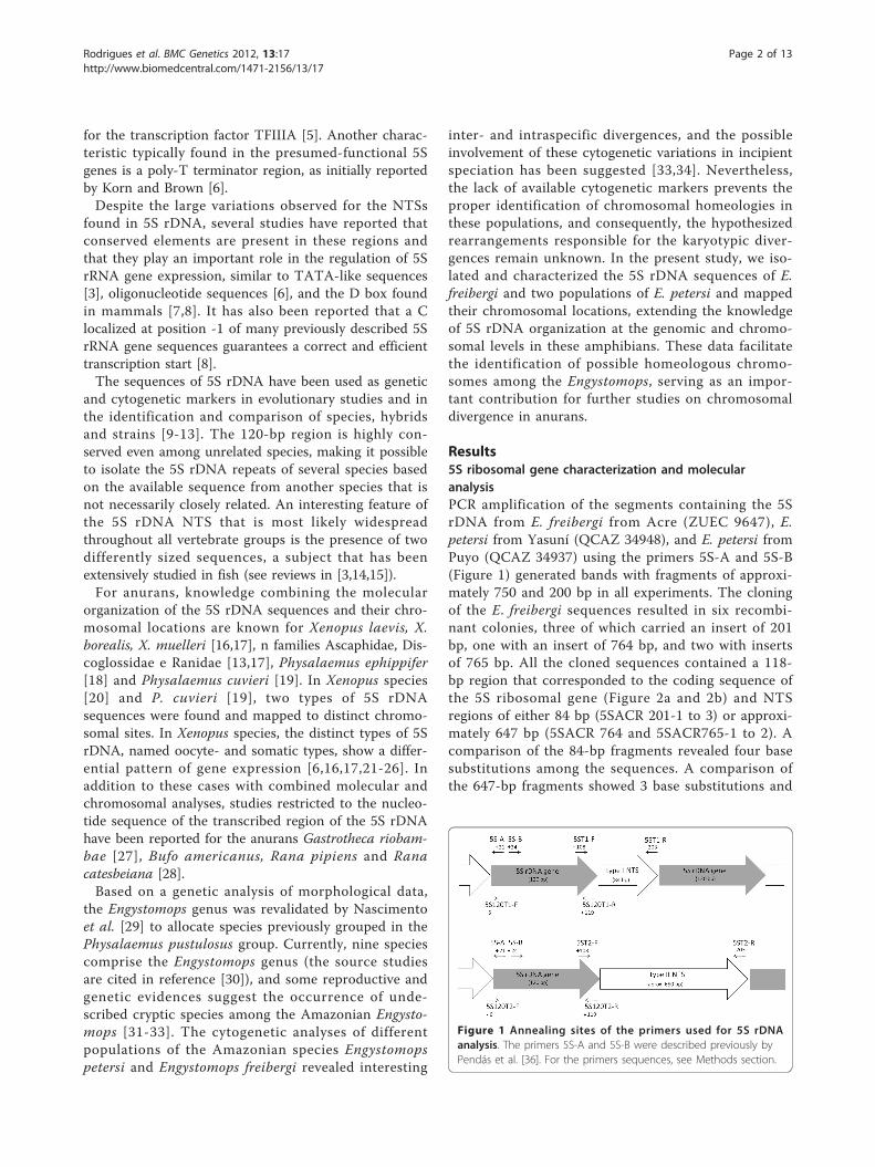
Results5S ribosomal gene characterization and molecularanalysisPCR amplification of the segments containing the 5SrDNA from E. freibergi from Acre (ZUEC 9647), E.petersi from Yasuní (QCAZ 34948), and E. petersi fromPuyo (QCAZ 34937) using the primers 5S-A and 5S-B(Figure 1) generated bands with fragments of approxi-mately 750 and 200 bp in all experiments. The cloningof the E. freibergi sequences resulted in six recombi-nant colonies, three of which carried an insert of 201bp, one with an insert of 764 bp, and two with insertsof 765 bp. All the cloned sequences contained a 118-bp region that corresponded to the coding sequence ofthe 5S ribosomal gene (Figure 2a and 2b) and NTSregions of either 84 bp (5SACR 201-1 to 3) or approxi-mately 647 bp (5SACR 764 and 5SACR765-1 to 2). Acomparison of the 84-bp fragments revealed four basesubstitutions among the sequences. A comparison ofthe 647-bp fragments showed 3 base substitutions and
Figure 1 Annealing sites of the primers used for 5S rDNAanalysis. The primers 5S-A and 5S-B were described previously byPendás et al. [36]. For the primers sequences, see Methods section.
Rodrigues et al. BMC Genetics 2012, 13:17http://www.biomedcentral.com/1471-2156/13/17
Page 2 of 13
a deletion-insertion at position 292 (Table 1; Figure 2aand 2b).Three recombinant clones containing the 5S rDNA
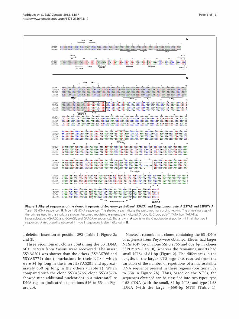
of E. petersi from Yasuní were recovered. The insert5SYAS201 was shorter than the others (5SYAS766 and5SYAS774) due to variations in their NTSs, whichwere 84 bp long in the insert 5SYAS201 and approxi-mately 650 bp long in the others (Table 1). Whencompared with the clone 5SYAS766, clone 5SYAS774showed nine additional nucleotides in a microsatelliteDNA region (indicated at positions 546 to 554 in Fig-ure 2b).
Nineteen recombinant clones containing the 5S rDNAof E. petersi from Puyo were obtained. Eleven had largerNTSs (649 bp in clone 5SPUY766 and 652 bp in clones5SPUY769-1 to 10), whereas the remaining inserts hadsmall NTSs of 84 bp (Figure 2). The differences in thelengths of the larger NTS segments resulted from thevariation of the number of repetitions of a microsatelliteDNA sequence present in these regions (positions 552to 554 in Figure 2b). Thus, based on the NTSs, thesequences obtained can be classified into two types: typeI 5S rDNA (with the small, 84-bp NTS) and type II 5SrDNA (with the large, ~650-bp NTS) (Table 1).
Figure 2 Aligned sequences of the cloned fragments of Engystomops freibergi (5SACR) and Engystomops petersi (5SYAS and 5SPUY). A.Type I 5S rDNA sequences. B. Type II 5S rDNA sequences. The shaded areas indicate the presumed transcribing regions. The annealing sites ofthe primers used in this study are shown. Presumed regulatory elements are indicated (A box, IE, C box, poly-T, TATA box, TATA-like,hexanucleotides AGAAGC and GCAAGT, and GAACAAA sequence). The arrow in A points to the C nucleotide at position -1 in all the type Isequences. A microsatellite observed in type II sequences is also indicated in B.
Rodrigues et al. BMC Genetics 2012, 13:17http://www.biomedcentral.com/1471-2156/13/17
Page 3 of 13

Interestingly, the presumed 5S rDNA coding region wasalso characteristic for each of these types (see Table 1),as could be inferred from the maximum likelihood ana-lysis of this specific region, in which all the type Isequences were clustered together and apart from thetype II sequences group (Figure 3).As expected, comparison of the 120 bp of the coding
regions of the type I and type II 5S rDNA of Engystomopswith those of the other anuran species and selected fishspecies available in GenBank revealed great similarity(Figure 4). When the type I and type II sequences of P.cuvieri were excluded from the analysis, there was ahigher similarity between the Engystomops type I 5S RNAgene and the remaining 5S sequences (88-90%) thanbetween the latter and the Engystomops type II gene(82%). When the Engystomops and P. cuvieri 5S geneswere compared, a higher similarity was observed amongthe sequences of the same type (Figure 4).
A higher similarity was also observed between theEngystomops and the P. cuvieri type I NTS (97% -100%)than between the type II NTS of the Engystomops spe-cies (~650 bp) and that of P. cuvieri (~580 bp) (90% -92%). Because the type I NTS sequences were the samein all the Engystomops analyzed, and the type II NTSsequences were quite similar (average pairwise similarity:98%; Figure 2), to better illustrate the comparisonbetween the Engystomops and P. cuvieri NTSs, only thepairwise alignments between the sequences of singlespecimens of E. petersi from Puyo and of P. cuvieri areshown in Figure 5. The three elements (A box, inter-mediate element and C box) of the 5S gene internalcontrol region (ICR) were identified in the presumedcoding regions of all the sequences isolated from thethree species of Engystomops (Figure 4). Interestingly,the control elements of the Engystomops type IIsequences differed more from those available in
Table 1 Engystomops type I and II 5S rDNA sequence data
Sequence name Specimen source(Voucher number, species identification, and locality)
GenBank accession number NTS size (bp)
5SACR201-1 ZUEC9651 - E. freibergi/Acre, Brazil JF325868 84
5SACR201-2 ZUEC9651 - E. freibergi/Acre, Brazil JF325869 84
5SACR201-3 ZUEC9651 - E. freibergi/Acre, Brazil JF325870 84
5SYAS201 QCAZ34948 - E. Petersi/Yasuní, Ecuador JF325859 84
5SPUY201-1 QCAZ34937 - E. petersi/Puyo, Ecuador JF325860 84
5SPUY201-2 QCAZ34937- E. petersi/Puyo, Ecuador JF325861 84
5SPUY201-3 QCAZ34937- E. petersi/Puyo, Ecuador JF325862 84
5SPUY201-4 QCAZ34937- E. petersi/Puyo, Ecuador JF325863 84
5SPUY201-5 QCAZ34937- E. petersi/Puyo, Ecuador JF325864 84
5SPUY201-6 QCAZ34937- E. petersi/Puyo, Ecuador JF325865 84
5SPUY201-7 QCAZ34937- E. petersi/Puyo, Ecuador JF325866 84
5SPUY201-8 QCAZ34937- E. petersi/Puyo, Ecuador JF325867 84
5SACR764 ZUEC9647 - E. freibergi/Acre, Brazil JF325843 647
5SACR765-1 ZUEC9647 - E. freibergi/Acre, Brazil JF325844 648
5SACR765-2 ZUEC9647 - E. freibergi/Acre, Brazil JF325845 648
5SYAS766 QCAZ34948 - E. Petersi/Yasuní, Ecuador JF325846 649
5SYAS774 QCAZ34948 - E. Petersi/Yasuní, Ecuador JF325847 658
5SPUY766 QCAZ34937- E. petersi/Puyo, Ecuador JF325848 648
5SPUY769-1 QCAZ34937- E. petersi/Puyo, Ecuador JF325849 651
5SPUY769-2 QCAZ34937- E. petersi/Puyo, Ecuador JF325850 651
5SPUY769-3 QCAZ34937- E. petersi/Puyo, Ecuador JF325851 651
5SPUY769-4 QCAZ34937- E. petersi/Puyo, Ecuador JF325852 651
5SPUY769-5 QCAZ34937- E. petersi/Puyo, Ecuador JF325853 651
5SPUY769-6 QCAZ34937- E. petersi/Puyo, Ecuador JF325854 651
5SPUY769-7 QCAZ34937- E. petersi/Puyo, Ecuador JF325855 651
5SPUY769-8 QCAZ34937- E. petersi/Puyo, Ecuador JF325856 651
5SPUY769-9 QCAZ34937- E. petersi/Puyo, Ecuador JF325857 651
5SPUY769-10 QCAZ34937- E. petersi/Puyo, Ecuador JF325858 651
Identification name, specimen source, GenBank accession number and NTS size of all cloned 5S rDNA sequences of Engystomops freibergi and Engystomopspetersi. ZUEC: Museu de Zoologia Prof. Adão José Cardoso, at the State University of Campinas, Brazil; QCAZ: Museo de Zoología de la Pontificia UniversidadCatólica del Ecuador.
Rodrigues et al. BMC Genetics 2012, 13:17http://www.biomedcentral.com/1471-2156/13/17
Page 4 of 13

GenBank than did the elements of the Engystomops typeI sequences.All sequences of the type I 5S rDNA contained the
control element TATA-box in their NTS regions, whichwas located 25 bp upstream from the coding region. Inaddition, the type I sequences also had a TATA-like ele-ment located 13 nucleotides upstream from the TATA-box (Figure 2a). In the NTS of the type II sequences, anelement similar to the TATA-box motif was alsodetected, but it was very distant from the +1 position ofthe presumed coding region of the 5S gene, occupyingpositions -345 to -350 (Figure 2b). Additionally, in thetype II 5S rDNA sequences, were found two hexanu-cleotides (at positions -53 to -48 and -21 to -16; Figure2b) resembling the regulatory hexanucleotides previouslydescribed for Xenopus [6].Both the type I and II Engystomops 5S sequences
showed a T-rich sequence containing 4 to 5T-residuesstarting at position -119, which may correspond to thepoly-T termination region of the 5S gene described inthe literature [6,35]. Interestingly, in addition to the poly-T region, the type I sequences showed a GAACAAAsequence very similar to the sequence GAAACAA, whichis found downstream from the 5S rRNA gene in fish andhas been suggested to act as a terminal region [3].
The consensus secondary structures for the presumedtype I and type II 5S rRNA is shown in Figure 6. Thesecondary structure of all analyzed 5S rRNAs consists offive helices (I-V), two hairpin loops (C and E), twointernal loops (B and D), and a hinge region (A),arranged into the three-helix junction.
Chromosome mapping of 5S rDNAAs expected, the karyotypes of the specimens of E. frei-bergi and E. petersi analyzed here were the same asthose described previously by Targueta et al. [33].Therefore, our study includes two of the three karyolo-gical groups recognized by Targueta et al. [33] amongE. petersi specimens: the Puyo and Yasuní karyologicalgroups. It is interesting to notice that the karyotypes ofthe specimens of E. petersi from Puyo are more similarto those of the specimens of E. freibergi than to the kar-yotype of E. petersi from Yasuní. To describe the map-ping of the 5S rDNA sequences in these karyotypes, thepreviously proposed chromosome classification scheme[33] was used.In the E. freibergi karyotype, a FISH probe containing
the entire repeating unit of the type I 5S rDNA hybri-dized pericentromerically to the short arm of Chromo-some 3 and also distally to the long arm ofChromosome 6 (Figure 7a). When using the probe con-taining the entire repeating unit of the type II 5S rDNA,only the distal region of the long arm of Chromosome 6was detected (Figure 7b). Neither the site detected inChromosome 3 nor the site on Chromosome 6 coin-cided with any nucleolar organizer regions (NORs)reported by Targueta et al. [33] (Figure 7).Because these data suggested a differential localiza-
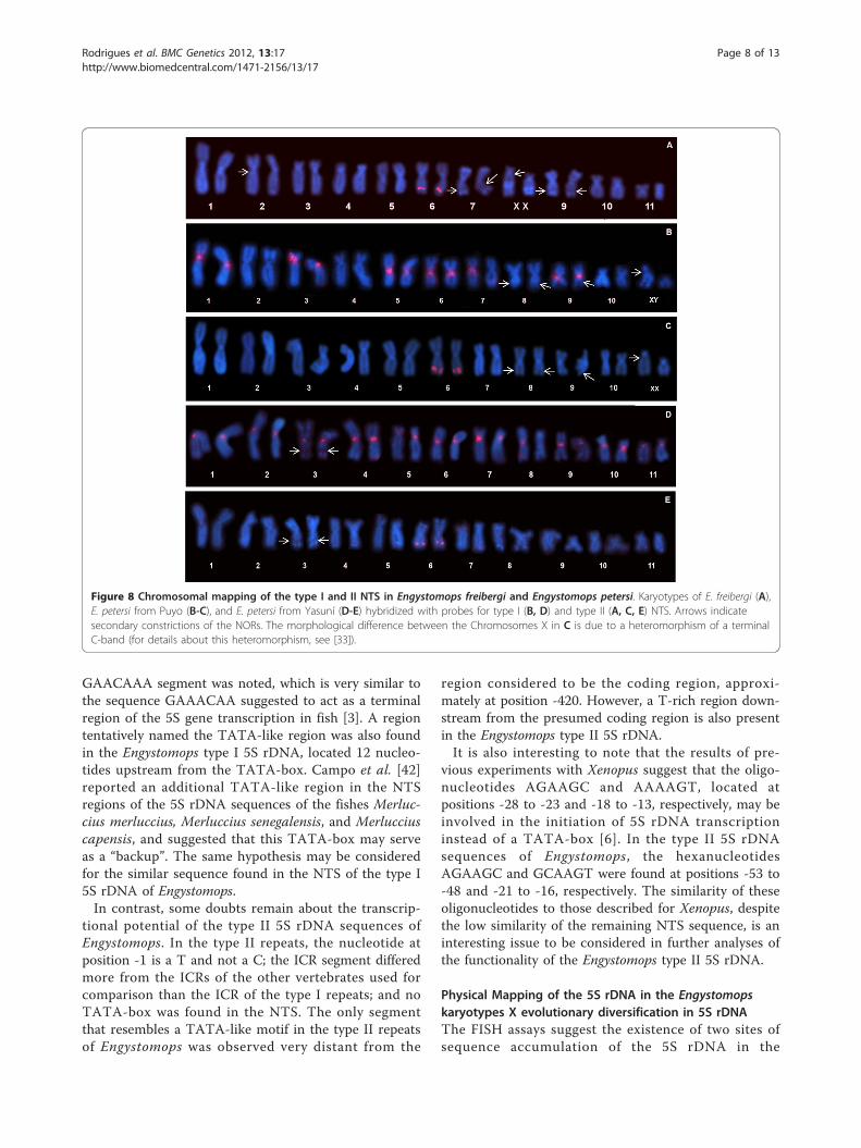
tion of both types of 5S rDNA sequences in the E. frei-bergi karyotype, further analyses were performed usingmore specific probes that exclusively contained eitherthe type I or type II NTS. In the karyotypes of E. frei-bergi and E. petersi from Puyo, which are morphologi-cally very similar (for details, see [33]), the type I NTSprobe detected the proximal region of the short arm ofChromosome 3 but did not detect a distal region ofChromosome 6. This probe also detected the centro-meric region of several chromosomes (Figure 8b). Incontrast, the type II NTS probe detected only a distalsite in the long arm of Chromosome 6 in the karyo-types of both E. freibergi (Figure 8a) and E. petersifrom Puyo (Figure 8c). In the karyotype of E. petersifrom Yasuní, the type I NTS probe detected the peri-centromeric region of the short arm of Chromosome 5and the centromeric regions of several chromosomes(Figure 8d), whereas the type II NTS probe detectedonly a distal site on the long arm of Chromosome 6(Figure 8e).
Figure 3 Maximum likelihood dendrogram inferred from thecoding region of the 5S rDNA sequences of Engystomops. Thelikelihood score was 265.2362. See Table 1 for a detailed descriptionof the sequence symbols. The dotted circle groups the type I 5SrDNA and the dashed circle groups the type II 5S rDNA sequences.Numbers above branches are bootstrap values from 1000pseudoreplicates. Bootstrap values under 0.5 were omitted.
Rodrigues et al. BMC Genetics 2012, 13:17http://www.biomedcentral.com/1471-2156/13/17
Page 5 of 13

DiscussionMolecular Organization of the 5S rDNA in EngystomopsThis study verified the occurrence of two types of 5SrDNA in the genomes of the Amazonian species ofEngystomops, a feature widely documented in many ver-tebrates, including fish [10,11,36-42], the anurans Xeno-pus laevis and Xenopus borealis [6,16,21-24,43,44]chickens [45-47], and mammals [12,48,49]. Similarly tothe findings reported in these studies, the two types of5S rDNA sequences found in E. freibergi and E. petersivaried slightly in their corresponding coding regions,with the main difference between them found in theNTS region, which varied in length (84 bp for the type I5S rDNA and approximately 650 bp for the type II 5SrDNA) and nucleotide composition. These two types of5S rDNA do not, however, appear to be related to thedual system observed in Xenopus (i.e., the oocyte andsomatic types) [6,13,16,20-22,24] because we found nosimilarities between the NTSs of the oocyte- or somatic-type sequences to those in the present study.
Since Ohno’s publication [50], the origin of genic var-iants has been attributed to events of sequence duplicationfollowed by processes that result in the divergence of theduplicated sequences. This hypothesis has been corrobo-rated by many studies (reviewed in references [51-55]) andmay also explain the presence of two types of 5S rDNA.Gene duplication may result from unequal crossing over,retropositioning, or chromosomal (or genomic) duplica-tion [51], and the outcomes of these events are quite dif-ferent, including neofunctionalization, pseudogene originor simple preservation of gene duplicates [50,56,57].Although our data for the Amazonian Engystomops do
not allow us to elucidate the events involved in the ori-gin of either type of the 5S rDNA, those events mayhave preceded the divergence of Engystomops and Phy-salaemus. Such an inference follows from the observa-tion of higher nucleotide divergence between thesequences of the type I and type II 5S rDNAs of eachspecies in these genera than among the sequences of thesame type found in distinct species.
Figure 4 Comparison of the 5S rDNA sequences of Engystomops with 5S rDNA sequences available in the GenBank. Alignment of thepresumed coding regions of the type I and II 5S rDNA sequences of Engystomops freibergi (Acre) and Engystomops petersi (Yasuní and Puyo)with the 5S rDNA sequences of other vertebrates obtained from the GenBank (accession numbers: AF250511, AF284728, AF284742, AY271269,S73107, M24954, V00647, J01009, M35055, J01010, M30904, M35176, M63899, V01425, V01426, X12622, X12623, X12624, M74438, X58365, X58368,X58367, M10817, X01309, V00589). The internal control regions are in gray (A box = positions 50 to 64; intermediate element = positions 67 to72; C box = position 80 to 97). Note that the control regions of the Engystomops type I 5S rDNA sequences are more similar to those found forthe other vertebrate 5S rDNA sequences than are those of the Engystomops type II 5S rDNA. Ooc: oocyte-type. Smc: somatic-type.
Rodrigues et al. BMC Genetics 2012, 13:17http://www.biomedcentral.com/1471-2156/13/17
Page 6 of 13
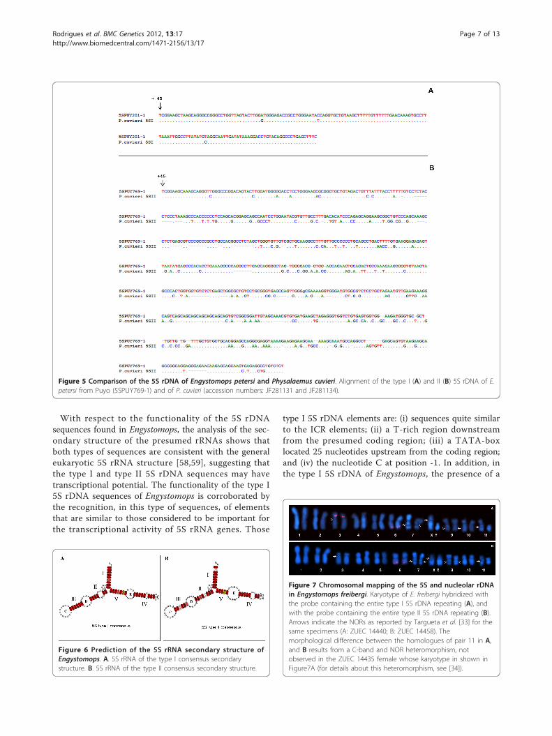
With respect to the functionality of the 5S rDNAsequences found in Engystomops, the analysis of the sec-ondary structure of the presumed rRNAs shows thatboth types of sequences are consistent with the generaleukaryotic 5S rRNA structure [58,59], suggesting thatthe type I and type II 5S rDNA sequences may havetranscriptional potential. The functionality of the type I5S rDNA sequences of Engystomops is corroborated bythe recognition, in this type of sequences, of elementsthat are similar to those considered to be important forthe transcriptional activity of 5S rRNA genes. Those
type I 5S rDNA elements are: (i) sequences quite similarto the ICR elements; (ii) a T-rich region downstreamfrom the presumed coding region; (iii) a TATA-boxlocated 25 nucleotides upstream from the coding region;and (iv) the nucleotide C at position -1. In addition, inthe type I 5S rDNA of Engystomops, the presence of a
Figure 5 Comparison of the 5S rDNA of Engystomops petersi and Physalaemus cuvieri. Alignment of the type I (A) and II (B) 5S rDNA of E.petersi from Puyo (5SPUY769-1) and of P. cuvieri (accession numbers: JF281131 and JF281134).
Figure 6 Prediction of the 5S rRNA secondary structure ofEngystomops. A. 5S rRNA of the type I consensus secondarystructure. B. 5S rRNA of the type II consensus secondary structure.
Figure 7 Chromosomal mapping of the 5S and nucleolar rDNAin Engystomops freibergi. Karyotype of E. freibergi hybridized withthe probe containing the entire type I 5S rDNA repeating (A), andwith the probe containing the entire type II 5S rDNA repeating (B).Arrows indicate the NORs as reported by Targueta et al. [33] for thesame specimens (A: ZUEC 14440; B: ZUEC 14458). Themorphological difference between the homologues of pair 11 in A,and B results from a C-band and NOR heteromorphism, notobserved in the ZUEC 14435 female whose karyotype in shown inFigure7A (for details about this heteromorphism, see [34]).
Rodrigues et al. BMC Genetics 2012, 13:17http://www.biomedcentral.com/1471-2156/13/17
Page 7 of 13
GAACAAA segment was noted, which is very similar tothe sequence GAAACAA suggested to act as a terminalregion of the 5S gene transcription in fish [3]. A regiontentatively named the TATA-like region was also foundin the Engystomops type I 5S rDNA, located 12 nucleo-tides upstream from the TATA-box. Campo et al. [42]reported an additional TATA-like region in the NTSregions of the 5S rDNA sequences of the fishes Merluc-cius merluccius, Merluccius senegalensis, and Merlucciuscapensis, and suggested that this TATA-box may serveas a “backup”. The same hypothesis may be consideredfor the similar sequence found in the NTS of the type I5S rDNA of Engystomops.In contrast, some doubts remain about the transcrip-
tional potential of the type II 5S rDNA sequences ofEngystomops. In the type II repeats, the nucleotide atposition -1 is a T and not a C; the ICR segment differedmore from the ICRs of the other vertebrates used forcomparison than the ICR of the type I repeats; and noTATA-box was found in the NTS. The only segmentthat resembles a TATA-like motif in the type II repeatsof Engystomops was observed very distant from the
region considered to be the coding region, approxi-mately at position -420. However, a T-rich region down-stream from the presumed coding region is also presentin the Engystomops type II 5S rDNA.It is also interesting to note that the results of pre-
vious experiments with Xenopus suggest that the oligo-nucleotides AGAAGC and AAAAGT, located atpositions -28 to -23 and -18 to -13, respectively, may beinvolved in the initiation of 5S rDNA transcriptioninstead of a TATA-box [6]. In the type II 5S rDNAsequences of Engystomops, the hexanucleotidesAGAAGC and GCAAGT were found at positions -53 to-48 and -21 to -16, respectively. The similarity of theseoligonucleotides to those described for Xenopus, despitethe low similarity of the remaining NTS sequence, is aninteresting issue to be considered in further analyses ofthe functionality of the Engystomops type II 5S rDNA.
Physical Mapping of the 5S rDNA in the Engystomopskaryotypes X evolutionary diversification in 5S rDNAThe FISH assays suggest the existence of two sites ofsequence accumulation of the 5S rDNA in the
Figure 8 Chromosomal mapping of the type I and II NTS in Engystomops freibergi and Engystomops petersi. Karyotypes of E. freibergi (A),E. petersi from Puyo (B-C), and E. petersi from Yasuní (D-E) hybridized with probes for type I (B, D) and type II (A, C, E) NTS. Arrows indicatesecondary constrictions of the NORs. The morphological difference between the Chromosomes X in C is due to a heteromorphism of a terminalC-band (for details about this heteromorphism, see [33]).
Rodrigues et al. BMC Genetics 2012, 13:17http://www.biomedcentral.com/1471-2156/13/17
Page 8 of 13
karyotypes of E. freibergi and E. petersi, one on 3p (E.freibergi and E. petersi from Puyo) or 5p (E. petersi fromYasuní) and another on 6q. The results of these assaysalso suggest that the former site is exclusive to or pre-ferentially constitutes type I sequences, whereas the lat-ter, on 6q, is associated with type II sequences.Ribosomal DNA repeating units are evolutionarily
dynamic and appear to be able to spread throughout thegenome, creating new rDNA loci [3,60-62]. The pre-sence of two distinct 5S rDNA sequence types organizedin different chromosomal regions or even on differentchromosomes has been described for several fish, e.g.,Salmo solar [36], Oncorhynchus mykiss [37], Coregonusartedi, C. zenithicus [63], and Oreochromis niloticus [40].In anurans, the first chromosome mapping experi-
ments for the 5S rRNA genes were conducted in Xeno-pus laevis. Using specific probes, Harper et al. [16]revealed a differential distribution of the two types of 5SrDNA in the Xenopus karyotypes, mapping the somatic-type 5S rDNA to the distal end of the long arm ofChromosome 9 in Xenopus laevis and X. borealis, andthe oocyte-type to the distal ends of the majority ofXenopus laevis chromosomes. The authors also mappeda trace oocyte-type 5S rDNA in X. laevis, which is aminor class of the oocyte type, to the distal end of thelong arm of Chromosome 13.In addition to these data for the Xenopus karyotypes,
only the 5S rDNA chromosomal sites were detected inthe karyotypes of Physalaemus ephippifer [18] and Phy-salaemus cuvieri [19]. A probe containing the entirerepeat of the type I 5S rDNA of P. cuvieri detected apericentromeric region of the short arm of Chromosome3 in both Physalaemus species karyotypes [18,19]whereas a probe with the entire repeat of the type II 5SrDNA of P. cuvieri preferentially detected a distal regionof the long arm of Chromosome 6 [19]. Similarly to theabove-mentioned cases, a differential localization of thetwo types of 5S rDNA found in the Engystomops specieswas observed in this study.As mentioned above, taking into account the similarity
of the sequences, probably the origin of the two types of5S rDNA found in Engystomops and Physalaemus spe-cies preceded the divergence of these genera. Appar-ently, the origin of this dual-system involvedtranslocation or transposition events that lead to theseparation of two groups of 5S rDNA sequences, favor-ing the dominance of divergence forces over homogeni-zation processes between these groups. On the otherhand, the homogeneity of the 5S rDNA repeats clus-tered in the same chromosomal site was maintained,what may be explained by concerted evolution. As aresult of these processes, two distinct types of 5S rDNAsequences, occupying different chromosomal sites, havearisen. It is worth mentioning that purifying selection
may also have been involved in this scenario. In additionto concerted evolution, purifying selection has beeninvoked to justify the homogenization in a gene family[64,65]. Since the comparison between both types of 5SrDNA sequences of Engystomops showed a higher varia-tion between the presumed coding-regions than betweentheir NTSs, it is likely that purifying selection has beenacting over these coding-regions, avoiding high level ofdivergence.Another intriguing finding of this study was the hybri-
dization of the probe that corresponds to the type INTS to the centromeric region of various chromosomes,sites which were not detected in this analysis by theprobes that potentially contain the transcribed region ofthe 5S rDNA. A possible explanation for this result isthat the sequences associated with the centromericregions are segments of satellite DNA derived from the5S rDNA, a phenomenon previously reported for thefish Hoplias malabaricus [66] and the frog Physalaemuscuvieri [19].The 5S rDNA cluster has been reported to be linked
to the major rDNA sequences [4,36,37,67-69] but, insome cases, is localized to different chromosomes[18,63,70-73]. The differential chromosomal localizationof the 5S and 45S rDNAs is the prevalent condition, notonly in the cited examples but also in other groups,including plants [74-77].Several authors have previously discussed the preva-
lence of different chromosomal sites for the 45S and 5SrDNAs over their linkage in other organisms, and aprobable explanation is intrinsically related to the repeti-tive nature of these sequences. Others have suggestedthat because tandem repeated sequences are frequentlyinvolved in events of unequal crossing-over and geneconversion, the separation of the two great families ofribosomal DNA at different chromosomal sites wouldavoid disruptive interference in its organization such asundesired rearrangements between the 45S and 5Sarrays [3,11].The 5S rDNA chromosomal sites unlinked to the
NORs of the Engystomops species represent new cytoge-netic markers to be considered for their karyotypic com-parison. Based on classic cytogenetic techniques, CMA3
and DAPI staining, and in situ localization of nucleolarrDNA, Targueta et al. [33] described the three Engysto-mops karyotypes of the present study and noted thatrecognizing chromosomal homeology was difficult, espe-cially between the Yasuní karyotype and the other two.In addition, difficulties in differentiating the three verymorphologically similar chromosome pairs (pairs 3, 6,and 8) in the Yasuní karyotype have been reported. Inthe present study, we were able to suggest a homeologybetween Chromosome 6 of the E. petersi karyotype fromYasuní and Chromosome 6 of E. freibergi and E. petersi
Rodrigues et al. BMC Genetics 2012, 13:17http://www.biomedcentral.com/1471-2156/13/17
Page 9 of 13
from Puyo based on the mapping of the type II 5SrDNA sequences. Additionally, this chromosome sitealso constitutes a distinctive marker for Chromosome 6in the karyotype of E. petersi from Yasuní, distinguishingit from Chromosome 8 and the NOR-bearing Chromo-some 3. Finally, the mapping of the type I 5S rDNAsequence to Chromosome 5 of the specimens fromYasuní and Chromosome 3 of E. freibergi and of E.petersi from Puyo, which are similar chromosomes insize and morphology, may suggests that these chromo-somes are homeologous.In addition to the recognition of chromosomal home-
ologies among the Engystomops species, the 5S rDNAmapping performed here allows for a better cytogeneticcomparison of Engystomops with its sister genus, Physa-laemus. Chromosomes 3 of P. cuvieri [19] and P. ephip-pifer [18], bearing the type I 5S rDNA sequence, aremorphologically similar to Chromosome 5 of E. petersifrom Yasuní and Chromosome 3 of E. freibergi and E.petersi from Puyo. Therefore, the homeology of all thesechromosomes can be strongly inferred. Similarly, we candeduce homeology among the metacentric Chromosome6 of the Engystomops species and Chromosome 5 of P.cuvieri, which all carry the type II 5S rDNA sequences.Chromosome 5 or 6 of P. ephippifer likely also bearsthis type of sequence; however, the presence of thissequence has not been verified with a specific probe fortype II 5S rDNA sequences [18]. These cytogenetic datasuggest that the chromosomal sites of the 5S rDNA maybe conserved in these leiuperid genera, which has notbeen observed for the NORs [18,33,78-84]. Therefore,the two 5S rDNA arrays appear to be independent unitsof evolution in Engystomops species, and further studiesof their functionality and their relation to a possiblecentromeric DNA satellite sequence are necessary toprovide a better understanding of the evolution of thesesequences.
ConclusionsIn Amazonian Engystomops, two types of 5S rDNA werefound and mapped to distinct chromosomal sites.Because these rDNA types are related to those found inPhysalaemus with respect to their nucleotide sequencesand chromosomal locations, their origin likely precededthe evolutionary divergence of these genera. In addition,our data revealed chromosomal homeologies among thethree karyotypes of the Amazonian Engystomops, repre-senting an important contribution for further studies ofkaryotype evolution in this genus.
MethodsSpecimensSpecimens of E. petersi from Puyo (Provincia de Pastaza)and Estación Científica de Yasuní (Provincia de
Olleana), located in the Ecuadorian Amazon, and speci-mens of E. freibergi from the Tejo estuary, state of Acre,Brazil were studied. The vouchers specimens are at theMuseo de Zoología de la Pontificia Universidad Católicadel Ecuador (QCAZ), Quito, Ecuador or at the Museude Zoologia “Prof. Adão José Cardoso” (ZUEC) at theUniversidade Estadual de Campinas (UNICAMP), Cam-pinas, state of São Paulo, Brazil.
Isolation and cloning of the 5S geneGenomic DNA was isolated from liver and muscle sam-ples stored at -80°C in the tissue collection deposited atthe Laboratório de Estudos Cromossômicos em Anurosat IB-UNICAMP, Brazil. The genomic DNA isolationwas performed as described [33], with a TNES solution(250 mM Tris-HCl pH 7.5, 2 M NaCl, 100 mM EDTApH 8.0, and 2.5% SDS). After electrophoresis in 1% agar-ose, the DNA quality was evaluated, and its quantity wasestimated. The entire repeating unit of the 5S rDNA(which includes the presumed transcribed region and theNTS) was isolated by PCR using the primers 5S-A (5’-TACGCCCGATCTCGTCCGATC-3’) and 5S-B (5’-CAGGCTGGTATGGCCGTAAGC-3’) [36]. The ampli-fied fragments were purified using the GFX PCR and GelBand DNA Purification kit (GE Healthcare - Little Chal-font, Buckinghamsire, UK) and cloned into the pGEM-Tvector (pGEM-T easy Vector - Promega - Madison, WI,USA) according to the manufacturer’s instructions.Recombinant vectors were used to transform competentEscherichia coli of the JM109 strain (Fermentas), and thecloned fragments were sequenced. Repeating units of twodifferent sizes were obtained and named type I 5S rDNAand type II 5S rDNA. Based on their nucleotidesequences, specific primers were designed to exclusivelyisolate the presumed 120-bp transcribed region from thetype I 5S rDNA (5S120T1-F: 5’-GCTTTCGCTTACGGC-CATACC-3’; 5S120T1-R: 5’-AGCTTACAGCACCTGG-TATTC-3’) and the type II 5S rDNA (5S120T2-F: 5’-GTCTCTGCTTACGGCCATACC-3’; 5S120T2-R: 5’-AGTCTACAGCACCCGCGCTTC-3’). Specific primerswere also designed to isolate the NTS region from thetype I (5ST1-F: 5’-GCTGTAAGCTTTTTGTTTTTT-GAA-3’; 5ST1-R: 5’-GAAAGCTCAGGGCCTGTACAG-3’) and type II (5ST2-F: 5’-GCTGTAGACCGTTTATT-TACCTT-3’; 5ST2-R: 5’-AGAGACAGGCCTCT-CACTTGC-3’) 5S rDNA repeats. The annealing sites ofthe primers used in this study are indicated in Figure 1.The resulting amplified fragments were all sequencedand analyzed.
DNA sequencing and analysisThe cloned fragments were amplified by PCR using the T7and SP6 primers. The amplified products were purifiedusing the GFX PCR and Gel Band DNA Purification kit
Rodrigues et al. BMC Genetics 2012, 13:17http://www.biomedcentral.com/1471-2156/13/17
Page 10 of 13
(GE Healthcare - Little Chalfont, Buckinghamsire, UK)and used directly as templates in amplification reactionsusing the BigDye Terminator chemistry version 3.1(Applied Biosystems - Austin, TX, USA) according to themanufacturer’s recommendations. Each cloned fragmentwas bi-directionally sequenced in an automatic DNAsequencer. The sequences were edited using BioEdit ver-sion 7.0.1 [85] and aligned with ClustalW software. Thefragments obtained were compared with other sequencesavailable in the GenBank database [86] and in the 5S Ribo-somal RNA Database [87]. All presumed coding regions ofthe generated 5S rDNA sequences were assembled into adata matrix, and a Maximum Likelihood (ML) analysiswas conducted using the PAUP* 4.0b10 software [88] withthe evolution model K80, which was selected by Modeltest3.7 [89] for this data set. Nodal support for the MLarrangement was assessed through a non-parametric boot-strap analysis [88], with a heuristic search based on 1,000pseudoreplicates.
Chromosome preparations and Fluorescent in situHybridization (FISH)Chromosome preparations were made from the intestineand testis cell suspensions obtained from the E. freibergispecimens ZUEC 14435, ZUEC 14439, ZUEC 14440,and ZUEC14458, and the E. petersi specimens QCAZ34946, QCAZ 34947, QCAZ 34940, and QCAZ 34942.All of these cell suspensions were available at the collec-tion of amphibian material deposited at the Laboratóriode Estudos Cromossômicos em Anuros at IB-UNI-CAMP, Brazil, and were used previously by Targueta etal. [33] to describe the karyotypes of E. freibergi and E.petersi. The cell suspensions were spotted onto cleanslides, and the chromosome preparations were hybri-dized with the 5S rDNA fragments isolated as describedabove and PCR-labeled with either biotin or digoxigenin.The hybridization was performed as described elsewhere[90]. The biotin-labeled probes were detected with ananti-biotin antibody (goat anti-biotin - Vector - Burlin-game CA, USA), which is recognized by a FITC-conju-gated secondary antibody (anti-goat IgG-FITC - Vector- Burlingame CA, USA). The digoxigenin-labeled probeswere detected with an anti-digoxigenin antibody conju-gated with rhodamine. The chromosomes were counter-stained with DAPI (0.5 μg/mL).
AbbreviationsrDNA: ribosomal DNA; NTS: non-transcribed spacer; DAPI: 4 6-diamidino-2-phenylindole; 3p: short arm of Chromosome 3; 5p: short arm ofChromosome 5; 6q: long arm of Chromosome 6.
Acknowledgements and fundingThe authors gratefully acknowledge Ailín Blasco for helping in the field workand Shirlei M. Recco Pimentel for the constant encouragement andassistance in obtaining some tissue samples. This work was supported by
the Brazilian agencies FAPESP (Fundação de Amparo à Pesquisa do Estadode São Paulo), CNPq (Conselho Nacional de Desenvolvimento Científico eTecnológico) and CAPES (Coordenação de Aperfeiçoamento de Pessoal deNível Superior), and the Ecuadorian agency SENESCYT (Secretaría Nacionalde Educación Superior, Ciencia, Tecnología y Innovación).
Author details1Department of Structural and Functional Biology, Institute of Biology,University of Campinas (UNICAMP), Campinas, SP 13083-863, Brazil. 2Escuelade Ciencias Biológicas, Pontifícia Universidad Católica Del Ecuador, Quito,Ecuador.
Authors’ contributionsDSR acquired the data and drafted the manuscript. MR helped to collect thespecimens and obtain the chromosome preparations and revised themanuscript. LBL designed and coordinated the study and helped draft themanuscript. All authors read and approved the final manuscript.
Competing interestsThe authors declare that they have no competing interests.
Received: 1 December 2011 Accepted: 20 March 2012Published: 20 March 2012
References1. Long EO, Dawid IB: Repeated genes in eukaryotes. Ann Rev Biochem 1980,
49:727-764.2. Drouin G, de Sá MM: The concerted evolution of 5S ribosomal genes
linked to the repeat units of other multigene families. Mol Biol Evol 1995,12(3):481-493.
3. Martins C, Wasko AP: Organization and evolution of 5S ribosomal DNA inthe fish genome. In Focus on Genome Research Chapter X. Edited by: ClydeR. Williams: Nova Science Publishers, New York, USA, Inc; 2004:335-363.
4. Gerbi SA: Evolution of Ribosomal DNA. In Molecular Evolutionary Genetics.Edited by: MacIntyre RJ. New York: Plenum Publishing; 1985:419-517.
5. Pieler T, Hamm J, Roeder RG: The 5S gene internal control region iscomposed of three distinct sequence elements, organized as twofunctional domains with variable spacing. Cell 1987, 48(1):91-100.
6. Korn LJ, Brown DD: Nucleotide sequence of Xenopus borealis oocyte 5SDNA: Comparison of sequences that flank several related eukaryoticgenes. Cell 1978, 15:1145-1156.
7. Hallenberg C, Frederiksen S: Effect of mutations in the upstreampromoter on the transcription of human 5S rRNA genes. Biochim BiophysActa 2001, 1520(2):169-173.
8. Nederby-Nielsen J, Hallenberg C, Frederiksen S, Sorensen PD, Lomholt B:Transcription of human 5S rRNA genes is influenced by an upstreamDNA sequence. Nucleic Acids Res 1993, 21(16):3631-3636.
9. Fernández-Tajes J, Méndez J: Identification of the razor clam species Ensisarcuatus, E. siliqua, E. directus, E. macha, and Solen marginatus usingPCR-RFLP analysis of the 5S rDNA region. J Agric Food Chem 2007,55:7278-7282.
10. Ferreira IA, Oliveira C, Venere PC, Galetti PM Jr, Martins C: 5S rDNAvariation and its phylogenetic inference in the genus Leporinus(Characiformes: Anostomidae). Genetica 2007, 129:253-257.
11. Martins C, Galetti PM Jr: Chromosomal localization of 5S rDNA genes inLeporinus fish (Anostomidae, Characiformes). Chrom Res 1999, 7:363-367.
12. Suzuki H, Moriwaki K, Sakurai S: Sequences and evolutionary analysis ofmouse 5S rDNAs. Mol Biol Evol 1994, 11(4):704-710.
13. Vitelli L, Batistoni R, Andronico F, Nardi I, Barsacchipilone G: Chromosomallocalization of 18S + 28S and 5S ribosomal-RNA genes in evolutionarilydiverse anuran amphibians. Chromosoma 1982, 84(4):475-491.
14. Pasolini P, Costagliola D, Rocco L, Tinti F: Molecular Organization of 5SrDNAs in Rajidae (Chondrichthyes): Structural Features and Evolution ofPiscine 5S rRNA Genes and Nontranscribed Intergenic Spacers. J Mol Evol2006, 62:564-574.
15. Pinhal D1, Araki CS, Gadig OBF, Martins C: Molecular organization of 5SrDNA in sharks of the genus Rhizoprionodon: insights into theevolutionary dynamics of 5S rDNA in vertebrate genomes. Genet Res2009, 91:61-72.
16. Harper ME, Price J, Korn LJ: Chromosonal mapping of Xenopus 5S genes:somatic-type versus oocyte-type. Nucleic Acids Res 1983, 11(8):2313-2323.
Rodrigues et al. BMC Genetics 2012, 13:17http://www.biomedcentral.com/1471-2156/13/17
Page 11 of 13

17. Nietfeld W, Digweedt M, Mentzel H, Meyerhof W, Köster M, Knöchel W,Erdmannt VA, Pieler T: Oocyte and somatic 5S ribosomal RNA and 5SRNA encoding genes in Xenopus tropicalis. Nucleic Acids Res 1988,16:8803-8815.
18. Nascimento J, Quinderé YRSD, Recco-Pimentel SM, Lima JRF, Lourenço LB:Heteromorphic Z and W sex chromosomes in Physalaemus ephippifer(Steindachner, 1864) (Anura, Leiuperidae). Gentica 2010, 138:1127-1132.
19. Vittorazzi SE, Lourenço LB, Del-Grande ML, Recco-Pimentel SM: SatelliteDNA Derived from 5S rDNA in Physalaemus cuvieri (Anura, Leiuperidae).Cytogenet Genome Res 2011, 134(2):101-107.
20. Schmid M, Vitelli I, Batistoni R: Chromosome Banding in Amphibia XI.Constitutive heterochromatin, nucleolus organizers, 18S+28S and 5Sribossomal RNA genes in Ascaphidae, Pipidae, Discoglossidae andPelobatidae. Chromosoma 1987, 95:271-284.
21. Carroll D, Brown DD: Repeating units of Xenopus laevis oocyte-type 5SDNA are heterogeneous in length. Cell 1976, 7:467-475.
22. Fedoroff NV, Brown DD: The nucleotide sequence of oocyte 5S DNA inXenopus laevis. I. The AT-rich spacer. Cell 1978, 13:701-716.
23. Miller JR, Cartwright EM, Brownlee GG, Fedoroff NV, Brown DD: Thenucleotide sequence of oocyte 5S DNA in Xenopus laevis. II. The GC-richregion. Cell 1978, 13:717-725.
24. Peterson RC, Doering JL, Brown DD: Characterization of two Xenopussomatic 5S DNAs and one minor oocyte-specific 5S DNA. Cell 1980,20:131-141.
25. Wolffe AP, Brown DD: Developmental regulation of two 5S ribosomalRNA genes. Science 1988, 241(4873):1626-1632.
26. Chipev CC, Wolffe AP: Chromosomal Organization of Xenopus laevisOocyte and Somatic 5S rRNA Genes In Vivo. Mol Cell Biol 1992,12(1):45-55.
27. del Pino EM, Murphy C, Masson PH, Gall JG: 5S rRNA-encoding genes ofthe marsupial frog Gastrotheca riobambae. Gene 1992, 111:235-238.
28. Gaskins CJ, Smith JF, Ogilvie MK, Hanas JS: Comparison of the sequenceand structure of Transcription Factor-IIIA from Bufo americanus andRana pipiens. Gene 1992, 120(2):197-206.
29. Nascimento LB, Caramaschi U, Cruz CAG: Taxonomic review of the speciesgroup of the genus Physalameus Fitzinger, 1826 with revalidation of thegenera Engystomops Jimenez-de-la-Espada, 1872 and EupemphixSteindachner, 1836 (Amphibia, Anura, Leptodactylidae). Arquivos doMuseu Nacional do Rio de Janeiro 2005, 63:297-320.
30. Frost DR: Amphibian Species of the World: An online reference.[http://research.amnh.org/vz/herpetology/amphibia/].
31. Funk CW, Caldwell JP, Peden CE, Padial JM, la Riva ID, Cannatella DC: Testsof biogeographic hypotheses for diversiWcation in the Amazonian forestfrog, Physalaemus petersi. Mol Phylogen Evol 2007, 44(2):825-837.
32. Ron SR, Guerra MA: Mate choice and courtship signal differentiationpromotes speciation in an Amazonian frog. Behav Ecol 2008,19(6):1128-1135.
33. Targueta CP, Rivera M, Souza MB, Recco-Pimentel SM, Lourenço LB:Cytogenetic contributions for the study of the Amazonian Engystomops(Anura; Leiuperidae) assessed in the light of phylogenetic relationships.Mol Phylogenet Evol 2010, 54(3):709-725.
34. Lourenço LB, Recco-Pimentel SM, Cardoso AJ: Two karyotypes andheteromorphic sex chromosomes in Physalaemus petersi (Anura,Leptodactylidae). Can J Zool 1999, 77(4):624-631.
35. Bogenhaggen DF, Brown DD: Nucleotide sequences in Xenopus 5S DNArequired for transcription termination. Cell 1981, 24(1):261-270.
36. Pendás AM, Moran P, Freije JP, Garcia-Vazquez E: Chromosomal mappingand nucleotide sequence of two tandem repeats of Atlantic salmon 5SrDNA. Cytogenet Cell Genet 1994, 67:31-36.
37. Móran P, Martinez JL, Garcia-Vazquez E, Pendás AM: Sex chromosomelinkage of 5S rDNA in rainbow trout (Oncorhynchus mykiss). CytogenetCell Genet 1996, 75(2-3):145-150.
38. Martins C, Galetti PM Jr: Organization of 5S rDNA in species of the fishLeporinus: two different genomic locations are characterized by distinctnontranscribed spacers. Genome 2001, 44:903-910.
39. Martins C, Galetti PM Jr: Two 5S rDNA arrays in Neotropical fish species:is it a general rule for fishes? Genetica 2001, 111:439-446.
40. Martins C, Wasko AP, Oliveira C, Porto-Foresti F, Parise-Maltempi PP,Wrightb JM, Foresti F: Dynamics of 5S rDNA in the tilapia (Oreochromisniloticus) genome: repeat units, inverted sequences, pseudogenes andchromosome loci. Cytogenet Genome Res 2002, 98:78-85.
41. Alves-Costa FA, Wasko AP, Oliveira C, Foresti F, Martins C: Genomicorganization and evolution of the 5S ribosomal DNA in Tilapiini fishes.Genetica 2006, 127:243-252.
42. Campo D, Machado-Schiaffino G, Horreo JL, Garcia-Vazquez E: Molecularorganization and evolution of 5S rDNA in the genus Merluccius and theirphylogenetic implications. J Mol Evol 2009, 68:208-216.
43. Pardue ML, Brown DD, Birnstiel ML: Location of the genes for 5Sribosomal RNA in Xenopus laevis. Chromosoma 1973, 42:191-203.
44. Komiya H, Hasegawa M, Takemura S: Differentiation of oocyte- andsomatic-type 5S rRNAs in animals. J Biochem 1986, 100:369-374.
45. Brownlee GG, Cartwright EM: The nucleotide sequence of the 5S RNA ofchicken embryo fibroblasts. Nucleic Acids Res 1975, 2:2279-2288.
46. Lazar E, Haendler B, Jacob M: Two 5S genes are expressed in chickensomatk cells. Nucleic Acids Res 1983, 11(22):7735-7741.
47. Daniels LM, Delany ME: Molecular and cytogenetic organization of the 5Sribosomal DNA array in chicken (Gallus gallus). Chrom Res 2003,11:305-317.
48. Hart RP, Folk WR: Structure and Organization of a Mammalian 5 S Gene.Cluster J Biol Chem 1982, 257(19):11706-11711.
49. Jensen LR, Frederiksen S: The 5S rRNA genes in Macaca fascicularis areorganized in two large tandem repeats. Biochim Biophys Acta 2000,1492(2-3):537-542.
50. Ohno S: Evolution by Gene Duplication. New York, Heidelberg, Berlin:Springer-Verlag; 1970.
51. Zhang J: Evolution by gene duplication: an update. Trends Ecol Evol 2003,18(6):292-298.
52. Lynch M: The Origins of Genome Architecture. Indiana University: SinauerAssociates Inc; 2007.
53. Zhou Q, Wang W: On the origin and evolution of new genes - Agenomic and experimental perspective. J Genet Genomics 2008,35:639-648.
54. Van de Peer Y, Maere S, Meyer A: The evolutionary significance of ancientgenome duplications. Nat Rev Genet 2009, 10:725-732.
55. Kaessmann H, Vinckenbosch N, Long M: RNA-based gene duplication:mechanistic and evolutionary insights. Nat Rev Genet 2009, 10(1):19-31.
56. Ohno S: So much ‘junk DNA’ in our genome. Brookhaven Symp Biol 1972,23:366-370.
57. Kaessmann H: Origins, evolution, and phenotypic impact of new genes.Genome Res 2010, 20:1313-1326.
58. Delihas N, Andersen J: Generalized structures of the 5S ribosomal RNAs.Nucl Acids Res 1982, 10:7323-7344.
59. Barciszewska MZ, Szymanski M, Erdmann VA, Barciszewski J: 5S ribosomalRNA. Biomacromolecules 2000, 1:297-302.
60. Iborra FJ, Cook PR: The interdependence of nuclear structure andfunction. Curr Opin Cell Biol 2002, 14:780-785.
61. Raskina O, Barber JC, Nevo E, Belyayev A: Repetitive DNA andchromosomal rearrangements: speciation-related events in plantgenomes. Chrom Evol 2008, 120:351-357.
62. Zhang X, Eickbush MT, Eickbush TH: Role of recombination in the long-term retention of transposable elements in rRNA gene loci. Genetics2008, 180:1617-1626.
63. Sajdak SL, Reed KM, Phillips RB: Intraindividual and interspecies variationin the 5S rDNA of Coregonid fish. J Mol Evol 1998, 46:680-688.
64. Nei M, Rogozin IB, Piontkivska H: Purifying selection and birth-and-deathevolution in the ubiquitin gene family. PNAS 2000, 97(20):10866-10871.
65. Pinhal D, Yoshimura TS, Araki CS, Martins C: The 5S rDNA family evolvesthrough concerted and birth-and-death evolution in fish genomes: anexample from freshwater stingrays. BMC Evol Biol 2011, 11:151-165.
66. Martins C, Ferreira IA, Oliveira C, Foresti F, Galetti PM Jr: A tandemlyrepetitive centromeric DNA sequence of the fish Hoplias malabaricus(Characiformes: Erythrinidae) is derived from 5S rDNA. Genetica 2006,127:133-141.
67. Fujiwara A, Abe S, Yamaha E, Yamazaki F, Yoshida MC: Chromosomallocalization and heterochromatin association of ribosomal RNA gene lociand silver-stained nucleolar organizer regions in salmonid fishes. ChromRes 1998, 6:463-471.
68. Dobigny G, Ozouf-Costaz C, Bonillo C, Volobouev V: Evolution of rRNAgene clusters and telomeric repeats during explosive genomerepatterning in Taterillus X (Rodentia, Gerbillinae). Cytogenet Genome Res2003, 103:94-103.
Rodrigues et al. BMC Genetics 2012, 13:17http://www.biomedcentral.com/1471-2156/13/17
Page 12 of 13
69. Cabral-de-Mello DC, Oliveira SG, Moura RC, Martins C: Chromosomalorganization of the 18S and 5S rRNAs and histone H3 genes inScarabaeinae coleopterans: insights into the evolutionary dynamics ofmultigene families and heterochromatin. BMC Genet 2011, 12:88-100.
70. Martinez JL, Morán P, Garcia-Vazquez E, Pendás AM: Chromosomallocalization of the major and 5S rRNA genes in the European eel(Anguilla anguilla). Cytogen Cell Genet 1996, 73:149-152.
71. Zijlstra C, Mellink CHM, deHaan NA, Bosma AA: Localization of the 18S,5.8S and 28S rRNA genes and the 5S rRNA genes in the babirusa andthe white-lipped peccary. Cytogenet Cell Genet 1997, 77:273-277.
72. Gornung E, Innocentiis SD, Annesi F, Sola L: Zebrafish 5S rRNA genes mapto the long arms of chromosome 3. Chrom Res 2008, 8:362.
73. Srikulnath K, Matsubara K, Uno Y, Thongpan A, Suputtitada S,Apisitwanich S, Matsuda Y, Nishida C: Karyological characterization of thebutterfly lizard (Leiolepis reevesii rubritaeniata, Agamidae, Squamata) bymolecular cytogenetic approach. Cytogenet Gen Res 2009, 125(3):213-223.
74. Li X, Guo R, Pedersen C, Hayman D, Langridge P: Physical localization ofrRNA genes by two-colour fluorescent in-situ hybridization andsequence analysis of the 5s rRNA gene in Phalaris coerulescens. Hereditas1997, 126:289-294.
75. Ansari HA, Ellison NW, Reader SM, Badaeva ED, Friebe B, Miller TE,Williams WE: Molecular cytogenetic organization of 5S and 18S-26S rDNAloci in white clover (Trifolium repens L.) and Related Species. Ann Bot1999, 83:199-206.
76. Siljak-Yakovlev S, Cerbah M, Coulaud J, Stoian V, Brown SC, Zoldos V,Jelenic S, Papes D: Nuclear DNA content, base composition,heterochromatin and rDNA in Picea omorika and Picea abies. Theor ApplGenet 2002, 104:505-512.
77. Puizina J, Weiss-Schneeweiss H, Pedrosa-Harand A, Kamenjarin J, Trinajstic I,Riha K, Schweizer D: Karyotype analysis in Hyacinthella dalmatica(Hyacinthaceae) reveals vertebrate-type telomere repeats at thechromosome ends. Genome 2003, 46(6):1070-1076.
78. Lourenço LB, Recco-Pimentel SM, Cardoso AJ: Polymorphism of thenucleolus organizer regions (NORs) in Physalaemus petersi (Amphibia,Anura, Leptodactylidae) detected by silver staining and fluorescence insitu hybridization. Chrom Res 1998, 6:621-628.
79. Silva APZ, Haddad CFB, Kasahara S: Nucleolus organizer regions inPhysalaemus cuvievi (Anura, Leptodactylidae), with evidence of aunique case of Ag-NOR variability. Hereditas 1999, 131:135-141.
80. Silva APZ, Baldissera FA Jr, Haddad CFB, Kasahara S: Karyotypes andnucleolar organizer regions in four species of the genus Physalaemus(Anura, Leptodactylidae). IheringiaSér Zool 2000, 88:159-164.
81. Amaral MJLV, Cardoso AJ, Recco-Pimentel SM: Cytogenetic analysis ofthree Physalaemus species (Amphibia, Anura). Caryologia 2000,53(2):283-288.
82. Quinderé YRSD, Lourenço LB, Andrade GV, Tomatis C, Baldo D, Recco-Pimentel SM: Polytypic and polymorphic cytogenetic variations in thewidespread anuran Physalaemus cuvieri (Anura, Leiuperidae) withemphasis on nucleolar organizing regions. Biol Res 2009, 42:79-92.
83. Tomatis C, Baldo D, Kolenc F, Borteiro C: Chromosomal Variation in theSpecies of the Physalaemus henselii Group (Anura: Leiuperidae). J Herpet2009, 43:555-560.
84. Milani M, Cassini CS, Recco-Pimentel SM, Lourenço LB: Karyotypic datadetect interpopulational variation in Physalaemus olfersiiand the firstcase of supernumerary chromosome in the genus. Animal Biol J 2011,2(1):21-28.
85. Hall T: Bioedit.[http://www.mbio.ncsu.edu/BioEdit/bioedit.html].86. GenBank [http://www.ncbi.nlm.nih.gov].87. Szymanski M, Barciszewska MZ, Erdmann VA, Barciszewski J: 5S Ribosomal
RNA Database.[http://rose.man.poznan.pl/5SData].88. Swofford D: PAUP 4.0β10.[http://paup.csit.fsu.edu/index.html].89. Posada D:[http://darwin.uvigo.es/software/modeltest.html].90. Viegas-Péquignot E: In situ Hybridization to chromosomes biotinylated
probes. In situ Hybridization: A practical approach. Edited by: Wilkinson DG.The Practical Approaches Series; 1992:137-157.
doi:10.1186/1471-2156-13-17Cite this article as: Rodrigues et al.: Molecular organization andchromosomal localization of 5S rDNA in Amazonian Engystomops(Anura, Leiuperidae). BMC Genetics 2012 13:17.
Submit your next manuscript to BioMed Centraland take full advantage of:
• Convenient online submission
• Thorough peer review
• No space constraints or color figure charges
• Immediate publication on acceptance
• Inclusion in PubMed, CAS, Scopus and Google Scholar
• Research which is freely available for redistribution
Submit your manuscript at www.biomedcentral.com/submit
Rodrigues et al. BMC Genetics 2012, 13:17http://www.biomedcentral.com/1471-2156/13/17
Page 13 of 13
Related Documents


