IOSR Journal of Dental and Medical Sciences (IOSR-JDMS ) e-ISSN: 2279-0853, p-ISSN: 2279-0861.Volume 14, Issue 10 Ver. X (Oct. 2015), PP 22-24 www.iosrjournals.org DOI: 10.9790/0853-1410102224 www.iosrjournals.org 22 | Page Maple Syrup Urine Disease: A Case Report Dr.N.L.N.Moorthy 1 , Dr.Veena 2* Dr. K. Priyanka 3 1 Professor, 2 Assistant Professor, 3 Junior Resident, 1,2,3 Department of Radio diagnosis, Gandhi Medical College and Hospital, Secunderabad, Telangana, 500003. INDIA. Abstract: Maple syrup urine disease is a rare autosomal recessive inborn error of metabolism of branch chain amino acids the condition is named because of distinctive sweet odour of affected infants urine. MSUD is caused by deficiency of branch chain alpha keto acid dehydrogenase enzyme complex, leading to accumulation of the branch chain amino acids (leucine, isoleucine, valine) and their toxic by products(keto acids) in the blood and urine .Imaging is characterised by MSUD affecting myelinated white matter.The typical site of involvement is cerebellar white matter, brainstem, globuspallidus, thalamus, and cerebral peduncles. The disease classically presents in early neonatal period. we report a case of MSUD pres enting in a 15 day neonate with classical MRI and Biochemical findings. Keywords: Maple syrup urine Disease, T2 Hyper intensities, cerebellum, brainstem, internal capsule. I. Introduction: Maple syrup urine disease is a auto somal recessive disorder in new born caused by abnormal oxidative decarboxylation of branch chain amino acids, leucine, isoleucine and valine. It causes accumulation of the corresponding ketoacids which results in oxidation of metabolites with a characteristic odour[1]. MSUD is classified into five clinical phenotypes: classic, intermediate, intermittent, thiamine responsive and dihydro lipoyl dehydrogenase deficient forms of which classic type is most common and most severe type which presents in new born period as poor feeding, dystonia, vomitings and seizures. Transcranial neurosonography, CT scan and MRI show charecteristic features of the disease. If not treated earlier, they progress to raised intracranial pressure and die in a few weeks. II. Case Report: A 15 day old female baby presented with seizure like activity, tonic posturing, status epilepticus, not accepting feeds with associated excessive cry. There was no history of fever or cough. Routine laboratory investgations were normal. Serum Ammonia was elevated. Serum lactate levels were normal.C reactive protein was positive..Non contr ast CT brain was normal . MRI showed bilateral symmetrical T2 hyperintensities in posterior limb of internal capsule, dorsal midbrain, cerebral peduncles. Bilateral symmetrical diffusion restriction noted in centrum semiovale ,the posterior limb of internal capsule, bilateral thalamus, midbrain, pons, cerebellar deep white matter and brain stem. MR Spectroscopy revealed lactate peak at 0.9 ppm [f ig 1,2,3,4]. Plasma aminoacid profile revealed glutamic acid-400 umol/L, valine -802 umol/L,isoleucine-290 umol/L ,leucine-1100umol/L confirming the diagnosis as Maple syrup urine dis ease. This is the third b orn child with normal hospital delivery ,second child of the parents expired on 26 th day and the child was not investigated previously. III. Discussion: MSUD with classical neuro radiologic al findings are reported in the literature frequen tly[2,3,4,5]. The severity of the symptoms in the neonates presenting with the features MSUD is related to the duration of acute toxic phase. If it develo ps in the late part of childhood, the disease prese nt with lethargy, irritability, vomitings. The classical neurosonographic features in neonates in this condition include bilateral symmetrical increased echogenicity of periventricular white matter, thalami, basal ganglia. The CT and MRI reveal typical signs in the form of localised edema in brainstem, cerebellar whitematter , cerebral peduncles, posterior limb of internal capsule, globus pallidus, perirolandic white matter. There may be generalised edema [7]. In MSUD encephalopathy ,there are two types of edema seen in MRI. First is intramyelinic edema and other is vasogenic edema. It has been hypothesised that myelinated areas show hyperintensity on DW images because of intra myelinic e dema, whereas unmyelinated areas show hypointensity because of vaso genic interstitial ede ma. Hence DW imaging is more useful than other sequences as it detects both types of edema. MRS show slight reduction of NAA , slight elevation of lactate, broad peak at 0.9 ppm due to methyl protons[8] . Janbrismar et al. described the imaging features of MSUD in 12 children and the related changes in the course of the treatment[9]. Wajanat jan et al. found that the abnormal findings on DWI are reversible and

Welcome message from author
This document is posted to help you gain knowledge. Please leave a comment to let me know what you think about it! Share it to your friends and learn new things together.
Transcript
7/24/2019 Maple Syrup Urine Disease: A Case Report
http://slidepdf.com/reader/full/maple-syrup-urine-disease-a-case-report 1/3
IOSR Journal of Dental and Medical Sciences (IOSR-JDMS)e-ISSN: 2279-0853, p-ISSN: 2279-0861.Volume 14, Issue 10 Ver. X (Oct. 2015), PP 22-24
www.iosrjournals.org
DOI: 10.9790/0853-1410102224 www.iosrjournals.org 22 | Page
Maple Syrup Urine Disease: A Case Report
Dr.N.L.N.Moorthy1
, Dr.Veena2*
Dr. K. Priyanka3
1 Professor, 2 Assistant Professor, 3 Junior Resident,1,2,3
Department of Radio diagnosis, Gandhi Medical College and Hospital, Secunderabad, Telangana, 500003.
INDIA.
Abstract: Maple syrup urine disease is a rare autosomal recessive inborn error of metabolism of branch
chain amino acids the condition is named because of distinctive sweet odour of affected infants urine. MSUD is
caused by deficiency of branch chain alpha keto acid dehydrogenase enzyme complex, leading to accumulation
of the branch chain amino acids (leucine, isoleucine, valine) and their toxic by products(keto acids) in the blood
and urine .Imaging is characterised by MSUD affecting myelinated white matter.The typical site of involvement
is cerebellar white matter, brainstem, globuspallidus, thalamus, and cerebral peduncles. The disease classically
presents in early neonatal period. we report a case of MSUD presenting in a 15 day neonate with classical MRI
and Biochemical findings.
Keywords: Maple syrup urine Disease, T2 Hyper intensities, cerebellum, brainstem, internal capsule.
I. Introduction:Maple syrup urine disease is a autosomal recessive disorder in new born caused by abnormal oxidative
decarboxylation of branch chain amino acids, leucine, isoleucine and valine. It causes accumulation of the
corresponding ketoacids which results in oxidation of metabolites with a characteristic odour[1]. MSUD is
classified into five clinical phenotypes: classic, intermediate, intermittent, thiamine responsive and dihydro
lipoyl dehydrogenase deficient forms of which classic type is most common and most severe type which
presents in new born period as poor feeding, dystonia, vomitings and seizures. Transcranial neurosonography,
CT scan and MRI show charecteristic features of the disease. If not treated earlier, they progress to raised
intracranial pressure and die in a few weeks.
II.
Case Report:A 15 day old female baby presented with seizure like activity, tonic posturing, status epilepticus, not
accepting feeds with associated excessive cry. There was no history of fever or cough. Routine laboratory
investgations were normal. Serum Ammonia was elevated. Serum lactate levels were normal.C reactive protein
was positive..Non contrast CT brain was normal . MRI showed bilateral symmetrical T2 hyperintensities in
posterior limb of internal capsule, dorsal midbrain, cerebral peduncles. Bilateral symmetrical diffusion
restriction noted in centrum semiovale ,the posterior limb of internal capsule, bilateral thalamus, midbrain, pons,
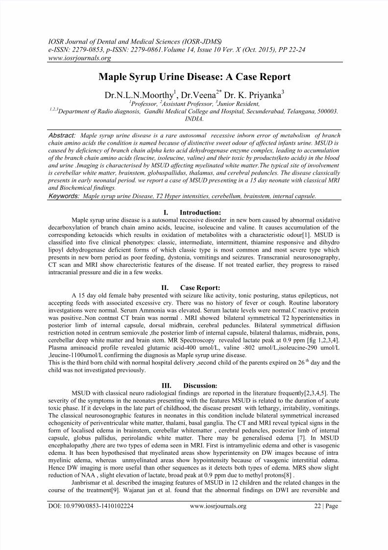
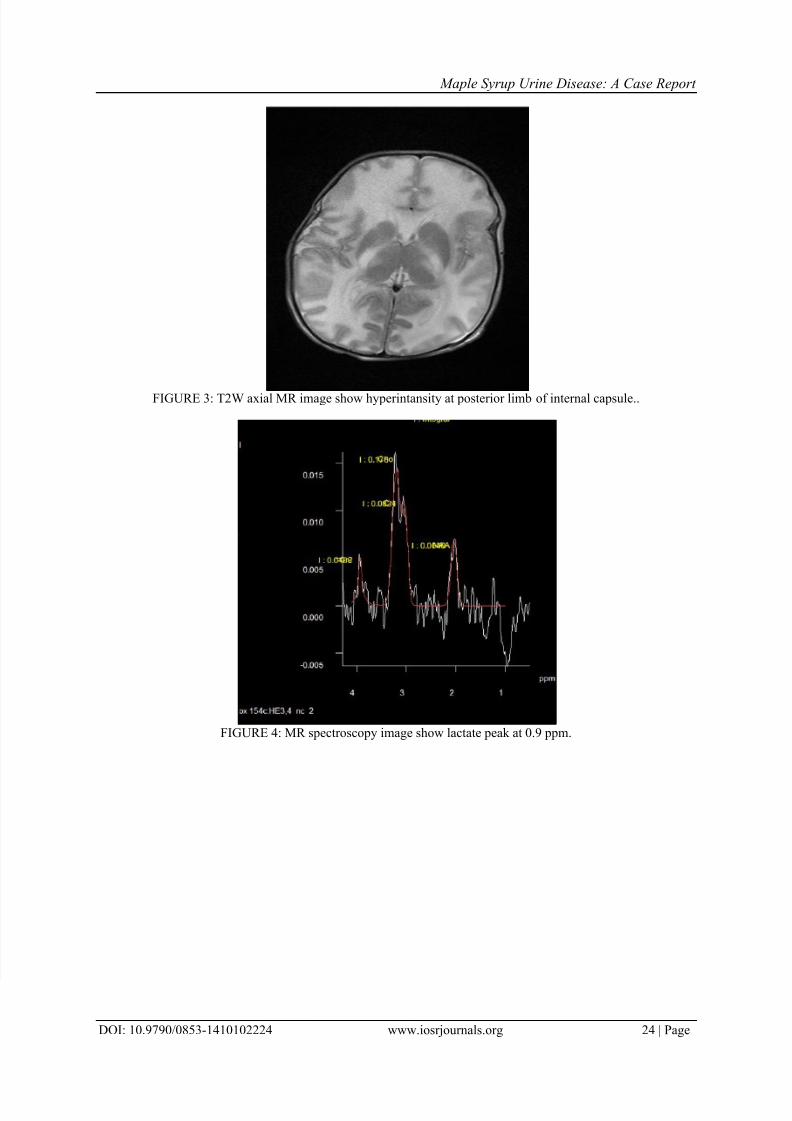
cerebellar deep white matter and brain stem. MR Spectroscopy revealed lactate peak at 0.9 ppm [fig 1,2,3,4].
Plasma aminoacid profile revealed glutamic acid-400 umol/L, valine -802 umol/L,isoleucine-290 umol/L
,leucine-1100umol/L confirming the diagnosis as Maple syrup urine disease.
This is the third born child with normal hospital delivery ,second child of the parents expired on 26 th day and the
child was not investigated previously.
III.
Discussion:MSUD with classical neuro radiological findings are reported in the literature frequently[2,3,4,5]. The
severity of the symptoms in the neonates presenting with the features MSUD is related to the duration of acute
toxic phase. If it develops in the late part of childhood, the disease present with lethargy, irritability, vomitings.
The classical neurosonographic features in neonates in this condition include bilateral symmetrical increased
echogenicity of periventricular white matter, thalami, basal ganglia. The CT and MRI reveal typical signs in the
form of localised edema in brainstem, cerebellar whitematter , cerebral peduncles, posterior limb of internal
capsule, globus pallidus, perirolandic white matter. There may be generalised edema [7]. In MSUD
encephalopathy ,there are two types of edema seen in MRI. First is intramyelinic edema and other is vasogenic
edema. It has been hypothesised that myelinated areas show hyperintensity on DW images because of intra
myelinic edema, whereas unmyelinated areas show hypointensity because of vasogenic interstitial edema.
Hence DW imaging is more useful than other sequences as it detects both types of edema. MRS show slight
reduction of NAA , slight elevation of lactate, broad peak at 0.9 ppm due to methyl protons[8] .
Janbrismar et al. described the imaging features of MSUD in 12 children and the related changes in thecourse of the treatment[9]. Wajanat jan et al. found that the abnormal findings on DWI are reversible and

7/24/2019 Maple Syrup Urine Disease: A Case Report
http://slidepdf.com/reader/full/maple-syrup-urine-disease-a-case-report 2/3
Maple Syrup Urine Disease: A Case Report
DOI: 10.9790/0853-1410102224 www.iosrjournals.org 23 | Page
elevated lactate, low NAA levels , elevated branched chain amino acid levels indicate mitochondrial
dysfunction during metabolic decompensation[10]. The other condition with similar diffusion imaging changes
include canavans disease, non ketotic hyperglycemia. The typical restriction on DWI is due to intramyelinic
edema corresponding to areas that are myelinated at birth[11,12].
IV.
Conclusion:Our findings suggest that during the acute phase and early encephalopathic crisis stage of MSUD ,
DWI can demonstrate the involvement of myelinated white matter in new born and thus can be a valuable
diagnostic tool.
References:[1]. Toxic and Metabolic Brain Disorders.chapter 3,4 th edition,A.James Barkovich Pediactric Neuroimaging. Lipincott Williams and
Wilkins;p 90.
[2]. Aditi Jain, K. Jagdeesh, Ranoji Mane, and Saurabh Singla Imaging in Classic Form of Maple Syrup UrineDisease:A Rare Metabolic
Central Nervous System J Clin Neonatol. 2013 Apr-Jun; 2(2): 98 – 100.
[3]. Venkatraman and Gunaseelan: Neuroradiological findings in maple syrup urine disease Journal of Pediatric Neurosciences 2013 /Jan-Apr / Volume 8 / / 31
[4]. Leema Pauline Cornelius,B.Kannan,Viveka saravanan,Eswaradass prasanna venkatesan.intramyelinic edemain maple syrup urine
disease.Ann Indian Acad Neurol 2014;17:211-3.
[5]. masudi sheetal, vishal, s., nimbal, satish patil, praveen m. m. and saurabh rathi case report of neuroradiological imaging - maple
syrup urine disease international journal of current research vol. 7, issue, 03, pp.14045-14047, march, 2015[6]. Fariello G,Dionisi-vici C,Orazi C,et al.cranial ultrasonography in maple syrup urine disease.AJNR Am Neuroradiol 1996;17:311-
315.
[7]. Brismar, J., Aqeel, A., Brismar, G., Coates, R., Gascon, G.,Ozand, P. 1990. Maple syrup urine disease. AJNR Am. J.Neuroradiol,
11:1219‑28.
[8]. Felber SR,Sperl W,Chemelli A,Murr C,Wendel U.maple syrup urine disease:metabolic decompensation monitored by proton
magnetic resonance imaging and spectroscopy.Ann Neurol 1993;33:396-401.
[9]. Brismar, J., Aqeel, A., Brismar, G., Coates, R., Gascon, G.,Ozand, P. 1990. Maple syrup urine disease: Findings on CT and MR
scans of the brain in 10 infants. AJNR Am. J.Neuroradiol, 11:1219‑28.
[10]. Jan W, Zimmerman RA, Wang ZJ, Berry GT, Kaplan PB, Kaye EM.MR diffusion imaging and MR spectroscopy of maple syrup
urinedisease during acute metabolic decompensation. Neuroradiology 2003;45:393‑9.
[11]. Bindu PS, Kovoor JM, Christopher R. Teaching neuro images: MRI in maple syrup urine disease. Neurology 2010;74:e12.
FIGURE 1,2,: DIFFUSION WEIGHTED MRI Brain show restriction at bilateral thalamus,Midbrain,pons
,cerebral peduncles,cerebellar deep white matter.
7/24/2019 Maple Syrup Urine Disease: A Case Report
http://slidepdf.com/reader/full/maple-syrup-urine-disease-a-case-report 3/3
Maple Syrup Urine Disease: A Case Report
DOI: 10.9790/0853-1410102224 www.iosrjournals.org 24 | Page
FIGURE 3: T2W axial MR image show hyperintansity at posterior limb of internal capsule..
FIGURE 4: MR spectroscopy image show lactate peak at 0.9 ppm.
Related Documents


![Maple Syrup | Maple Candies | Gift Boxes Candles ...1].pdfMaple Syrup | Maple Candies | Gift Boxes Candles & Souvenirs | Sugarhouse Tours ... Maple Syrup may be one of the most unique](https://static.cupdf.com/doc/110x72/5af59d9b7f8b9a74448e6f4c/maple-syrup-maple-candies-gift-boxes-candles-1pdfmaple-syrup-maple-candies.jpg)