Clark and Jonathan L. E. Dean Corina Tudor, Jeremy Saklatvala, Andrew R. Francesco P. Marchese, Anna Aubareda, Inhibiting CAF1 Deadenylase Recruitment Tristetraprolin-directed mRNA Decay by MAPKAP Kinase 2 Blocks Signal Transduction: doi: 10.1074/jbc.M110.136473 originally published online July 1, 2010 2010, 285:27590-27600. J. Biol. Chem. 10.1074/jbc.M110.136473 Access the most updated version of this article at doi: . JBC Affinity Sites Find articles, minireviews, Reflections and Classics on similar topics on the Alerts: When a correction for this article is posted • When this article is cited • to choose from all of JBC's e-mail alerts Click here Supplemental material: http://www.jbc.org/content/suppl/2010/07/01/M110.136473.DC1.html http://www.jbc.org/content/285/36/27590.full.html#ref-list-1 This article cites 44 references, 30 of which can be accessed free at by guest on October 31, 2013 http://www.jbc.org/ Downloaded from by guest on October 31, 2013 http://www.jbc.org/ Downloaded from by guest on October 31, 2013 http://www.jbc.org/ Downloaded from by guest on October 31, 2013 http://www.jbc.org/ Downloaded from by guest on October 31, 2013 http://www.jbc.org/ Downloaded from by guest on October 31, 2013 http://www.jbc.org/ Downloaded from by guest on October 31, 2013 http://www.jbc.org/ Downloaded from by guest on October 31, 2013 http://www.jbc.org/ Downloaded from by guest on October 31, 2013 http://www.jbc.org/ Downloaded from by guest on October 31, 2013 http://www.jbc.org/ Downloaded from by guest on October 31, 2013 http://www.jbc.org/ Downloaded from by guest on October 31, 2013 http://www.jbc.org/ Downloaded from

Welcome message from author
This document is posted to help you gain knowledge. Please leave a comment to let me know what you think about it! Share it to your friends and learn new things together.
Transcript

Clark and Jonathan L. E. DeanCorina Tudor, Jeremy Saklatvala, Andrew R. Francesco P. Marchese, Anna Aubareda, Inhibiting CAF1 Deadenylase RecruitmentTristetraprolin-directed mRNA Decay by MAPKAP Kinase 2 BlocksSignal Transduction:
doi: 10.1074/jbc.M110.136473 originally published online July 1, 20102010, 285:27590-27600.J. Biol. Chem.
10.1074/jbc.M110.136473Access the most updated version of this article at doi:
.JBC Affinity SitesFind articles, minireviews, Reflections and Classics on similar topics on the
Alerts:
When a correction for this article is posted•
When this article is cited•
to choose from all of JBC's e-mail alertsClick here
Supplemental material:
http://www.jbc.org/content/suppl/2010/07/01/M110.136473.DC1.html
http://www.jbc.org/content/285/36/27590.full.html#ref-list-1
This article cites 44 references, 30 of which can be accessed free at
by guest on October 31, 2013http://www.jbc.org/Downloaded from by guest on October 31, 2013http://www.jbc.org/Downloaded from by guest on October 31, 2013http://www.jbc.org/Downloaded from by guest on October 31, 2013http://www.jbc.org/Downloaded from by guest on October 31, 2013http://www.jbc.org/Downloaded from by guest on October 31, 2013http://www.jbc.org/Downloaded from by guest on October 31, 2013http://www.jbc.org/Downloaded from by guest on October 31, 2013http://www.jbc.org/Downloaded from by guest on October 31, 2013http://www.jbc.org/Downloaded from by guest on October 31, 2013http://www.jbc.org/Downloaded from by guest on October 31, 2013http://www.jbc.org/Downloaded from by guest on October 31, 2013http://www.jbc.org/Downloaded from

MAPKAP Kinase 2 Blocks Tristetraprolin-directed mRNADecay by Inhibiting CAF1 Deadenylase Recruitment□S
Received for publication, April 20, 2010, and in revised form, June 30, 2010 Published, JBC Papers in Press, July 1, 2010, DOI 10.1074/jbc.M110.136473
Francesco P. Marchese, Anna Aubareda, Corina Tudor1, Jeremy Saklatvala, Andrew R. Clark, andJonathan L. E. Dean2
From the Kennedy Institute of Rheumatology Division, Faculty of Medicine, Imperial College London,London W6 8LH, United Kingdom
Tristetraprolin (TTP) directs its target AU-rich element(ARE)-containing mRNAs for degradation by promoting re-moval of the poly(A) tail. The p38 MAPK pathway regulatesmRNA stability via the downstream kinase MAPK-activatedprotein kinase 2 (MAPKAP kinase 2 or MK2), which phosphor-ylates and prevents the mRNA-destabilizing function of TTP.We show that deadenylation of endogenous ARE-containingtumor necrosis factor mRNA is inhibited by p38 MAPK. Toinvestigate whether phosphorylation of TTP by MK2 regulatesTTP-directed deadenylation of ARE-containing mRNAs, weused a cell-free assay that reconstitutes the mechanism in vitro.We find that phosphorylation of Ser-52 and Ser-178 of TTP byMK2 results in inhibition of TTP-directed deadenylation ofARE-containing RNA. The use of 14-3-3 protein antagonistsshowed that regulation of TTP-directed deadenylation by MK2is independent of 14-3-3 binding to TTP. To investigate themechanism whereby TTP promotes deadenylation, it was nec-essary to identify the deadenylases involved. The carbon catab-olite repressor protein (CCR)4�CCR4-associated factor (CAF)1complexwas identified as themajor source of deadenylase activ-ity in HeLa cells responsible for TTP-directed deadenylation.CAF1a and CAF1b were found to interact with TTP in an RNA-independent fashion. We find that MK2 phosphorylationreduces the ability of TTP to promote deadenylation by inhibit-ing the recruitment of CAF1 deadenylase in a mechanism thatdoes not involve sequestration of TTP by 14-3-3. Cyclooxygen-ase-2 mRNA stability is increased in CAF1-depleted cells inwhich it is no longer p38 MAPK/MK2-regulated.
Many mammalian mRNAs contain adenosine/uridine-richelements (AREs)3 in their 3�-untranslated regions (UTRs)that target mRNAs for rapid degradation. The importance ofAREs in regulation of mRNA stability has been demon-strated particularly for mRNAs of the inflammatory re-
sponse. The p38 MAPK pathway inhibits ARE-mediated decayallowing for dynamic control of the expression of thesemRNAs(1–3). p38MAPK regulatesmRNAstability via the downstreamkinase MAPKAP kinase 2 (MK2), which phosphorylates (4, 5)and prevents the function (6, 7) of the mRNA-destabilizingARE-binding protein, tristetraprolin (TTP). We recentlyshowed that dual control ofmRNA stability byTTP and the p38MAPK pathway is a general mechanism, which operates formany mRNAs of the inflammatory response (8). The impor-tance of TTP in the control of inflammatory gene expressionis demonstrated by spontaneous inflammatory arthritis inTTP�/� mice arising mainly from increased tumor necrosisfactor (TNF) production (9). p38 MAPK also regulates mRNAstability by direct phosphorylation and inactivation of anotherARE-binding protein, KH domain-splicing regulatory protein(10, 11). p38 MAPK inhibitors fail to destabilize mRNAs of theinflammatory response in macrophages from TTP�/� mice (8,12). Thus, it appears that in these cells, at least, TTP is entirelyresponsible for the effects on mRNA stability mediated by thep38 MAPK pathway.In addition to controlling inflammatory mediator mRNA
stability, the p38 MAPK pathway stabilizes both TTP mRNA(13) and protein (14), thereby increasing TTP expression. Thecurrent understanding is that the mRNA destabilizing protein,TTP, is induced and held in a latent state by the p38 MAPKpathway and is poised to direct mRNA destabilization whenactivity in the pathway dissipates. Thus a proinflammatorystimulus triggers the induction of inflammatory genes and thesubsequent resolution of their expression, the latter being cru-cial to prevent chronic inflammation. It has long been knownthat treatment of cells with p38 MAPK inhibitor following aninflammatory stimulus destabilizes inflammatory mediatormRNAs (1). Recently, we showed that treatment of cells withp38 MAPK inhibitor prior to an inflammatory stimulus fails todestabilize TNF mRNA (8), presumably because under theseconditions, TTP protein is not induced (14). Thus, prolongedtreatment of cells with p38 MAPK inhibitors in vivo may nottake advantage of blocking this important post-transcriptionalmechanismof gene regulation. This is one potential reasonwhyp38 MAPK inhibitors have failed recently in clinical trials forrheumatoid arthritis (15). Elucidation of the post-transcrip-tional mechanism downstream of p38 MAPK may offer futurepromise for the therapy of such diseases.It is known that TTP directs its target mRNAs for degrada-
tion by promoting removal of the poly(A) tail or deadenylation(16), the first step in mRNA decay. The p38 MAPK pathway
□S The on-line version of this article (available at http://www.jbc.org) containssupplemental Figs. S1–S4.
1 Present address: Airway Disease Section, National Heart and Lung Institute,Imperial College, Dovehouse St., SW3 6LY London, United Kingdom.
2 To whom correspondence should be addressed: Kennedy Institute of Rheu-matology Division, Imperial College London, 65 Aspenlea Rd, Hammer-smith, London, W6 8LH, United Kingdom. Tel.: 44-208-383-4470; Fax:44-208-383-4499; E-mail: [email protected].
3 The abbreviations used are: ARE, adenosine/uridine-rich element; PAN,poly(A) nuclease; PARN, poly(A) ribonuclease; MK2, MAPK-activated pro-tein kinase 2; nt, nucleotide(s); Q-RT-PCR, quantitative reverse-transcrip-tion PCR; PB, processing body.
THE JOURNAL OF BIOLOGICAL CHEMISTRY VOL. 285, NO. 36, pp. 27590 –27600, September 3, 2010© 2010 by The American Society for Biochemistry and Molecular Biology, Inc. Printed in the U.S.A.
27590 JOURNAL OF BIOLOGICAL CHEMISTRY VOLUME 285 • NUMBER 36 • SEPTEMBER 3, 2010

stabilizes mRNAs by inhibiting deadenylation (17, 18) but theprecise mechanism whereby phosphorylation of TTP by MK2inhibits poly(A) tail shortening is not known. Phosphorylationof TTP by MK2 at Ser-52 and Ser-178 results in binding of14-3-3 to TTP (6, 19), and the formation of this complex hasbeen suggested to prevent TTP from interacting with mRNAdecay factors (6).Twodistinctdeadenylase complexes, poly(A)nuclease (PAN)2-
PAN3, and carbon catabolite repressor protein (CCR)4-CCR4-associated factor (CAF)1, originally were discovered in yeast(20, 21). Human orthologues of both complexes exist (22).In humans, the CCR4�CAF1 complex comprises two subunitswith deadenylase activity (CCR4 and CAF1) together withseven otherCNOTproteins (23). HumanCCR4 andCAF1 eachhave two different paralogues: CCR4a (CNOT6) and CCR4b(CNOT6L); and CAF1a (CNOT7) and CAF1b (CNOT8). Ingeneral, for mRNA decay in mammalian cells, PAN2-PAN3 isthought to catalyze initial poly(A) shortening, andCCR4-CAF1then removes the remaining �110 nucleotides (nt) of thepoly(A) tail (24).CAF1 deadenylase has been implicated in ARE-mediated
deadenylation. Knockdown of CAF1 by RNA interference(RNAi) has been shown to impair the deadenylation and decayof an ARE-containing �-globin mRNA (25, 26). In contrast,CCR4 depletion has been reported to have no effect on dead-enylation of anARE reportermRNA (26).Mammalian cells alsocontain another, predominantly nuclear enzyme, poly(A) ribo-nuclease (PARN) (27). It has been suggested to be involved inARE-mediated deadenylation (28) and to promote TTP-di-rected deadenylation in vitro (29). TTP has been reported tointeract with mRNA decay factors including the exosome (30),Dcp1a, Dcp2, Xrn1, and also CCR4 (31) but not PARN (29). It isthus not clear which deadenylase is involved in TTP-directeddeadenylation in cells.To elucidate the mechanism whereby MK2 inactivates TTP,
it was necessary to first identify which deadenylase is involvedin TTP-directed deadenylation. To investigate this, we modi-fied an in vitro ARE-dependent and TTP-directed deadenyla-tion assay described by Lai et al. (29) to use bacterially ex-pressed recombinant TTP. This allowed the involvement ofdeadenylases to be determined by assaying extracts from cellsdepleted of different deadenylases by RNAi in the presence of aconstant amount of TTP. The use of recombinant TTP in the invitro systemalso allowed us to investigate the role ofMK2 in theabsence of changes in TTP protein expression, which occurs incells following activation or inhibition of this kinase (7, 14). Theassay uses TNF and granulocyte/macrophage-colony stimulat-ing factor (GM-CSF) ARE RNA substrates with 100-nt poly(A)tails. Deadenylation of both of these mRNAs has been shownpreviously to be regulated by TTP (16, 32). Both mRNAs alsoare stabilized by the p38MAPK/MK2pathway (33, 34). R18 anddifopein (dimeric fourteen-three-three peptide inhibitor) arehigh affinity 14-3-3 antagonists that allow for essentially com-plete inhibition of 14-3-3 binding to target proteins (35). Thein vitro deadenylation assay enabled us to use R18 and difo-pein to test the function of 14-3-3 in deadenylation and todetermine a novel mechanism whereby MK2 inhibits TTP-directed deadenylation.
EXPERIMENTAL PROCEDURES
Materials—General laboratory reagents were from Sigma.4-(4-Fluorophenyl)-2-(4-hydroxyphenyl)-5-(4-pyridyl)1H-imidazole (SB202190) was from Calbiochem-Novabiochem.pSP73-GM-CSF ARE-p(A)� and -GM-CSF ARE mut-p(A)�plasmids were described previously (36). The GM-CSF AREsequence was removed by digestion with XbaI and replacedwith a SpeI XbaI fragment from pBluescript-hTNF 75 (37) tocreate pSP73-TNFARE-p(A)� and -TNFARE antisense-p(A)�plasmids. Glutathione S-transferase (GST)-TTP expressionplasmids were constructed by subcloning the previously de-scribed wild-type and a S52A/S178A mutated form of mu-rine TTP cDNA (13) into pGEX-6P-3 (GE Healthcare) at theEcoRI site. GM-CSF ARE and GM-CSF ARE mut oligoribo-nucleotides were synthesized commercially (Dharmacon). Thesequences were as follows: GM-CSF ARE, AGUAAUAUUUA-UAUAUUUAUAUUUUUAAAAUAUUUAUUUAUUUAUU-UAUUUAA; andGM-CSFAREmut, GUAAUAUGAAUACA-UCUGAAUGUCUGGAAUAUUGAUUGAUAAGCUUAGU-CGAC. The antibodies used were as follows: anti-�-tubulin(Sigma), anti-MK2 (Upstate), anti-CAF1a (Abnova), anti-CAF1b(LifeSpan BioSciences), anti-pan-14-3-3 (Chemicon), and anti-TTP (4). Anti-PARN, anti-PAN2, and anti-PABP1 antibodieswere gifts from Professor Anders Virtanen (Uppsala Univer-sity), Professor Shin-ichi Hoshino (University of Tokyo), andDr. Matt Brook (University of Edinburgh), respectively.Cell Culture—HeLa (gift from Yamanouchi) and RAW 264.7
(ATCC) cells were cultured in Dulbecco’s modified Eagle’smedium (PAA Laboratories) supplemented with 10% fetal calfserum (PAA Laboratories). Penicillin/streptomycin (PAA Lab-oratories)was included inHeLa cell culturemedium.Cellsweremaintained at 37 °C in the presence of 5% CO2.Preparation of Recombinant TTP—GST-TTP expression plas-
mids were used to transform Escherichia coli TOP10 (Invitro-gen). Bacteria were grown in LB containing 100 �g/ml ampicil-
FIGURE 1. p38 MAPK regulates the poly(A) tail length of endogenous TNFmRNA. A and B, RAW 264.7 cells were treated with LPS (10 ng ml�1) for 2 h,and then actinomycin D (Act D; 10 �g ml�1) was added together with vehicle(0.1% dimethyl sulfoxide) or 1 �M SB202190 (SB). Cells were harvested at thetime intervals shown, and RNA was extracted. A, 10 �g RNA was incubatedwith TNF 1246 (lanes 2–9) or TNF 1246 oligodeoxynucleotide and oligo(dT)(lanes 1 and 10) and cleaved with RNase H. The samples were Northern blot-ted with an antisense riboprobe against the TNF 3�-UTR. B, Northern blot offull-length TNF mRNA. GAPDH is shown as a loading control.
Mechanism for Stabilization of mRNA by MK2
SEPTEMBER 3, 2010 • VOLUME 285 • NUMBER 36 JOURNAL OF BIOLOGICAL CHEMISTRY 27591
lin, and 1 mM isopropyl 1-thio-�-D-galactopyranoside wasadded atmid-exponential phase to induce expression for 12 h at28 °C. Cells were harvested and suspended in 20 mM HEPES,pH 7.9, with 10% (v/v) glycerol, 0.5 M KCl, 2 mM DTT, 1 mM
PMSF, 1 �g/ml pepstatin A, 13.5 �g/ml aprotinin, and 10 �M
E-64. Cells were lysed by four passages through a French pres-sure cell at 15,000 psi. Cell debris was removed by centrifuga-tion at 30,000 � g for 20 min, and the supernatant was incu-bated with glutathione-Sepharose 4B (GE Healthcare) at 4 °Cfor 30 min with shaking. The resin was washed with 15 columnvolumes of PBS, and bound protein was eluted with 50 mM
Tris-HCl, pH 8.0, 10 mM reduced glutathione. On-columncleavage of the GST tag was performed with PreScission prote-ase (GE Healthcare) treatment following the manufacturer’sinstructions. Glycerol was added to a final concentration of 10%(v/v), and the protein was stored at �80 °C until use. TTP pro-tein concentration was determined by Bradford assay.In Vitro Deadenylation Assay—This was performed accord-
ing to Lai et al. (29) using HeLa cells lysed by Dounce homog-enization. Briefly, RNA substrates with 100 nt poly(A) tails
were prepared by in vitro transcrip-tion in the presence of [�-32P]UTP(PerkinElmer) as described previ-ously (37). A radiolabeled poly(A)tail was added to the GM-CSF AREor GM-CSF ARE mut oligoribo-nucleotides using a poly(A) tailingkit (Epicentre) and [�-32P]ATP(PerkinElmer) according to themanufacturer’s instructions. RNAspecies with poly(A) tails of �100nt were obtained by gel purifica-tion. HeLa S100, GST-TTP, andradiolabeled substrate RNA weremixed and incubated at 37 °C for thetimes indicated, and EDTA (finalconcentration, 20 mM) was addedto terminate the reaction. RNAwas isolated by phenol-chloroformextraction, electrophoresed on poly-acrylamide-urea gels, and visualizedand quantified by phosphorimaging(FLA-5100 imager, Fuji, Japan;AIDA 1D quantification software,Raytest, Germany).In Vitro Phosphorylation of GST-
TTP by MK2—In vitro phosphory-lation of TTP by MK2 was per-formed with immunoprecipitatedMK2orwith recombinantMK2 (Mil-lipore). 1 �g of GST-TTP was com-binedwith immunoprecipitatedMK2or with recombinant activated MK2(0.1 unit) in a final volume of 30 �lcontaining 20 �M ATP and incu-bated at 30 °C. To assess phosphor-ylation of GST-TTP, 4 �Ci of[�-32P]ATP (PerkinElmer Life Sci-
ences) was included in the reaction, and it was stopped by theaddition of 10�l of 4� SDS-PAGE sample buffer and subjectedto electrophoresis. 32P incorporation was visualized and quan-tified by phosphorimaging. The stoichiometry of phosphory-lation was measured by excising the radioactive bands andscintillation counting.GST Pulldown Assay—2 �g of GST-TTP was incubated with
30�l of a 50% slurry of glutathione-Sepharose 4B and 200�g ofcytoplasmic extract in 1 ml of binding buffer (10 mM HEPES,pH 7.6, 100 mM KCl, 6 mM MgCl2, 1 mM DTT, 1% (v/v) IgepalCA-360) for 60 min at 4 °C in the presence or absence of 150units of benzonase (Sigma). The beads were washed with bind-ing buffer and boiled in SDS-PAGE sample buffer for 5min, andeluted proteins were analyzed byWestern blotting as describedpreviously (38).RNase H Mapping and Northern Blot—RNase H mapping
was performed as described previously (17) using an antisensemurine TNF 3�-UTR oligodeoxynucleotide (TNF 1246) span-ning nt 1246–1276 of the TNF mRNA (5�-GCTGGCTCTGT-GAGGAAGGCTGTGCATTGC-3�). RNA was detected by
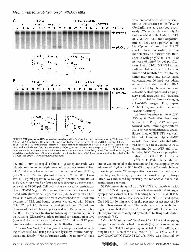
FIGURE 2. TTP promotes ARE-dependent deadenylation in vitro. A, in vitro deadenylation of 32P-labeled TNFARE or TNF ARE antisense RNA substrates were incubated in the presence of HeLa S100 (5 �g) and 100 ng GSTor GST-TTP at 37 °C for the times indicated. Representative phosphorimage of urea-PAGE of 32P-labeled reac-tion products is shown. Graphs show mean poly(A)100 expressed as a percentage of t � 0 � S.E. from threeindependent experiments. Where not shown, error bars are smaller than the symbols. The positions of polya-denylated substrate (poly(A)100) and deadenylated product (poly(A)0(*)) are indicated. B, as for A but withGM-CSF ARE or GM-CSF ARE mut RNA substrates.
Mechanism for Stabilization of mRNA by MK2
27592 JOURNAL OF BIOLOGICAL CHEMISTRY VOLUME 285 • NUMBER 36 • SEPTEMBER 3, 2010

Northern blotting using a murine antisense riboprobe thathybridizes with TNF mRNA between the cleavage site and thepoly(A) tail. This was generated by PCR amplification of a250-bp sequence spanning nt 1359–1609 of the 3�-UTRofTNFmRNA and in vitro transcribed as described previously (37).Full-length TNF andGAPDHmRNAswere detected byNorth-ern blotting as described previously (17).Electrophoretic Mobility Shift Assay (EMSA)—A GM-CSF
ARE oligonucleotide was end-labeled with [�-32P]ATP (3000Ci/mmol) using T4 polynucleotide kinase. A 32P-labeled RNAprobe (�0.1 pmol) was incubated in the presence or absence ofcold competitors for 15 min at room temperature with GST,unphosphorylated TTP, or TTP in vitro phosphorylated byMK2 in 20�l of bandshift buffer (20mMHEPES, pH7.2, 100�M
ZnCl2, 50 mMKCl, 1 mMDTT, 5% glycerol) containing heparinsulfate (5 mg/ml). Two �l of loading buffer (80% glycerol, 0.1%bromphenol blue) was then added, and RNA�protein com-plexes were resolved by electrophoresis on a 4% (w/v) acrylam-ide/Tris borate gel usingTris borate running buffer. Complexeswere visualized using a phosphorimaging device. Quantifieddata were plotted as fraction bound versus protein concentra-tion and fit with the Hill equation using Prism 5 (GraphPadSoftware).RNAi—The following siRNAs were used: scramble, 5�-
CAGUCGCGUUUGCGACUGGdTdT-3� and PARN, de-scribed previously by Lin et al. (28) were obtained from Euro-fins MWG Operon; and CCR4a (s33100), CCR4b (s48341),CAF1a (s26638), CAF1b (s17849), PAN2 (s19252), and PAN3(s48721) were purchased from Ambion (Applied Biosystems).HeLa cells were transfected twice using Oligofectamine
(Invitrogen), 24 h apart, as described previously (38), exceptthat 15 nM siRNA (final concentration) was used. The efficiencyof protein depletion was determined by Western blotting (38)and quantitative reverse-transcription PCR (Q-RT-PCR).Q-RT-PCR—RNA was isolated using the RNeasy Kit (Qia-
gen), and cDNAwas generated using the Reverse Transcriptionsystem kit and oligo(dT) (Promega). This cDNA was analyzedby Q-RT-PCR using TaqMan technology and primer-probesets for CCR4a (Hs01019492_m1), CCR4b (Hs00375913_m1), CAF1a (Hs01020564_m1), CAF1b (Hs00231841_m1),PARN (Hs00377733_m1), PAN2 (Hs00208356_m1), PAN3(Hs00107000_m1), COX-2 (Hs00153133_m1), and GAPDH(Hs99999905_m1) from Applied Biosystems. A Rotor-Gene6000 thermal cycler and software (Corbett Research) wereused. The ��Ct method and relative quantitation (with stan-dard curves) were used for mRNA quantitation using GAPDHas internal control (39).
RESULTS
The p38 MAPK Pathway Inhibits Deadenylation of Endoge-nous TNF mRNA—We showed previously that p38 MAPKstabilizes ARE-containing reporter mRNAs by inhibitingdeadenylation (17). To investigate whether p38 MAPK reg-ulates deadenylation of an endogenous mRNA, the poly(A)tail length of TNF mRNA induced by lipopolysaccharide (LPS)in the macrophage-like cell line RAW 264.7 in the presence orabsence of the p38MAPK inhibitor SB202190was assessed. Forthis, RNase H mapping was used because full-length TNF
mRNA is too long for accurate resolution of mRNAs with dif-ferent poly(A) tail lengths. RNase H cleaves RNA/DNA hetero-duplexes allowing mRNAs to be shortened in a specific fashionin the presence of oligodeoxyribonucleotides. Cells weretreated with LPS for 2 h, and then actinomycin D was added toblock transcription together with dimethyl sulfoxide vehicle or1 �M SB202190. RNA was isolated from the cells and treatedwith RNase H and an oligodeoxyribonucleotide against theTNFmRNA3�-UTR to cleave TNFmRNA. Some samples wereadditionally treated with oligo(dT) to completely removepoly(A) tails. Cleaved TNF mRNA was detected by Northernblot with a probe against the 3�-end of the mRNA. In cells
FIGURE 3. Phosphorylation of TTP by MK2 inhibits deadenylation. MK2was immunoprecipitated from lysates of HeLa cells stimulated with IL-1� andused to phosphorylate recombinant GST-TTP in vitro. Phosphorylation reac-tions were performed for different times (30, 60, and 120 min) at 30 °C, and anonimmune antibody (N.I.) served as a control. A, phosphorylated GST-TTPassayed by in vitro deadenylation assay for 60 min at 37 °C. B, phosphorimageof GST-TTP or GST phosphorylation by immunoprecipitated MK2 or nonim-mune control (N.I.) for different times in the presence of [�-32P]ATP. C, graphshowing correlation between GST-TTP phosphorylation by MK2 (% maxincorp. of 32P) and inhibition of deadenylation.
Mechanism for Stabilization of mRNA by MK2
SEPTEMBER 3, 2010 • VOLUME 285 • NUMBER 36 JOURNAL OF BIOLOGICAL CHEMISTRY 27593

treated with LPS and vehicle, TNF mRNA was relatively stableand little poly(A) shortening occurred (Fig. 1A). The mRNAunderwent rapid simultaneous decay and deadenylation in thepresence of SB202190 (Fig. 1A), confirming an important rolefor p38 MAPK in blocking deadenylation of an endogenoustranscript. It was not possible to quantify GAPDH mRNA onthe samemembranes as TNFmRNA owing to nonspecific deg-radation of GAPDHmRNA during RNase H treatment. There-fore, an identical actinomycin D chase experiment was per-formed in parallel, and full-length TNF and GAPDH mRNAswere detected by Northern blotting. The even GAPDH signalson the gel (Fig. 1B) confirm that the identical TNFmRNAdecayprofiles in Fig. 1 (A and B) are an accurate assessment of TNFmRNA decay.TTP-directed in Vitro Deadenylation of ARE-containing
RNA—p38 MAPK stabilizes inflammatory mRNAs via acti-vation of the downstream kinase MK2, which phosphory-lates and blocks the function of the mRNA destabilizing fac-tor, TTP. To investigate the mechanism wherebyMK2 inhibitspoly(A) shortening, it was necessary to modify an ARE-depen-dent and TTP-directed in vitro deadenylation assay developedby Lai et al. (29) to use recombinant bacterially expressed GST-TTP rather than TTP overexpressed in 293 cells. In vitro dead-enylation reactions contained a 100,000 � g supernatant of
HeLa cytoplasmic extract (S100),which served as a source of dead-enylases, GST-TTP or GST, and aradiolabeled RNA substrate com-prisingARE-containing RNA linkedto a 100 nt poly(A) tail. Reactionmixtures were incubated for differ-ent times in the presence of GST-TTP, GST or S100 alone. Thisshowed time- and TTP-dependentdisappearance of full-length TNFRNA substrate (poly(A)100) and theaccumulation of an intermediate ofhigher mobility on the gel (Fig. 2A).The increase in the deadenylatedproduct did not fully correspond tothe reduction of full-length sub-strate (Fig. 2A), suggesting that it isan intermediate that undergoes fur-ther decay. No TTP-directed dead-enylation was seen in in vitro dead-enylation assay with antisense TNFARE (Fig. 2A). Similar results wereobtained for a GM-CSF ARE RNAand mutant form in which the AREwas disrupted with multiple substi-tutions of A and U to G and C (Fig.2B), consistent with ARE binding byTTP being a mandatory event forTTP-directed deadenylation. Titra-tion of GST-TTP confirmed that100 ng is the optimum amount ofTTP to use in the assay and oli-go(dT) and RNase H treatment
showed that the GM-CSF ARE RNA intermediate representsan RNA species that lacks the poly(A) tail (poly(A)0(*)) and anadditional �5 nt (supplemental Fig. S1).Phosphorylation of TTP byMK2 Inhibits Deadenylation—To
elucidate how phosphorylation of TTP by MK2 regulatesdeadenylation of ARE-containing mRNAs, inhibition ofTTP-directed deadenylation by MK2 was reconstituted invitro. In initial experiments, MK2 was immunoprecipitatedfrom lysates of HeLa cells stimulated with IL-1�, a potentactivator of the p38 MAPK pathway, and used to phosphor-ylate recombinant GST-TTP. Phosphorylation reactionswere performed for different times, and phosphorylatedGST-TTP was assayed for its ability to promote deadenyla-tion following a 1 h incubation at 37 °C. A dummy phosphor-ylation reaction with a nonimmune antibody immunopre-cipitate served as a control. Phosphorylation of GST-TTP byMK2 for 1 or 2 h inhibited the ability of TTP to promoteGM-CSF ARE substrate deadenylation (Fig. 3, A and C).Analysis of GST-TTP phosphorylated for different times inthe presence of [�-32P]ATP showed that phosphorylation wasessentially complete at 2 h, and little phosphorylation of GST-TTPwas seen with a nonimmune immunoprecipitate (Fig. 3B).Thus, the inhibition of deadenylation appeared to closely cor-relate with phosphorylation of GST-TTP by MK2 (Fig. 3C).
FIGURE 4. Phosphorylation of TTP by MK2 does not affect ARE binding by TTP. A, 32P end-labeled GM-CSFARE RNA probe (0.1 pmol) was incubated with different concentrations of either unphosphorylated TTP or TTPphosphorylated by recombinant active MK2 for 30 min at 30 °C. B, graph of bound RNA against [TTP] and bestfit with the Hill equation from three independent experiments. Where not shown, error bars are smaller than thesymbols. C, 32P end-labeled GM-CSF ARE was incubated with TTP (25 nM) or GST (25 nM) in the presence orabsence of increasing amounts (1, 10, and 20�) of cold GM-CSF ARE (self) or GM-CSF ARE mut (non-self) RNAcompetitors. Results are representative of three experiments. RNA�protein complexes were resolved by elec-trophoresis and visualized using a phosphorimaging device. The free probe and the TTP�RNA complexes areindicated.
Mechanism for Stabilization of mRNA by MK2
27594 JOURNAL OF BIOLOGICAL CHEMISTRY VOLUME 285 • NUMBER 36 • SEPTEMBER 3, 2010

Phosphorylation of TTP by MK2 Has No Effect on GM-CSFARERNABinding—It has been suggested that phosphorylationof TTP by MK2 reduces its affinity for mRNA (7). To test this,EMSAwas performed with a GM-CSFARE RNA probe lackinga poly(A) tail and either unphosphorylated TTP or TTP phos-phorylated by recombinant activeMK2 in vitro. For this exper-iment, cleaved TTP lacking the GST tag was used to avoidformation of complexes that may be formed due to dimer-ization of GST. Bands corresponding to two distinctRNA�protein complexes were observed and there was no sig-nificant difference in binding between unphosphorylatedand phosphorylated TTP (Fig. 4, A and B). ARE-dependentbinding of TTP was confirmed by competition experimentsusing unlabeled GM-CSF ARE (self) and GM-CSF ARE mut(non-self) probes as competitors in the binding reaction. Com-petition was strongest with GM-CSF ARE RNA, consistentwith specific ARE binding (Fig. 4C). Incubation of GM-CSFARE RNA with a bacterially expressed GST preparation con-firmed that complexes were formed by TTP and not E. colicontaminants (Fig. 4C). Phosphorylation of TTP by MK2 wasconfirmed to be complete by the incorporation of 32P into TTP(supplemental Fig. S2). The stoichiometry of phosphorylationwas 0.48 mol/mol TTP. This value is approximate as TTP pro-
tein concentration wasmeasured byBradford assay and not all of thepreparation was full-length (seeFig. 3B).14-3-3 Binding Does Not Mediate
the Effect of MK2 on TTP-directedDeadenylation—The involvementof 14-3-3 in sequestering TTP fromdeadenylases was tested using theantagonists R18 and difopein (35).GST pulldown was used to deter-mine the concentrations of R18and difopein needed to disrupt the14-3-3�TTP complex. Wild-type andS52A/S178A GST-TTP were phos-phorylated by recombinant MK2as before or left unphosphorylated.Fusion proteins or GST alone wereincubated with glutathione-Sepha-rose 4B beads and HeLa S100 inthe presence or absence of 14-3-3binding inhibitors. Western blots ofpulled downmaterial for 14-3-3 andTTP showed, as expected, thatphosphorylation of TTP at Ser-52and Ser-178 by MK2 gave rise to14-3-3 binding (Fig. 5A). 5 �M R18or 1 �M difopein efficiently inhib-ited 14-3-3 binding to phosphory-lated TTP (Fig. 5A). Phosphoryla-tion of GST-TTP by recombinantactive MK2 inhibited deadenylation(Fig. 5B). Inactive MK2 had noeffect (data not shown). Inclusion ofR18 or difopein in in vitro deadeny-
lation assay did not significantly impair the ability of MK2 toinhibit TTP-directed deadenylation, indicating that this proc-ess is not regulated by 14-3-3 (Fig. 5B). Moreover, GST-TTPS52A/S178A did not display MK2-dependent inhibition ofdeadenylation (Fig. 5B), indicating that Ser-52 and Ser-178mediate the effect of MK2 on deadenylation directed byTTP. Both wild-type and S52A/S178A GST-TTP migratedwith reduced mobility on SDS-PAGE (Fig. 5A), consistentwith phosphorylation of residues other than Ser-52 and Ser-178 by MK2. This was confirmed by significant 32P incorpo-ration into the mutant form of the protein in the presence ofMK2 (supplemental Fig. S2).The CCR4�CAF1 Complex Is the Major TTP-directed Dead-
enylase in HeLa S100—To further probe the mechanism it wasnecessary to identify the major deadenylase responsible forTTP-directed deadenylation. The known mammalian dead-enylases (CCR4-CAF1, PAN2-PAN3 and PARN)were depletedin HeLa cells in two independent experiments by RNAi andhigh speed supernatants prepared and analyzed by duplicate invitro deadenylation assays. First, HeLa cells were transfectedwith siRNAs against the two CCR4 paralogues (CCR4a andCCR4b alone or in combination), a scrambled double-strandedoligonucleotide as a control, or left untransfected. Cells were
FIGURE 5. 14-3-3 binding does not mediate the effect of MK2 on TTP-directed deadenylation. A, GST-TTP(WT or S52A/S178A) was phosphorylated with MK2 as in Fig. 4 or left unphosphorylated and used in a GSTpulldown assay. S100 extracts from HeLa cells were incubated with recombinant GST-TTP or GST (negativecontrol) immobilized on glutathione-Sepharose in the presence or absence of R18 or difopein. Pulled downproteins were resolved by SDS-PAGE and Western blotted for 14-3-3 and TTP. Results are representative ofthree experiments. B, in vitro deadenylation assay using unphosphorylated and phosphorylated GST-TTP (wild-type and S52A/S178A) in the presence or absence of R18 (5 �M) or difopein (1 �M). Graphs of mean (�S.E.) forthree independent experiments are shown.
Mechanism for Stabilization of mRNA by MK2
SEPTEMBER 3, 2010 • VOLUME 285 • NUMBER 36 JOURNAL OF BIOLOGICAL CHEMISTRY 27595
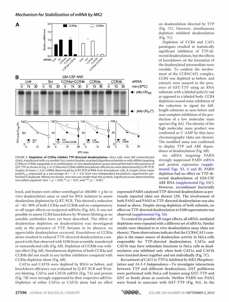
lysed, and lysates were either centrifuged at 100,000 � g for invitro deadenylation assay or used for RNA isolation to assessdeadenylase depletion by Q-RT-PCR. This showed a reductionof �85–90% of both CCR4a and CCR4b and no compensatoryor off-target effects on reciprocal mRNAs (Fig. 6A). It was notpossible to assess CCR4 knockdown byWestern blotting as nosuitable antibodies have yet been described. The effect ofdeadenylase depletion on deadenylation was investigatedonly in the presence of TTP, because in its absence, noappreciable deadenylation occurred. Knockdown of CCR4aalone resulted in reduced TTP-directed deadenylation com-pared with that observed with S100 from scramble-transfectedor untransfected cells (Fig. 6B). Depletion of CCR4b was with-out effect (Fig. 6B). Simultaneous depletion of both CCR4a andCCR4b did not result in any further inhibition compared withCCR4a depletion alone (Fig. 6B).CAF1a and CAF1b were depleted by RNAi as before, and
knockdown efficiency was evaluated by Q-RT-PCR and West-ern blotting. CAF1a and CAF1b mRNA (Fig. 7A) and protein(Fig. 7B) were strongly suppressed by their respective siRNAs.Depletion of either CAF1a or CAF1b alone had no effect
on deadenylation directed by TTP(Fig. 7C). However, simultaneousdepletion inhibited deadenylation(Fig. 7C).Depletion of CCR4 and CAF1
paralogues resulted in statisticallysignificant inhibition of TTP-di-rected deadenylation, but the effectsof knockdown on the formation ofthe deadenylated intermediate werevariable. To confirm the involve-ment of the CCR4�CAF1 complex,CCR4 was depleted as before, andextracts were assayed in the pres-ence of GST-TTP using an RNAsubstrate with a labeled poly(A) tailas opposed to a labeled body. CCR4depletion caused some inhibition ofthe reduction in signal for full-length substrate as seen before andnear complete inhibition of the pro-duction of a low molecular massspecies (Fig. 8A). The identity of thehigh molecular mass product wasconfirmed as 5�-AMP by thin layerchromatography (data not shown).The modified assay was confirmedto display TTP and ARE depen-dence of deadenylation (Fig. 8B).An siRNA targeting PARN
strongly suppressed PARN mRNAand protein expression (supple-mental Figs. S3, A and B). PARNdepletion had no effect on TTP-di-rected deadenylation of GM-CSFARE RNA (supplemental Fig. S3C).However, recombinant bacterially
expressed PARN catalyzed TTP-directed deadenylation as pre-viously reported (data not shown) (29). The involvement ofboth PAN2 and PAN3 in TTP-directed deadenylation was alsotested as above. Despite strong depletion of both subunits, noeffect onTTP-directed deadenylation in S100 supernatantswasobserved (supplemental Fig. S4).To control for possible off-target effects, all siRNA-mediated
depletions were repeated with a different set of siRNAs. Similarresults were obtained in in vitro deadenylation assay (data notshown). These observations indicate that theCCR4�CAF1 com-plex is the major source of deadenylase activity in HeLa cellsresponsible for TTP-directed deadenylation. CAF1a andCAF1b may have redundant functions in HeLa cells as dead-enylation was inhibited only when both CAF1a and CAF1bwere knocked down together and not individually (Fig. 7C).Recruitment of CAF1 to TTP Is Inhibited byMK2 Phosphory-
lation and 14-3-3 Independence—To investigate interactionsbetween TTP and different deadenylases, GST pulldownswere performed with HeLa cell lysates using GST-TTP andGST or beads alone as controls. Neither PARN nor PAN2were found to associate with GST-TTP (Fig. 9A). In the
FIGURE 6. Depletion of CCR4a inhibits TTP-directed deadenylation. HeLa cells were left untransfected(Untr), transfected with a scramble (Scr) control double-stranded oligoribonucleotide or with siRNAs targetingCCR4a or CCR4b, separately or in combination. In vitro deadenylation assays were performed in the presence ofGST-TTP as shown in Fig. 2. A, CCR4a and CCR4b mRNA knockdown efficiencies were determined by Q-RT-PCR.Graphs of mean (�S.D.) mRNA determined by Q-RT-PCR of RNA from knockdown cells. B, Graphs show meanpoly(A)100 expressed as a percentage of t � 0 � S.D. from two independent knockdown experiments per-formed in duplicate. Where not shown, error bars are smaller than the symbols. Significance was determined bytwo-tailed unpaired t test. *, p 0.05; **, p 0.01; and ***, p 0.001.
Mechanism for Stabilization of mRNA by MK2
27596 JOURNAL OF BIOLOGICAL CHEMISTRY VOLUME 285 • NUMBER 36 • SEPTEMBER 3, 2010
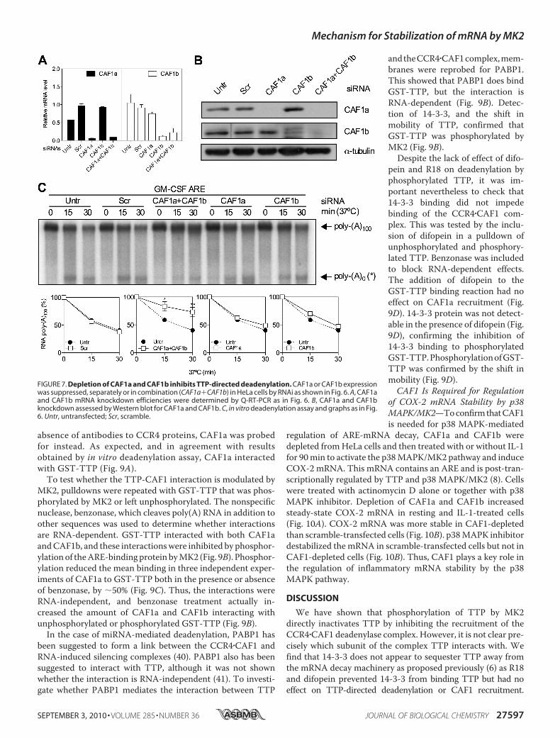
absence of antibodies to CCR4 proteins, CAF1a was probedfor instead. As expected, and in agreement with resultsobtained by in vitro deadenylation assay, CAF1a interactedwith GST-TTP (Fig. 9A).To test whether the TTP-CAF1 interaction is modulated by
MK2, pulldowns were repeated with GST-TTP that was phos-phorylated by MK2 or left unphosphorylated. The nonspecificnuclease, benzonase, which cleaves poly(A) RNA in addition toother sequences was used to determine whether interactionsare RNA-dependent. GST-TTP interacted with both CAF1aandCAF1b, and these interactionswere inhibited by phosphor-ylationof theARE-bindingprotein byMK2 (Fig. 9B). Phosphor-ylation reduced the mean binding in three independent exper-iments of CAF1a to GST-TTP both in the presence or absenceof benzonase, by �50% (Fig. 9C). Thus, the interactions wereRNA-independent, and benzonase treatment actually in-creased the amount of CAF1a and CAF1b interacting withunphosphorylated or phosphorylated GST-TTP (Fig. 9B).
In the case of miRNA-mediated deadenylation, PABP1 hasbeen suggested to form a link between the CCR4�CAF1 andRNA-induced silencing complexes (40). PABP1 also has beensuggested to interact with TTP, although it was not shownwhether the interaction is RNA-independent (41). To investi-gate whether PABP1 mediates the interaction between TTP
andtheCCR4�CAF1complex,mem-branes were reprobed for PABP1.This showed that PABP1 does bindGST-TTP, but the interaction isRNA-dependent (Fig. 9B). Detec-tion of 14-3-3, and the shift inmobility of TTP, confirmed thatGST-TTP was phosphorylated byMK2 (Fig. 9B).Despite the lack of effect of difo-
pein and R18 on deadenylation byphosphorylated TTP, it was im-portant nevertheless to check that14-3-3 binding did not impedebinding of the CCR4�CAF1 com-plex. This was tested by the inclu-sion of difopein in a pulldown ofunphosphorylated and phosphory-lated TTP. Benzonase was includedto block RNA-dependent effects.The addition of difopein to theGST-TTP binding reaction had noeffect on CAF1a recruitment (Fig.9D). 14-3-3 protein was not detect-able in the presence of difopein (Fig.9D), confirming the inhibition of14-3-3 binding to phosphorylatedGST-TTP.PhosphorylationofGST-TTP was confirmed by the shift inmobility (Fig. 9D).CAF1 Is Required for Regulation
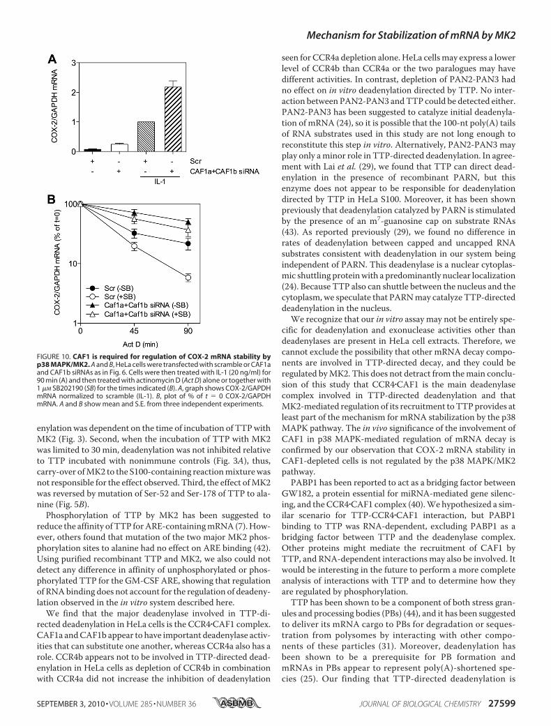
of COX-2 mRNA Stability by p38MAPK/MK2—ToconfirmthatCAF1is needed for p38 MAPK-mediated
regulation of ARE-mRNA decay, CAF1a and CAF1b weredepleted fromHeLa cells and then treated with or without IL-1for 90min to activate the p38MAPK/MK2 pathway and induceCOX-2 mRNA. This mRNA contains an ARE and is post-tran-scriptionally regulated by TTP and p38 MAPK/MK2 (8). Cellswere treated with actinomycin D alone or together with p38MAPK inhibitor. Depletion of CAF1a and CAF1b increasedsteady-state COX-2 mRNA in resting and IL-1-treated cells(Fig. 10A). COX-2 mRNA was more stable in CAF1-depletedthan scramble-transfected cells (Fig. 10B). p38MAPK inhibitordestabilized the mRNA in scramble-transfected cells but not inCAF1-depleted cells (Fig. 10B). Thus, CAF1 plays a key role inthe regulation of inflammatory mRNA stability by the p38MAPK pathway.
DISCUSSION
We have shown that phosphorylation of TTP by MK2directly inactivates TTP by inhibiting the recruitment of theCCR4�CAF1 deadenylase complex. However, it is not clear pre-cisely which subunit of the complex TTP interacts with. Wefind that 14-3-3 does not appear to sequester TTP away fromthe mRNA decay machinery as proposed previously (6) as R18and difopein prevented 14-3-3 from binding TTP but had noeffect on TTP-directed deadenylation or CAF1 recruitment.
FIGURE 7. Depletion of CAF1a and CAF1b inhibits TTP-directed deadenylation. CAF1a or CAF1b expressionwas suppressed, separately or in combination (CAF1a�CAF1b) in HeLa cells by RNAi as shown in Fig. 6. A, CAF1aand CAF1b mRNA knockdown efficiencies were determined by Q-RT-PCR as in Fig. 6. B, CAF1a and CAF1bknockdown assessed by Western blot for CAF1a and CAF1b. C, in vitro deadenylation assay and graphs as in Fig.6. Untr, untransfected; Scr, scramble.
Mechanism for Stabilization of mRNA by MK2
SEPTEMBER 3, 2010 • VOLUME 285 • NUMBER 36 JOURNAL OF BIOLOGICAL CHEMISTRY 27597

14-3-3 binding to TTP has been reported to cause TTP to shuttlefrom the nucleus to the cytoplasm (19) and to prevent TTP fromassociating with stress granules (6).We cannot exclude that 14-3-3-mediated TTP subcellular localization might play a role in theregulationofARE-mediateddecay, but it doesnot appear tobe themechanism to explain the regulation of TTP-directed deadenyla-tion byMK2 under the conditions used in this study.Regulation of TTP-directed deadenylation by MK2 requires
phosphorylation of Ser-52 and Ser-178 because no effect ondeadenylation was observed for a mutant form in which thesetwo serines were mutated to alanine. Moreover, no MK2-de-pendent difference in binding of CAF1a and CAF1b wasobserved with the mutant form of TTP. MK2 still caused sig-nificant phosphorylation of the S52A/S178A mutant form ofTTP, consistent with phosphorylation of other residues, as
reported previously (5). However, because regulation of dead-enylation andCAF1 binding was blocked bymutation of Ser-52and Ser-178 to alanine, the other MK2 phosphorylation sitesmay have a different function.Several observations indicate that the effect ofMK2 on dead-
enylation is caused by phosphorylation of TTP and not of otherproteins in the S100 extract. First, the effect of MK2 on dead-
FIGURE 8. Depletion of CCR4 inhibits TTP-directed deadenylation andformation of 5�-AMP. A, cells were transfected with scramble or CCR4a andCCR4b together, cells were lysed, and extracts were assayed in the presenceof GST-TTP using an [�-32P]ATP-labeled RNA substrate. Two portions of thesame gel are shown. B, same as A, but GM-CSF ARE or GM-CSF ARE mut RNAsubstrates were incubated in the presence or absence of GST-TTP (100 ng)and untransfected cell extracts for the times indicated. The position of the5�-AMP product of the deadenylation reaction is indicated.
FIGURE 9. Recruitment of CAF1 to TTP is inhibited by MK2 phosphoryla-tion and 14-3-3 independent. A–D, GST-TTP or GST alone was bound toglutathione -Sepharose 4B beads in presence of HeLa cell lysates. A, pulleddown material probed for PAN2, PARN, or CAF1a. B, GST pulldown assay ofGST-TTP (wild-type or S52A/S178A mutant) phosphorylated by MK2 in vitro orleft unphosphorylated as in Fig. 4, in the presence or absence of 150 units ofbenzonase. C, a graph of mean (�S.E.) for three independent experiments ofCaf1a protein quantified by densitometry. D, GST pulldown assay of wild-typeGST-TTP phosphorylated by MK2 or left unphosphorylated, in the presence of150 units of benzonase (Benz.) and in the presence or absence of 1 �M
difopein.
Mechanism for Stabilization of mRNA by MK2
27598 JOURNAL OF BIOLOGICAL CHEMISTRY VOLUME 285 • NUMBER 36 • SEPTEMBER 3, 2010
enylationwas dependent on the time of incubation of TTPwithMK2 (Fig. 3). Second, when the incubation of TTP with MK2was limited to 30 min, deadenylation was not inhibited relativeto TTP incubated with nonimmune controls (Fig. 3A), thus,carry-over ofMK2 to the S100-containing reactionmixturewasnot responsible for the effect observed. Third, the effect ofMK2was reversed by mutation of Ser-52 and Ser-178 of TTP to ala-nine (Fig. 5B).
Phosphorylation of TTP by MK2 has been suggested toreduce the affinity ofTTP forARE-containingmRNA (7).How-ever, others found that mutation of the two major MK2 phos-phorylation sites to alanine had no effect on ARE binding (42).Using purified recombinant TTP and MK2, we also could notdetect any difference in affinity of unphosphorylated or phos-phorylated TTP for the GM-CSF ARE, showing that regulationof RNAbinding does not account for the regulation of deadeny-lation observed in the in vitro system described here.
We find that the major deadenylase involved in TTP-di-rected deadenylation in HeLa cells is the CCR4�CAF1 complex.CAF1a andCAF1b appear to have important deadenylase activ-ities that can substitute one another, whereas CCR4a also has arole. CCR4b appears not to be involved in TTP-directed dead-enylation in HeLa cells as depletion of CCR4b in combinationwith CCR4a did not increase the inhibition of deadenylation
seen for CCR4a depletion alone. HeLa cellsmay express a lowerlevel of CCR4b than CCR4a or the two paralogues may havedifferent activities. In contrast, depletion of PAN2-PAN3 hadno effect on in vitro deadenylation directed by TTP. No inter-action between PAN2-PAN3 andTTP could be detected either.PAN2-PAN3 has been suggested to catalyze initial deadenyla-tion of mRNA (24), so it is possible that the 100-nt poly(A) tailsof RNA substrates used in this study are not long enough toreconstitute this step in vitro. Alternatively, PAN2-PAN3 mayplay only aminor role in TTP-directed deadenylation. In agree-ment with Lai et al. (29), we found that TTP can direct dead-enylation in the presence of recombinant PARN, but thisenzyme does not appear to be responsible for deadenylationdirected by TTP in HeLa S100. Moreover, it has been shownpreviously that deadenylation catalyzed by PARN is stimulatedby the presence of an m7-guanosine cap on substrate RNAs(43). As reported previously (29), we found no difference inrates of deadenylation between capped and uncapped RNAsubstrates consistent with deadenylation in our system beingindependent of PARN. This deadenylase is a nuclear cytoplas-mic shuttling proteinwith a predominantly nuclear localization(24). Because TTP also can shuttle between the nucleus and thecytoplasm, we speculate that PARNmay catalyze TTP-directeddeadenylation in the nucleus.We recognize that our in vitro assay may not be entirely spe-
cific for deadenylation and exonuclease activities other thandeadenylases are present in HeLa cell extracts. Therefore, wecannot exclude the possibility that other mRNA decay compo-nents are involved in TTP-directed decay, and they could beregulated byMK2. This does not detract from themain conclu-sion of this study that CCR4�CAF1 is the main deadenylasecomplex involved in TTP-directed deadenylation and thatMK2-mediated regulation of its recruitment toTTPprovides atleast part of the mechanism for mRNA stabilization by the p38MAPK pathway. The in vivo significance of the involvement ofCAF1 in p38 MAPK-mediated regulation of mRNA decay isconfirmed by our observation that COX-2 mRNA stability inCAF1-depleted cells is not regulated by the p38 MAPK/MK2pathway.PABP1 has been reported to act as a bridging factor between
GW182, a protein essential for miRNA-mediated gene silenc-ing, and theCCR4�CAF1 complex (40).We hypothesized a sim-ilar scenario for TTP-CCR4�CAF1 interaction, but PABP1binding to TTP was RNA-dependent, excluding PABP1 as abridging factor between TTP and the deadenylase complex.Other proteins might mediate the recruitment of CAF1 byTTP, and RNA-dependent interactionsmay also be involved. Itwould be interesting in the future to perform a more completeanalysis of interactions with TTP and to determine how theyare regulated by phosphorylation.TTP has been shown to be a component of both stress gran-
ules and processing bodies (PBs) (44), and it has been suggestedto deliver its mRNA cargo to PBs for degradation or seques-tration from polysomes by interacting with other compo-nents of these particles (31). Moreover, deadenylation hasbeen shown to be a prerequisite for PB formation andmRNAs in PBs appear to represent poly(A)-shortened spe-cies (25). Our finding that TTP-directed deadenylation is
FIGURE 10. CAF1 is required for regulation of COX-2 mRNA stability byp38 MAPK/MK2. A and B, HeLa cells were transfected with scramble or CAF1aand CAF1b siRNAs as in Fig. 6. Cells were then treated with IL-1 (20 ng/ml) for90 min (A) and then treated with actinomycin D (Act D) alone or together with1 �M SB202190 (SB) for the times indicated (B). A, graph shows COX-2/GAPDHmRNA normalized to scramble (IL-1). B, plot of % of t � 0 COX-2/GAPDHmRNA. A and B show mean and S.E. from three independent experiments.
Mechanism for Stabilization of mRNA by MK2
SEPTEMBER 3, 2010 • VOLUME 285 • NUMBER 36 JOURNAL OF BIOLOGICAL CHEMISTRY 27599
mediated by the CCR4�CAF1 complex, together with evi-dence that CCR4 and CAF1 are predominantly not PB-asso-ciated (25), suggests that TTP-directed deadenylation also maybe largely PB-independent. We cannot, however, exclude theinvolvement of stress granules and PBs in TTP function, and itwould be interesting to understand the role of these foci in theTTP-directed deadenylation mechanism.
Acknowledgments—We are most grateful to J. Steitz, S. Hoshino, A.Virtanen, and M. Brook for provision of reagents. We also acknowl-edge Arthritis Research UK for support.
REFERENCES1. Dean, J. L., Brook, M., Clark, A. R., and Saklatvala, J. (1999) J. Biol. Chem.
274, 264–2692. Winzen, R., Kracht, M., Ritter, B., Wilhelm, A., Chen, C. Y., Shyu, A. B.,
Muller, M., Gaestel, M., Resch, K., and Holtmann, H. (1999) EMBO J. 18,4969–4980
3. Lasa, M., Mahtani, K. R., Finch, A., Brewer, G., Saklatvala, J., and Clark,A. R. (2000)Mol. Cell. Biol. 20, 4265–4274
4. Mahtani, K. R., Brook, M., Dean, J. L., Sully, G., Saklatvala, J., and Clark,A. R. (2001)Mol. Cell. Biol. 21, 6461–6469
5. Chrestensen, C. A., Schroeder, M. J., Shabanowitz, J., Hunt, D. F., Pelo,J. W., Worthington, M. T., and Sturgill, T. W. (2004) J. Biol. Chem. 279,10176–10184
6. Stoecklin, G., Stubbs, T., Kedersha, N., Wax, S., Rigby, W. F., Blackwell,T. K., and Anderson, P. (2004) EMBO J. 23, 1313–1324
7. Hitti, E., Iakovleva, T., Brook, M., Deppenmeier, S., Gruber, A. D., Radzi-och, D., Clark, A. R., Blackshear, P. J., Kotlyarov, A., andGaestel,M. (2006)Mol. Cell. Biol. 26, 2399–2407
8. Tudor, C., Marchese, F. P., Hitti, E., Aubareda, A., Rawlinson, L., Gaestel,M., Blackshear, P. J., Clark, A. R., Saklatvala, J., and Dean, J. L. (2009) FEBSLett. 583, 1933–1938
9. Taylor, G. A., Carballo, E., Lee, D. M., Lai, W. S., Thompson, M. J., Patel,D. D., Schenkman, D. I., Gilkeson, G. S., Broxmeyer, H. E., Haynes, B. F.,and Blackshear, P. J. (1996) Immunity 4, 445–454
10. Gherzi, R., Lee, K. Y., Briata, P.,Wegmuller, D., Moroni, C., Karin,M., andChen, C. Y. (2004)Mol. Cell 14, 571–583
11. Winzen, R., Thakur, B. K., Dittrich-Breiholz, O., Shah, M., Redich, N.,Dhamija, S., Kracht, M., and Holtmann, H. (2007) Mol. Cell. Biol. 27,8388–8400
12. Datta, S., Biswas, R., Novotny,M., Pavicic, P. G., Jr., Herjan, T.,Mandal, P.,and Hamilton, T. A. (2008) J. Immunol. 180, 2545–2552
13. Tchen, C. R., Brook,M., Saklatvala, J., andClark, A. R. (2004) J. Biol. Chem.279, 32393–32400
14. Brook, M., Tchen, C. R., Santalucia, T., McIlrath, J., Arthur, J. S., Sak-latvala, J., and Clark, A. R. (2006)Mol. Cell. Biol. 26, 2408–2418
15. Clark, A. R., Dean, J. L., and Saklatvala, J. (2009) Arthritis Rheum. 60,3513–3514
16. Lai, W. S., Carballo, E., Strum, J. R., Kennington, E. A., Phillips, R. S., andBlackshear, P. J. (1999)Mol. Cell. Biol. 19, 4311–4323
17. Dean, J. L., Sarsfield, S. J., Tsounakou, E., and Saklatvala, J. (2003) J. Biol.
Chem. 278, 39470–3947618. Winzen, R., Gowrishankar, G., Bollig, F., Redich, N., Resch, K., and Holt-
mann, H. (2004)Mol. Cell. Biol. 24, 4835–484719. Johnson, B. A., Stehn, J. R., Yaffe, M. B., and Blackwell, T. K. (2002) J. Biol.
Chem. 277, 18029–1803620. Boeck, R., Tarun, S., Jr., Rieger, M., Deardorff, J. A., Muller-Auer, S., and
Sachs, A. B. (1996) J. Biol. Chem. 271, 432–43821. Tucker, M., Valencia-Sanchez, M. A., Staples, R. R., Chen, J., Denis, C. L.,
and Parker, R. (2001) Cell 104, 377–38622. Garneau, N. L.,Wilusz, J., andWilusz, C. J. (2007)Nat. Rev. Mol. Cell Biol.
8, 113–12623. Lau, N. C., Kolkman, A., van Schaik, F. M., Mulder, K. W., Pijnappel,
W. W., Heck, A. J., and Timmers, H. T. (2009) Biochem. J. 422, 443–45324. Yamashita, A., Chang, T. C., Yamashita, Y., Zhu, W., Zhong, Z., Chen,
C. Y., and Shyu, A. B. (2005) Nat. Struct. Mol. Biol. 12, 1054–106325. Zheng, D., Ezzeddine, N., Chen, C. Y., Zhu, W., He, X., and Shyu, A. B.
(2008) J. Cell. Biol. 182, 89–10126. Schwede, A., Ellis, L., Luther, J., Carrington,M., Stoecklin,G., andClayton,
C. (2008) Nucleic Acids Res. 36, 3374–338827. Korner, C. G., and Wahle, E. (1997) J. Biol. Chem. 272, 10448–1045628. Lin, W. J., Duffy, A., and Chen, C. Y. (2007) J. Biol. Chem. 282,
19958–1996829. Lai, W. S., Kennington, E. A., and Blackshear, P. J. (2003) Mol. Cell. Biol.
23, 3798–381230. Chen, C. Y., Gherzi, R., Ong, S. E., Chan, E. L., Raijmakers, R., Pruijn, G. J.,
Stoecklin, G., Moroni, C., Mann, M., and Karin, M. (2001) Cell 107,451–464
31. Lykke-Andersen, J., and Wagner, E. (2005) Genes Dev. 19, 351–36132. Carballo, E., Lai, W. S., and Blackshear, P. J. (2000) Blood 95, 1891–189933. Brook, M., Sully, G., Clark, A. R., and Saklatvala, J. (2000) FEBS Lett. 483,
57–6134. Tebo, J., Der, S., Frevel, M., Khabar, K. S., Williams, B. R., and Hamilton,
T. A. (2003) J. Biol. Chem. 278, 12085–1209335. Masters, S. C., and Fu, H. (2001) J. Biol. Chem. 276, 45193–4520036. Voeltz, G. K., Ongkasuwan, J., Standart, N., and Steitz, J. A. (2001) Genes
Dev. 15, 774–78837. Dean, J. L., Wait, R., Mahtani, K. R., Sully, G., Clark, A. R., and Saklatvala,
J. (2001)Mol. Cell. Biol. 21, 721–73038. Alford, K. A., Glennie, S., Turrell, B. R., Rawlinson, L., Saklatvala, J., and
Dean, J. L. E. (2007) J. Biol. Chem. 282, 6232–624139. Livak, K. J., and Schmittgen, T. D. (2001)Methods 25, 402–40840. Zekri, L., Huntzinger, E., Heimstadt, S., and Izaurralde, E. (2009)Mol. Cell.
Biol. 29, 6220–623141. Rowlett, R. M., Chrestensen, C. A., Schroeder, M. J., Harp, M. G., Pelo,
J. W., Shabanowitz, J., DeRose, R., Hunt, D. F., Sturgill, T.W., andWorth-ington, M. T. (2008) Am. J. Physiol. Gastrointest. Liver Physiol. 295,G421–430
42. Sun, L., Stoecklin, G., Van Way, S., Hinkovska-Galcheva, V., Guo, R. F.,Anderson, P., and Shanley, T. P. (2007) J. Biol. Chem. 282, 3766–3777
43. Dehlin, E., Wormington,M., Korner, C. G., andWahle, E. (2000) EMBO J.19, 1079–1086
44. Kedersha, N., Stoecklin, G., Ayodele, M., Yacono, P., Lykke-Andersen, J.,Fritzler, M. J., Scheuner, D., Kaufman, R. J., Golan, D. E., and Anderson, P.(2005) J. Cell. Biol. 169, 871–884
Mechanism for Stabilization of mRNA by MK2
27600 JOURNAL OF BIOLOGICAL CHEMISTRY VOLUME 285 • NUMBER 36 • SEPTEMBER 3, 2010
Related Documents