1 Product Information Storage: Store kits in the dark at 4ºC. Stability: The components in the kits are stable for a minimum of 3 months under proper storage conditions. Application: TSA Plus Fluorescence kits are intended for high sensitivity detection in immunohistochemistry (IHC), immunocytochemistry (ICC) and in situ hybridization (ISH) experiments. Final detection may be fluorescent or chromogenic (with TSA Plus Fluorescein kits only). FOR RESEARCH USE ONLY. Safety Note All reagents are classified as nonhazardous. However, it is strongly recommended to wear disposable gloves and safety glasses while working. Thorough washing of hands after handling is also recommended. Quality Control We certify that QC results of these reagents meet our quality release criteria. What is TSA technology? TSA ® from PerkinElmer is a technology that improves fluorescent and chromogenic signals by up to 100-fold while allowing reduced consumption of primary antibodies or probes. TSA Plus is an optimized and enhanced version that is up to 20 times more sensitive than standard tyramide reagents. TSA Plus Fluorescence Kits use horseradish peroxidase (HRP) to catalyze covalent deposition of fluorophores directly adjacent to the immobilized enzyme. The labeling reaction is quick (less than 10 minutes) and deposited labels can then be viewed directly using standard or confocal microscopy. TSA Plus Fluorescein may also be used in combination with anti-fluorescein enzyme conjugates and appropriate chromogenic substrates for brightfield microscopy. The use of TSA reagents results in a significant increase in sensitivity over standard detection methods, while maintaining specificity and resolution. Moreover, TSA reagents allow drastically reduced consumption of primary antibody or probe. TSA Plus Fluorescence kits are ideal for multiplexed IHC or ISH in combination. The Opal™ protocol allows simultaneous measurement of 6 or more biomarkers in FFPE tissue. For more information, please see www.perkinelmer.com/opal. MANUAL TSA ® Research Reagents Caution: For Laboratory Use. A research chemical for research purposes only. TSA Plus Fluorescence Kits

Welcome message from author
This document is posted to help you gain knowledge. Please leave a comment to let me know what you think about it! Share it to your friends and learn new things together.
Transcript
1
Product Information
Storage: Store kits in the dark at 4ºC.
Stability: The components in the kits are stable for a minimum of 3 months under proper storage
conditions. Application: TSA Plus Fluorescence kits are intended for high sensitivity detection in immunohistochemistry
(IHC), immunocytochemistry (ICC) and in situ hybridization (ISH) experiments. Final detection may be fluorescent or chromogenic (with TSA Plus Fluorescein kits only).
FOR RESEARCH USE ONLY.
Safety Note All reagents are classified as nonhazardous. However, it is strongly recommended to wear
disposable gloves and safety glasses while working. Thorough washing of hands after handling is also recommended.
Quality Control We certify that QC results of these reagents meet our quality release criteria.
What is TSA technology?
TSA® from PerkinElmer is a technology that improves fluorescent and chromogenic signals by up to 100-fold while
allowing reduced consumption of primary antibodies or probes. TSA Plus is an optimized and enhanced version that is up to 20 times more sensitive than standard tyramide reagents. TSA Plus Fluorescence Kits use horseradish peroxidase (HRP) to catalyze covalent deposition of fluorophores directly adjacent to the immobilized enzyme. The labeling reaction is quick (less than 10 minutes) and deposited labels can then be viewed directly using standard or confocal microscopy. TSA Plus Fluorescein may also be used in combination with anti-fluorescein enzyme conjugates and appropriate chromogenic substrates for brightfield microscopy. The use of TSA reagents results in a significant increase in sensitivity over standard detection methods, while maintaining specificity and resolution. Moreover, TSA reagents allow drastically reduced consumption of primary antibody or probe. TSA Plus Fluorescence kits are ideal for multiplexed IHC or ISH in combination. The Opal™ protocol allows simultaneous measurement of 6 or more biomarkers in FFPE tissue. For more information, please see www.perkinelmer.com/opal.
MANUAL TSA® Research Reagents
Caution: For Laboratory Use. A research chemical for research purposes only.
TSA Plus Fluorescence Kits

2
Material Provided
Format* Catalog # Kit Components
TSA Plus Fluorescein
50-150 slides NEL741001KT 1X Plus Amplification Diluent (15 mL)
Fluorescein Plus Amplification Reagent (2 tubes, add 150 µL DMSO / tube)
250-750 slides NEL741B001KT 1X Plus Amplification Diluent (5 x 15 mL)
Fluorescein Plus Amplification Reagent (10 tubes, add 150 µL DMSO / tube)
25-75 slides NEL741E001KT 1X Plus Amplification Diluent (7.5 mL)
Fluorescein Plus Amplification Reagent (1 tube, add 150 µL DMSO)
TSA Plus Tetramethylrhodamine
50-150 slides NEL742001KT 1X Plus Amplification Diluent (15 mL)
TMR Plus Amplification Reagent (2 tubes, add 150 µL DMSO / tube)
250-750 slides NEL742B001KT 1X Plus Amplification Diluent (5 x 15 mL)
TMR Plus Amplification Reagent (10 tubes, add 150 µL DMSO / tube)
TSA Plus Cyanine 3
50-150 slides NEL744001KT 1X Plus Amplification Diluent (15 mL)
Cyanine 3 Plus Amplification Reagent (2 tubes, add 150 µL DMSO / tube)
250-750 slides NEL744B001KT 1X Plus Amplification Diluent (5 x 15 mL)
Cyanine 3 Plus Amplification Reagent (10 tubes, add 150 µL DMSO / tube)
25-75 slides NEL744E001KT 1X Plus Amplification Diluent (7.5 mL)
Cyanine 3 Plus Amplification Reagent (1 tube, add 150 µL DMSO)
TSA Plus Cyanine 3.5
50-150 slides NEL763001KT 1X Plus Amplification Diluent (15 mL)
Cyanine 3.5 Plus Amplification Reagent (2 tubes, add 150 µL DMSO / tube)
250-750 slides NEL763B001KT 1X Plus Amplification Diluent (5 x 15 mL)
Cyanine 3.5 Plus Amplification Reagent (10 tubes, add 150 µL DMSO / tube)
25-75 slides NEL763E001KT 1X Plus Amplification Diluent (7.5 mL)
Cyanine 3.5 Plus Amplification Reagent (1 tube, add 150 µL DMSO)
TSA Plus Cyanine 5
50-150 slides NEL745001KT 1X Plus Amplification Diluent (15 mL)
Cyanine 5 Plus Amplification Reagent (2 tubes, add 150 µL DMSO / tube)
250-750 slides NEL745B001KT 1X Plus Amplification Diluent (5 x 15 mL)
Cyanine 5 Plus Amplification Reagent (10 tubes, add 150 µL DMSO / tube)
25-75 slides NEL745E001KT 1X Plus Amplification Diluent (7.5 mL)
Cyanine 5 Plus Amplification Reagent (1 tube, add 150 µL DMSO)
TSA Plus Cyanine 5.5
50-150 slides NEL766001KT 1X Plus Amplification Diluent (15 mL)
Cyanine 5.5 Plus Amp Reagent (2 tubes, add 150 µL DMSO / tube)
250-750 slides NEL766B001KT 1X Plus Amplification Diluent (5 x 15 mL)
Cyanine 5.5 Plus Amplification Reagent (10 tubes, add 150 µL DMSO / tube)
25-75 slides NEL766E001KT 1X Plus Amplification Diluent (7.5 mL)
Cyanine 5.5 Plus Amplification Reagent (1 tube, add 150 µL DMSO)
TSA Plus Cyanine 3 / Cyanine 5
50-150 slides NEL752001KT 1X Plus Amplification Diluent (2 x15 mL)
Cyanine 3 Plus Amplification Reagent (2 tubes, add 150 µL DMSO / tube)
Cyanine 5 Plus Amplification Reagent (2 tubes, add 150 µL DMSO / tube)
TSA Plus Cyanine 3 / Fluorescein
50-150 slides NEL753001KT 1X Plus Amplification Diluent (2 x15 mL)
Cyanine 3 Plus Amplification Reagent (2 tubes, add 150 µL DMSO / tube)
Fluorescein Plus Amplification Reagent (2 tubes, add 150 µL DMSO / tube)
TSA Plus Cyanine 5 / Fluorescein
50-150 slides NEL754001KT 1X Plus Amplification Diluent (2 x15 mL)
Cyanine 5 Plus Amplification Reagent (2 tubes, add 150 µL DMSO / tube)
Fluorescein Plus Amplification Reagent (2 tubes, add 150 µL DMSO / tube)
TSA Plus Fluorescein / Tetramethylrhodamine
50-150 slides NEL756001KT 1X Plus Amplification Diluent (2 x15 mL)
TMR Plus Amplification Reagent (2 tubes, add 150 µL DMSO / tube)
Fluorescein Plus Amplification Reagent (2 tubes, add 150 µL DMSO / tube)
TSA Plus Fluorescence Palette Kit
10-35 slides NEL760001KT
1X Plus Amplification Diluent (2 x15 mL)
Fluorescein Plus Amplification Reagent (1 tubes, add 60 µL DMSO)
TMR Plus Amplification Reagent (1 tube, add 60 µL DMSO)
Cyanine 3 Plus Amplification Reagent (1 tube, add 60 µL DMSO)
Cyanine 5 Plus Amplification Reagent (1 tube, add 60 µL DMSO)
*The format of the kit is based on 100 - 300 µL per slide of TSA Plus Working Solution (see page 3).
3
Reagents and Materials
Critical Reagents Required but not Supplied
HRP-labelled reagent to drive the signal amplification reaction is required. For example: o Anti-digoxigenin-HRP for use with digoxigenin labeled probes or antibodies (cat. no. NEF832001EA) o Anti-fluorescein-HRP for use with fluorescein labeled probes or antibodies (cat. no. NEF710001EA) o HRP-conjugated anti-species secondary antibody
Anti-rabbit IgG (goat) HRP (cat. no. NEF812001EA) Anti-mouse IgG (goat) HRP (cat. no. NEF822001EA)
DMSO (molecular biology or HPLC grade)
Buffer components and detergents such as PBS, Triton-X100, Tween-20
Blocking Reagent (cat. no. FP1012 or equivalent)
Solutions to prepare
The following buffers and reagents are required for slide preparation and signal amplification
TSA Plus Stock Solution TSA Plus Amplification Reagent is supplied as a solid. Each vial must be reconstituted with molecular biology or HPLC grade DMSO to make TSA Plus Stock Solution. TSA Plus Stock Solution is stable for at least 3 months when stored at 4ºC. (Note: DMSO freezes at 4ºC; therefore, thaw Stock Solution before each use). Here are recommended dissolution volumes for stock solutions.
For the TSA Plus Fluorescence Palette Kit (NEL760001KT), add 60 µL DMSO to each tube of Amplification Reagent.
Add 150 µL DMSO per tube Amplification Reagent for all other TSA Plus Fluorescence Kits. TNT Wash Buffer 0.1 M TRIS-HCl, pH 7.5 0.15 M NaCl 0.05% Tween®20 Other wash buffers (such as PBS) may be used. Substitution of 0.3% Triton X-100 for the 0.05% Tween-20 is also possible. Users should validate the use of alternative wash buffers with their own systems. TNB Blocking Buffer 0.1 M TRIS-HCl, pH 7.5 0.15 M NaCl 0.5% Blocking Reagent (available separately, catalog number FP1012) Add Blocking Reagent slowly in small increments to buffer while stirring. Heat gradually to 55°C with continuous stirring to completely dissolve the Blocking Reagent. (This should take no longer than 30-60 minutes.) The solution will appear milky. Bring to room temperature before using. Aliquot and store at -20°C for long term use. TSA Plus Working Solution Before each procedure, dilute TSA Plus Stock Solution1:50 in 1X Amplification Diluent to make TSA Plus Working Solution. Approximately 100-300 µL of TSA Plus Working Solution is required per slide. Discard any unused portion of TSA Plus Working Solution.
4
Recommendations
Do not let slides dry out between steps.
A humidified chamber is recommended for all incubation steps (i.e., a damp paper towel in a covered box).
Drain off as much of the incubation solutions as possible, before the addition of the next solution, to prevent reagent dilution and uneven staining. Blot area around, but not on, tissue section using a tissue.
Be sure to use enough volume of each reagent to completely cover sections or cells.
Optional: Use of coverslips may reduce reagent evaporation, especially during steps which require long incubation at elevated temperatures (such as probe hybridization). However care must be taken upon removal to prevent damage to tissues or cells.
If your assay includes streptavidin conjugates, check for endogenous biotin which may be a source of non-specific background.
If there is too much signal, dilute the primary antibody, probe or HRP conjugate further. TSA Plus kits are designed for use at 1:50 dilution of TSA Plus Stock Solution for optimal results.
First time users should apply TSA to a proven system.
Quenching Endogenous Peroxidase Activation and covalent binding of TSA reagent is catalyzed by peroxidase. Endogenous peroxidase will be a source of background, and should be quenched if present. Users should establish the need for doing this and optimal methodology specific to the tissues or cells being stained. Options include:
0.3% H2O2 to 3% H2O2 in PBS, incubation for 10 to 60 minutes
0.3% H2O2 to 3% H2O2 in Methanol, incubation for 10 to 60 minutes
For paraffin-embedded tissues, quenching can be done after dewaxing and alcohol rehydration but before the protease digestion step. For frozen tissue or cell preps, quenching can be done following fixation and before the protease digestion step. After quenching wash with TNT or 1X PBS buffer for 5 minutes. See Li et al from references section for more suggestions.
TSA-ISH Optimization Inadequate optimization of probe and HRP conjugate dilution may be a source of high background and reduced signal. TSA is ideal for detection of targets that have weak signals using standard detection methods. If the signal is strong with standard detection methods, dilute the probe until the signal begins to disappear and then proceed with TSA optimization. Probe Optimization
Slide 1: use same probe dilution as standard method.
Slide 2: 5-fold dilution from slide 1
Slide 3: 2-fold dilution from slide 2 (further dilution may be necessary)
Slide 4: negative control (probe omitted) HRP Conjugate Dilution
SA-HRP included with TSA Systems: 1:100 dilution
Streptavidin-HRP (catalog number NEL750001EA): 1:250-1:1000 dilution
Anti-fluorescein-HRP (catalog number NEF710001EA): 1:100-1:500 dilution
Anti-DNP-HRP (catalog number FP1129): 1:100-1:500 dilution
Anti-digoxigenin-HRP (catalog number NEF832001EA): 1:500-1:5000 dilution
HRP reagents from other manufacturers: Begin with recommended range for slide applications. Further dilution may be needed
5
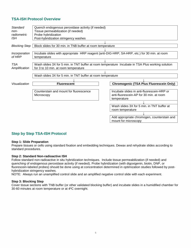
TSA-ISH Protocol Overview Standard non-radiometric ISH
Quench endogenous peroxidase activity (if needed) Tissue permeabilization (if needed) Probe hybridization Post-hybridization stringency washes
↓
Blocking Step Block slides for 30 min. in TNB buffer at room temperature ↓
Incorporation of HRP
Incubate slides with appropriate HRP reagent (anti-DIG-HRP, SA-HRP, etc.) for 30 min. at room temperature
↓
TSA Amplification
Wash slides 3X for 5 min. in TNT buffer at room temperature Incubate in TSA Plus working solution for 3 to 10 min. at room temperature
↓
Wash slides 3X for 5 min. in TNT buffer at room temperature
↓ ↓
Visualization Fluorescent Chromogenic (TSA Plus Fluorescein Only)
Counterstain and mount for fluorescence Microscopy
Incubate slides in anti-fluorescein-HRP or anti-fluorescein-AP for 30 min. at room temperature
↓
Wash slides 3X for 5 min. in TNT buffer at
room temperature
↓
Add appropriate chromogen, counterstain and
mount for microscopy
Step by Step TSA-ISH Protocol Step 1: Slide Preparation Prepare tissues or cells using standard fixation and embedding techniques. Dewax and rehydrate slides according to standard procedures. Step 2: Standard Non-radioactive ISH Follow standard non-radioactive in situ hybridization techniques. Include tissue permeabilization (if needed) and quenching of endogenous peroxidase activity (if needed). Probe hybridization (with digoxigenin, biotin, DNP, or fluorescein-labeled probes) should be done using at concentration determined in optimization studies followed by post-hybridization stringency washes. NOTE: Always run an unamplified control slide and an amplified negative control slide with each experiment. Step 3: Blocking Step Cover tissue sections with TNB buffer (or other validated blocking buffer) and incubate slides in a humidified chamber for 30-60 minutes at room temperature or at 4ºC overnight.
6
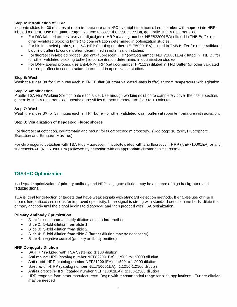
Step 4: Introduction of HRP Incubate slides for 30 minutes at room temperature or at 4ºC overnight in a humidified chamber with appropriate HRP-labeled reagent. Use adequate reagent volume to cover the tissue section, generally 100-300 µL per slide.
For DIG-labeled probes, use anti-digoxigenin-HRP (catalog number NEF832001EA) diluted in TNB Buffer (or other validated blocking buffer) to concentration determined in optimization studies.
For biotin-labeled probes, use SA-HRP (catalog number NEL750001EA) diluted in TNB Buffer (or other validated blocking buffer) to concentration determined in optimization studies.
For fluorescein-labeled probes, use anti-fluorescein-HRP (catalog number NEF710001EA) diluted in TNB Buffer (or other validated blocking buffer) to concentration determined in optimization studies.
For DNP-labeled probes, use anti-DNP-HRP (catalog number FP1129) diluted in TNB Buffer (or other validated blocking buffer) to concentration determined in optimization studies.
Step 5: Wash Wash the slides 3X for 5 minutes each in TNT Buffer (or other validated wash buffer) at room temperature with agitation. Step 6: Amplification Pipette TSA Plus Working Solution onto each slide. Use enough working solution to completely cover the tissue section, generally 100-300 µL per slide. Incubate the slides at room temperature for 3 to 10 minutes. Step 7: Wash Wash the slides 3X for 5 minutes each in TNT buffer (or other validated wash buffer) at room temperature with agitation. Step 8: Visualization of Deposited Fluorophores For fluorescent detection, counterstain and mount for fluorescence microscopy. (See page 10 table, Fluorophore Excitation and Emission Maxima.) For chromogenic detection with TSA Plus Fluorescein, incubate slides with anti-fluorescein-HRP (NEF710001EA) or anti-fluorescein-AP (NEF709001PK) followed by detection with an appropriate chromogenic substrate.
TSA-IHC Optimization Inadequate optimization of primary antibody and HRP conjugate dilution may be a source of high background and reduced signal. TSA is ideal for detection of targets that have weak signals with standard detection methods. It enables use of much more dilute antibody solutions for improved specificity. If the signal is strong with standard detection methods, dilute the primary antibody until the signal begins to disappear and then proceed with TSA optimization. Primary Antibody Optimization
Slide 1: use same antibody dilution as standard method.
Slide 2: 5-fold dilution from slide 1
Slide 3: 5-fold dilution from slide 2
Slide 4: 5-fold dilution from slide 3 (further dilution may be necessary)
Slide 4: negative control (primary antibody omitted) HRP Conjugate Dilution
SA-HRP included with TSA Systems: 1:100 dilution
Anti-mouse-HRP (catalog number NEF822001EA): 1:500 to 1:2000 dilution
Anti-rabbit-HRP (catalog number NEF812001EA): 1:500 to 1:2000 dilution
Streptavidin-HRP (catalog number NEL750001EA): 1:1250-1:2500 dilution
Anti-fluorescein-HRP (catalog number NEF710001EA): 1:100-1:500 dilution
HRP reagents from other manufacturers: Begin with recommended range for slide applications. Further dilution may be needed

7
TSA-IHC Protocol Overview
Quench endogenous peroxidase activity (if needed) ↓
Block slides for 30 min. in TNB buffer at room temperature
Standard IHC
↓
Incubate slides in primary antibody for 30-60 minutes at room temperature
↓
Wash slides 3X for 5 min. in TNT buffer at room temperature
↓
Incorporation of HRP
Incubate slides in HRP labeled secondary antibody for 30 min. at room temperature
Incubate slides in biotinylated secondary antibody 30-60 min. at room temperature
or Wash slides 3X for 5 min. in TNT buffer at room temperature
Incubate slides in SA-HRP for 30 min. at room temperature
↓
Wash slides 3X for 5 min. in TNT buffer at room temperature
↓
TSA Amplification
Incubate in TSA Plus Working Solution for 3 to 10 min. at room temperature
↓
Wash slides 3X for 5 min. in TNT buffer at room temperature
↓
Visualization Fluorescent Chromogenic (TSA Plus Fluorescein Only)
Counterstain and mount for fluorescence microscopy
Incubate slides in anti-fluorescein-HRP or anti-fluorescein-AP for 30 min. at room temperature
↓
Wash slides 3X for 5 min. in TNT buffer at
room temperature
↓
Add appropriate chromogen, counterstain and
mount for microscopy
Step by Step TSA-IHC Protocol Step 1: Slide Preparation Prepare tissues or cells for detection with TSA using standard fixation and embedding techniques. Dewax and rehydrate using standard protocols. Quench endogenous peroxidase activity if necessary. NOTE: Always run an unamplified control slide and an amplified negative control slide with each experiment. Step 2: Blocking Step Cover tissue sections with TNB buffer (or other validated blocking buffer) and incubate slides in a humidified chamber for 30-60 minutes at room temperature or at 4ºC overnight. Step 3: Primary Antibody Incubation Drain off the blocking buffer and apply primary antibody, diluted in TNB Buffer (or other validated blocking buffer). Incubate the primary antibody preparation per the manufacturer's instructions regarding incubation time and temperature requirements. Use enough volume to completely cover the tissue section (generally 100-300 µL per slide) at the concentration determined in optimization studies.

8
Step 4: Wash Wash the slides 3X for 5 minutes each in TNT Buffer (or other validated wash buffer) at room temperature with agitation. Step 5: Introduction of HRP Incubate slides for 30 minutes at room temperature or at 4ºC overnight in a humidified chamber with appropriate HRP-labeled reagent. Use adequate reagent volume to cover the tissue section, generally 100-300 µL per slide. Options include.
HRP labeled secondary antibody diluted in TNB Buffer (or other validated blocking buffer).
100-300 µL of biotinylated secondary antibody diluted in TNB Buffer (or other validated blocking buffer). Incubate 30-60 minutes in a humidified chamber. Wash the slides for 3 X 5 minutes TNT buffer at room temperature with agitation. Follow by 100-300 µL of SA-HRP diluted in TNB Buffer. Use SA-HRP at 1:100 dilution, or at 1:2000 if using PerkinElmer Cat. # NEL750001EA.
When using alternative suppliers, reagents should be optimized for use with TSA starting with manufacturer's recommended dilutions. Incubate slides in a humidified chamber for 30-60 minutes at room temperature or at 4ºC overnight.
Step 6: Wash Wash the slides 3X for 5 minutes each in TNT Buffer (or other validated wash buffer) at room temperature with agitation. Step 7: Amplification Pipette 100-300 µL of TSA Plus Working Solution onto each slide. Incubate the slides at room temperature for 3 to 10 minutes. Step 8: Wash Wash the slides 3X for 5 minutes each in TNT Buffer (or other validated wash buffer) at room temperature with agitation. Step 9: Visualization of Deposited Fluorophores For fluorescent detection, counterstain and mount for fluorescence microscopy. (See page 10 table, Fluorophore Excitation and Emission Maxima.) For chromogenic detection with TSA Plus Fluorescein, incubate slides with anti-fluorescein-HRP (NEF710001EA) or anti-fluorescein-AP (NEF709001PK) followed by detection with an appropriate chromogenic substrate.

9
Troubleshooting Technical Support Resources
Assay Support Knowledge Base: www.perkinelmer.com/askTSA
Email: [email protected]
Telephone o USA toll-free 800-762-4000 o EU toll-free 00800 33 29 0000 o Finland toll-free 999 800 33 29 0000 o China toll-free 800 820 5046
ISH Troubleshooting
PROBLEM REMEDY
Low Signal Optimize probe concentration.
Titer HRP conjugate to determine optimum concentration for signal amplification.
Add tissue permeabilization step to facilitate penetration of reagents.
Lengthen incubation time for TSA Plus Working Solution.
Excess Signal Decrease concentration of HRP conjugate introduced prior to amplification.
Decrease probe concentration.
Decrease TSA Plus Working Solution incubation time.
Decrease concentration of anti-fluorescein-enzyme conjugate used for chromogenic visualization.
High Background
Decrease probe concentration.
Decrease concentration of HRP conjugate.
Check for endogenous biotin (if using streptavidin conjugates)
Shorten chromogenic development time.
Lengthen endogenous peroxidase quenching step.
Filter buffers.
Increase number and/or length of washes.
Nonqualified or contaminated blocking reagent used. Use PerkinElmer Blocking Reagent (FP1020 or FP1012).
IHC Troubleshooting
PROBLEM REMEDY
Low Signal Titer primary and/or secondary antibodies to determine optimum concentration for signal amplification
Lengthen incubation time for TSA Plus Working Solution.
Use antigen retrieval techniques to unmask the target.
Excess Signal Decrease concentration of primary and/or secondary antibody or HRP conjugates.
Decrease TSA Plus Working Solution incubation time.
Decrease concentration of anti-fluorescein-enzyme conjugate used for chromogenic visualization.
High Background
Filter buffers
Decrease concentration of primary and/or secondary antibody or HRP conjugates.
Lengthen endogenous peroxidase quenching step.
Check for endogenous biotin (if using streptavidin conjugates)
Increase number and/or length of washes.
Shorten chromogenic development time.
Nonqualified or contaminated blocking reagent used. Use PerkinElmer Blocking Reagent (FP1020 or FP1012).

10
Selected References Brend, Tim, and Scott A. Holley. "Zebrafish whole mount high-resolution double fluorescent in situ hybridization." Journal of visualized experiments: JoVE 25 (2009). Brown, Jason, Hallie Wimberly, Donald R. Lannin, Christian Nixon, David L. Rimm, and Veerle Bossuyt. "Multiplexed Quantitative Analysis of CD3, CD8, and CD20 Predicts Response to Neoadjuvant Chemotherapy in Breast Cancer."Clinical Cancer Research (2014): clincanres-1622.
Lee, Seung-won, Song Eun Lee, Seong Hyuk Ko, Eun Kyoung Hong, Kwang Il Nam, Kei-ichiro Nakamura, Shuhei Imayama et al. "Introduction of tyramide signal amplification (TSA) to pre-embedding nanogold-silver staining at the electron microscopic level." Journal of Histochemistry & Cytochemistry 53, no. 2 (2005): 249-252. Lein, Ed S., Michael J. Hawrylycz, Nancy Ao, Mikael Ayres, Amy Bensinger, Amy Bernard, Andrew F. Boe et al. "Genome-wide atlas of gene expression in the adult mouse brain." Nature 445, no. 7124 (2006): 168-176. Liu, Gang, Sejal Amin, Nataly N. Okuhama, Guoning Liao, and Lisa A. Mingle. "A quantitative evaluation of peroxidase inhibitors for tyramide signal amplification mediated cytochemistry and histochemistry." Histochemistry and cell biology 126, no. 2 (2006): 283-291. Silahtaroglu, Asli N., Dorrit Nolting, Lars Dyrskjøt, Eugene Berezikov, Morten Møller, Niels Tommerup, and Sakari Kauppinen. "Detection of microRNAs in frozen tissue sections by fluorescence in situ hybridization using locked nucleic acid probes and tyramide signal amplification." Nature protocols 2, no. 10 (2007): 2520-2528. Stack, Edward C., Chichung Wang, Kristin Roman, and Clifford C. Hoyt. "Multiplexed immunohistochemistry, imaging, and quantitation: a review, with an assessment of Tyramide signal amplification, multispectral imaging and multiplex analysis." Methods (2014).
Zaidi, Aliya U., Hideki Enomoto, Jeffrey Milbrandt, and Kevin A. Roth. "Dual fluorescent in situ hybridization and immunohistochemical detection with tyramide signal amplification." Journal of Histochemistry & Cytochemistry 48, no. 10 (2000): 1369-1375.
TSA Fluorophore Excitation and Emission Maxima
Fluorophore Excitation Emission
Coumarin 402 nm 443 nm
Fluorescein 494 nm 517 nm
Tetramethylrhodamine 550 nm 570 nm
Cyanine 3 550 nm 570 nm
Cyanine 3.5 581 nm 596 nm
Cyanine 5 648 nm 667 nm
Cyanine 5.5 673 nm 692 nm
11
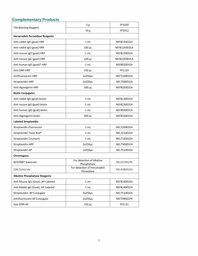
Complementary Products
TSA Blocking Reagent 3 g FP1020
10 g FP1012
Horseradish Peroxidase Reagents
Anti-rabbit IgG (goat) HRP 1 mL NEF812001EA
Anti-rabbit IgG (goat) HRP 100 µL NEF812E001EA
Anti-mouse IgG (goat) HRP 1 mL NEF822001EA
Anti-mouse IgG (goat) HRP 100 µL NEF822E001EA
Anti-human IgG (goat)* HRP 1 mL NEF802001EA
Anti-DNP-HRP 150 µL FP1129
Antifluorescein-HRP 2x250µL NEF710001EA
Streptavidin-HRP 2x250µL NEL750001EA
Anti-digoxigenin HRP 500 µL NEF832001EA
Biotin Conjugates
Anti-rabbit IgG (goat) biotin 1 mL NEF813001EA
Anti-mouse IgG (goat) biotin 1 mL NEF823001EA
Anti-human IgG (goat) biotin 1 mL NEF803001EA
Anti-digoxigenin biotin 500 µL NEF833001EA
Labeled Streptavidin
Streptavidin Fluorescein 1 mL NEL720001EA
Streptavidin Texas Red® 1 mL NEL721001EA
Streptavidin Coumarin 1 mL NEL722001EA
Streptavidin-HRP 2x250µL NEL750001EA
Streptavidin-AP 2x250µL NEL751001EA
Chromogens
BCIP/NBT Substrate For detection of Alkaline
Phosphatase NEL937001PK
DAB Substrate For detection of Horseradish
Peroxidase NEL938001EA
Alkaline Phosphatase Reagents
Anti-Mouse IgG (Goat), AP-Labeled 1 mL NEF814001EA
Anti-Rabbit IgG (Goat), AP-Labeled 1 mL NEF824001EA
Streptavidin- AP Conjugate 2x250µL NEL751001EA
Antifluorescein-AP Conjugate 2x250µL NEF709001PK
Anti-DNP-AP 150 µL FP1131

12
Hapten Labeled Deoxynucleotides (25 nmol, for labeling of ISH probes)
3-Amino-3-Deoxydigoxigenin-9-dCTP 25 nmol NEL562001EA
Biotin-11-dATP 25 nmol NEL540001EA
Biotin-11-dCTP 25 nmol NEL538001EA
Biotin-11-dGTP 25 nmol NEL541001EA
Biotin-11-dUTP 25 nmol NEL539001EA
DNP-11-dUTP 25 nmol NEL551001EA
Fluorescein-12-dATP 25 nmol NEL465001EA
Fluorescein-12-dCTP 25 nmol NEL424001EA
Fluorescein-12-dGTP 25 nmol NEL429001EA
Fluorescein-12-dUTP 25 nmol NEL413001EA
Hapten Labeled Ribonucleotides (25 nmol, for labeling of ISH probes)
Biotin-11-ATP 250 nmol NEL544001EA
Biotin-11-CTP 250 nmol NEL542001EA
Biotin-11-GTP 250 nmol NEL545001EA
Biotin-11-UTP 250 nmol NEL543001EA
Fluorescein-12-ATP 250 nmol NEL439001EA
Fluorescein-12-CTP 250 nmol NEL434001EA
Fluorescein-12-GTP 250 nmol NEL496001EA
Fluorescein-12-UTP 250 nmol NEL414001EA
TSA and its use are protected under U.S. Patents 5,688,966, 5,863,748, 5,767,287, 6,372,937, 6,399,299, 6,593,100, 6,617,125 and patents pending, and foreign equivalents thereof.. For Research use only. This product is distributed and sold for research purposes only by the end-user in the research market, and, to that extent, by purchasing this product the end-user is granted a limited license to use this product for research use only. This product is not intended for diagnostic or therapeutic use and no license or right is granted for use of this product for diagnostic or therapeutic purposes. Purchase does not include or carry any right or license to use, develop or otherwise exploit this product commercially. Any commercial use, development or exploitation of this product without the express prior written authorization of PerkinElmer is strictly prohibited and may constitute infringement of the intellectual property rights of PerkinElmer under the aforementioned patents. TSA is a registered trademark of PerkinElmer. Other trademarks are property of their respective owners.
Related Documents