Mandible Fractures: Evaluation and Management Joseph L. Russell, MD Faculty Advisor: Tammara Watts, MD Department of Otolaryngology—Head & Neck Surgery The University of Texas Medical Branch (UTMB Health) Grand Rounds Presentation March 29, 2013

Welcome message from author
This document is posted to help you gain knowledge. Please leave a comment to let me know what you think about it! Share it to your friends and learn new things together.
Transcript

Mandible Fractures: Evaluation and Management
Joseph L. Russell, MD
Faculty Advisor: Tammara Watts, MD
Department of Otolaryngology—Head & Neck Surgery
The University of Texas Medical Branch (UTMB Health)
Grand Rounds Presentation
March 29, 2013

Overview
• Review of anatomy
• Review of occlusion
• Types and locations of fractures
• Patient evaluation and initial management
• Definitive Management
• Timing of fracture repair
• Maxillomandibular fixation
• Open reduction and internal fixation (ORIF)
• Post-operative care
• Complications

Anatomy
• Regions
• Symphysis
• Parasymphyseal region—spans canine to canine
• Body—from canine to angle
• Alveolar process—contains teeth, resorbs if teeth are lost
• Angle—non-tooth bearing region between body and ramus
• Ramus
• Condyle
• Coronoid process
• Inferior alveolar/mental nerves
• Muscle attachments
• Weak points
• Condylar neck
• Angle—especially if third molar is present
• Mental foramen Cummings/Ballenger

Dingman RO, Natvig P: Surgery of facial fractures, Philadelphia, 1964, Saunders


Cummings Fig 12-15

Occlusion Angle’s Classification
Class I: mesiobuccal cusp of the maxillary first molar rests within the mesiobuccal groove of the mandibular first molar
Class II: maxillary molar is more anterior (retrognathic)
Class III: maxillary molar is more posterior (prognathic)
Small Figure—Ballenger’s Fig 55-1
Large Figure—Myer’s Fig 92-5

Crossbite
• Normal: maxillary buccal cusps lie lateral to the mandibular buccal
cusps
• Lingual crossbite: maxillary buccal cusps are positioned medially
• Buccal crossbite: maxillary buccal cusps are positioned laterally
Ballenger’s

Fracture classification
• Simple/Compound/Comminuted/Greenstick
• Anatomic location
• Dentition status
• Stability

Fracture classification
• Simple
• Oral mucosa and external skin are intact
• Compound (open)
• Laceration of mucosa or skin is present
• Fracture passes into a tooth root
• Greenstick
• Involves only one cortex
• Most common in children
• Comminuted
• Multiple fragments of bone

Fracture
Classification
Anatomic location
- Condyle
- Body
- Angle

Fracture classification
• Dentition status
• Dentulous
• Edentulous
• Decreased mandibular height
• Tenuous blood supply
• Superficial location of the mental forarmen
• Accuracy of re-approximation less important
• Pediatric
• Unerrupted dentition must be avoided
• Deciduous teeth hold wire poorly

- Stability: Favorable vs unfavorable
• Due to upward forces of the temporalis and masseter and downward forces of the suprahyoid musculature
• Almost all angle fractures are unfavorable
Favorable (A) vs
unfavorable (B)
fractures.
Temporalis and
masseter forces on the
proximal fragment, and
igastric/suprahyoid
musculature forces on
the distal segment,
stabilize the fracture in
A but destabilize it in B.
Myer’s Operative Otolaryngology Fig 92-2
FRACTIRE CLASSIFICATION
Patient evaluation
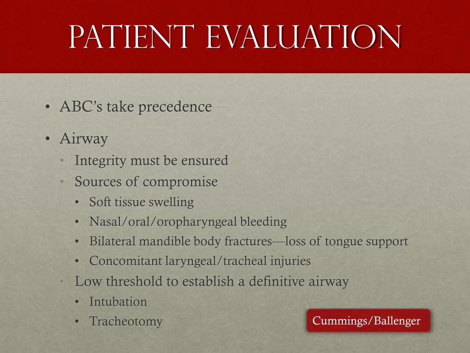
• ABC’s take precedence
• Airway
• Integrity must be ensured
• Sources of compromise
• Soft tissue swelling
• Nasal/oral/oropharyngeal bleeding
• Bilateral mandible body fractures—loss of tongue support
• Concomitant laryngeal/tracheal injuries
• Low threshold to establish a definitive airway
• Intubation
• Tracheotomy Cummings/Ballenger

Patient evaluation
• Cervical spine injury
• Must be ruled out in all patients
• Rate is 5% to 8% if an isolated facial fracture is present
• Rate is 7 to 11% if 2 or more facial fractures are present
• Stabilize cervical spine with C-collar at presentation
Mulligan RP, Mahabir RC. The prevalence of cervical spine injury,
head injury, or both with isolated and multiple craniomaxillofacial
fractures. Plast Reconstr Surg 2010;126:1647.

Patient Evaluation
• History
• Often limited by patient being intubated, unconscious, intoxicated, etc
• Mechanism
• Assault, motor vehicle collision, fall, gunshot
• If patient is conscious and cooperative, assess for:
• Presence/location of pain
• Subjective malocclusion
• Trismus
• Intraoral bleeding
• Numbness, particularly in the mental nerve region

Patient evaluation
• Physical Examination
• May be limited by swelling, hematoma, C-collar, tubes
• First assess the general appearance of the face
• Note lacerations, ecchymoses, edema, areas of distortion
• Chin lacerations indicate possible subcondylar fractures
• Palpate the mandible, note sensitive regions, mobility
• Include palpation of the condylar heads through the external auditory canals—pain may indicate presence of condylar head fracture
• Note intraoral mucosal tears, ecchymoses, bleeding
• Occlusion
• Quality of dentition, tooth involvement in fracture lines, fractured teeth
• Missing teeth—if acute, chest x-ray is needed
• Look for deviation of the mandible, premature molar contact, open bite, crossbite, trismus
• Full cranial nerve examination if patient is conscious, with special focus on cranial nerves V and VII

Patient evaluation
• Radiography
• Panoramic tomography (Panorex)
• Superior sensitivity for mandible fractures than 3 mm CT scans
Preferred by some surgeons as the sole imaging modality of the mandible
• Non-contrasted maxillofacial computed tomography (CT)
• 1 mm fine cuts have superior sensitivity to Panorex (100% vs 86%)
• Usually obtained in patients due to need to evaluate for other facial fractures
• 3-D reconstructions can be rendered and are helpful in some cases, such as comminuted fractures
Cummings

Grainger & Allison’s, Fig 63.41; right parasympheseal
and left angle fractures; note full condyle view

• Right parasymphyseal and left ramus fractures.
• Avery LL, Susarla SM, Novelline RA. Multidetector and three-dimensional CT evaluation
of the patient with maxillofacial injury. Radiol Clin N Am 49 (2011) 183–203.

• Right ramus and left anterior body fractures
• Marx: Rosen’s Emergency Medicine, Figure 39-7

Management

Initial management
• With rare exception, mandible fractures are not surgical emergencies
• If surgical intervention is needed, it should be undertaken as soon as it is safe to do so
• Soft diet
• Pain control
• Antibiotics for all open fractures (includes fractures involving tooth roots)
• Penicillins, cephalosporins, and clindamycin are appropriate options

Timing of repair
• Champy and others once advocated repair within 24 hours
• Often not feasible, commonly resulting in delays
• Barker et al, Laryngoscope 2011, University of Virginia
• Retrospective chart review of 83 patients over 5 years
• Mean time to fixation was 6.7 days
• No correlation found between increased time to repair and rate
of complications (infection, nonunion, malunion)
• Trend found between fewer complications and increasing time
to surgical repair
Barker DA, Oo KK, Allak A, et al. Timing for repair of mandible fractures.
Laryngoscope 2011;121:1160-3.

Antibiotics
• Numerous studies have shown no benefit with routine use of post-operative antibiotic therapy beyond 24 hours
• There is, however, evidence to support pre and perioperative antibiotic use
• Any fracture extending to the dentoalveolar ridge is an open fracture and requires antibiotic therapy
• Closed condyle fractures do not require antibiotics
• Ancef 1 g IV prior to surgery then one additional dose 8 hours later
• Reduced mandible fracture infection rate from 42% to 14% in a prospective clinical trial by Chole and Yee
• Andreasen JO, Jensen SS, Schwartz O, et al. A systematic review of prophylactic antibiotics in the treatment
of maxillofacial fractures. J Oral Maxillofac Surg 2006;64:1664-8
• Miles BA, Potter JK, Ellis E. The efficacy of postoperative antibiotic regimens in the open treatment of
mandibular fractures: a prospective randomized trial. J Oral Maxillofac Surg 2006;64:576-82
• Chole RA, Yee J. Antibiotic prophylaxis for facial fractures. Arch Otolaryngol Head Neck Surg
1987;113:1055

Teeth in the
fracture line
• Prior to routine antibiotic use, pulling an involved
tooth was recommended to reduce the incidence of
infection
• Current recommendations are to remove a tooth in the
fracture line if it is carious, if the tooth or its root are
fractured, or if the presence of the tooth is preventing
proper fracture reduction
• Removal of the third molar tends to destabilize angle
fractures and should be avoided if possible
Cummings

Definitive
management
• Soft diet
• Optimal treatment for some non-displaced ramus fractures and subcondylar fractures
• Must be no malocclusion
• Closed reduction—maxillomandibular fixation (MMF)
• Arch bars
• Eyelet wires
• IMF screws
• Open reduction and internal fixation (ORIF)
• Mini plates vs larger reconstruction plates
• Lag screws

General Principles
• Restore the patient’s pre-morbid occlusion
• Repair both skeletal and soft tissue injuries
• Use lacerations when possible
• Use mucosal incisions when possible
• Reduce all fractures
• Stabilize fractures
• Fixate all fractures adequately to allow bone healing

Closed Reduction:
Indications
• Nondisplaced favorable fractures
• Pediatric fractures
• Open reduction best avoided due to risk of injuring tooth buds
• Grossly comminuted fractures
• Avoids periosteal stripping of the bone fragments
• Condyle fractures
• Controversial, though closed treatment is generally appropriate
• Bilateral condyle fractures can result in loss of mandibular
height if closed reduction is the only treatment

Closed Reduction: ContraIndications
• Compromised pulmonary function
• Severe asthma
• Severe COPD
• Poorly controlled seizures
• Severe nausea
• Psychiatric or neurologic disorders

Closed
Reduction
Erich arch bars
- Standard for MMF
- Use 24 gauge circumdental wire to
fasten each bar to the dentition
- Lugs must open AWAY from the
crowns of the teeth
- Place patient in maximum
intercuspation and place MMF
wires or elastics
Ballengers: Fig 55-17
Text from Resident Manual
Text from Resident Manual

Closed
reduction
IMF screws
- 2.5 mm self-drilling and
self-tapping screws
- Placed adjacent to the
canine tooth roots
- Alternative if arch bars
cannot be applied or for
temporary MMF during
ORIF AO foundation website

ORIF: Indications
• Displaced unfavorable angle fractures
• Complex facial fractures requiring a stable mandibular base
• Atrophic edentulous mandibles with minimal cancellous
bone, and poor osteogenesis and healing potential
• Some condyle fractures
• Absolute indications
• Displacement into the middle cranial fossa or EAC
• Inability to obtain adequate occlusion
• Lateral extracapsular dislocation
• Contaminated open joint wound

ORIF: Approaches
• Intraoral
• Preferred
• More direct
• No external scars
• Low risk to the facial nerve
• Disadvantage—decreased exposure
• External
• Improved exposure of the posterior body, angle, and ramus
• Often required for severely comminuted fractures
• Disadvantages—cervical scar, risk of injury to branches of the facial nerve
• Approach chosen should allow adequate exposure to reduce and immobilize the fracture(s)

ORIF: competing principles
• Arbeitsgemenschaft fur Osteosynthesefragen (AO)
• Large, load-bearing plates
• Bicortical screws
• Champy
• Small, load-sharing plates
• Monocortical screws

ORIF: competing principles
• Bouloux et al (Emory), J Oral Maxillofac Surg, 2012
• Randomized, nonblinded, prospective trial over 2 years
• 127 initial patients, 53 of whom completed the required follow up
• Exclusions: clinical infection, previous treatment, comminuted fractures, condyle fractures, gunshot injuries, mandibular atrophy
• Two groups: AO (control) and Champy (experimental)
• All received perioperative antibiotics, follow up was at least six weeks, and no MMF was used
• Top fracture types
• 42% had a left angle fracture
• 23% had a right parasymphyseal fracture
• 19% had a right angle fracture
• Primary outcome: fracture union status at 6 weeks
• 81% union in the AO group and 89% in the Champy group (P = 0.95, not significant)
• Complications—no significant difference between groups
• Infection—AO 15% vs Champy 11% (P = 0.70)
• Hardware removal—AO 4% vs Champy 19% (P = 0.19)
• Prolonged antibiotic therapy—AO 12% vs Champy 4% (P = 0.35)
Bouloux GF, Chen S, Threadgill JM. Small and large titanium plates are equally
effective for treating mandible fractures. J Oral and Maxillofac Surg 2012;70:1613-21

Symphyseal &
Parasymphyseal
fractures
- Intraoral vestibular approach best
- Arch bars placed and MMF wires secured prior to incision
- Incision is made from canine to canine, leaving at least a 1 cm cuff of tissue from the mucogingival junction for closure
- Dissection carried to bone
- Freer used to dissect below the periosteum to the inferior border of the mandible
- Mental nerves identified and preserved
- Fracture line debrided, reduced, and MMF tightened
- Four hole 2.0 mm system titanium plate secured just below the tooth roots with monocortical screws
- Four to six hole 2.3 to 2.7 mm system titanium plate is shaped to the lower border of the mandible and secured with at least two screws on each side
- MMF wires cut and occlusion checked; if satisfactory, incision is closed.
- If no other fractures require the arch bars, they are also removed prior to closing

Body Fractures - Intraoral approach best
- Arch bars placed and MMF wires secured prior to incision
- 5 cm incision made in the gingivobuccal sulcus over the
fracture, leaving a 1 cm cuff of tissue from the
mucogingival junction for closure
- Mucosa only incision made anteriorly; blunt dissection
used to identify and dissect out the mental nerve from the
labial flap of tissue
- Dissection carried to bone
- Freer used to dissect below the periosteum to the inferior
border of the mandible
- Mental nerves identified and preserved
- Fracture line debrided, reduced, and MMF tightened
- Four hole tension band secured between the tooth roots
and mental foramen with monocortical screws
- Larger 2.3 to 2.7 mm system titanium plate is shaped to
the lower border of the mandible and secured anteriorly
through the intraoral incision with 2-3 screws. If needed,
a percutaneous drill guide is placed in the cheek to reach
the posterior screw holes and 2-3 screws are placed
- MMF wires cut and occlusion checked; if satisfactory,
incision is closed.
- If no other fractures require the arch bars, they are also
removed prior to closure of the incision

Angle Fractures
- Non-displaced fractures can be
treated with MMF x 6 weeks
- Intra-oral approach is similar to
approach for a body fracture, with
incision more posterior and
extended superiorly over the
external oblique ridge
- Arch bars placed prior to incision,
but MMF is performed after
dissection
- Fracture line is exposed and
reduced
- Plating options include two mini
plates, a superior mini plate and
heavier lower plate, and a 2 mm 8
hole strut plate
- Percutaneous drill guide is required
to drill holes and place the screws

Angle Fractures - External approach
- 5 cm incision is made 2 to 2.5 cm below the angle of the
mandible and extended posterosuperiorly toward the
earlobe
- Deep subcutaneous dissection is performed inferiorly on
top of the platysma
- Platysma is incised inferiorly and dissection is carried
down to the posterior belly of the digastric
- Once the digastric is identified, dissection is carried
superiorly to the inferior border of the mandible
- Periosteum is incised and a freer is used to elevate the
periosteum from the bone, exposing the fracture
- Fracture is debrided and reduced, and the patient is
placed in MMF
- Plating options include two mini plates, a superior mini
plate and heavier lower plate, and a 2 mm 8 hole strut
plate
- After plating, MMF is released and occlusion checked;
if satisfactory arch bars are removed unless needed for
other fractures
- Intraoral incisions closed with chromic or vicryl suture
- For external approach, platysma is closed with a
running locking vicryl suture, deep dermal interrupted
monocryl sutures are placed, and the skin is closed with
5-0 FAST gut or 5-0 nylon/prolene running suture.

Ramus Fractures
• Most ramus fractures can be treated with MMF x 6
weeks
• Intraoral plating is very difficult
• External approach for plating is similar to that of angle
fractures

Condyle Fractures
- Most condyle fractures can be
treated with MMF x 2-4 weeks
- If the head is involved, MMF is
limited to 2 weeks to prevent TMJ
ankylosis
- Frequent scenario is a condyle
fracture with contralateral
parasymphyseal, body, or angle
fracture—treat contralateral fracture
with ORIF and condyle fracture
with MMF x 2-4 weeks
- When plating is required, an
external approach is preferred
- External approach is similar to the
angle approach but with the
superior end of the incision brought
to about 2 cm from the earlobe

Coronoid fractures
• Uncommon, usually associated with a ZMC fracture
• No treatment is required in the majority of cases

Postoperative care
• Wire cutters kept at bedside upon leaving the OR and sent home with the patient
• No benefit to extending antibiotics beyond 24 hours post-op
• Oral hygiene is stressed to the patient
• Daily brushing of teeth and arch bars
• Water pick is very effective
• Dental wax to protect the buccal mucosa
• Some authors advocate weekly follow up until arch bars are removed

Complications

Infection
• Occurs in 10-15% of patients
• No significant decrease in the infection rate with extended post-op antibiotics
• Thought to result from fracture instability and movement instead of contamination with oral flora
• Predisposing factors
• Local
• Poor reduction/immobilization
• Poorly closed oral wounds
• Fractured teeth in the fracture line
• Diminished blood supply
• Devitalized tissue
• Comminuted fractures
• Systemic
• Alcoholism
• Poorly controlled DM
• Immunocompromise
• Treatment is surgical drainage, removal of infected hardware, and prolonged antibiotic therapy
• Placement of a heavy reconstruction plate may be required

nonunion
• Occurs in 3-5% of fractures
• Most common cause is inadequate reduction and
immobilization
• Treatment
• Control infection if present
• Debride devitalized tissues
• Remove hardware, freshen fracture ends, and place a new
heavy plate
• Place bone graft if needed

Malunion
• Improper alignment of the healed bony segments
• Caused by improper reduction, inadequate occlusal
alignment, and inadequate stability of the fracture
• May be clinically insignificant if occlusion is good
• Treatment
• Minor discrepancies can be treated with orthodontics
• Major discrepancies require open surgical repair with
refracturing and/or osteotomies

TMJ ankylosis
• Mandibular condyle fuses to the glenoid fossa
• Predisposing factors
• Intra-articular hemorrhage
• Condyle head fracture
• Prevention
• Shorter period of MMF (2-3 weeks)
• Physiotherapy
• Definitive treatment may require arthroplasty or total joint replacement

trigeminal nerve
injury
• Inferior alveolar nerve frequently injured when fracture
involves the mandibular canal
• Mental nerve at iatrogenic risk especially during
parasymphyseal and body fracture repairs
• Important to document deficits prior to surgery

Facial nerve injury
• In a trauma patient, the nerve can be injured anywhere alone its course
• Main trunk can be injured from fractures or surgery around the condylar neck
• Marginal mandibular branch at risk during submandibular external approaches
• Frontal branch at risk if an external pre-auricular approach is used
• Facial nerve function should be documented prior to surgery

References
• Donald PJ. Facial fractures. In Ballenger’s Otorhinolaryngology Head and Neck Surgery. 17th ed 2009
• Kellman RM. Maxillofacial trauma. In Cumming’s Otolaryngology: Head and Neck Surgery, 5th ed. 2010
• Dingman RO, Natvig P: Surgery of facial fractures, Philadelphia, 1964, Saunders
• Mulligan RP, Mahabir RC. The prevalence of cervical spine injury, head injury, or both with isolated and multiple craniomaxillofacial fractures. Plast Reconstr Surg 2010;126:1647.
• Avery LL, Susarla SM, Novelline RA. Multidetector and three-dimensional CT evaluation of the patient with maxillofacial injury. Radiol Clin N Am 49 (2011) 183–203.
• Barker DA, Oo KK, Allak A, et al. Timing for repair of mandible fractures. Laryngoscope 2011;121:1160-3.
• Andreasen JO, Jensen SS, Schwartz O, et al. A systematic review of prophylactic antibiotics in the treatment of maxillofacial fractures. J Oral Maxillofac Surg 2006;64:1664-8
• Miles BA, Potter JK, Ellis E. The efficacy of postoperative antibiotic regimens in the open treatment of mandibular fractures: a prospective randomized trial. J Oral Maxillofac Surg 2006;64:576-82
• Bouloux GF, Chen S, Threadgill JM. Small and large titanium plates are equally effective for treating mandible fractures. J Oral and Maxillofac Surg 2012;70:1613-21
• Ochs MW. Fractures of the mandible. In Myers, EN ed. Operative Otolaryngology., 2nd ed 2008.
• Eusterman VD. Mandibular trauma. In Resident Manual of Trauma to the Face, Head, and Neck. Aao—HNS Foundation,, 2012
Related Documents