Managing Incidental Findings on Abdominal CT: White Paper of the ACR Incidental Findings Committee Lincoln L. Berland, MD a , Stuart G. Silverman, MD b , Richard M. Gore, MD c , William W. Mayo-Smith, MD d , Alec J. Megibow, MD, MPH e , Judy Yee, MD f , James A. Brink, MD g , Mark E. Baker, MD h , Michael P. Federle, MD i , W. Dennis Foley, MD j , Isaac R. Francis, MD k , Brian R. Herts, MD h , Gary M. Israel, MD g , Glenn Krinsky, MD l , Joel F. Platt, MD k , William P. Shuman, MD m , Andrew J. Taylor, MD n As multidetector CT has come to play a more central role in medical care and as CT image quality has improved, there has been an increase in the frequency of detecting “incidental findings,” defined as findings that are unrelated to the clinical indication for the imaging examination performed. These “incidentalomas,” as they are also called, often confound physicians and patients with how to manage them. Although it is known that most incidental findings are likely benign and often have little or no clinical significance, the inclination to evaluate them is often driven by physician and patient unwillingness to accept uncertainty, even given the rare possibility of an important diagnosis. The evaluation and surveil- lance of incidental findings have also been cited as among the causes for the increased utilization of cross-sectional imaging. Indeed, incidental findings may be serious, and hence, when and how to evaluate them are unclear. The workup of incidentalomas has varied widely by physician and region, and some standardization is desirable in light of the current need to limit costs and reduce risk to patients. Subjecting a patient with an incidentaloma to unnecessary testing and treatment can result in a potentially injurious and expensive cascade of tests and procedures. With the participation of other radiologic organizations listed herein, the ACR formed the Incidental Findings Committee to derive a practical and medically appropriate approach to managing incidental findings on CT scans of the abdomen and pelvis. The committee has used a consensus method based on repeated reviews and revisions of this document and a collective review and interpretation of relevant literature. This white paper provides guidance developed by this committee for addressing incidental findings in the kidneys, liver, adrenal glands, and pancreas. Key Words: Incidental findings, incidentaloma, pancreatic cyst, renal cyst, liver lesion, adrenal nodule J Am Coll Radiol 2010;7:754-773. Copyright © 2010 American College of Radiology FOREWORD This white paper is meant not to comprehensively review the interpretation and management of solid masses in each organ system but to provide general guidance for managing incidentally discovered masses, appreciating that individual care will vary depending on each patient’s specific circum- stances; the clinical environment, available resources; and the judgment of the practitioner. Also, the term guidelines has not a Department of Radiology, University of Alabama at Birmingham, Birming- ham, Alabama. b Department of Radiology, Brigham and Women’s Hospital, Boston, Massa- chusetts. c Department of Radiology, Evanston Hospital, Evanston, Illinois. d Department of Radiology, Brown University School of Medicine, Provi- dence, Rhode Island. e Department of Radiology, NYU-Langone Medical Center, New York, New York. f Department of Radiology, University of California, San Francisco, San Fran- cisco, California. g Department of Diagnostic Radiology, Yale University School of Medicine, New Haven, Connecticut. h Department of Radiology, Cleveland Clinic, Cleveland, Ohio. i Department of Radiology, Stanford University Medical Center, Stanford, California. j Department of Radiology, Medical College of Wisconsin, Milwaukee, Wisconsin. k Department of Radiology, University of Michigan, Ann Arbor, Michigan. l Radiology Associates of Ridgewood, PA, Waldwick, New Jersey. m Department of Radiology, University of Washington School of Medicine, Seattle, Washington. n Department of Radiology, Virginia Commonwealth University Medical Center, Richmond, Virginia. Corresponding author and reprints: Lincoln L. Berland, MD, University of Alabama at Birmingham, Department of Radiology, 619 S 19th Street, N348, Birmingham, AL 35249-1900; e-mail: [email protected]. © 2010 American College of Radiology 0091-2182/10/$36.00 ● DOI 10.1016/j.jacr.2010.06.013 754

Welcome message from author
This document is posted to help you gain knowledge. Please leave a comment to let me know what you think about it! Share it to your friends and learn new things together.
Transcript
F
Tt
a
hb
cc
d
de
f
cg
Nh
7
Managing Incidental Findings onAbdominal CT: White Paper of theACR Incidental Findings Committee
Lincoln L. Berland, MDa, Stuart G. Silverman, MDb, Richard M. Gore, MDc,William W. Mayo-Smith, MDd, Alec J. Megibow, MD, MPHe, Judy Yee, MDf,
James A. Brink, MDg, Mark E. Baker, MDh, Michael P. Federle, MDi,W. Dennis Foley, MDj, Isaac R. Francis, MDk, Brian R. Herts, MDh,
Gary M. Israel, MDg, Glenn Krinsky, MDl, Joel F. Platt, MDk,William P. Shuman, MDm, Andrew J. Taylor, MDn
As multidetector CT has come to play a more central role in medical care and as CT image quality has improved, there hasbeen an increase in the frequency of detecting “incidental findings,” defined as findings that are unrelated to the clinicalindication for the imaging examination performed. These “incidentalomas,” as they are also called, often confoundphysicians andpatientswithhowtomanage them.Although it is knownthatmost incidentalfindings are likelybenignandoften have little or no clinical significance, the inclination to evaluate them is often driven by physician and patientunwillingness to accept uncertainty, even given the rare possibility of an important diagnosis. The evaluation and surveil-lance of incidental findings have also been cited as among the causes for the increased utilization of cross-sectional imaging.Indeed, incidental findings may be serious, and hence, when and how to evaluate them are unclear. The workup ofincidentalomas has varied widely by physician and region, and some standardization is desirable in light of the current needto limit costs and reduce risk to patients. Subjecting a patient with an incidentaloma to unnecessary testing and treatmentcan result in a potentially injurious and expensive cascade of tests and procedures. With the participation of other radiologicorganizations listed herein, the ACR formed the Incidental Findings Committee to derive a practical and medicallyappropriate approach to managing incidental findings on CT scans of the abdomen and pelvis. The committee has used aconsensus method based on repeated reviews and revisions of this document and a collective review and interpretation ofrelevant literature.Thiswhitepaperprovidesguidancedevelopedbythiscommittee foraddressing incidentalfindings inthekidneys, liver, adrenal glands, and pancreas.
Key Words: Incidental findings, incidentaloma, pancreatic cyst, renal cyst, liver lesion, adrenal nodule
J Am Coll Radiol 2010;7:754-773. Copyright © 2010 American College of Radiology
oicsj
i
Cj
k
l
m
Sn
C
A
OREWORD
his white paper is meant not to comprehensively reviewhe interpretation and management of solid masses in each
Department of Radiology, University of Alabama at Birmingham, Birming-am, Alabama.
Department of Radiology, Brigham and Women’s Hospital, Boston, Massa-husetts.
Department of Radiology, Evanston Hospital, Evanston, Illinois.
Department of Radiology, Brown University School of Medicine, Provi-ence, Rhode Island.
Department of Radiology, NYU-Langone Medical Center, New York, New York.
Department of Radiology, University of California, San Francisco, San Fran-isco, California.
Department of Diagnostic Radiology, Yale University School of Medicine,ew Haven, Connecticut.
Department of Radiology, Cleveland Clinic, Cleveland, Ohio. B
54
rgan system but to provide general guidance for managingncidentally discovered masses, appreciating that individualare will vary depending on each patient’s specific circum-tances; the clinical environment, available resources; and theudgment of the practitioner. Also, the term guidelines has not
Department of Radiology, Stanford University Medical Center, Stanford,alifornia.
Department of Radiology, Medical College of Wisconsin, Milwaukee, Wisconsin.
Department of Radiology, University of Michigan, Ann Arbor, Michigan.
Radiology Associates of Ridgewood, PA, Waldwick, New Jersey.
Department of Radiology, University of Washington School of Medicine,eattle, Washington.
Department of Radiology, Virginia Commonwealth University Medicalenter, Richmond, Virginia.
Corresponding author and reprints: Lincoln L. Berland, MD, University oflabama at Birmingham, Department of Radiology, 619 S 19th Street, N348,
irmingham, AL 35249-1900; e-mail: [email protected].© 2010 American College of Radiology0091-2182/10/$36.00 ● DOI 10.1016/j.jacr.2010.06.013
brTiiaapFronu
I
Tibcir[tmmtpantbdSo
ctiaaArheitaClOtp
sripptdpssab
P
T
●
●
●
PT
B
●
●
●
●
●
H
BaoAfm
Berland et al/Managing Incidentalomas on Abdominal CT 755
een used in this white paper to avoid the implication that thisepresents a component of the ACR Practice Guidelines andechnicalStandards (whichrepresentofficialACRpolicy,hav-
ngundergonearigorousdraftingandreviewprocess culminat-ng in approval by the ACR Council), or the ACR Appropri-teness Criteria® (which use a formal consensus-buildingpproach using a modified Delphi technique). This white pa-er, which represents the collective experience of the Incidentalindings Committee, using a less formal process of repeatedeviews and revisions of the draft document, does not representfficial ACR policy. For these reasons, this white paper shouldot be used to establish the legal standard of care in any partic-lar situation.
NTRODUCTION
he rapid increase in the utilization of cross-sectionalmaging examinations over the past two decades, com-ined with the ongoing improvement in the spatial andontrast resolution of these studies, has led to a markedncrease in the number of findings detected that are un-elated to the primary objectives of the examinations1-4]. An incidental finding, also known as an inciden-aloma, may be defined as “an incidentally discoveredass or lesion, detected by CT, MRI, or other imagingodality performed for an unrelated reason” [5]. Al-
hough such findings are incidental to the primary pur-ose of the study, one analysis suggested, “Some researchnd clinical activities are so prone to generating findingsot intentionally sought that it is disingenuous to termhem ‘unanticipated’ even if their precise nature cannote anticipated in advance” [6]. More important than theefinition is the action that each such finding invokes.o, we are asked to consider, “What is the responsible usef information that nobody asked for?” [7].
The burden of extra costs with incidental findings onross-sectional imaging has also raised concerns withinhe government and third-party payers as medical imag-ng utilization and expenditures have risen. A recent ex-mple of this was seen in the May 2009 CMS noncover-ge decision regarding screening CT colonography [8].lthough CT colonography focuses on detecting colo-
ectal polyps to prevent colorectal carcinoma, an unen-anced, low–radiation dose CT scan of the lower chest,ntire abdomen, and pelvis contains clinically significantncidental findings in 5% to 16% of asymptomatic pa-ients [1,4,9-14], with a higher frequency in symptom-tic patients [9,10,12-14]. The noncoverage decision byMS cited concern for the costs of evaluating extraco-
onic findings that are diagnostically indeterminate.ther existing or developing technologies may face this
ype of economic scrutiny as CMS and other third-partyayers become more focused on cost containment.
Although countless studies have been devoted to de- ecribing findings related to specific medical conditions,elatively little research has been devoted to understand-ng incidental findings. The most common reason toursue incidental findings is to differentiate benign fromotentially serious (including malignant) lesions. Al-hough most incidental findings prove to be benign, theiriscovery often leads to a cascade of testing that is costly,rovokes anxiety, exposes patients to radiation unneces-arily, and may even cause morbidity [15]. Articles de-cribing criteria for detecting, categorizing, reporting,nd managing such findings have been inconsistent atest and leave many unanswered questions [1,9-14].
ROJECT OBJECTIVES
he objectives of this project were:
to develop a consensus on sets of organ-specific imagingfeatures for some commonly affected organ systemswithin the abdomen, which will lead to consistent defini-tions for, and identification of, incidental findings;to develop medically appropriate approaches to managingincidentalfindings thatarediagnostically indeterminate; andto address the differences between unenhanced, low–radiation dose CT examinations and contrast-en-hanced CT examinations using standard radiationdoses for detecting and managing incidental findings.
OTENTIAL BENEFICIAL OUTCOMES OFHE PROJECT
enefits anticipated from this effort included:
reducing risks to patients from additional unnecessaryexaminations, including the risks of radiation and risksassociated with interventional procedures;limiting the costs of managing incidental findings topatients and the health care system;achieving greater consistency in recognizing, report-ing, and managing incidental findings, as a componentof formal quality improvement efforts;providing guidance to radiologists who are concernedabout the risk for litigation for missing incidental find-ings that later prove to be clinically important; andhelping focus research efforts to lead to an evidence-based approach to incidental findings.
ISTORY OF THE PROJECT
ecause of the increasing recognition of the problemsnd opportunities of incidental findings, considerationf a formal approach to these issues began within theCR in 2006. The Incidental Findings Committee was
ormed under the auspices of the Body Imaging Com-ission of the ACR. After several meetings and confer-
nce calls, the concepts and objectives described above
wlcft
arfmiCCA(t
C
Ecmabotntfincteamaab
gssfdcdamiTimiw
ER
CcpS[pttr
uatswab
CI
Oicsncmatpetasoeahbco[sefmi
nT
756 Journal of the American College of Radiology/Vol. 7 No. 10 October 2010
ere formulated. The initial intent was to develop guide-ines analogous to those produced by the Fleischner So-iety on pulmonary nodules [16] and the consensus con-erences of the Society of Radiologists in Ultrasound onhyroid nodules [17] and carotid imaging [18].
Because of the keen interest among groups both withinnd outside the ACR, the committee’s participants wereecruited from members of the ACR, all of who were alsoellows or members of the Society of Computed Body To-ography and Magnetic Resonance, the Society of Gastro-
ntestinal Radiologists, and the Society of Uroradiology.ontacts from other groups within the ACR, including theolon Cancer Committee, the Appropriateness Criteria–drenal Panel and the Appropriateness Criteria–GI Panel
Liver Lesion Topic) also helped ensure the consistency ofhe guidance produced among these groups.
ONSENSUS PROCESS
xpert radiologists in relevant organ systems were re-ruited to participate in the Incidental Findings Com-ittee and its subcommittees. We plan to further review
nd revise these recommendations periodically, on theasis of comments and new research. Although the scopef a project to address incidental findings on CT is large,he committee decided to develop guidance for a limitedumber of organ systems. Four subcommittees were es-ablished to address the largest number of incidentalndings within the abdomen, in the kidneys, liver, adre-al glands, and pancreas. A fifth subcommittee washarged with attempting to ensure the use of commonerminology and a common format. The committeelected to defer considering other incidental findingsrising in the abdomen and pelvis, such as ovarianasses, splenic lesions, lymphadenopathy, and vascular
bnormalities, including arterial stenoses, abdominalortic aneurysms, and renal artery aneurysms. The mem-ership of each subcommittee is listed in the Appendix.Each subcommittee was tasked to develop organ-specific
uidance, which was initially formulated primarily by theubcommittee chairs. When this was complete, these sub-ections were distributed to the subcommittee members forurther comments and discussion. Revisions of the entireocument were then distributed to the subcommitteehairs, and multiple revisions ensued. Finally, the draft wasistributed to the entire Incidental Findings Committee fordditional review to achieve consensus and to arrive at a finalanuscript. Reviews by other ACR committees were also
ntegrated into drafts at appropriate points in the process.o facilitate rapidly formulating and clearly communicat-
ng this guidance, and to provide convenient graphic sum-aries for easy reference, the committee decided to express
ts recommendations in flowcharts and tables, buttressed
ith explanatory text. bLEMENTS OF THESEECOMMENDATIONS AND FLOWCHARTS
ertain subspecialties within radiology have addressed in-onsistencies of documentation by creating structured re-orting, such as the Breast Imaging Reporting and Dataystem® classification [19]. In an analogous way, Zalis et al20], for the Working Group on Virtual Colonoscopy, pro-osed “C-RADS,” which includes an “E” classification sys-em for extracolonic findings. Although this latter classifica-ion system has elements in common with theseecommendations, it is not included with them here.
In the flowcharts within this white paper, the algorithmsse yellow boxes for steps that involve data to affect man-gement, such as categorization, demographics, history, andhe results of studies. Green boxes represent action steps,uch as performing a study, following up, or interveningith a biopsy or surgery. Red boxes indicate that the evalu-
tion process should stop, with no further action required,ecause the lesion can be concluded to be benign.
HALLENGES OF ADDRESSINGNCIDENTAL FINDINGS
ne of the crucial obstacles to managing incidental find-ngs cost-effectively is the unwillingness of many physi-ians to accept uncertainty even when the chance of aerious diagnosis is extremely unlikely. This unwilling-ess is in part driven by a paucity of data, the lack oflear-cut algorithms with regard to diagnostic and treat-ent strategies, fear of potential malpractice litigation,
nd the desire of patients and their families to adhere tohe adage “better safe than sorry.” It may be difficult forhysicians or patients to appreciate at an intellectual ormotional level that an incidental finding might not needo undergo further examinations or follow-up. Not onlyre further tests likely to yield a benign diagnosis, butuch testing could even lead to morbidity [15]. On thether hand, an incidental finding could represent a ser-ndipitous discovery of a serious diagnosis, such as a largebdominal aortic aneurysm, and be potentially lifesaving;ence the conundrum. The discussion of cost is alsourdened with strong opinions, with some believing thatost should be no obstacle to reaching a comfortable levelf medical certainty for a positive or negative diagnosis21,22]. Others might argue that medical resourceshould be best applied where they are known to be mostffective. However, there is strong scientific validationor applying medical strategies that optimize results whileinimizing costs and applying “evidence-based” reason-
ng to medical decisions [21].Unfortunately, information about the cost-effective-
ess of pursuing incidental findings is largely lacking.herefore, achieving a consensus of experts, supported
y available literature, is a reasonable interim objective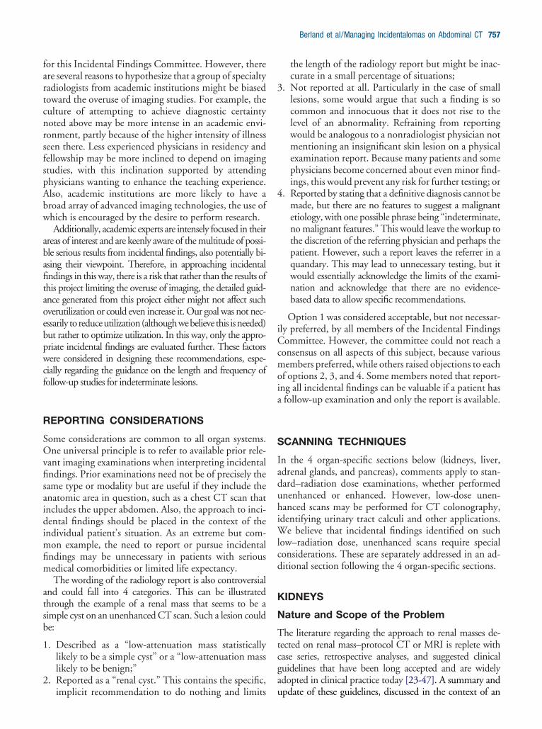
fartcnrsfspAbw
abafitaoebpwcf
R
SOvfisaidimfim
atsb
1
2
3
4
iCcmoia
S
IaduhiWlcd
K
N
Ttcga
Berland et al/Managing Incidentalomas on Abdominal CT 757
or this Incidental Findings Committee. However, therere several reasons to hypothesize that a group of specialtyadiologists from academic institutions might be biasedoward the overuse of imaging studies. For example, theulture of attempting to achieve diagnostic certaintyoted above may be more intense in an academic envi-onment, partly because of the higher intensity of illnesseen there. Less experienced physicians in residency andellowship may be more inclined to depend on imagingtudies, with this inclination supported by attendinghysicians wanting to enhance the teaching experience.lso, academic institutions are more likely to have aroad array of advanced imaging technologies, the use ofhich is encouraged by the desire to perform research.Additionally, academic experts are intensely focused in their
reas of interest and are keenly aware of the multitude of possi-le serious results from incidental findings, also potentially bi-sing their viewpoint. Therefore, in approaching incidentalndings in this way, there is a risk that rather than the results ofhis project limiting the overuse of imaging, the detailed guid-nce generated from this project either might not affect suchverutilization or could even increase it. Our goal was not nec-ssarily to reduceutilization(althoughwebelieve this isneeded)ut rather to optimize utilization. In this way, only the appro-riate incidental findings are evaluated further. These factorsere considered in designing these recommendations, espe-
ially regarding the guidance on the length and frequency ofollow-up studies for indeterminate lesions.
EPORTING CONSIDERATIONS
ome considerations are common to all organ systems.ne universal principle is to refer to available prior rele-
ant imaging examinations when interpreting incidentalndings. Prior examinations need not be of precisely theame type or modality but are useful if they include thenatomic area in question, such as a chest CT scan thatncludes the upper abdomen. Also, the approach to inci-ental findings should be placed in the context of the
ndividual patient’s situation. As an extreme but com-on example, the need to report or pursue incidental
ndings may be unnecessary in patients with seriousedical comorbidities or limited life expectancy.The wording of the radiology report is also controversial
nd could fall into 4 categories. This can be illustratedhrough the example of a renal mass that seems to be aimple cyst on an unenhanced CT scan. Such a lesion coulde:
. Described as a “low-attenuation mass statisticallylikely to be a simple cyst” or a “low-attenuation masslikely to be benign;”
. Reported as a “renal cyst.” This contains the specific,
implicit recommendation to do nothing and limits uthe length of the radiology report but might be inac-curate in a small percentage of situations;
. Not reported at all. Particularly in the case of smalllesions, some would argue that such a finding is socommon and innocuous that it does not rise to thelevel of an abnormality. Refraining from reportingwould be analogous to a nonradiologist physician notmentioning an insignificant skin lesion on a physicalexamination report. Because many patients and somephysicians become concerned about even minor find-ings, this would prevent any risk for further testing; or
. Reported by stating that a definitive diagnosis cannot bemade, but there are no features to suggest a malignantetiology, with one possible phrase being “indeterminate,no malignant features.” This would leave the workup tothe discretion of the referring physician and perhaps thepatient. However, such a report leaves the referrer in aquandary. This may lead to unnecessary testing, but itwould essentially acknowledge the limits of the exami-nation and acknowledge that there are no evidence-based data to allow specific recommendations.
Option 1 was considered acceptable, but not necessar-ly preferred, by all members of the Incidental Findingsommittee. However, the committee could not reach a
onsensus on all aspects of this subject, because variousembers preferred, while others raised objections to each
f options 2, 3, and 4. Some members noted that report-ng all incidental findings can be valuable if a patient hasfollow-up examination and only the report is available.
CANNING TECHNIQUES
n the 4 organ-specific sections below (kidneys, liver,drenal glands, and pancreas), comments apply to stan-ard–radiation dose examinations, whether performednenhanced or enhanced. However, low-dose unen-anced scans may be performed for CT colonography,
dentifying urinary tract calculi and other applications.e believe that incidental findings identified on such
ow–radiation dose, unenhanced scans require specialonsiderations. These are separately addressed in an ad-itional section following the 4 organ-specific sections.
IDNEYS
ature and Scope of the Problem
he literature regarding the approach to renal masses de-ected on renal mass–protocol CT or MRI is replete withase series, retrospective analyses, and suggested clinicaluidelines that have been long accepted and are widelydopted in clinical practice today [23-47]. A summary and
pdate of these guidelines, discussed in the context of an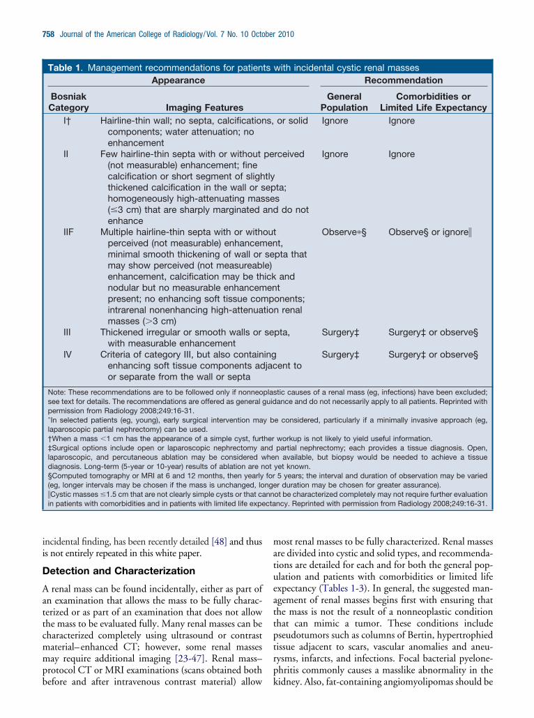
ii
D
Aattcmmpb
matueattptrp
cta
758 Journal of the American College of Radiology/Vol. 7 No. 10 October 2010
ncidental finding, has been recently detailed [48] and thuss not entirely repeated in this white paper.
etection and Characterization
renal mass can be found incidentally, either as part ofn examination that allows the mass to be fully charac-erized or as part of an examination that does not allowhe mass to be evaluated fully. Many renal masses can beharacterized completely using ultrasound or contrastaterial– enhanced CT; however, some renal massesay require additional imaging [23-47]. Renal mass–
rotocol CT or MRI examinations (scans obtained both
Table 1. Management recommendations for patienAppearance
BosniakCategory Imaging Features
I† Hairline-thin wall; no septa, calcificationcomponents; water attenuation; noenhancement
II Few hairline-thin septa with or without(not measurable) enhancement; finecalcification or short segment of slighthickened calcification in the wall or shomogeneously high-attenuating mas(�3 cm) that are sharply marginatedenhance
IIF Multiple hairline-thin septa with or withperceived (not measurable) enhancemminimal smooth thickening of wall ormay show perceived (not measureabenhancement, calcification may be thnodular but no measurable enhancempresent; no enhancing soft tissue comintrarenal nonenhancing high-attenuamasses (�3 cm)
III Thickened irregular or smooth walls orwith measurable enhancement
IV Criteria of category III, but also containenhancing soft tissue components ador separate from the wall or septa
Note: These recommendations are to be followed only if nonneosee text for details. The recommendations are offered as general gpermission from Radiology 2008;249:16-31.�In selected patients (eg, young), early surgical intervention malaparoscopic partial nephrectomy) can be used.†When a mass �1 cm has the appearance of a simple cyst, furt‡Surgical options include open or laparoscopic nephrectomy alaparoscopic, and percutaneous ablation may be considered wdiagnosis. Long-term (5-year or 10-year) results of ablation are n§Computed tomography or MRI at 6 and 12 months, then yearly(eg, longer intervals may be chosen if the mass is unchanged, lo�Cystic masses �1.5 cm that are not clearly simple cysts or that cain patients with comorbidities and in patients with limited life expe
efore and after intravenous contrast material) allow k
ost renal masses to be fully characterized. Renal massesre divided into cystic and solid types, and recommenda-ions are detailed for each and for both the general pop-lation and patients with comorbidities or limited lifexpectancy (Tables 1-3). In general, the suggested man-gement of renal masses begins first with ensuring thathe mass is not the result of a nonneoplastic conditionhat can mimic a tumor. These conditions includeseudotumors such as columns of Bertin, hypertrophiedissue adjacent to scars, vascular anomalies and aneu-ysms, infarcts, and infections. Focal bacterial pyelone-hritis commonly causes a masslike abnormality in the
with incidental cystic renal massesRecommendation
GeneralPopulation
Comorbidities orLimited Life Expectancy
or solid Ignore Ignore
rceived
ta;s
do not
Ignore Ignore
t,pta that
andt
onents;n renal
Observe�§ Observe§ or ignore�
pta, Surgery‡ Surgery‡ or observe§
ent toSurgery‡ Surgery‡ or observe§
tic causes of a renal mass (eg, infections) have been excluded;ance and do not necessarily apply to all patients. Reprinted with
e considered, particularly if a minimally invasive approach (eg,
workup is not likely to yield useful information.partial nephrectomy; each provides a tissue diagnosis. Open,n available, but biopsy would be needed to achieve a tissueyet known.5 years; the interval and duration of observation may be variedr duration may be chosen for greater assurance).
ot be characterized completely may not require further evaluationncy. Reprinted with permission from Radiology 2008;249:16-31.
ts
s,
pe
tlyepseand
outen
sele)ickenp
tio
se
ingjac
plasuid
y b
herndheotforngenn
idney. Also, fat-containing angiomyolipomas should be
epaabfl
tbieicmmcecnflamIIt
dt
on[aa3csa
1spmrltmltmmrccug
mis
Berland et al/Managing Incidentalomas on Abdominal CT 759
xcluded. With rare exceptions, a mass that contains fat,articularly when not calcified, can be diagnosed as anngiomyolipoma with confidence. The subsequent man-gement then can be derived and is summarized in Ta-les 1 to 3. These tables are reconfigured in the form ofowchart algorithms in Figures 1 and 2.The approach to the cystic renal mass follows the
ime-tested approach of Bosniak [23,25,27-32]. The ta-les and flowcharts are constructed so that both patientsn the general population and those with limited lifexpectancy can be managed. In general, size is not a factorn the Bosniak classification of cystic renal masses, be-ause large cystic masses are often benign, and small onesay be malignant. However, the smaller the mass, theore likely it is benign. Therefore, the commonly en-
ountered cystic-appearing renal mass that is too small tovaluate all of its features, including its CT attenuation,an be presumed to be benign if it does not display anyonsimple features. In the green “action boxes” in theowcharts (Figures 1 and 2), observation with imaging,lso known as active surveillance [49,50], is recom-ended for indeterminate masses in Bosniak category
IF and is also an option for masses in categories III andV in patients with limited life expectancy or comorbidi-ies that would increase the risk of treatment.
There is no known interval of time that can be used toiagnose an indeterminate renal mass with certainty, al-
Table 2. Management recommendations for incidenpopulation
Mass Size Probable Diagnosis ReLarge (�3 cm) Renal cell carcinoma† Surg
Small (1-3 cm) Renal cell carcinoma† Surg
Very small (�1 cm) Renal cell carcinoma,oncocytoma,angiomyolipoma�
Obs
Note: These recommendations are best followed after nonneoplatext for details. The recommendations are offered as general gui�Benign entities are more likely in small renal masses than large†Provided there is no detectable fat by CT or MRI using protoco‡Surgical options include open or laparoscopic nephrectomylaparoscopic, and percutaneous ablation may be considered wdiagnosis. Long-term (5-year or 10-year) results of ablation are n§Computed tomography or MRI at 3 to 6 months, 12 months, and(eg, shorter intervals if the mass is enlarging). Reprinted with per
hough 5 years has been suggested as a reasonable length m
f time to diagnose an indeterminate renal mass as be-ign on the basis of the lack of morphologic change36,48]. Depending on the level of suspicion, and patientnd referrer comfort with observation, both the durationnd interval may be altered. As indicated in Tables 1 toand the flowcharts (Figures 1 and 2), growth alone
annot be used to definitively diagnose a mass (whetherolid or cystic) as malignant. Benign masses may grow,nd malignant ones may grow little, if at all [51,52].
Regarding the flowchart for cystic renal masses (Figure), both Bosniak category III and IV masses are managedurgically; however, category IV masses have a greaterrobability of malignancy than category III masses, andanagement approaches other than resection carry more
isk. Because many Bosniak category III masses are ma-ignant, surgery is recommended for the general popula-ion. Percutaneous biopsy of Bosniak category III renalasses, although controversial, may be helpful, particu-
arly in patients with comorbidities that would pose risko patients undergoing surgery [34,46]. If a definitivealignant result can be obtained with biopsy, surgeryay be planned with confidence. For a benign biopsy
esult to be useful, it should be both definitive and spe-ific of a benign entity. Biopsy results that reveal nonspe-ific cells should be viewed with caution and cannot besed alone to guide management. Because Bosniak cate-ory III masses typically contain few solid elements, it
l solid renal masses in patients in the general
mmendation Commenty‡ Angiomyolipoma with minimal fat,
oncocytoma, other benignneoplasms may be found atsurgery
y‡ If hyperattenuating, andhomogenously enhancing,consider MRI and percutaneousbiopsy to diagnoseangiomyolipoma with minimalfat
e until 1 cm§ Thin (�3 mm) sections helpconfirm enhancement
causes of a renal mass (eg, infections) have been excluded; seece and do not necessarily apply to all patients.s.esigned to evaluate renal masses.partial nephrectomy; both provide a tissue diagnosis. Open,
n available, but biopsy would be needed to achieve a tissueyet known.en yearly; the interval and duration of observation may be variedsion from Radiology 2008;249:16-31.
ta
coer
er
erv
sticdanonels d
andheotth
ay be difficult to both target and procure diagnostic
ttct
fln(mspsetmmstpammcb
gaistwHrv
itopomsmtopad
inte
760 Journal of the American College of Radiology/Vol. 7 No. 10 October 2010
issue for biopsy, limiting the ability to achieve defini-ively benign or malignant results. However, even if aonfident diagnosis of a benign entity can be made inhese patients, observation is still warranted.
We define solid masses as those that contain little or nouid attenuating (�20 Hounsfield units [HU]) compo-ents and usually consist predominantly of enhancing tissueTables 2 and 3, Figure 2). As described for cystic renalasses, all solid masses should be evaluated first for features
uggesting a nonneoplastic etiology, such as focal bacterialyelonephritis or other conditions noted above. A thoroughearch for fat cells using CT or MRI protocols designed tovaluate renal masses should also be undertaken. Althoughhere are rare exceptions, fat-containing noncalcified renalasses in adults can be diagnosed as benign angiomyolipo-as with confidence [48]. The subsequent approach to a
olid renal mass is then predicated mostly on size. Althoughhere is no single feature of a renal mass that can be used toredict its biologic behavior accurately, size is a reasonablend practical approach. In general, large (�3 cm) solid renalasses are likely malignant; similarly, the smaller a solidass, the more likely it is benign. In addition, a small renal
ell carcinoma is more likely to be low grade and indolent
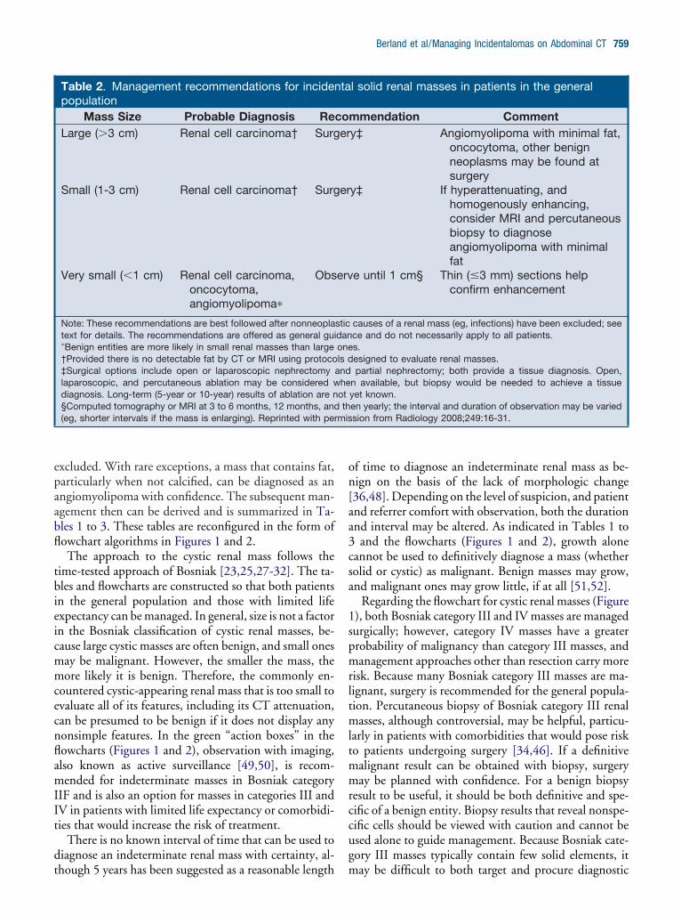
Table 3. Management recommendations for incidenexpectancy or comorbidities that increase the risk o
Mass Size Probable Diagnosis RecLarge (�3 cm) Renal cell
carcinoma†Surge
Small (1-3 cm) Renal cellcarcinoma†
Surge
Very small (�1 cm) Renal cell carcinoma,oncocytoma,angiomyolipoma�
Obse
Note: These recommendations are best followed after nonneoplatext for details. The recommendations are offered as general gui�Benign entities are more likely in small renal masses than large†Provided there is no detectable fat by CT or MRI using protoco‡Surgical options include open or laparoscopic nephrectomylaparoscopic, and percutaneous ablation may be considered wdiagnosis. Long-term (5-year or 10-year) results of ablation are n§Computed tomography or MRI at 3 to 6 months, 12 months, anintervals if the mass is enlarging); the duration of observation mamass of any size in a patient with limited life expectancy or comorbis small. It may be safe to observe a solid renal mass beyorecommendations on the risks and benefits of observation. Repr
ehaving than a larger one [53]. Therefore, we have sug- o
ested that solid masses �1 cm be observed [48]. Thispproach is further supported by the difficulty of confirm-ng that masses of this size are enhancing and are thereforeolid. Partial volume effects can mimic enhancement. Thus,he use of thin-section (�3 mm) CT and MRI is advisedhen both evaluating and observing such small masses.owever, there are rare cases of aggressively behaving small
enal cell carcinomas, even those �1 cm. Therefore, obser-ation is not completely without risk [54].
Solid renal masses between 1 and 3 cm can be character-zed as enhancing with confidence. Unlike masses �1 cm,hese masses are large enough to be targeted for percutane-us biopsy. Although still somewhat controversial, in someatients, biopsy can be used to provide a definitive diagnosisf oncocytoma and angiomyolipoma, the two most com-on benign neoplasms found after surgical resection of a
olid renal mass [53,55]. Because an angiomyolipoma withinimal fat typically presents as a hyperdense, T2-hypoin-
ense, homogeneously enhancing mass, MRI, with or with-ut CT, can be used to identify such masses and lead toercutaneous biopsy [41,47,48]. Although oncocytomasre typically homogeneously enhancing masses and mayisplay a central scar, these features may also be found in
l solid renal masses in patients with limited lifereatment
mendation Commentor observe Angiomyolipoma with minimal fat,
oncocytoma, other benignneoplasms may be found atsurgery; biopsy can be usedpreoperatively to confirm renalcell carcinoma
or observe If hyperattenuating, andhomogenously enhancing,consider MRI and percutaneousbiopsy to diagnoseangiomyolipoma with minimalfat
until 1.5 cm§ Thin (�3 mm) sections helpconfirm enhancement
causes of a renal mass (eg, infections) have been excluded; seece and do not necessarily apply to all patients.s.esigned to evaluate renal masses.partial nephrectomy; both provide a tissue diagnosis. Open,
n available, but biopsy would be needed to achieve a tissueyet known.en yearly; the interval of observation may be varied (eg, shorterindividualized. Observation may be considered for a solid renal
ies that increase the risk of treatment, particularly when the mass1.5 cm, but there are insufficient data to provide definitived with permission from Radiology 2008;249:16-31.
taf tomry‡
ry‡
rve
sticdanonels d
andheotd thy beiditnd
ncocytic renal cell carcinomas. Therefore, specific recom-
mo
L
N
Radormpcnmwfpod
bs
IF
Sswttee2
m3ptppe
F ct
Berland et al/Managing Incidentalomas on Abdominal CT 761
endations as to which masses should undergo percutane-us biopsy cannot be made.
IVER
ature and Scope of the Problem
ecent advances in multidetector CT, MRI, ultrasoundnd 2-[18F]fluoro-2-deoxyglucose PET have led to theetection of incidental hepatic masses in both the oncol-gy and nononcology patient population that in the pastemained undiscovered. This has engendered a manage-ent dilemma that is particularly pertinent to oncology
atients, in whom any hepatic mass, clinical or subclini-al, warrants attention. At autopsy, as many as 52% ofoncancer patients have benign hepatic lesions, and liveretastases are found in as many as 36% of patients dyingith cancer [56]. Key questions to answer include the
ollowing: (1) Does the hepatic incidentaloma put theatient at risk for an adverse outcome? (2) Can a primaryr metastatic malignancy be accurately and confidently
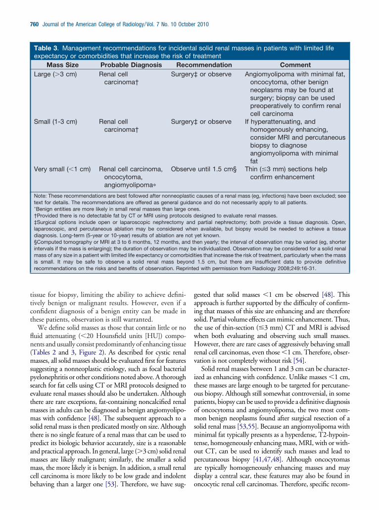
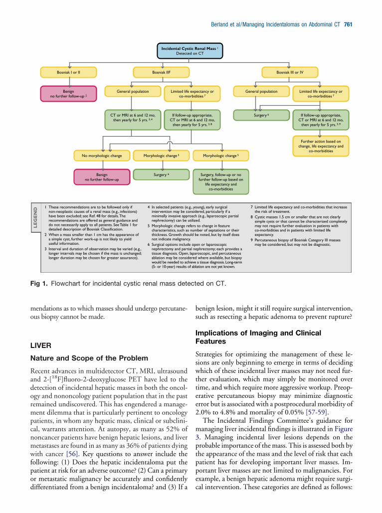
1 These recommendations are to be followed only if non-neoplastic causes of a renal mass (e.g., infections) have been excluded; see Ref. 48 for details. The recommendations are offered as general guidance and do not necessarily apply to all patients. See Table 1 for detailed description of Bosniak Classification.
2 When a mass smaller than 1 cm has the appearance of a simple cyst, further work-up is not likely to yield useful information.
3 Interval and duration of observation may be varied (e.g., longer intervals may be chosen if the mass is unchanged; longer duration may be chosen for greater assurance).
4 In selected patients (eintervention may be cominimally invasive apprnephrectomy) can be
5 Morphologic change rcharacteristics, such asthickness. Growth shonot indicate malignanc
6 Surgical options includnephrectomy and parttissue diagnosis. Openablation may be considwould be needed to ac(5- or 10-year) results o
LE
GE
ND
Incidental CysDetect
Bosniak IIF
No morphologic change
Surgery 6
Bosniak I or II
Benignno further follow-up 2
Limited lifeco-m
General population
If follow-uCT or MRI
then year
CT or MRI at 6 and 12 mo, then yearly for 5 yrs. 3, 4
Benignno further follow-up
Morphologic change 5
ig 1. Flowchart for incidental cystic renal mass dete
ifferentiated from a benign incidentaloma? and (3) If a c
enign lesion, might it still require surgical intervention,uch as resecting a hepatic adenoma to prevent rupture?
mplications of Imaging and Clinicaleatures
trategies for optimizing the management of these le-ions are only beginning to emerge in terms of decidinghich of these incidental liver masses may not need fur-
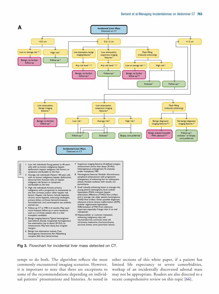
her evaluation, which may simply be monitored overime, and which require more aggressive workup. Preop-rative percutaneous biopsy may minimize diagnosticrror but is associated with a postprocedural morbidity of.0% to 4.8% and mortality of 0.05% [57-59].The Incidental Findings Committee’s guidance foranaging liver incidental findings is illustrated in Figure
. Managing incidental liver lesions depends on therobable importance of the mass. This is assessed both byhe appearance of the mass and the level of risk that eachatient has for developing important liver masses. Im-ortant liver masses are not limited to malignancies. Forxample, a benign hepatic adenoma might require surgi-
ung), early surgical ered, particularly if a (e.g., laparoscopic partial d.
to change in feature ber of septations or their e noted, but by itself does
en or laparoscopic phrectomy; each provides a roscopic, and percutaneous where available, but biopsy a tissue diagnosis. Long-term ation are not yet known.
7 Limited life expectancy and co-morbidities that increase the risk of treatment.
8 Cystic masses 1.5 cm or smaller that are not clearly simple cysts or that cannot be characterized completely may not require further evaluation in patients with co-morbidities and in patients with limited life expectancy.
9 Percutaneous biopsy of Bosniak Category III masses may be considered, but may not be diagnostic.
enal Mass 1 CT
Limited life expectancy or co-morbidities 7
General populationectancy or ities 7
Surgery 6
Further action based on change, life expectancy and
co-morbidities
Bosniak III or IV
propriate,and 12 mo, 5 yrs. 3, 8
If follow-up appropriate,CT or MRI at 6 and 12 mo,
then yearly for 5 yrs. 3, 9
Morphologic change 5
Surgery, follow-up or no further follow-up based on
life expectancy and co-morbidities
ed on CT.
.g., yonsidoach
utilizeefers numuld by.e opial ne, lapaered
hievef abl
tic Red on
exporbid
p ap at 6 ly for
al intervention. These categories are defined as follows:

1
2
3
A
N
Andara
6dnwmclgmettm
maltnalu
F te
762 Journal of the American College of Radiology/Vol. 7 No. 10 October 2010
. Low-risk individuals: Young patients (aged �40 years),with no malignancies, hepatic dysfunction, hepatic malig-nant risk factors, or symptoms attributable to the liver.
. Average-risk individuals: Patients aged �40 years, with noknown malignancies, hepatic dysfunction, hepatic malig-nant risk factors, or symptoms attributable to the liver.
. High-risk individuals: Patients with known primarymalignancies with a propensity to metastasize to theliver, cirrhosis, or other hepatic risk factors. Hepaticrisk factors include hepatitis, chronic active hepati-tis, sclerosing cholangitis, primary biliary cirrhosis,hemochromatosis, hemosiderosis, hepatic dysfunc-tion, and long-term oral contraceptive use.
DRENAL GLANDS
ature and Scope of the Problem
n incidental adrenal mass, often referred to as an adre-al incidentaloma, is defined as an adrenal mass (�1 cm)iscovered incidentally on a cross-sectional imaging ex-mination performed for another reason. Incidental ad-enal masses are very common, estimated to occur in
1 These recommendations are to be followed only if non-neoplastic causes of a renal mass (e.g., infections and fat-containing angiomyolipomas) have been excluded; see Ref. 48 for details. The recommendations are offered as general guidance and do not necessarily apply to all patients.
2 Differential diagnosis includes renal cell carcinoma, oncocytoma, angiomyolipoma. Benign entities are more likely in small renal masses than large ones.
3 Limited life expectancy and co-morbidities that increase the risk of treatment.
4 Interval and duration of observation may be varied (e.g., shorter interval if the mass is enlarging).
5 Probable diagnosis reis no detectable fat atdesigned to evaluate r
6 If hyperattenuating anconsider MRI and perangiomyolipoma with
7 Surgical options includnephrectomy and partissue diagnosis. Openablation may be consiwould be needed to aLong-term (5- or 10-yyet known.
LE
GE
ND
Incidental SoDetect
1-3<1 cm 2
General population
Sur
General population
Follow-up until 1 cm:CT or MRI at 3-6 mo and 12
mo, then yearly 4
Follow-up until 1.5 cm:CT or MRI at 3-6 mo and 12
mo, then yearly 4
Hyperattenuating, homogeneously enhancing:
consider MRI, biopsy 6
Limited life expectancy and co-morbidities 3
ig 2. Flowchart for incidental solid renal mass detec
pproximately 3% to 7% of the adult population [60- (
3]. The most frequent pathology for an incidentallyiscovered adrenal mass is a nonhyperfunctioning ade-oma [64]. It was shown in one study that the over-helming majority of incidentally discovered adrenalasses are benign in patients with no known malignan-
ies [65]. Statistics indicate that given the high preva-ence of nonhyperfunctioning adrenal adenomas in theeneral population, an incidentally discovered adrenalass in an oncology patient is most likely benign. How-
ver, the adrenal gland is also a common site for metas-ases and, somewhat less commonly, primary adrenalumors, including pheochromocytomas, aldosterono-as, and adrenal cortical carcinomas.The goal of imaging when an incidental adrenalass is discovered is to differentiate a benign “leave-
lone” mass (eg, nonhyperfunctioning tumor, myelo-ipoma, hemorrhage, cyst) from a mass that warrantsreatment (eg, metastasis, pheochromocytoma, adre-al cortical carcinoma). From an imaging perspective,n optimal algorithm should be used to diagnose botheave-alone masses and masses that need treatment,sing as few tests as possible. The adrenal flowchart
ll carcinoma, provided there or MRI using protocols masses. mogeneously enhancing, eous biopsy to diagnose
mal fat.en or laparoscopic ephrectomy; both provide a roscopic, and percutaneous
d where available, but biopsy e a tissue diagnosis.
results of ablation are not
8 Observation may be considered for a solid renal mass of any size in a patient with limited life expectancy or co-morbidities that increase the risk of treatment, particularly when the mass is small. It may be safe to observe a solid renal mass beyond 1.5 cm; however, there are insufficient data to provide definitive recommendations on the risks and benefits of observation. Thin (≤3 mm) sections help confirm enhancement.
9 Probable diagnosis renal cell carcinoma. Angiomyolipoma with minimal fat, oncocytoma, and other benign neoplasms may be found at surgery.
10 Percutaneous biopsy can be utilized preoperatively to confirm renal cell carcinoma.
enal Mass 1 CT
Surgery 7
Surgery 7, 10 Follow-up 8
Limited life expectancy or co-morbidities 3
General population
7 Follow-up 8
>3 cm 9
Limited life expectancy and co-morbidities 3
d on CT.
nal ce CT enal d hocutanminie op
tial n, lapaderechievear)
lid Red on
cm 5
gery
Figure 4) and recommendations described here at-
tcisu
olwm
F n C
Berland et al/Managing Incidentalomas on Abdominal CT 763
empt to do both. The algorithm reflects the mostommonly encountered imaging scenarios. However,t is important to note that there are exceptions toome of the recommendations depending on individ-
IncidentaDetect
0.5-
Follow-up 4
Follo
<0.5 cm
Low attenuation, benign imaging features 5
Low attsuspicio
feat
Low attenuation, benign imaging
features 5
Low attenuatsuspicious ima
features 6
Low or average risk 1, 2
Any risk level 1, 2, 3 Any risk
Low risk 1
Follow-up 4 Evaluate 7
Average ris
Benign, no further follow-up
Benign, no further follow-up 6
Benign, no further follow-up 6
High risk 3
A
B
1 Low risk individuals: Young patient (≤ 40 years old), with no known malignancy, hepatic dysfunction, hepatic malignant risk factors, or symptoms attributable to the liver.
2 Average risk individuals: Patient >40 years old, with no known malignancy, hepatic dysfunction, abnormal liver function tests or hepatic malignant risk factors or symptoms attributable to the liver.
3 High risk individuals: Known primary malignancy with a propensity to metastasize to the liver, cirrhosis, and/or other hepatic risk factors. Hepatic risk factors include hepatitis, chronic active hepatitis, sclerosing cholangitis, primary biliary cirrhosis, hemochromatosis, hemosiderosis, oral contraceptive use, anabolic steroid use.
4 Follow-up CT or MRI in 6 months. May need more frequent follow-up in some situations, such as a cirrhotic patient who is a liver transplant candidate.
5 Benign imaging features: Typical hemangioma (see below), sharply marginated, homogeneous low attenuation (up to about 20 HU), no enhancement. May have sharp, but irregular margins.
6 Benign low attenuation masses: Cyst, hemangioma, hamartoma, Von Meyenberg complex (bile duct hamartomas).
7 Suspicious imaging features: Ill-deenhancement (more than about 2heterogeneous, enlargement. To eprefer multiphasic MRI.
8 Hemangioma features: Nodular dperipheral enhancement with proenlargement of enhancing foci onphases. Nodule isodense with vesparenchyma.
9 Small robustly enhancing lesion inyoung patient: hemangioma, focal hyperplasia (FNH), transient hepaattenuation difference (THAD) fland in average risk, older patient:THAD flow artifact. Other possibadenoma, arterio-venous malformnodular regenerative hyperplasia.Differentiation of FNH from adenimportant especially if larger thansubcapsular.
10 Hepatocellular or common metaenhancing malignancy: islet cell, neuroendocrine, carcinoid, renal carcinoma, melanoma, choriocarcsarcoma, breast, some pancreatic
LE
GE
ND
Incidental Liver MassDetected on CT
ig 3. Flowchart for incidental liver mass detected o
al patients’ presentations and histories. As noted in r
ther sections of this white paper, if a patient hasimited life expectancy or severe comorbidities,orkup of an incidentally discovered adrenal massay not be appropriate. Readers are also directed to a
r Mass CT
>1.5 cm
4
tion, aging 7
Flash filling (robustly enhancing)
Benign diagnostic imaging features 8, 9
No benign diagnostic imaging features 10
Flash filling (robustly enhancing)
l 1, 2, 3 Low or average risk 1, 2 High risk 3
Biopsy, core preferredFollow-up 4,
evaluate 7 or biopsy, core preferred
High risk 3
Benign, evaluate if possible FNH, adenoma 8, 9
Evaluate 7 Follow-up 4
Benign, no further follow-up 8, 9
margins, ),
te,
tinuous ive equent not
age risk, lar
tifact, angioma, agnoses: (AVM),
and
a, ns.
T.
l Liveed on
1.5 cm
w-up
enuaus imures
ion, ging
leve
k 2
fined0 HUvalua
iscongress subssels,
avernodutic ow ar hemle diation
oma 4 cm
static
cell inom lesio
ecent comprehensive review on this topic [66].
IA
IlfmnHoeapi
e(mecno
eamw
ipumnhc
ddaUld1
F d o
764 Journal of the American College of Radiology/Vol. 7 No. 10 October 2010
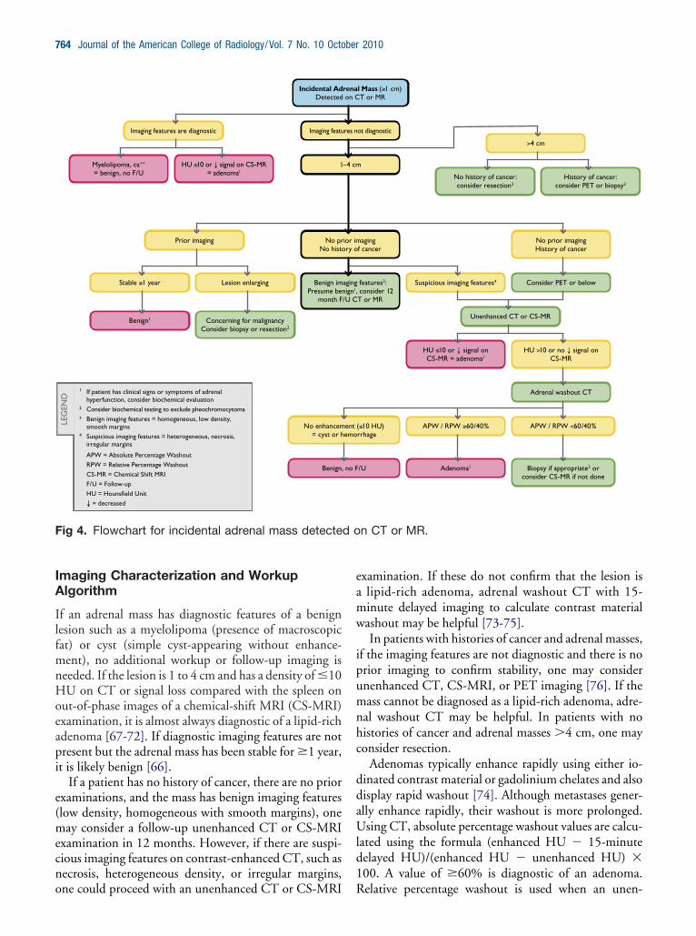
maging Characterization and Workuplgorithm
f an adrenal mass has diagnostic features of a benignesion such as a myelolipoma (presence of macroscopicat) or cyst (simple cyst-appearing without enhance-ent), no additional workup or follow-up imaging is
eeded. If the lesion is 1 to 4 cm and has a density of �10U on CT or signal loss compared with the spleen on
ut-of-phase images of a chemical-shift MRI (CS-MRI)xamination, it is almost always diagnostic of a lipid-richdenoma [67-72]. If diagnostic imaging features are notresent but the adrenal mass has been stable for �1 year,
t is likely benign [66].If a patient has no history of cancer, there are no prior
xaminations, and the mass has benign imaging featureslow density, homogeneous with smooth margins), oneay consider a follow-up unenhanced CT or CS-MRI
xamination in 12 months. However, if there are suspi-ious imaging features on contrast-enhanced CT, such asecrosis, heterogeneous density, or irregular margins,
1 If patient has clinical signs or symptoms of adrenal hyperfunction, consider biochemical evaluation
2 Consider biochemical testing to exclude pheochromocytoma3 Benign imaging features = homogeneous, low density,
smooth margins4 Suspicious imaging features = heterogeneous, necrosis,
irregular margins
APW = Absolute Percentage Washout
RPW = Relative Percentage Washout
CS-MR = Chemical Shift MRI
F/U = Follow-up
HU = Hounsfield Unit
↓ = decreased
LEG
END
Incidental AdDetected
Imaging featu
1
No histo
Benign imPresume be
month F
Prior imaging
Lesion enlarging
Consider biopsy or resection2
Imaging features are diagnostic
HU ≤10 or ↓ signal on CS-MR = adenoma1
Stable ≥1 year
Benign1
Myelolipoma, ca++
= benign, no F/U
No enhancem= cyst or h
Benign,
No pr
Concerning for malignancy
ig 4. Flowchart for incidental adrenal mass detecte
ne could proceed with an unenhanced CT or CS-MRI R
xamination. If these do not confirm that the lesion islipid-rich adenoma, adrenal washout CT with 15-inute delayed imaging to calculate contrast materialashout may be helpful [73-75].In patients with histories of cancer and adrenal masses,
f the imaging features are not diagnostic and there is norior imaging to confirm stability, one may considernenhanced CT, CS-MRI, or PET imaging [76]. If theass cannot be diagnosed as a lipid-rich adenoma, adre-
al washout CT may be helpful. In patients with noistories of cancer and adrenal masses �4 cm, one mayonsider resection.
Adenomas typically enhance rapidly using either io-inated contrast material or gadolinium chelates and alsoisplay rapid washout [74]. Although metastases gener-lly enhance rapidly, their washout is more prolonged.sing CT, absolute percentage washout values are calcu-
ated using the formula (enhanced HU � 15-minuteelayed HU)/(enhanced HU � unenhanced HU) �00. A value of �60% is diagnostic of an adenoma.
l Mass (≥1 cm)T or MR
ot diagnostic
f cancer
features3: consider 12 T or MR
No prior imagingHistory of cancer
Suspicious imaging features4
Unenhanced CT or CS-MR
HU >10 or no ↓ signal on CS-MR
Adrenal washout CT
APW / RPW ≥60/40%
Adenoma1
>4 cm
consider resection2History of cancer:
consider PET or biopsy2
Consider PET or below
HU ≤10 or ↓ signal on CS-MR = adenoma1
APW / RPW <60/40%
Biopsy if appropriate2
consider CS-MR if not done
( 10 HU)≤rrhage
/U
aging
No history of cancer:
or
n CT or MR.
rena on C
res n
−4 cm
ry o
agingnign1,/U C
ent emo
no F
ior im
elative percentage washout is used when an unen-
haph1[dai
CarlapmHmaacfe
atfopfrf[rrwwsa
P
N
TnM1af
niw
tcmctma3I5s
np[iuanma
D
Tintlwcucf
a“aWntSacmHow
tdapg
Berland et al/Managing Incidentalomas on Abdominal CT 765
anced CT value is not available and the enhanced valuesre compared with 15-minute delayed scans. Relativeercentage washout is calculated using the formula (en-anced HU � 15-minute delayed HU)/enhanced HU �00; a value of �40% is diagnostic for an adenoma73-75]. Adrenal washout CT was used successfully toistinguish adenomas from nonadenomas in 160 of 166drenal masses with 98% sensitivity and 92% specific-ty [73].
Recent advances in imaging characterization withT, MRI, and PET have decreased the need for im-
ge-guided percutaneous biopsies to characterize ad-enal masses [77]. However, if an adrenal mass is en-arging, it may be prudent to proceed to percutaneousdrenal biopsy or surgical resection. In an oncologyatient, a new adrenal mass in a patient with knownetastases elsewhere is most likely another metastasis.owever, an isolated adrenal mass could be benign oralignant. If the mass cannot be characterized as an
denoma using CT, MRI, or PET, a biopsy may beppropriate. If there are signs or symptoms of pheo-hromocytoma, it may be prudent to obtain plasma-ractionated metanephrine and normetanephrine lev-ls before biopsy [78].
Imaging examinations are useful to separate adrenaldenomas from other masses but cannot be used to dis-inguish hyperfunctioning adenomas from nonhyper-unctioning adenomas. One approach would be to relyn history and physical examination to determine whichatients should undergo biochemical testing for hyper-unctioning adrenal neoplasms. Some endocrinologistsecommend excluding an occult, asymptomatic hyper-unctioning neoplasm in all adrenal incidentalomas60,62,63]. This approach would be costly and is notoutinely performed by many physicians. Regarding theadiology report, when an adenoma can be diagnosedith imaging, we suggest stating, “Findings consistentith a benign adenoma. If there are clinical signs or
ymptoms of adrenal hyperfunction, biochemical evalu-tion may be appropriate.”
ANCREAS
ature and Scope of the Problem
he frequency of detection of pancreatic cysts by CT scan-ing is reported between 1.2% [79] and 2.6% [80]. ForRI, the reported frequency is significantly higher, at
9.9% of MRI examinations [81]. Because pancreatic cystsre quite prevalent, a practicing radiologist may see severalor every 100 abdominal imaging cases performed.
Cystic pancreatic tumors are most often frankly be-ign or low-grade indolent neoplasms. In one study that
ncluded asymptomatic patients with pancreatic cysts in
hom there was operative correlation, 17% of asymp- tomatic cysts were serous cystadenomas, 28% were mu-inous cystic neoplasms, 27% were intraductal papillaryucinous neoplasms (IPMNs), 2.5% were ductal adeno-
arcinomas, and 3.8% were pseudocysts [82]. Intraduc-al papillary mucinous neoplasms were the most com-on cystic neoplasm when both symptomatic and
symptomatic patients were evaluated. In another series,9% of IPMNs were incidentally detected, and 50% ofPMNs were side branch or branch duct IPMNs with a-year risk for developing high-grade dysplasia or inva-ive carcinoma of 15% [83].
Mucinous cystic masses, namely IPMNs and muci-ous cystic neoplasms, have a well-established malignantotential likened to an adenoma-carcinoma sequence84]. Because of this malignant potential, it has becomencreasingly difficult for radiologists evaluating individ-al cases to know how to frame the report to help guideppropriate management. We believe that the guida-ce below will help in the evaluation and reporting of theajority of these lesions. These recommendations are
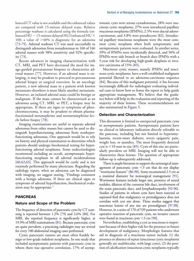
lso summarized in Figure 5.
etection and Characterization
his discussion is limited to unexpected pancreatic cystsn asymptomatic patients. Asymptomatic patients haveo clinical or laboratory indication directly referable tohe pancreas, including but not limited to hyperamy-asemia, recent-onset diabetes, severe epigastric pain,eight loss, or jaundice. The most frequently detected
yst is �10 mm in size [85]. Cysts of this size are partic-larly prevalent on MRI. Imaging will not be able toharacterize these lesions. The question of appropriateollow-up is subsequently addressed.
There is ample literature to support the nonsurgical man-gement of pancreatic cysts �3 cm that do not displayworrisome features” [86-90]. Some recommend 2.5 cm asmaximal diameter for nonsurgical management [91].orrisome features include larger size, presence of mural
odules, dilation of the common bile duct, involvement ofhe main pancreatic duct, and lymphadenopathy [92-96].tudies of patients in whom cysts have been resected orspirated find that malignancy or premalignancy does notorrelate with cyst size alone. These studies suggest thatucinous lesions of any size are premalignant [97,98].owever, in a series of 170 of 539 patients who underwent
perative resection of pancreatic cysts, no invasive cancersere found in mucinous cysts �3 cm [90].Nevertheless, establishing a cyst as mucinous is impor-
ant because of their higher risk for the presence or futureevelopment of malignancy. Morphologic features thatid in diagnosis of a mucinous tumor include (1) theresence or absence of septae (mucinous cystic neoplasmsenerally are multilocular, with large cysts), (2) the posi-
ion of calcification (mucinous cystic neoplasms typically
hcampaommbtcntct
tpcad(
aap
IF
Miin“rbda
1lsoae
F( �
766 Journal of the American College of Radiology/Vol. 7 No. 10 October 2010
ave peripheral calcification, whereas serous tumors haveentral calcification), (3) location within the pancreas,nd (4) the presence of main pancreatic duct involve-ent [99-102]. Mucinous cystic tumors can be sus-
ected when a cyst is present in the tail of the pancreas inperimenopausal woman [84]. The presence or absencef direct communication with the main pancreatic ductust be established to distinguish a mucinous cystic tu-or (with relatively high malignant potential) from a
ranch duct IPMN (with relatively low malignant poten-ial). Three-dimensional imaging with either MRI or CTan address this question. Conversely, serous cystade-oma characteristically displays variably dense radial sep-ae in a honeycombed or spongiform pattern and centralalcification. The more peripheral cysts are larger thanhe more central cysts.
A simple but useful imaging-based classification sys-em differentiates pancreatic cystic masses into 4 mor-hologic types: (1) unilocular (pseudocysts, mucinousystic neoplasms, lymphoepithelial cysts, small IPMNs,nd small serous tumors), (2) microcystic (serous cysta-enomas and lymphoepithelial cysts), (3) macrocystic
<2 cm
Stable Growth Uncharacterized cystic mass
B
Single follow-up in 1 yr, preferably MRI 2
Follow-up yearly Follo6 mo
Imaging cpreferab
Asymptomatic 1 Patient with Detected on CT, MRI (w
Benign, no further follow-up
ig 5. Flow chart for an asymptomatic patient with anwith or without contrast), or ultrasound (US). MRCP
mucinous cystic neoplasms, oligocystic serous tumors, r
nd IPMNs) and (4) cysts with solid components (solid-ppearing serous tumors, solid pseudopapillary neo-lasms, and cystic islet cell tumors) [103].
mplications of Imaging and Clinicaleatures
ost incidental cysts can be detected on routine abdom-nal studies. However, if a cyst needs to be characterized,t is recommended that a diagnosis of a specific cyst typeot be made unless the patient undergoes a dedicatedpancreas-style” study. For multidetector CT, this wouldequire a dual-phase contrast-enhanced acquisition inoth pancreatic and portal venous phases using a narrowetector configuration. Thin-section images should bevailable on a workstation that can perform 3-D analysis.
Magnetic resonance imaging should be performed at.5 T. Phased-array torso coils enhance signal and paral-
el imaging increases speed and improves resolution. Thetudy should include sequences that display in-phase andut-of-phase T1, T2 (preferably with fat suppression),nd 3-D, fat-saturated, gradient-echo T1 gadolinium-nhanced sequences in pancreatic, portal, and equilib-
1 Signs and symptoms include hyperamylasemia, recent onset diabetes, severe epigastric pain, weight loss, steatorrhea or jaundice.
2 Consider decreasing interval if younger, omitting with limited life expectancy. Recommend limited T2-weighted MRI for routine follow-ups.
3 Recommend pancreas-dedicated MRI with MRCP.
4 If no growth after 2 years, follow yearly. If growth OR suspicious features develop, consider resection.
5 BD-IPMN = branch duct intraductal papillary mucinous neoplasm.
LE
GE
ND
m >3 cm
N Serous cystadenoma
Serous cystadenoma Uncharacterized cystic mass or other
cystic neoplasm
every years 4
Follow-up every 2 yr
Consider resection when ≥ 4 cm
Cyst aspiration
Resect, depending on co-morbidities
and risk
terization, I/MRCP 3
ental Pancreatic Cystic Mass without contrast) or US
cidental pancreatic cystic mass detected on CT, MRIMR cholangiopancreatography.
2-3 c
D-IPM
w-upfor 2
haracly MR
Incidith or
in
ium phases. Additionally, MR cholangiopancreatogra-
ptpipFacMd
vcvtgbaeasnppthn[la
iaonpm
ctiIgmtsudoOcsho
pfa8ptofc([t
stdcmiwa
imIbstdowidi
lSdc1ssawrr1ymym1t
Berland et al/Managing Incidentalomas on Abdominal CT 767
hy is necessary. Current MRI scanners have respiratoryriggered 3-D sequences for MR cholangiopancreatogra-hy [104]. Secretin administration may facilitate visual-
zation of the communication of a cyst with the mainancreatic duct [105]. By consensus, the Incidentalindings Committee suggests dedicated MRI as the im-ging procedure of choice to characterize a pancreaticyst. This reflects the superior contrast resolution of
RI, facilitating the recognition of septae, nodules, anduct communication [106].The pretest likelihood that a given lesion in an indi-
idual patient is a malignant neoplasm is of paramountonsideration when deciding on management. Contro-ersy exists between using dedicated imaging or an at-empt at aspiration of a cyst under endoscopic ultrasounduidance. Most often, this decision will be made on theasis of the size of the cyst, location within the pancreas,ccessibility to the endoscopic ultrasound approach, andxpertise of the endosonographer. A carcinoembryonicntigen level in the aspirate of 192 ng/mL has a highpecificity for discriminating mucinous from nonmuci-ous cysts, demonstrating higher accuracy than cyst mor-hology [107]. Amylase levels of �250 U/L excludeseudocysts. There is a high degree of overlap betweenhe values obtained at aspiration [108]. Recent reportsave documented the development of ductal adenocarci-oma in a remote site in the pancreas from an IPMN109,110]. Many believe that the presence of a mucinousesion is a signal of increased risk for pancreatic neoplasmnywhere within the gland.
The consensus of the Incidental Findings Committees that if surgery is contemplated, aspiration of a pancre-tic cyst �3 cm should be attempted. It is a widely heldpinion, shared by this committee, that cysts �1.5 cmeed not be immediately characterized, whereas it is ap-ropriate to characterize other cysts, depending on co-orbid conditions and life expectancy.Imaging surveillance of pancreatic cystic neoplasms is
ontroversial. However, emerging consensus suggestshat selective nonoperative management in patients withncidental pancreatic cysts is appropriate [96,111,112].n a series of 369 of 539 patients with a mean radio-raphic follow-up period of 24 months (range, 1-172onths), 8% developed changes that prompted resec-
ion. Malignancies were present in 38% [90]. In a retro-pective case series of 79 patients with long-term follow-p, either 5 years by imaging or 8 years clinically,iagnosed with small (�2 cm), simple pancreatic cystsn sonography or CT from 1985 to 1996 were reviewed.f the 22 patients with radiologic follow-up, 59% had
ysts that remained unchanged or became smaller (meanize, 8 mm; mean follow-up period, 9 years), and 41%ad cysts that enlarged, from a mean of 14 mm to a mean
f 26 mm (mean follow-up period, 8 years). Of the 27 catients with clinical or questionnaire follow-up (meanollow-up period, 10 years), none developed symptom-tic pancreatic disease. Twenty-three percent died withinyears without adequate radiologic follow-up, none of
ancreas-related causes [113]. Another series of 90 pa-ients with incidental cysts with a mean follow-up periodf 48 months revealed malignancy in 1 patient 7 yearsrom diagnosis [86]. The frequency of cancer in surgi-ally resected cysts �3 cm has been reported as 19%including symptomatic and asymptomatic patients)114], but when only truly incidental cysts are evaluated,he frequency is reported as only 3.5% [82].
A follow-up examination must clearly establish thetability of a cyst. Therefore, patients should be advisedo undergo serial imaging at facilities with protocols foredicated pancreatic imaging. Although there is no clearonsensus among pancreatic experts regarding the opti-al imaging test for follow-up of pancreatic cysts, a lim-
ted MRI examination relying exclusively on T2-eighted unenhanced acquisitions has been proposed aspractical follow-up strategy [115].Careful evaluation of the imaging findings is directed at
nspecting the lesion for changes in the thickness of the wall,ural irregularities, or frank solid nodules. For branch duct
PMNs, the adjacent main pancreatic duct diameter shoulde recorded. The lesion should be carefully measured withlice number and series appearing in the report, and elec-ronic calipers should be placed on the exact image used toetermine the diameters. Currently, there is no consensusn defining what increment of growth is important. It isell known that the precision of manual measurement is
nversely related to the lesion diameter. Thus, it may beifficult to determine if the reported growth of a small lesion
s true growth or measurement error.As of this writing, there is no universally accepted fol-
ow-up protocol. Most proposed programs are based on theendai criteria that arose from a consensus conference ad-ressing the management and follow-up of mucinous pan-reatic cysts. Cysts �1 cm are followed yearly, cysts betweenand 3 cm are sent for further imaging (endoscopic ultra-
ound or MRI) looking for septae and mural nodules, andimple cysts are followed at 6-month intervals for 2 yearsnd then yearly. If they grow above 3 cm or develop anyorrisome features, patients are considered candidates for
esection [88]. In contradistinction, a recommendation de-ived from reviewing 166 cysts with a mean size of 2 cm in50 patients revealed that 89% showed no growth over 2ears. The only predictor of cyst growth was the presence ofural nodules. This study suggested no follow-up until 2
ears after detection [85]. In the Incidental Findings Com-ittee’s recommendations, cysts �2 cm may be followed at
-year intervals, and if there is no growth, follow-up ceases ifhe patient remains asymptomatic. A cyst that is �3 cm is
onsidered a surgical lesion unless it is a serous cystadenoma
oAla
hT�spcipti
S
Tf
1
2
3
SL
Baaooti
dcdaf
K
ThkahoiFdc
btuabnsclwfim
cswpttnasntbita
fr
1
768 Journal of the American College of Radiology/Vol. 7 No. 10 October 2010
r if patient comorbidities preclude benefit from resection.cyst between 2 and 3 cm may be characterized and fol-
owed semiannually if mucinous, yearly if uncharacterized,nd every 2 years if it is a serous cystadenoma.
Serous cystadenoma is a benign lesion. However, studiesave clearly documented that these lesions may grow.herefore, some recommend resecting serous cystadenomas4 cm regardless of the presence of symptoms [116], or in
ymptomatic patients regardless of size [90]. Solid pseudo-apillary epithelial neoplasm is a low-grade malignancy thatan present with cystic-appearing components. The major-ty are found in young women. They frequently containeripheral calcification and variable content (most charac-eristically hemorrhages) within the cysts. Solid pseudopap-llary epithelial neoplasm lesions should undergo resection.
ummary
he Incidental Findings Committee recommends theollowing for managing incidental pancreatic cysts:
. Surgery should be considered for patients with cysts�3 cm.a. If the lesion is a serous cystadenoma, surgery is
deferred until the cyst is �4 cm.b. Solid pseudopapillary epithelial neoplasm tumors
should be resected.c. Patient factors ultimately determine the appropri-
ateness of surgical treatment.
. Patients with simple (not containing any solid ele-ments) cysts �3 cm can be followed.a. Attempts should be made to characterize all cysts
�2 cm at the time of detection. Magnetic reso-nance imaging is the imaging procedure of choice.
b. Cyst aspiration is strongly advised before any surgeryis undertaken in a patient with a cyst of this size.
c. Cysts �2 cm can be followed less frequently thanthose between 2 and 3 cm.
d. Avoid characterizing cysts �1.5 to 2 cm unlessabsolutely characteristic.
. The presence of symptoms is a critical factor in decid-ing appropriate therapy.a. The frequency of malignancy in small cysts is sig-
nificantly higher in symptomatic patients.
PECIAL CONSIDERATIONS FOROW-DOSE UNENHANCED CT
ecause of the advent of screening CT examinations suchs CT colonography and heightened concern about radi-tion exposure, low-dose unenhanced CT examinationsf the abdomen are increasing in use. The managementf incidental findings discovered either on such examina-ions or on conventional-dose unenhanced examinations
s controversial, and there are different challenges. Low-ose techniques will increase image noise but should nothange the mean HU values to determine adrenal massensity. The following sections describe organ-specificpproaches for these types of examinations that may varyrom those described above.
idneys
he management of a renal mass detected on an unen-anced CT scan is controversial. To the best of ournowledge, no studies have addressed how best to man-ge non-fat-containing renal masses detected with unen-anced CT, and thus, these recommendations reflect ourpinions on the basis of our experience and understand-ng of the prevalence and natural history of such findings.urthermore, other than angiomyolipomas, renal massesetected incidentally on unenhanced CT scans oftenannot be accurately characterized.
Our experience suggests that when a renal mass seems toe a simple cyst on an unenhanced CT scan, the chance thathe mass is benign is extremely high. However, careful eval-ation of the mass’s features is important. To be consideredprobable simple cyst on unenhanced CT, the mass shoulde well marginated, contain contents that are homoge-eous, and water attenuation (0-20 HU), and display noepta, wall thickening, calcification (unless minimal, thinalcification within the wall), or nodularity. If any of theseatter features is present, a renal mass–protocol CT or MRIould be needed to diagnose the mass with complete con-dence. Sonography may be helpful, but in some cases itay not be definitive.To our knowledge, no studies in the literature have spe-
ifically addressed the likelihood of cancers in lesions thateem to be simple cysts on unenhanced CT. Furthermore,hen low-dose CT techniques are used, nonsimple (andotentially malignant) features that otherwise would be de-ected with standard-dose CT may not be detectable. As aheoretical example, the heterogeneity of a renal cell carci-oma may be incorrectly attributed to noise of low-dose CTnd undergo no further evaluation or follow-up. Also, someimple cysts may not appear homogeneous, because ofoise, so differentiating heterogeneity sometimes encoun-ered on low-dose CT from a heterogeneous solid mass maye difficult. Hence, although the possibility of misinterpret-ng a renal cancer as a simple cyst exists, it is well understoodhat the technical factors used to perform an examinationffect sensitivity and specificity.
The Incidental Findings Committee recommends theollowing for low-dose unenhanced CT examinations forenal masses:
. It may be appropriate to interpret incidental renalmasses as simple cysts unless suspicious features notedabove are convincingly present. The argument for
adopting this approach is even stronger when consid-
2
L
Ttr
fl
1
2
A
Tdoshctco
fa
1
2
3
P
T(tMaba
F
Trscmterere
C
Igofifpafl
ddtoiawoif
A
Te
Berland et al/Managing Incidentalomas on Abdominal CT 769
ering small (�3 cm) masses, particularly those �1cm. The smaller the mass (even when solid), the morelikely it is benign. Furthermore, masses �1 cm maynot be able to be fully characterized, even if renalmass–protocol CT or MRI was performed. Althoughthis represents a consensus opinion of the committee,no data are yet available to support this approach.
. If a renal mass is small (�3 cm), homogeneous, and�70 HU, recent data suggest that the mass can beconfidently diagnosed as a benign hyperattenuatingcyst (Bosniak category II) [43].
iver
he recommendations in the flowchart in Figure 3 applyo low-dose unenhanced procedures as well as standard–adiation dose enhanced examinations.
The Incidental Findings Committee recommends theollowing for low-dose unenhanced CT examinations foriver masses:
. In low-risk and average-risk patients, sharply margin-ated, low-attenuation (�20 HU) solitary or multiplemasses may typically not need further evaluation.
. Small, solitary masses �1.5 cm that are not cystic andare discovered on unenhanced or standard-dose orlow-dose scans in low-risk and average-risk patientsmay typically not need further evaluation.
drenal Glands
he low-dose unenhanced technique is less sensitive foretermining the internal architecture and heterogeneityf an adrenal mass than contrast-enhanced CT with atandard radiation exposure. We are not aware of anyelpful literature addressing the topic of adrenal massharacterization on low-dose unenhanced CT examina-ions. Therefore, these recommendations represent theonsensus opinion on the basis of the clinical experiencef the committee members.
The Incidental Findings Committee recommends theollowing for low-dose unenhanced CT examinations fordrenal masses:
. Because attenuation should not be altered by a low-dose technique, if the mean attenuation of an adrenalmass is �10 HU on a low-dose CT examination, onemay conclude that the adrenal mass is likely to be abenign adenoma.
. If a lesion is �10 HU and 1 to 4 cm in an asymptom-atic patient without cancer, 1-year follow-up CT orMRI may be considered, if no prior studies for com-parison are available. Prior examinations that showstability for �1 year can eliminate the need for furtherworkup, so every effort should be made to obtain
prior CT or MRI examinations in these situations. m. For adrenal masses �4 cm, dedicated adrenal MRI orCT should be considered to further characterize.
ancreas
he recommendations shown in the pancreatic flowchartFigure 5) also apply to low-dose unenhanced CT examina-ions. The importance of comparison with prior CT or
RI examinations cannot be overemphasized to potentiallyvoid further workup. Specifically, for lesions �2 cm, sta-ility over �1 year is highly suggestive of a benign lesionnd may eliminate the need for follow-up imaging.
UTURE COMMITTEE OBJECTIVES
he Incidental Findings Committee hopes that theseecommendations will become widely applied and willearch for additional methods to disseminate them. Theommittee also expects to refine and adapt these recom-endations and to develop additional guidance for other
ypes of incidental findings. To advance the scientificvidence regarding incidental findings, the committeeecommends that the concepts, terminology, and param-ters discussed in this paper become the basis for futureesearch, to help the results of such research be moreasily applied within a common framework.
ONCLUSIONS
ncidental findings on imaging during daily practice haverown in number related to the rapid increase in the utilizationf CT and to its improved image quality. These incidentalndingspotentially lead to increasedrisk to thepatientandcostrom additional procedures. Underscoring concern amonghysicians is the fear that failure to report incidental findingsnd recommend follow-up will place radiologists in jeopardyor malpractice litigation, should a lesion eventually lead to aife-threatening health problem.
This effort within the ACR, conducted by the Inci-ental Findings Committee, attempts to systematicallyescribe a variety of the most common potential inciden-al findings on abdominal CT and provide detailed rec-mmendations to assist practicing radiologists in makingnformed decisions about reporting such masses anddvising their referring clinicians and patients abouthether and how these should be managed. This is partf an ongoing project to develop, refine, and disseminatenformation about incidental findings and to collect in-ormation and support research about them.
CKNOWLEDGMENTS
o N. Reed Dunnick, MD, who supported and helpedstablish the Committee within the Body Imaging Com-
ission of the ACR and to David Kurth of the ACR staff,
wt
R
770 Journal of the American College of Radiology/Vol. 7 No. 10 October 2010
ho helped support and coordinate the activities that ledo this manuscript.
EFERENCES
1. Pickhardt PJ, Hanson ME, Vanness DJ, et al. Unsuspected extracolonicfindings at screening CT colonography: clinical and economic impact.Radiology 2008;249:151-9.
2. Bovio S, Cataldi A, Reimondo G, et al. Prevalence of adrenal incidenta-loma in a contemporary computerized tomography series. J EndocrinolInvest 2006;29:298-302.
3. Wagner SC, Morrison WB, Carrino JA, Schweitzer ME, Nothnagel H.Picture archiving and communication system: effect on reporting ofincidental findings. Radiology 2002;225:500-5.
4. Yee J, Kumar NN, Godara S, et al. Extracolonic abnormalities discov-ered incidentally at CT colonography in a male population. Radiology2005;236:519-26.
5. Incidentaloma. Available at: http://medical-dictionary.thefreedictionary.com/Incidental�finding. Accessed May 20, 2010.
6. Parker LS. The future of incidental findings: should they be viewed asbenefits? J Law Med Ethics 2008;36:341-51.
7. Fletcher RH, Pignone M. Extracolonic findings with computed tomo-graphic colonography: asset or liability? Arch Intern Med 2008;168:685-6.
8. Centers for Medicare and Medicaid Services. Decision memo for screen-ing computed tomography colonography (CTC) for colorectal cancer(CAG-00396N). Baltimore, Md: Centers for Medicare and MedicaidServices.
9. Berland LL. Incidental extracolonic findings on CT colonography: theimpending deluge and its implications. J Am Coll Radiol 2009;6:14-20.
10. Hara AK, Johnson CD, MacCarty RL, Welch TJ. Incidental extraco-lonic findings at CT colonography. Radiology 2000;215:353-7.
11. Hassan C, Pickhardt PJ, Laghi A, et al. Computed tomographiccolonography to screen for colorectal cancer, extracolonic cancer, andaortic aneurysm: model simulation with cost-effectiveness analysis. ArchIntern Med 2008;168:696-705.
12. Hellstrom M, Svensson MH, Lasson A. Extracolonic and incidentalfindings on CT colonography (virtual colonoscopy). AJR Am J Roent-genol 2004;182:631-8.
13. Xiong T, McEvoy K, Morton DG, Halligan S, Lilford RJ. Resources andcosts associated with incidental extracolonic findings from CT colonogra-phy: a study in a symptomatic population. Br J Radiol 2006;79:948-61.
14. Xiong T, Richardson M, Woodroffe R, Halligan S, Morton D, LilfordRJ. Incidental lesions found on CT colonography: their nature andfrequency. Br J Radiol 2005;78:22-9.
15. Casarella WJ. A patient’s viewpoint on a current controversy. Radiology2002;224:927.
16. MacMahon H, Austin JH, Gamsu G, et al. Guidelines for managementof small pulmonary nodules detected on CT scans: a statement from theFleischner Society. Radiology 2005;237:395-400.
17. Frates MC, Benson CB, Charboneau JW, et al. Management of thyroidnodules detected at US: Society of Radiologists in Ultrasound consensusconference statement. Radiology 2005;237:794-800.
18. Grant EG, Benson CB, Moneta GL, et al. Carotid artery stenosis: gray-scale and Doppler US diagnosis—Society of Radiologists in UltrasoundConsensus Conference. Radiology 2003;229:340-6.
19. D’Orsi CJ, Bassett LW, Berg WA, et al. Breast Imaging Reporting andData System: ACR BI-RADS-Mammography. 4th ed. Reston, Va:
American College of Radiology; 2003.20. Zalis ME, Barish MA, Choi JR, et al. CT colonography reporting anddata system: a consensus proposal. Radiology 2005;236:3-9.
21. Elstein AS. On the origins and development of evidence-based medicineand medical decision making. Inflamm Res 2004;53(suppl):S184-9.
22. Wolf SM, Lawrenz FP, Nelson CA, et al. Managing incidental findingsin human subjects research: analysis and recommendations. J Law MedEthics 2008;36:219-48.
23. Birnbaum BA, Bosniak MA, Megibow AJ, Lubat E, Gordon RB. Observa-tions on the growth of renal neoplasms. Radiology 1990;176:695-701.
24. Birnbaum BA, Hindman N, Lee J, Babb JS. Renal cyst pseudoenhance-ment: influence of multidetector CT reconstruction algorithm and scan-ner type in phantom model. Radiology 2007;244:767-75.
25. Bosniak M. Problematic renal masses. In: RSNA categorical course indiagnostic radiology: genitourinary radiology. Oak Brook, Ill: Radiolog-ical Society of North America; 1994:183-91.
26. Bosniak MA. The current radiological approach to renal cysts. Radiol-ogy 1986;158:1-10.
27. Bosniak MA. The small (less than or equal to 3.0 cm) renal parenchymaltumor: detection, diagnosis, and controversies. Radiology 1991;179:307-17.
28. Bosniak MA. Diagnosis and management of patients with complicatedcystic lesions of the kidney. AJR Am J Roentgenol 1997;169:819-21.
29. Bosniak MA. Should we biopsy complex cystic renal masses (Bosniakcategory III)? AJR Am J Roentgenol 2003;181:1425-6.
30. Bosniak MA, Birnbaum BA, Krinsky GA, Waisman J. Small renal pa-renchymal neoplasms: further observations on growth. Radiology 1995;197:589-97.
31. Bosniak MA, Megibow AJ, Hulnick DH, Horii S, Raghavendra BN. CTdiagnosis of renal angiomyolipoma: the importance of detecting smallamounts of fat. AJR Am J Roentgenol 1988;151:497-501.
32. Bosniak MA, Rofsky NM. Problems in the detection and characteriza-tion of small renal masses. Radiology 1996;198:638-41.
33. Curry NS, Cochran ST, Bissada NK. Cystic renal masses: accurateBosniak classification requires adequate renal CT. AJR Am J Roentgenol2000;175:339-42.
34. Harisinghani MG, Maher MM, Gervais DA, et al. Incidence of malig-nancy in complex cystic renal masses (Bosniak category III): shouldimaging-guided biopsy precede surgery? AJR Am J Roentgenol 2003;180:755-8.
35. Hartman DS, Weatherby E III, Laskin WB, Brody JM, Corse W, BaluchJD. Cystic renal cell carcinoma: CT findings simulating a benign hyper-dense cyst. AJR Am J Roentgenol 1992;159:1235-7.
36. Israel GM, Bosniak MA. Follow-up CT of moderately complex cysticlesions of the kidney (Bosniak category IIF). AJR Am J Roentgenol2003;181:627-33.
37. Israel GM, Bosniak MA. Calcification in cystic renal masses: is it impor-tant in diagnosis? Radiology 2003;226:47-52.
38. Israel GM, Bosniak MA. How I do it: evaluating renal masses. Radiology2005;236:441-50.
39. Israel GM, Hindman N, Bosniak MA. Evaluation of cystic renal masses:comparison of CT and MR imaging by using the Bosniak classificationsystem. Radiology 2004;231:365-71.
40. Israel GM, Hindman N, Hecht E, Krinsky G. The use of opposed-phasechemical shift MRI in the diagnosis of renal angiomyolipomas. AJRAm J Roentgenol 2005;184:1868-72.
41. Jinzaki M, McTavish JD, Zou KH, Judy PF, Silverman SG. Evaluationof small (� 3 cm) renal masses with MDCT: benefits of thin overlapping
reconstructions. AJR Am J Roentgenol 2004;183:223-8.
Berland et al/Managing Incidentalomas on Abdominal CT 771
42. Jinzaki M, Tanimoto A, Narimatsu Y, et al. Angiomyolipoma: imagingfindings in lesions with minimal fat. Radiology 1997;205:497-502.
43. Jonisch AI, Rubinowitz AN, Mutalik PG, Israel GM. Can high-attenu-ation renal cysts be differentiated from renal cell carcinoma at unen-hanced CT? Radiology 2007;243:445-50.
44. Kim JK, Park SY, Shon JH, Cho KS. Angiomyolipoma with minimalfat: differentiation from renal cell carcinoma at biphasic helical CT.Radiology 2004;230:677-84.
45. Silverman S, Gan, YU, Mortele, KJ, Tuncali, K, Cibas, ES. Renal massbiopsy in the new millennium: an important diagnostic procedure. In:RSNA categorical course in diagnostic radiology: genitourinary radiol-ogy. Chicago, Ill: Radiological Society of North America; 2006:219-36.
46. Silverman SG, Gan YU, Mortele KJ, Tuncali K, Cibas ES. Renal massesin the adult patient: the role of percutaneous biopsy. Radiology 2006;240:6-22.
47. Silverman SG, Mortele KJ, Tuncali K, Jinzaki M, Cibas ES. Hyperat-tenuating renal masses: etiologies, pathogenesis, and imaging evaluation.Radiographics 2007;27:1131-43.
48. Silverman SG, Israel GM, Herts BR, Richie JP. Management of theincidental renal mass. Radiology 2008;249:16-31.
49. Campbell SC, Novick AC, Belldegrun A, et al. Guideline for manage-ment of the clinical T1 renal mass. J Urol 2009;182:1271-9.
50. American Urological Association. Guideline for management of theclinical stage 1 renal mass. Available at: http://www.auanet.org/content/media/renalmass09.pdf?CFID�2980781&CFTOKEN�53338937&jsessionid�84305943038372e237f85a47377557304155. AccessedMay 15, 2010.
51. Kassouf W, Aprikian AG, Laplante M, Tanguay S. Natural history ofrenal masses followed expectantly. J Urol 2004;171:111-3.
52. Kunkle DA, Crispen PL, Chen DY, Greenberg RE, Uzzo RG. Enhanc-ing renal masses with zero net growth during active surveillance. J Urol2007;177:849-53.
53. Thompson RH, Kurta JM, Kaag M, et al. Tumor size is associated withmalignant potential in renal cell carcinoma cases. J Urol 2009;181:2033-6.
54. Nguyen MM, Gill IS. Effect of renal cancer size on the prevalence ofmetastasis at diagnosis and mortality. J Urol 2009;181:1020-7.
55. Frank I, Blute ML, Cheville JC, Lohse CM, Weaver AL, Zincke H. Solidrenal tumors: an analysis of pathological features related to tumor size.J Urol 2003;170:2217-20.
56. Washington K. Masses of the liver. In: Odze R, Goldblum JR, eds.Surgical pathology of the GI tract, liver, biliary tract and pancreas. 2nded. New York: Elsevier; 2009:657-789.
57. Padia SA, Baker ME, Schaeffer CJ, et al. Safety and efficacy of sono-graphic-guided random real-time core needle biopsy of the liver. J ClinUltrasound 2009;37:138-43.
58. Appelbaum L, Kane RA, Kruskal JB, Romero J, Sosna J. Focal hepaticlesions: US-guided biopsy-lessons from review of cytologic and patho-logic examination results. Radiology 2009;250:453-8.
59. Whitmire LF, Galambos JT, Phillips VM, et al. Imaging guided percu-taneous hepatic biopsy: diagnostic accuracy and safety. J Clin Gastroen-terol 1985;7:511-5.
60. NIH state-of-the-science statement on management of the clinicallyinapparent adrenal mass (“incidentaloma”). NIH Consens State SciStatements 2002;19:1-25.
61. Choyke PL. ACR Appropriateness Criteria® on incidentally discovered
adrenal mass. J Am Coll Radiol 2006;3:498-504.62. Grumbach MM, Biller BM, Braunstein GD, et al. Management of theclinically inapparent adrenal mass (“incidentaloma”). Ann Intern Med2003;138:424-9.
63. Young WF Jr. Clinical practice. The incidentally discovered adrenalmass. N Engl J Med 2007;356:601-10.
64. Mansmann G, Lau J, Balk E, Rothberg M, Miyachi Y, Bornstein SR.The clinically inapparent adrenal mass: update in diagnosis and manage-ment. Endocr Rev 2004;25:309-40.
65. Song JH, Chaudhry FS, Mayo-Smith WW. The incidental adrenal masson CT: prevalence of adrenal disease in 1,049 consecutive adrenal massesin patients with no known malignancy. AJR Am J Roentgenol 2008;190:1163-8.
66. Boland GW, Blake MA, Hahn PF, Mayo-Smith WW. Incidental adre-nal lesions: principles, techniques, and algorithms for imaging charac-terization. Radiology 2008;249:756-75.
67. Boland GW, Lee MJ, Gazelle GS, Halpern EF, McNicholas MM, Muel-ler PR. Characterization of adrenal masses using unenhanced CT: ananalysis of the CT literature. AJR Am J Roentgenol 1998;171:201-4.
68. Fujiyoshi F, Nakajo M, Fukukura Y, Tsuchimochi S. Characterizationof adrenal tumors by chemical shift fast low-angle shot MR imaging:comparison of four methods of quantitative evaluation. AJR Am JRoentgenol 2003;180:1649-57.
69. Israel GM, Korobkin M, Wang C, Hecht EN, Krinsky GA. Comparisonof unenhanced CT and chemical shift MRI in evaluating lipid-richadrenal adenomas. AJR Am J Roentgenol 2004;183:215-9.
70. Korobkin M, Giordano TJ, Brodeur FJ, et al. Adrenal adenomas: rela-tionship between histologic lipid and CT and MR findings. Radiology1996;200:743-7.
71. Lee MJ, Hahn PF, Papanicolaou N, et al. Benign and malignant adrenalmasses: CT distinction with attenuation coefficients, size, and observeranalysis. Radiology 1991;179:415-8.
72. Mayo-Smith WW, Lee MJ, McNicholas MM, Hahn PF, Boland GW,Saini S. Characterization of adrenal masses (�5 cm) by use of chemicalshift MR imaging: observer performance versus quantitative measures.AJR Am J Roentgenol 1995;165:91-5.
73. Caoili EM, Korobkin M, Francis IR, et al. Adrenal masses: characteriza-tion with combined unenhanced and delayed enhanced CT. Radiology2002;222:629-33.
74. Korobkin M, Brodeur FJ, Francis IR, Quint LE, Dunnick NR, Londy F.CT time-attenuation washout curves of adrenal adenomas and nonade-nomas. AJR Am J Roentgenol 1998;170:747-52.
75. Pena CS, Boland GW, Hahn PF, Lee MJ, Mueller PR. Characterizationof indeterminate (lipid-poor) adrenal masses: use of washout character-istics at contrast-enhanced CT. Radiology 2000;217:798-802.
76. Boland GW, Blake MA, Holalkere NS, Hahn PF. PET/CT for thecharacterization of adrenal masses in patients with cancer: qualitativeversus quantitative accuracy in 150 consecutive patients. AJR Am JRoentgenol 2009;192:956-62.
77. Paulsen SD, Nghiem HV, Korobkin M, Caoili EM, Higgins EJ. Chang-ing role of imaging-guided percutaneous biopsy of adrenal masses: eval-uation of 50 adrenal biopsies. AJR Am J Roentgenol 2004;182:1033-7.
78. Silverman SG, Mueller PR, Pinkney LP, Koenker RM, Seltzer SE.Predictive value of image-guided adrenal biopsy: analysis of results of101 biopsies. Radiology 1993;187:715-8.
79. Spinelli KS, Fromwiller TE, Daniel RA, et al. Cystic pancreatic neo-plasms: observe or operate. Ann Surg 2004;239:651-9.
80. Laffan TA, Horton KM, Klein AP, et al. Prevalence of unsuspected
pancreatic cysts on MDCT. AJR Am J Roentgenol 2008;191:802-7.
1
1
1
1
1
1
1
1
1
1
1
1
1
1
1
1
1
A
C
772 Journal of the American College of Radiology/Vol. 7 No. 10 October 2010
81. Zhang XM, Mitchell DG, Dohke M, Holland GA, Parker L. Pancreaticcysts: depiction on single-shot fast spin-echo MR images. Radiology2002;223:547-53.
82. Fernández-del Castillo C, Targarona J, Thayer SP, Rattner DW, BruggeWR, Warshaw AL. Incidental pancreatic cysts: clinicopathologic char-acteristics and comparison with symptomatic patients. Arch Surg 2003;138:427-34.
83. Levy P, Jouannaud V, O’Toole D, et al. Natural history of intraductalpapillary mucinous tumors of the pancreas: actuarial risk of malignancy.Clin Gastroenterol Hepatol 2006;4:460-8.
84. Adsay NV. Cystic neoplasia of the pancreas: pathology and biology. JGastrointest Surg 2008;12:401-4.
85. Das A, Wells CD, Nguyen CC. Incidental cystic neoplasms of pancreas:what is the optimal interval of imaging surveillance? Am J Gastroenterol2008;103:1657-62.
86. Lahav M, Maor Y, Avidan B, Novis B, Bar-Meir S. Nonsurgical man-agement of asymptomatic incidental pancreatic cysts. Clin Gastroen-terol Hepatol 2007;5:813-7.
87. Sahani DV, Saokar A, Hahn PF, Brugge WR, Fernández-Del Castillo C.Pancreatic cysts 3 cm or smaller: how aggressive should treatment be?Radiology 2006;238:912-9.
88. Tanaka M, Chari S, Adsay V, et al. International consensus guidelinesfor management of intraductal papillary mucinous neoplasms and mu-cinous cystic neoplasms of the pancreas. Pancreatology 2006;6:17-32.
89. Walsh RM, Vogt DP, Henderson JM, et al. Natural history of indeter-minate pancreatic cysts. Surgery 2005;138:665-71.
90. Allen PJ, D’Angelica M, Gonen M, et al. A selective approach to theresection of cystic lesions of the pancreas: results from 539 consecutivepatients. Ann Surg 2006;244:572-82.
91. Gomez D, Rahman SH, Wong LF, Verbeke CS, Menon KV. Predictorsof malignant potential of cystic lesions of the pancreas. Eur J Surg Oncol2008;34:876-82.
92. Lee SH, Shin CM, Park JK, et al. Outcomes of cystic lesions in thepancreas after extended follow-up. Dig Dis Sci 2007;52:2653-9.
93. Salvia R, Crippa S, Falconi M, et al. Branch-duct intraductal papillarymucinous neoplasms of the pancreas: to operate or not to operate? Gut2007;56:1086-90.
94. Bassi C, Crippa S, Salvia R. Intraductal papillary mucinous neoplasms(IPMNs): is it time to (sometimes) spare the knife? Gut 2008;57:287-9.
95. Javle M, Shah P, Yu J, et al. Cystic pancreatic tumors (CPT): predictorsof malignant behavior. J Surg Oncol 2007;95:221-8.
96. Brounts LR, Lehmann RK, Causey MW, Sebesta JA, Brown TA. Nat-ural course and outcome of cystic lesions in the pancreas. Am J Surg2009;197:619-23.
97. Goh BK, Tan YM, Cheow PC, et al. Cystic lesions of the pancreas: anappraisal of an aggressive resectional policy adopted at a single institu-tion during 15 years. Am J Surg 2006;192:148-54.
98. Walsh RM, Vogt DP, Henderson JM, et al. Management of suspectedpancreatic cystic neoplasms based on cyst size. Surgery 2008;144:677-85.
99. Procacci C, Graziani R, Bicego E, et al. Serous cystadenoma of thepancreas: report of 30 cases with emphasis on the imaging findings.J Comput Assist Tomogr 1997;21:373-82.
00. Procacci C, Biasiutti C, Carbognin G, et al. Characterization of cystictumors of the pancreas: CT accuracy. J Comput Assist Tomogr 1999;23:906-12.
01. Rautou PE, Levy P, Vullierme MP, et al. Morphologic changes inbranch duct intraductal papillary mucinous neoplasms of the pancreas: a
midterm follow-up study. Clin Gastroenterol Hepatol 2008;6:807-14.02. Procacci C, Carbognin G, Accordini S, et al. CT features of malignantmucinous cystic tumors of the pancreas. Eur Radiol 2001;11:1626-30.
03. Sahani DV, Kadavigere R, Saokar A, Fernández-del Castillo C, Brugge WR,Hahn PF. Cystic pancreatic lesions: a simple imaging-based classificationsystem for guiding management. Radiographics 2005;25:1471-84.
04. Sodickson A, Mortele KJ, Barish MA, Zou KH, Thibodeau S, TempanyCM. Three-dimensional fast-recovery fast spin-echo MRCP: compari-son with two-dimensional single-shot fast spin-echo techniques. Radi-ology 2006;238:549-59.
05. Carbognin G, Pinali L, Girardi V, Casarin A, Mansueto G, Mucelli RP.Collateral branches IPMTs: secretin-enhanced MRCP. Abdom Imaging2007;32:374-80.
06. Waters JA, Schmidt CM, Pinchot JW, et al. CT vs MRCP: optimal classi-fication of IPMN type and extent. J Gastrointest Surg 2008;12:101-9.
07. Brugge WR, Lewandrowski K, Lee-Lewandrowski E, et al. Diagnosis ofpancreatic cystic neoplasms: a report of the cooperative pancreatic cyststudy. Gastroenterology 2004;126:1330-6.
08. van der Waaij LA, van Dullemen HM, Porte RJ. Cyst fluid analysis inthe differential diagnosis of pancreatic cystic lesions: a pooled analysis.Gastrointest Endosc 2005;62:383-9.
09. Tada M, Kawabe T, Arizumi M, et al. Pancreatic cancer in patients withpancreatic cystic lesions: a prospective study in 197 patients. Clin Gas-troenterol Hepatol 2006;4:1265-70.
10. Uehara H, Nakaizumi A, Ishikawa O, et al. Development of ductalcarcinoma of the pancreas during follow-up of branch duct intraductalpapillary mucinous neoplasm of the pancreas. Gut 2008;57:1561-5.
11. Edirimanne S, Connor SJ. Incidental pancreatic cystic lesions. WorldJ Surg 2008;32:2028-37.
12. Bassi C, Sarr MG, Lillemoe KD, Reber HA. Natural history of intra-ductal papillary mucinous neoplasms (IPMN): current evidence andimplications for management. J Gastrointest Surg 2008;12:645-50.
13. Handrich SJ, Hough DM, Fletcher JG, Sarr MG. The natural history ofthe incidentally discovered small simple pancreatic cyst: long-term fol-low-up and clinical implications. AJR Am J Roentgenol 2005;184:20-3.
14. Lee CJ, Scheiman J, Anderson MA, et al. Risk of malignancy in resectedcystic tumors of the pancreas � or �3 cm in size: is it safe to observeasymptomatic patients? A multi-institutional report. J Gastrointest Surg2008;12:234-42.
15. Macari M, Lee T, Kim S, et al. Is gadolinium necessary for MRI fol-low-up evaluation of cystic lesions in the pancreas? Preliminary results.AJR Am J Roentgenol 2009;192:159-64.
16. Tseng JF, Warshaw AL, Sahani DV, Lauwers GY, Rattner DW, Fernán-dez-del Castillo C. Serous cystadenoma of the pancreas: tumor growthrates and recommendations for treatment. Ann Surg 2005;242:413-21.
PPENDIX
ommittee Members
Incidental Findings Committee: Lincoln L. Berland,MD, chair.
Kidney Subcommittee: Stuart G. Silverman, MD,chair, Joel F. Platt, MD, Brian R. Herts, MD, andGary M. Israel, MD.
Liver Subcommittee: Richard M. Gore, MD, chair,Michael P. Federle, MD, Mark E. Baker, MD, and
W. Dennis Foley, MD.
Berland et al/Managing Incidentalomas on Abdominal CT 773
Adrenal Gland Subcommittee: William W. Mayo-Smith, MD, chair, Joel F. Platt, MD, Brian R.Herts, MD, Glenn A. Krinsky, MD, and Isaac R.Francis, MD.
Pancreas Subcommittee: Alec J. Megibow, MD,MPH, chair, Richard M. Gore, MD, Mark E.
Baker, MD, and Andrew J. Taylor, MD.Lexicon Subcommittee: Lincoln L. Berland, MD,chair, William P. Shuman, MD, Stuart G. Silver-man, MD, and Alec J. Megibow, MD, MPH.
ACR Colon Colonography Committee: Judy Yee,MD, chair.
Ex Officio: James A. Brink, MD, chair, Body Imaging
Commission.To access the article and take the exam, log in to www.acr.org and click on the CME iconlocated next to the JACR cover. Follow the instructions and answer 3 questions tocomplete the requirement for CME. Claim the credit and print your CME certificateonline. Note: CME for ACR members is free, however you will need to click on the “Buy Now”button and proceed through the shopping cart process in order to receive the credit.
Accreditation StatementThe American Roentgen Ray Society is accredited by the Accreditation Council onContinuing Medical Education to provide continuing medical education for physicians.
Designation StatementThe American Roentgen Ray Society designates this educational activity for a maximum of1 AMA PRA Category 1 Credit(S)™. Physicians should only claim credit commensuratewith the extent of their participation in the activity.
The American Medical Association has determined that physicians not licensed in the U.S.who participate in this CME activity are eligible for AMA PRA Category 1 Credit(S)™.
The American Roentgen Ray Society is a strategic partner of the American College ofRadiology.
Faculty Disclosure StatementsThe faculty members listed below have indicated that they have no relevant financial relation-ships or potential conflicts of interest related to the material presented, and they do not discussthe use of a medical device or pharmaceutical that is classified by the FDA as investigational forthe intended use, or that is “off-label,” e.g., a use not described on the product’s label.
● Richard M. Gore, MD● William W. Mayo-Smith, MD● Alec J. Megibow, MD, MPH● James A. Brink, MD● Michael P. Federle, MD● Issac R. Francis, MD● Brian R. Herts, MD● Gary M. Israel, MD● Glenn Krinsky, MD● Joel F. Platt, MD● Andrew J. Taylor, MD
The faculty members listed below have disclosed the following financial relationship and there are noconflicts of interest related to their material, and they do not discuss the use of a medical device orpharmaceutical that is classified by the FDA as investigational for the intended use, or that is “off-label,”:e.g., a use not described in the product’s label.
● Lincoln L. Berland, MD - Consultant, Nuance, Inc.● Stuart G. Silverman, MD - Book Royalties, Lippincott, Williams, & Wilkins● Judy Yee, MD - Research Grant, GE Healthcare● Mark E. Baker, MD - Informal Consultant on Volumen, Bracco; Research Agreement, Siemen’s
Medical Systems● W. Dennis Foley, MD - Medical Advisory Board Honorarium, GE Healthcare● William P. Shuman, MD - Clinical Research Grant, GE Healthcare
Related Documents