Management of Thyroid Nodules, Parathyroid and Lymph Nodes Rajeev H. Mehta, MD, FACS ENT Surgical Consultants, Ltd Assistant Clinical Professor Department of Otolaryngology-Head and Neck Surgery University of Illinois-Chicago

Welcome message from author
This document is posted to help you gain knowledge. Please leave a comment to let me know what you think about it! Share it to your friends and learn new things together.
Transcript
Management of Thyroid Nodules, Parathyroid and
Lymph Nodes
Rajeev H. Mehta, MD, FACS ENT Surgical Consultants, Ltd
Assistant Clinical Professor Department of Otolaryngology-Head and Neck Surgery
University of Illinois-Chicago
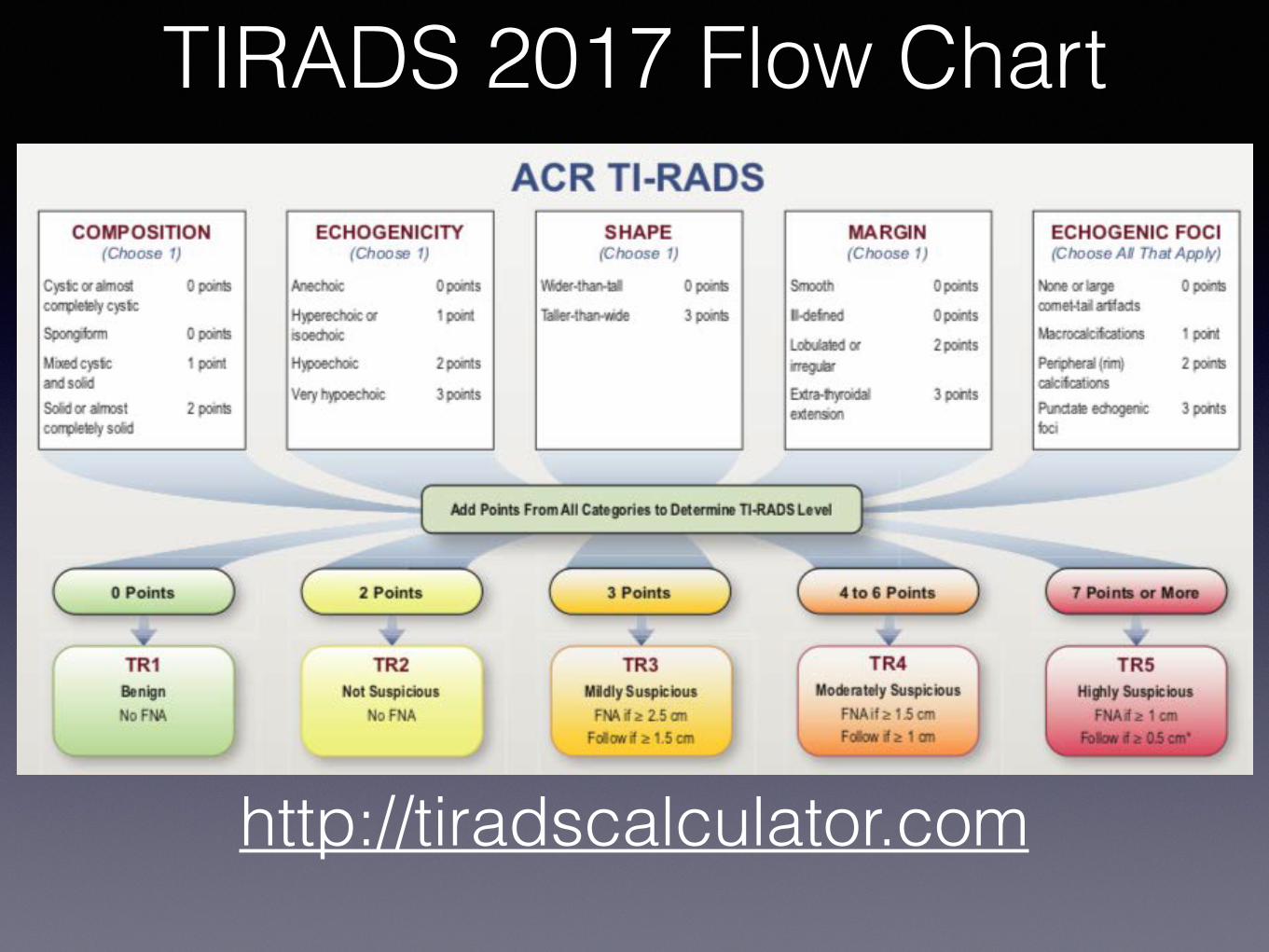
Thyroid US is a poor screening tool because:
• 66% of benign nodules have at least one positive US predictor of papillary thyroid cancer
• 66% of papillary cancers have at least one non-suspicious US feature
So US guided FNA is a vital adjunct.
2015 ATA Guidelines RECOMMENDATION 7: FNA is the procedure of choice in the evaluation of thyroid nodules, when clinically indicated.
Ultrasound Features 2015 American Thyroid Association Management Guidelines for Adult Patients with
Thyroid Nodules and Differentiated Thyroid Cancer: The American Thyroid Association Guidelines Task Force on Thyroid Nodules and Differentiated Thyroid
Cancer. Thyroid. 2016 Jan 1; 26(1): 1–133
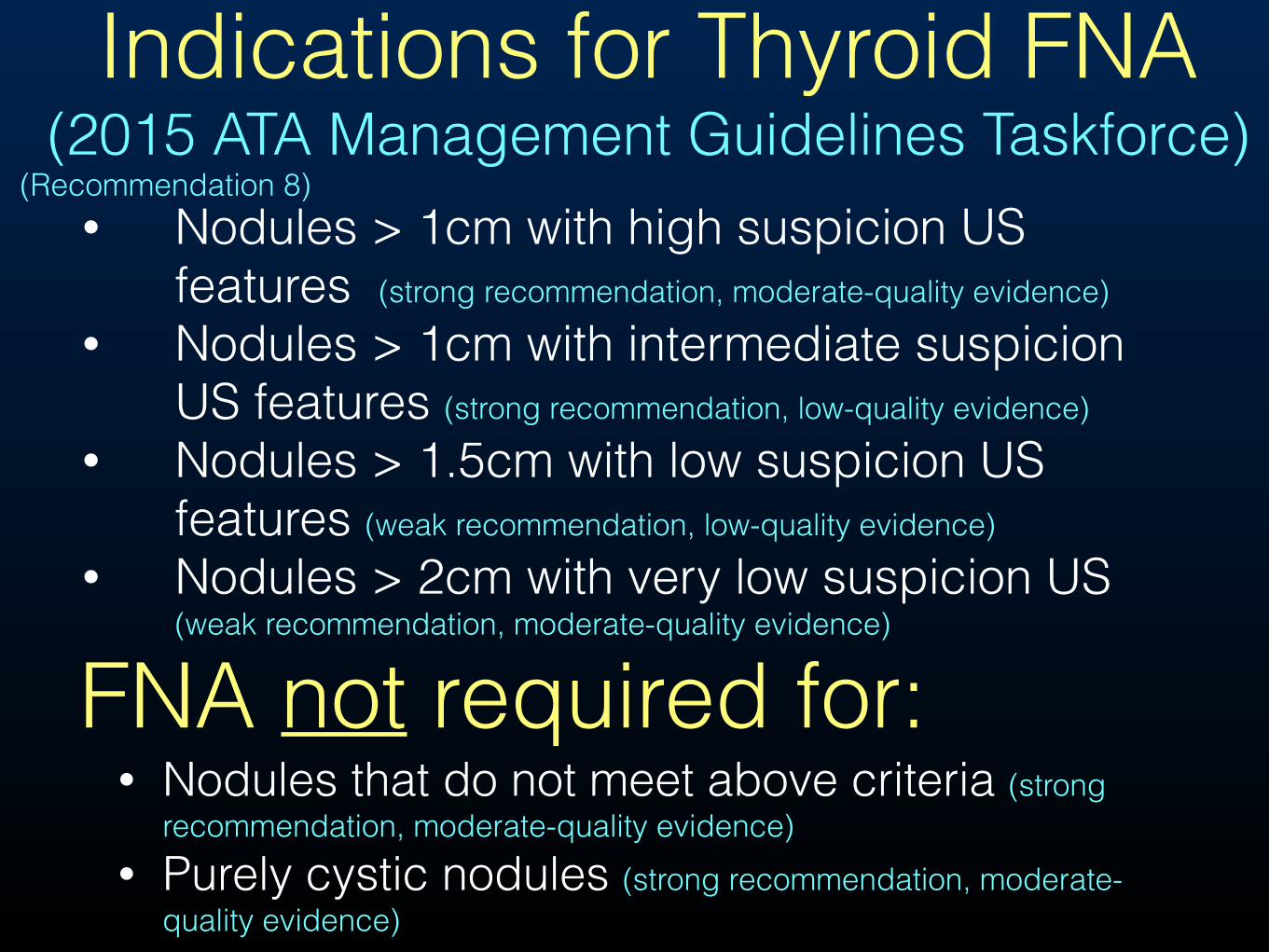
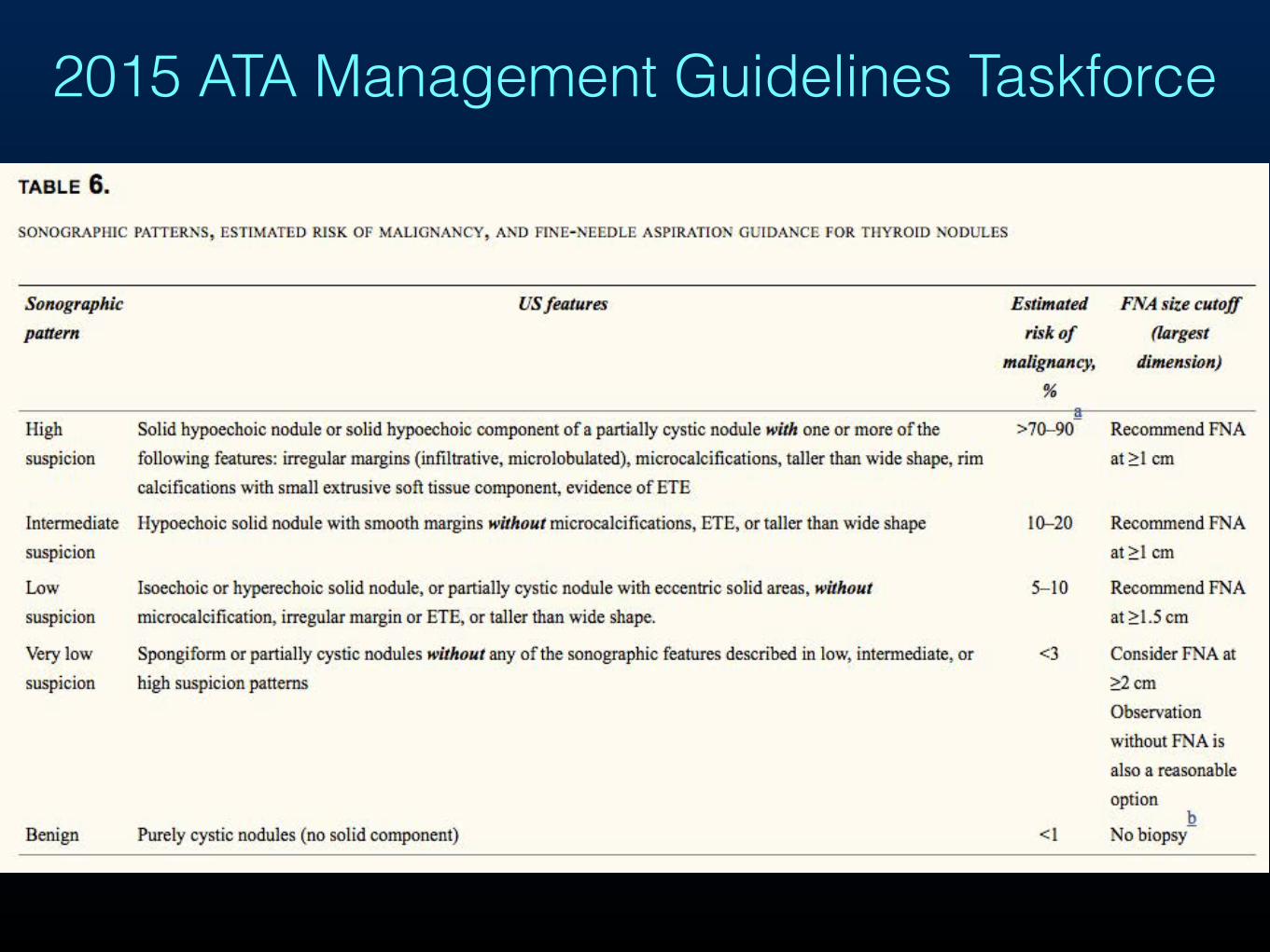
• Nodules > 1cm with high suspicion US features (strong recommendation, moderate-quality evidence)
• Nodules > 1cm with intermediate suspicion US features (strong recommendation, low-quality evidence)
• Nodules > 1.5cm with low suspicion US features (weak recommendation, low-quality evidence)
• Nodules > 2cm with very low suspicion US (weak recommendation, moderate-quality evidence)
Indications for Thyroid FNA (2015 ATA Management Guidelines Taskforce)
FNA not required for:• Nodules that do not meet above criteria (strong
recommendation, moderate-quality evidence) • Purely cystic nodules (strong recommendation, moderate-
quality evidence)
• (Recommendation 8)
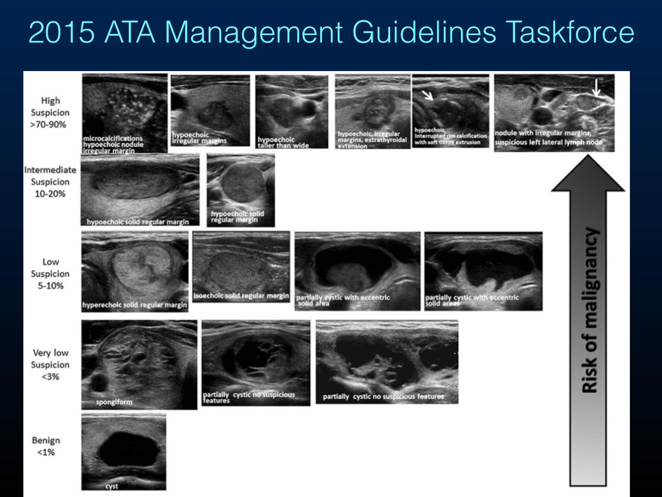
2015 ATA Management Guidelines Taskforce
2015 ATA Management Guidelines Taskforce
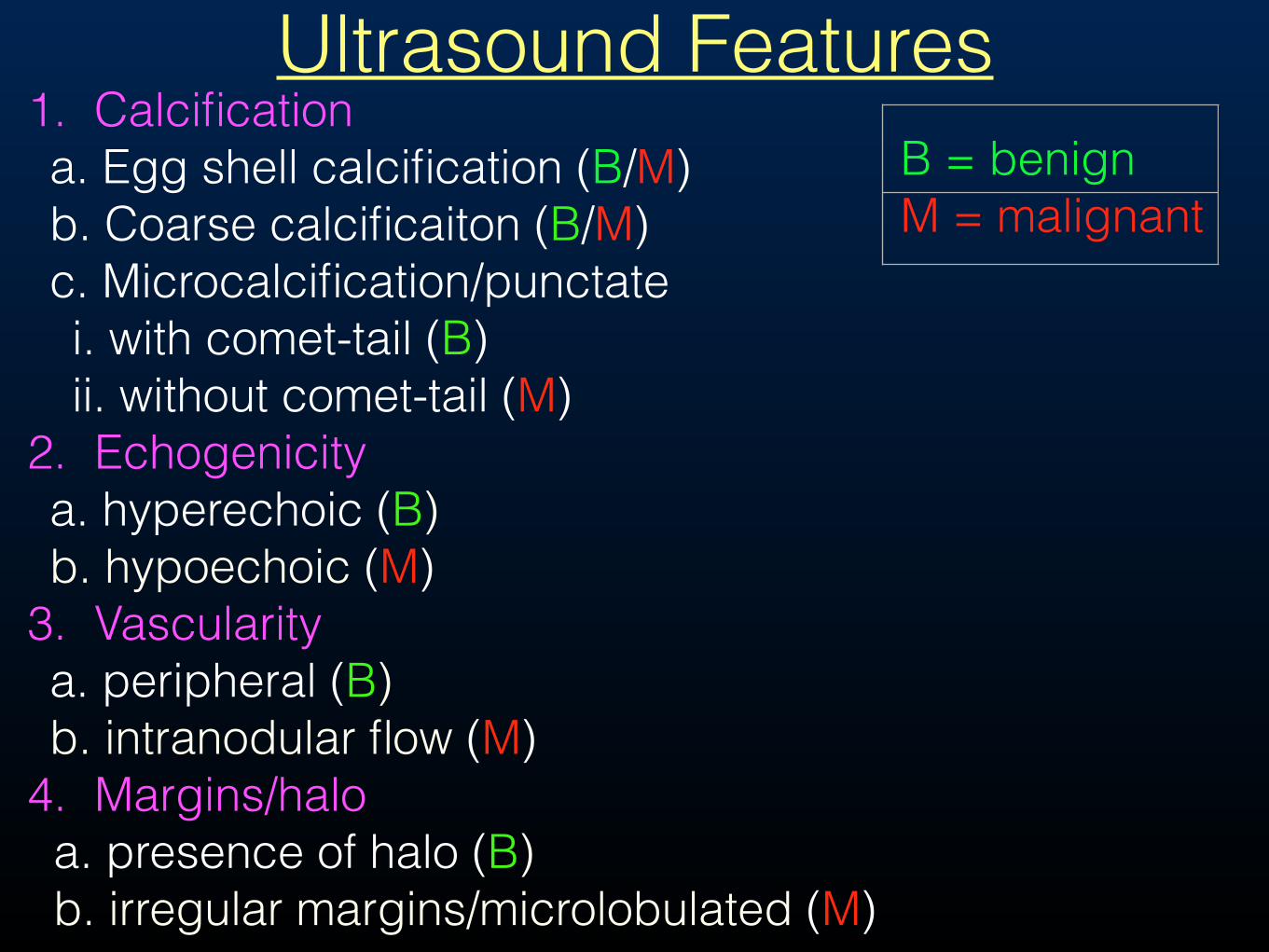
Ultrasound Features1. Calcification a. Egg shell calcification (B/M) b. Coarse calcificaiton (B/M) c. Microcalcification/punctate i. with comet-tail (B) ii. without comet-tail (M)
2. Echogenicity a. hyperechoic (B) b. hypoechoic (M)
3. Vascularity a. peripheral (B) b. intranodular flow (M)
4. Margins/halo a. presence of halo (B) b. irregular margins/microlobulated (M)
B = benign M = malignant
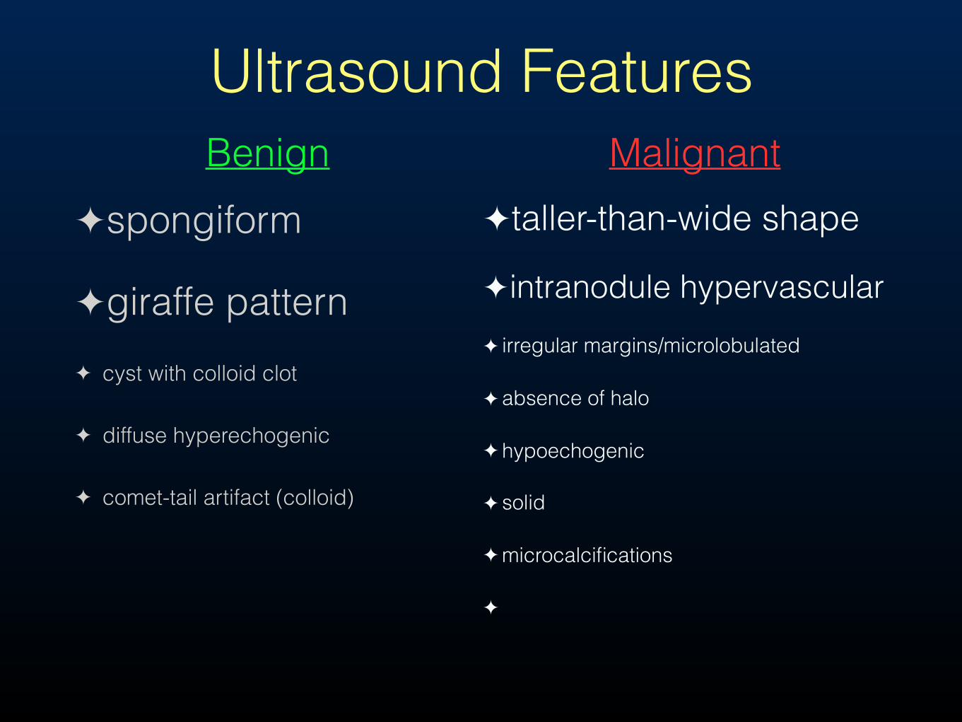
Ultrasound Features
✦spongiform
✦giraffe pattern ✦ cyst with colloid clot
✦ diffuse hyperechogenic
✦ comet-tail artifact (colloid)
✦taller-than-wide shape
✦intranodule hypervascular ✦ irregular margins/microlobulated
✦absence of halo
✦hypoechogenic
✦solid
✦microcalcifications
✦
Benign Malignant
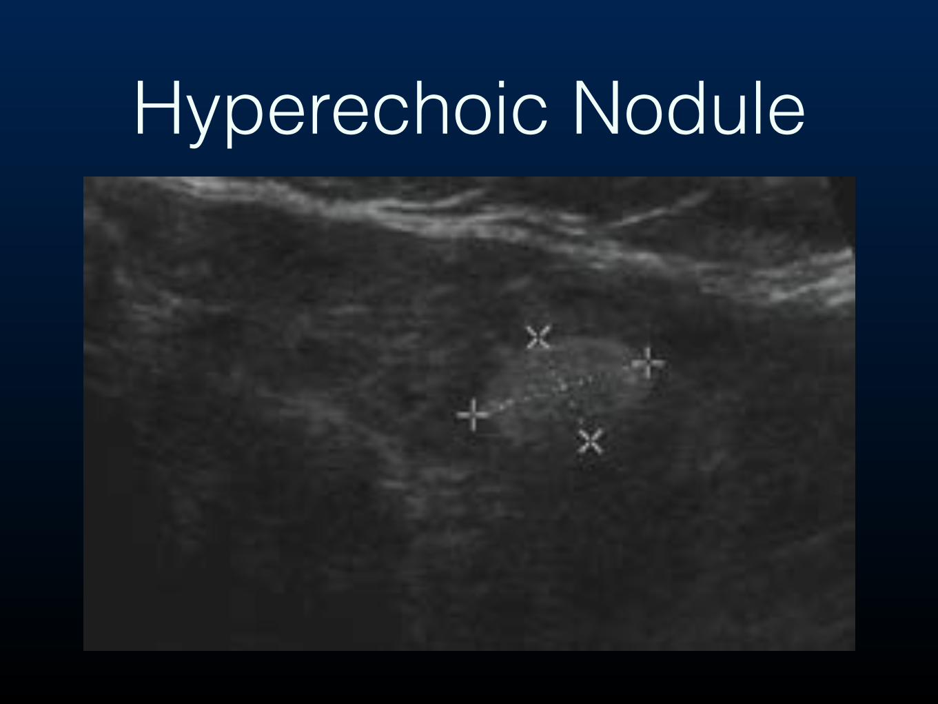
Hyperechoic Nodule
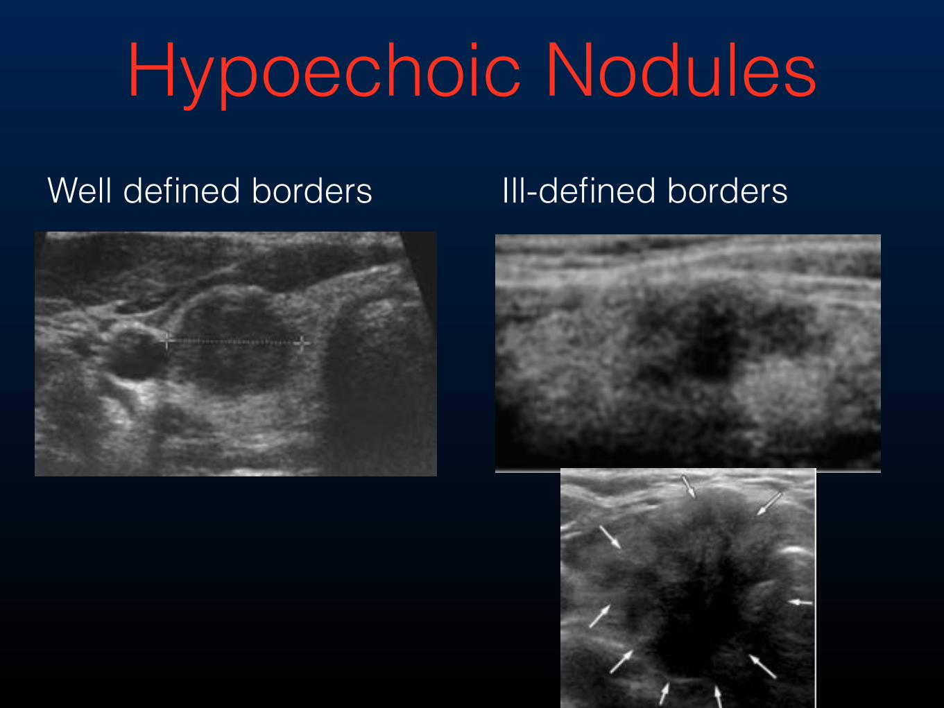
Hypoechoic Nodules Well defined borders Ill-defined borders
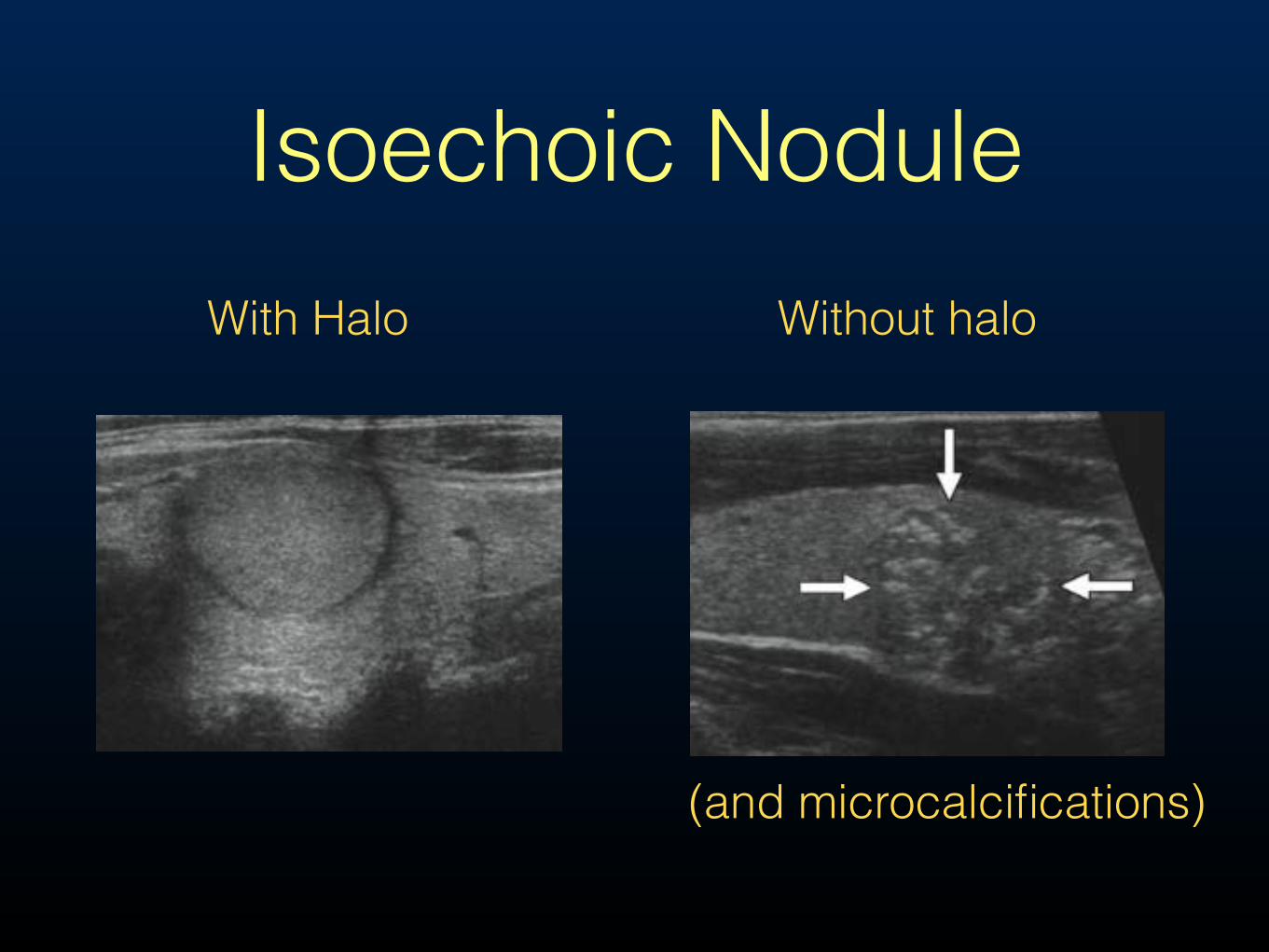
Isoechoic NoduleWith Halo Without halo
(and microcalcifications)
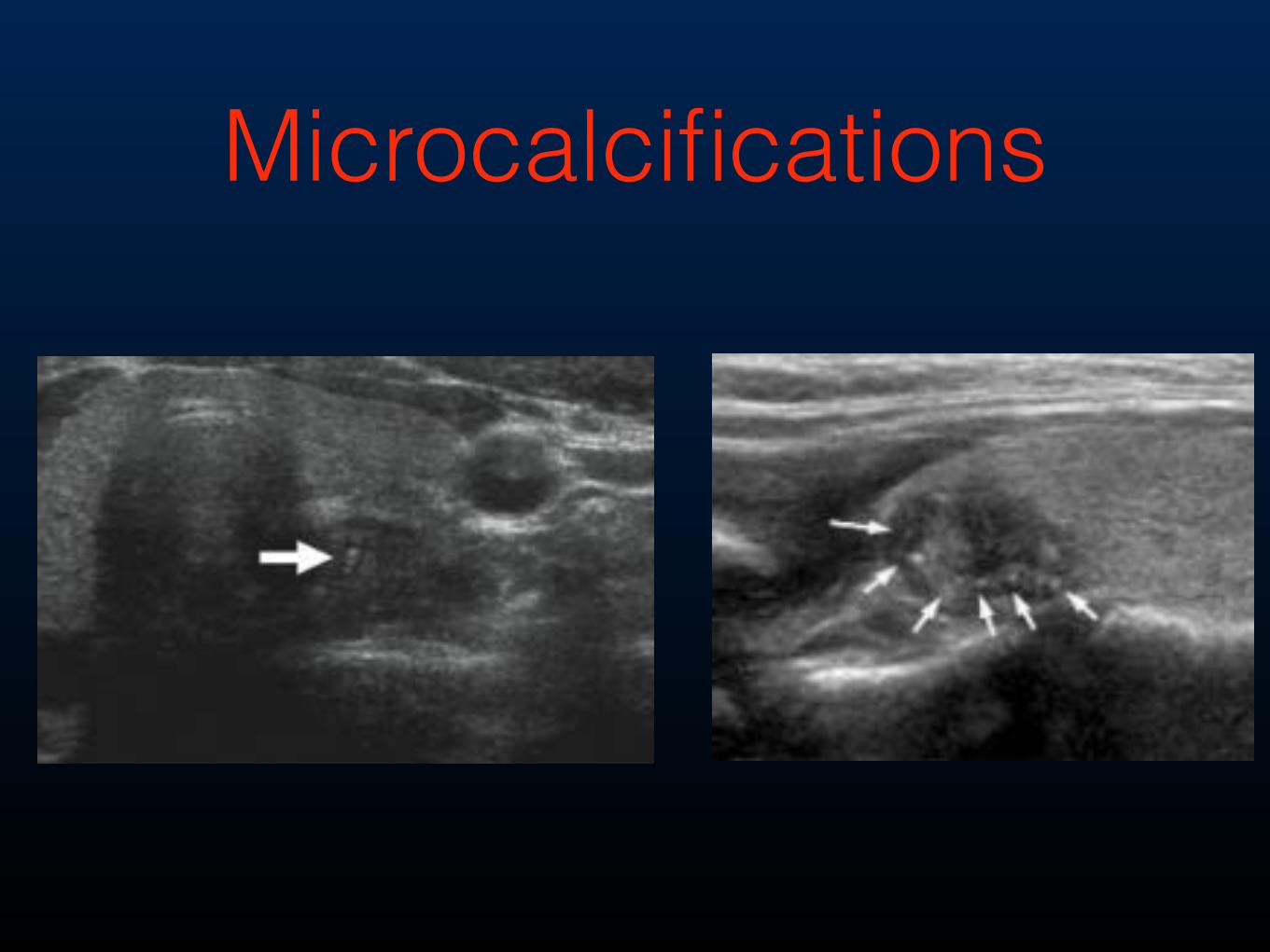
Microcalcifications
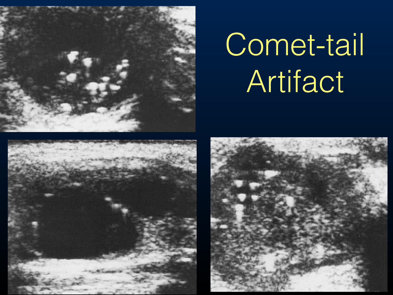
Comet-tail Artifact
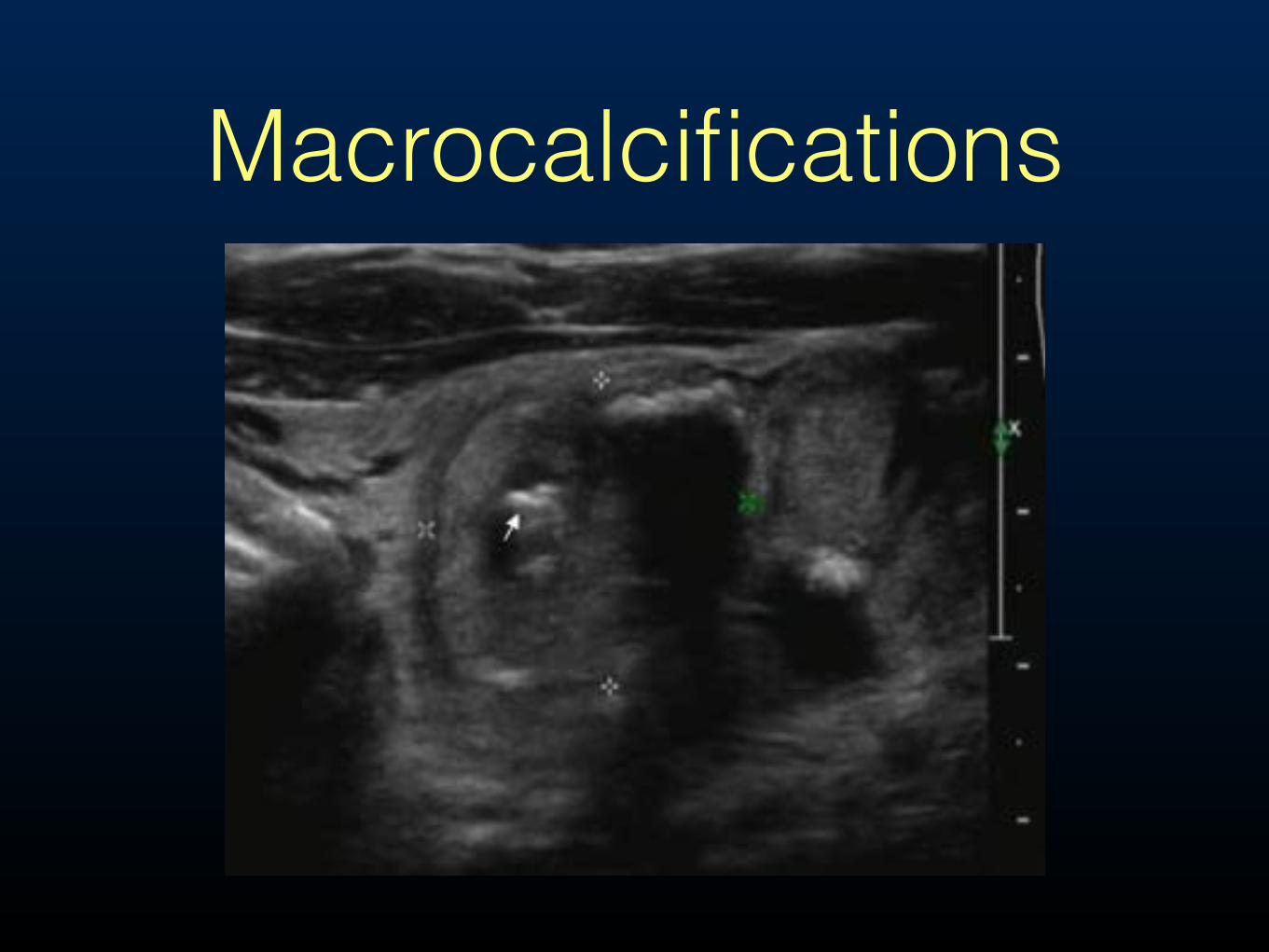
Macrocalcifications
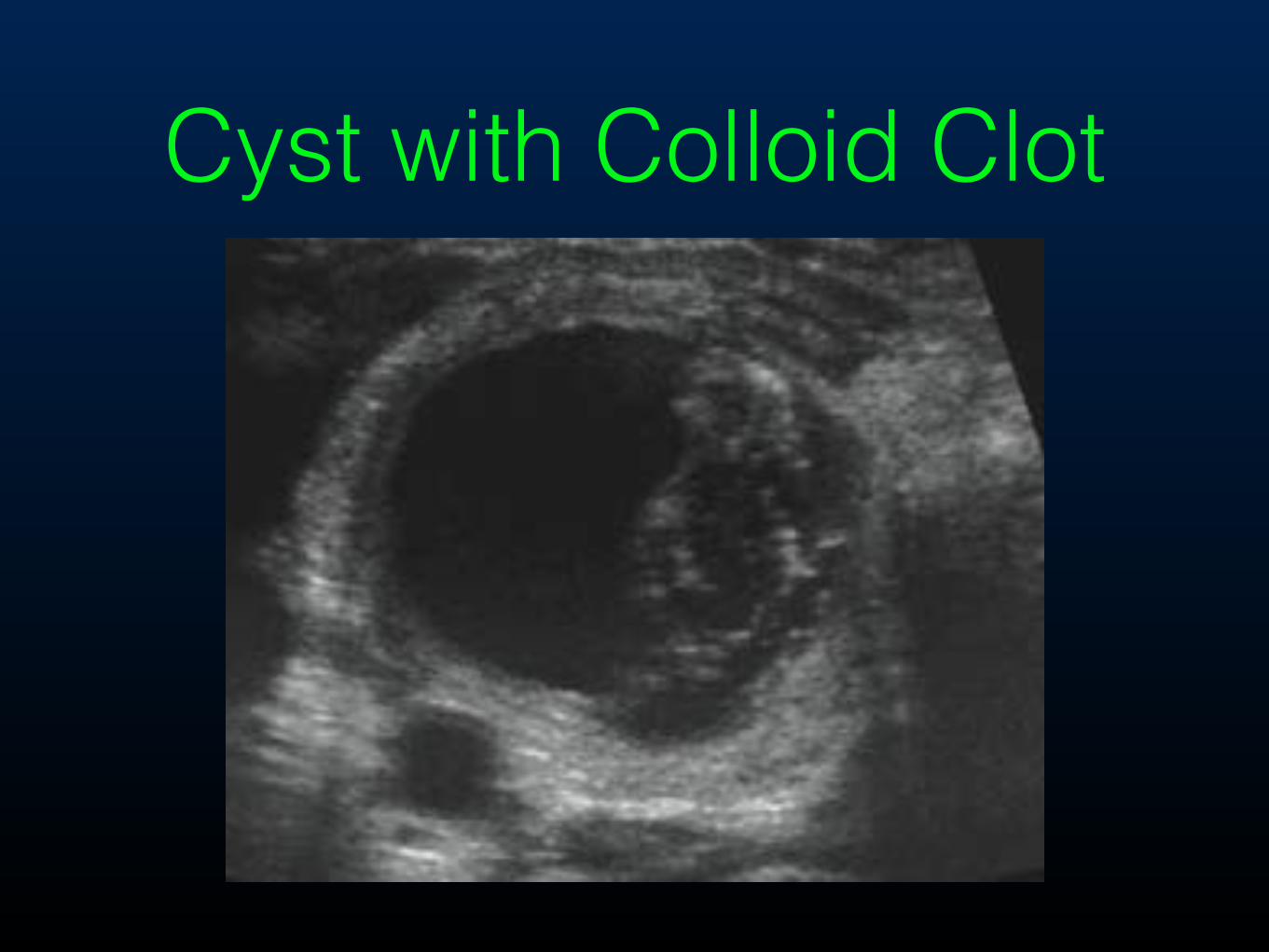
Cyst with Colloid Clot
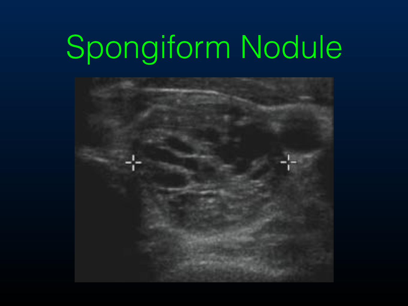
Spongiform Nodule
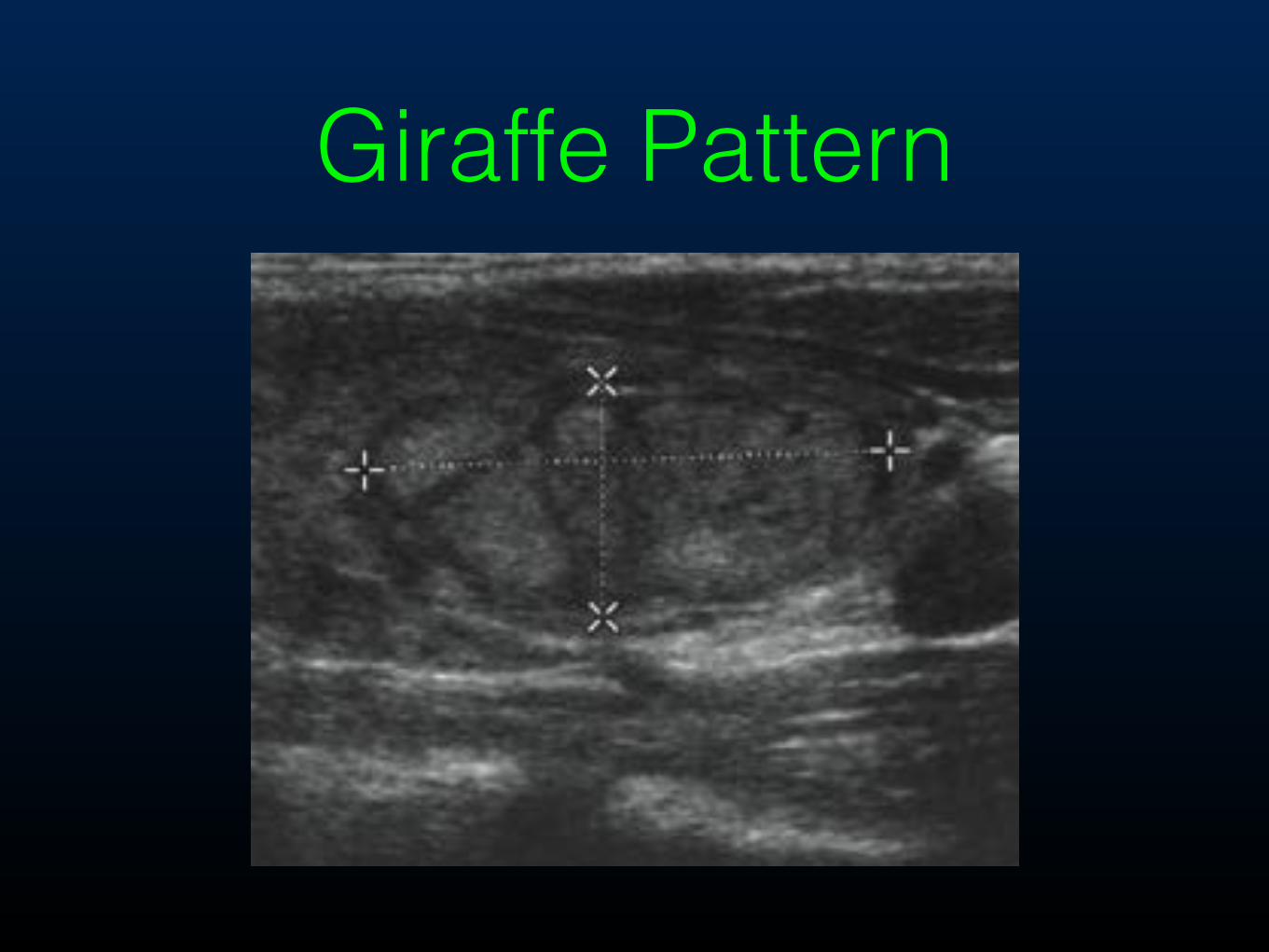
Giraffe Pattern
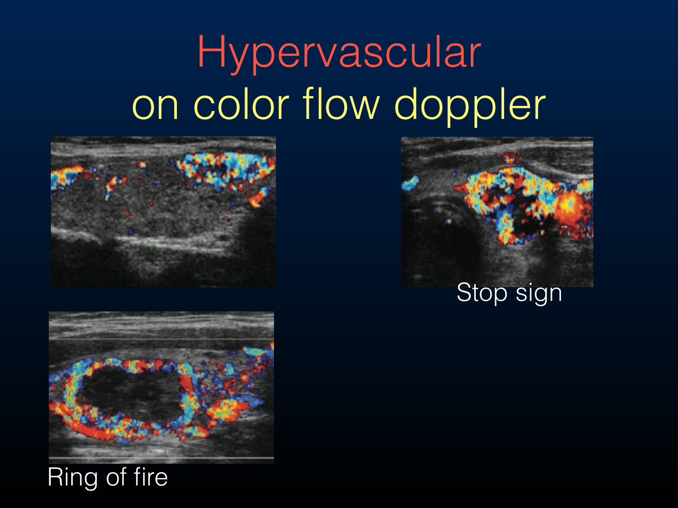
Hypervascular on color flow doppler
Ring of fire
Stop sign
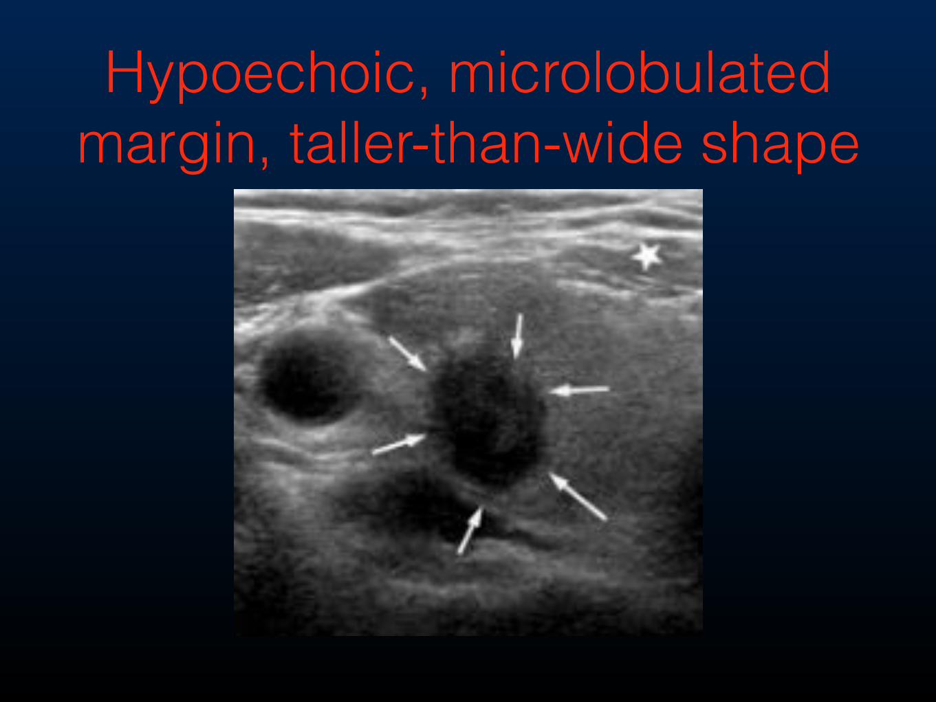
Hypoechoic, microlobulated margin, taller-than-wide shape
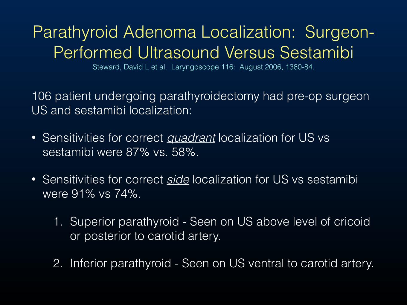
Parathyroid Adenoma Localization: Surgeon-Performed Ultrasound Versus Sestamibi
Steward, David L et al. Laryngoscope 116: August 2006, 1380-84.
106 patient undergoing parathyroidectomy had pre-op surgeon US and sestamibi localization:
• Sensitivities for correct quadrant localization for US vs sestamibi were 87% vs. 58%.
• Sensitivities for correct side localization for US vs sestamibi were 91% vs 74%.
1. Superior parathyroid - Seen on US above level of cricoid or posterior to carotid artery.
2. Inferior parathyroid - Seen on US ventral to carotid artery.
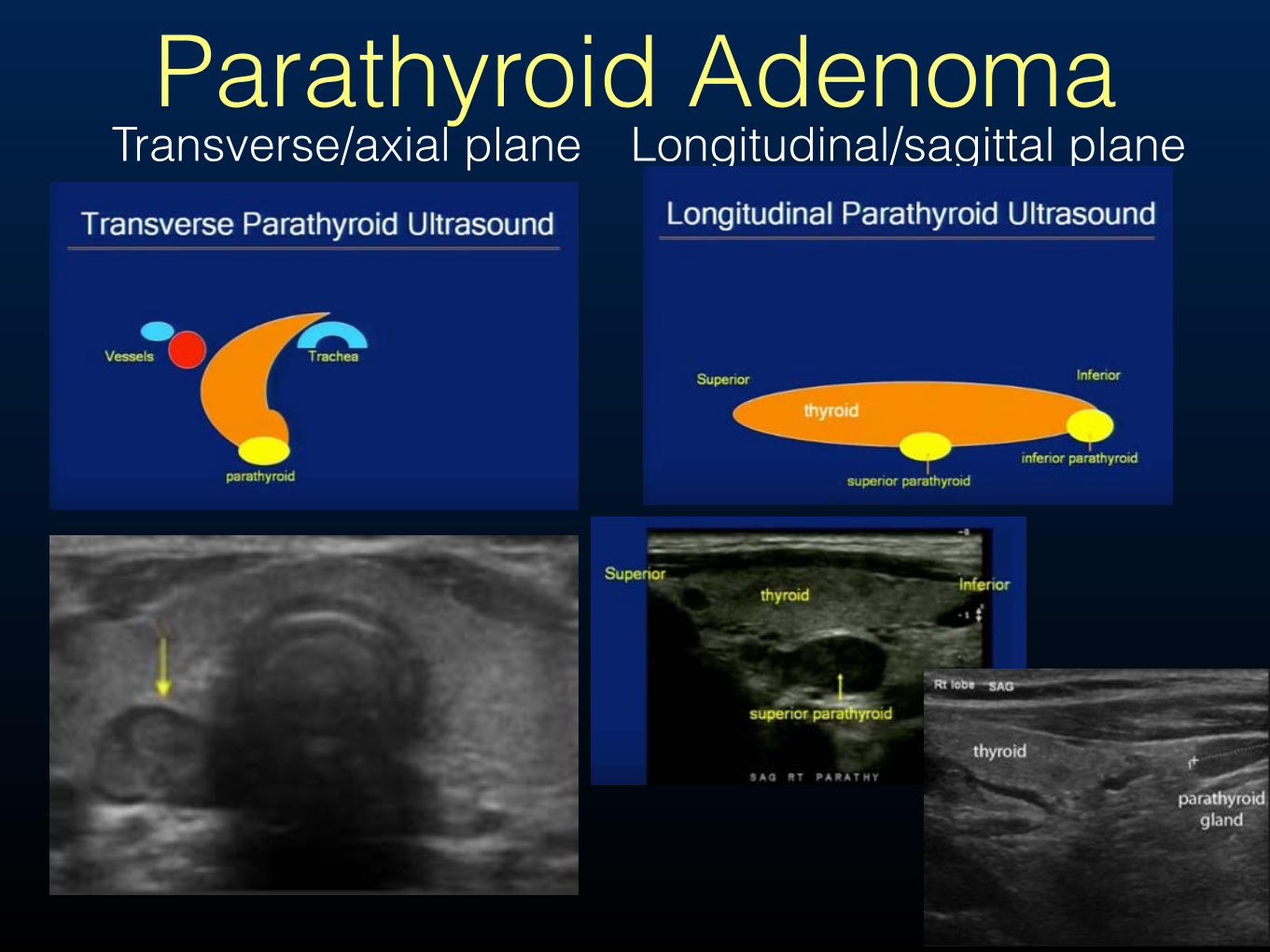
Parathyroid AdenomaTransverse/axial plane Longitudinal/sagittal plane
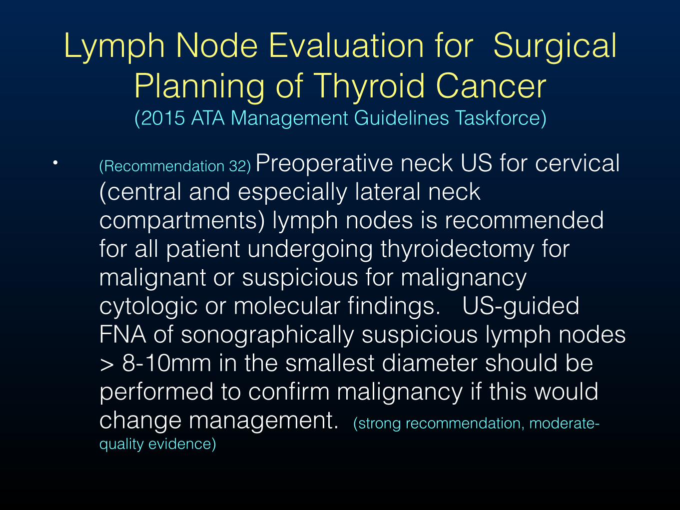
Lymph Node Evaluation for Surgical Planning of Thyroid Cancer (2015 ATA Management Guidelines Taskforce)
• (Recommendation 32) Preoperative neck US for cervical (central and especially lateral neck compartments) lymph nodes is recommended for all patient undergoing thyroidectomy for malignant or suspicious for malignancy cytologic or molecular findings. US-guided FNA of sonographically suspicious lymph nodes > 8-10mm in the smallest diameter should be performed to confirm malignancy if this would change management. (strong recommendation, moderate-quality evidence)
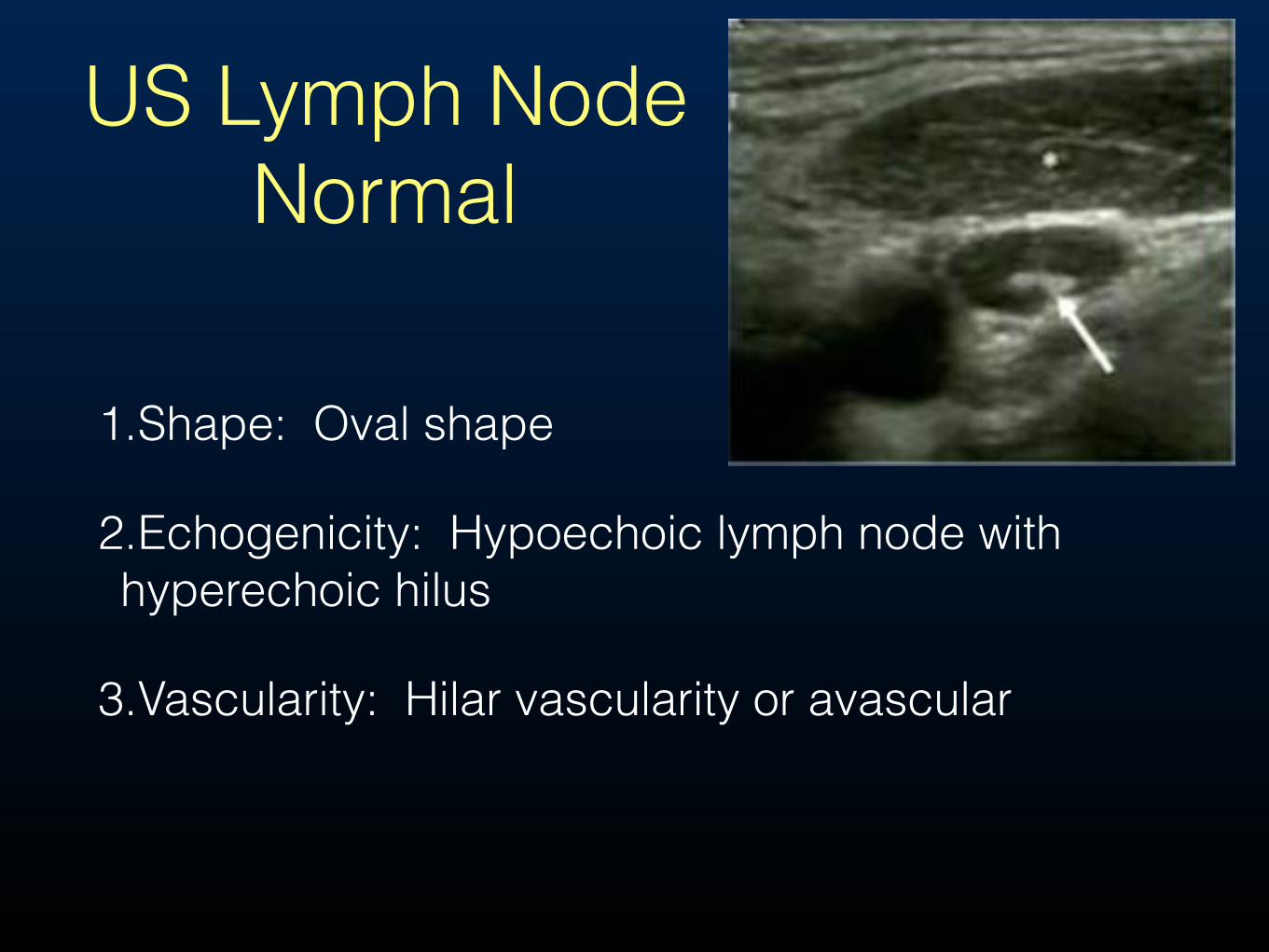
US Lymph Node Normal
1.Shape: Oval shape
2.Echogenicity: Hypoechoic lymph node with hyperechoic hilus
3.Vascularity: Hilar vascularity or avascular
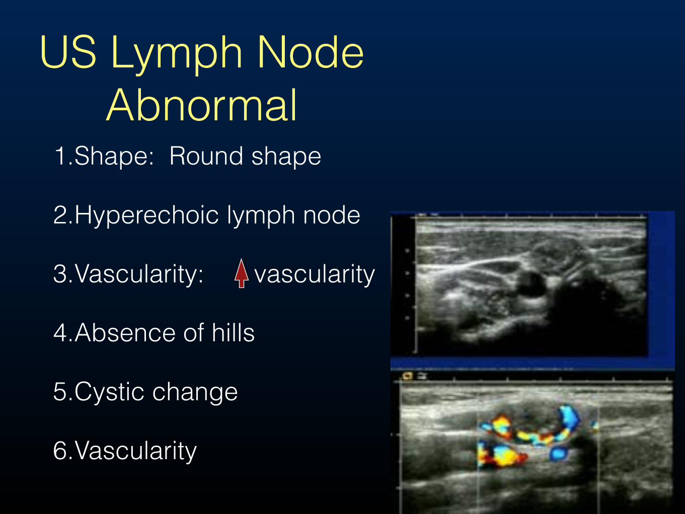
US Lymph Node Abnormal
1.Shape: Round shape
2.Hyperechoic lymph node
3.Vascularity: vascularity
4.Absence of hills
5.Cystic change
6.Vascularity
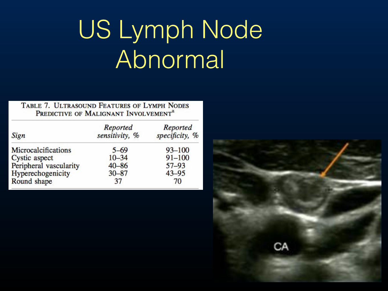
US Lymph Node Abnormal
🚵
🚴
That's all folks!
Related Documents