Management of right ventricular failure J. Parissis, Attikon University Hospital Disclosures: received horonaria from ORION PHARMA , Finland

Welcome message from author
This document is posted to help you gain knowledge. Please leave a comment to let me know what you think about it! Share it to your friends and learn new things together.
Transcript

Management of right ventricular failure
J. Parissis,
Attikon University Hospital
Disclosures: received horonaria from ORION PHARMA , Finland

Definition
The clinical condition associated with
any structural or functional process
that restricts the ability of the RV to fill
with blood and/or to eject blood into
the pulmonary vasculature.

Main causes of RV failure
1. LV failure (biventricular failure, most common)
2. Severe pulmonary embolism
3. ARDS (acute lung injury)
4. Sepsis induced RV dysfunction
5. Idiopathic or secondary forms of pulmonary hypertension
6.Right ventricle infarction or ischemia
7. Pericardial diseases (constrictive pericarditis, tamponade)
8. RV failure after cardiac surgery (e.g. cardiac transplant or LVAD implantation)
9.Congenital heart disease (e.g. Ebstein’s anomaly)
10. Valvulopathies (e.g. pulmonary valve stenosis, TR)
11. Rare cardiomyopathies (e.g Arrhythmiogenic RV dysplasia)
12. Arrhythmias
13. Hematologic disorders (e.g. Acute chest syndrome in sicle celldisease)

4% 1%
12%
7%
39%
37%
AdHF Pulmonary oedema Cardiogenic shock
Hypertensive HF Right HF High cardiac output failure
3%
4%
11%
65%
16%
EHS HF II vs ALARM-HF clinical classification according to ESC AHF
Guidelines
EHS HF II: 3,580 patients, ALARM-HF: 4,953 patients (1911 AdHF, 1820 p-oed, 581 C-shock, 365 Hyp AHF, 222 RV AHF, 54 High cardiac output)
ALARM-HFEHS HFII
Follath F, Yilmaz MB, Delgado JF, Parissis JT, et al. Intensive Care Medicine 2011

ALARM-HF: mortality across
classification
7%
11% 10%12%
6% 7%
40% 40%
2%
13%
0%
5%
10%
15%
20%
25%
30%
35%
40%
45%
EHS HF
II
All AHF ADCHF De
NOVO
AHF
ADHF P-OE Cardio
shock
EHS HF
II C-
Shock
HT AHF RV HF
Sample = EHS HF II (3.580), All ALARM-HF patients (4.953)
Follath F, Yilmaz B, Delgado J, Parissis J, Mebazaa A. Intens Care Med 2011


Prognostic Value of Tissue Doppler Right Ventricular Systolic and
Diastolic Function Indexes Combined With Plasma B-Type
Natriuretic Peptide in Patients With Advanced Heart Failure
V Bistola, JT. Parissis, ... , E. Iliodromitis, D. Kremastinos. Am J Cardiol 2010;105:249–254*

Pathophysiology of RV failure. *The time course (acute or chronic) and time of onset of the disease
process (newborn, pediatric, or adult years) also influence RV adaptation to disease.
Haddad F et al. Circulation. 2008;117:1717-1731

Pathophysiology of failing RV
Piazza, G. et al. Chest 2005;128:1836-1852

Ventricular interdependence
• During systole, LV protrudes in RV• Surrounding pericardium with limited distensibility• Compliance of one ventricle can modify the other = Diastolic ventricular interaction
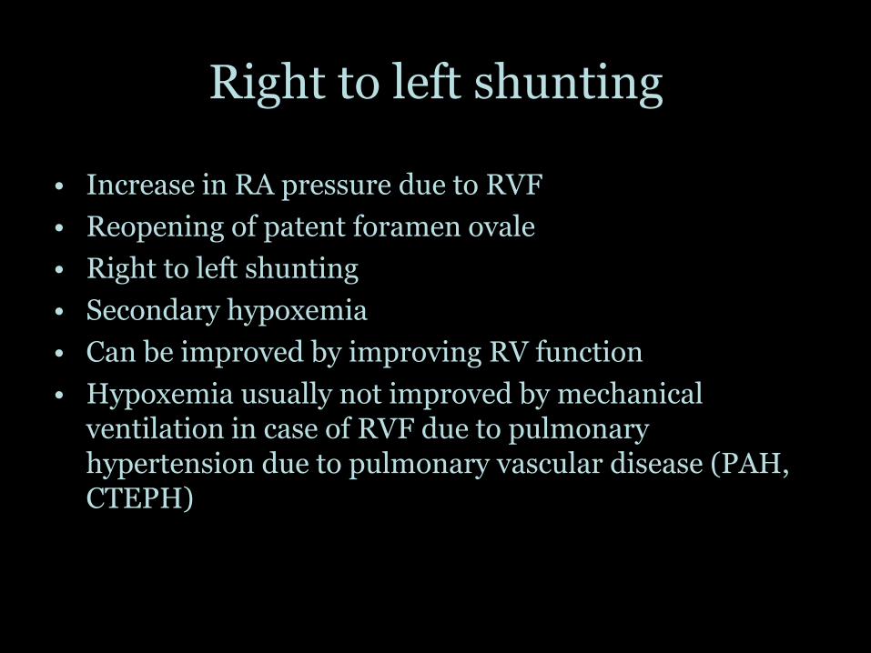
Right to left shunting
• Increase in RA pressure due to RVF
• Reopening of patent foramen ovale
• Right to left shunting
• Secondary hypoxemia
• Can be improved by improving RV function
• Hypoxemia usually not improved by mechanical ventilation in case of RVF due to pulmonary hypertension due to pulmonary vascular disease (PAH, CTEPH)
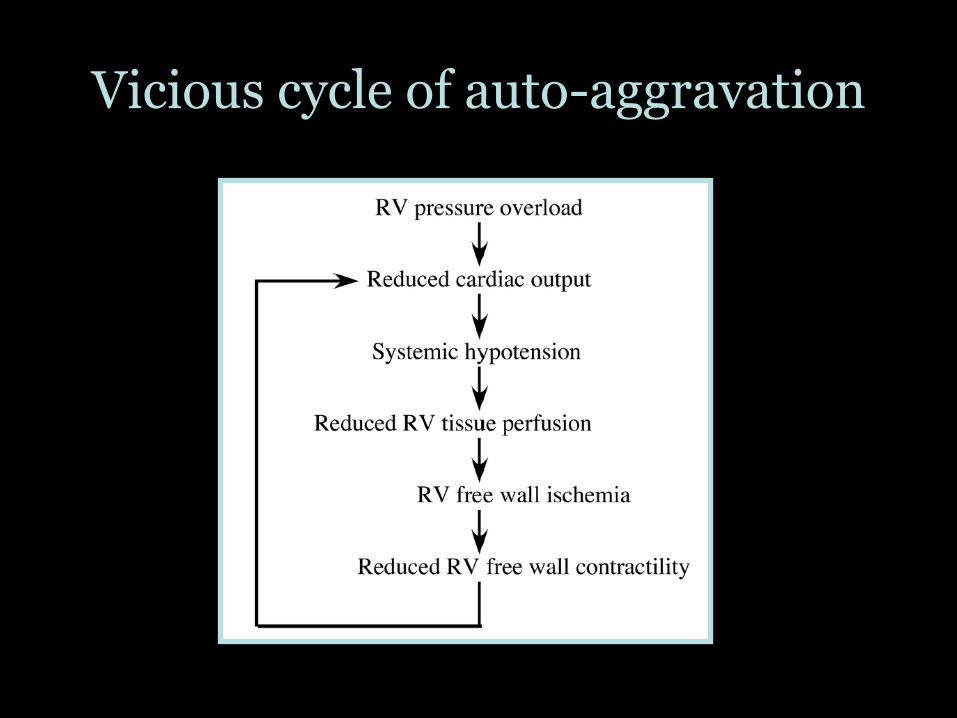
Vicious cycle of auto-aggravation

Clinical and biological signs of acute RV failure
CLINICAL
- Signs of systemic congestion
Jugular venous distension, hepatojugular reflex, peripheral oedema, pleural effusions, congestive liver/hepatomegaly, ascites, anasarca
- Signs of RV dysfunction
Third heart sound, systolic murmur of TR regurgitation, hepaticpulse, signs of concomitant LV dysfunction
- Signs of low cardiac output state
Hypotension, tachycardia, cool extremities, central nevrous system abnormalities, oliguria
BIOLOGICAL
- Hypoxemia, hyper- or hypocapnia, increased lactate , elevatednatriuretic peptides and /or Hs-troponins and/or d-dimers, abnormal liver biochemistry (elevated ALP, GGT, bilirubin, INR, transaminases), abnormal renal function (urine output, BUN, creatinine), increased inflammatory markers (e.g. CRP)

Management
• Control of trigerring factors
• Supportive treatment:
– Optimization of preload
– Improving contractility
– Pulmonary vasodilators
• Specific therapies addressing the cause of RVF

Treatment of triggering factors(acute on chronic)
• Arrhytmias
• Infections
• Pulmonary embolism
• Thyroïd dysfunction

Optimization of preload
Frank-Starling relationship between preload and stroke volume: preload dependance (A) and preload independance (B)
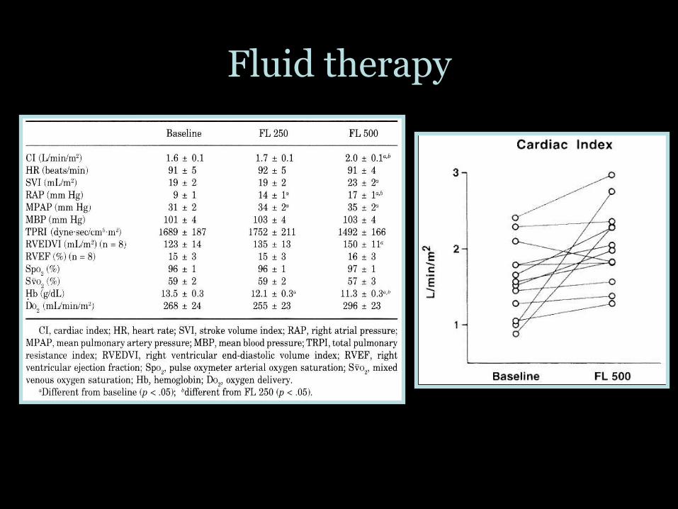
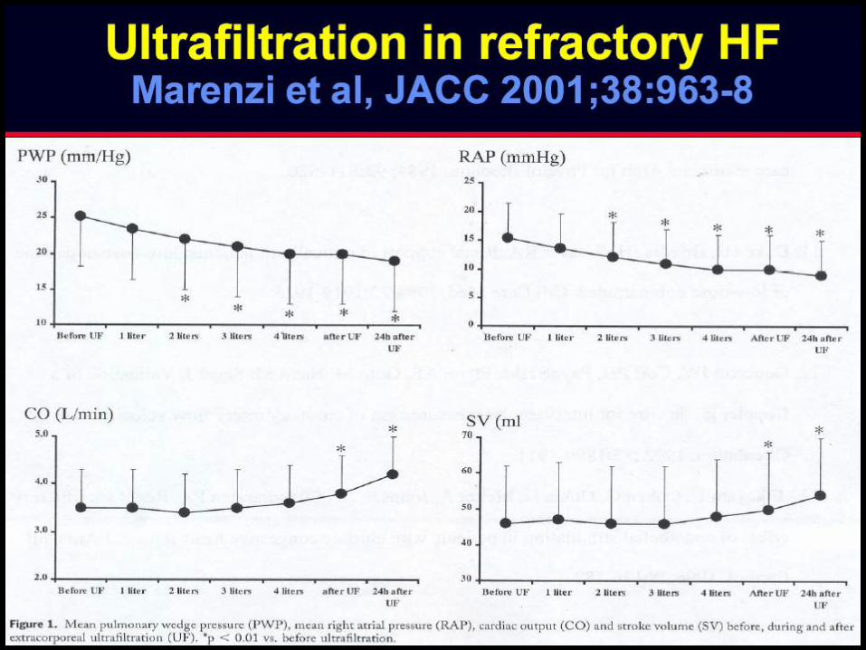
Fluid therapy

Diuretics
• Frequent volume overload
• At a point of Frank-Starling curve where there is no more reserve on contractility
• Ventricular interdependance
• Diuretics to be considered
• Sometimes with continuous high dose infusion
• (plus high dose of MRAs and /or metolazone especialy in biventricular failure)
• If fails, consider CVVHF

Dobutamine
• 1 adrenergic stimulation
• CI PVR at 5 g/kg/mn
• At higher dose HR without subsequent in PVR
• Experimental models Dobutamine Norepinephrine to improve right-ventricular –pulmonary artery coupling
• Improves CI, PVR and PaO2/FiO2 in combination with Inhaled nitric oxyde
Norepinephrine
• 1 and 1 adrenergic stimulation
• Increases mPAP and PVR
• But marked improvement in CO
• Useful in combination with Dobutamine for hypotensive patients
• Causes less tachycardia than other inotropes
• Second choice after Dobutamine in normotensive patients

Levosimendan
• Calcium sentitizer: increases the sensitivity of troponin C for Ca2+ within cardiac myocyte
• Dilatation of pulmonary vasculature by activation of adenosin tri-phosphate potassium channel
• Animal studies and pilot studies support its efficacy in right ventricle failure associated with pulmonary hypertension
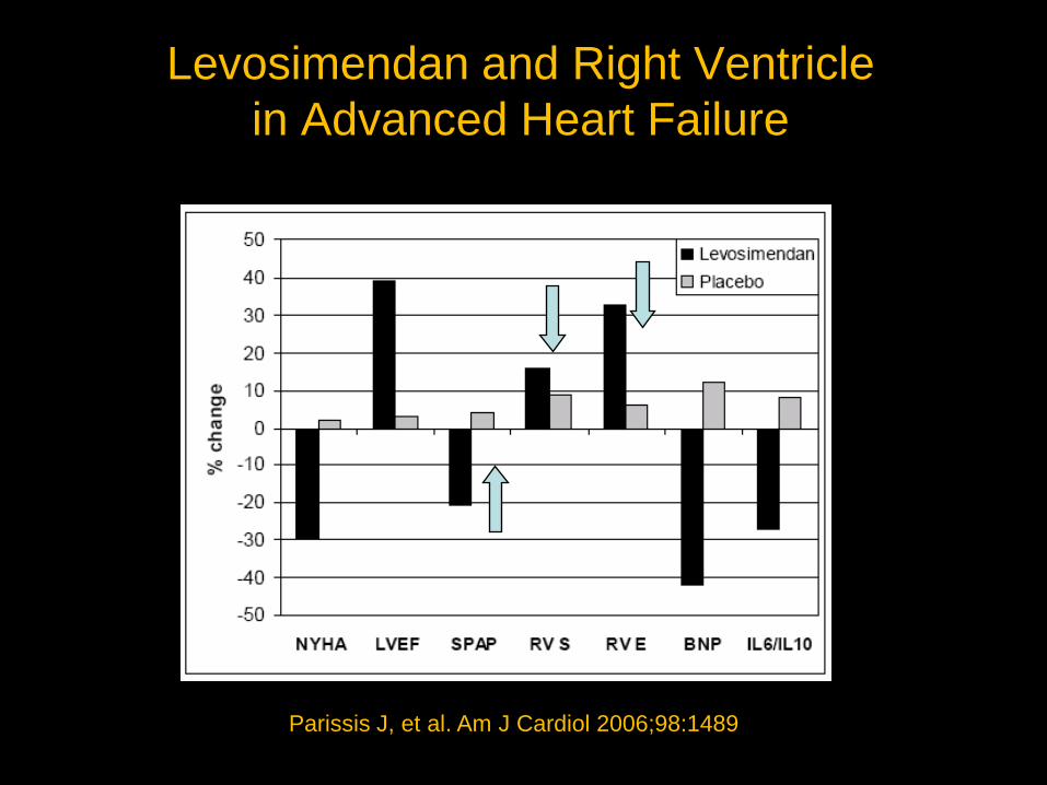
Levosimendan and Right Ventricle
in Advanced Heart Failure
Parissis J, et al. Am J Cardiol 2006;98:1489

• 35 ICU patients with ARDS and sepsis randomized to receive placebo or levosimendan 0.2g/kg/mn
• Mean arterial pressure 80 to 90 mmHg (sustained by norepinephrine infusion)
• Improvement of right ventricle performance:
– CI (from 3.8 1.1 to 4.2 1.0 L/min/m2)
– PAPm (from 29 3 to 25 3 mm Hg)
– RVESV, RVEF, SvO2
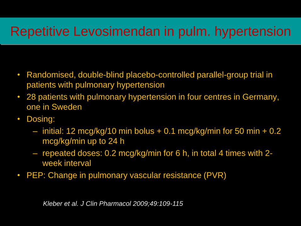
• Randomised, double-blind placebo-controlled parallel-group trial in
patients with pulmonary hypertension
• 28 patients with pulmonary hypertension in four centres in Germany,
one in Sweden
• Dosing:
– initial: 12 mcg/kg/10 min bolus + 0.1 mcg/kg/min for 50 min + 0.2
mcg/kg/min up to 24 h
– repeated doses: 0.2 mcg/kg/min for 6 h, in total 4 times with 2-
week interval
• PEP: Change in pulmonary vascular resistance (PVR)
Kleber et al. J Clin Pharmacol 2009;49:109-115
Repetitive Levosimendan in pulm. hypertension

-40
-20
0
20
40
1 h 2 h 4 h 6 h 8 h 24 h 1 h 2 h 4 h 6 h
Baseline Day 0 Week 8
Time
Δ
mPAP
(%) levosimendan
placebo
Kleber et al. J Clin Pharmacol 2009;49:109-115
Change in mPAP (mean SEM)
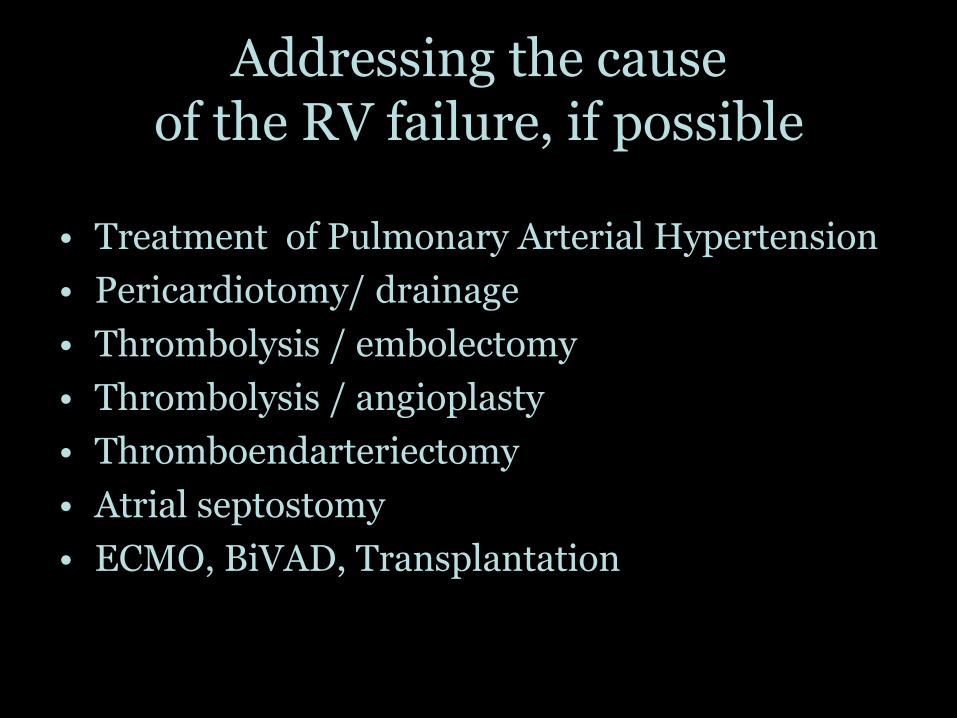
Addressing the cause of the RV failure, if possible
• Treatment of Pulmonary Arterial Hypertension
• Pericardiotomy/ drainage
• Thrombolysis / embolectomy
• Thrombolysis / angioplasty
• Thromboendarteriectomy
• Atrial septostomy
• ECMO, BiVAD, Transplantation

Pulmonary vasodilators

Inhaled nitric oxyde
• Dilate pulmonary vessels in ventilated units of the lung
• Reverses hypoxic pulmonary vasoconstriction
• In acutely decompensated RV improves PVR, increase CO improve PaO2/FiO2 (Benker KA et Al. Am J Crit Care. 1997 Mar;6(2):127-31)
• Beware of methemoglobinemia (high concentraton, prolonged use)

Effect of abrupt discontinuation of NO
2002 Yearbook of Intensive Care and Emergency Medicine,
Acute right ventricular failure: physiology and therapy by Renaud E, Karpati P, Mebazaa A

Prostanoids
• Intravenous Epoprostenol
• Effect on survival in stable patients with PAH
• Reduces mPAP and improves CO
• Systemic side effects
• Worsening PaO2/FiO2
• Systemic effects (hypotension)
• Inhaled prostacyclin / nebulized iloprost: case series
(Shock associated with PAH, Olschewski H. Intensive Care
Med. 1998 Jun;24(6):631-4)

Sidenafil
• Phosphodiesterase-5 inhibitor
• Approved for treatment of PAH (stable patients)
• Only case reports for use in critically ill (RVF after transplant: De Santo LS et Al.Transplant Proc. 2008 Jul-Aug; 40(6):
2015-8)
• May be useful for weaning from inhaled nitric oxyde
• Effect start 15mn after administration, peak effects within 30-60mn
• Systemic hypotension

Effects of mechanical ventilation
• Increased RV afterload due to positive pressure ventilation
• Hemodynamic failure frequently refractory in PAH patient put on MV
• In ARDS increase in mPAP while increasing tidal volume and PEEP
• Permissive hypercapnia is deleterious (increase in mPAP)
•
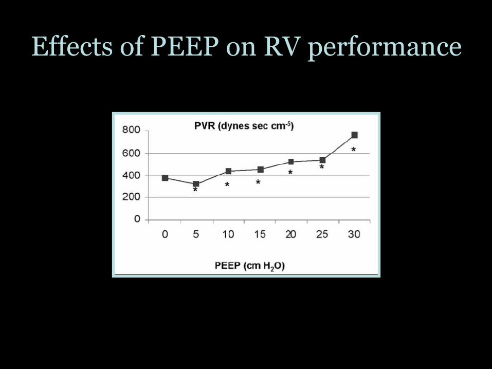
Effects of PEEP on RV performance

Effect of high PEEP on RV

Risk factors for RHF

Predictors of RHF in the
recent VAD era
CRITT Score
SCORE 0-1: LVAD ONLY
SCORE 4-5: BiVAD
SCORE 2-3: LVAD + pharmacologic
therapy for RHF OR temporary
BiVAD

Management of Acute RV failure
JACC 2010;56:1435

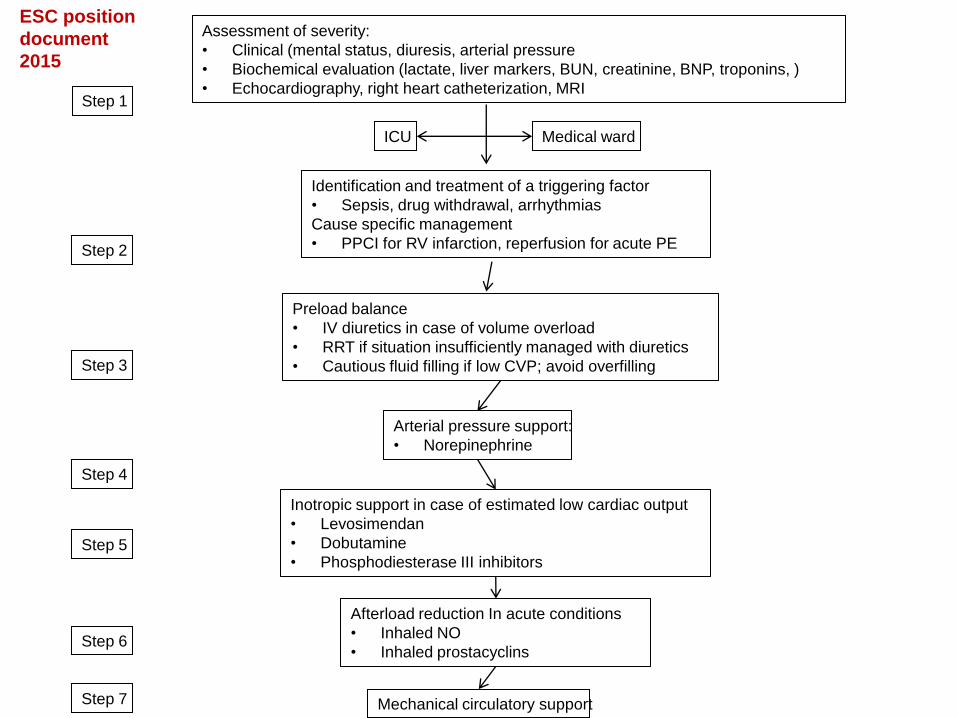
ESC position
document
2015
Assessment of severity:
• Clinical (mental status, diuresis, arterial pressure
• Biochemical evaluation (lactate, liver markers, BUN, creatinine, BNP, troponins, )
• Echocardiography, right heart catheterization, MRI
Identification and treatment of a triggering factor
• Sepsis, drug withdrawal, arrhythmias
Cause specific management
• PPCI for RV infarction, reperfusion for acute PE
ICU Medical ward
Step 1
Step 2
Step 3
Preload balance
• IV diuretics in case of volume overload
• RRT if situation insufficiently managed with diuretics
• Cautious fluid filling if low CVP; avoid overfilling
Step 4
Arterial pressure support:
• Norepinephrine
Step 5
Inotropic support in case of estimated low cardiac output
• Levosimendan
• Dobutamine
• Phosphodiesterase III inhibitors
Step 6
Afterload reduction In acute conditions
• Inhaled NO
• Inhaled prostacyclins
Step 7 Mechanical circulatory support

First-in-Human Transcatheter
Tricuspid Valve Repair in a Patient With
Severely Regurgitant Tricuspid Valve

TAKE HOME MESSAGES
-Patients with RV failure have an increased risk for major CV outcomes
-- Identification of underlying cause and pathophysiology is essential for the optimal management
-- Treat the underlying cause supporting also central hemoduynamis and optimizing volume status
-- Biventricular failure needs therapeutic approaches of advanced HF (inotropic support, mechanical support, ultrafiltration)
-- Isolatet RV failure may need specific treatment with pulmonary vascular bed vasodilators
Related Documents