Management of hematological malignancies during pregnancy Tina Rizack, 1 * Anthony Mega, 1 Robert Legare, 2 and Jorge Castillo 1 The management of hematological malignancies during pregnancy is a challenging endeavor, which not only requires technical skills and knowledge by the clinicians but also requires sound clinical judgment and compassion, keeping in mind the patient and family preferences and, ultimately, the wellbeing of the neonate. The incidence of hematological malignancies during pregnancy is rare, ranging from 1 in 1,000 to 1 in 10,000 deliveries, impeding the design and execution of large prospective studies. The purpose of this review is to evaluate the limited existing data and make useful suggestions in the management of acute and chronic leukemias, Hodgkin and non-Hodgkin lymphomas, plasma cell myeloma, and other hematologi- cal malignancies, such as myelodysplastic syndromes and hairy cell leukemia, during pregnancy. Am. J. Hematol. 84:830–841, 2009. V V C 2009 Wiley-Liss, Inc. Introduction Malignancy affects 1 in every 1,000 pregnancies and is the second leading cause of maternal death in the United States. However, the incidence is expected to rise with the increasing trend to postpone pregnancy. The majority of cases are solid tumors with hematologic malignancies rep- resenting 25% of cancers affecting pregnancy [1]. A low incidence of cases makes prospective trials difficult to exe- cute. Data is often scarce and antiquated; few guidelines for the management of hematological malignancies during pregnancy exist and are mainly based on retrospective data and case reports. Management of the pregnant patient with a malignancy is a diagnostic, therapeutic, and social challenge requiring a multidisciplinary team approach. The care of the pregnant patient with a malignancy necessitates a difficult balance of trying to cure the mother while minimizing the effects on not only the fetus but future gestations. Hematological malignancies often require prompt therapy. In the pregnant patient this poses a difficult situation of considering the treatment effects on the fetus versus the natural progres- sion of the disease on both the fetus and the mother. Treat- ment should mimic that of nonpregnant patients as much as possible, taking into consideration the gestational age at presentation, the clinical stage of disease, and the prefer- ence of the patient. Medical Therapy During Pregnancy Chemotherapy during pregnancy Whenever possible, treatment should be deferred until the second trimester, after the completion of organogene- sis. Chemotherapy in the first trimester can induce a spontaneous abortion or significantly increase the risk of congenital abnormalities. In the first trimester, the risk of congenital malformations is 10% with the use of a single agent and 15–25% with combination therapy [2,3]. Alkyl- ating agents and antimetabolites carry the greatest risk, while vincristine is associated with the lowest risk [3]. Inad- vertent therapy given in the first 2 weeks of pregnancy, before fetal circulation has been established, will generally have no effect on the pregnancy (Table I). After the first trimester, the risk of congenital malforma- tions from chemotherapy is about 3%, which approaches the baseline population risk [4]. Chemotherapeutic effects on pregnancy after the first trimester include low birth weight, intrauterine growth restriction (IUGR), premature birth, stillborn fetus, impaired functional development, myocardial toxicity, and spontaneous abortion [5,6]. Earlier studies have shown that late manifestations of in utero exposure to chemotherapeutic agents may rarely include impaired growth and possibly diminish neurologic and/or in- tellectual function [7,8]. Toxicities such as anemia, neutro- penia, pancytopenia, and alopecia have been observed in newborns [9]. However, longer follow-up and review of existing data have shown that chemotherapy does not have an effect on late neurodevelopment, fertility, or future malig- nancies in the exposed fetus [10]. Furthermore, a large study investigated the effects of therapy for hematological malignancies on 84 children with a mean follow-up of 18.7 years [11]. The study found no physical, neurological, or psychological abnormalities, and normal cognitive devel- opment. In addition, no childhood cancer was reported in the first- or second-generation children [11]. Standard doses of drugs adjusted to continuing weight gain should be used, since studies determining drug levels in utero or fetal tissue are lacking, although the physiologic changes in pregnancy may alter the drug metabolism. These changes include increased plasma volume, decreased serum albumin concentration, enhanced hepatic detoxification of drugs, increased hepatic and renal clear- ance, and diminished gastric motility. The amniotic sac may be a third space for drugs such as methotrexate and cispla- tin. Agents to avoid include aminopterin (rarely used any- more), melphalan, and procarbazine. Methotrexate toxicity has a dose- and trimester-dependent effect [12]. In the first trimester, it is an abortificant often used in the treatment of ectopic pregnancy. High doses of methotrexate are associ- ated with the aminopterin syndrome (i.e., cranial dysotosis, delayed ossification, hypertelorism, wide nasal bridge, Conflict of interest: Nothing to report. *Correspondence to: Tina Rizack, MD, MPH, 164 Summit Ave, Fain Build- ing, Providence, RI 02906. E-mail: [email protected] 1 Division of Hematology and Oncology, The Miriam Hospital, The Warren Alpert Medical School of Brown University, Providence, Rhode Island; 2 Department of Obstetrics and Gynecology and Division of Hematology and Oncology, Women and Infant’s Hospital, The Warren Alpert Medical School of Brown University, Providence, Rhode Island Received for publication 4 May 2009; Revised 8 September 2009; Accepted 12 September 2009 Am. J. Hematol. 84:830–841, 2009. Published online 16 September 2009 in Wiley InterScience (www.interscience. wiley.com). DOI: 10.1002/ajh.21547 Critical Review V V C 2009 Wiley-Liss, Inc. American Journal of Hematology 830 http://www3.interscience.wiley.com/cgi-bin/jhome/35105

Welcome message from author
This document is posted to help you gain knowledge. Please leave a comment to let me know what you think about it! Share it to your friends and learn new things together.
Transcript
Management of hematological malignancies during pregnancy
Tina Rizack,1* Anthony Mega,1 Robert Legare,2 and Jorge Castillo1
The management of hematological malignancies during pregnancy is a challenging endeavor, which notonly requires technical skills and knowledge by the clinicians but also requires sound clinical judgmentand compassion, keeping in mind the patient and family preferences and, ultimately, the wellbeing of theneonate. The incidence of hematological malignancies during pregnancy is rare, ranging from 1 in 1,000 to1 in 10,000 deliveries, impeding the design and execution of large prospective studies. The purpose of thisreview is to evaluate the limited existing data and make useful suggestions in the management of acuteand chronic leukemias, Hodgkin and non-Hodgkin lymphomas, plasma cell myeloma, and other hematologi-cal malignancies, such as myelodysplastic syndromes and hairy cell leukemia, during pregnancy. Am. J.Hematol. 84:830–841, 2009. VVC 2009 Wiley-Liss, Inc.
IntroductionMalignancy affects �1 in every 1,000 pregnancies and is
the second leading cause of maternal death in the UnitedStates. However, the incidence is expected to rise with theincreasing trend to postpone pregnancy. The majority ofcases are solid tumors with hematologic malignancies rep-resenting 25% of cancers affecting pregnancy [1]. A lowincidence of cases makes prospective trials difficult to exe-cute. Data is often scarce and antiquated; few guidelinesfor the management of hematological malignancies duringpregnancy exist and are mainly based on retrospectivedata and case reports.Management of the pregnant patient with a malignancy
is a diagnostic, therapeutic, and social challenge requiringa multidisciplinary team approach. The care of the pregnantpatient with a malignancy necessitates a difficult balance oftrying to cure the mother while minimizing the effects onnot only the fetus but future gestations. Hematologicalmalignancies often require prompt therapy. In the pregnantpatient this poses a difficult situation of considering thetreatment effects on the fetus versus the natural progres-sion of the disease on both the fetus and the mother. Treat-ment should mimic that of nonpregnant patients as muchas possible, taking into consideration the gestational age atpresentation, the clinical stage of disease, and the prefer-ence of the patient.
Medical Therapy During Pregnancy
Chemotherapy during pregnancyWhenever possible, treatment should be deferred until
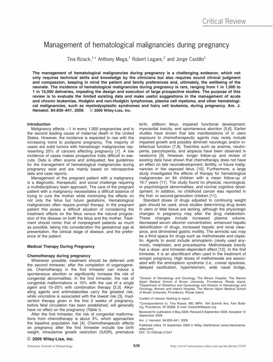
the second trimester, after the completion of organogene-sis. Chemotherapy in the first trimester can induce aspontaneous abortion or significantly increase the risk ofcongenital abnormalities. In the first trimester, the risk ofcongenital malformations is 10% with the use of a singleagent and 15–25% with combination therapy [2,3]. Alkyl-ating agents and antimetabolites carry the greatest risk,while vincristine is associated with the lowest risk [3]. Inad-vertent therapy given in the first 2 weeks of pregnancy,before fetal circulation has been established, will generallyhave no effect on the pregnancy (Table I).After the first trimester, the risk of congenital malforma-
tions from chemotherapy is about 3%, which approachesthe baseline population risk [4]. Chemotherapeutic effectson pregnancy after the first trimester include low birthweight, intrauterine growth restriction (IUGR), premature
birth, stillborn fetus, impaired functional development,myocardial toxicity, and spontaneous abortion [5,6]. Earlierstudies have shown that late manifestations of in uteroexposure to chemotherapeutic agents may rarely includeimpaired growth and possibly diminish neurologic and/or in-tellectual function [7,8]. Toxicities such as anemia, neutro-penia, pancytopenia, and alopecia have been observed innewborns [9]. However, longer follow-up and review ofexisting data have shown that chemotherapy does not havean effect on late neurodevelopment, fertility, or future malig-nancies in the exposed fetus [10]. Furthermore, a largestudy investigated the effects of therapy for hematologicalmalignancies on 84 children with a mean follow-up of18.7 years [11]. The study found no physical, neurological,or psychological abnormalities, and normal cognitive devel-opment. In addition, no childhood cancer was reported inthe first- or second-generation children [11].Standard doses of drugs adjusted to continuing weight
gain should be used, since studies determining drug levelsin utero or fetal tissue are lacking, although the physiologicchanges in pregnancy may alter the drug metabolism.These changes include increased plasma volume,decreased serum albumin concentration, enhanced hepaticdetoxification of drugs, increased hepatic and renal clear-ance, and diminished gastric motility. The amniotic sac maybe a third space for drugs such as methotrexate and cispla-tin. Agents to avoid include aminopterin (rarely used any-more), melphalan, and procarbazine. Methotrexate toxicityhas a dose- and trimester-dependent effect [12]. In the firsttrimester, it is an abortificant often used in the treatment ofectopic pregnancy. High doses of methotrexate are associ-ated with the aminopterin syndrome (i.e., cranial dysotosis,delayed ossification, hypertelorism, wide nasal bridge,
Conflict of interest: Nothing to report.
*Correspondence to: Tina Rizack, MD, MPH, 164 Summit Ave, Fain Build-ing, Providence, RI 02906. E-mail: [email protected]
1Division of Hematology and Oncology, The Miriam Hospital, The WarrenAlpert Medical School of Brown University, Providence, Rhode Island;2Department of Obstetrics and Gynecology and Division of Hematology andOncology, Women and Infant’s Hospital, The Warren Alpert Medical Schoolof Brown University, Providence, Rhode Island
Received for publication 4 May 2009; Revised 8 September 2009; Accepted 12September 2009
Am. J. Hematol. 84:830–841, 2009.
Published online 16 September 2009 in Wiley InterScience (www.interscience.wiley.com).DOI: 10.1002/ajh.21547
Critical Review
VVC 2009 Wiley-Liss, Inc.
American Journal of Hematology 830 http://www3.interscience.wiley.com/cgi-bin/jhome/35105
TABLEI.
MaternalandFetalComplicationsReportedwithSelectedAnticancerAgents
Usedto
TreatHematologicalMalignancies
Agent
Use
Majormaternal
complications
Fetaleffects
Pregnancy
category
Alkylatingagents
Chlorambucil
CLL,NHL
Mye
losuppression
Firsttrim
ester:Afew
casereportswithnoadve
rseeffects,twocasesofunilateralrenalandureteragenesis,onecaseofaretinaldefect.
DSecondto
thirdtrim
ester:Seve
ralcasereportswith
noadve
rseeffects.
Cisplatin
AML,NHL,HL
Mye
losuppression;
nephrotoxicity;
neurotoxicity
Firsttrim
ester:Twocaseswithnoadve
rseeffects.
Secondto
thirdtrim
ester:Twocaseswithnoadve
rseeffects;onecasewithexposure
at26weeksanddelivered6dayslaterwithneutropenia,
hairloss,
andhearingim
pairment.
D
Cyclophosphamide
NHL,CLL,MM,ALL
Mye
losuppression
Caseseries:Twenty-onecases(11in
firsttrim
ester)
withnoanomalies,
anotherseriesofninepatients
(threein
firsttrim
ester)
withno
anomaliesbutonepatienttreatedin
allthreetrim
esters
deliveredaninfantwithpancytopenia
andlow
birth
weight.a
D
Firsttrim
ester:Fourcaseswithdigitabnorm
alities,twowithhernias,onesingle
leftcoronary
artery,onestillbirth,onegrowth
retardation,one
imperforate
anuswithrectova
ginalfistula,onefacialabnorm
ality,
deve
lopmentaldelay.a
Secondto
thirdtrim
ester:Few
caseseriesoflong-term
follow-upinfants
withnorm
algrowth
andmentaldeve
lopment.Low
birth
weightcommon
sideeffect.Twoinfants
withpancytopenia
withexposure
inthirdtrim
ester.
Dacarbazine
HL
Mye
losuppression
Caseseries:Seve
nteencases,
seve
nin
firsttrim
ester,nocongenitalabnorm
alities.
CSecondto
thirdtrim
ester:Onecaseexposedin
fourthmonth
withnoabnorm
alities.
Mechlorethamine
HL
Mye
losuppression;infertility
Caseseries:Six
patients,onewithfirsttrim
esterexposure,nocongenita
lanomalies.
Seriesof13caseswithfirsttrim
esterexposure:one
spontaneousabortion,oneelective
term
ination,oneinfantborn
withhydrocephalusdied4hrafterbirth.a
D
Melphalan
MM
Mye
losuppression
Firsttrim
ester:Onereportedmiscarriage.
DProcarbazine
HL
Mye
losuppression;infertility
Caseseries:Thirteenpatients,threereceivedcombinationchemotherapy,
onespontaneousabortion,oneelective
term
ination,onewith
hydrocephalus,
died4hrafterbirth.Seriesofsix
children,onein
firsttrim
ester,withnoabnorm
alities.Twenty-six
pregnancies:Oneinfant
withcleftlip
andpalate.a
D
Firsttrim
ester:Atleastsix
casesonly
twowithnorm
alinfants,low
birth
weightmale
diedafterbirth
foundto
have
atrialseptaldefect,aelective
abortionwithmale
infantwithmarkedly
reducedmalpositionedkidneys,spontaneousabortionat24weekswithdigitalabnorm
alities,a
norm
al
infantwithmultiple
hemangiomas.
Secondto
thirdtrim
ester:Noabnorm
alitiesdescribed.
Anthracyclines
Daunorubicin
AML,ALL,APML
Mye
losuppression;acute
andchronic
cardiac
toxicities
Caseseries:Ninecases,
onewith
firsttrim
esterexposure,noteratogenic
effects,twoelective
term
inations,
twopremature
deliveries,two
stillbirths(oneofwhichhadmyo
cardialnecrosis).
Firsttrim
ester:Fourpatients,twonorm
al,twospontaneousabortions.
D
Secondto
thirdtrim
ester:Onecaseofpremature
delivery,onecaseof‘‘fetaldistress’’andtransientneonatalmarrow
suppression.
Doxorubicin
HL,NHL
Mye
losuppression;acute
andchronic
cardiac
toxicities
Firsttrim
ester:Nine/tencaseswithnoadve
rseoutcomes,
onecasewithim
perforate
anusandrectova
ginalfistula.
D
Idarubicin
AML,ALL,APML
Mye
losuppression;acute
andchronic
cardiac
toxicities
Fivecasesallwith
complications,
includingonecaseofIUFD
2daysafterinfusionforconsolidation,anothercaseofIUFD,IUGR,andtwo
casesofcardiomyo
pathy.a
D
Antibiotics
Bleomycin
HL,NHL
Pneumonitis
Caseseries:Twenty-threepatients
(11in
firsttrim
ester)
withnoanomalies.
aD
Secondto
thirdtrim
ester:Isolatedcasereportsofregim
ensincludingbleomycin
withnocongenitalabnorm
alities.Onecaseoftransient
leucopenia
andneutropenia
inapremature
infantwhosemotherreceivedBEP7–10dayspriorto
delivery.
Antimetabolites
Cladribine
HCL
Mye
losuppression
Nocasesin
literature.
DFludarabine
CLL,NHL,MM
Mye
losuppression
Nocasesin
literature.
DHydroxyurea
AML,ALL,CML,CLL
Mye
losuppression
Caseseries:Ninepatients,seve
nwithnoadve
rseeffects,oneelective
abortionofapparentlynorm
alfetus,
onestillborn
withoutgross
abnorm
alities.
Single
casereportofanorm
alinfantwithexposure
inalltrim
esters,whichalsocommentedonfourothercases:One
premature
delivery
butnocongenitaldefects.Thirty-onecasesnotedincreasedriskofIUGR,IUFD,andprematurity,butmayhave
beendue
tounderlyingdisease.
D
Methotrexate
ALL,NHL
Mye
losuppressionand
Acute
renalfailure
Caseseries:Ninepatients,five
infirsttrim
ester,nocongenitalabnorm
alities,fourlow
birth
weight,onewithlow
birth
weightandpancytopenia.a
Seriesofeightpatients
(tenpregnancies)with
exposure
priorto
andduringpregnancy:Threespontaneousabortions,
twoelective
abortions,
fourfull-term
infants
withnoabnorm
alities(allwithfirsttrim
esterexposure).Prospective
seriesof21patients,withfive
exposuresduring
pregnancy(fourin
firsttrim
ester)
withnoabnorm
alities.
X
Firsttrim
ester:Atleastseve
nchildrenwithaminopterin-typesyndrome.
American Journal of Hematology 831
critical review

TABLE
I.(C
ontinued)
Agent
Use
Majormaternal
complications
Fetaleffects
Pregnancy
category
Secondto
thirdtrim
ester:Onecaseofseve
repancytopenia
withexposure
inallthreetrim
esters,five
caseswithnoanomalies,
oneinfantwith
46chromosomesbutwith
presenceofgapsandaringchromosomeofunknownsignificance.a
Nucleosideanalogs
Cytarabine
AML,ALL,CML
Mye
losuppression
Caseseries:Caseseriesofninepatients
allnorm
al,five
infirsttrim
ester.Caseseriesofninepatients,fourin
firsttrim
ester,nonewith
malfo
rmations,twowithlow
birth
weight,onewithseve
repancytopenia
andlow
weight.Caseseriesofseve
npatients,twowithlow
birth
weight,onewiththrombocytopenia.a
D
Firsttrim
ester:Manycasereportswithnorm
alinfants.Onecasewithlow
birth
weight,digitabnorm
alitiesbutmotherhadsim
ilarexposure
insubsequentpregnancyanddeliveredanorm
alinfant.aOneinfantwithsingle
agentexposure
with
bilateralmicrotiawithatresia
ofauditory
canals,lim
b,anddigitdeform
ities.
Secondto
thirdtrim
ester:Threecaseswithchromosomalabnorm
alities.Oneinfantwithnorm
alphenotypebutpresenceofgapsandaring
chromosomeofunknownsignificance,a
threecasesofIUFD.Oneinfantborn
withbruisingandpetechiae,oneinfantwithseve
repancytopenia
exposedin
allthreetrim
esters,onecaseofseve
repancytopenia,a
twocasesofneutropenia
withthrombocytopenia.
TopoisomeraseIIinhibitors
Etoposide
AML,NHL,HL
Mye
losuppression,May
prolongPT,INR
Caseseries:Eleve
npatie
nts,twoin
firsttrim
esterwithnocongenitalabnorm
alities,
oneinfantborn
withleucopenia
andhearinglossat27
weeks,
transientpancytopenia
ininfantborn
at32weeksaftermaternaltreatm
entin
thirdtrim
ester.
D
Vincaalkaloids
Vinblastine
HL
Mye
losuppression
Caseseries:Tenchildren,fourwithexposure
duringfirsttrim
ester,nonewithabnorm
alities.Seriesof26patients,oneinfantborn
with
hydrocephaluswithexposure
atWeek3,oneinfantwithcleftpalate
exposedduringfirsttrim
ester.aSix
patients
withnocongenitalanomalies
abnorm
alities.
D
Firsttrim
ester:More
than15casesreported,onespontaneousabortionwithdigitalabnorm
alities,
onelow
birth
weightinfantdiedshortly
after
birth
from
smallsecundum
atrialdefect.a
Secondto
thirdtrim
ester:More
thantencasesreportedwithnoabnorm
alities.
Vincristine
HL,NHL,ALL
Neurotoxicity
Caseseries:Noanomaliesobservedin
28children,12womentreatedin
thefirsttrim
ester.Seriesofninepatients,five
treatedduringfirst
trim
ester,hadnoanomalies:fourwithlow
birth
weight,onewithseve
reneutropenia
andlow
birth
weight.aSeriesofseve
npatients,three
duringsecondorthirdtrim
ester,nocongenitalabnorm
alities.Seriesof26pregnancies,
onecleftlip
andpalate.a
Seriesof13patients
with
firsttrim
esterexposure,outoffourpregnanciesthere
wasonenorm
allivebirth,onespontaneousabortion,oneelective
term
ination,andone
hydrocephalus,
died4hrafterbirth.a
D
Firsttrim
ester:Twomalfo
rmedinfants
reported:onelow
birth
weightinfantdiedshortly
afterbirth
from
smallsecundum
atrialdefectandone
elective
abortionwithmarkedly
reducedmalfo
rmedkidneys.
aTwoothermalfo
rmedinfants
were
reportedin
acasesseries.
aOnehealthy
infanttreatedafter22ndweekwith46chromosomeswiththepresenceofgapsandaringchromosomeofunknownsignificance.a
Onecase
ofseve
repancytopenia
aandonecaseoftransientseve
rebonemarrow
suppressiona(feltto
bedueto
mercaptopurineexposure).Onecase
ofIUFD.
Monoclonalantibodies
Ibritumomab
NHL
Infusion-relatedreactions;
mye
losuppression
Contraindicatedin
pregnancydueto
prohibitivefetalradiationexposure.
D
Rituxim
ab
NHL
Infusion-relatedreactions;
mye
losuppression
Few
reportsshow
safety
inalltrim
esters.TransientB-celldepletionreportedfollowedbyfullim
munologic
recove
ryandnorm
alresponseto
vaccines.
C
Tositumomab
NHL
Infusion-relatedreactions;
mye
losuppression
Contraindicateddueto
prohibitivefetalradiationexposure.
X
Tyrosinekinaseinhibitors
Dasatinib
CML
Fluid
retention
Caseseries:Eightpatients,threeelective
term
inations,twospontaneousabortions,
threehealthydeliveries,
oneunknownoutcome.
DIm
atinib
CML
Fatigue;fluid
retention
Caseseries:Onehundredandeighty
patients,103withfirsttrim
esterexposure,38exposedin
allthreetrim
esters,125withava
ilable
data.Fifty
percentdeliverednorm
alinfants,35electedterm
ination(threeforknowncongenitalabnorm
alities).Twelveinfants
born
with
abnorm
alities:
Threewithsim
ilardefects
includingexomphalos,
cardiac,
andrenalanomalies.
D
Miscellaneous
All-transretinoic
acid
(ATRA)
APML
Hemorrhage
DIC
Firsttrim
ester:Eighty-five
percentriskofteratogenicity,
includingseve
reneurologicalandcardiova
scularcomplications.
Increasedrate
of
miscarriage.a
D
Secondto
thirdtrim
esters:Nocongenitalabnorm
alitiesreported.
Asparaginase
ALL
Allergic
reactions;
hypofibrinogenemia
Caseseries:Ninecases,
afourin
firsttrim
ester,withnocongenitalabnorm
alities,onecaseoftransientoligohydramnios,
meconium,andlow
birth
weight,andoneinfantwithnorm
alphenotypebutpresenceofgapsandaringchromosomeofunknownsignificance.
C
Bortezo
mib
MM
Neuropathy;hypotension
Nocasesin
literature.
D
832 American Journal of Hematology
critical review
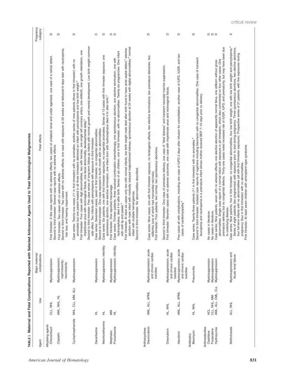
micrognatia, and ear anomalies) [9,13]. Most experts rec-ommend against the use of methotrexate in pregnancyunless no acceptable alternative exists.
Supportive therapy during pregnancyThe use of supportive therapy should be the same as for
the general population. Antihistamines, ondansetron, phe-nothiazines, metoclopramide, and corticosteroids have fairlygood safety profiles in pregnancy [14].Antiemetics. A large retrospective study of 3,458
patients exposed to metoclopramide during their first tri-mester found no increase in adverse outcomes on the fetusor pregnancy when compared to pregnant patients who didnot receive metoclopramide [15]. Ondansetron, a 5-HT3-se-rotonin antagonist, has been reported to be safe in preg-nancy. Four cases were exposed to ondansetron duringfirst, second, and/or third trimesters; there were no fetaladverse outcomes with a highly efficacious control of hyper-emesis in the mother [16–19]. The only prospective trial ofondansetron included 176 women exposed in the first tri-mester and did not appear to be associated with anincreased risk of major malformations above baseline, butthe sample size was felt to be limited [20]. Granisetron hasbeen studied prospectively to prevent nausea and vomitingduring cesarean section and has been shown to be safeand effective in this setting [21]. There is not enough datato support the use of other 5-HT3-serotonin antagonists oraprepitant.Growth factors. Erythropoietin does not cross the pla-
centa and its use is felt to be safe in pregnancy [22]. Gran-ulocyte colony-stimulating factor use in pregnancy hasbeen reported in a registry series of 20 patients with severechronic neutropenia with a median dose of 2.7 mcg/kg/dayadministered daily or every other day during all three tri-mesters with an average duration of three trimesters.These data, although limited, did not reveal an increase inadverse congenital abnormalities or fetal death comparedto pregnant patients that did not receive the drug [23].Bisphosphonates. Although the use of bisphosphonates
during pregnancy has not been evaluated prospectively, arecent literature search including 51 patients exposed tobisphosphonates shortly prior to conception or during preg-nancy did not find evidence of skeletal abnormalities ormalformations in the products of the exposed mothers [24].Leukapheresis. Leukapheresis has been used in both
acute and chronic leukemia to rapidly reduce high whiteblood cell counts in patients with impending vascular occlu-sion. Experience with leukapheresis during pregnancy islimited to only a handful of cases used to treat both chronicand acute leukemias [25–31]. In general, the therapy wastolerated well by the mother and the fetus. Although experi-ence is limited, leukapheresis may be used as a short-termtemporizing measure when no other options exist or inpatients refusing other therapies during pregnancy.
Management of Lymphoma During PregnancyWhenever possible, diagnosis should be made with an
excisional biopsy of a lymph node, a procedure that is easyto perform and has not shown increased morbidity or mor-tality to the mother or the fetus [32,33]. Staging should bedone with a posterior/anterior chest X-ray with abdominalshielding, and routine blood work, including complete bloodcount, erythrocyte sedimentation rate, serum creatinine,and liver enzymes, including lactate dehydrogenase andalkaline phosphatase [34]. However, serum alkaline phos-phatase is significantly elevated in the third trimester andmay not be useful. A pathologic placental evaluation afterdelivery should be done routinely to assess the need forappropriate staging and therapy in the neonate [1]. A boneT
ABLE
I.(C
ontinued)
Agent
Use
Majormaternal
complications
Fetaleffects
Pregnancy
category
Interferonalpha
MM,HCL,CML
Flu-likeillness;fatigue;
infertility
Fortycasesdocumentedin
literature,eightin
firsttrim
ester,only
onecaseoffetalmalfo
rmationswithconcurrentuseofhydroxyurea.
C
Lenalidomide
MM,MDS
Mye
losuppression;
thrombosis
Perinatalrisksbasedonexperiencewiththalidomide.
X
Thalidomide
MM
Neuropathy;thrombosis
Seve
remalfo
rmationsincludedefects
ofthelim
bs,
axialske
leton,headandface,eye
s,ears,tongue,teeth,centralnervous,
respiratory,
cardiova
scular,andgenitourinary
systems,
andthegastrointestinaltractwithriskofcongenita
lmalfo
rmationsbetween20and50%.
X
Table
adaptedandmodifiedfrom
http://www.m
otherisk.org/women/cancerC
ategory.jsp?category_id527.
ALL,acute
lymphocyticleuke
mia;AML,acute
mye
loid
leuke
mia;APML,acute
promye
locyticleuke
mia;BEP,
bleomycin,etoposide,cisplatin;CLL,chronic
lymphocyticleuke
mia;CML,chronic
mye
loid
leuke
mia;DIC,disseminatedintra-
vascularcoagulation;HCL,hairycellleuke
mia;HL,Hodgkin
lymphoma;NHL,non-H
odgkin
lymphoma;MDS,mye
lodysplasticsyndrome;MM,multiple
mye
loma;IUFD,intrauterinefetaldemise;IUGR,intrauterinegrowth
retardation.
aMultiple
drugregim
enused.
Pregnancycategory
interpretation:A:Controlledstudiesshow
norisk;adequate,well-controlledstudiesin
pregnantwomenhave
failedto
demonstrate
riskto
thefetus.
B:Noevidenceofriskin
humans;eitheranim
alfindingsshow
risk
(buthumanfindingsdonot)or,ifnoadequate
humanstudieshave
beendone,anim
alfindingsare
negative
.C:Riskcannotberuledout;humanstudiesare
lackingandanim
alstudiesare
eitherpositiveforfetalriskorlackingaswell;
how-
eve
r,potentialbenefits
mayjustify
thepotentialrisk.D:Positiveevidenceofrisk;inve
stigationalorpostm
arketingdata
show
riskto
fetus;neve
rtheless,
potentialbenefits
mayoutweightherisk.X:Contraindicatedin
pregnancy;studiesin
anim
als
orhumans,
orinve
stigationalorpostm
arketingreportshave
shownfetalrisk,whichclearlyoutweighsanypossible
benefitto
thepatient.
American Journal of Hematology 833
critical review

marrow biopsy is recommended for non-Hodgkin lymphoma(NHL) and only in patients with B symptoms or cytopeniasin Hodgkin lymphoma (HL). Intrabdominal disease can beevaluated with noncontrast MRI or ultrasound [35,36]. PETand gallium have not proven to be safe in pregnancy [37].Bone scans are generally not recommended unless thereare no other means of detecting bone metastases andresults would change management. Baker et al. describedabout a series of three pregnant patients with breast cancerwho received a modified bone scan to reduce fetal expo-sure with normal outcomes in the offspring. The authorssuggest using 10 mCI rather than 20 mCI and doubling theimaging time to reduce fetal exposure [38]. A Foley cathe-ter should be placed while hydrating the patient to promoterapid washout of the excreted radiopharmaceutical from thepatient’s bladder. On the other hand, MRI has a diagnosticaccuracy of 91% for the detection of bone metastases inhematological malignancies [39].Delivery should be delayed until fetal maturity is
achieved, without compromising the health of the mother orfetus. If bleomycin was used during pregnancy, it has beensuggested that the women should not receive oxygen dur-ing delivery not to exacerbate possible pulmonary toxicity[40]. A cesarean delivery is not warranted unless otherwiseindicated, because staging laparotomy is no longer recom-mended. Delivery should be planned no less than 2 weeksbut ideally 3–4 weeks after chemotherapy to allow formaternal and fetal blood count recovery and completechemotherapy metabolism by the fetus and placenta. A full-staging assessment should be performed postpartum in allpatients, usually with a PET-CT; 18F-FDG can cross theplacenta and is concentrated in breast tissue and in breastmilk [41]. The patient should not hold the infant for 24 hr af-ter the PET scan to decrease radiation exposure of thenewborn. If breastfeeding, the patient should discontinuegiving the baby breast milk for 72 hr after a PET scan. It isrecommended that patients treated for cancer wait 1 or 2years after the completion of chemotherapy before conceiv-ing, as this is the most likely period of disease recurrence.In lymphoma patients felt to have a high likelihood of cure,1 year may be adequate.
Hodgkin lymphomaHL is more commonly seen during pregnancy than NHL
because the peak incidence of HL coincides with the repro-ductive years. HL affects one in 1,000–6,000 pregnancies[42] and concurrent pregnancy has been identified in 3.2%of all patients with HL [43]. HL is more commonly seen inwomen with lower parity and advanced maternal age, butthe clinical behavior and prognosis are similar to those ofnonpregnant women [44]. Pathologically, the majority ofcases present with nodular sclerosing HL, which is also themost common histology seen in women younger than 40years [45]. The largest study to date, consisting of 48 preg-nant women matched to nonpregnant controls with HL,showed a median age of 26 years without difference instage distribution at diagnosis [46]. See Fig. 1 for an algo-rithmic approach to the management of HL in pregnancy.Early-stage HL. In general terms, treatment of early-
stage HL should and can be deferred until after the secondtrimester [34,47]. In fact, more than 50% of patients cancontinue pregnancy to term without treatment [48]. Thus, ifan early-stage HL diagnosis is made early in pregnancy, awatchful waiting approach is reasonable. If treatment isrequired, it is usually possible to control the lymphoma withsingle-agent chemotherapy, such as vinblastine or anthra-cyclines, allowing the pregnancy to go to term [34,47,48].The combination of doxorubicin, vincristine, bleomycin, and
dacarbazine (ABVD) is the most frequently employed regi-men in the US and is considered the standard of care [49].Recently, a study about a series of 17 patients treated overthe past 21 years was published favoring the use of single-agent vinblastine; 11 patients required no treatment duringpregnancy and 6 required single-agent vinblastine to con-trol disease until delivery. This series found a greater than75% response rate to single-agent vinblastine at doses of6 mg/m2 used in treatment-naı̈ve patients at intervals ofseveral weeks or longer with minimal toxicity to mother andchild. All 17 women treated delivered normal infants nowranging in age from 2 to 21 years [48]. Several otherarticles report the use of single agent vinblastine in the firsttrimester with no adverse effects on the fetus (except forone case of fetal syndactyly in a child whose mother alsoreceived oral cyclophosphamide during all three trimesters)and favorable disease response [50]. Patients who progressdespite vinblastine can be treated with ABVD during thesecond or third trimester.Several studies have documented the efficacy of radia-
tion for early-stage HL during pregnancy [43,45,47]. Radia-tion should be reserved for cases in which it is absolutelynecessary and extreme precaution should be undertaken tolimit the whole body fetal dose to less than or equal to0.1 Gy and to protect the uterus by using 10 half-valuelayer shielding [5]. Prior to treatment, the maximal dose tothe fetus should be calculated and then monitored through-out treatment [51]. The aim is for partial rather than defini-tive therapy until after delivery. Early stage supradiagphrag-matic disease such as isolated cervical or axillary sitesmay be treated with involved-field radiation therapy [43,52].Mediastinal sites or mantle field irradiation are feasible inpregnant women with early stage HL during the secondand third trimester, if special attention is paid to shieldingand radiation delivery techniques [43,52]. Healthy offspringwas seen in all the cases treated with mediastinal irradia-tion for supradiaphragmatic HL, with midfetal doses of lessthan 0.05 Gy [43,52]. However, other studies using adummy have shown that the embryo is exposed to doseshigher than 0.1 Gy, despite appropriate shielding [53]. Withrecent advances in radiation oncology techniques, espe-cially smaller portal sites, the risk is likely to be lower thanpreviously reported [51].Treatment delay may also be considered in limited clinical
stage IA or IIA HL presenting during the second or third tri-mester, stable nonurgent HL diagnosed after 20 weeksgestation, or clinically accessible sites of disease that canbe easily monitored [5,42]. Accumulating but still limiteddata have shown the feasibility and safety of administering
Figure 1. Proposed management of Hodgkin lymphoma during pregnancy.
834 American Journal of Hematology
critical review
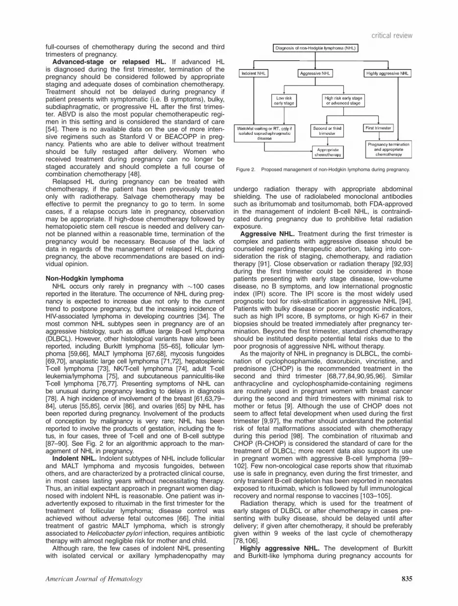
full-courses of chemotherapy during the second and thirdtrimesters of pregnancy.Advanced-stage or relapsed HL. If advanced HL
is diagnosed during the first trimester, termination of thepregnancy should be considered followed by appropriatestaging and adequate doses of combination chemotherapy.Treatment should not be delayed during pregnancy ifpatient presents with symptomatic (i.e. B symptoms), bulky,subdiaphragmatic, or progressive HL after the first trimes-ter. ABVD is also the most popular chemotherapeutic regi-men in this setting and is considered the standard of care[54]. There is no available data on the use of more inten-sive regimens such as Stanford V or BEACOPP in preg-nancy. Patients who are able to deliver without treatmentshould be fully restaged after delivery. Women whoreceived treatment during pregnancy can no longer bestaged accurately and should complete a full course ofcombination chemotherapy [48].Relapsed HL during pregnancy can be treated with
chemotherapy, if the patient has been previously treatedonly with radiotherapy. Salvage chemotherapy may beeffective to permit the pregnancy to go to term. In somecases, if a relapse occurs late in pregnancy, observationmay be appropriate. If high-dose chemotherapy followed byhematopoietic stem cell rescue is needed and delivery can-not be planned within a reasonable time, termination of thepregnancy would be necessary. Because of the lack ofdata in regards of the management of relapsed HL duringpregnancy, the above recommendations are based on indi-vidual opinion.
Non-Hodgkin lymphomaNHL occurs only rarely in pregnancy with �100 cases
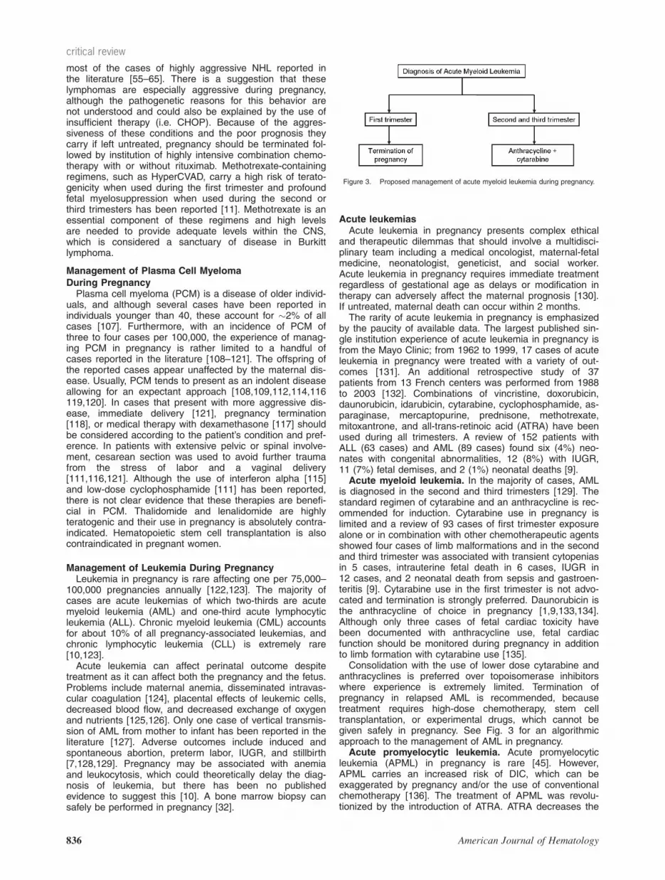
reported in the literature. The occurrence of NHL during preg-nancy is expected to increase due not only to the currenttrend to postpone pregnancy, but the increasing incidence ofHIV-associated lymphoma in developing countries [34]. Themost common NHL subtypes seen in pregnancy are of anaggressive histology, such as diffuse large B-cell lymphoma(DLBCL). However, other histological variants have also beenreported, including Burkitt lymphoma [55–65], follicular lym-phoma [59,66], MALT lymphoma [67,68], mycosis fungoides[69,70], anaplastic large cell lymphoma [71,72], hepatosplenicT-cell lymphoma [73], NK/T-cell lymphoma [74], adult T-cellleukemia/lymphoma [75], and subcutaneous panniculitis-likeT-cell lymphoma [76,77]. Presenting symptoms of NHL canbe unusual during pregnancy leading to delays in diagnosis[78]. A high incidence of involvement of the breast [61,63,79–84], uterus [55,85], cervix [86], and ovaries [65] by NHL hasbeen reported during pregnancy. Involvement of the productsof conception by malignancy is very rare; NHL has beenreported to involve the products of gestation, including the fe-tus, in four cases, three of T-cell and one of B-cell subtype[87–90]. See Fig. 2 for an algorithmic approach to the man-agement of NHL in pregnancy.Indolent NHL. Indolent subtypes of NHL include follicular
and MALT lymphoma and mycosis fungoides, betweenothers, and are characterized by a protracted clinical course,in most cases lasting years without necessitating therapy.Thus, an initial expectant approach in pregnant women diag-nosed with indolent NHL is reasonable. One patient was in-advertently exposed to rituximab in the first trimester for thetreatment of follicular lymphoma; disease control wasachieved without adverse fetal outcomes [66]. The initialtreatment of gastric MALT lymphoma, which is stronglyassociated to Helicobacter pylori infection, requires antibiotictherapy with almost negligible risk for mother and child.Although rare, the few cases of indolent NHL presenting
with isolated cervical or axillary lymphadenopathy may
undergo radiation therapy with appropriate abdominalshielding. The use of radiolabeled monoclonal antibodiessuch as ibritumomab and tositumomab, both FDA-approvedin the management of indolent B-cell NHL, is contraindi-cated during pregnancy due to prohibitive fetal radiationexposure.Aggressive NHL. Treatment during the first trimester is
complex and patients with aggressive disease should becounseled regarding therapeutic abortion, taking into con-sideration the risk of staging, chemotherapy, and radiationtherapy [91]. Close observation or radiation therapy [92,93]during the first trimester could be considered in thosepatients presenting with early stage disease, low-volumedisease, no B symptoms, and low international prognosticindex (IPI) score. The IPI score is the most widely usedprognostic tool for risk-stratification in aggressive NHL [94].Patients with bulky disease or poorer prognostic indicators,such as high IPI score, B symptoms, or high Ki-67 in theirbiopsies should be treated immediately after pregnancy ter-mination. Beyond the first trimester, standard chemotherapyshould be instituted despite potential fetal risks due to thepoor prognosis of aggressive NHL without therapy.As the majority of NHL in pregnancy is DLBCL, the combi-
nation of cyclophosphamide, doxorubicin, vincristine, andprednisone (CHOP) is the recommended treatment in thesecond and third trimester [68,77,84,90,95,96]. Similaranthracycline and cyclophosphamide-containing regimensare routinely used in pregnant women with breast cancerduring the second and third trimesters with minimal risk tomother or fetus [9]. Although the use of CHOP does notseem to affect fetal development when used during the firsttrimester [9,97], the mother should understand the potentialrisk of fetal malformations associated with chemotherapyduring this period [98]. The combination of rituximab andCHOP (R-CHOP) is considered the standard of care for thetreatment of DLBCL; more recent data also support its usein pregnant women with aggressive B-cell lymphoma [99–102]. Few non-oncological case reports show that rituximabuse is safe in pregnancy, even during the first trimester, andonly transient B-cell depletion has been reported in neonatesexposed to rituximab, which is followed by full immunologicalrecovery and normal response to vaccines [103–105].Radiation therapy, which is used for the treatment of
early stages of DLBCL or after chemotherapy in cases pre-senting with bulky disease, should be delayed until afterdelivery; if given after chemotherapy, it should be preferablygiven within 9 weeks of the last cycle of chemotherapy[78,106].Highly aggressive NHL. The development of Burkitt
and Burkitt-like lymphoma during pregnancy accounts for
Figure 2. Proposed management of non-Hodgkin lymphoma during pregnancy.
American Journal of Hematology 835
critical review
most of the cases of highly aggressive NHL reported inthe literature [55–65]. There is a suggestion that theselymphomas are especially aggressive during pregnancy,although the pathogenetic reasons for this behavior arenot understood and could also be explained by the use ofinsufficient therapy (i.e. CHOP). Because of the aggres-siveness of these conditions and the poor prognosis theycarry if left untreated, pregnancy should be terminated fol-lowed by institution of highly intensive combination chemo-therapy with or without rituximab. Methotrexate-containingregimens, such as HyperCVAD, carry a high risk of terato-genicity when used during the first trimester and profoundfetal myelosuppression when used during the second orthird trimesters has been reported [11]. Methotrexate is anessential component of these regimens and high levelsare needed to provide adequate levels within the CNS,which is considered a sanctuary of disease in Burkittlymphoma.
Management of Plasma Cell MyelomaDuring PregnancyPlasma cell myeloma (PCM) is a disease of older individ-
uals, and although several cases have been reported inindividuals younger than 40, these account for �2% of allcases [107]. Furthermore, with an incidence of PCM ofthree to four cases per 100,000, the experience of manag-ing PCM in pregnancy is rather limited to a handful ofcases reported in the literature [108–121]. The offspring ofthe reported cases appear unaffected by the maternal dis-ease. Usually, PCM tends to present as an indolent diseaseallowing for an expectant approach [108,109,112,114,116119,120]. In cases that present with more aggressive dis-ease, immediate delivery [121], pregnancy termination[118], or medical therapy with dexamethasone [117] shouldbe considered according to the patient’s condition and pref-erence. In patients with extensive pelvic or spinal involve-ment, cesarean section was used to avoid further traumafrom the stress of labor and a vaginal delivery[111,116,121]. Although the use of interferon alpha [115]and low-dose cyclophosphamide [111] has been reported,there is not clear evidence that these therapies are benefi-cial in PCM. Thalidomide and lenalidomide are highlyteratogenic and their use in pregnancy is absolutely contra-indicated. Hematopoietic stem cell transplantation is alsocontraindicated in pregnant women.
Management of Leukemia During PregnancyLeukemia in pregnancy is rare affecting one per 75,000–
100,000 pregnancies annually [122,123]. The majority ofcases are acute leukemias of which two-thirds are acutemyeloid leukemia (AML) and one-third acute lymphocyticleukemia (ALL). Chronic myeloid leukemia (CML) accountsfor about 10% of all pregnancy-associated leukemias, andchronic lymphocytic leukemia (CLL) is extremely rare[10,123].Acute leukemia can affect perinatal outcome despite
treatment as it can affect both the pregnancy and the fetus.Problems include maternal anemia, disseminated intravas-cular coagulation [124], placental effects of leukemic cells,decreased blood flow, and decreased exchange of oxygenand nutrients [125,126]. Only one case of vertical transmis-sion of AML from mother to infant has been reported in theliterature [127]. Adverse outcomes include induced andspontaneous abortion, preterm labor, IUGR, and stillbirth[7,128,129]. Pregnancy may be associated with anemiaand leukocytosis, which could theoretically delay the diag-nosis of leukemia, but there has been no publishedevidence to suggest this [10]. A bone marrow biopsy cansafely be performed in pregnancy [32].
Acute leukemiasAcute leukemia in pregnancy presents complex ethical
and therapeutic dilemmas that should involve a multidisci-plinary team including a medical oncologist, maternal-fetalmedicine, neonatologist, geneticist, and social worker.Acute leukemia in pregnancy requires immediate treatmentregardless of gestational age as delays or modification intherapy can adversely affect the maternal prognosis [130].If untreated, maternal death can occur within 2 months.The rarity of acute leukemia in pregnancy is emphasized
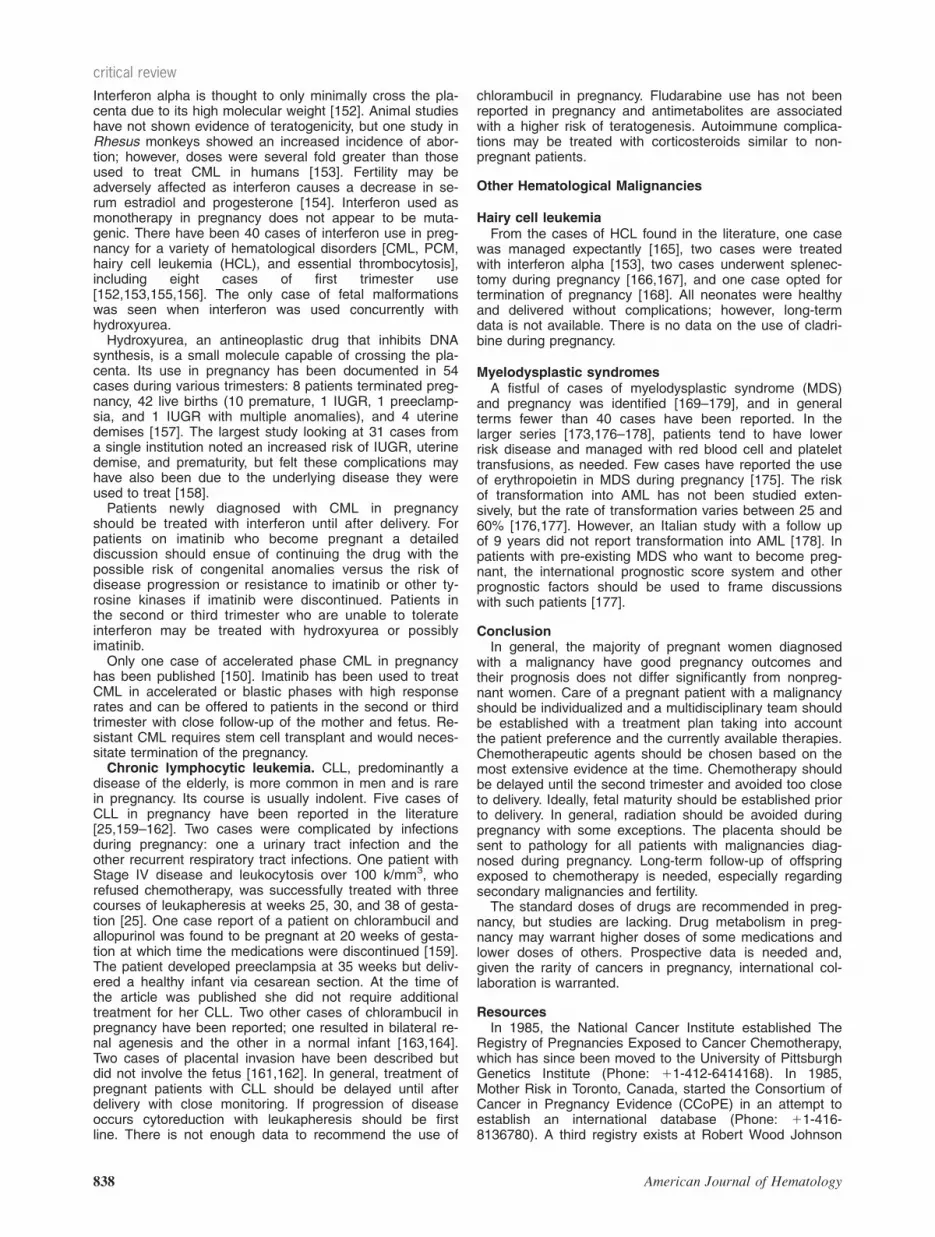
by the paucity of available data. The largest published sin-gle institution experience of acute leukemia in pregnancy isfrom the Mayo Clinic; from 1962 to 1999, 17 cases of acuteleukemia in pregnancy were treated with a variety of out-comes [131]. An additional retrospective study of 37patients from 13 French centers was performed from 1988to 2003 [132]. Combinations of vincristine, doxorubicin,daunorubicin, idarubicin, cytarabine, cyclophosphamide, as-paraginase, mercaptopurine, prednisone, methotrexate,mitoxantrone, and all-trans-retinoic acid (ATRA) have beenused during all trimesters. A review of 152 patients withALL (63 cases) and AML (89 cases) found six (4%) neo-nates with congenital abnormalities, 12 (8%) with IUGR,11 (7%) fetal demises, and 2 (1%) neonatal deaths [9].Acute myeloid leukemia. In the majority of cases, AML
is diagnosed in the second and third trimesters [129]. Thestandard regimen of cytarabine and an anthracycline is rec-ommended for induction. Cytarabine use in pregnancy islimited and a review of 93 cases of first trimester exposurealone or in combination with other chemotherapeutic agentsshowed four cases of limb malformations and in the secondand third trimester was associated with transient cytopeniasin 5 cases, intrauterine fetal death in 6 cases, IUGR in12 cases, and 2 neonatal death from sepsis and gastroen-teritis [9]. Cytarabine use in the first trimester is not advo-cated and termination is strongly preferred. Daunorubicin isthe anthracycline of choice in pregnancy [1,9,133,134].Although only three cases of fetal cardiac toxicity havebeen documented with anthracycline use, fetal cardiacfunction should be monitored during pregnancy in additionto limb formation with cytarabine use [135].Consolidation with the use of lower dose cytarabine and
anthracyclines is preferred over topoisomerase inhibitorswhere experience is extremely limited. Termination ofpregnancy in relapsed AML is recommended, becausetreatment requires high-dose chemotherapy, stem celltransplantation, or experimental drugs, which cannot begiven safely in pregnancy. See Fig. 3 for an algorithmicapproach to the management of AML in pregnancy.Acute promyelocytic leukemia. Acute promyelocytic
leukemia (APML) in pregnancy is rare [45]. However,APML carries an increased risk of DIC, which can beexaggerated by pregnancy and/or the use of conventionalchemotherapy [136]. The treatment of APML was revolu-tionized by the introduction of ATRA. ATRA decreases the
Figure 3. Proposed management of acute myeloid leukemia during pregnancy.
836 American Journal of Hematology
critical review

risk of DIC and increases the cure rate to more than 70%.Use of ATRA in pregnancy was first published in 1994 andsince then there have been 29 case reports of ATRA use inpregnancy [136–139]. ATRA given in the first trimester car-ries an 85% risk of teratogenicity, including severe neuro-logical and cardiovascular complications, and together withchemotherapy is associated with an increased rate of mis-carriage [10]. ATRA, alone and in combination, given in thesecond and third trimesters has generally shown favorableoutcomes in the mother and fetus.In the first trimester, a therapeutic abortion is recom-
mended after a detailed discussion with the patient andwhen clinically feasible. ATRA should be initiated with orwithout the use of an anthracycline if pregnancy terminationis planned. If termination is not planned, an anthracyclinealone should be used in the first trimester [10].In the second and third trimester, clinical decisions
regarding treatment should be weighed against the gesta-tional age, presence of DIC, and the leukocyte count.ATRA should be initiated as soon as the disease is con-firmed. If the leukocyte count is less than 10 k/mm3, ATRAwith or without an anthracycline is recommended. If leuko-cyte count is greater than 10 k/mm3, an anthracycline aloneis recommended to decrease the risk of differentiation syn-drome [140]. Frequent hematological and molecular moni-toring is recommended for the mother. Chemotherapyshould be avoided 3–4 weeks prior to delivery to reducematernal and fetal cytopenias. Vaginal delivery after 32weeks should be attempted, if feasible. Arsenic trioxide isteratogenic and contraindicated in pregnancy. See Fig. 4for an algorithmic approach to the management of APML inpregnancy.Acute lymphocytic leukemia. ALL is rare among adults
and only 21 cases of ALL in pregnancy have been pub-lished in the literature with poor follow-up data in manypatients [131,141–145]. Because of its aggressive nature,prompt chemotherapy is recommended once the diagnosisof ALL is made. Recently, studies about two cases of ALLdiagnosed in the third trimester were published with deliv-ery at 32.4 weeks due to IUGR in one case and at 33weeks due to preterm premature rupture of the membranesin the other [141,144]. Despite both infants suffering respi-ratory distress after delivery, both were reportedly doingwell several months after delivery. Both patients receivedinduction chemotherapy that included prednisolone, vincris-tine, daunorubicin, and L-asparaginase.Methotrexate, a crucial component of most ALL regi-
mens, is highly teratogenic and an abortifacient in the firsttrimester. Intrathecal methotrexate use has only been
reported in one case [144]. Termination is suggested fol-lowed shortly thereafter by chemotherapy for patients priorto the 20th week of gestation. After the 20th week, a modi-fied protocol that does not use methotrexate may be useduntil the third trimester. Cytarabine, cyclophosphamide, Vincaalkaloids, L-asparaginase, anthracyclines, and steroids haveall been used in pregnancy. In the third trimester, treatmentprotocols similar to nonpregnant patient should be followed[145]. Whenever possible, delivery should be planned whenthe patient is not cytopenic, usually after the 32nd week ofgestation. See Fig. 5 for an algorithmic approach to the man-agement of ALL in pregnancy.
Chronic leukemiasChronic myelogenous leukemia. Only 10% of cases of
CML occur in women of childbearing age [146]. Imatinibmesylate, a tyrosine kinase inhibitor, has revolutionized thetreatment of CML. Imatinib, however, is felt to be terato-genic in animal models and contraception is recommendedwith its use. Use of imatinib in the treatment of CML duringpregnancy had been limited until a recent publication byPye et al. of 180 women exposed during pregnancy [147].Outcome data were available for 125 of these women; 50%delivered normal infants and 28% elected termination (threefor known abnormalities of the fetus). Of 12 infants bornwith abnormalities, 3 had similar complex combinations ofdefects leading the authors to conclude that although themajority of patients had normal outcomes there remains arisk that exposure may result in serious fetal malformations.Data on prognosis with cessation of imatinib is limited toonly a small number of patients with rapid recurrence ofdisease in many patients but remission achievable in mostwith reinitiating imatinib [148,149]. Pye also presented fol-low-up on 10 patients who had discontinued imatinib due topregnancy [147,150]. Nine of the 10 patients had a com-plete hematological response (CHR) prior to pregnancy.Five out of the nine patients lost their CHR while off imati-nib, but all were able to achieve CHR within 18 months.Eight patients have become pregnant on dasatinib:
three had induced abortions, two had first-trimester spon-taneous abortions, and three delivered. Among the deliv-eries were a healthy infant at term, a cesarean section forunknown reasons at 7 months with a reported healthyinfant, and the third case had not delivered at the time ofpublication, but had a healthy pregnancy [151]. Given thelimited data, it is recommended that patients on dasatinibavoid pregnancy.Prior to the era of imatinib, interferon alpha was the
treatment of choice for patients ineligible for transplant.
Figure 5. Proposed management of acute lymphocytic leukemia duringpregnancy.Figure 4. Proposed management of acute promyelocytic leukemia during
pregnancy.
American Journal of Hematology 837
critical review
Interferon alpha is thought to only minimally cross the pla-centa due to its high molecular weight [152]. Animal studieshave not shown evidence of teratogenicity, but one study inRhesus monkeys showed an increased incidence of abor-tion; however, doses were several fold greater than thoseused to treat CML in humans [153]. Fertility may beadversely affected as interferon causes a decrease in se-rum estradiol and progesterone [154]. Interferon used asmonotherapy in pregnancy does not appear to be muta-genic. There have been 40 cases of interferon use in preg-nancy for a variety of hematological disorders [CML, PCM,hairy cell leukemia (HCL), and essential thrombocytosis],including eight cases of first trimester use[152,153,155,156]. The only case of fetal malformationswas seen when interferon was used concurrently withhydroxyurea.Hydroxyurea, an antineoplastic drug that inhibits DNA
synthesis, is a small molecule capable of crossing the pla-centa. Its use in pregnancy has been documented in 54cases during various trimesters: 8 patients terminated preg-nancy, 42 live births (10 premature, 1 IUGR, 1 preeclamp-sia, and 1 IUGR with multiple anomalies), and 4 uterinedemises [157]. The largest study looking at 31 cases froma single institution noted an increased risk of IUGR, uterinedemise, and prematurity, but felt these complications mayhave also been due to the underlying disease they wereused to treat [158].Patients newly diagnosed with CML in pregnancy
should be treated with interferon until after delivery. Forpatients on imatinib who become pregnant a detaileddiscussion should ensue of continuing the drug with thepossible risk of congenital anomalies versus the risk ofdisease progression or resistance to imatinib or other ty-rosine kinases if imatinib were discontinued. Patients inthe second or third trimester who are unable to tolerateinterferon may be treated with hydroxyurea or possiblyimatinib.Only one case of accelerated phase CML in pregnancy
has been published [150]. Imatinib has been used to treatCML in accelerated or blastic phases with high responserates and can be offered to patients in the second or thirdtrimester with close follow-up of the mother and fetus. Re-sistant CML requires stem cell transplant and would neces-sitate termination of the pregnancy.Chronic lymphocytic leukemia. CLL, predominantly a
disease of the elderly, is more common in men and is rarein pregnancy. Its course is usually indolent. Five cases ofCLL in pregnancy have been reported in the literature[25,159–162]. Two cases were complicated by infectionsduring pregnancy: one a urinary tract infection and theother recurrent respiratory tract infections. One patient withStage IV disease and leukocytosis over 100 k/mm3, whorefused chemotherapy, was successfully treated with threecourses of leukapheresis at weeks 25, 30, and 38 of gesta-tion [25]. One case report of a patient on chlorambucil andallopurinol was found to be pregnant at 20 weeks of gesta-tion at which time the medications were discontinued [159].The patient developed preeclampsia at 35 weeks but deliv-ered a healthy infant via cesarean section. At the time ofthe article was published she did not require additionaltreatment for her CLL. Two other cases of chlorambucil inpregnancy have been reported; one resulted in bilateral re-nal agenesis and the other in a normal infant [163,164].Two cases of placental invasion have been described butdid not involve the fetus [161,162]. In general, treatment ofpregnant patients with CLL should be delayed until afterdelivery with close monitoring. If progression of diseaseoccurs cytoreduction with leukapheresis should be firstline. There is not enough data to recommend the use of
chlorambucil in pregnancy. Fludarabine use has not beenreported in pregnancy and antimetabolites are associatedwith a higher risk of teratogenesis. Autoimmune complica-tions may be treated with corticosteroids similar to non-pregnant patients.
Other Hematological Malignancies
Hairy cell leukemiaFrom the cases of HCL found in the literature, one case
was managed expectantly [165], two cases were treatedwith interferon alpha [153], two cases underwent splenec-tomy during pregnancy [166,167], and one case opted fortermination of pregnancy [168]. All neonates were healthyand delivered without complications; however, long-termdata is not available. There is no data on the use of cladri-bine during pregnancy.
Myelodysplastic syndromesA fistful of cases of myelodysplastic syndrome (MDS)
and pregnancy was identified [169–179], and in generalterms fewer than 40 cases have been reported. In thelarger series [173,176–178], patients tend to have lowerrisk disease and managed with red blood cell and platelettransfusions, as needed. Few cases have reported the useof erythropoietin in MDS during pregnancy [175]. The riskof transformation into AML has not been studied exten-sively, but the rate of transformation varies between 25 and60% [176,177]. However, an Italian study with a follow upof 9 years did not report transformation into AML [178]. Inpatients with pre-existing MDS who want to become preg-nant, the international prognostic score system and otherprognostic factors should be used to frame discussionswith such patients [177].
ConclusionIn general, the majority of pregnant women diagnosed
with a malignancy have good pregnancy outcomes andtheir prognosis does not differ significantly from nonpreg-nant women. Care of a pregnant patient with a malignancyshould be individualized and a multidisciplinary team shouldbe established with a treatment plan taking into accountthe patient preference and the currently available therapies.Chemotherapeutic agents should be chosen based on themost extensive evidence at the time. Chemotherapy shouldbe delayed until the second trimester and avoided too closeto delivery. Ideally, fetal maturity should be established priorto delivery. In general, radiation should be avoided duringpregnancy with some exceptions. The placenta should besent to pathology for all patients with malignancies diag-nosed during pregnancy. Long-term follow-up of offspringexposed to chemotherapy is needed, especially regardingsecondary malignancies and fertility.The standard doses of drugs are recommended in preg-
nancy, but studies are lacking. Drug metabolism in preg-nancy may warrant higher doses of some medications andlower doses of others. Prospective data is needed and,given the rarity of cancers in pregnancy, international col-laboration is warranted.
ResourcesIn 1985, the National Cancer Institute established The
Registry of Pregnancies Exposed to Cancer Chemotherapy,which has since been moved to the University of PittsburghGenetics Institute (Phone: 11-412-6414168). In 1985,Mother Risk in Toronto, Canada, started the Consortium ofCancer in Pregnancy Evidence (CCoPE) in an attempt toestablish an international database (Phone: 11-416-8136780). A third registry exists at Robert Wood Johnson
838 American Journal of Hematology
critical review
Medical School in Camden, New Jersey (Phone: 11-856-7577876). Support for pregnant patients with cancer canbe found at www.pregnantwithcancer.org.
MethodsA search in Pubmed/Medline was carried out from January 1, 1980,
to December 31, 2008, looking for English-only articles using the for-mula ‘‘(pregnancy OR gestation) AND (leukemia OR lymphoma OR my-eloma)’’ within titles and abstracts. A total of 896 articles wereobtained, and after careful review of abstracts, 247 articles weredeemed to be relevant for the present review. From the found articles,117 focused on leukemia, 94 on lymphoma, 12 on myeloma, and 24 onother topics, such as MDSs and use of radiotherapy or chemotherapyduring pregnancy in cancer patients.
A separate search was undertaken looking for abstracts presented atthe American Societies of Hematology and Oncology (ASH and ASCO,respectively) Annual Meetings using a similar strategy. Fifty-twoabstracts were obtained (43 abstracts from ASH and 9 from ASCO).Upon review, 10 ASH abstracts were considered relevant, while noASCO abstracts were included in the present review.
References1. Hurley TJ, McKinnell JV, Irani MS. Hematologic malignancies in pregnancy.
Obstet Gynecol Clin North Am 2005;32:595–614.2. Doll DC, Ringenberg QS, Yarbro JW. Management of cancer during preg-
nancy. Arch Intern Med 1988;148:2058–2064.3. Doll DC, Ringenberg QS, Yarbro JW. Antineoplastic agents and pregnancy.
Semin Oncol 1989;16:337–346.4. Kalter H, Warkany J. Medical progress. Congenital malformations: Etiologic
factors and their role in prevention (first of two parts). N Engl J Med 1983;308:424–431.
5. Barnicle MM. Chemotherapy and pregnancy. Semin Oncol Nurs 1992;8:124–132.
6. Ebert U, Loffler H, Kirch W. Cytotoxic therapy and pregnancy. PharmacolTher 1997;74:207–220.
7. Reynoso EE, Shepherd FA, Messner HA, et al. Acute leukemia during preg-nancy: The Toronto Leukemia Study Group experience with long-term fol-low-up of children exposed in utero to chemotherapeutic agents. J ClinOncol 1987;5:1098–1106.
8. Garber JE. Long-term follow-up of children exposed in utero to antineoplas-tic agents. Semin Oncol 1989;16:437–444.
9. Cardonick E, Iacobucci A. Use of chemotherapy during human pregnancy.Lancet Oncol 2004;5:283–291.
10. Shapira T, Pereg D, Lishner M. How I treat acute and chronic leukemia inpregnancy. Blood Rev 2008;22:247–259.
11. Aviles A, Neri N. Hematological malignancies and pregnancy: A final reportof 84 children who received chemotherapy in utero. Clin Lymphoma 2001;2:173–177.
12. Feldkamp M, Carey JC. Clinical teratology counseling and consultation casereport: Low dose methotrexate exposure in the early weeks of pregnancy.Teratology 1993;47:533–539.
13. Warkany J, Beaudry PH, Hornstein S. Attempted abortion with aminopterin(4-amino-pteroylglutamic acid); malformations of the child. AMA J Dis Child1959;97:274–281.
14. Einarson A, Maltepe C, Boskovic R, et al. Treatment of nausea and vomit-ing in pregnancy: An updated algorithm. Can Fam Physician 2007;53:2109–2111.
15. Matok I, Gorodischer R, Koren G, et al. The safety of metoclopramide usein the first trimester of pregnancy. N Engl J Med 2009;360:2528–2535.
16. Tincello DG, Johnstone MJ. Treatment of hyperemesis gravidarum with the5-HT3 antagonist ondansetron (Zofran). Postgrad Med J 1996;72:688–689.
17. Siu SS, Yip SK, Cheung CW, et al. Treatment of intractable hyperemesis grav-idarum by ondansetron. Eur J Obstet Gynecol Reprod Biol 2002;105:73–74.
18. Guikontes E, Spantideas A, Diakakis J. Ondansetron and hyperemesis grav-idarum. Lancet 1992;340:1223.
19. World MJ. Ondansetron and hyperemesis gravidarum. Lancet 1993;341:185.20. Einarson A, Maltepe C, Navioz Y, et al. The safety of ondansetron for nau-
sea and vomiting of pregnancy: A prospective comparative study. BJOG2004;111:940–943.
21. Fujii Y. Prevention of emetic episodes during cesarean delivery performedunder regional anesthesia in parturients. Curr Drug Saf 2007;2:25–32.
22. Briggs GC FR, Yaffee SJ. A Reference Guide to Fetal and Neonatal Risk:Drugs in Pregnancy and Lactation. Philadelphia: Lippincott, Williams andWilkins; 2005.
23. Dale DC, Cottle TE, Fier CJ, et al. Severe chronic neutropenia: Treatmentand follow-up of patients in the Severe Chronic Neutropenia InternationalRegistry. Am J Hematol 2003;72:82–93.
24. Djokanovic N, Klieger-Grossmann C, Koren G. Does treatment withbisphosphonates endanger the human pregnancy? J Obstet Gynaecol Can2008;30:1146–1148.
25. Ali R, Ozkalemkas F, Ozkocaman V, et al. Successful labor in the course ofchronic lymphocytic leukemia (CLL) and management of CLL during preg-nancy with leukapheresis. Ann Hematol 2004;83:61–63.
26. Ali R, Ozkalemkas F, Ozkocaman V, et al. Successful pregnancy and deliv-ery in a patient with chronic myelogenous leukemia (CML), and manage-ment of CML with leukapheresis during pregnancy: A case report and reviewof the literature. Jpn J Clin Oncol 2004;34:215–217.
27. Bazarbashi MS, Smith MR, Karanes C, et al. Successful management ofPh chromosome chronic myelogenous leukemia with leukapheresis duringpregnancy. Am J Hematol 1991;38:235–237.
28. Broccia G, Casula P, Andria M. Chronic myelocytic leukemia in pregnancy:Report of a case treated with leukapheresis. Tumori 1984;70:371–374.
29. Fitzgerald D, Rowe JM, Heal J. Leukapheresis for control of chronic myelog-enous leukemia during pregnancy. Am J Hematol 1986;22:213–218.
30. Nolan TE, Ross WB, Caldwell C. Chronic granulocytic leukemia in preg-nancy. A case report. J Reprod Med 1988;33:661–663.
31. Fitzgerald JM, McCann SR. The combination of hydroxyurea and leucaphe-resis in the treatment of chronic myeloid leukaemia in pregnancy. Clin LabHaematol 1993;15:63–65.
32. Weisz B, Meirow D, Schiff E, et al. Impact and treatment of cancer duringpregnancy. Expert Rev Anticancer Ther 2004;4:889–902.
33. Cohen-Kerem R, Railton C, Oren D, et al. Pregnancy outcome followingnon-obstetric surgical intervention. Am J Surg 2005;190:467–473.
34. Pereg D, Koren G, Lishner M. The treatment of Hodgkin’s and non-Hodgkin’slymphoma in pregnancy. Haematologica 2007;92:1230–1237.
35. Kawabata I, Takahashi Y, Iwagaki S, et al. MRI during pregnancy. J PerinatMed 2003;31:449–458.
36. Levine D. Obstetric MRI. J Magn Reson Imaging 2006;24:1–15.
37. Hicks RJ, Binns D, Stabin MG. Pattern of uptake and excretion of (18)F-FDG in the lactating breast. J Nucl Med 2001;42:1238–1242.
38. Baker J, Ali A, Groch MW, et al. Bone scanning in pregnant patients withbreast carcinoma. Clin Nucl Med 1987;12:519–524.
39. Schmidt GP, Reiser MF, Baur-Melnyk A. Whole-body MRI for the stagingand follow-up of patients with metastasis. Eur J Radiol 2009;70:393–400.
40. Sorosky JI, Sood AK, Buekers TE. The use of chemotherapeutic agents dur-ing pregnancy. Obstet Gynecol Clin North Am 1997;24:591–599.
41. Benveniste H, Fowler JS, Rooney WD, et al. Maternal-fetal in vivo imaging:A combined PET and MRI study. J Nucl Med 2003;44:1522–1530.
42. Stewart HL Jr, Monto RW. Hodgkin’s disease and pregnancy. Am J ObstetGynecol 1952;63:570–578.
43. Woo SY, Fuller LM, Cundiff JH, et al. Radiotherapy during pregnancy forclinical stages IA–IIA Hodgkin’s disease. Int J Radiat Oncol Biol Phys 1992;23:407–412.
44. Yahalom J. Treatment options for Hodgkin’s disease in pregnancy. LeukLymphoma 1990;2:151–161.
45. Jacobs C, Donaldson SS, Rosenberg SA, et al. Management of the preg-nant patient with Hodgkin’s disease. Ann Intern Med 1981;95:669–675.
46. Lishner M, Zemlickis D, Degendorfer P, et al. Maternal and foetal out-come following Hodgkin’s disease in pregnancy. Br J Cancer 1992;65:114–117.
47. Yahalom J. Treatment options for Hodgkin’s disease during pregnancy. LeukLymphoma 1990;2:151–161.
48. Connors JM. Challenging problems: Coincident pregnancy, HIV infection,and older age. Hematology Am Soc Hematol Educ Program 2008;2008:334–339.
49. Connors JM, Noordijk EM, Horning SJ. Hodgkin’s lymphoma: Basing thetreatment on the evidence. Hematology Am Soc Hematol Educ Program2001:178–193.
50. Bachanova V, Connors JM. How is Hodgkin lymphoma in pregnancy besttreated? ASH evidence-based review 2008. Hematology Am Soc HematolEduc Program 2008:33–34.
51. Kal HB, Struikmans H. Radiotherapy during pregnancy: Fact and fiction.Lancet Oncol 2005;6:328–333.
52. Nisce LZ, Tome MA, He S, et al. Management of coexisting Hodgkin’s dis-ease and pregnancy. Am J Clin Oncol 1986;9:146–151.
53. Mazonakis M, Varveris H, Fasoulaki M, et al. Radiotherapy of Hodgkin’s dis-ease in early pregnancy: Embryo dose measurements. Radiother Oncol2003;66:333–339.
54. Canellos GP, Anderson JR, Propert KJ, et al. Chemotherapy of advancedHodgkin’s disease with MOPP, ABVD, or MOPP alternating with ABVD. NEngl J Med 1992;327:1478–1484.
55. Antic N, Colovic M, Cemerikic V, et al. Disseminated Burkitt’s-like lymphomaduring pregnancy. Med Oncol 2000;17:233–236.
56. Armitage JO, Feagler JR, Skoog DP. Burkitt lymphoma during pregnancywith bilateral breast involvement. JAMA 1977;237:151.
57. Barnes MN, Barrett JC, Kimberlin DF, et al. Burkitt lymphoma in pregnancy.Obstet Gynecol 1998;92(4 Pt 2):675–678.
58. Berrebi A, Schattner A, Mogilner BM. Disseminated Burkitt’s lymphoma dur-ing pregnancy. Acta Haematol 1983;70:139–140.
59. Bobrow LG, Richards MA, Happerfield LC, et al. Breast lymphomas: A clini-copathologic review. Hum Pathol 1993;24:274–278.
60. Bornkamm GW, Kaduk B, Kachel G, et al. Epstein–Barr virus-positive Bur-kitt’s lymphoma in a German woman during pregnancy. Blut 1980;40:167–177.
61. Fadiora SO, Mabayoje VO, Aderoumu AO, et al. Generalised Burkitt’s lym-phoma involving both breasts—A case report. West Afr J Med 2005;24:280–282.
62. Friedrichs B, Tiemann M, Salwender H, et al. The effects of rituximab treat-ment during pregnancy on a neonate. Haematologica 2006;91:1426–1427.
American Journal of Hematology 839
critical review

63. Illes A, Banyai A, Jenei K, et al. Bilateral primary malignant lymphoma ofthe breast during pregnancy. Haematologia (Budap) 1996;27:99–105.
64. Lam MS. Treatment of Burkitt’s lymphoma during pregnancy. Ann Pharmac-other 2006;40:2048–2052.
65. Magloire LK, Pettker CM, Buhimschi CS, et al. Burkitt’s lymphoma of theovary in pregnancy. Obstet Gynecol 2006;108(3 Pt 2):743–745.
66. Kimby E, Sverrisdottir A, Elinder G. Safety of rituximab therapy during the firsttrimester of pregnancy: A case history. Eur J Haematol 2004;72:292–295.
67. Okabe S, Kuriyama Y, Kawanishi Y, et al. MALT lymphoma originating inbreast and uvula. Leuk Lymphoma 2001;41(3/4):461–463.
68. Silva PT, de Almeida HM, Principe F, et al. Non-Hodgkin lymphoma duringpregnancy. Eur J Obstet Gynecol Reprod Biol 1998;77:249–251.
69. Echols KT, Gilles JM, Diro M. Mycosis fungoides in pregnancy: Remissionafter treatment with alpha-interferon in a case refractory to conventionaltherapy: A case report. J Matern Fetal Med 2001;10:68–70.
70. Castelo-Branco C, Torne A, Cararach V, et al. Mycosis fungoides and preg-nancy. Oncol Rep 2001;8:197–199.
71. Kisacik B, Akdogan A, Maras Y, et al. Anaplastic large cell lymphoma pre-senting with symmetric polyarthritis in pregnancy. Rheumatol Int 2008;28:909–911.
72. Iyengar P, Reid-Nicholson M, Moreira AL. Pregnancy-associated anaplasticlarge-cell lymphoma of the breast: A rare mimic of ductal carcinoma. DiagnCytopathol 2006;34:298–302.
73. Niitsu N, Kohri M, Togano T, et al. Development of hepatosplenic gamma-delta T-cell lymphoma with pancytopenia during early pregnancy: A casereport and review of the literature. Eur J Haematol 2004;73:367–371.
74. Kato M, Ichimura K, Hayami Y, et al. Pregnancy-associated cytotoxic lym-phoma: A report of 4 cases. Int J Hematol 2001;74:186–192.
75. Ohba T, Matsuo I, Katabuchi H, et al. Adult T-cell leukemia/lymphoma inpregnancy. Obstet Gynecol 1988;72(3 Pt 2):445–447.
76. Ravikanti L, Singh V. Subcutaneous panniculitic T-cell lymphoma presentingas pyrexia of unknown origin in pregnancy: A case report and literaturereview. Aust N Z J Obstet Gynaecol 2003;43:166–168.
77. Reimer P, Rudiger T, Muller J, et al. Subcutaneous panniculitis-like T-celllymphoma during pregnancy with successful autologous stem cell transplan-tation. Ann Hematol 2003;82:305–309.
78. Pohlman B, Macklis RM. Lymphoma and pregnancy. Semin Oncol 2000;27:657–666.
79. Vasilakaki T, Zizi-Sermpetzoglou A, Katsamagkou E, et al. Bilateral primarybreast lymphoma—Case report. Eur J Gynaecol Oncol 2006;27:623–624.
80. Shulman LN, Hitt RA, Ferry JA. Case records of the Massachusetts GeneralHospital. Case 4-2008. A 33-year-old pregnant woman with swelling of theleft breast and shortness of breath. N Engl J Med 2008;358:513–523.
81. Sherer DM, Stimphil RG, Santoso P, et al. Stage IV large B cell lymphomapresenting as gigantomastia and pulmonary hypertension. Obstet Gynecol2004;103(5 Pt 2):1061–1064.
82. Kitawaki T, Yonetani N, Akasaka H, et al. Non-Hodgkin’s lymphoma involv-ing bilateral breasts. Intern Med 1998;37:311–315.
83. Kirkpatrick AW, Bailey DJ, Weizel HA. Bilateral primary breast lymphoma inpregnancy: A case report and literature review. Can J Surg 1996;39:333–335.
84. Bhele S, Gujral S. Bilateral peripheral T-cell lymphoma of breast: A casereport. Indian J Pathol Microbiol 2007;50:816–818.
85. Roumen FJ, de Leeuw JW, van der Linden PJ, et al. Non-Hodgkin lym-phoma of the puerperal uterus. Obstet Gynecol 1990;75(3 Pt 2):527–529.
86. Wang PH, Chao KC, Lin G, et al. Primary malignant lymphoma of the cer-vix in pregnancy. A case report. J Reprod Med 1999;44:630–632.
87. Pollack RN, Sklarin NT, Rao S, et al. Metastatic placental lymphoma associ-ated with maternal human immunodeficiency virus infection. Obstet Gynecol1993;81(5 ( Pt 2)):856–857.
88. Tsujimura T, Matsumoto K, Aozasa K. Placental involvement by maternalnon-Hodgkin’s lymphoma. Arch Pathol Lab Med 1993;117:325–327.
89. Maruko K, Maeda T, Kamitomo M, et al. Transplacental transmission ofmaternal B-cell lymphoma. Am J Obstet Gynecol 2004;191:380–381.
90. Meguerian-Bedoyan Z, Lamant L, Hopfner C, et al. Anaplastic large celllymphoma of maternal origin involving the placenta: Case report and litera-ture survey. Am J Surg Pathol 1997;21:1236–1241.
91. Koren G, Weiner L, Lishner M, et al. Cancer in pregnancy: Identification ofunanswered questions on maternal and fetal risks. Obstet Gynecol Surv1990;45:509–514.
92. Spitzer M, Citron M, Ilardi CF, et al. Non-Hodgkin’s lymphoma during preg-nancy. Gynecol Oncol 1991;43:309–312.
93. Ba-Thike K, Oo N. Non-Hodgkin’s lymphoma in pregnancy. Asia Oceania JObstet Gynaecol 1990;16:229–232.
94. A predictive model for aggressive non-Hodgkin’s lymphoma. The Interna-tional Non-Hodgkin’s Lymphoma Prognostic Factors Project. N Engl J Med1993;329:987–994.
95. Lees CC, Tsirigotis M, Carr JV, et al. T cell non-Hodgkin’s lymphoma pre-senting in the first trimester of pregnancy. Postgrad Med J 1994;70:371–372.
96. Toki H, Okabe K, Kamei H, et al. Successful chemotherapy on a pregnantnon-Hodgkin’s lymphoma patient. Acta Med Okayama 1990;44:321–323.
97. Lishner M, Zemlickis D, Sutcliffe SB, et al. Non-Hodgkin’s lymphoma andpregnancy. Leuk Lymphoma 1994;14(5/6):411–413.
98. Weisz B, Schiff E, Lishner M. Cancer in pregnancy: Maternal and fetal impli-cations. Hum Reprod Update 2001;7:384–393.
99. Decker M, Rothermundt C, Hollander G, et al. Rituximab plus CHOP fortreatment of diffuse large B-cell lymphoma during second trimester of preg-nancy. Lancet Oncol 2006;7:693–694.
100. Guven S, Ozcebe OI, Tuncer ZS. Non-Hodgkin’s lymphoma complicatingpregnancy: A case report. Eur J Gynaecol Oncol 2005;26:457–458.
101. Hanaoka M, Tsukimori K, Hojo S, et al. B-cell lymphoma during pregnancyassociated with hemophagocytic syndrome and placental involvement. ClinLymphoma Myeloma 2007;7:486–490.
102. Rey J, Coso D, Roger V, et al. Rituximab combined with chemotherapy forlymphoma during pregnancy. Leuk Res 2009;33:e8–e9.
103. Klink DT, van Elburg RM, Schreurs MW, et al. Rituximab administration inthird trimester of pregnancy suppresses neonatal B-cell development. ClinDev Immunol 2008;2008:271363.
104. Scully M, Starke R, Lee R, et al. Successful management of pregnancy inwomen with a history of thrombotic thrombocytopaenic purpura. Blood Coa-gul Fibrinolysis 2006;17:459–463.
105. Ojeda-Uribe M, Gilliot C, Jung G, et al. Administration of rituximab duringthe first trimester of pregnancy without consequences for the newborn. JPerinatol 2006;26:252–255.
106. Turchi JJ, Villasis C. Anthracyclines in the treatment of malignancy in preg-nancy. Cancer 1988;61:435–440.
107. Smith A, Wisloff F, Samson D. Guidelines on the diagnosis and managementof multiple myeloma 2005. Br J Haematol 2006;132:410–451.
108. Kosova LA, Schwartz SO. Multiple myeloma and normal pregnancy. Reportof a case. Blood 1966;28:102–111.
109. Rosner F, Soong BC, Krim M, et al. Normal pregnancy in a patient withmultiple myeloma. Obstet Gynecol 1968;31:811–820.
110. Talerman A, Serjeant GR, Milner PF. Normal pregnancy in a patient withmultiple myeloma and sickle cell anaemia. West Indian Med J 1971;20:97–100.
111. Lergier JE, Jimenez E, Maldonado N, et al. Normal pregnancy in multiplemyeloma treated with cyclophosphamide. Cancer 1974;34:1018–1022.
112. Caudle MR, Dodd S, Solomon A. Multiple myeloma in pregnancy: A casereport. Obstet Gynecol 1990;75(3 Pt 2):516–518.
113. Malee MP. Multiple myeloma in pregnancy: A case report. Obstet Gynecol1990;75(3 Pt 2):513–515.
114. Pajor A, Kelemen E, Mohos Z, et al. Multiple myeloma in pregnancy. Int JGynaecol Obstet 1991;35:341–342.
115. Sakata H, Karamitsos J, Kundaria B, et al. Case report of interferon alfatherapy for multiple myeloma during pregnancy. Am J Obstet Gynecol 1995;172(1 Pt 1):217–219.
116. Maglione A, Di Giorgio G, Petruzzelli F, et al. Multiple myeloma diagnosedduring early pregnancy: A case report. Eur J Obstet Gynecol Reprod Biol2003;111:214–215.
117. Forthman CL, Ponce BA, Mankin HJ. Multiple myeloma with a pathologicfracture during pregnancy. A case report. J Bone Joint Surg Am 2004;86:1284–1288.
118. Malik S, Oliver R, Odejinmi F. A rare association with hyperemesis: Preg-nancy and multiple myeloma. J Obstet Gynaecol 2006;26:693–695.
119. Lee JC, Francis RS, Smith S, et al. Renal failure complicating myeloma inpregnancy. Nephrol Dial Transplant 2007;22:3652–3655.
120. Zun KH, Choi HM. Multiple myeloma presenting as vertebral compressionduring pregnancy. Int J Gynaecol Obstet 2008;100:89–90.
121. Quinn J, Rabin N, Rodriguez-Justo M, et al. Multiple myeloma presentingwith spinal cord compression during pregnancy. Ann Hematol 2009;88:181–182.
122. Haas JF. Pregnancy in association with a newly diagnosed cancer: A popu-lation-based epidemiologic assessment. Int J Cancer 1984;34:229–235.
123. Pavlidis NA. Coexistence of pregnancy and malignancy. Oncologist 2002;7:279–287.
124. Carradice D, Austin N, Bayston K, et al. Successful treatment of acute pro-myelocytic leukaemia during pregnancy. Clin Lab Haematol 2002;24:307–311.
125. Dildy GA 3rd, Moise KJ Jr, Carpenter RJ Jr, et al. Maternal malignancymetastatic to the products of conception: A review. Obstet Gynecol Surv1989;44:535–540.
126. Sheikh SS, Khalifa MA, Marley EF, et al. Acute monocytic leukemia (FABM5) involving the placenta associated with delivery of a healthy infant: Casereport and discussion. Int J Gynecol Pathol 1996;15:363–366.
127. Osada S, Horibe K, Oiwa K, et al. A case of infantile acute monocytic leu-kemia caused by vertical transmission of the mother’s leukemic cells. Cancer1990;65:1146–1149.
128. Catanzarite VA, Ferguson JE 2nd. Acute leukemia and pregnancy: A reviewof management and outcome, 1972–1982. Obstet Gynecol Surv 1984;39:663–678.
129. Caligiuri MA, Mayer RJ. Pregnancy and leukemia. Semin Oncol 1989;16:388–396.
130. Iseminger KA, Lewis MA. Ethical challenges in treating mother and fetuswhen cancer complicates pregnancy. Obstet Gynecol Clin North Am 1998;25:273–285.
131. Greenlund LJ, Letendre L, Tefferi A. Acute leukemia during pregnancy: Asingle institutional experience with 17 cases. Leuk Lymphoma 2001;41(5/6):571–577.
132. Chelghoum Y, Vey N, Raffoux E, et al. Acute leukemia during pregnancy: Areport on 37 patients and a review of the literature. Cancer 2005;104:110–117.
840 American Journal of Hematology
critical review
133. Aviles A, Neri N, Nambo MJ. Long-term evaluation of cardiac function in chil-dren who received anthracyclines during pregnancy. Ann Oncol 2006;17:286–288.
134. Hahn KM, Johnson PH, Gordon N, et al. Treatment of pregnant breast can-cer patients and outcomes of children exposed to chemotherapy in utero.Cancer 2006;107:1219–1226.
135. Germann N, Goffinet F, Goldwasser F. Anthracyclines during pregnancy:Embryo-fetal outcome in 160 patients. Ann Oncol 2004;15:146–150.
136. Fadilah SA, Hatta AZ, Keng CS, et al. Successful treatment of acute promy-elocytic leukemia in pregnancy with all-trans retinoic acid. Leukemia 2001;15:1665–1666.
137. Consoli U, Figuera A, Milone G, et al. Acute promyelocytic leukemia duringpregnancy: Report of 3 cases. Int J Hematol 2004;79:31–36.
138. Stentoft J, Nielsen JL, Hvidman LE. All-trans retinoic acid in acute promyelo-cytic leukemia in late pregnancy. Leukemia 1994;8:1585–1588.
139. Valappil S, Kurkar M, Howell R. Outcome of pregnancy in women treatedwith all-trans retinoic acid; a case report and review of literature. Hematology2007;12:415–418.
140. Culligan DJ, Merriman L, Kell J, et al. The management of acute promyelo-cytic leukemia presenting during pregnancy. Clin Leuk 2007;1:183–191.
141. Ali R, Ozkalemkas F, Kimya Y, et al. Acute leukemia and pregnancy. LeukRes 2009;33:e26–e28.
142. Bergstrom SK, Altman AJ. Pregnancy during therapy for childhood acutelymphoblastic leukemia: Two case reports and a review of the literature. JPediatr Hematol Oncol 1998;20:154–159.
143. Hansen WF, Fretz P, Hunter SK, et al. Leukemia in pregnancy and fetalresponse to multiagent chemotherapy. Obstet Gynecol 2001;97(5 Pt 2):809–812.
144. Matsouka C, Marinopoulos S, Barbaroussi D, et al. Acute lymphoblastic leu-kemia during gestation. Med Oncol 2008;25:190–193.
145. Molkenboer JF, Vos AH, Schouten HC, et al. Acute lymphoblastic leukaemiain pregnancy. Neth J Med 2005;63:361–363.
146. Mesquita MM, Pestana A, Mota A. Successful pregnancy occurring withinterferon-alpha therapy in chronic myeloid leukemia. Acta Obstet GynecolScand 2005;84:300–301.
147. Pye SM, Cortes J, Ault P, et al. The effects of imatinib on pregnancy out-come. Blood 2008;111:5505–5508.
148. Cortes J, O’Brien S, Kantarjian H. Discontinuation of imatinib therapy afterachieving a molecular response. Blood 2004;104:2204–2205.
149. Rousselot P, Huguet F, Rea D, et al. Imatinib mesylate discontinuation inpatients with chronic myelogenous leukemia in complete molecular remis-sion for more than 2 years. Blood 2007;109:58–60.
150. Ault P, Kantarjian H, O’Brien S, et al. Pregnancy among patients withchronic myeloid leukemia treated with imatinib. J Clin Oncol 2006;24:1204–1208.
151. Cortes J, O’Brien S, Ault P, et al. Pregnancy outcomes among patients withchronic myeloid leukemia treated with dasatinib. Blood (ASH Annual MeetingAbstracts) 2008;112:Abstract 3230.
152. Roth MS, Foon KA. Alpha interferon in the treatment of hematologic malig-nancies. Am J Med 1986;81:871–882.
153. Baer MR, Ozer H, Foon KA. Interferon-alpha therapy during pregnancy inchronic myelogenous leukaemia and hairy cell leukaemia. Br J Haematol1992;81:167–169.
154. Kauppila A, Cantell K, Janne O, et al. Serum sex steroid and peptide hor-mone concentrations, and endometrial estrogen and progestin receptor lev-els during administration of human leukocyte interferon. Int J Cancer 1982;29:291–294.
155. Hiratsuka M, Minakami H, Koshizuka S, et al. Administration of interferon-alpha during pregnancy: Effects on fetus. J Perinat Med 2000;28:372–376.
156. Vantroyen B, Vanstraelen D. Management of essential thrombocythemia dur-ing pregnancy with aspirin, interferon alpha-2a and no treatment. A compar-ative analysis of the literature. Acta Haematol 2002;107:158–169.
157. Harrison C. Pregnancy and its management in the Philadelphia negativemyeloproliferative diseases. Br J Haematol 2005;129:293–306.
158. Thauvin-Robinet C, Maingueneau C, Robert E, et al. Exposure to hydrox-yurea during pregnancy: A case series. Leukemia 2001;15:1309–1311.
159. Ali R, Ozkalemkas F, Kimya Y, et al. Pregnancy in chronic lymphocytic leu-kemia: Experience with fetal exposure to chlorambucil. Leuk Res 2009;33:567–569.
160. Chrisomalis L, Baxi LV, Heller D. Chronic lymphocytic leukemia in pregnancy.Am J Obstet Gynecol 1996;175:1381–1382.
161. Gurman G. Pregnancy and successful labor in the course of chronic lympho-cytic leukemia. Am J Hematol 2002;71:208–210.
162. Welsh TM, Thompson J, Lim S. Chronic lymphocytic leukemia in pregnancy.Leukemia 2000;14:1155.
163. Steege JF, Caldwell DS. Renal agenesis after first trimester exposure tochlorambucil. South Med J 1980;73:1414–1415.
164. Shotton D, Monie IW. Possible teratogenic effect of chlorambucil on a humanfetus. JAMA 1963;186:74–75.
165. Williams JK. Hairy cell leukemia in pregnancy: A case report. Am J ObstetGynecol 1987;156:210–211.
166. Stiles GM, Stanco LM, Saven A, et al. Splenectomy for hairy cell leukemiain pregnancy. J Perinatol 1998;18:200–201.
167. Alothman A, Sparling TG. Managing hairy cell leukemia in pregnancy. AnnIntern Med 1994;120:1048–1049.
168. Patsner B, Penney RW, Walsh CM. Recurrent hairy cell leukemia duringpregnancy: A case report. Am J Obstet Gynecol 1994;170(5 Pt 1):1380–1381.
169. Biswas A, Sanyal P. Myelodysplastic syndrome in pregnancy—A rare causeof severe anaemia in pregnancy. J Indian Med Assoc 2006;104:639–640, 644.
170. Essien EM, Sharma U, Upadhaya K, et al. Myelodysplastic syndrome andsuccessful pregnancy. Int J Hematol 1998;68:449–452.
171. Furukawa Y, Enomoto M, Sato Y, et al. Myelodysplastic syndrome in preg-nancy with hematological improvement following delivery. Nippon KetsuekiGakkai Zasshi 1988;51:76–80.
172. Hara K, Saito Y, Morimoto N, et al. Anaesthetic management of caesareansection in a patient with myelodysplastic syndrome. Can J Anaesth 1998;45:157–163.
173. Ikeda Y, Masuzaki H, Nakayama D, et al. Successful management and peri-natal outcome of pregnancy complicated with myelodysplastic syndrome.Leuk Res 2002;26:255–260.
174. Pagliuca A, Mufti GJ, Fenaux P, et al. Myelodysplastic syndromes duringpregnancy. Eur J Haematol 1991;47:310–312.
175. Sackmann Massa F, Pavlovsky S. Myelodysplastic syndrome and preg-nancy: Case report. Leuk Res 2009;33:e23–e25.
176. Siddiqui T, Elfenbein GJ, Noyes WD, et al. Myelodysplastic syndromes pre-senting in pregnancy. A report of five cases and the clinical outcome. Can-cer 1990;66:377–381.
177. Steensma DP, Tefferi A. Myelodysplastic syndrome and pregnancy: TheMayo Clinic experience. Leuk Lymphoma 2001;42:1229–1234.
178. Volpicelli P, Latagliata R, Breccia M, et al. Pregnancy in patients with myelo-dysplastic syndromes (MDS). Leuk Res 2008;32:1605–1607.
179. Vorst EJ, Levene NA, Nisani R, et al. Fragile X syndrome and myelodyspla-sia discovered during pregnancy. Br J Haematol 1993;85:415–416.
American Journal of Hematology 841
critical review
Related Documents