MANAGEMENT OF A SUSPICIOUS ADNEXAL MASS e244 CURRENT ONCOLOGY—VOLUME 19, NUMBER 4, AUGUST 2012 Copyright © 2012 Multimed Inc. Following publication in Current Oncology, the full text of each article is available immediately and archived in PubMed Central (PMC). PRACTICE GUIDELINE SERIES Management of a suspicious adnexal mass: a clinical practice guideline J.E. Dodge MD,* A.L. Covens MD, † C. Lacchetti MHSc, ‡ L.M. Elit MD, § T. Le MD, || M. Devries–Aboud PhD, # M. Fung-Kee-Fung MD || and the Gynecology Cancer Disease Site Group ultrasonography), there is currently no screening strategy for ovarian cancer. The purpose of this document is to identify evidence that would inform optimal recommended protocols for the identification and surgical manage- ment of adnexal masses suspicious for malignancy. Outcomes Outcomes of interest for the identification question included sensitivity and specificity. Outcomes of interest for the surgical question included optimal surgery, overall survival, progression-free or disease- free survival, reduction in the number of surgeries, morbidity, adverse events, and quality of life. Methodology After a systematic review, a practice guideline con- taining clinical recommendations relevant to patients in Ontario was drafted. The practice guideline was reviewed and approved by the Gynecology Disease Site Group and the Report Approval Panel of the Program in Evidence-based Care. External review by Ontario practitioners was obtained through a survey, the results of which were incorporated into the practice guideline. Practice Guideline These recommendations apply to adult women presenting with a suspicious adnexal mass, either symptomatic or asymptomatic. Identification of an Adnexal Mass Suspicious for Ovarian Cancer Sonography (particularly 3-dimensional sonog- raphy), magnetic resonance imaging (MRI), and computed tomography ( CT ) imaging are each recom- mended for differentiating malignant from benign ovarian masses. However, the working group offers the following further recommendations, based on ABSTRACT Questions What is the optimal strategy for preoperative identification of the adnexal mass suspicious for ovarian cancer? What is the most appropriate surgical procedure for a woman who presents with an adnexal mass suspicious for malignancy? Perspectives In Canada in 2010, 2600 new cases of ovarian can- cer were estimated to have been diagnosed, and of those patients, 1750 were estimated to have died, making ovarian cancer the 7th most prevalent form of cancer and the 5th leading cause of cancer death in Canadian women. Women with ovarian cancer typically have subtle, nonspecific symptoms such as abdominal pain, bloating, changes in bowel fre- quency, and urinary or pelvic symptoms, making early detection difficult. Thus, most ovarian cancer cases are diagnosed at an advanced stage, when the cancer has spread outside the pelvis. Because of late diagnosis, the 5-year relative survival ratio for ovarian cancer in Canada is only 40%. Unfortu- nately, because of the low positive predictive value of potential screening tests (cancer antigen 125 and Curr Oncol, Vol. 19, pp. e244-257; doi: http://dx.doi.org/10.3747/co.19.980 Cancer Care Ontario’s Program in Evidence-based Care is spon- sored by Cancer Care Ontario and the Ministry of Health and Long-term Care. Please see the Program in Evidence-Based Care section of Cancer Care Ontario’s Web site for a complete list of current Gynecology Cancer Disease Site Group members (http:// www.cancercare.on.ca/cms/one.aspx?pageId=10245)

Welcome message from author
This document is posted to help you gain knowledge. Please leave a comment to let me know what you think about it! Share it to your friends and learn new things together.
Transcript

MANAGEMENT OF A SUSPICIOUS ADNEXAL MASS
e244Current OnCOlOgy—VOlume 19, number 4, August 2012Copyright © 2012 Multimed Inc. Following publication in Current Oncology, the full text of each article is available immediately and archived in PubMed Central (PMC).
PRACTICE GUIDELINE SERIES
Management of a suspicious adnexal mass: a clinical practice guidelineJ.E. Dodge md,* A.L. Covens md,† C. Lacchetti mhsc,‡ L.M. Elit md,§ T. Le md,|| M. Devries–Aboud phd,# M. Fung-Kee-Fung md|| and the Gynecology Cancer Disease Site Group
ultrasonography), there is currently no screening strategy for ovarian cancer.
The purpose of this document is to identify evidence that would inform optimal recommended protocols for the identification and surgical manage-ment of adnexal masses suspicious for malignancy.
Outcomes
Outcomes of interest for the identification question included sensitivity and specificity. Outcomes of interest for the surgical question included optimal surgery, overall survival, progression-free or disease-free survival, reduction in the number of surgeries, morbidity, adverse events, and quality of life.
Methodology
After a systematic review, a practice guideline con-taining clinical recommendations relevant to patients in Ontario was drafted. The practice guideline was reviewed and approved by the Gynecology Disease Site Group and the Report Approval Panel of the Program in Evidence-based Care. External review by Ontario practitioners was obtained through a survey, the results of which were incorporated into the practice guideline.
Practice Guideline
These recommendations apply to adult women presenting with a suspicious adnexal mass, either symptomatic or asymptomatic.
Identification of an Adnexal Mass Suspicious for Ovarian CancerSonography (particularly 3-dimensional sonog-raphy), magnetic resonance imaging (mri), and computed tomography (ct) imaging are each recom-mended for differentiating malignant from benign ovarian masses. However, the working group offers the following further recommendations, based on
ABSTRACT
Questions
What is the optimal strategy for preoperative identification of the adnexal mass suspicious for ovarian cancer?
What is the most appropriate surgical procedure for a woman who presents with an adnexal mass suspicious for malignancy?
Perspectives
In Canada in 2010, 2600 new cases of ovarian can-cer were estimated to have been diagnosed, and of those patients, 1750 were estimated to have died, making ovarian cancer the 7th most prevalent form of cancer and the 5th leading cause of cancer death in Canadian women. Women with ovarian cancer typically have subtle, nonspecific symptoms such as abdominal pain, bloating, changes in bowel fre-quency, and urinary or pelvic symptoms, making early detection difficult. Thus, most ovarian cancer cases are diagnosed at an advanced stage, when the cancer has spread outside the pelvis. Because of late diagnosis, the 5-year relative survival ratio for ovarian cancer in Canada is only 40%. Unfortu-nately, because of the low positive predictive value of potential screening tests (cancer antigen 125 and
Curr Oncol, Vol. 19, pp. e244-257; doi: http://dx.doi.org/10.3747/co.19.980
Cancer Care Ontario’s Program in Evidence-based Care is spon-sored by Cancer Care Ontario and the Ministry of Health and Long-term Care. Please see the Program in Evidence-Based Care section of Cancer Care Ontario’s Web site for a complete list of current Gynecology Cancer Disease Site Group members (http://www.cancercare.on.ca/cms/one.aspx?pageId=10245)
DODGE et al.
e245Current OnCOlOgy—VOlume 19, number 4, August 2012Copyright © 2012 Multimed Inc. Following publication in Current Oncology, the full text of each article is available immediately and archived in PubMed Central (PMC).
their expert consensus opinion and a consideration of availability, access, and harm:
• Where technically feasible, transvaginal sonog-raphy should be the modality of first choice in patients with a suspicious isolated ovarian mass.
• To help clarify malignant potential in patients in whom ultrasonography may be unreliable, mri is the most appropriate test.
• In cases in which extra-ovarian disease is sus-pected or needs to be ruled out, ct is the most useful technique.
• Evaluation of an adnexal mass by Doppler technology alone is not recommended. Doppler technology should be combined with a morphol-ogy assessment.
• Ultrasonography-based morphology scoring sys-tems can be used to differentiate benign from ma-lignant adnexal masses. These scoring systems are based on specific ultrasound parameters, each with several scores base on determined features. All evaluated scoring systems were found to have an acceptable level of sensitivity and specificity; the choice of scoring system may therefore be made based on clinician preference.
• As a standalone modality, serum cancer antigen 125 is not recommended for distinguishing be-tween benign and malignant adnexal masses.
• Frozen sections for the intraoperative diagnosis of a suspicious adnexal mass is recommended in settings in which availability and patient prefer-ence allow.
Surgical Procedures for an Adnexal Mass Suspicious for MalignancyTo improve survival, comprehensive surgical stag-ing with lymphadenectomy is recommended for the surgical management of patients with early-stage ovarian cancer.
Laparoscopy is a reasonable alternative to lapa-rotomy, provided that appropriate surgery and stag-ing can be done. The choice between laparoscopy and laparotomy should be based on patient and clinician preference. Discussion with a gynecologic oncologist is recommended.
Fertility-preserving surgery is an acceptable al-ternative to more extensive surgery in patients with low-malignant-potential tumours and those with well-differentiated surgical stage i ovarian cancer. Discus-sion with a gynecologic oncologist is recommended.
KEY WORDS
Adnexal mass, identification, ultrasonography, surgery
1. QUESTIONS
What is the optimal strategy for preoperative identifica-tion of an adnexal mass suspicious for ovarian cancer?
What is the most appropriate surgical procedure for a woman who presents with an adnexal mass suspicious for malignancy?
2. BACKGROUND
In Canada in 2010, 2600 new cases of ovarian can-cer were estimated to have been diagnosed, and of those patients, 1750 were estimated to have died, making ovarian cancer the 7th most prevalent form of cancer and the 5th leading cause of cancer death in Canadian women1. Women with ovarian cancer typically have subtle, nonspecific symptoms such as abdominal pain, bloating, changes in bowel fre-quency, and urinary or pelvic symptoms2, making early detection difficult. Thus, most ovarian cancer cases are diagnosed at an advanced stage, when the cancer has spread outside the pelvis3. Because of late diagnosis, the 5-year relative survival ratio for ovarian cancer in Canada is only 40%1. Unfortu-nately, because of the low positive predictive value of potential screening tests [cancer antigen 125 (CA125) and ultrasonography], there is currently no screening strategy for ovarian cancer4.
The purpose of the present document is to identify evidence that can inform optimal recom-mended protocols for the identification and sur-gical management of adnexal masses suspicious for malignancy.
3. METHODS
3.1 Guideline Development
The evidence-based series guidelines developed by Cancer Care Ontario’s Program in Evidence-based Care (pebc) use the methods of the practice guide-lines development cycle5. For the present project, the core methodology used to develop the evidentiary base was an update of two previously published systematic reviews: the Agency for Healthcare Research and Quality (ahrq) report, 20063, and the Australian Cancer Network Clinical Practice Guide-line, 20046. Evidence was selected and reviewed by 5 members of the pebc Gynecology Disease Site Group (dsg) and 1 methodologist.
This practice guideline is a convenient and up-to-date source of the best available evidence on the management of an adnexal mass suspicious for malignancy. It was developed by systematic review, data synthesis, internal review by a clinician and a methodologist, and external review by clinical experts and Ontario practitioners. The systematic review evidence (manuscript under development) forms the basis of the recommendations developed by the Gynecology dsg. The systematic review and companion recommendations are intended to pro-mote evidence-based practice in Ontario, Canada. The pebc is supported by the Ontario Ministry of
MANAGEMENT OF A SUSPICIOUS ADNEXAL MASS
e246Current OnCOlOgy—VOlume 19, number 4, August 2012Copyright © 2012 Multimed Inc. Following publication in Current Oncology, the full text of each article is available immediately and archived in PubMed Central (PMC).
Health and Long-Term Care through Cancer Care Ontario. All work produced by the pebc is editorially independent of its funding source.
3.2 Literature Search Strategy
As a first step, an Internet search of Canadian and international health organizations and the National Guidelines Clearinghouse was conducted for existing guidelines and systematic reviews relevant to the re-search question. Guidelines were included if they had been published since 1999 in English. This initial en-vironmental scan yielded eleven practice guidelines; however, one guideline was excluded because the full guideline was available only in French, and another guideline was excluded because only the National Guidelines Clearinghouse summary was available. One evidence report and technology assessment and one clinical practice guideline identified through the environmental scan were deemed to be the most appropriate to answer the guideline questions. The 2006 ahrq report3 addresses the identification question concerning an adnexal mass suspicious for malignancy. The 2004 Australian Cancer Network Clinical Practice Guideline6 addresses the surgical management question concerning an adnexal mass suspicious for malignancy.
The literature search from the ahrq report was updated using medline (Ovid: January 2004 through week 3, March 2009). Because an exact search strat-egy for the Australian Cancer Network report was not available, an update of that literature search (using the key words provided in the report) was approximated using medline (Ovid: January 2004 through week 3, April 2009). This literature search combined disease-specific terms (“pelvic mass,” “adnexal mass,” “pelvic neoplasms,” “ovarian cancer,” “ovarian neoplasm,” “ovarian carcinoma,” “epithelial ovarian cancer,” “borderline ovarian tumours,” and “tumours of low malignant potential”) with surgery-specific terms (“intraoperative pathological examination,” “frozen section,” “debulking surgery,” “fertility sparing,” “surgical staging,” “bilateral salpingo-oophorectomy,” “total hysterectomy,” “node or nodal dissection,” “surgical management,” “treatment,” “cytoreduction,” “secondary cytoreduction,” “interval cytoreduction,” “laparotomy,” and “laparoscopy”) for all study designs.
Relevant articles and abstracts were selected and reviewed by 2 reviewers. The reference lists of included studies, together with the personal reference lists of the guideline working group, were searched for additional studies.
4. RESULTS
Four meta-analyses7–10 and sixty-seven primary studies pertaining to the identification of an adnexal mass suspicious for malignancy met the inclusion criteria and were included in the review. A total of
1809 articles were identified in the updated search for the most appropriate surgical procedure, of which sixteen met the inclusion criteria11–26.
5. DSG CONSENSUS PROCESS
The draft guideline and systematic review were circu-lated to the Gynecology dsg for review and approval. The dsg consists of medical oncologists, radiation oncologists, surgical oncologists, and a methodologist.
6. INTERNAL REVIEW
Before submission of this evidence-based series draft report for external review, the report was reviewed and approved by the pebc Report Approval Panel, which consists of 2 members, including an oncologist with expertise in clinical and methodology issues. The key issues raised by the Report Approval Panel are noted below. Modifications to the guideline were made accordingly.
• If pathology is still the “gold standard,” what is the role of the other diagnostic technologies?
• The authors’ first recommendation concludes that 3-dimensional ultrasonography, computed tomography (ct), and magnetic resonance imag-ing (mri) are “all recommended,” with consid-erations of more “local factors” then suggested as determinants of the modality of choice. The authors should reconsider whether they have missed an opportunity to make a more definitive recommendation that accounts for the “equality” in diagnostic efficacy and what can be reasonably assumed about cost, access, harm (for example, radiation exposure), and patient inconvenience.
• The authors consider various diagnostic tools separately (for example, imaging, CA125). Is there a risk that, in practice, these modalities are used in combination and in doing so, diagnostic properties are changed? Related to this theme, are there important differences in the eligibility of patients included in any analysis of a single modality in which a second-modality criterion was required for inclusion?
• In contrast to the diagnostic efficacy section, the section that deals with “therapy” does not include conventional guideline methodology or reporting. The authors should reconsider their approach to that question.
• The authors might wish to clarify whether the post-diagnostic therapeutic pathway includes multiple modalities that require systematic re-view to assess linkage.
7. EXTERNAL REVIEW
The pebc external review process is two-pronged and includes a targeted peer review that is intended to

DODGE et al.
e247Current OnCOlOgy—VOlume 19, number 4, August 2012Copyright © 2012 Multimed Inc. Following publication in Current Oncology, the full text of each article is available immediately and archived in PubMed Central (PMC).
obtain direct feedback on the draft report from a small number of specified content experts, and a professional consultation that is intended to facilitate dissemination of the final guidance report to Ontario practitioners.
7.1 Methods
7.1.1 Targeted Peer ReviewDuring the guideline development process, 2 targeted peer reviewers from Ontario and 1 from the United States considered to be clinical or methodological experts (or both) on the topic were identified by the working group. Several weeks before completion of the draft report, the nominees were contacted by e-mail and asked to serve as reviewers. The 3 review-ers agreed, and the draft report and a questionnaire were sent by e-mail for review. The questionnaire consisted of items evaluating the methods, results, and interpretive summary used to inform the draft recommendations and whether the draft recommen-dations should be approved as a guideline. Written comments were invited. The questionnaire and draft document were sent April 8, 2011. Follow-up reminders were sent at 2 weeks (e-mail) and at 4 weeks (telephone call). One reviewer of the invited 3 provided a response to the questionnaire. A score of 5 out of 5 was assigned to the guideline by that reviewer on all 8 questions.
7.1.2 Professional ConsultationFeedback was obtained through a brief online survey of health care professionals who are the intended users of the guideline. Gynecologists and gynecologic oncologists in the pebc database were contacted by e-mail to inform them of the survey. Participants were asked to rate the overall quality of the guideline (Section 1) and whether they would use and recommend it. Written comments were invited. Participants were contacted by e-mail and directed to the survey Web site, where they were provided with access to the survey, the guideline recommendations (Section 1), and the evidentiary base (Section 2). The notification e-mail was sent April 13, 2011. The consultation period ended June 10, 2011. The working group reviewed the results of the survey.
7.2 Results
7.2.1 Summary of Written Comments from the Targeted Peer ReviewOf the 3 invited reviewers, 1 provided a response. The responding reviewer advised that references by L. Cohen and A. Fleischer be added to the evidence base. The authors were not able to gather more information from the reviewer regarding exactly which publica-tions had been missed. The authors examined whether references by Cohen and Fleischer (independently or together) had been considered at any time during the
guideline development process. Cohen et al. (2001)27 was considered by the ahrq review and reported in Section 2 under “Other Scoring Systems.” One Fleischer paper (Wilson et al., 200628) was included in the evidence base for the guideline. In the end, no modifications to the evidence base were made on the basis of the reviewer’s comment.
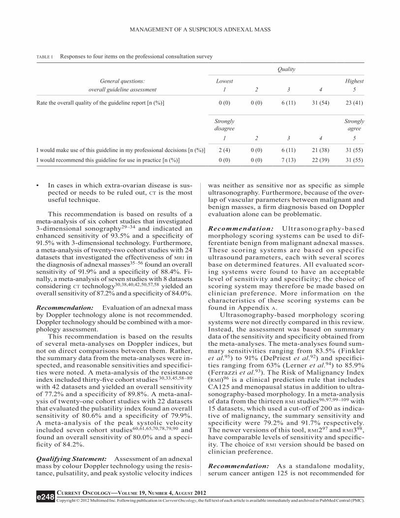
7.2.2 Summary of Written Comments from the Professional ConsultationAs a result of the professional consultation, 60 responses were received. Table i summarizes key results of the feedback survey. Modifications to the guideline were made accordingly.
Of the 60 responders, 20 provided additional written comments. Most indicated that the document was of high quality and would be of use to practitio-ners. Suggestions for improvements or additions to the document included several comments relating to the scoring systems described in the report. The feed-back generally indicated that many practitioners in the province are not aware of the scoring systems. A direct link from the recommendations to the scoring systems was requested. It was also suggested that the guideline recommend one scoring system that would be the most reliable. Other comments related to scor-ing systems include making the Risk of Malignancy Index (rmi) available as an appendix to the guideline. There was also a request for an appendix setting out the ultrasonography features of malignancy and the definitions of resistance index, pulsatility index, and peak systolic velocity.
8. PRACTICE GUIDELINE
The present report integrates the feedback obtained through the external review process, with final ap-proval given by the Gynecology dsg and the Report Approval Panel of the pebc.
8.1 Recommendations and Key Evidence
8.1.1 Identification of an Adnexal Mass Suspicious for Ovarian CancerRecommendation: Sonography (particularly 3-dimensional sonography), magnetic resonance im-aging (mri), and computed tomography (ct) imaging are each recommended for differentiating malignant from benign ovarian masses. However, the working group offers the following further recommendations, based on their expert consensus opinion and a con-sideration of availability, access, and harm:
• Where technically feasible, transvaginal sonog-raphy should be the modality of first choice in patients with a suspicious isolated ovarian mass.
• To help clarify malignant potential in patients in whom ultrasonography may be unreliable, mri is the most appropriate test.
MANAGEMENT OF A SUSPICIOUS ADNEXAL MASS
e248Current OnCOlOgy—VOlume 19, number 4, August 2012Copyright © 2012 Multimed Inc. Following publication in Current Oncology, the full text of each article is available immediately and archived in PubMed Central (PMC).
• In cases in which extra-ovarian disease is sus-pected or needs to be ruled out, ct is the most useful technique.
This recommendation is based on results of a meta-analysis of six cohort studies that investigated 3-dimensional sonography29–34 and indicated an enhanced sensitivity of 93.5% and a specificity of 91.5% with 3-dimensional technology. Furthermore, a meta-analysis of twenty-two cohort studies with 24 datasets that investigated the effectiveness of mri in the diagnosis of adnexal masses35–56 found an overall sensitivity of 91.9% and a specificity of 88.4%. Fi-nally, a meta-analysis of seven studies with 8 datasets considering ct technology30,38,40,42,50,57,58 yielded an overall sensitivity of 87.2% and a specificity of 84.0%.
Recommendation: Evaluation of an adnexal mass by Doppler technology alone is not recommended. Doppler technology should be combined with a mor-phology assessment.
This recommendation is based on the results of several meta-analyses on Doppler indices, but not on direct comparisons between them. Rather, the summary data from the meta-analyses were in-spected, and reasonable sensitivities and specifici-ties were noted. A meta-analysis of the resistance index included thirty-five cohort studies 30,33,45,58–89 with 42 datasets and yielded an overall sensitivity of 77.2% and a specificity of 89.8%. A meta-anal-ysis of twenty-one cohort studies with 22 datasets that evaluated the pulsatility index found an overall sensitivity of 80.6% and a specificity of 79.9%. A meta-analysis of the peak systolic velocity included seven cohort studies60,61,65,70,78,79,90 and found an overall sensitivity of 80.0% and a speci-ficity of 84.2%.
Qualifying Statement: Assessment of an adnexal mass by colour Doppler technology using the resis-tance, pulsatility, and peak systolic velocity indices
was neither as sensitive nor as specific as simple ultrasonography. Furthermore, because of the over-lap of vascular parameters between malignant and benign masses, a firm diagnosis based on Doppler evaluation alone can be problematic.
Recommendation: Ult rasonography-based morphology scoring systems can be used to dif-ferentiate benign from malignant adnexal masses. These scoring systems are based on specif ic ultrasound parameters, each with several scores base on determined features. All evaluated scor-ing systems were found to have an acceptable level of sensitivity and specificity; the choice of scoring system may therefore be made based on clinician preference. More information on the characteristics of these scoring systems can be found in Appendix a.
Ultrasonography-based morphology scoring systems were not directly compared in this review. Instead, the assessment was based on summary data of the sensitivity and specificity obtained from the meta-analyses. The meta-analyses found sum-mary sensitivities ranging from 83.5% (Finkler et al.95) to 91% (DePriest et al.92) and specifici-ties ranging from 63% (Lerner et al.94) to 85.9% (Ferrazzi et al.93). The Risk of Malignancy Index (rmi)96 is a clinical prediction rule that includes CA125 and menopausal status in addition to ultra-sonography-based morphology. In a meta-analysis of data from the thirteen rmi studies96,97,99–109 with 15 datasets, which used a cut-off of 200 as indica-tive of malignancy, the summary sensitivity and specificity were 79.2% and 91.7% respectively. The newer versions of this tool, rmi297 and rmi398, have comparable levels of sensitivity and specific-ity. The choice of rmi version should be based on clinician preference.
Recommendation: As a standalone modality, serum cancer antigen 125 is not recommended for
table i Responses to four items on the professional consultation survey
Quality
General questions: Lowest Highestoverall guideline assessment 1 2 3 4 5
Rate the overall quality of the guideline report [n (%)] 0 (0) 0 (0) 6 (11) 31 (54) 23 (41)
Strongly Stronglydisagree agree
1 2 3 4 5
I would make use of this guideline in my professional decisions [n (%)] 2 (4) 0 (0) 6 (11) 21 (38) 31 (55)
I would recommend this guideline for use in practice [n (%)] 0 (0) 0 (0) 7 (13) 22 (39) 31 (55)

DODGE et al.
e249Current OnCOlOgy—VOlume 19, number 4, August 2012Copyright © 2012 Multimed Inc. Following publication in Current Oncology, the full text of each article is available immediately and archived in PubMed Central (PMC).
distinguishing between benign and malignant ad-nexal masses.
This recommendation is based on a meta-analysis of forty-nine cohort studies 45,59,63,67,80,90,95,101,103,107, 108,110–147 and two case–control studies 148,149 with a total of 52 datasets that found, at a threshold of 35 U/mL, an overall sensitivity of 78.7% and a specificity of 77.9%.
Qualifying Statement: Elevated serum CA125 has been reported in a variety of benign conditions. Because the incidence of ovarian cancer relative to benign gynecologic conditions is lower in premeno-pausal women, serum CA125 is of limited use in that population3. Serum CA125 is elevated in only 50% of early-stage ovarian cancers150. Caution should be used in interpreting values in such patients.
Recommendation: Frozen sections for the intra-operative diagnosis of a suspicious adnexal mass is recommended in settings in which availability and patient preference allow.
This recommendation is based on a meta-analysis of frozen section diagnoses that included fifteen co-hort studies 35,151–164 and yielded an overall sensitivity of 89.2% and a specificity of 97.9%.
8.1.2 Surgical Procedures for an Adnexal Mass Suspicious for MalignancyRecommendation: To improve survival, compre-hensive surgical staging with lymphadenectomy is recommended for the surgical management of patients with early-stage ovarian cancer.
This recommendation is based on the results of five retrospective cohort studies11,12,14,16,17. Two large population-based studies11,12 found improved 3-year (p < 0.001)12 and 5-year disease-specific survival (p < 0.001)11 for surgical staging with lymphadenectomy compared with staging proce-dures without lymphadenectomy. Oksefjell et al.16 reported a statistically significant improvement in 5-year overall survival rates in patients that under-went lymphadenectomy compared with those that did not (87% vs. 64%; p = 0.02). Survival analyses performed by both Skirnisdottir et al.17 and Hornung et al.14 also demonstrated a statistically significant benefit in disease-free survival (p = 0.004 and p = 0.0007 respectively) for patients that underwent lymphadenectomy compared with patients that did not. Hornung and colleagues14 also considered overall survival and reported a statistically significant dif-ference (p = 0.0008) between the groups in favour of the patients undergoing a lymphadenectomy. The one randomized controlled trial15 that was identified reported no statistically significant effect of lymphad-enectomy on progression-free survival (hazard ratio: 0.72; 95% confidence interval: 0.46 to 1.14) or overall survival (hazard ratio: 0.85; 95% confidence interval: 0.49 to 1.47). However, the study was underpowered
to detect a difference in survival, the study’s second-ary outcome. Rather, the sample size calculation was undertaken to detect a difference in the prevalence of lymph node positivity. The study was deemed inadequate to inform the recommendation.
Recommendation: Laparoscopy is a reasonable alternative to laparotomy, provided that appropriate surgery and staging can be done. The choice between laparoscopy and laparotomy should be based on patient and clinician preference. Discussion with a gynecologic oncologist is recommended.
This recommendation is based on the results of six retrospective cohort studies20–25. In the three studies21–23 that considered patients with early epithelial ovarian cancer, no statistical difference in survival rates was detected between patients under-going laparoscopy and those undergoing laparotomy. In the management of patients with early border-line ovarian tumours, Romangnolo et al.24, Park et al.25, and Desfeux et al.20 found that the surgical approach—laparoscopic or laparotomic—did not appear to influence survival rates.
Recommendation: Fertility-preserving surgery is an acceptable alternative to more extensive surgery in patients with low malignant-potential tumours and those with well-differentiated surgical stage i ovarian cancer. Discussion with a gynecologic on-cologist is recommended.
This recommendation is based on two cohort studies that compared the impacts of conservative fertility-sparing surgeries and more radical surgical approaches. Yinon et al.26 specifically compared rates of recurrence in 40 patients who underwent unilateral salpingo-oophorectomy with those in 22 patients who underwent cystectomy only. No statistical difference in recurrence rates was detected (27.5% vs. 22.7%, p = 0.8). Similarly, in a larger study of 360 women with low malignant-potential tumours, Park et al.25 found no difference in disease-free survival between patients who underwent radical or fertility-sparing surgery (p = 0.651).
Qualifying Statement: The Gynecology Cancer dsg acknowledges that, despite definitions and cri-teria, it is unrealistic to expect that 100% of ovarian cancers will be identified as suspicious preopera-tively. Pathology remains the clinical standard.
9. CONFLICT OF INTEREST DISCLOSURES
The authors declare that there are no financial con-flicts of interest.
10. REFERENCES
1. Canadian Cancer Society’s Steering Committee. Canadian Cancer Statistics 2010. Toronto, ON: Canadian Cancer Society; 2010.

MANAGEMENT OF A SUSPICIOUS ADNEXAL MASS
e250Current OnCOlOgy—VOlume 19, number 4, August 2012Copyright © 2012 Multimed Inc. Following publication in Current Oncology, the full text of each article is available immediately and archived in PubMed Central (PMC).
2. Scottish Intercollegiate Guidelines Network (sign). Epithel-ial Ovarian Cancer: A National Clinical Guideline. No. 75. Edinburgh, Scotland: sign; 2003.
3. Myers ER, Bastian LA, Havrilesky LJ, et al. Management of adnexal mass. Evid Rep Technol Assess (Full Rep) 2006;:1–145.
4. Fung Kee Fung M, Bryson P, Johnston M, Chambers A, and the members of the Gynecology Cancer Disease Site Group. Screening Postmenopausal Women for Ovarian Cancer. Evi-dence summary report no. 4–6a. Toronto, ON: Cancer Care Ontario; 2004.
5. Browman GP, Levine MN, Mohide EA, et al. The practice guidelines development cycle: a conceptual tool for practice guidelines development and implementation. J Clin Oncol 1995;13:502–12.
6. Australia, Department of Health and Ageing, Australian Cancer Network, National Breast Cancer Centre (nbcc). Clinical Practice Guidelines for the Management of Women with Epithelial Ovarian Cancer. Camperdown, Australia: nbcc; 2004.
7. Geomini P, Bremer G, Kruitwagen R, Mol BW. Diagnostic accuracy of frozen section diagnosis of the adnexal mass: a metaanalysis. Gynecol Oncol 2005;96:1–9.
8. Liu J, Xu Y, Wang J. Ultrasonography, computed tomography and magnetic resonance imaging for diagnosis of ovarian carcinoma. Eur J Radiol 2007;62:328–34.
9. Geomini P, Kruitwagen R, Bremer GL, Cnossen J, Mol BW. The accuracy of risk scores in predicting ovarian malignancy: a systematic review. Obstet Gynecol 2009;113:384–94.
10. Medeiros LR, Rosa DD, Edelweiss MI, et al. Accuracy of frozen-section analysis in the diagnosis of ovarian tumors: a systematic quantitative review. Int J Gynecol Cancer 2005;15:192–202.
11. Chan JK, Munro EG, Cheung MK, et al. Association of lymph-adenectomy and survival in stage i ovarian cancer patients. Obstet Gynecol 2007;109:12–19.
12. Chan J, Fuh K, Shin J, et al. The treatment and outcomes of early-stage epithelial ovarian cancer: have we made any progress? Br J Cancer 2008;98:1191–6.
13. Cho YH, Kim DY, Kim JH, et al. Is complete surgical staging necessary in patients with stage i mucinous epithelial ovarian tumors? Gynecol Oncol 2006;103:878–82.
14. Hornung R, Urs E, Serenella E, et al. Analysis of potential prognostic factors in 111 patients with ovarian cancer. Cancer Lett 2004;206:97–106.
15. Maggioni A, Benedetti Panici P, Dell’Anna T, et al. Random-ised study of systematic lymphadenectomy in patients with epithelial ovarian cancer macroscopically confined to the pelvis. Br J Cancer 2006;95:699–704.
16. Oksefjell H, Sandstad B, Tropé C. Is the watch and wait ap-proach adequate after comprehensive surgical staging in inva-sive stage i epithelial ovarian cancer? The Norwegian Radium Hospital experience. Eur J Gynaecol Oncol 2008;29:583–9.
17. Skírnisdóttir I, Sorbe B. Lymph node sampling is of prog-nostic value in early stage epithelial ovarian carcinoma. Eur J Gynaecol Oncol 2005;26:181–5.
18. Suzuki S, Kajiyama H, Shibata K, et al. Is there any associa-tion between retroperitoneal lymphadenectomy and survival benefit in ovarian clear cell carcinoma patients? Ann Oncol 2008;19:1284–7.
19. Wong HF, Low JJ, Chua Y, Busmanis I, Tay EH, Ho TH. Ovarian tumors of borderline malignancy: a review of 247 patients from 1991 to 2004. Int J Gynecol Cancer 2007;17:342–9.
20. Desfeux P, Camatte S, Chatellier G, Blanc B, Querleu D, Lécuru F. Impact of surgical approach on the management of macroscopic early ovarian borderline tumors. Gynecol Oncol 2005;98:390–5.
21. Ghezzi F, Cromi A, Uccella S, et al. Laparoscopy versus laparotomy for the surgical management of apparent early stage ovarian cancer. Gynecol Oncol 2007;105:409–13.
22. Lécuru F, Desfeux P, Camatte S, Bissery A, Blanc B, Querleu D. Impact of initial surgical access on staging and survival of patients with stage i ovarian cancer. Int J Gynecol Cancer 2006;16:87–94.
23. Park JY, Bae J, Lim MC, et al. Laparoscopic and lapa-rotomic staging in stage i epithelial ovarian cancer: a comparison of feasibility and safety. Int J Gynecol Cancer 2008;18:1202–9.
24. Romagnolo C, Gadducci A, Sartori E, Zola P, Maggino T. Management of borderline ovarian tumors: results of an Italian multicenter study. Gynecol Oncol 2006;101:255–60.
25. Park JY, Kim DY, Kim JH, Kim YM, Kim YT, Nam JH. Surgical management of borderline ovarian tumors: the role of fer tility-sparing surgery. Gynecol Oncol 2009;113:75–82.
26. Yinon Y, Beiner ME, Gotlieb WH, Korach Y, Perri T, Ben-Baruch G. Clinical outcome of cystectomy compared with uni-lateral salpingo-oophorectomy as fertility-sparing treatment of borderline ovarian tumors. Fertil Steril 2007;88:479–84.
27. Cohen LS, Escobar PF, Scharm C, Glimco B, Fishman DA. Three-dimensional power Doppler ultrasound improves the diagnostic accuracy for ovarian cancer prediction. Gynecol Oncol 2001;82:40–8.
28. Wilson WD, Valet AS, Andreotti RF, Green–Jarvis B, Lysh-chik A, Fleischer AC. Sonographic quantification of ovarian tumor vascularity. J Ultrasound Med 2006;25:1577–81.
29. Geomini PM, Kluivers KB, Moret E, Bremer GL, Kruit-wagen RF, Mol BW. Evaluation of adnexal masses with three-dimensional ult rasonography. Obstet Gynecol 2006;108:1167–75.
30. Laban M, Metawee H, Elyan A, Kamal M, Kamel M, Man-sour G. Three-dimensional ultrasound and three-dimensional power Doppler in the assessment of ovarian tumors. Int J Gynaecol Obstet 2007;99:201–5.
31. Alcázar JL, Galán MJ, García–Manero M, Guerriero S. Three-dimensional sonographic morphologic assessment in complex adnexal masses: preliminary experience. J Ultrasound Med 2003;22:249–54.
32. Kurjak A, Kupesić S. Three dimensional ultrasound and power Doppler in assessment of uterine and ovarian angiogenesis: a prospective study. Croat Med J 1999;40:413–20.
33. Kurjak A, Kupesic S, Sparac V, Kosuta D. Three-dimensional ultrasonographic and power Doppler characterization of ovar-ian lesions. Ultrasound Obstet Gynecol 2000;16:365–71.
34. Alcázar JL, Castillo G. Comparison of 2-dimensional and 3-dimensional power-Doppler imaging in complex adnexal masses for the prediction of ovarian cancer. Am J Obstet Gynecol 2005;192:807–12.
DODGE et al.
e251Current OnCOlOgy—VOlume 19, number 4, August 2012Copyright © 2012 Multimed Inc. Following publication in Current Oncology, the full text of each article is available immediately and archived in PubMed Central (PMC).
35. Bazot M, Nassar–Slaba J, Thomassin–Naggara I, Cortez A, Uzan S, Daraï E. mr imaging compared with intrao-perative frozen-section examination for the diagnosis of adnexal tumors; correlation with final histology. Eur Radiol 2006;16:2687–99.
36. Chen M, Wang WC, Zhou C, et al. Differentiation between malignant and benign ovarian tumors by magnetic resonance imaging. Chin Med Sci J 2006;21:270–5.
37. Guerra A, Cunha TM, Félix A. Magnetic resonance evaluation of adnexal masses. Acta Radiol 2008;49:700–9.
38. Tsili AC, Tsampoulas C, Argyropoulou M, et al. Comparative evaluation of multidetector ct and mr imaging in the differ-entiation of adnexal masses. Eur Radiol 2008;18:1049–57.
39. Booth SJ, Turnbull LW, Poole DR, Richmond I. The accur-ate staging of ovarian cancer using 3T magnetic resonance imaging—a realistic option. BJOG 2008;115:894–901.
40. Umemoto M, Shiota M, Shimono T, Hoshiai H. Preoperative diagnosis of ovarian tumors, focusing on the solid area based on diagnostic imaging. J Obstet Gynaecol Res 2006;32:195–201.
41. Scoutt LM, McCarthy SM, Lange R, Bourque A, Schwartz PE. mr evaluation of clinically suspected adnexal masses. J Comput Assist Tomogr 1994;18:609–18.
42. Buist MR, Golding RP, Burger CW, et al. Comparative evaluation of diagnostic methods in ovarian carcinoma with emphasis on ct and mri. Gynecol Oncol 1994;52:191–8.
43. Fenchel S, Grab D, Nuessle K, et al. Asymptomatic adnexal masses: correlation of fdg pet and histopathologic findings. Radiology 2002;223:780–8.
44. Grab D, Flock F, Stöhr I, et al. Classification of asymptom-atic adnexal masses by ultrasound, magnetic resonance imaging, and positron emission tomography. Gynecol Oncol 2000;77:454–9.
45. Hata K, Hata T, Manabe A, Sugimura K, Kitao M. A critical evaluation of transvaginal Doppler studies, transvaginal sonography, magnetic resonance imaging, and CA125 in detecting ovarian cancer. Obstet Gynecol 1992;80:922–6.
46. Hricak H, Chen M, Coakley FV, et al. Complex adnexal masses: detection and characterization with MR imaging—multivariate analysis. Radiology 2000;214:39–46.
47. Huber S, Medl M, Baumann L, Czembirek H. Value of ultra-sound and magnetic resonance imaging in the preoperative evaluation of suspected ovarian masses. Anticancer Res 2002;22:2501–7.
48. Jain KA, Friedman DL, Pettinger TW, Alagappan R, Jeffrey RB Jr, Sommer FG. Adnexal masses: comparison of speci-ficity of endovaginal us and pelvic mr imaging. Radiology 1993;186:697–704.
49. Komatsu T, Konishi I, Mandai M, et al. Adnexal masses: transvaginal us and gadolinium-enhanced mr imaging assess-ment of intratumoral structure. Radiology 1996;198:109–15.
50. Kurtz AB, Tsimikas JV, Tempany CM, et al. Diagnosis and staging of ovarian cancer: comparative values of Doppler and conventional us, ct, and mr imaging correlated with surgery and histopathologic analysis—report of the Radiology Diag-nostic Oncology Group. Radiology 1999;212:19–27.
51. Medl M, Kulenkampff KJ, Stiskal M, Peters–Engl C, Leod-olter S, Czembirek H. Magnetic resonance imaging in the preoperative evaluation of suspected ovarian masses. Anti-cancer Res 1995;15:1123–5.
52. Reuter M, Steffens J, Schüppler U, et al. Critical evaluation of the specificity of mri and tvus for differentiation of malignant from benign adnexal lesions. Eur Radiol 1998;8:39–44.
53. Yamashita Y, Torashima M, Hatanaka Y, et al. Adnexal masses: accuracy of characterization with transvaginal us and precontrast and postcontrast mr imaging. Radiology 1995;194:557–65.
54. Kawahara K, Yoshida Y, Kurokawa T, et al. Evaluation of posi-tron emission tomography with tracer 18-fluorodeoxyglucose in addition to magnetic resonance imaging in the diagnosis of ovarian cancer in selected women after ultrasonography. J Comput Assist Tomogr 2004;28:505–16.
55. Sohaib SA, Mills TD, Sahdev A, et al. The role of magnetic resonance imaging and ultrasound in patients with adnexal masses. Clin Radiol 2005;60:340–8.
56. Rieber A, Nüssle K, Stöhr I, et al. Preoperative diagnosis of ovarian tumors with mr imaging: comparison with transvagin-al sonography, positron emission tomography, and histologic findings. AJR Am J Roentgenol 2001;177:123–9.
57. Kitajima K, Murakami K, Yamasaki E, et al. Diagnostic ac-curacy of integrated fdg-pet/contrast-enhanced ct in staging ovarian cancer: comparison with enhanced ct. Eur J Nucl Med Mol Imaging 2008;35:1912–20.
58. Lin JY, Angel C, DuBeshter B, Walsh CJ. Diagnoses after laparotomy for a mass in the pelvic area in women. Surg Gynecol Obstet 1993;176:333–8.
59. Alcázar JL, Errasti T, Zornoza A, Mínguez JA, Galán MJ. Transvaginal color Doppler ultrasonography and CA-125 in suspicious adnexal masses. Int J Gynaecol Obstet 1999;66:255–61.
60. Alcázar JL, Ruiz–Perez ML, Errasti T. Transvaginal color Doppler sonography in adnexal masses: which parameter performs best? Ultrasound Obstet Gynecol 1996;8:114–19.
61. Alcázar JL, López–García G. Transvaginal color Doppler assessment of venous flow in adnexal masses. Ultrasound Obstet Gynecol 2001;17:434–8.
62. Anandakumar C, Chew S, Wong YC, Chia D, Ratnam SS. Role of transvaginal ultrasound color flow imaging and Dop-pler waveform analysis in differentiating between benign and malignant ovarian tumors. Ultrasound Obstet Gynecol 1996;7:280–4.
63. Berlanda N, Ferrari MM, Mezzopane R, et al. Impact of a multiparameter, ultrasound-based triage on surgical man-agement of adnexal masses. Ultrasound Obstet Gynecol 2002;20:181–5.
64. Bromley B, Goodman H, Benacerraf BR. Comparison between sonographic morphology and Doppler waveform for the diag-nosis of ovarian malignancy. Obstet Gynecol 1994;83:434–7.
65. Buy JN, Ghossain MA, Hugol D, et al. Characterization of ad-nexal masses: combination of color Doppler and conventional sonography compared with spectral Doppler analysis alone and conventional sonography alone. AJR Am J Roentgenol 1996;166:385–93.
66. Carter JR, Lau M, Fowler JM, Carlson JW, Carson LF, Twiggs LB. Blood flow characteristics of ovarian tumors: implica-tions for ovarian cancer screening. Am J Obstet Gynecol 1995;172:901–7.
67. Chou CY, Chang CH, Yao BL, Kuo HC. Color Doppler ultrasonography and serum CA125 in the differentiation of

MANAGEMENT OF A SUSPICIOUS ADNEXAL MASS
e252Current OnCOlOgy—VOlume 19, number 4, August 2012Copyright © 2012 Multimed Inc. Following publication in Current Oncology, the full text of each article is available immediately and archived in PubMed Central (PMC).
benign and malignant ovarian tumors. J Clin Ultrasound 1994;22:491–6.
68. Franchi M, Beretta P, Ghezzi F, Zanaboni F, Goddi A, Salva-tore S. Diagnosis of pelvic masses with transabdominal color Doppler, CA125 and ultrasonography. Acta Obstet Gynecol Scand 1995;74:734–9.
69. Guerriero S, Ajossa S, Piras S, et al. Three-dimensional quantification of tumor vascularity as a tertiary test after B-mode and power Doppler evaluation for detection of ovarian cancer. J Ultrasound Med 2007;26:1271–8.
70. Hata K, Hata T, Kitao M. Intratumoral peak systolic velocity as a new possible predictor for detection of adnexal malignancy. Am J Obstet Gynecol 1995;172:1496–500.
71. Jain KA. Prospective evaluation of adnexal masses with endovaginal gray-scale and duplex and color Doppler us: cor-relation with pathologic findings. Radiology 1994;191:63–7.
72. Kurjak A, Zalud I, Alfirevic Z. Evaluation of adnexal masses with transvaginal color ultrasound. J Ultrasound Med 1991;10:295–7.
73. Kurjak A, Predanić M. New scoring system for prediction of ovarian malignancy based on transvaginal color Doppler sonography. J Ultrasound Med 1992;11:631–8.
74. Leeners B, Schild RL, Funk A, et al. Colour Doppler sonog-raphy improves the pre-operative diagnosis of ovarian tumours made using conventional transvaginal sonography. Eur J Obstet Gynecol Reprod Biol 1996;64:79–85.
75. Marchesini AC, Magrio FA, Berezowski AT, Neto OB, No-gueira AA, Candido dos Reis FJ. A critical analysis of Dop-pler velocimetry in the differential diagnosis of malignant and benign ovarian masses. J Womens Health (Larchmt) 2008;17:97–102.
76. Marret H, Sauget S, Giraudeau B, et al. Contrast-enhanced sonography helps in discrimination of benign from malignant adnexal masses. J Ultrasound Med 2004;23:1629–39.
77. Mercé LT, Caballero RA, Barco MJ, Bau S, López G. B-Mode, utero-ovarian and intratumoural transvaginal colour Doppler ultrasonography for differential diagnosis of ovarian tumours. Eur J Obstet Gynecol Reprod Biol 1998;76:97–107.
78. Mousavi AS, Borna S, Moeinoddini S. Estimation of prob-ability of malignancy using a logistic model combining col-or Doppler ultrasonography, serum CA125 level in women with a pelvic mass. Int J Gynecol Cancer 2006;16(suppl 1):92–8.
79. Prömpeler HJ, Madjar H, Sauerbrei W. Classification of ad-nexal tumors by transvaginal color Doppler. Gynecol Oncol 1996;61:354–63.
80. Schneider VL, Schneider A, Reed KL, Hatch KD. Comparison of Doppler with two-dimensional sonography and CA 125 for prediction of malignancy of pelvic masses. Obstet Gynecol 1993;81:983–8.
81. Stein SM, Laifer–Narin S, Johnson MB, et al. Differentiation of benign and malignant adnexal masses: relative value of gray-scale, color Doppler, and spectral Doppler sonography. AJR Am J Roentgenol 1995;164:381–6.
82. Takac I. Analysis of blood flow in adnexal tumors by using color Doppler imaging and pulsed spectral analysis. Ultra-sound Med Biol 1998;24:1137–41.
83. Tekay A, Jouppila P. Validity of pulsatility and resistance indices in classification of adnexal tumors with transvaginal
color Doppler ultrasound. Ultrasound Obstet Gynecol 1992;2:338–44.
84. Tepper R, Lerner–Geva L, Altaras MM, et al. Transvaginal color flow imaging in the diagnosis of ovarian tumors. J Ultrasound Med 1995;14:731–4.
85. Timor–Tritsch LE, Lerner JP, Monteagudo A, Santos R. Transvaginal ultrasonographic characterization of ovarian masses by means of color flow-directed Doppler measure-ments and a morphologic scoring system. Am J Obstet Gynecol 1993;168:909–13.
86. Valentin L. Comparison of Lerner score, Doppler ultrasound examination, and their combination for discrimination be-tween benign and malignant adnexal masses. Ultrasound Obstet Gynecol 2000;15:143–7.
87. Valentin L. Pattern recognition of pelvic masses by gray-scale ultrasound imaging: the contribution of Doppler ultrasound. Ultrasound Obstet Gynecol 1999;14:338–47.
88. Wu CC, Lee CN, Chen TM, Lai JI, Hsieh CY, Hsieh FJ. Factors contributing to the accuracy in diagnosing ovarian malignancy by color Doppler ultrasound. Obstet Gynecol 1994;84:605–8.
89. Zanetta G, Vergani P, Lissoni A. Color Doppler ultrasound in the preoperative assessment of adnexal masses. Acta Obstet Gynecol Scand 1994;73:637–41.
90. Hillaby K, Aslam N, Salim R, Lawrence A, Raju KS, Jurkovic D. The value of detection of normal ovarian tissue (the “ovar-ian crescent sign”) in the differential diagnosis of adnexal masses. Ultrasound Obstet Gynecol 2004;23:63–7.
91. Sassone AM, Timor–Tritsch IE, Artner A, Westhoff C, Warren WB. Transvaginal sonographic characterization of ovarian disease: evaluation of a new scoring system to predict ovarian malignancy. Obstet Gynecol 1991;78:70–6.
92. DePriest PD, Shenson D, Fried A, et al. A morphology index based on sonographic findings in ovarian cancer. Gynecol Oncol 1993;51:7–11.
93. Ferrazzi E, Zanetta G, Dordoni D, Berlanda N, Mezzopane R, Lissoni AA. Transvaginal ultrasonographic characterization of ovarian masses: comparison of five scoring systems in a multicenter study. Ultrasound Obstet Gynecol 1997;10:192–7.
94. Lerner JP, Timor–Tritsch IE, Federman A, Abramovich G. Transvaginal ultrasonographic characterization of ovarian masses with an improved, weighted scoring system. Am J Obstet Gynecol 1994;170:81–5.
95. Finkler NJ, Benacerraf B, Lavin PT, Wojciechowski C, Knapp RC. Comparison of serum CA125, clinical impression, and ultrasound in the preoperative evaluation of ovarian masses. Obstet Gynecol 1988;72:659–64.
96. Jacobs I, Oram D, Fairbanks J, Turner J, Frost C, Grudzin-skas JG. A risk of malignancy index incorporating CA 125, ultrasound and menopausal status for the accurate preopera-tive diagnosis of ovarian cancer. Br J Obstet Gynaecol 1990;97:922–9.
97. Tingulstad S, Hagen B, Skjeldestad FE, et al. Evaluation of a risk of malignancy index based on serum CA125, ultrasound findings and menopausal status in the pre-operative diagnosis of pelvic masses. Br J Obstet Gynaecol 1996;103:826–31.
98. Tingulstad S, Hagen B, Skjeldestad FE, Halvorsen T, Nustad K, Onsrud M. The risk-of-malignancy index to evaluate potential ovarian cancers in local hospitals. Obstet Gynecol 1999;93:448–52.
DODGE et al.
e253Current OnCOlOgy—VOlume 19, number 4, August 2012Copyright © 2012 Multimed Inc. Following publication in Current Oncology, the full text of each article is available immediately and archived in PubMed Central (PMC).
99. Bensaid C, Le Frère Belda MA, et al. Performance of lapa-roscopy in identifying malignant ovarian cysts. Surg Endosc 2006;20:1410–14.
100. Van Holsbeke C, Van Calster B, Valentin L, et al. External validation of mathematical models to distinguish between benign and malignant adnexal tumors: a multicenter study by the International Ovarian Tumor Analysis Group. Clin Cancer Res 2007;13:4440–7.
101. Asif N, Sattar A, Dawood MM, Rafi T, Aamir M, Anwar M. Pre-operative evaluation of ovarian mass: risk of malignancy index. J Coll Physicians Surg Pak 2004;14:128–31.
102. Davies AP, Jacobs I, Woolas R, Fish A, Oram D. The adnexal mass: benign or malignant? Evaluation of a risk of malignancy index. Br J Obstet Gynaecol 1993;100:927–31.
103. Manjunath AP, Pratapkumar, Sujatha K, Vani R. Comparison of three risk of malignancy indices in evaluation of pelvic masses. Gynecol Oncol 2001;81:225–9.
104. Morgante G, la Marca A, Ditto A, De Leo V. Comparison of two malignancy risk indices based on serum CA125, ultra-sound score and menopausal status in the diagnosis of ovarian masses. Br J Obstet Gynaecol 1999;106:524–7.
105. Aslam N, Tailor A, Lawton F, Carr J, Savvas M, Jurkovic D. Prospective evaluation of three different models for the pre-operative diagnosis of ovarian cancer. BJOG 2000;107:1347–53.
106. Mol BW, Boll D, De Kanter M, et al. Distinguishing the be-nign and malignant adnexal mass: an external validation of prognostic models. Gynecol Oncol 2001;80:162–7.
107. Timmerman D, Bourne TH, Tailor A, et al. A comparison of methods for preoperative discrimination between malignant and benign adnexal masses: the development of a new logistic regression model. Am J Obstet Gynecol 1999;181:57–65.
108. Engelen MJ, Bongaerts AH, Sluiter WJ, et al. Distinguishing benign and malignant pelvic masses: the value of different diagnostic methods in everyday clinical practice. Eur J Obstet Gynecol Reprod Biol 2008;136:94–101.
109. Obeidat BR, Amarin ZO, Latimer JA, Crawford RA. Risk of malignancy index in the preoperative evaluation of pelvic masses. Int J Gynaecol Obstet 2004;85:255–8.
110. Patsner B, Mann WJ. The value of preoperative serum CA 125 levels in patients with a pelvic mass. Am J Obstet Gynecol 1988;159:873–6.
111. Adonakis GL, Paraskevaidis E, Tsiga S, Seferiadis K, Lolis DE. A combined approach for the early detection of ovarian cancer in asymptomatic women. Eur J Obstet Gynecol Reprod Biol 1996;65:221–5.
112. Benjapibal M, Neungton C. Pre-operative prediction of serum CA125 level in women with ovarian masses. J Med Assoc Thai 2007;90:1986–91.
113. Romagnolo C, Trivella G, Bonacina M, Fornalè M, Maggino T, Ferrazzi E. Preoperative diagnosis of 221 consecutive ovarian masses: scoring system and expert evaluation. Eur J Gynaecol Oncol 2006;27:487–9.
114. Zhang Z, Yu Y, Xu F, et al. Combining multiple serum tumor markers improves detection of stage i epithelial ovarian cancer. Gynecol Oncol 2007;107:526–31.
115. Milojkovic M, Hrgovic Z, Hrgovic I, Jonat W, Maass N, Buković D. Significance of CA125 serum level in discrimina-tion between benign and malignant masses in the pelvis. Arch Gynecol Obstet 2004;269:176–80.
116. Erdoğan N, Ozçelik B, Serin IS, Akgün M, Oztürk F. Dop-pler ultrasound assessment and serum cancer antigen 125 in the diagnosis of ovarian tumors. Int J Gynaecol Obstet 2005;91:146–50.
117. Balbi GC, Musone R, Menditto A, et al. Women with a pel-vic mass: indicators of malignancy. Eur J Gynaecol Oncol 2001;22:459–62.
118. Chalas E, Welshinger M, Engellener W, Chumas J, Barbieri R, Mann WJ. The clinical significance of thrombocytosis in women presenting with a pelvic mass. Am J Obstet Gynecol 1992;166:974–7.
119. Chen DX, Schwartz PE, Li XG, Yang Z. Evaluation of CA125 levels in differentiating malignant from benign tumors in patients with pelvic masses. Obstet Gynecol 1988;72:23–7.
120. Doed JR, Quinn MA, Rome R, Koh H. Women with a pelvic mass—when to perform an ultrasound. Aust N Z J Obstet Gynaecol 1993;33:404–7.
121. Einhorn N, Bast RC Jr, Knapp RC, Tjernberg B, Zurawski VR Jr. Preoperative evaluation of serum CA 125 levels in pa-tients with primary epithelial ovarian cancer. Obstet Gynecol 1986;67:414–16.
122. Gadducci A, Capriello P, Bartolini T, et al. The associa-tion of ultrasonography and CA-125 test in the preoperative evaluation of ovarian carcinoma. Eur J Gynaecol Oncol 1988;9:373–6.
123. Gadducci A, Ferdeghini M, Prontera C, et al. The concomitant determination of different tumor markers in patients with epi-thelial ovarian cancer and benign ovarian masses: relevance for differential diagnosis. Gynecol Oncol 1992;44:147–54.
124. Gadducci A, Ferdeghini M, Rispoli G, Prontera C, Bianchi R, Fioretti P. Comparison of tumor-associated trypsin inhibitor (tati) with CA125 as a marker for diagnosis and monitoring of epithelial ovarian cancer. Scand J Clin Lab Invest Suppl 1991;207:19–24.
125. Hogdall EV, Høgdall CK, Tingulstad S, et al. Predictive values of serum tumour markers tetranectin, OVX1, CASA and CA125 in patients with a pelvic mass. Int J Cancer 2000;89:519–23.
126. Woolas RP, Conaway MR, Xu F, et al. Combinations of multiple serum markers are superior to individual assays for discriminating malignant from benign pelvic masses. Gynecol Oncol 1995;59:111–16.
127. Zhang Z, Barnhill SD, Zhang H, et al. Combination of multiple serum markers using an artificial neural network to improve specificity in discriminating malignant from benign pelvic masses. Gynecol Oncol 1999;73:56–61.
128. Hurteau JA, Woolas RP, Jacobs IJ, et al. Soluble interleukin-2 receptor alpha is elevated in sera of patients with benign ovarian neoplasms and epithelial ovarian cancer. Cancer 1995;76:1615–20.
129. Kawai M, Kikkawa F, Ishikawa H, et al. Differential diag-nosis of ovarian tumors by transvaginal color-pulse Doppler sonography. Gynecol Oncol 1994;54:209–14.
130. Maggino T, Gadducci A, D’Addario V, et al. Prospective multicenter study on CA125 in postmenopausal pelvic masses. Gynecol Oncol 1994;54:117–23.
131. Malkasian GD Jr, Knapp RC, Lavin PT, et al. Preoperative evaluation of serum CA 125 levels in premenopausal and postmenopausal patients with pelvic masses: discrimination

MANAGEMENT OF A SUSPICIOUS ADNEXAL MASS
e254Current OnCOlOgy—VOlume 19, number 4, August 2012Copyright © 2012 Multimed Inc. Following publication in Current Oncology, the full text of each article is available immediately and archived in PubMed Central (PMC).
of benign from malignant disease. Am J Obstet Gynecol 1988;159:341–6.
132. Mancuso A, De Vivo A, Triolo O, Irato S. The role of trans-vaginal ultrasonography and serum CA 125 assay combined with age and hormonal state in the differential diagnosis of pelvic masses. Eur J Gynaecol Oncol 2004;25:207–10.
133. O’Connell GJ, Ryan E, Murphy KJ, Prefontaine M. Predictive value of CA125 for ovarian carcinoma in patients presenting with pelvic masses. Obstet Gynecol 1987;70:930–2.
134. Pyrgiotis E, Salamalekis E, Loghis C, Dima C, Zourlas PA. CA 125 in preoperative evaluation of pelvic masses. Eur J Gynaecol Oncol 1993;14:279–82.
135. Roman LD, Muderspach LI, Stein SM, Laifer–Narin S, Groshen S, Morrow CP. Pelvic examination, tumor marker level, and gray-scale and Doppler sonography in the prediction of pelvic cancer. Obstet Gynecol 1997;89:493–500.
136. Schutter EM, Davelaar EM, van Kamp GJ, Verstraeten RA, Kenemans P, Verheijen RH. The differential diagnostic potential of a panel of tumor markers (CA125, CA 15-3, and CA 72-4 antigens) in patients with a pelvic mass. Am J Obstet Gynecol 2002;187:385–92.
137. Schutter EM, Kenemans P, Sohn C, et al. Diagnostic value of pelvic examination, ultrasound, and serum CA125 in postmenopausal women with a pelvic mass. An international multicenter study. Cancer 1994;74:1398–406.
138. Schutter EM, Sohn C, Kristen P, et al. Estimation of probabil-ity of malignancy using a logistic model combining physical examination, ultrasound, serum CA125, and serum CA 72-4 in postmenopausal women with a pelvic mass: an international multicenter study. Gynecol Oncol 1998;69:56–63.
139. Sengoku K, Satoh T, Saitoh S, Abe M, Ishikawa M. Evalua-tion of transvaginal color Doppler sonography, transvaginal sonography and CA125 for prediction of ovarian malignancy. Int J Gynaecol Obstet 1994;46:39–43.
140. Smikle CB, Lunt CC, Hankins GD. Clinical predictors in the evaluation of a pelvic mass. Mil Med 1995;160:233–5.
141. Soper JT, Hunter VJ, Daly L, Tanner M, Creasman WT, Bast RC Jr. Preoperative serum tumor-associated anti-gen levels in women with pelvic masses. Obstet Gynecol 1990;75:249–54.
142. Tay SK, Chua EK. Correlation of serum, urinary and salivary CA125 levels in patients with adnexal masses. Ann Acad Med Singapore 1994;23:311–14.
143. Tian J, Zhang J, Jiao L, Li Y, Cao L. A prospective study of Tc-99m mibi in the differential diagnosis of pelvic masses in female patients. Clin Nucl Med 2000;25:614–18.
144. Torres JC, Derchain SF, Faundes A, Gontijo RC, Martinez EZ, Andrade LA. Risk-of-malignancy index in preoperative evaluation of clinically restricted ovarian cancer. Sao Paulo Med J 2002;120:72–6.
145. Vasilev SA, Schlaerth JB, Campeau J, Morrow CP. Serum CA 125 levels in preoperative evaluation of pelvic masses. Obstet Gynecol 1988;71:751–6.
146. Wakahara F, Kikkawa F, Nawa A, et al. Diagnostic ef-ficacy of tumor markers, sonography, and intraoperative frozen section for ovarian tumors. Gynecol Obstet Invest 2001;52:147–52.
147. Weiner Z, Thaler I, Beck D, Rottem S, Deutsch M, Brandes JM. Differentiating malignant from benign ovarian tumors
with transvaginal color f low imaging. Obstet Gynecol 1992;79:159–62.
148. El-Shalakany A, Abou-Talib Y, Shalaby HS, Sallam M. Pre-operative serum inhibin levels in patients with ovarian tumors. J Obstet Gynaecol Res 2004;30:155–61.
149. Nakae M, Iwamoto I, Fujino T, et al. Preoperative plasma osteopontin level as a biomarker complementary to carbo-hydrate antigen 125 in predicting ovarian cancer. J Obstet Gynaecol Res 2006;32:309–14.
150. nih consensus conference. Ovarian cancer. Screening, treat-ment, and follow-up. nih Consensus Development Panel on Ovarian Cancer. JAMA 1995;273:491–7.
151. Naik R, Cross P, Lopes A, Godfrey K, Hatem MH. “True” versus “apparent” stage i epithelial ovarian cancer: value of frozen section analysis. Int J Gynecol Cancer 2006;16(suppl 1):41–6.
152. Boriboonhirunsarn D, Sermboon A. Accuracy of frozen sec-tion in the diagnosis of malignant ovarian tumor. J Obstet Gynaecol Res 2004;30:394–9.
153. Brun JL, Cortez A, Rouzier R, et al. Factors influencing the use and accuracy of frozen section diagnosis of epithelial ovarian tumors. Am J Obstet Gynecol 2008;199:244.e1–7.
154. Wootipoom V, Dechsukhum C, Hanprasertpong J, Lim A. Accuracy of intraoperative frozen section in diagnosis of ovarian tumors. J Med Assoc Thai 2006;89:577–82.
155. Yarandi F, Eftekhar Z, Izadi–Mood N, Shojaei H. Accuracy of intraoperative frozen section in the diagnosis of ovarian tumors. Aust N Z J Obstet Gynaecol 2008;48:438–41.
156. Ghaemmaghami F, Fakour F, Karimi Zarchi M, et al. Clinical assessment, gross examination, frozen section of ovarian masses: do patients benefit? Arch Gynecol Obstet 2008;278:209–13.
157. Wasinghon P, Suthippintawong C, Tuipae S. The accuracy of intraoperative frozen sections in the diagnosis of ovarian tumors. J Med Assoc Thai 2008;91:1791–5.
158. Ilvan S, Ramazanoglu R, Ulker Akyildiz E, Calay Z, Bese T, Oruc N. The accuracy of frozen section (intraoperative con-sultation) in the diagnosis of ovarian masses. Gynecol Oncol 2005;97:395–9.
159. Stewart CJ, Brennan BA, Hammond IG, Leung YC, McCart-ney AJ. Intraoperative assessment of ovarian tumors: a 5-year review with assessment of discrepant diagnostic cases. Int J Gynecol Pathol 2006;25:216–22.
160. Geomini PM, Zuurendonk LD, Bremer GL, de Graaff J, Kruitwagen RF, Mol BW. The impact of size of the adnexal mass on the accuracy of frozen section diagnosis. Gynecol Oncol 2005;99:362–6.
161. Fanfani F, Zannoni GF, Fagotti A, et al. Importance of a spe-cialized pathologist for the examination of frozen sections of adnexal masses. Int J Gynecol Cancer 2007;17:1034–9.
162. Tangjitgamol S, Jesadapatrakul S, Manusirivithaya S, Sheanakul C. Accuracy of frozen section in diagnosis of ovarian mass. Int J Gynecol Cancer 2004;14:212–19.
163. Taskiran C, Erdem O, Onan A, et al. The role of frozen section evaluation in the diagnosis of adnexal mass. Int J Gynecol Cancer 2008;18:235–40.
164. Canis M, Mashiach R, Wattiez A, et al. Frozen section in laparoscopic management of macroscopically suspicious ovar-ian masses. J Am Assoc Gynecol Laparosc 2004;11:365–9.

DODGE et al.
e255Current OnCOlOgy—VOlume 19, number 4, August 2012Copyright © 2012 Multimed Inc. Following publication in Current Oncology, the full text of each article is available immediately and archived in PubMed Central (PMC).
Correspondence to: Michael Fung-Kee-Fung, Cancer Care Ontario’s Program in Evidence-based Care, c/o Christina Lacchetti, McMaster University–Henderson Site, 60(G) Wing, 2nd Floor, Room 227, 711 Concession Street, Hamilton, Ontario L8V 1C3.E-mail: [email protected]
* Division of Gynaecologic Oncology, Princess Margaret Hospital, University Health Network, Department of Obstetrics and Gynaecology, Toronto, ON.
† Division of Gynecologic Oncology, Odette Can-cer Centre, Sunnybrook Health Sciences Centre, Toronto, ON.
‡ Cancer Care Ontario, Program in Evidence-Based Care, McMaster University, Hamilton, ON.
§ Department of Obstetrics and Gynecology, Mc-Master University, Hamilton, ON.
|| Division of Gynecologic Oncology, Department of Obstetrics and Gynecology, University of Ot-tawa, Ottawa, ON.
# Guelph Family Health Team, Guelph, ON.

MANAGEMENT OF A SUSPICIOUS ADNEXAL MASS
e256Current OnCOlOgy—VOlume 19, number 4, August 2012Copyright © 2012 Multimed Inc. Following publication in Current Oncology, the full text of each article is available immediately and archived in PubMed Central (PMC).
APPENDIX A SCORING SYSTEMS FOR DISTINGUISHING BENIGN FROM MALIGNANT ADNEXAL MASSES
Ultrasonography-Based Morphology Scoring Systems
table a.i Detailed description of ultrasonography-based scoring systems3
Scoring system Score
Sassone et al., 199191
Morphology 1 2 3 4 5Inner wall structure (mm) Smooth Irregularities ≤3 Papillarities >3 na, —
mostly solidWall thickness (mm) Thin (≤3) Thick (>3) na, — —
mostly solidSepta (mm) None Thin (≤3) Thick (>3) — —Echogenicity Sonolucent Low echogenicity Low echogenicity — High echogenicity
with echogenic core;mixed echogenicity
Cut-off suggestive of malignancy: >9
DePriest et al., 199392
Morphology 0 1 2 3 4Cystic wall structure (mm) Smooth Smooth Papillary projection Predominately
(<3 thick) (>3 thick) (<3) (≥3) solidVolume (cm3) <10 10 to 50 >50 to 200 >200 to 500 >500Septa structure (mm) None Thin septa Thick septa Solid area Predominately
(<3) (3 to 10) (≥10) solidCut-off suggestive of malignancy: ≥5
Ferrazzi et al., 199793
Morphology 1 2 3 4 5Wall (mm) ≤3 >3 — Irregular, Irregular,
mostly solid na
Septa (mm) None ≤3 >3 — —Vegetations None — — ≤3 >3Echogenicity Sonolucent Low echogenicity — With With
echogenic areas heterogeneousechogenic areas,
solidCut-off suggestive of malignancy: >9
Lerner et al., 199494
Morphology 0 1 2 3 —Wall structure (mm) Smooth or small — Solid or Papillarities —
irregularities na (≥3) —(<3)
Shadowing Yes No — — —Septa (mm) None or thin Thick — — —
(<3) (≥3)Echogenicity Sonolucent or — — Mixed or high —
low-level echo orechogenic core
Cut-off suggestive of malignancy: ≥3
DODGE et al.
e257Current OnCOlOgy—VOlume 19, number 4, August 2012Copyright © 2012 Multimed Inc. Following publication in Current Oncology, the full text of each article is available immediately and archived in PubMed Central (PMC).
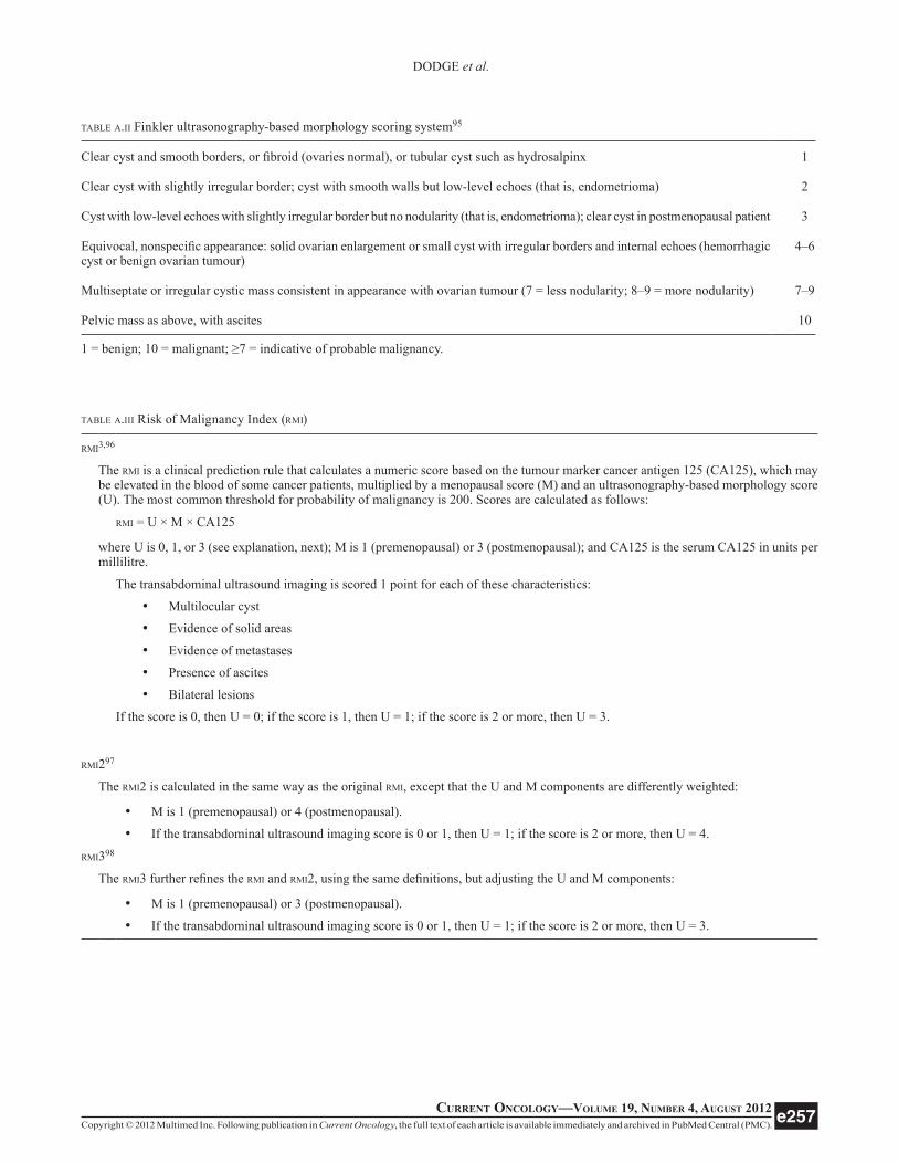
table a.ii Finkler ultrasonography-based morphology scoring system95
Clear cyst and smooth borders, or fibroid (ovaries normal), or tubular cyst such as hydrosalpinx 1
Clear cyst with slightly irregular border; cyst with smooth walls but low-level echoes (that is, endometrioma) 2
Cyst with low-level echoes with slightly irregular border but no nodularity (that is, endometrioma); clear cyst in postmenopausal patient 3
Equivocal, nonspecific appearance: solid ovarian enlargement or small cyst with irregular borders and internal echoes (hemorrhagic cyst or benign ovarian tumour)
4–6
Multiseptate or irregular cystic mass consistent in appearance with ovarian tumour (7 = less nodularity; 8–9 = more nodularity) 7–9
Pelvic mass as above, with ascites 10
1 = benign; 10 = malignant; ≥7 = indicative of probable malignancy.
table a.iii Risk of Malignancy Index (rmi)
rmi3,96
The rmi is a clinical prediction rule that calculates a numeric score based on the tumour marker cancer antigen 125 (CA125), which may be elevated in the blood of some cancer patients, multiplied by a menopausal score (M) and an ultrasonography-based morphology score (U). The most common threshold for probability of malignancy is 200. Scores are calculated as follows:
rmi = U × M × CA125
where U is 0, 1, or 3 (see explanation, next); M is 1 (premenopausal) or 3 (postmenopausal); and CA125 is the serum CA125 in units per millilitre.
The transabdominal ultrasound imaging is scored 1 point for each of these characteristics:
• Multilocular cyst
• Evidence of solid areas
• Evidence of metastases
• Presence of ascites
• Bilateral lesions
If the score is 0, then U = 0; if the score is 1, then U = 1; if the score is 2 or more, then U = 3.
rmi297
The rmi2 is calculated in the same way as the original rmi, except that the U and M components are differently weighted:
• M is 1 (premenopausal) or 4 (postmenopausal).
• If the transabdominal ultrasound imaging score is 0 or 1, then U = 1; if the score is 2 or more, then U = 4.
rmi398
The rmi3 further refines the rmi and rmi2, using the same definitions, but adjusting the U and M components:
• M is 1 (premenopausal) or 3 (postmenopausal).
• If the transabdominal ultrasound imaging score is 0 or 1, then U = 1; if the score is 2 or more, then U = 3.
Related Documents


![[1].pdf - ResearchGate](https://static.cupdf.com/doc/110x72/62129a6cbbf9242e6965a6a7/1pdf-researchgate.jpg)