Citation: Hobi, S.; Cafarchia, C.; Romano, V.; Barrs, V.R. Malassezia: Zoonotic Implications, Parallels and Differences in Colonization and Disease in Humans and Animals. J. Fungi 2022, 8, 708. https://doi.org/ 10.3390/jof8070708 Academic Editor: Hans de Cock Received: 15 June 2022 Accepted: 30 June 2022 Published: 4 July 2022 Publisher’s Note: MDPI stays neutral with regard to jurisdictional claims in published maps and institutional affil- iations. Copyright: © 2022 by the authors. Licensee MDPI, Basel, Switzerland. This article is an open access article distributed under the terms and conditions of the Creative Commons Attribution (CC BY) license (https:// creativecommons.org/licenses/by/ 4.0/). Fungi Journal of Review Malassezia: Zoonotic Implications, Parallels and Differences in Colonization and Disease in Humans and Animals Stefan Hobi 1, * , Claudia Cafarchia 2 , Valentina Romano 2 and Vanessa R. Barrs 1,3, * 1 Department of Veterinary Clinical Sciences, Jockey Club College of Veterinary Medicine and Life Sciences, City University, Tat Chee Avenue, Kowloon, Hong Kong, China 2 Department of Veterinary Medicine, University of Bari, Str. Prov. per Casamassima Km 3, Valenzano, (Bari), 70010, Italy; [email protected] (C.C.); [email protected] (V.R.) 3 Centre for Animal Health and Welfare, City University of Hong Kong, Kowloon Tong, Hong Kong, China * Correspondence: [email protected] (S.H.); [email protected] (V.R.B.) Abstract: Malassezia spp. are commensals of the skin, oral/sinonasal cavity, lower respiratory and gastrointestinal tract. Eighteen species have been recovered from humans, other mammals and birds. They can also be isolated from diverse environments, suggesting an evolutionary trajectory of adaption from an ecological niche in plants and soil to the mucocutaneous ecosystem of warm- blooded vertebrates. In humans, dogs and cats, Malassezia-associated dermatological conditions share some commonalities. Otomycosis is common in companion animals but is rare in humans. Systemic infections, which are increasingly reported in humans, have yet to be recognized in animals. Malassezia species have also been identified as pathogenetic contributors to some chronic human diseases. While Malassezia species are host-adapted, some species are zoophilic and can cause fungemia, with outbreaks in neonatal intensive care wards associated with temporary colonization of healthcare worker’s hands from contact with their pets. Although standardization is lacking, susceptibility testing is usually performed using a modified broth microdilution method. Antifungal susceptibility can vary depending on Malassezia species, body location, infection type, disease duration, presence of co-morbidities and immunosuppression. Antifungal resistance mechanisms include biofilm formation, mutations or overexpression of ERG11, overexpression of efflux pumps and gene rearrangements or overexpression in chromosome 4. Keywords: dermatology; zoonotic diseases; fungi; Malassezia; yeasts; resistance; treatment; transmission; animals; humans 1. Introduction Malassezia are small thick-walled ovoid, ellipsoid or cylindrical commensal yeasts of warm-blooded vertebrates. Their genome of approximately 10 Mb is almost half the size of Cryptococcus, another basidiomycete of medical and veterinary importance [1,2]. The mycelial phase of Malassezia spp. has been observed naturally in some skin lesions and induced in specialized culture media incubated at 30 ◦ C[2–6]. Malassezia species reproduce asexually by unipolar broad-based budding. The sexual form has not been detected, although the mating-type locus region has been identified [7]. An important characteristic of all Malassezia is their dependence on lipids for growth due to an absent fatty-acid synthetase gene and consequent inability to synthesize long- chain fatty acids. Although one species, M. pachydermatis, can readily grow on Sabauraud’s dextrose agar (SDA), a medium without lipid supplementation, it is still lipid dependent and its growth in this medium is due to the use of lipid fractions within the peptone, a component of SDA [2,8–11]. M. furfur was first identified on human skin in 1846 [12], but recently the genus has received more attention, not only because of its association with dermatological diseases in animals (dermatitis, otitis externa) and humans (pityriasis versicolor, atopic dermatitis, J. Fungi 2022, 8, 708. https://doi.org/10.3390/jof8070708 https://www.mdpi.com/journal/jof

Welcome message from author
This document is posted to help you gain knowledge. Please leave a comment to let me know what you think about it! Share it to your friends and learn new things together.
Transcript
Citation: Hobi, S.; Cafarchia, C.;
Romano, V.; Barrs, V.R. Malassezia:
Zoonotic Implications, Parallels and
Differences in Colonization and
Disease in Humans and Animals. J.
Fungi 2022, 8, 708. https://doi.org/
10.3390/jof8070708
Academic Editor: Hans de Cock
Received: 15 June 2022
Accepted: 30 June 2022
Published: 4 July 2022
Publisher’s Note: MDPI stays neutral
with regard to jurisdictional claims in
published maps and institutional affil-
iations.
Copyright: © 2022 by the authors.
Licensee MDPI, Basel, Switzerland.
This article is an open access article
distributed under the terms and
conditions of the Creative Commons
Attribution (CC BY) license (https://
creativecommons.org/licenses/by/
4.0/).
FungiJournal of
Review
Malassezia: Zoonotic Implications, Parallels and Differences inColonization and Disease in Humans and AnimalsStefan Hobi 1,* , Claudia Cafarchia 2 , Valentina Romano 2 and Vanessa R. Barrs 1,3,*
1 Department of Veterinary Clinical Sciences, Jockey Club College of Veterinary Medicine and Life Sciences,City University, Tat Chee Avenue, Kowloon, Hong Kong, China
2 Department of Veterinary Medicine, University of Bari, Str. Prov. per Casamassima Km 3, Valenzano, (Bari),70010, Italy; [email protected] (C.C.); [email protected] (V.R.)
3 Centre for Animal Health and Welfare, City University of Hong Kong, Kowloon Tong, Hong Kong, China* Correspondence: [email protected] (S.H.); [email protected] (V.R.B.)
Abstract: Malassezia spp. are commensals of the skin, oral/sinonasal cavity, lower respiratory andgastrointestinal tract. Eighteen species have been recovered from humans, other mammals andbirds. They can also be isolated from diverse environments, suggesting an evolutionary trajectoryof adaption from an ecological niche in plants and soil to the mucocutaneous ecosystem of warm-blooded vertebrates. In humans, dogs and cats, Malassezia-associated dermatological conditionsshare some commonalities. Otomycosis is common in companion animals but is rare in humans.Systemic infections, which are increasingly reported in humans, have yet to be recognized in animals.Malassezia species have also been identified as pathogenetic contributors to some chronic humandiseases. While Malassezia species are host-adapted, some species are zoophilic and can causefungemia, with outbreaks in neonatal intensive care wards associated with temporary colonizationof healthcare worker’s hands from contact with their pets. Although standardization is lacking,susceptibility testing is usually performed using a modified broth microdilution method. Antifungalsusceptibility can vary depending on Malassezia species, body location, infection type, diseaseduration, presence of co-morbidities and immunosuppression. Antifungal resistance mechanismsinclude biofilm formation, mutations or overexpression of ERG11, overexpression of efflux pumpsand gene rearrangements or overexpression in chromosome 4.
Keywords: dermatology; zoonotic diseases; fungi; Malassezia; yeasts; resistance; treatment; transmission;animals; humans
1. Introduction
Malassezia are small thick-walled ovoid, ellipsoid or cylindrical commensal yeastsof warm-blooded vertebrates. Their genome of approximately 10 Mb is almost half thesize of Cryptococcus, another basidiomycete of medical and veterinary importance [1,2].The mycelial phase of Malassezia spp. has been observed naturally in some skin lesionsand induced in specialized culture media incubated at 30 ◦C [2–6]. Malassezia speciesreproduce asexually by unipolar broad-based budding. The sexual form has not beendetected, although the mating-type locus region has been identified [7].
An important characteristic of all Malassezia is their dependence on lipids for growthdue to an absent fatty-acid synthetase gene and consequent inability to synthesize long-chain fatty acids. Although one species, M. pachydermatis, can readily grow on Sabauraud’sdextrose agar (SDA), a medium without lipid supplementation, it is still lipid dependentand its growth in this medium is due to the use of lipid fractions within the peptone, acomponent of SDA [2,8–11].
M. furfur was first identified on human skin in 1846 [12], but recently the genus hasreceived more attention, not only because of its association with dermatological diseasesin animals (dermatitis, otitis externa) and humans (pityriasis versicolor, atopic dermatitis,
J. Fungi 2022, 8, 708. https://doi.org/10.3390/jof8070708 https://www.mdpi.com/journal/jof

J. Fungi 2022, 8, 708 2 of 33
Malassezia folliculitis, seborrheic dermatitis) [2,13,14], but also due to its increased detectionin systemic infections, especially in neonates and immunocompromised patients [15–18].
M. pachydermatis, originally thought to be part of the mycobiome in dogs and cats only,has now also been isolated from humans, production animals and from multiple exoticand wildlife species such as the sea lion, scarlet macaw, brown bear, American black bear,Eurasian badger, big anteater, common wombat, Mangaliza pig, wide-mouthed rhinoceros,Indian elephant, red fox, porcupine and coyote [19–22].
In this review, we use the One-Health paradigm to explore similarities and differencesregarding carriage of Malassezia species in humans and companion animals, antifungalsusceptibility, resistance mechanisms, Malassezia-associated diseases and treatment. Theavailable evidence for transmission between animals and humans, directionality of trans-mission, and clinical relevance are also discussed.
2. Classification of Malassezia Yeasts
Malassezia yeasts belong to the family Malasseziaceae, order Malasseziales and classMalasseziomycetes. They are included in the morphologically highly diverse subdivisionof Ustilaginomycotina, and due to their filament (hyphae) and reproduction characteristics,they are contained in the division of Basidiomycota [23–26].
Thus far, 18 Malassezia species have been identified from a variety of mammalian hostsand birds (Table 1) and further expansion of the genus is likely [27]. For species differentia-tion, locus analysis of specific ribosomal gene sequences, such as ITS, D1/D2, ß-tubulin,chitin synthetase 2 and large subunit polymerase 2, is used. For phylogenetic stem eval-uation and species delimitation, whole genome sequencing (WGS) is necessary [1,27–35].
Table 1. Classification of Malassezia by species, reference strain, hosts and phylogenetic clades.
Species Reference Strain/GenBankAccession Genome Number Described Hosts Clade
M. furfur CBS 14141, GCA_009938135 Human, Cat, Dog, Cattle, Pig, Goat, Elk, Horse,Sheep, Elephant, Monkey, Ostrich, Pelican A
M. brasiliensis * MA 1455 Parrot AM. yamatoensis MY9725, GCA_001264885 Human, Cat AM. psittaci * MA 1454 Parrot AM. obtusa CBS 7876, GCA_001264985 Human, Cat, Dog, Goat, Horse AM. japonica CBS 9431, GCA_001264785 Human, Cat AM. vespertilionis CBS 15041, GCA_002818225 Bat A
M. globosa CBS 7966, GCA_001264805 Human, Cat, Dog, Cattle, Goat, Horse,Sheep, Cheetah B
M. restricta CBS 7877, GCA_001264765 Human, Cat, Dog, Cattle, Goat, Horse, Sheep BM. arunalokei CBS 13387, GCA_020085095 Human, Dog B
M. sympodialis ATCC 42132, GCA_001264925 Human, Dog, Cat, Pig, Cattle, Goat, Horse, Sheep,Chicken B
M. dermatis CBS 9169, GCA_001264665 Human, Cat BM. caprae CBS 10434, GCA_001264625 Goat, Horse, Human BM. equina CBS 9969, GCA_001264685 Horse, Cattle BM. nana JCM 12085, GCA_001600835 Cat, Dog, Cattle, Horse B
M. pachydermatis CBS 1879, GCA_001264975 Human, Dog, Cat, Pig, Goat, Rabbit, Various exoticand wild mammals, Birds (Thraupidae, Macaw) B
M. cuniculi CBS 11721, GCA_001264635 Rabbit CM. slooffiae CBS 7956, GCA_001264965 Human, Cat Cattle, Sheep, Pig, Goat, Horse C
* = whole genome not available.
Recently, after the WGS of 28 representative isolates from 15 Malassezia species, con-catenated protein sequences of 254 conserved orthologues were included in a phylogeneticanalysis to resolve the taxonomy of the genus [27]. Similar to previous analyses [1,26], allspecies fell into three distinct clades [27] (Table 1).

J. Fungi 2022, 8, 708 3 of 33
3. Malassezia Species in the Environment and Possible Vectors
Although first isolated from the skin of humans, followed by other warm-bloodedvertebrates, recent data have shown that Malassezia species have a much broader spectrumof ecological diversity than originally thought [36–38]. These yeasts have now been isolatedfrom a range of environments, including marine water, anoxic oceans, hydrothermalvents, deep-sea to high arctic marine sediment and Antarctic soil [36,39–52]. Malasseziaspecies also dominate the mycobiome of marine invertebrates, such as sponges and corals,and have been identified in healthy and diseased marine algae [36,53,54]. In addition,Malassezia species have been isolated from soil nematodes, cone-snails, olive fruit-flies andorchid roots [55–58]. A potential role for nematodes and flies as vectors for Malassezia hasbeen speculated [55,58–60].
It is now apparent that Malassezia species are among the most widespread fungi onEarth [36–38]. Their evolutionary trajectory involves adaptation from an ecological niche inplants and soil to the mucocutaneous ecosystem of animals [36–38]. This has been facilitatedby the loss of complex carbohydrate metabolism genes (glycosyl hydrolase encoding) and agenus-wide gain of lipid hydrolases including lipases, phospholipases and acid sphingomyeli-nases that are required to degrade and use skin- or mucosa-associated lipids [1,36–38].
4. Malassezia Species and Their Role as Commensals in Humans
Twelve Malassezia species have been isolated from human skin [6,16,34,61–75] (Table 1).Malassezia arunalokei is the only species isolated from humans that has not been isolatedfrom animals, with the exception of dogs [74,76].
Malassezia species colonization of the skin starts directly after birth, increases untilaround 12 months of age, and then remains relatively static until puberty, when anothersignificant quantitative increase in colonization occurs, associated with increased sebaceousgland activity and changes in the lipid composition of the skin [6,77]. After puberty,Malassezia species comprise 50 to 80% of the human mycobiome [78–80,100]. The limiteddata currently available about cutaneous mycobiomes in preterm and term neonates showsthat Malassezia species distribution on the skin of neonates and children varies betweenstudies, but M. globosa, M. furfur, M. sympodialis and M. restricta are the most prevalentspecies described [14].
In contrast, M. restricta and M. globosa dominate the mycobiome of both healthy anddiseased skin in adult humans, followed by M. sympodialis, albeit at a much lower frequencythan the former two [1,62,69,81–91]. M. furfur can be common at certain body sites (e.g., toe-web space) in healthy individuals but is not a dominating species overall. Instead, thisspecies is more frequently isolated from skin diseases, such as psoriasis vulgaris andpityriasis versicolor [1,5,73,89–93].
Climate and ethnicity also impact the carriage of Malassezia species [94,95]. In a studyby Leong et al. in 2019, people in Singapore of four different ethnicities (Chinese, Malay,Indian and Caucasian) carried a higher number of Malassezia species and showed greaterspecies diversity and evenness than Caucasians in Switzerland. The predominant species(isolated by culture from the skin of the side of the nose) in the latter were M. restricta andM. sympodialis, while M. globosa was absent. In contrast, sampling from the same site inthe four ethnic groups in Singapore showed M. globosa, M. furfur and M. restricta were thedominant species. Caucasians from the two locations showed different species distributions,with M. restricta being twice as common among those in Singapore, while M. globosa wasabsent in Swiss Caucasians. The same study associated the lower temperature and humidityof Switzerland compared to Singapore with a lower positive culture rate and lower speciesdiversity [95] (Figure 1). From other studies, it can be concluded that M. restricta plays adominant role as a skin commensal in Europe, whereas M. globosa comparatively dominatesin Asia [13,96].
J. Fungi 2022, 8, 708 4 of 33
J. Fungi 2022, 8, x FOR PEER REVIEW 4 of 33
distributions, with M. restricta being twice as common among those in Singapore, while M. globosa was absent in Swiss Caucasians. The same study associated the lower temper-ature and humidity of Switzerland compared to Singapore with a lower positive culture rate and lower species diversity [96] (Figure 1). From other studies, it can be concluded that M. restricta plays a dominant role as a skin commensal in Europe, whereas M. globosa comparatively dominates in Asia [13,97].
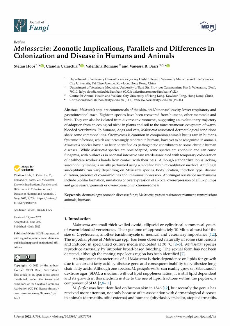
Figure 1. Predominant Malassezia species found as commensals in the sinonasal cavity (A), on the skin of the forehead and external ear canal (B), in the lungs and gastrointestinal tract (C) and on the skin of the occiput, back and groin (D). The relative diversity of Malassezia species found in healthy skin is shown in (E,F). The predominant species isolated from the culture of the skin on the side of the nose of Caucasians in Switzerland were M. restricta and M. sympodialis, while M. globosa was absent. In contrast, sampling from the skin of the noses of 4 ethnic groups (Chinese, Malay, Indian and Caucasian) in Singapore overall revealed M. globosa, M. furfur and M. restricta to be the domi-nant species [96].
Several studies have shown that sex and body site also influence the species of Malas-sezia species present on the skin and their abundance [1,89,92,98–101]. Site-specific species include M. restricta, which favors colonization of the external ear canals, retroauricular crease and forehead and M. globosa, which is most commonly isolated from the back, oc-ciput and groin [100,102].
A Japanese study in 2010 quantified Malassezia colonization of the skin of the cheek using real-time PCR and determined associations with gender and age in 770 healthy in-dividuals [100]. Total Malassezia DNA in males stayed constant from age 0 until around 9 years of age, with a progressive increase each year thereafter until the age of 16 to 18. In females, total Malassezia DNA increased until the age of 12, decreased between the ages of 19 and 22, and then increased again between the ages of 30 and 39. In both genders, there was a gradual decrease in Malassezia species abundance over the course of life. Overall, males tended to have more abundant Malassezia DNA than females, and M. globosa and M. restricta were the dominant species for both for all ages.
Malassezia species carriage at different skin locations was investigated using culture-based methods. No significant differences between the genders were found. While M. re-stricta dominated the scalp and M. sympodialis dominated the trunk, M. globosa was about equally common at both locations [103].
Figure 1. Predominant Malassezia species found as commensals in the sinonasal cavity (A), on theskin of the forehead and external ear canal (B), in the lungs and gastrointestinal tract (C) and onthe skin of the occiput, back and groin (D). The relative diversity of Malassezia species found inhealthy skin is shown in (E,F). The predominant species isolated from the culture of the skin on theside of the nose of Caucasians in Switzerland were M. restricta and M. sympodialis, while M. globosawas absent. In contrast, sampling from the skin of the noses of 4 ethnic groups (Chinese, Malay,Indian and Caucasian) in Singapore overall revealed M. globosa, M. furfur and M. restricta to be thedominant species [95].
Several studies have shown that sex and body site also influence the species ofMalassezia species present on the skin and their abundance [1,88,91,97–100]. Site-specificspecies include M. restricta, which favors colonization of the external ear canals, retroauric-ular crease and forehead and M. globosa, which is most commonly isolated from the back,occiput and groin [100,101].
A Japanese study in 2010 quantified Malassezia colonization of the skin of the cheekusing real-time PCR and determined associations with gender and age in 770 healthyindividuals [99]. Total Malassezia DNA in males stayed constant from age 0 until around9 years of age, with a progressive increase each year thereafter until the age of 16 to 18. Infemales, total Malassezia DNA increased until the age of 12, decreased between the ages of19 and 22, and then increased again between the ages of 30 and 39. In both genders, therewas a gradual decrease in Malassezia species abundance over the course of life. Overall,males tended to have more abundant Malassezia DNA than females, and M. globosa andM. restricta were the dominant species for both for all ages.
Malassezia species carriage at different skin locations was investigated using culture-based methods. No significant differences between the genders were found. WhileM. restricta dominated the scalp and M. sympodialis dominated the trunk, M. globosa wasabout equally common at both locations [102].
Other factors that may influence the colonization of Malassezia species include hostfactors (immune response, body secretion, skin occlusion), other skin inhabitants (e.g. par-asites, other microbes) and environmental parameters, including exposure to ultravioletlight [96]. Even the birth process itself has a significant impact. If a baby is born via naturaldelivery, its skin microbiota resembles the mother’s vaginal communities, but if deliveredvia caesarian section, it represents the mother’s skin surface population [103–107]. Inaddition, vaginal birth is associated with a higher abundance of Malassezia [108,109].
J. Fungi 2022, 8, 708 5 of 33
Malassezia species were previously thought to be commensals of the skin only. Al-though the skin is the primary ecological niche, more recent data demonstrate that theseyeasts also colonize the mucosa of the sinonasal and oral cavities, as well as the gastroin-testinal and lower respiratory tract [110–115]. Malassezia species are dominant membersof the mycobiome of the sinuses, with M. restricta and M. sympodialis most frequentlydetected [116]. Malassezia also comprise 30% of the gastrointestinal mycobiome, with threespecies detected—M. globosa, M. restricta and M. pachydermatis [117]. The fungal burden inthe lungs of healthy people is relatively low. In one study, using a metagenomic approach,the lung mycobiome was characterized by a high proportion of basidiomycetes, includingM. restricta and M. globosa [118], while in another ascomycetes, including Candida species,were most abundant [119] (Figure 1).
5. Malassezia Species and Their Role as Commensals in Companion Animals
Using culture-based techniques, Malassezia species have been identified as the mostcommon yeast colonizing healthy canine skin [120,121]. Metagenomic approaches revealthat, in contrast to humans, Ascomycota, especially Alternaria and Cladosporium species, arethe most abundant fungal species on the skin of healthy dogs and cats [122,123].
Overall, eleven Malassezia species have been isolated from cats and seven fromdogs [2,124–142] (Table 1). Culture-based studies clearly favor M. pachydermatis as thedominant species colonizing the skin of dogs and cats [124–126,128,135,143–146]. In onerecent study using metagenomics and quantitative PCR (qPCR), M. restricta and M. globose,but not M. pachydermatis, were identified as the dominant species colonizing healthy fe-line skin [139].
Malassezia abundance and species diversity are influenced by body site, genetic pre-dispositions and concurrent diseases [2,135–137,140,141,146–148]. M. pachydermatis is morefrequently isolated from dogs from perioral and interdigital skin than from the back orventral body sites, such as the axillae or groin [2] (Figure 2).
J. Fungi 2022, 8, x FOR PEER REVIEW 5 of 33
Other factors that may influence the colonization of Malassezia species include host factors (immune response, body secretion, skin occlusion), other skin inhabitants (e.g. par-asites, other microbes) and environmental parameters, including exposure to ultraviolet light [97]. Even the birth process itself has a significant impact. If a baby is born via natural delivery, its skin microbiota resembles the mother’s vaginal communities, but if delivered via caesarian section, it represents the mother’s skin surface population [104–108]. In ad-dition, vaginal birth is associated with a higher abundance of Malassezia [109,110].
Malassezia species were previously thought to be commensals of the skin only. Alt-hough the skin is the primary ecological niche, more recent data demonstrate that these yeasts also colonize the mucosa of the sinonasal and oral cavities, as well as the gastroin-testinal and lower respiratory tract [111–116]. Malassezia species are dominant members of the mycobiome of the sinuses, with M. restricta and M. sympodialis most frequently de-tected [117]. Malassezia also comprise 30% of the gastrointestinal mycobiome, with three species detected—M. globosa, M. restricta and M. pachydermatis [118]. The fungal burden in the lungs of healthy people is relatively low. In one study, using a metagenomic approach, the lung mycobiome was characterized by a high proportion of basidiomycetes, including M. restricta and M. globosa [119], while in another ascomycetes, including Candida species, were most abundant [120] (Figure 1).
5. Malassezia Species and Their Role as Commensals in Companion Animals Using culture-based techniques, Malassezia species have been identified as the most
common yeast colonizing healthy canine skin [121,122]. Metagenomic approaches reveal that, in contrast to humans, Ascomycota, especially Alternaria and Cladosporium species, are the most abundant fungal species on the skin of healthy dogs and cats [123,124].
Overall, eleven Malassezia species have been isolated from cats and seven from dogs [2,125–143] (Table 1). Culture-based studies clearly favor M. pachydermatis as the dominant species colonizing the skin of dogs and cats [125–127,129,136,144–147]. In one recent study using metagenomics and quantitative PCR (qPCR), M. restricta and M. globose, but not M. pachydermatis, were identified as the dominant species colonizing healthy feline skin [140].
Malassezia abundance and species diversity are influenced by body site, genetic pre-dispositions and concurrent diseases [2,136–138,141,142,147–149]. M. pachydermatis is more frequently isolated from dogs from perioral and interdigital skin than from the back or ventral body sites, such as the axillae or groin [2] (Figure 2).
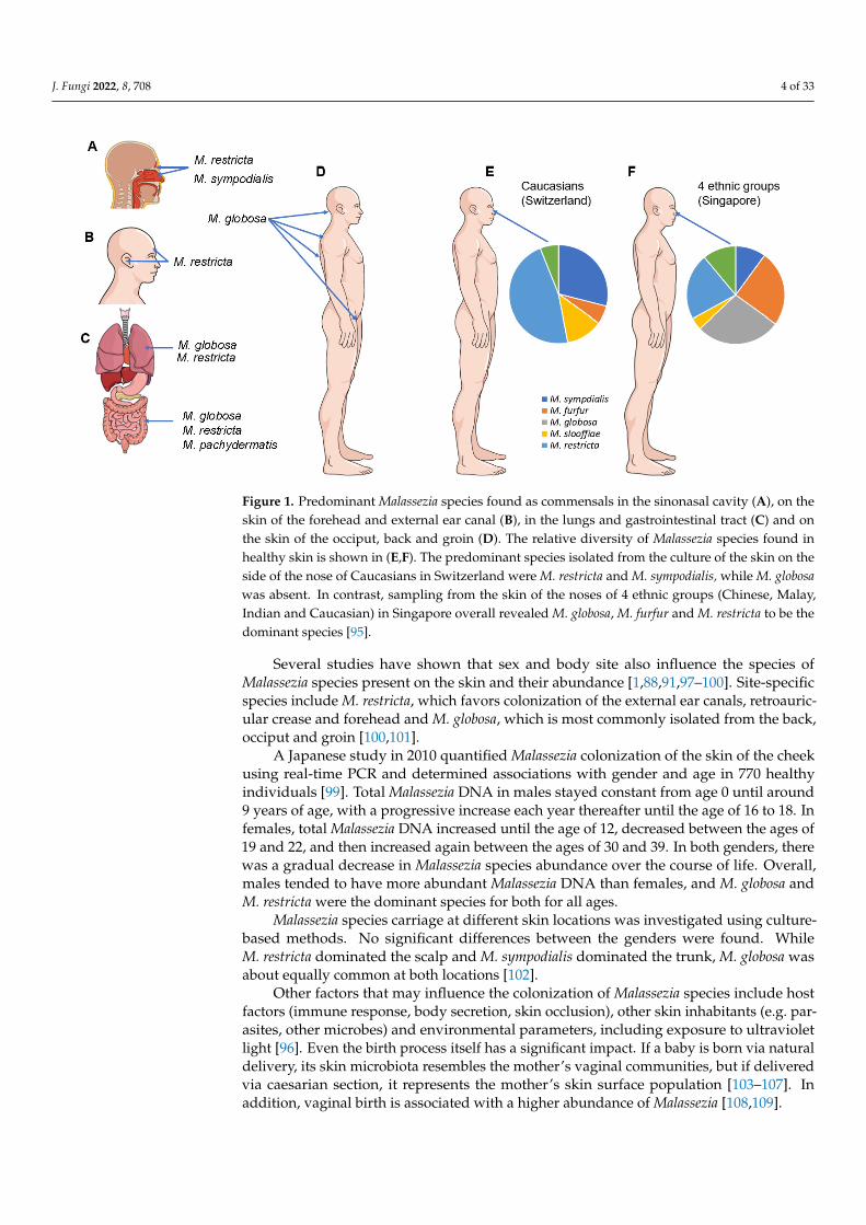
Figure 2. Malassezia species colonization in healthy dogs and cats. Figure 2. Malassezia species colonization in healthy dogs (A) and cats (B).
In cats, the external ear canal is most commonly colonized by M. pachydermatis,followed by other species such as M. furfur, M. globosa, M. sympodialis, M. obtusa andM. nana [134,142,149–152]. M. nana is the most common skin and ear isolate after M. pachy-dermatis, with one specific genotype dominating [136,137]. Claw folds of cats are a particularniche for M. slooffiae [135–137] (Figure 2).
Two specific feline breeds, the Devon Rex and Sphynx, harbor high Malassezia speciesloads, with a dominance of M. pachydermatis [135,146,147]. Cats with otitis externa alsohave a higher abundance of Malassezia species in the ear canals compared to healthy
J. Fungi 2022, 8, 708 6 of 33
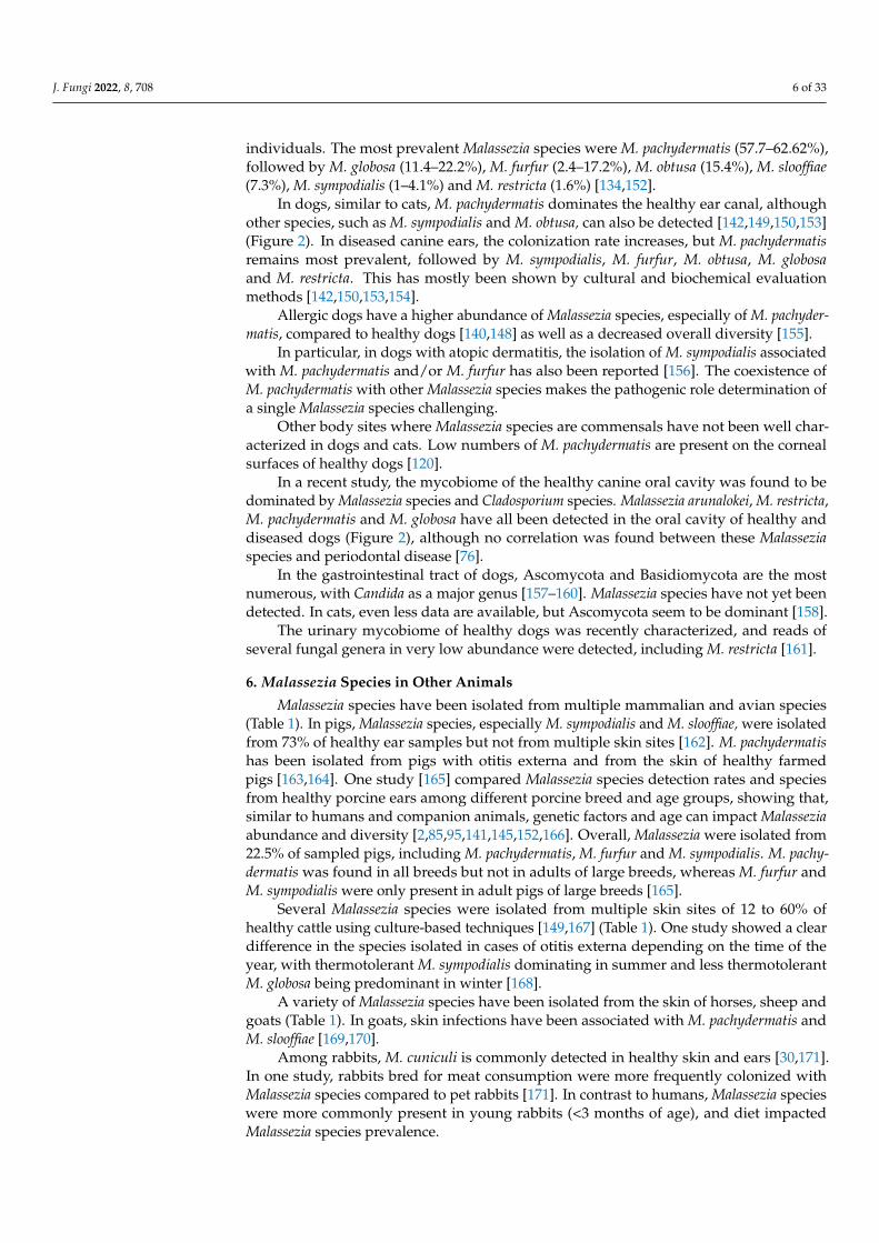
individuals. The most prevalent Malassezia species were M. pachydermatis (57.7–62.62%),followed by M. globosa (11.4–22.2%), M. furfur (2.4–17.2%), M. obtusa (15.4%), M. slooffiae(7.3%), M. sympodialis (1–4.1%) and M. restricta (1.6%) [134,152].
In dogs, similar to cats, M. pachydermatis dominates the healthy ear canal, althoughother species, such as M. sympodialis and M. obtusa, can also be detected [142,149,150,153](Figure 2). In diseased canine ears, the colonization rate increases, but M. pachydermatisremains most prevalent, followed by M. sympodialis, M. furfur, M. obtusa, M. globosaand M. restricta. This has mostly been shown by cultural and biochemical evaluationmethods [142,150,153,154].
Allergic dogs have a higher abundance of Malassezia species, especially of M. pachyder-matis, compared to healthy dogs [140,148] as well as a decreased overall diversity [155].
In particular, in dogs with atopic dermatitis, the isolation of M. sympodialis associatedwith M. pachydermatis and/or M. furfur has also been reported [156]. The coexistence ofM. pachydermatis with other Malassezia species makes the pathogenic role determination ofa single Malassezia species challenging.
Other body sites where Malassezia species are commensals have not been well char-acterized in dogs and cats. Low numbers of M. pachydermatis are present on the cornealsurfaces of healthy dogs [120].
In a recent study, the mycobiome of the healthy canine oral cavity was found to bedominated by Malassezia species and Cladosporium species. Malassezia arunalokei, M. restricta,M. pachydermatis and M. globosa have all been detected in the oral cavity of healthy anddiseased dogs (Figure 2), although no correlation was found between these Malasseziaspecies and periodontal disease [76].
In the gastrointestinal tract of dogs, Ascomycota and Basidiomycota are the mostnumerous, with Candida as a major genus [157–160]. Malassezia species have not yet beendetected. In cats, even less data are available, but Ascomycota seem to be dominant [158].
The urinary mycobiome of healthy dogs was recently characterized, and reads ofseveral fungal genera in very low abundance were detected, including M. restricta [161].
6. Malassezia Species in Other Animals
Malassezia species have been isolated from multiple mammalian and avian species(Table 1). In pigs, Malassezia species, especially M. sympodialis and M. slooffiae, were isolatedfrom 73% of healthy ear samples but not from multiple skin sites [162]. M. pachydermatishas been isolated from pigs with otitis externa and from the skin of healthy farmedpigs [163,164]. One study [165] compared Malassezia species detection rates and speciesfrom healthy porcine ears among different porcine breed and age groups, showing that,similar to humans and companion animals, genetic factors and age can impact Malasseziaabundance and diversity [2,85,95,141,145,152,166]. Overall, Malassezia were isolated from22.5% of sampled pigs, including M. pachydermatis, M. furfur and M. sympodialis. M. pachy-dermatis was found in all breeds but not in adults of large breeds, whereas M. furfur andM. sympodialis were only present in adult pigs of large breeds [165].
Several Malassezia species were isolated from multiple skin sites of 12 to 60% ofhealthy cattle using culture-based techniques [149,167] (Table 1). One study showed a cleardifference in the species isolated in cases of otitis externa depending on the time of theyear, with thermotolerant M. sympodialis dominating in summer and less thermotolerantM. globosa being predominant in winter [168].
A variety of Malassezia species have been isolated from the skin of horses, sheep andgoats (Table 1). In goats, skin infections have been associated with M. pachydermatis andM. slooffiae [169,170].
Among rabbits, M. cuniculi is commonly detected in healthy skin and ears [30,171].In one study, rabbits bred for meat consumption were more frequently colonized withMalassezia species compared to pet rabbits [171]. In contrast to humans, Malassezia specieswere more commonly present in young rabbits (<3 months of age), and diet impactedMalassezia species prevalence.
J. Fungi 2022, 8, 708 7 of 33
Among different bird species, Malassezia species have been isolated from healthy anddiseased sites, including beak (M. brasiliensis, M. psittaci), feathers and wings (M. pachyder-matis, M. furfur), oropharynx (M. pachydermatis, M. furfur, M. brasiliensis, M. psittaci), andfeces (M. pachydermatis, M. furfur) [20,125,172]. M. sympodialis has also been commonlyfound in diseased combs of adult chickens [173].
7. Zoonotic and Reverse Zoonotic Transmission of Malassezia Species
There is now ample evidence that different Malassezia species are shared betweenhumans and animals (Table 1). However, some genotypes within a species might be hostadapted or linked to a particular host site location or skin disorder [27,30,31,74,110,174]. Inparticular, sequence analyses of the LSU rDNA showed distinct Malassezia species subtypeson different host species [110]. Sequence analysis of IGS1 distinguished specific M. globosa,M. restricta, and M. pachydermatis variants in seborrheic dermatitis and atopic eczema andon the healthy skin of humans and animals [85,174]. Among M. pachydermatis, eight IGS1subtypes were identified and subtype 3D was mainly associated with skin lesions [175].Additionally, M. pachydermatis, frequently isolated from cats and dogs [176–180], but rarelyfrom human skin [62,181,182] was known to cause fungemia in people, especially inneonates [34,63,183–191]. However, newborn babies have skin colonization by M. sympo-dialis and M. globosa, but not by M. pachydermatis [78–80,100]. Thus, the ease with whichthese yeasts can be transmitted from one body site to another [192] or between animals andtheir owners [182] makes us hypothesize that zoonotic and reverse zoonotic transmissionof these yeast species can occur.
In particular, the carriage of M. pachydermatis in healthy and diseased dogs with allergicdermatitis or otitis externa was compared to healthy human owners [182]. M. pachydermatisDNA was identified on the palms of over 90% of pet owners, regardless of the diseasestate of their dogs. Based on culture results indicating the relative abundance of Malasseziaspecies, owners of affected dogs were 11 times more likely to be culture positive thanowners of healthy dogs [182].
The zoophilic potential of M. pachydermatis was first postulated by Dr. Gueho [193] butwas clearly confirmed ten years later when an outbreak of neonatal fungemia caused by M.pachydermatis was investigated [184]. The strain implicated in the outbreak was isolatedfrom a health care worker’s hands, from contaminated equipment and from dogs belongingto three health care workers working in the involved intensive care nursery unit. One orseveral healthcare workers likely contaminated the nursery environment and their patientsafter transient colonization of their hands by the organism. After optimizing hand hygiene,no further cases were reported and all cultures from staff members tested negative [184].
Other studies have also demonstrated that hospitalized infants can be colonizedby Malassezia species, especially M. pachydermatis and M. furfur, via contact with theirparents or healthcare workers or indirectly via incubator surfaces [16,77,183,184,194–196].Healthcare workers can then further transmit the organism from one infant to another viatheir hands. Through this mechanism, several Malassezia species outbreaks have occurredin the past [184,197,198].
Carriage of M. pachydermatis in humans was detected in low numbers on the scalpand palms of 12% of healthy individuals in one study [181], and on the skin of 5% ofhealthy medical students in another [62]. In other studies it was not detected at allin healthy individuals, and overall appears to be a rare, transient colonizer of humanskin [6,102]. Similarly, other studies have found no causal associations between M. pachy-dermatis and human Malassezia-associated skin conditions, including seborrheic dermatitisand pityriasis versicolor [62,199].
While there is evidence that M. pachydermatis can be transmitted between dogs andhumans, further investigations into the genotypes involved, and the strain characteristicsare warranted [184,188–190,200]. The relatively recent discovery of M. pachydermatis as acommensal of the human gut introduces another potential reservoir of infection in humansby this species [117].
J. Fungi 2022, 8, 708 8 of 33
There is phenotypic and phylogenetic evidence that species with high host diver-sity, such as M. furfur, are undergoing diversification to enable successful adaptation todifferent hosts [201]. Strains from different animal species remain closely genetically re-lated, but the extent and frequency of zoonotic or reverse zoonotic transmission have notbeen investigated.
8. Superficial Malassezia-Associated Diseases in Humans and Animals8.1. Malassezia-Associated Dermatological Diseases in Humans
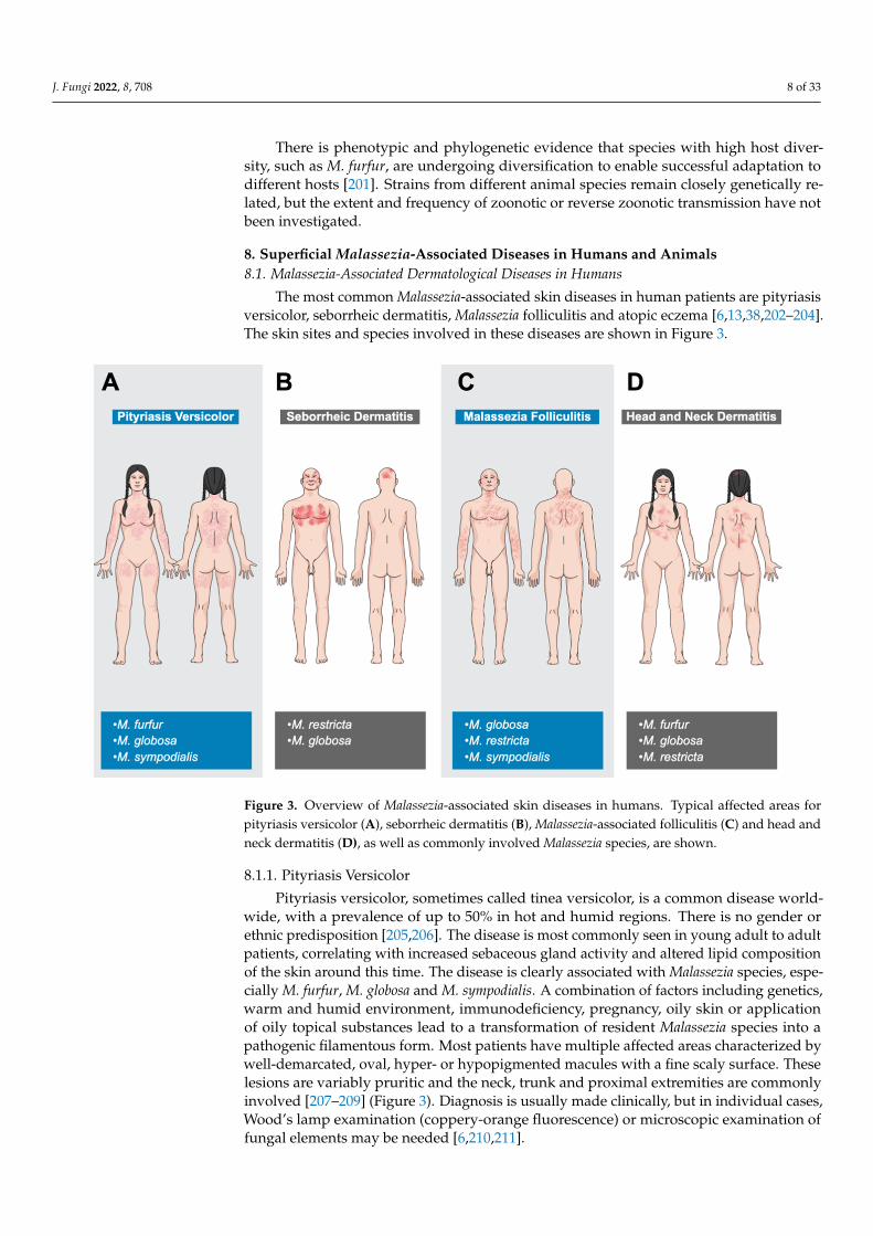
The most common Malassezia-associated skin diseases in human patients are pityriasisversicolor, seborrheic dermatitis, Malassezia folliculitis and atopic eczema [6,13,38,202–204].The skin sites and species involved in these diseases are shown in Figure 3.
J. Fungi 2022, 8, x FOR PEER REVIEW 8 of 33
healthy individuals, and overall appears to be a rare, transient colonizer of human skin [6,103]. Similarly, other studies have found no causal associations between M. pachyder-matis and human Malassezia-associated skin conditions, including seborrheic dermatitis and pityriasis versicolor [62,200].
While there is evidence that M. pachydermatis can be transmitted between dogs and humans, further investigations into the genotypes involved, and the strain characteristics are warranted [185,189–191,201]. The relatively recent discovery of M. pachydermatis as a commensal of the human gut introduces another potential reservoir of infection in hu-mans by this species [118].
There is phenotypic and phylogenetic evidence that species with high host diversity, such as M. furfur, are undergoing diversification to enable successful adaptation to differ-ent hosts [202]. Strains from different animal species remain closely genetically related, but the extent and frequency of zoonotic or reverse zoonotic transmission have not been investigated.
8. Superficial Malassezia-Associated Diseases in Humans and Animals 8.1. Malassezia-Associated Dermatological Diseases in Humans
The most common Malassezia-associated skin diseases in human patients are pityria-sis versicolor, seborrheic dermatitis, Malassezia folliculitis and atopic eczema [6,13,38,203–205]. The skin sites and species involved in these diseases are shown in Figure 3.
Figure 3. Overview of Malassezia-associated skin diseases in humans. Typical affected areas for pit-yriasis versicolor (A), seborrheic dermatitis (B), Malassezia-associated folliculitis (C) and head and neck dermatitis (D), as well as commonly involved Malassezia species, are shown.
8.1.1. Pityriasis Versicolor Pityriasis versicolor, sometimes called tinea versicolor, is a common disease world-
wide, with a prevalence of up to 50% in hot and humid regions. There is no gender or ethnic predisposition [206,207]. The disease is most commonly seen in young adult to adult patients, correlating with increased sebaceous gland activity and altered lipid
Figure 3. Overview of Malassezia-associated skin diseases in humans. Typical affected areas forpityriasis versicolor (A), seborrheic dermatitis (B), Malassezia-associated folliculitis (C) and head andneck dermatitis (D), as well as commonly involved Malassezia species, are shown.
8.1.1. Pityriasis Versicolor
Pityriasis versicolor, sometimes called tinea versicolor, is a common disease world-wide, with a prevalence of up to 50% in hot and humid regions. There is no gender orethnic predisposition [205,206]. The disease is most commonly seen in young adult to adultpatients, correlating with increased sebaceous gland activity and altered lipid compositionof the skin around this time. The disease is clearly associated with Malassezia species, espe-cially M. furfur, M. globosa and M. sympodialis. A combination of factors including genetics,warm and humid environment, immunodeficiency, pregnancy, oily skin or applicationof oily topical substances lead to a transformation of resident Malassezia species into apathogenic filamentous form. Most patients have multiple affected areas characterized bywell-demarcated, oval, hyper- or hypopigmented macules with a fine scaly surface. Theselesions are variably pruritic and the neck, trunk and proximal extremities are commonlyinvolved [207–209] (Figure 3). Diagnosis is usually made clinically, but in individual cases,Wood’s lamp examination (coppery-orange fluorescence) or microscopic examination offungal elements may be needed [6,210,211].

J. Fungi 2022, 8, 708 9 of 33
8.1.2. Seborrheic Dermatitis
Seborrheic dermatitis also occurs worldwide, with ‘normal’ and dandruff forms af-fecting around 5% and up to 50% of the population, respectively. There is also an HIV-associated form. There is no ethnic predisposition, but males are clearly predisposed.Disease is mainly seen in infants and adults [212–214]. The etiology is not completely clearbut involves an interplay of skin flora, lipid composition on the skin surface, skin barrierintegrity, immune response to Malassezia species and individual host factors. Increased se-baceous gland activity, immunodeficiency, neurological and psychological diseases, certaindrugs and environmental factors such as low humidity and temperature are risk factorsfor seborrheic dermatitis [213,215,216]. M. restricta or M. globosa are typically isolatedfrom active lesions and antifungal treatment usually leads to significant clinical improve-ment. Other species can be isolated, including M. furfur, M. sympodialis, M. obtusa andM. slooffiae [38,217–219]. The scalp, face and chest are most commonly affected, although ininfants, the diaper area, neck and axillae may also be involved (Figure 3). Skin lesions are of-ten inflamed, pruritic and present at one or multiple locations. They include poorly definedfollicular papules and plaques, fine white scales, and yellow crusts. In the mild dandruffform, no inflammation but a fine, mild scaling on the scalp and beard dominates [220,221].
8.1.3. Malassezia Folliculitis
Malassezia folliculitis is another common worldwide disease with a prevalence of 1to 17%. It occurs more commonly in young to middle-aged adult males [222–224]. Fol-licular occlusion or a disturbance of the normal cutaneous flora leads to an abnormalproliferation of Malassezia species and the development of disease. Common associatedspecies include M. globosa, M. restricta and M. sympodialis [6,202,224–228]. Predispos-ing factors include hot and humid climate, excessive sweating, non-breathable clothing,application of make-up or sunscreens, certain drugs (antibiotics, glucocorticoids) andimmunosuppression [6,224,229,230]. The disease typically involves the face, upper back,extensor surfaces of the arms, chest and neck (Figure 3). In almost 75% of cases, more thanone location is affected. Lesions include small but pruritic follicular papules and pustules.This presentation is often mistaken for acne or bacterial folliculitis [223,224,231,232].
8.1.4. Atopic Dermatitis (Head and Neck Dermatitis)
Atopic dermatitis (AD) is a common, chronic, inflammatory and pruritic disease,affecting 10 to 25% of children and 1 to 2% of adults. Head and neck dermatitis (HND), asubtype of AD, mostly occurs in adolescence and adulthood in individuals with a historyof IgE-mediated AD. There is no gender or ethnic predisposition [233–236]. The etiology isincompletely understood, but it is clear that Malassezia species play an important role indisease pathogenesis. The high activity of sebaceous glands at affected sites, together withthe skin barrier disruption of the atopic disease, allow Malassezia species to proliferate, lead-ing to increased exposure to the immune system, triggering a humoral and cell-mediatedimmune response [237–243]. Some involved Malassezia antigens have been well character-ized (M. globosa—MGL_1304; M. sympodialis—Mala s 8; M. restricta—Mala r 8) and havebeen identified in the sweat of patients, leading to aggravated clinical signs, especially afterintense sweating [244,245]. These antigens have also shown variable histamine-releasingproperties [246]. Malassezia species isolated from disease-associated sites have includedM. furfur, M. obtusa, M. globosa, M. restricta and M. sympodialis, but there was no signif-icant difference in isolation compared to healthy individuals [240,247]. Others found ahigher colonization rate by M. furfur, as well as a lower colonization rate by M. globosa andM. sympodialis, in affected AD patients [62]. Specific genotypes of M. globosa and M. restrictahave also been identified as colonizing AD skin [65,174].
HND patients have erythema and erythematous plaques on the forehead, eyelids,perioral, neck and upper trunk together with variable pruritus (Figure 3). In severe cases,the whole face may be involved, leading to the term “red face”. With the chronicity of the
J. Fungi 2022, 8, 708 10 of 33
disease, lichenification and scaling can occur [245,248]. Wheal-like, edematous changeshave also been described [245].
8.2. Malassezia Dermatitis and Otitis Externa in Animals
In dogs and cats, Malassezia dermatitis and otitis externa are commonly encounteredin daily practice [141,156,249] but they can also be seen in farm animals, especially horsesand goats. The prevalence of Malassezia-associated skin diseases in farm animals maybe underestimated [169,170,250–256]. Malassezia dermatitis and otitis externa have alsobeen reported in many other animals, including sea lions, fennecs, okapi, dromedaries,rhinoceros, canaries and pinnipeds [21,163,257–262].
Concurrent Malassezia dermatitis and sarcoptic or demodectic mange are occasionallyseen in lagomorphs or hamsters, respectively [263–265]. Some specific dog and cat breedshave a higher risk of Malassezia dermatitis [135,145,266–269] (Table 2).
Table 2. Breed predisposition for Malassezia dermatitis in companion animals.
Dog Breeds Cat Breeds
West Highland White Terrier Devon RexEnglish Setter SphynxBasset HoundBoxerAmerican Cocker SpanielPoodleDachshundAustralian Silky TerrierShih Tzu
In veterinary Malassezia-associated dermatitis, typical cutaneous manifestations includealopecia, erythema, scaling, crusts and accumulation of greasy, malodorous, brown to blackkeratosebaceous debris. In chronic infections, lichenification and hyperpigmentation may alsobe present. The intensity of pruritus is variable [2,141,179,254–256,260,265,266,270,271].
In canine patients, an infection or overgrowth with Malassezia species is most com-monly associated with allergic diseases (flea bite hypersensitivity, food allergy, atopic der-matitis), ectoparasitic infestations, superficial pyoderma, occasionally with endocrinopathies(hypothyroidism, hyperadrenocorticism, diabetes mellitus), keratinization disorders andrarely with autoimmune diseases [272–275]. Common involved areas include the externalear canal, pinnae, lips, muzzle, ventral neck, ventral body sites, medial hind limbs, perianalsite and paws [270,276] (Figure 4).
In dogs with environmental allergies, the clinical signs of Malassezia dermatitis oftenmimic, or even worsen, those of atopic disease [270]. It has been shown that affectedpatients show elevated levels of Malassezia-specific IgG and IgE in their serum [277]. Inaddition, immediate hypersensitivity reactions were observed in canine atopic patientsin which M. pachydermatis extracts were intradermally injected or after passive trans-fer of atopic serum to healthy recipient dogs using the Prausnitz–Küstner (P-K) tech-nique [278,279]. Together with the frequent isolation and higher colonization rate ofMalassezia species on and from the skin of these patients, their relevance and contribution,especially M. pachydermatis, to disease pathogenesis has been demonstrated [140]. Four ma-jor allergens of M. pachydermatis with a size of 45, 52, 56 and 63 kDa were detected in morethan 50% of atopic dogs in a study by Chen et al. in 2002 [280].
J. Fungi 2022, 8, 708 11 of 33J. Fungi 2022, 8, x FOR PEER REVIEW 11 of 33
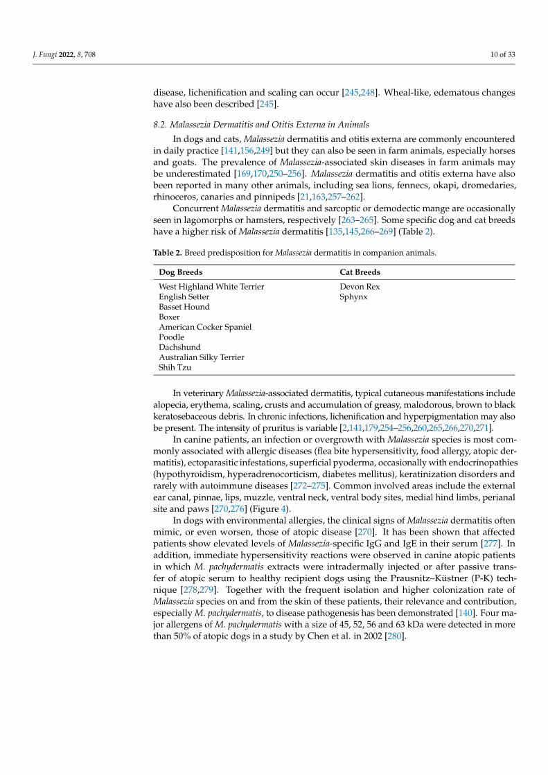
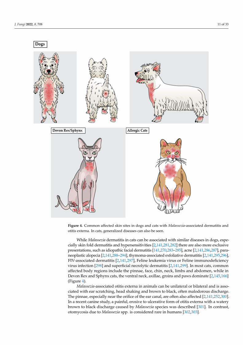
Figure 4. Common affected skin sites in dogs and cats with Malassezia-associated dermatitis and otitis externa. In cats, generalized diseases can also be seen.
In dogs with environmental allergies, the clinical signs of Malassezia dermatitis often mimic, or even worsen, those of atopic disease [272]. It has been shown that affected pa-tients show elevated levels of Malassezia-specific IgG and IgE in their serum [279]. In ad-dition, immediate hypersensitivity reactions were observed in canine atopic patients in which M. pachydermatis extracts were intradermally injected or after passive transfer of atopic serum to healthy recipient dogs using the Prausnitz–Küstner (P-K) technique [280,281]. Together with the frequent isolation and higher colonization rate of Malassezia species on and from the skin of these patients, their relevance and contribution, especially M. pachydermatis, to disease pathogenesis has been demonstrated [141]. Four major aller-gens of M. pachydermatis with a size of 45, 52, 56 and 63 kDa were detected in more than 50% of atopic dogs in a study by Chen et al. in 2002 [282].
While Malassezia dermatitis in cats can be associated with similar diseases in dogs, especially skin fold dermatitis and hypersensitivities [2,142,283,284] there are also more exclusive presentations, such as idiopathic facial dermatitis [142,272,285–287], acne [2,142,288,289], paraneoplastic alopecia [2,142,290–296], thymoma-associated exfoliative dermatitis [2,142,297,298], FIV-associated dermatitis [2,142,299], Feline leukemia virus or
Figure 4. Common affected skin sites in dogs and cats with Malassezia-associated dermatitis andotitis externa. In cats, generalized diseases can also be seen.
While Malassezia dermatitis in cats can be associated with similar diseases in dogs, espe-cially skin fold dermatitis and hypersensitivities [2,141,281,282] there are also more exclusivepresentations, such as idiopathic facial dermatitis [141,270,283–285], acne [2,141,286,287], para-neoplastic alopecia [2,141,288–294], thymoma-associated exfoliative dermatitis [2,141,295,296],FIV-associated dermatitis [2,141,297], Feline leukemia virus or Feline immunodeficiencyvirus infection [298] and superficial necrolytic dermatitis [2,141,299]. In most cats, commonaffected body regions include the pinnae, face, chin, neck, limbs and abdomen, while inDevon Rex and Sphynx cats, the ventral neck, axillae, groins and paws dominate [2,145,166](Figure 4).
Malassezia-associated otitis externa in animals can be unilateral or bilateral and is asso-ciated with ear scratching, head shaking and brown to black, often malodorous discharge.The pinnae, especially near the orifice of the ear canal, are often also affected [2,141,252,300].In a recent canine study, a painful, erosive to ulcerative form of otitis externa with a waterybrown to black discharge caused by Malassezia species was described [301]. In contrast,otomycosis due to Malassezia spp. is considered rare in humans [302,303].
J. Fungi 2022, 8, 708 12 of 33
8.3. Miscellaneous Forms of Superficial Malassezia-Associated Diseases
Occasionally, Malassezia species can also infect the nails of humans and the claws ofanimals. In companion animals, paronychia with erythema, swelling and a waxy brown toblack discharge is common, while in humans, subungual hyperkeratosis and onycholysiscan be seen [85,141,304–306].
Another potential site of superficial Malassezia infection is the cornea. There are sparsereports of keratomycosis in humans and dogs associated with M. furfur and M. restricta inhumans and M. pachydermatis in dogs [307–309]. Interestingly, one affected dog [309] and ahuman patient [308] both had diabetes mellitus and in all described cases immunomodulatoryor antibiotic drugs were used. These predisposing factors could have facilitated Malasseziaspecies overgrowth. The burden of corneal colonization by Malassezia species significantlyincreases in cases of corneal ulceration [120,310]. Whether Malassezia species could have aprimary pathogenic role in some cases of corneal ulceration requires further investigation.
9. Systemic Infections and Chronic Malassezia-Associated Diseases in Humansand Animals9.1. Fungemia and Systemic Infections
Of the 18 Malassezia species, only three are known to cause fungemia—M. furfur,M. pachydermatis and M. sympodialis. In the former two, one specific genotype isinvolved [14,198,311,312]. In fungemic patients, M. furfur is most frequently isolated, fol-lowed by M. pachdermatis and M. sympodialis [14,16,313].
Since the first report of systemic infection by an unspecified Malassezia species in1979 [314], systemic infections have been described with increasing frequency [14–16,18,198],likely due to growing recognition of the pathogenic potential of Malassezia species, as wellas improved detection methods [14,110].
The skin plays a significant role in the development of fungemia as both a reservoir ofMalassezia species and a portal of entry into the bloodstream by Malassezia species when it iscompromised [14]. Predisposing factors for fungemia include premature birth, hospitaliza-tion and duration of stay in a neonatal intensive care unit, immunosuppression, peritonealdialysis, presence of central venous catheter, total parenteral nutrition with lipid supplemen-tation (especially in neonates), invasive surgical procedures, long-term or broad-spectrumantimicrobial administration, chronic illnesses and topical application of soybean oil con-taining products [16,184,185,315]. Parenteral lipids are not only favorable for Malasseziaspecies growth but can also reduce the immune response of a patient by the generation ofreactive oxygen species, which decrease neutrophil phagocytosis [183,184,316,317].
The pathogenesis of Malassezia fungemia is not fully understood. Since only particulargenotypes of M. furfur or M. pachydermatis are associated with fungemia, pathogen virulencefactors are likely important determinants of systemic infection [198,311,312]. Malasseziaspecies possess a number of virulence factors, including lipases, phospholipases, metabo-lites (indirubin, indole carbazole, pityriacitrin and others), nanovesicles, cell membraneµ-opioid receptors, hydrophobicity, adherence and the ability to form biofilm [38,318–325].Of these, increased phospholipase activity and the release of allergen-enriched nanovesiclesare often related to more severe disease and fungemia [312,318,321,322,326].
Pathogenic Malassezia strains associated with fungemia are either already presentcolonizing the patient’s skin or are transmitted to the skin through interactions withhealthcare worker’s hands or contaminated medical devices, materials and/or parenteralsolutions [14,16,77,183,184,194,196].
Severe illness, the administration of immunosuppressive, antifungal or broad-spectrumantimicrobial drugs or parenteral lipids, poor anatomic conformation and/or prematureage lead to an impaired immune state. Different combinations of such factors enableinvasion of the body at an entrance point, such as a surgical wound or an intravenouscatheter site [14,38,194,327,328].
Hematogenous dissemination of Malassezia species can involve infection of the heart,lungs and, less commonly, the kidneys, pancreas, liver, spleen, brain and skin (multiple

J. Fungi 2022, 8, 708 13 of 33
cutaneous pustules) [183,184,316,317]. Biofilm formation facilitates local replication andfurther shedding of the organism into the blood system [317,329–331].
Systemic infections with Malassezia species include a broad range of presentations,from single-organ infection to fungemia, and can be fatal. Single-site infections includemeningitis [332,333], endocardial mass [334], pneumonia [335,336], peritonitis [314,337,338],osteomyelitis [339], septic arthritis [339], sinusitis [340] and mastitis [341].
Clinical signs of systemic Malassezia species infection in infants include fever, respira-tory distress from pneumonia or bronchopneumonia, lethargy, bradycardia, seizures andcyanosis. Infected infants often show hepato- and splenomegaly. The main hematologicalfindings are leukocytosis or leukopenia and thrombocytopenia [183–185,342–345].
Infections in children and adults are characterized by fever, chills, myalgia, nausea,vomiting and respiratory distress. Haematological findings include leukopenia (rarelyleukocytosis) and thrombocytosis [329,346–348].
The diagnosis of Malassezia-associated fungemia is challenging due to its special needsfor growth including lipid dependency. It is recommended to directly culture blood orcentral venous catheter tips on lipid-rich culture media via blood culture specimen tubesand not to use an automated blood culture system [14,313,349]. In addition, since humanblood can have inhibitory and toxic effects on yeasts, the addition of 3% palmitic acid mayfavor positive detection [350].
Thus far, Malassezia-associated fungemia has not been reported in animals.
9.2. Chronic Diseases in Humans and Animals
In patients with HIV infection, the burden of the Malassezia species in the gut and onthe skin of individuals with seborrheic dermatitis is significantly increased, associated withlow numbers of CD4+ helper cells/Th17 cells. This overgrowth of Malassezia species is a riskfactor for fungemia and other Malassezia-associated infections, including HIV-associatedseborrheic dermatitis [115,351–353].
In patients with inflammatory bowel disease (IBD), including Crohn’s disease, Malasseziaspecies dominate the gastrointestinal mycobiome [354–356]. M. restricta colonization, espe-cially in the sigmoid colon, can increase disease severity by intensifying the inflammatoryresponse [356]. This effect is strongly associated with the presence of the Crohn’s diseaserisk allele altered caspase recruitment domain 9 (CARD9 S12N). CARD9 is an adapterprotein of the CARD-CC family that mediates pattern recognition signaling and is essentialfor fungal defense [115,354–359]. In mice models, the same authors showed the capabilityof M. restricta to cause significant changes to the colon, including colon shortening, mucosalerosion and crypt destruction [356].
It has been speculated that Malassezia species could have a pathogenic role in the devel-opment or progression of colorectal cancer since affected individuals have gastrointestinalmycobiome dysbiosis with a higher burden of Malassezia species compared to healthyindividuals [115,360–362]. However, whether this is an effect or a cause of cancer remainsto be proven. Malassezia species have been found to play a causal role in pancreatic ductaladenocarcinoma (PDA) associated with migration from the gut to the pancreas [361]. In hu-man and murine PDA, cancerous pancreatic tissue contained a 3000-fold higher burden offungi compared to healthy pancreatic tissue and was specifically enriched for the Malasseziaspecies. The oncogenic pathway was also identified as the activation of mannose-bindinglectin, which drives the complement cascade and promotes oncogenesis [361].
A role for Malassezia species in neurodegenerative diseases, such as Alzheimer’sdisease and Parkinson’s disease, has been speculated due to their frequent detection inaffected areas of brain tissue [363–366]. The source of Malassezia species is fungemia due tobreaches of the cutaneous or gastrointestinal barriers. However, whether their presencereflects opportunistic colonization of damaged tissue or is causal has not been determined.Similar studies in veterinary medicine are lacking.
J. Fungi 2022, 8, 708 14 of 33
10. Antifungal Susceptibility Testing10.1. Methodology
Usually, established testing concentrations are used as a reference for systemicallyapplied drugs at their recommended doses [367,368]. Topically, much higher concentrationscan be reached, important for topical therapy and thereby susceptibility testing methodswould need to be adjusted [369–371]. Since Malassezia species are involved in commondiseases and can potentially cause deep infections, fungemia or even death, susceptibilitytesting becomes a necessary and very important tool [14–16,18,110]. Even if there arestandard proposed guidelines for testing the susceptibility profile of filamentous fungiand yeasts, it is difficult to implement these methods with Malassezia species due to theirspecial needs and growth characteristics [372,373]. As a consequence, and due to the lackof standardization, different procedures were proposed with culture medium, inoculumsize, incubation time, and criteria used to determine MIC endpoints largely vary amongthe studies, thus making it difficult to interpret the data in the literature [374]. The sus-ceptibility of Malassezia species to antifungal compounds has been tested using differentmethods, including a modified Clinical and Laboratory Standards Institute (CLSI) brothmicrodilution protocol [375–378] and agar-based diffusion methods (Disk Diffusion – DDand the E test—ET) [379–383]. However, the agreement analysis between agar-based dif-fusion methods and modified CLSI standard reference procedures still needs to be betterinvestigated. Overall, DD might not represent a valid alternative for determining thesusceptibility of Malassezia yeasts to azoles and amphotericin B (AmB), and ET should beused with specific media and longer reading times and only for specific drugs [374].
A completely different approach has been described via corneofungimetry. Stratumcorneum cells coated with olive oil form the basement of this testing process, mimickingan in vivo situation [384–386]. There is no comparison of this principle with commonlyused methods.
Overall, clear international standard guidelines for susceptibility testing of Malasseziaare urgently warranted to effectively compare and analyze data, but the authors considerthe broth microdilution method the most suitable one and regard this as the gold standard.
10.2. Patterns of Antifungal Susceptibility
For clinical usability, not only the MIC distribution but also other factors such asserum concentration of the drug, pharmacodynamics, resistance mechanisms and clinicalefficacy need to be considered [387–389]. These are encompassed by clinical breakpointvalues established by the CLSI and EUCAST [372,388–390]. These breakpoints are regularlyupdated and if not available, usually the ones for Candida, including C. krusei, C. parapsilosis,C. tropicalis and C. albicans, are considered [391].
Nevertheless, the final proof of resistance is through the detection of the underlyingmechanism. For Malassezia yeasts, clinical breakpoint values are still not established, butproof of the underlying mechanisms of resistance has been verified for some Malasseziaspecies (see below).
Overall, Malassezia species antifungal susceptibility profiles against azoles, AmB andterbinafine (TER) vary between species or intraspecies, regardless of the media or otherconditions employed [374]. M. sympodialis and M. pachydermatis are reported to havelower MICs of antifungals AmB, TER and azoles, in general compared to M. furfur andM. globosa [38,195,378,392–394].
MIC variation can also be seen within a given species, as shown for M. sympodialis,M. globosa and M. furfur [376,378,393,395]. Similar results are reflected in a canine studyinvolving M. pachydermatis, indicating less variation within the same patient but moredissimilarity between different patients [394,396].
Malassezia spp. bloodstream isolates have higher MICs for the same antifungal drugcompared to skin-origin isolates [392,393,397–400]. Accordingly, the disease status canaffect the MIC, as shown in dogs [394,401–403]. Patients with prior antifungal expo-sure showed higher values than healthy individuals. In an in vitro evaluation, strains

J. Fungi 2022, 8, 708 15 of 33
from diseased dogs showed higher MIC values across several azole drugs, including flu-conazole (FCZ), ketoconazole (KZ), miconazole (MIZ), itraconazole (ITZ), voriconazole(VCZ) and posaconazole (PSZ), compared to strains from healthy individuals [404]. Weilerand colleagues found M. pachydermatis isolates from diseased animals to be less suscep-tible to AmB, nystatin, FCZ, clotrimazole (CL) and MIZ [402]. In an Asian study, highMIC values for KZ and ITZ were found among isolates of atopic dogs compared to theirhealthy counterparts [403].
Not surprisingly, the duration of a disease influences the MIC, as reflected in a caninestudy on otitis externa, in which patients with chronic disease had higher MIC valuesassociated with MIZ and CL than those with an acute form [394,405]. This could also berelated to prior antifungal exposure.
Studies focusing on fungemia have shown a better efficacy of AmB against M. pachyder-matis than against M. furfur [393,395,406]. For M. furfur, better effects can be achieved whenusing the liposomal version of the drug or when combined with FCZ [393,407]. TER worksbetter for M. pachydermatis and M. sympodialis than for M. furfur [395–397]. ConsideringMalassezia species overall, ITZ and KZ are reported to be more effective than FCZ, VCZor AmB [195,392,393,395].
Nevertheless, looking at various reports, it can be concluded that for M. pachydermatis,ITZ and PSZ show the highest activity compared to other antifungals, with an MIC 90 ofmostly less than 0.5 µg/mL. On the other hand, CL (up to 16 µg/mL) and thiabendazole(up to 32 µg/mL) show relatively high values [382,393,396,408,409]. However, from aclinical perspective, MIC 90 (values at which the growth of 90% of the tested isolates isinhibited) warrants careful interpretation since tissue concentrations are not included inthe calculation.
11. Resistance Mechanisms
Antifungal resistance can be primary (intrinsic) or secondary (acquired) [410]. Theformer occurs naturally without previous exposure to antifungal drugs. Acquired resistancetakes place after or during interactions with antimicrobials [410].
An early study in 1994 showed that resistant-induced mutant strains of M. pachyderma-tis exhibited significantly decreased levels of membrane sterols but increased amounts offecosterol, indicating a possible evasion mechanism of polyene antifungals by replacementof sterol with a precursor product [411]. Mutations in the gene ERG11 (CYP51), encod-ing for lanosterol-14α-demethylase, which converts lanosterol to ergosterol, have beendetected for induced KZ-resistant M. pachydermatis and for clinically resistant M. globosastrains. These mutations include missense mutations, amino acid alterations and tandemquadruplication and confer azole resistance [412,413]. Chromosomal rearrangements andgene overexpression, leading to tandem quadruplication of genes within chromosome4, have been identified in some mutant-resistant strains. Since this region carries genes,including ERG 4 and 11, affecting ergosterol synthesis, azole resistance was conferred bythis resistance mechanism [413].
Overexpression of ERG11 can also lead to resistance due to the overwhelming presenceof the target protein, which has been demonstrated in clinical isolates of M. pachydermatisand M. restricta [413,414].
A different resistance mechanism affecting azole drugs involves efflux pumps. Theseoverexpressed proteins can actively transport accumulated intracellular antifungal drugsout of fungal cells. Around 30 different proteins have been described either belongingto the ATP-binding cassette (such as CDR1, CDR2 or PDR10) or the major facilitator(such as MDR1) superfamily. Such mechanisms have been detected among isolates ofM. pachydermatis, M. furfur and M. restricta (Pdr5) [393,414–416]. Mitochondrial dysfunctionin M. restricta strains involving ATM1, an iron-sulfur transporter, leading to the activationof the pleiotropic drug resistance (PDR) pathway, resulting in an increased expression ofefflux pump transporters, has also been described [414]. Interestingly, by using a Malasseziaspecies broth microdilution chequerboard analysis testing the in vitro efficacy of azoles in
J. Fungi 2022, 8, 708 16 of 33
combination with drug efflux pump modulators (i.e., haloperidol—HAL, promethazine—PTZ, and cyclosporine), FCZ MIC = 128 µg/mL for M. furfur, FCZ MIC = 64 µg/mL forM. pachydermatis and VOR MIC = 4 µg/mL for both Malassezia species were proposed ascut-off values to discriminate suscep2tible and resistant strains [415].
Finally, biofilm formation can also significantly decrease antifungal sensitivity, asshown in studies of M. pachydermatis [321,394,409].
12. Treatment of Malassezia-Related Diseases12.1. Treatment of Malassezia-Associated Skin Diseases12.1.1. Treatment in Animals
For topical therapy, preparations of chlorhexidine alone or in combination with anazole antifungal are mostly used [2,232,374]. For severe Malassezia-associated skin dis-eases or cases that do not respond to topical therapy alone, oral KZ or ITZ are favoredin dogs [2,374,417–420] and ITZ in cats [2,282,374,421,422]. Due to its high concentrationand persistence within the stratum corneum, pulse therapy of ITZ is used with 7 days on,7 days off, 7 days on, or twice weekly administration [421,423]. Terbinafine [423–426] andFCZ [427] have been prescribed in single case reports and clinical trials are warranted beforetreatment recommendations can be made. Even if clinical evidence indicates the efficacy ofazole for the control of skin infections, the common recurrence of skin disorders requiresthe recognition of underlying diseases or the use of prophylaxis systems for the manage-ment of these infections in animals [2]. As maintenance therapy, plant-based compounds(i.e., essential oils and phenolic compounds) and peptides have achieved interesting results,but future studies need to be done in order to propose them for clinical use [374].
12.1.2. Treatment in Humans
Pityriasis versicolor—A combination of keratomodulating (sulfur, salicylic acid, sele-nium sulfide, zinc pyrithione) and antifungal (azoles, ciclopirox olamine, TER) shampoos,sprays or solutions is usually effective, but in widespread, severe, refractory or recur-rent cases, systemic antifungal therapy with ITZ or FCZ may be required. Terbinafine isnot effective [428,429]. Relapses are common, even after successful initial treatment andlong-term management can be challenging [429–431] (Figure 3).
Seborrheic dermatitis—Topical treatment with a combination of keratomodulating(pine, tar, salicylic acid, sulfur), antifungal (KZ, ciclopirox, zinc pyrithione) and anti-inflammatory drugs (glucocorticoids, calcineurin inhibitors) together with brushing toremove and soften keratinous material is usually the first choice [219,232,432–437]. Insevere, widespread and refractory cases, systemic antifungal drugs including ITZ, FCZ,TER and rarely KZ are considered. In addition, it is always important to address theunderlying disease if it is present [219,232,432–437] (Figure 3).
Malassezia folliculitis—There is some evidence that systemic treatment is the mostefficient method, considering the location of the disease within the hair follicles [438].Itraconazole and FCZ show good efficacy [222,224,439–441]. Topical treatment (azoles,selenium sulfide and propylene glycol 50%) may be better used as a preventive measure-ment or for patients where systemic treatment is contraindicated [439–442]. Photodynamictherapy as an alternative treatment has also been mentioned [443,444] (Figure 3).
Atopic dermatitis (head and neck dermatitis, HND)—HND patients respond best tosystemic antifungal treatment, especially when using ITZ or KZ [243,445–450]. Affectedindividuals are often treated daily for one to two months and then twice weekly formaintenance [448]. Fluconazole can also be used, although some studies report that itwould not be as effective as the latter two mentioned drugs [448,451]. Limited data exist forsystemic TER [452]. Topical antifungal treatment has not been very promising, althoughciclopirox olamine twice daily may be an option for selected cases [453] (Figure 3).
With increased recognition of azole resistance in Malassezia species, there has also been anexpansion in the investigation of alternative treatment approaches, including photodynamictherapy, natural products, antifungal peptides and proteinase inhibitors [443,454–459].
J. Fungi 2022, 8, 708 17 of 33
12.2. Treatment of Systemic Malassezia Infections
For systemic infections in humans, rapid organism identification, together with anaggressive systemic treatment approach, is essential [14,110,196,460]. Intravenous ther-apy with AmB is effective in infants and adults [14,16,18,38,72,187,188,191,315,347]. FCZ,PSZ and VCZ have been administered, but careful considerations are necessary since fail-ure of the first two drugs are reported, especially due to reported or suspected reducedsusceptibility [18,34,187,188,191,404,461–463]. Flucytosine or echinocandins have no effi-cacy against Malassezia and should be avoided [18,185,191,464]. In addition to antifungaltherapy, it is of fundamental importance to remove any indwelling devices, such as cathetersand to temporarily stop parenteral lipid supplementation [14,18,110,191,196,229,460].
13. Conclusions
Malassezia species are among the most widespread fungi on our planet and it is ex-pected that new species and hosts will be discovered. While some Malassezia speciesare host adapted, many are shared between animals and humans. There is evidence ofzoonotic transmission, especially for M. pachydermatis, but more longitudinal data areneeded for further elucidation. Malassezia species can be associated with many differentskin diseases in companion, production, avian and exotic animals as well as in humans. Inpeople, Malassezia fungemia and internal infections are increasingly recognized, especiallyin immunocompromised individuals. In addition, these yeasts are associated with certainchronic diseases, such as Crohn’s disease, but also with some cancers, such as pancreaticductal adenocarcinoma. Malassezia species need special culture media to grow and inter-national standardization for susceptibility testing is urgently needed. In both human andveterinary medicine, topical treatment is preferred unless the type, severity or refractorystate of the disease doesn’t allow it. For systemic Malassezia species infections, AmB istypically used, while for other diseases, azole preparations dominate.
Author Contributions: Conceptualization, S.H. and V.R.B.; methodology, S.H. and V.R.B.; validation,S.H. and V.R.B.; formal analysis, S.H. and V.R.B.; investigation, S.H. and V.R.B.; resources, S.H., V.R.B.,V.R., and C.C.; data curation, S.H. and V.R.B.; writing—original draft preparation, S.H. and V.R.B.;writing—review and editing, S.H., V.R.B., V.R., and C.C.; visualization, S.H. and V.R.B.; supervision,V.R.B. and C.C.; project administration, S.H. and V.R.B.; funding acquisition, S.H. All authors haveread and agreed to the published version of the manuscript.
Funding: The authors thank the City University of Hong Kong for financial support through theUGC Block Grant.
Institutional Review Board Statement: This review does not require ethical approval.
Informed Consent Statement: Not applicable.
Data Availability Statement: Not applicable.
Conflicts of Interest: The authors declare no conflict of interest.
References1. Wu, G.; Zhao, H.; Li, C.; Rajapakse, M.P.; Wong, W.C.; Xu, J.; Saunders, C.W.; Reeder, N.L.; Reilman, R.A.; Scheynius, A.; et al.
Genus-Wide Comparative Genomics of Malassezia Delineates Its Phylogeny, Physiology, and Niche Adaptation on Human Skin.PLoS Genet. 2015, 11, e1005614. [CrossRef] [PubMed]
2. Bond, R.; Morris, D.O.; Guillot, J.; Bensignor, E.J.; Robson, D.; Mason, K.V.; Kano, R.; Hill, P.B. Biology, diagnosis and treatment ofMalassezia dermatitis in dogs and cats Clinical Consensus Guidelines of the World Association for Veterinary Dermatology. Vet.Dermatol. 2020, 31, 28–74. [CrossRef] [PubMed]
3. Saadatzadeh, M.R. The Immunology of the Mycelial Phase of Malassezia; University of Leeds: Leeds, UK, 1998.4. Saadatzadeh, M.; Ashbee, H.; Holland, K.; Ingham, E. Production of the mycelial phase of Malassezia in vitro. Sabouraudia 2001,
39, 487–493. [CrossRef] [PubMed]5. Saadatzadeh, M.; Ashbee, H.; Cunliffe, W.; Ingham, E. Cell-mediated immunity to the mycelial phase of Malassezia spp. in
patients with pityriasis versicolor and controls. Br. J. Dermatol. 2001, 144, 77–84. [CrossRef] [PubMed]6. Prohic, A.; Jovovic Sadikovic, T.; Krupalija-Fazlic, M.; Kuskunovic-Vlahovljak, S. Malassezia species in healthy skin and in
dermatological conditions. Int. J. Dermatol. 2016, 55, 494–504. [CrossRef]
J. Fungi 2022, 8, 708 18 of 33
7. Gioti, A.; Nystedt, B.R.; Li, W.; Xu, J.; Andersson, A.; Averette, A.F.; MRnch, K.; Wang, X.; Kappauf, C.; Kingsbury, J.M. Genomicinsights into the atopic eczema-associated skin commensal yeast Malassezia sympodialis. MBio 2013, 4, e00572-12. [CrossRef]
8. Shifrine, M.; Marr, A. The requirement of fatty acids by Pityrosporum ovale. Microbiology 1963, 32, 263–270. [CrossRef]9. Brunke, S.; Hube, B. MfLIP1, a gene encoding an extracellular lipase of the lipid-dependent fungus Malassezia furfur. Microbiology
2006, 152, 547–554. [CrossRef]10. Juntachai, W.; Oura, T.; Murayama, S.Y.; Kajiwara, S. The lipolytic enzymes activities of Malassezia species. Sabouraudia 2009, 47,
477–484. [CrossRef]11. Puig, L.; Bragulat, M.R.; Castella, G.; Cabanes, F.J. Characterization of the species Malassezia pachydermatis and re-evaluation of its
lipid dependence using a synthetic agar medium. PLoS ONE 2017, 12, e0179148. [CrossRef]12. Vest, B.E.; Krauland, K. Malassezia Furfur. In StatPearls; StatPearls Publishing: Treasure Island, FL, USA, 2020.13. Saunte, D.M.L.; Gaitanis, G.; Hay, R.J. Malassezia-Associated Skin Diseases, the Use of Diagnostics and Treatment. Front. Cell.
Infect. Microbiol. 2020, 10, 112. [CrossRef] [PubMed]14. Rhimi, W.; Theelen, B.; Boekhout, T.; Otranto, D.; Cafarchia, C. Malassezia spp. Yeasts of Emerging Concern in Fungemia. Front.
Cell Infect. Microbiol. 2020, 10, 370. [CrossRef] [PubMed]15. Miceli, M.H.; Díaz, J.A.; Lee, S.A. Emerging opportunistic yeast infections. Lancet Infect. Dis. 2011, 11, 142–151. [CrossRef]16. Iatta, R.; Cafarchia, C.; Cuna, T.; Montagna, O.; Laforgia, N.; Gentile, O.; Rizzo, A.; Boekhout, T.; Otranto, D.; Montagna, M.T.
Bloodstream infections by Malassezia and Candida species in critical care patients. Med. Mycol. 2014, 52, 264–269. [CrossRef][PubMed]
17. Ilahi, A.; Hadrich, I.; Neji, S.; Trabelsi, H.; Makni, F.; Ayadi, A. Real-Time PCR Identification of Six Malassezia Species. Curr.Microbiol. 2017, 74, 671–677. [CrossRef]
18. Pedrosa, A.F.; Lisboa, C.; Rodrigues, A.G. Malassezia infections with systemic involvement: Figures and facts. J. Dermatol. 2018,45, 1278–1282. [CrossRef]
19. Salkin, I.; Stone, W.; Gordon, M. Association of Malassezia (Pityrosporum) pachydermatis with sarcoptic mange in New York State. J.Wildl. Dis. 1980, 16, 509–514. [CrossRef]
20. Breuer-Strosberg, R.; Hochleithner, M.; Kuttin, E. Malassezia pachydermatis isolation from a scarlet macaw. Mycoses 1990, 33,247–250. [CrossRef]
21. Guillot, J.; Petit, T.; Degorce-Rubiales, F.; Guého, E.; Chermette, R. Dermatitis caused by Malassezia pachydermatis in a Californiasea lion (Zalophus californianus). Vet. Rec. 1998, 142, 311–312. [CrossRef]
22. Nakagaki, K.; Hata, K.; Iwata, E.; Takeo, K. Malassezia pachydermatis isolated from a South American sea lion (Otaria byronia) withdermatitis. J. Vet. Med. Sci. 2000, 62, 901–903. [CrossRef]
23. Begerow, D.; Bauer, R.; Boekhout, T. Phylogenetic placements of ustilaginomycetous anamorphs as deduced from nuclear LSUrDNA sequences. Mycol. Res. 2000, 104, 53–60. [CrossRef]
24. Fell, J.W.; Boekhout, T.; Fonseca, A.; Scorzetti, G.; Statzell-Tallman, A. Biodiversity and systematics of basidiomycetous yeasts asdetermined by large-subunit rDNA D1/D2 domain sequence analysis. Int. J. Syst. Evol. Microbiol. 2000, 50, 1351–1371. [CrossRef][PubMed]
25. Hibbett, D.S.; Binder, M.; Bischoff, J.F.; Blackwell, M.; Cannon, P.F.; Eriksson, O.E.; Huhndorf, S.; James, T.; Kirk, P.M.; Lücking, R.A higher-level phylogenetic classification of the Fungi. Mycol. Res. 2007, 111, 509–547. [CrossRef] [PubMed]
26. Wang, Q.-M.; Theelen, B.; Groenewald, M.; Bai, F.-Y.; Boekhout, T. Moniliellomycetes and Malasseziomycetes, two new classes inUstilaginomycotina. Pers. Mol. Phylogeny Evol. Fungi 2014, 33, 41. [CrossRef] [PubMed]
27. Lorch, J.M.; Palmer, J.M.; Vanderwolf, K.J.; Schmidt, K.Z.; Verant, M.L.; Weller, T.J.; Blehert, D.S. Malassezia vespertilionis sp. nov.:A new cold-tolerant species of yeast isolated from bats. Persoonia 2018, 41, 56–70. [CrossRef]
28. Castella, G.; Coutinho, S.D.; Cabanes, F.J. Phylogenetic relationships of Malassezia species based on multilocus sequence analysis.Med. Mycol. 2014, 52, 99–105. [PubMed]
29. Cabanes, F.J.; Theelen, B.; Castella, G.; Boekhout, T. Two new lipid-dependent Malassezia species from domestic animals. FEMSYeast Res. 2007, 7, 1064–1076. [PubMed]
30. Cabañes, F.; Vega, S.; Castellá, G. Malassezia cuniculi sp. nov. a novel yeast species isolated from rabbit skin. Med. Mycol. 2011, 49,40–48. [CrossRef] [PubMed]
31. Cabanes, F.J.; Coutinho, S.D.; Puig, L.; Bragulat, M.R.; Castella, G. New lipid-dependent Malassezia species from parrots. Rev.Iberoam. Micol. 2016, 33, 92–99. [CrossRef]
32. Park, M.; Cho, Y.J.; Lee, Y.W.; Jung, W.H. Whole genome sequencing analysis of the cutaneous pathogenic yeast Malassezia restrictaand identification of the major lipase expressed on the scalp of patients with dandruff. Mycoses 2017, 60, 188–197. [CrossRef][PubMed]
33. Morand, S.C.; Bertignac, M.; Iltis, A.; Kolder, I.; Pirovano, W.; Jourdain, R.; Clavaud, C. Complete Genome Sequence of Malasseziarestricta CBS 7877, an Opportunist Pathogen Involved in Dandruff and Seborrheic Dermatitis. Microbiol. Resour. Announc. 2019, 8,e01543-18. [CrossRef] [PubMed]
34. Chow, N.A.; Chinn, R.; Pong, A.; Schultz, K.; Kim, J.; Gade, L.; Jackson, B.R.; Beer, K.D.; Litvintseva, A.P. Use of whole-genome sequencing to detect an outbreak of Malassezia pachydermatis infection and colonization in a neonatal intensive careunit—California, 2015–2016. Infect. Control. Hosp. Epidemiol. 2020, 41, 851–853. [CrossRef] [PubMed]

J. Fungi 2022, 8, 708 19 of 33
35. D’Andreano, S.; Viñes, J.; Francino, O. Whole-Genome Sequencing and De Novo Assembly of Malassezia pachydermatis Isolatedfrom the Ear Canal of a Dog with Otitis. Microbiol. Resour. Announc. 2021, 10, e00205-21. [CrossRef] [PubMed]
36. Amend, A. From dandruff to deep-sea vents: Malassezia-like fungi are ecologically hyper-diverse. PLoS Pathog. 2014, 10, e1004277.[CrossRef] [PubMed]
37. Amend, A.; Burgaud, G.; Cunliffe, M.; Edgcomb, V.P.; Ettinger, C.L.; Gutiérrez, M.; Heitman, J.; Hom, E.F.; Ianiri, G.; Jones, A.C.Fungi in the marine environment: Open questions and unsolved problems. MBio 2019, 10, e01189-18. [CrossRef]
38. Theelen, B.; Cafarchia, C.; Gaitanis, G.; Bassukas, I.D.; Boekhout, T.; Dawson, T.L., Jr. Malassezia ecology, pathophysiology, andtreatment. Med. Mycol. 2018, 56 (Suppl. S1), S10–S25. [CrossRef]
39. Arenz, B.E.; Held, B.W.; Jurgens, J.A.; Farrell, R.L.; Blanchette, R.A. Fungal diversity in soils and historic wood from the Ross SeaRegion of Antarctica. Soil Biol. Biochem. 2006, 38, 3057–3064. [CrossRef]
40. Fell, J.W.; Scorzetti, G.; Connell, L.; Craig, S. Biodiversity of micro-eukaryotes in Antarctic Dry Valley soils with<5% soil moisture.Soil Biol. Biochem. 2006, 38, 3107–3119.
41. Bass, D.; Howe, A.; Brown, N.; Barton, H.; Demidova, M.; Michelle, H.; Li, L.; Sanders, H.; Watkinson, S.C.; Willcock, S.; et al.Yeast forms dominate fungal diversity in the deep oceans. Proc. Biol. Sci. 2007, 274, 3069–3077. [CrossRef] [PubMed]
42. Lai, X.; Cao, L.; Tan, H.; Fang, S.; Huang, Y.; Zhou, S. Fungal communities from methane hydrate-bearing deep-sea marinesediments in South China Sea. ISME J. 2007, 1, 756–762. [CrossRef] [PubMed]
43. Le Calvez, T.; Burgaud, G.; Mahe, S.; Barbier, G.; Vandenkoornhuyse, P. Fungal diversity in deep-sea hydrothermal ecosystems.Appl. Environ. Microbiol. 2009, 75, 6415–6421. [CrossRef] [PubMed]
44. Gao, Z.; Johnson, Z.I.; Wang, G. Molecular characterization of the spatial diversity and novel lineages of mycoplankton inHawaiian coastal waters. ISME J. 2010, 4, 111–120. [CrossRef] [PubMed]
45. Roy, M.; Watthana, S.; Stier, A.; Richard, F.; Vessabutr, S.; Selosse, M.A. Two mycoheterotrophic orchids from Thailand tropicaldipterocarpacean forests associate with a broad diversity of ectomycorrhizal fungi. BMC Biol. 2009, 7, 51. [CrossRef] [PubMed]
46. Jebaraj, C.S.; Raghukumar, C.; Behnke, A.; Stoeck, T. Fungal diversity in oxygen-depleted regions of the Arabian Sea revealed bytargeted environmental sequencing combined with cultivation. FEMS Microbiol. Ecol. 2010, 71, 399–412. [CrossRef] [PubMed]
47. Edgcomb, V.P.; Beaudoin, D.; Gast, R.; Biddle, J.F.; Teske, A. Marine subsurface eukaryotes: The fungal majority. Environ. Microbiol.2011, 13, 172–183. [CrossRef]
48. Singh, P.; Raghukumar, C.; Verma, P.; Shouche, Y. Fungal community analysis in the deep-sea sediments of the Central IndianBasin by culture-independent approach. Microb. Ecol. 2011, 61, 507–517. [CrossRef] [PubMed]
49. Richards, T.A.; Jones, M.D.; Leonard, G.; Bass, D. Marine fungi: Their ecology and molecular diversity. Ann. Rev. Mar. Sci. 2012, 4,495–522. [CrossRef]
50. Orsi, W.; Biddle, J.F.; Edgcomb, V. Deep sequencing of subseafloor eukaryotic rRNA reveals active Fungi across marine subsurfaceprovinces. PLoS ONE 2013, 8, e56335.
51. Zhang, T.; Wang, N.F.; Zhang, Y.Q.; Liu, H.Y.; Yu, L.Y. Diversity and distribution of fungal communities in the marine sedimentsof Kongsfjorden, Svalbard (High Arctic). Sci. Rep. 2015, 5, 14524. [CrossRef] [PubMed]
52. Pang, K.L.; Guo, S.Y.; Chen, I.A.; Burgaud, G.; Luo, Z.H.; Dahms, H.U.; Hwang, J.S.; Lin, Y.L.; Huang, J.S.; Ho, T.W.; et al. Insightsinto fungal diversity of a shallow-water hydrothermal vent field at Kueishan Island, Taiwan by culture-based and metabarcodinganalyses. PLoS ONE 2019, 14, e0226616. [CrossRef]
53. Gao, Z.; Li, B.; Zheng, C.; Wang, G. Molecular detection of fungal communities in the Hawaiian marine sponges Suberites zetekiand Mycale armata. Appl. Environ. Microbiol. 2008, 74, 6091–6101. [CrossRef] [PubMed]
54. Amend, A.S.; Barshis, D.J.; Oliver, T.A. Coral-associated marine fungi form novel lineages and heterogeneous assemblages. ISMEJ. 2012, 6, 1291–1301. [CrossRef]
55. Renker, C.; Alphei, J.; Buscot, F. Soil nematodes associated with the mammal pathogenic fungal genus Malassezia (Basidiomycota:Ustilaginomycetes) in Central European forests. Biol. Fertil. Soils 2003, 37, 70–72. [CrossRef]
56. Tondello, A.; Vendramin, E.; Villani, M.; Baldan, B.; Squartini, A. Fungi associated with the southern Eurasian orchid Spiranthesspiralis (L.) Chevall. Fungal Biol. 2012, 116, 543–549. [CrossRef] [PubMed]
57. Quandt, A.; Glasco, A.; James, T. Intestinal Mycobiome Variation Across Geography and Phylogeny in the Snail Genus Conus.Genetics Society of America. In Proceedings of the 29th Fungal Genetics Conference Asilomar, Asilomar, CA, USA, 14–19 March2017; Volume 20.
58. Malacrinò, A.; Schena, L.; Campolo, O.; Laudani, F.; Mosca, S.; Giunti, G.; Strano, C.P.; Palmeri, V. A metabarcoding survey on thefungal microbiota associated to the olive fruit fly. Microb. Ecol. 2017, 73, 677–684. [CrossRef] [PubMed]
59. Duarte, E.; Resende, J.; Rosa, C.; Hamdan, J. Prevalence of yeasts and mycelial fungi in bovine parasitic otitis in the State of MinasGerais, Brazil. J. Vet. Med. Ser. B 2001, 48, 631–635. [CrossRef] [PubMed]
60. Elhady, A.; Gine, A.; Topalovic, O.; Jacquiod, S.; Sorensen, S.J.; Sorribas, F.J.; Heuer, H. Microbiomes associated with infectivestages of root-knot and lesion nematodes in soil. PLoS ONE 2017, 12, e0177145. [CrossRef] [PubMed]
61. Simmons, R.B.; Gueho, E. A new species of Malassezia. Mycol. Res. 1990, 94, 1146–1149. [CrossRef]62. Nakabayashi, A.; Sei, Y.; Guillot, J. Identification of Malassezia species isolated from patients with seborrhoeic dermatitis, atopic
dermatitis, pityriasis versicolor and normal subjects. Med. Mycol. 2000, 38, 337–341. [CrossRef] [PubMed]

J. Fungi 2022, 8, 708 20 of 33
63. Bernier, V.; Weill, F.X.; Hirigoyen, V.; Elleau, C.; Feyler, A.; Labrèze, C.; Sarlangue, J.; Chène, G.; Couprie, B.; Taïeb, A. Skincolonization by Malassezia species in neonates: A prospective study and relationship with neonatal cephalic pustulosis. Arch.Dermatol. 2002, 138, 215–218. [CrossRef] [PubMed]
64. Sugita, T.; Takashima, M.; Shinoda, T.; Suto, H.; Unno, T.; Tsuboi, R.; Ogawa, H.; Nishikawa, A. New yeast species, Malasseziadermatis, isolated from patients with atopic dermatitis. J. Clin. Microbiol. 2002, 40, 1363–1367. [CrossRef] [PubMed]
65. Sugita, T.; Takashima, M.; Kodama, M.; Tsuboi, R.; Nishikawa, A. Description of a new yeast species, Malassezia japonica, and itsdetection in patients with atopic dermatitis and healthy subjects. J. Clin. Microbiol. 2003, 41, 4695–4699. [CrossRef] [PubMed]
66. Sugita, T.; Tajima, M.; Takashima, M.; Amaya, M.; Saito, M.; Tsuboi, R.; Nishikawa, A. A new yeast, Malassezia yamatoensis,isolated from a patient with seborrheic dermatitis, and its distribution in patients and healthy subjects. Microbiol. Immunol. 2004,48, 579–583. [CrossRef]
67. Gupta, A.K.; Boekhout, T.; Theelen, B.; Summerbell, R.; Batra, R. Identification and typing of Malassezia species by amplifiedfragment length polymorphism and sequence analyses of the internal transcribed spacer and large-subunit regions of ribosomalDNA. J. Clin. Microbiol. 2004, 42, 4253–4260. [CrossRef] [PubMed]
68. Batra, R.; Boekhout, T.; Guého, E.; Cabanes, F.J.; Dawson, T.L., Jr.; Gupta, A.K. Malassezia Baillon, emerging clinical yeasts. FEMSYeast Res. 2005, 5, 1101–1113. [CrossRef]
69. Boekhout, T.; Guého-Kellermann, E.; Mayser, P.; Velegraki, A. Malassezia and the Skin: Science and Clinical Practice; Springer Science& Business Media: Berlin/Heidelberg, Germany, 2010.
70. Gaitanis, G.; Velegraki, A.; Mayser, P.; Bassukas, I.D. Skin diseases associated with Malassezia yeasts: Facts and controversies. Clin.Dermatol. 2013, 31, 455–463. [CrossRef]
71. Cabañes, F. Malassezia Yeasts: How Many Species Infect Humans and Animals? PLoS Pathog. 2014, 10, e1003892. [CrossRef]72. Aguirre, C.; Euliarte, C.; Finquelievich, J.; de los Ángeles Sosa, M.; Giusiano, G. Fungemia and interstitial lung compromise
caused by Malassezia sympodialis in a pediatric patient. Rev. Iberoam. Micol. 2015, 32, 118–121. [CrossRef]73. Honnavar, P.; Chakrabarti, A.; Dogra, S.; Handa, S.; Rudramurthy, S.M. Phenotypic and molecular characterization of Malassezia
japonica isolated from psoriasis vulgaris patients. J. Med. Microbiol. 2015, 64, 232–236. [CrossRef]74. Honnavar, P.; Prasad, G.S.; Ghosh, A.; Dogra, S.; Handa, S.; Rudramurthy, S.M. Malassezia arunalokei sp. nov. a novel yeast species
isolated from seborrheic dermatitis patients and healthy individuals from India. J. Clin. Microbiol. 2016, 54, 1826–1834. [CrossRef]75. Patron, R.L. A 34-year-old man with cough, lung nodules, fever, and eosinophilia. Clin. Infect. Dis. 2016, 63, 1525–1526. [PubMed]76. Niemiec, B.A.; Gawor, J.; Tang, S.; Prem, A.; Krumbeck, J.A. The mycobiome of the oral cavity in healthy dogs and dogs with
periodontal disease. Am. J. Vet. Res. 2021, 83, 42–49. [CrossRef] [PubMed]77. Nagata, R.; Nagano, H.; Ogishima, D.; Nakamura, Y.; Hiruma, M.; Sugita, T. Transmission of the major skin microbiota, Malassezia,
from mother to neonate. Pediatrics Int. 2012, 54, 350–355. [CrossRef] [PubMed]78. Grice, E.A.; Segre, J.A. The skin microbiome. Nat. Rev. Microbiol. 2011, 9, 244–253. [CrossRef]79. Jo, J.-H.; Deming, C.; Kennedy, E.A.; Conlan, S.; Polley, E.C.; Ng, W.-I.; Segre, J.A.; Kong, H.H.; Program, N.C.S. Diverse human
skin fungal communities in children converge in adulthood. J. Investig. Dermatol. 2016, 136, 2356–2363. [CrossRef]80. Ward, T.L.; Dominguez-Bello, M.G.; Heisel, T.; Al-Ghalith, G.; Knights, D.; Gale, C.A. Development of the human mycobiome
over the first month of life and across body sites. MSystems 2018, 3, e00140-17. [CrossRef] [PubMed]81. Gueho, E.; Midgley, G.; Guillot, J. The genus Malassezia with description of four new species. Antonie Van Leeuwenhoek 1996, 69,
337–355. [CrossRef]82. Aspiroz, C.; Moreno, L.-A.; Rezusta, A.; Rubio, C. Differentiation of three biotypes of Malassezia species on human normal skin.
Correspondence with M. globosa, M. sympodialis and M. restricta. Mycopathologia 1999, 145, 69–74. [CrossRef]83. Gupta, A.; Kohli, Y.; Summerbell, R.; Faergemann, J. Quantitative culture of Malassezia species from different body sites of
individuals with or without dermatoses. Med. Mycol. 2001, 39, 243–251. [CrossRef]84. Gemmer, C.M.; DeAngelis, Y.M.; Theelen, B.; Boekhout, T.; Dawson, T.L., Jr. Fast, noninvasive method for molecular detection
and differentiation of Malassezia yeast species on human skin and application of the method to dandruff microbiology. J. Clin.Microbiol. 2002, 40, 3350–3357. [CrossRef]
85. Gupta, A.K.; Batra, R.; Bluhm, R.; Boekhout, T.; Dawson, T.L., Jr. Skin diseases associated with Malassezia species. J. Am. Acad.Dermatol. 2004, 51, 785–798. [CrossRef] [PubMed]
86. Gaitanis, G.; Velegraki, A.; Alexopoulos, E.; Chasapi, V.; Tsigonia, A.; Katsambas, A. Distribution of Malassezia species in pityriasisversicolor and seborrhoeic dermatitis in Greece. Typing of the major pityriasis versicolor isolate M. globosa. Br. J. Dermatol. 2006,154, 854–859. [CrossRef] [PubMed]
87. Sugita, T.; Tajima, M.; Tsubuku, H.; Tsuboi, R.; Nishikawa, A. Quantitative analysis of cutaneous Malassezia in atopic dermatitispatients using real-time PCR. Microbiol. Immunol. 2006, 50, 549–552. [CrossRef] [PubMed]
88. Akaza, N.; Akamatsu, H.; Sasaki, Y.; Takeoka, S.; Kishi, M.; Mizutani, H.; Sano, A.; Hirokawa, K.; Nakata, S.; Matsunaga, K.Cutaneous Malassezia microbiota of healthy subjects differ by sex, body part and season. J. Dermatol. 2010, 37, 786–792. [CrossRef][PubMed]
89. Clavaud, C.; Jourdain, R.; Bar-Hen, A.; Tichit, M.; Bouchier, C.; Pouradier, F.; El Rawadi, C.; Guillot, J.; Ménard-Szczebara, F.;Breton, L. Dandruff is associated with disequilibrium in the proportion of the major bacterial and fungal populations colonizingthe scalp. PLoS ONE 2013, 8, e58203. [CrossRef]
J. Fungi 2022, 8, 708 21 of 33
90. Tanaka, A.; Cho, O.; Saito, M.; Tsuboi, R.; Kurakado, S.; Sugita, T. Molecular characterization of the skin fungal microbiota inpatients with seborrheic dermatitis. J. Clin. Exp. Dermatol. Res. 2014, 5, 239.
91. Leung, M.H.; Chan, K.C.; Lee, P.K. Skin fungal community and its correlation with bacterial community of urban Chineseindividuals. Microbiome 2016, 4, 1–15. [CrossRef]
92. Aydogan, K.; Tore, O.; Akcaglar, S.; Oral, B.; Ener, B.; Tunalı, S.; Saricaoglu, H. Effects of Malassezia yeasts on serum Th1 and Th2cytokines in patients with guttate psoriasis. Int. J. Dermatol. 2013, 52, 46–52.
93. Gomez-Moyano, E.; Crespo-Erchiga, V.; Martínez-Pilar, L.; Diaz, D.G.; Martínez-García, S.; Navarro, M.L.; Casaño, A.V. DoMalassezia species play a role in exacerbation of scalp psoriasis? J. Mycol. Med. 2014, 24, 87–92. [CrossRef]
94. Zhang, H.; Zhang, R.; Ran, Y.; Dai, Y.; Lu, Y.; Wang, P. Genetic polymorphism of Malassezia furfur isolates from Han and Tibetanethnic groups in China using DNA fingerprinting. Med. Mycol. 2010, 48, 1034–1038. [CrossRef]
95. Leong, C.; Schmid, B.; Toi, M.J.; Wang, J.; Irudayaswamy, A.S.; Goh, J.P.Z.; Bosshard, P.P.; Glatz, M.; Dawson, T.L., Jr. Geographicaland ethnic differences influence culturable commensal yeast diversity on healthy skin. Front. Microbiol. 2019, 10, 1891. [CrossRef][PubMed]
96. Grice, E.A.; Dawson, T.L. Host–microbe interactions: Malassezia and human skin. Curr. Opin. Microbiol. 2017, 40, 81–87. [CrossRef][PubMed]
97. Gaitanis, G.; Velegraki, A.; Alexopoulos, E.C.; Kapsanaki-Gotsi, E.; Zisova, L.; Ran, Y.; Zhang, H.; Arsenis, G.; Bassukas, I.D.;Faergemann, J. Malassezia furfur fingerprints as possible markers for human phylogeography. ISME J. 2009, 3, 498–502. [CrossRef]
98. Giusiano, G.; de los Angeles Sosa, M.; Rojas, F.; Vanacore, S.T.; Mangiaterra, M. Prevalence of Malassezia species in pityriasisversicolor lesions in northeast Argentina. Rev. Iberoam. Micol. 2010, 27, 71–74. [CrossRef] [PubMed]
99. Sugita, T.; Suzuki, M.; Goto, S.; Nishikawa, A.; Hiruma, M.; Yamazaki, T.; Makimura, K. Quantitative analysis of the cutaneousMalassezia microbiota in 770 healthy Japanese by age and gender using a real-time PCR assay. Med. Mycol. 2010, 48, 229–233.[CrossRef] [PubMed]
100. Findley, K.; Oh, J.; Yang, J.; Conlan, S.; Deming, C.; Meyer, J.A.; Schoenfeld, D.; Nomicos, E.; Park, M.; Kong, H.H. Topographicdiversity of fungal and bacterial communities in human skin. Nature 2013, 498, 367–370. [CrossRef]
101. Oh, J.; Byrd, A.L.; Deming, C.; Conlan, S.; Kong, H.H.; Segre, J.A. Biogeography and individuality shape function in the humanskin metagenome. Nature 2014, 514, 59–64. [CrossRef]
102. Prohic, A.; Simic, D.; Sadikovic, T.J.; Krupalija-Fazlic, M. Distribution of Malassezia species on healthy human skin in Bosnia andHerzegovina: Correlation with body part, age and gender. Iran J. Microbiol. 2014, 6, 253–262.
103. Dominguez-Bello, M.G.; Costello, E.K.; Contreras, M.; Magris, M.; Hidalgo, G.; Fierer, N.; Knight, R. Delivery mode shapes theacquisition and structure of the initial microbiota across multiple body habitats in newborns. Proc. Natl. Acad. Sci. USA 2010, 107,11971–11975. [CrossRef]
104. Oh, K.J.; Lee, S.E.; Jung, H.; Kim, G.; Romero, R.; Yoon, B.H. Detection of ureaplasmas by the polymerase chain reaction in theamniotic fluid of patients with cervical insufficiency. J. Perinat. Med. 2010, 38, 261–268. [CrossRef]
105. Aagaard, K.; Ma, J.; Antony, K.M.; Ganu, R.; Petrosino, J.; Versalovic, J. The placenta harbors a unique microbiome. Sci. Transl.Med. 2014, 6, 237ra65. [CrossRef] [PubMed]
106. Dunn, A.B.; Jordan, S.; Baker, B.J.; Carlson, N.S. The maternal infant microbiome: Considerations for labor and birth. MCN. Am. J.Matern. Child Nurs. 2017, 42, 318. [CrossRef] [PubMed]
107. Georgountzou, A.; Papadopoulos, N.G. Postnatal innate immune development: From birth to adulthood. Front. Immunol. 2017,8, 957. [CrossRef] [PubMed]
108. Bellemain, E.; Carlsen, T.; Brochmann, C.; Coissac, E.; Taberlet, P.; Kauserud, H. ITS as an environmental DNA barcode for fungi:An in silico approach reveals potential PCR biases. BMC Microbiol. 2010, 10, 1–9. [CrossRef]
109. Bazzicalupo, A.L.; Bálint, M.; Schmitt, I. Comparison of ITS1 and ITS2 rDNA in 454 sequencing of hyperdiverse fungalcommunities. Fungal Ecol. 2013, 6, 102–109. [CrossRef]
110. Gaitanis, G.; Magiatis, P.; Hantschke, M.; Bassukas, I.D.; Velegraki, A. The Malassezia genus in skin and systemic diseases. Clin.Microbiol. Rev. 2012, 25, 106–141. [CrossRef]
111. Dupuy, A.K.; David, M.S.; Li, L.; Heider, T.N.; Peterson, J.D.; Montano, E.A.; Dongari-Bagtzoglou, A.; Diaz, P.I.; Strausbaugh, L.D.Redefining the human oral mycobiome with improved practices in amplicon-based taxonomy: Discovery of Malassezia as aprominent commensal. PLoS ONE 2014, 9, e90899. [CrossRef]
112. Nash, A.K.; Auchtung, T.A.; Wong, M.C.; Smith, D.P.; Gesell, J.R.; Ross, M.C.; Stewart, C.J.; Metcalf, G.A.; Muzny, D.M.; Gibbs,R.A. The gut mycobiome of the Human Microbiome Project healthy cohort. Microbiome 2017, 5, 1–13. [CrossRef]
113. Hoggard, M.; Zoing, M.; Biswas, K.; Taylor, M.W.; Douglas, R.G. The sinonasal mycobiota in chronic rhinosinusitis and controlpatients. Rhinology 2019, 57, 190–199. [CrossRef]
114. Pisa, D.; Alonso, R.; Carrasco, L. Parkinson’s Disease: A Comprehensive Analysis of Fungi and Bacteria in Brain Tissue. Int. J.Biol. Sci. 2020, 16, 1135. [CrossRef]
115. Abdillah, A.; Ranque, S. Chronic Diseases Associated with Malassezia Yeast. J. Fungi 2021, 7, 855. [CrossRef] [PubMed]116. Lee, K.; Zhang, I.; Kyman, S.; Kask, O.; Cope, E.K. Co-infection of Malassezia sympodialis with bacterial pathobionts Pseudomonas
aeruginosa or Staphylococcus aureus leads to distinct sinonasal inflammatory responses in a murine acute sinusitis model. Front.Cell. Infect. Microbiol. 2020, 10, 472. [CrossRef] [PubMed]
117. Hallen-Adams, H.E.; Suhr, M.J. Fungi in the healthy human gastrointestinal tract. Virulence 2017, 8, 352–358. [CrossRef] [PubMed]
J. Fungi 2022, 8, 708 22 of 33
118. Fraczek, M.G.; Chishimba, L.; Niven, R.M.; Bromley, M.; Simpson, A.; Smyth, L.; Denning, D.W.; Bowyer, P. Corticosteroidtreatment is associated with increased filamentous fungal burden in allergic fungal disease. J. Allergy Clin. Immunol. 2018, 142,407–414. [CrossRef]
119. Martinsen, E.M.; Eagan, T.M.; Leiten, E.O.; Haaland, I.; Husebø, G.R.; Knudsen, K.S.; Drengenes, C.; Sanseverino, W.; Paytuví-Gallart, A.; Nielsen, R. The pulmonary mycobiome—A study of subjects with and without chronic obstructive pulmonary disease.PLoS ONE 2021, 16, e0248967. [CrossRef]
120. Prado, M.R.; Brilhante, R.S.; Cordeiro, R.A.; Monteiro, A.J.; Sidrim, J.J.; Rocha, M.F. Frequency of yeasts and dermatophytes fromhealthy and diseased dogs. J. Vet. Diagn. Investig. 2008, 20, 197–202. [CrossRef]
121. Brito, E.H.; Fontenelle, R.O.; Brilhante, R.S.; Cordeiro, R.A.; Monteiro, A.J.; Sidrim, J.J.; Rocha, M.F. The anatomical distributionand antimicrobial susceptibility of yeast species isolated from healthy dogs. Vet. J. 2009, 182, 320–326. [CrossRef]
122. Meason-Smith, C.; Diesel, A.; Patterson, A.P.; Older, C.E.; Mansell, J.M.; Suchodolski, J.S.; Rodrigues Hoffmann, A. What is livingon your dog’s skin? Characterization of the canine cutaneous mycobiota and fungal dysbiosis in canine allergic dermatitis. FEMSMicrobiol. Ecol. 2015, 91, fiv139. [CrossRef]
123. Meason-Smith, C.; Diesel, A.; Patterson, A.P.; Older, C.E.; Johnson, T.J.; Mansell, J.M.; Suchodolski, J.S.; Hoffmann, A.R.Characterization of the cutaneous mycobiota in healthy and allergic cats using next generation sequencing. Adv. Vet. Dermatol.2017, 8, 84–94.
124. Gustafson, B.A. Otitis Externa in the Dog. A Bacteriological and Experimental Study; Gernandts Boktryckeri: Stockholm, Sweden,1955; p. 117.
125. Dufait, R. Presence of Malassezia pachydermatis (syn. Pityrosporum canis) on the hair and feathers of domestic animals. Bull. Soc.Franc. Mycol. Med. 1985, 14, 19–22.
126. Hajsig, M.; Tadic, V.; Lukman, P. Malassezia pachydermatis in dogs: Significance of its location. Vet Arhiv 1985, 55, 259–266.127. Bond, R.; Anthony, R.; Dodd, M.; Lloyd, D. Isolation of Malassezia sympodialis from feline skin. J. Med. Vet. Mycol. 1996, 34,
145–147. [CrossRef] [PubMed]128. Bond, R.; Howell, S.; Haywood, P.; Lloyd, D. Isolation of Malassezia sympodialis and Malassezia globosa from healthy pet cats. Vet.
Rec. 1997, 141, 200–201. [CrossRef] [PubMed]129. Bond, R.; Lloyd, D. Skin and mucosal populations of Malassezia pachydermatis in healthy and seborrhoeic basset hounds. Vet.
Dermatol. 1997, 8, 101–106. [CrossRef]130. Raabe, P.; Mayser, P.; Weiss, R. Demonstration of Malassezia furfur and M. sympodialis together with M. pachydermatis in
veterinary specimens: Nachweis von Malassezia furfur und M. sympodialis in veterinärmedizinischem Untersuchungsgut.Mycoses 1998, 41, 493–500. [CrossRef]
131. Crespo, M.; Abarca, M.; Cabanes, F. Isolation of Malassezia furfur from a cat. J. Clin. Microbiol. 1999, 37, 1573–1574. [CrossRef]132. Crespo, M.; Abarca, M.; Cabanes, F. Atypical lipid-dependent Malassezia species isolated from dogs with otitis externa. J. Clin.
Microbiol. 2000, 38, 2383–2385. [CrossRef]133. Crespo, M.; Abarca, M.; Cabanes, F. Otitis externa associated with Malassezia sympodialis in two cats. J. Clin. Microbiol. 2000, 38,
1263–1266. [CrossRef]134. Nardoni, S.; Mancianti, F.; Rum, A.; Corazza, M. Isolation of Malassezia species from healthy cats and cats with otitis. J. Feline Med.
Surg. 2005, 7, 141–145. [CrossRef]135. Åhman, S.; Perrins, N.; Bond, R. Carriage of Malassezia spp. yeasts in healthy and seborrhoeic Devon Rex cats. Sabouraudia 2007,
45, 449–455. [CrossRef]136. Bellis, F.D.; Castellá, G.; Cabañes, F.J.; Bond, R. Absence of DNA sequence diversity of the intergenic spacer 1 region in Malassezia
nana isolates from cats. Med. Mycol. 2010, 48, 427–429. [CrossRef] [PubMed]137. Castellá, G.; de Bellis, F.; Bond, R.; Cabañes, F.J. Molecular characterization of Malassezia nana isolates from cats. Vet. Microbiol.
2011, 148, 363–367. [CrossRef] [PubMed]138. Sihelská, Z.; Conková, E.; Váczi, P.; Harcárová, M.; Böhmová, E. Occurrence of Malassezia yeasts In dermatologically diseased
dogs. Folia Vet. 2017, 61, 17–21. [CrossRef]139. Older, C.E.; Diesel, A.B.; Lawhon, S.D.; Queiroz, C.R.; Henker, L.C.; Rodrigues Hoffmann, A. The feline cutaneous and oral
microbiota are influenced by breed and environment. PLoS ONE 2019, 14, e0220463. [CrossRef]140. Meason-Smith, C.; Olivry, T.; Lawhon, S.D.; Hoffmann, A.R. Malassezia species dysbiosis in natural and allergen-induced atopic
dermatitis in dogs. Med. Mycol. 2020, 58, 756–765. [CrossRef]141. Guillot, J.; Bond, R. Malassezia Yeasts in Veterinary Dermatology: An Updated Overview. Front. Cell Infect. Microbiol. 2020, 10, 79.
[CrossRef]142. Crespo, M.; Abarca, M.; Cabañes, F.J. Occurrence of Malassezia spp. in the external ear canals of dogs and cats with and without
otitis externa. Med. Mycol. 2002, 40, 115–121. [CrossRef]143. Hajsig, D.; Hajsig, M.; Svoboda Vukovic, D. Malassezia pachydermatis in healthy cats. Vet Arhiv 1990, 60, 69–73.144. Hirai, A.; Kano, R.; Makimura, K.; Duarte, E.R.; Hamdan, J.S.; Lachance, M.-A.; Yamaguchi, H.; Hasegawa, A. Malassezia nana sp.
nov. a novel lipid-dependent yeast species isolated from animals. Int. J. Syst. Evol. Microbiol. 2004, 54, 623–627. [CrossRef]145. Ahman, S.E.; Bergstrom, K.E. Cutaneous carriage of Malassezia species in healthy and seborrhoeic Sphynx cats and a comparison
to carriage in Devon Rex cats. J. Feline Med. Surg. 2009, 11, 970–976. [CrossRef]
J. Fungi 2022, 8, 708 23 of 33
146. Volk, A.V.; Belyavin, C.E.; Varjonen, K.; Cadiergues, M.-C.; Stevens, K.B.; Bond, R. Malassezia pachydermatis and M nanapredominate amongst the cutaneous mycobiota of Sphynx cats. J. Feline Med. Surg. 2010, 12, 917–922. [CrossRef] [PubMed]
147. Bond, R.; Stevens, K.; Perrins, N.; Åhman, S. Carriage of Malassezia spp. yeasts in Cornish Rex, Devon Rex and Domesticshort-haired cats: A cross-sectional survey. Vet. Dermatol. 2008, 19, 299–304. [CrossRef]
148. Korbelik, J.; Singh, A.; Rousseau, J.; Weese, J.S. Analysis of the otic mycobiota in dogs with otitis externa compared to healthyindividuals. Vet. Dermatol. 2018, 29, 417-e138. [CrossRef] [PubMed]
149. Crespo, M.; Abarca, M.; Cabanes, F. Occurrence of Malassezia spp. in horses and domestic ruminants. Mycoses 2002, 45, 333–337.[CrossRef] [PubMed]
150. Cafarchia, C.; Gallo, S.; Capelli, G.; Otranto, D. Occurrence and population size of Malassezia spp. in the external ear canal of dogsand cats both healthy and with otitis. Mycopathologia 2005, 160, 143–149. [CrossRef]
151. Dizotti, C.; Coutinho, S. Isolation of Malassezia pachydermatis and M. sympodialis from the external ear canal of cats with andwithout otitis externa. Acta Vet. Hung. 2007, 55, 471–477. [CrossRef]
152. Shokri, H.; Khosravi, A.; Rad, M.; Jamshidi, S. Occurrence of Malassezia species in Persian and domestic short hair cats with andwithout otitis externa. J. Vet. Med. Sci. 2010, 72, 293–296. [CrossRef]
153. Eidi, S.; Khosravi, A.R.; Jamshidi, S. A comparison of different kinds of Malassezia species in healthy dogs and dogs with otitisexterna and skin lesions. Turk. J. Vet. Anim. Sci. 2011, 35, 345–350. [CrossRef]
154. Bradley, C.W.; Lee, F.F.; Rankin, S.C.; Kalan, L.R.; Horwinski, J.; Morris, D.O.; Grice, E.A.; Cain, C.L. The otic microbiota andmycobiota in a referral population of dogs in eastern USA with otitis externa. Vet. Dermatol. 2020, 31, 225-e49. [CrossRef]
155. Bradley, C.W.; Morris, D.O.; Rankin, S.C.; Cain, C.L.; Misic, A.M.; Houser, T.; Mauldin, E.A.; Grice, E.A. Longitudinal evaluationof the skin microbiome and association with microenvironment and treatment in canine atopic dermatitis. J. Investig. Dermatol.2016, 136, 1182–1190. [CrossRef]
156. Nardoni, S.; Mancianti, F.; Corazza, M.; Rum, A. Occurrence of Malassezia species in healthy and dermatologically diseased dogs.Mycopathologia 2004, 157, 383–388. [CrossRef] [PubMed]
157. Suchodolski, J.S.; Morris, E.K.; Allenspach, K.; Jergens, A.E.; Harmoinen, J.A.; Westermarck, E.; Steiner, J.M. Prevalence andidentification of fungal DNA in the small intestine of healthy dogs and dogs with chronic enteropathies. Vet. Microbiol. 2008, 132,379–388. [CrossRef] [PubMed]
158. Handl, S.; Dowd, S.E.; Garcia-Mazcorro, J.F.; Steiner, J.M.; Suchodolski, J.S. Massive parallel 16S rRNA gene pyrosequencingreveals highly diverse fecal bacterial and fungal communities in healthy dogs and cats. FEMS Microbiol. Ecol. 2011, 76, 301–310.[CrossRef] [PubMed]
159. Swanson, K.S.; Dowd, S.E.; Suchodolski, J.S.; Middelbos, I.S.; Vester, B.M.; Barry, K.A.; Nelson, K.E.; Torralba, M.; Henrissat, B.;Coutinho, P.M. Phylogenetic and gene-centric metagenomics of the canine intestinal microbiome reveals similarities with humansand mice. ISME J. 2011, 5, 639–649. [CrossRef] [PubMed]
160. Foster, M.L.; Dowd, S.E.; Stephenson, C.; Steiner, J.M.; Suchodolski, J.S. Characterization of the fungal microbiome (mycobiome)in fecal samples from dogs. Vet. Med. Int. 2013, 2013, 658373. [CrossRef]
161. Melgarejo, T.; Oakley, B.B.; Krumbeck, J.A.; Tang, S.; Krantz, A.; Linde, A. Assessment of bacterial and fungal populations inurine from clinically healthy dogs using next-generation sequencing. J. Vet. Intern. Med. 2021, 35, 1416–1426. [CrossRef]
162. Garau, M.; del Palacio, A.; Garcia, J. Prevalence of Malassezia spp. in healthy pigs. Mycoses 2005, 48, 17–20. [CrossRef]163. Kuttin, E.; Glas, I. Mycotic otitis externa in animals. Mycoses 1985, 28, 61–68. [CrossRef]164. Pinter, L.; Anthony, R.M.; Glumac, N.; Hajsig, D.; Pogacnik, M.; Drobnic-Kosorok, M. Apparent cross-infection with a single
strain of Malassezia pachydermatis on a pig farm. Acta Vet. Hung. 2002, 50, 151–156. [CrossRef]165. Nardoni, S.; Merildi, V.; Frangioni, S.; Ariti, G.; Verin, R.; Vannucci, P.; Mancianti, F. Isolation and characterization of Malassezia
spp. in healthy swine of different breeds. Vet. Microbiol. 2010, 141, 155–158. [CrossRef]166. Colombo, S.; Nardoni, S.; Cornegliani, L.; Mancianti, F. Prevalence of Malassezia spp. yeasts in feline nail folds: A cytological and
mycological study. Vet. Dermatol. 2007, 18, 278–283. [CrossRef] [PubMed]167. Zia, M.; Mirhendi, H.; Toghyani, M. Detection and identification of Malassezia species in domestic animals and aquatic birds by
PCR-RFLP. Iran J. Vet. Res. 2015, 16, 36–41. [PubMed]168. Duarte, E.; Batista, R.; Hahn, R.; Hamdan, J. Factors associated with the prevalence of Malassezia species in the external ears of
cattle from the state of Minas Gerais, Brazil. Med. Mycol. 2003, 41, 137–142. [CrossRef] [PubMed]169. Pin, D. Seborrhoeic dermatitis in a goat due to Malassezia pachydermatis. Vet. Dermatol. 2004, 15, 53–56. [CrossRef]170. Uzal, F.A.; Paulson, D.; Eigenheer, A.L.; Walker, R.L. Malassezia slooffiae-associated dermatitis in a goat. Vet. Dermatol. 2007, 18,
348–352. [CrossRef]171. Galuppi, R.; Morandi, B.; Agostini, S.; Dalla Torre, S.; Caffara, M. Survey on the Presence of Malassezia spp. in Healthy Rabbit Ear
Canals. Pathogens 2020, 9, 696. [CrossRef]172. Mendes, J.F.; Albano, A.P.N.; Coimbra, M.A.A.; Ferreira, G.F.D.; Gonçalves, C.L.; Nascente, P.d.S.; Mello, J.R.B.d. Fungi isolated
from the excreta of wild birds in screening centers in Pelotas, RS, Brazil. Rev. Do Inst. Med. Trop. São Paulo 2014, 56, 525–528.[CrossRef]
173. Gründer, S.; Mayser, P.; Redmann, T.; Kaleta, E. Mycological examinations on the fungal flora of the chicken comb. Mycoses 2005,48, 114–119. [CrossRef]
J. Fungi 2022, 8, 708 24 of 33
174. Sugita, T.; Tajima, M.; Amaya, M.; Tsuboi, R.; Nishikawa, A. Genotype analysis of Malassezia restricta as the major cutaneous florain patients with atopic dermatitis and healthy subjects. Microbiol. Immunol. 2004, 48, 755–759. [CrossRef]
175. Kobayashi, T.; Kano, R.; Nagata, M.; Hasegawa, A.; Kamata, H. Genotyping of Malassezia pachydermatis isolates from caninehealthy skin and atopic dermatitis by internal spacer 1 (IGS1) region analysis. Vet. Dermatol. 2011, 22, 401–405. [CrossRef]
176. Åkerstedt, J.; Vollset, I. Malassezia pachydermatis with special referenceto canine skin disease. Br. Vet. J. 1996, 152, 269–281.[CrossRef]
177. Guillot, J.; Bond, R. Malassezia pachydermatis: A review. Med. Mycol. 1999, 37, 295–306. [CrossRef] [PubMed]178. Outerbridge, C.A. Mycologic disorders of the skin. Clin. Tech. Small Anim. Pract. 2006, 21, 128–134. [CrossRef]179. Bajwa, J. Canine Malassezia dermatitis. Can. Vet. J. 2017, 58, 1119–1121. [PubMed]180. Dawson, T.L., Jr. Malassezia: The forbidden kingdom opens. Cell Host Microbe 2019, 25, 345–347. [CrossRef] [PubMed]181. Bandhaya, M. The distribution of Malassezia furfur and Malassezia pachydermatis on normal human skin. Southeast Asian J. Trop.
Med. Public Health 1993, 24, 343–346.182. Morris, D.O.; O’Shea, K.; Shofer, F.S.; Rankin, S. Malassezia pachydermatis carriage in dog owners. Emerg. Infect. Dis. 2005, 11, 83.
[CrossRef]183. Welbel, S.F.; McNeil, M.M.; Pramanik, A.; Silberman, R.; Oberle, A.D.; Midgley, G.; Crow, S.; Jarvis, W.R. Nosocomial Malassezia
pachydermatis bloodstream infections in a neonatal intensive care unit. Pediatric Infect. Dis. J. 1994, 13, 104–108. [CrossRef]184. Chang, H.J.; Miller, H.L.; Watkins, N.; Arduino, M.J.; Ashford, D.A.; Midgley, G.; Aguero, S.M.; Pinto-Powell, R.; von Reyn, C.F.;
Edwards, W. An epidemic of Malassezia pachydermatis in an intensive care nursery associated with colonization of health careworkers’ pet dogs. N. Engl. J. Med. 1998, 338, 706–711. [CrossRef]
185. Chryssanthou, E.; Broberger, U.; Petrini, B. Malassezia pachydermatis fungaemia in a neonatal intensive care unit. Acta Paediatr.2001, 90, 323–327. [CrossRef]
186. Ayhan, M.; Sancak, B.; Karaduman, A.; Arıkan, S.; Sahin, S. Colonization of neonate skin by Malassezia species: Relationship withneonatal cephalic pustulosis. J. Am. Acad. Dermatol. 2007, 57, 1012–1018. [CrossRef] [PubMed]
187. Al-Sweih, N.; Ahmad, S.; Joseph, L.; Khan, S.; Khan, Z. Malassezia pachydermatis fungemia in a preterm neonate resistant tofluconazole and flucytosine. Med. Mycol. Case Rep. 2014, 5, 9–11. [CrossRef] [PubMed]
188. Choudhury, S.; Marte, R.L. Malassezia pachydermatis fungaemia in an adult on posaconazole prophylaxis for acute myeloidleukaemia. Pathol. J. RCPA 2014, 46, 466–467. [CrossRef] [PubMed]
189. Roman, J.; Bagla, P.; Ren, P.; Blanton, L.S.; Berman, M.A. Malassezia pachydermatis fungemia in an adult with multibacillary leprosy.Med. Mycol. Case Rep. 2016, 12, 1–3. [CrossRef] [PubMed]
190. Lee, J.; Cho, Y.G.; Im Choi, S.; Lee, H.S. First case of catheter-related Malassezia pachydermatis fungemia in an adult. Ann. Lab. Med.2019, 39, 99–101. [CrossRef]
191. Huang, C.-Y.; Peng, C.-C.; Hsu, C.-H.; Chang, J.-H.; Chiu, N.-C.; Chi, H. Systemic infection caused by Malassezia pachydermatis ininfants: Case series and review of the literature. Pediatric Infect. Dis. J. 2020, 39, 444–448. [CrossRef]
192. Cafarchia, C.; Gallo, S.; Romito, D.; Capelli, G.; Chermette, R.; Guillot, J.; Otranto, D. Frequency, body distribution, and populationsize of Malassezia species in healthy dogs and in dogs with localized cutaneous lesions. J. Vet. Diagn. Investig. 2005, 17, 316–322.[CrossRef]
193. Gueho, E.; Simmons, R.; Pruitt, W.; Meyer, S.; Ahearn, D. Association of Malassezia pachydermatis with systemic infections ofhumans. J. Clin. Microbiol. 1987, 25, 1789–1790. [CrossRef]
194. Van Belkum, A.; Boekhout, T.; Bosboom, R. Monitoring spread of Malassezia infections in a neonatal intensive care unit byPCR-mediated genetic typing. J. Clin. Microbiol. 1994, 32, 2528–2532. [CrossRef]
195. Iatta, R.; Figueredo, L.A.; Montagna, M.T.; Otranto, D.; Cafarchia, C. In vitro antifungal susceptibility of Malassezia furfur frombloodstream infections. J. Med. Microbiol. 2014, 63, 1467–1473. [CrossRef]
196. Velegraki, A.; Cafarchia, C.; Gaitanis, G.; Iatta, R.; Boekhout, T. Malassezia infections in humans and animals: Pathophysiology,detection, and treatment. PLoS Pathog. 2015, 11, e1004523. [CrossRef] [PubMed]
197. Archer-Dubon, C.; Icaza-Chivez, M.E.; Orozco-Topete, R.; Reyes, E.; Baez-Martinez, R.; Ponce de Leon, S. An epidemic outbreakof Malassezia folliculitis in three adult patients in an intensive care unit: A previously unrecognized nosocomial infection. Int. J.Dermatol. 1999, 38, 453–456. [CrossRef] [PubMed]
198. Ilahi, A.; Hadrich, I.; Goudjil, S.; Kongolo, G.; Chazal, C.; Léké, A.; Ayadi, A.; Chouaki, T.; Ranque, S. Molecular epidemiology ofa Malassezia pachydermatis neonatal unit outbreak. Med. Mycol. 2018, 56, 69–77. [CrossRef] [PubMed]
199. Prohic, A.; Kasumagic-Halilovic, E. Identification of Malassezia pachydermatis from healthy and diseased human skin. Med. Arh.2009, 63, 317–319.
200. Fan, Y.-M.; Huang, W.-M.; Li, S.-F.; Wu, G.-F.; Lai, K.; Chen, R.-Y. Granulomatous skin infection caused by Malassezia pachydermatisin a dog owner. Arch. Dermatol. 2006, 142, 1181–1184. [CrossRef]
201. Puig, L.; Bragulat, M.R.; Castella, G.; Cabanes, F.J. Phenotypic and genetic diversity of Malassezia furfur from domestic and zooanimals. Med. Mycol. 2018, 56, 941–949. [CrossRef]
202. Rubenstein, R.M.; Malerich, S.A. Malassezia (pityrosporum) folliculitis. J. Clin. Aesthetic Dermatol. 2014, 7, 37.203. Tucker, D.; Masood, S. Seborrheic Dermatitis. In StatPearls; StatPearls Publishing: Treasure Island, FL, USA, 2020.204. Karray, M.; McKinney, W.P. Tinea Versicolor. In StatPearls; StatPearls Publishing: Treasure Island, FL, USA, 2021.

J. Fungi 2022, 8, 708 25 of 33
205. Alvarado, Z.; Pereira, C. Fungal diseases in children and adolescents in a referral centre in Bogota, Colombia. Mycoses 2018, 61,543–548. [CrossRef]
206. De Luca, D.A.; Maianski, Z.; Averbukh, M. A study of skin disease spectrum occurring in Angola phototype V–VI population inLuanda. Int. J. Dermatol. 2018, 57, 849–855. [CrossRef]
207. Diongue, K.; Kébé, O.; Faye, M.; Samb, D.; Diallo, M.; Ndiaye, M.; Seck, M.; Badiane, A.; Ranque, S.; Ndiaye, D. MALDI-TOF MSidentification of Malassezia species isolated from patients with pityriasis versicolor at the Seafarers’ Medical Service in Dakar,Senegal. J. Mycol. Médicale 2018, 28, 590–593. [CrossRef]
208. Brandi, N.; Starace, M.; Alessandrini, A.; Piraccini, B.M. Tinea versicolor of the neck as side effect of topical steroids for alopeciaareata. J. Dermatol. Treat. 2019, 30, 757–759. [CrossRef] [PubMed]
209. Choi, F.D.; Juhasz, M.L.; Mesinkovska, N.A. Topical ketoconazole: A systematic review of current dermatological applicationsand future developments. J. Dermatol. Treat. 2019, 30, 760–771. [CrossRef] [PubMed]
210. Errichetti, E.; Stinco, G. Dermoscopy in general dermatology: A practical overview. Dermatol. Ther. 2016, 6, 471–507. [CrossRef]211. Rosen, T. Mycological Considerations in the Topical Treatment of Superficial Fungal Infections. J. Drugs Dermatol. JDD 2016,
15 (Suppl. 2), s49–s55. [PubMed]212. Palamaras, I.; Kyriakis, K.; Stavrianeas, N. Seborrheic dermatitis: Lifetime detection rates. J. Eur. Acad. Dermatol. Venereol. 2012,
26, 524–526. [CrossRef] [PubMed]213. Scognamiglio, P.; Chiaradia, G.; de Carli, G.; Giuliani, M.; Mastroianni, C.M.; Barbacci, S.A.; Buonomini, A.R.; Grisetti, S.;
Sampaolesi, A.; Corpolongo, A. The potential impact of routine testing of individuals with HIV indicator diseases in order toprevent late HIV diagnosis. BMC Infect. Dis. 2013, 13, 473. [CrossRef]
214. Sanders, M.; Pardo, L.; Franco, O.; Ginger, R.; Nijsten, T. Prevalence and determinants of seborrhoeic dermatitis in a middle-agedand elderly population: The Rotterdam Study. Br. J. Dermatol. 2018, 178, 148–153. [CrossRef]
215. Lally, A.; Casabonne, D.; Imko-Walczuk, B.; Newton, R.; Wojnarowska, F. Prevalence of benign cutaneous disease among Oxfordrenal transplant recipients. J. Eur. Acad. Dermatol. Venereol. 2011, 25, 462–470. [CrossRef]
216. Dessinioti, C.; Katsambas, A. Seborrheic dermatitis: Etiology, risk factors, and treatments: Facts and controversies. Clin. Dermatol.2013, 31, 343–351. [CrossRef]
217. Harada, K.; Saito, M.; Sugita, T.; Tsuboi, R. Malassezia species and their associated skin diseases. J. Dermatol. 2015, 42, 250–257.[CrossRef]
218. Celis, A.; Wösten, H.; Triana, S.; Restrepo, S.; de Cock, H. Malassezia spp. beyond the mycobiota. SM Dermatol. J. 2017, 3, 1–10.219. Kamamoto, C.; Nishikaku, A.; Gompertz, O.; Melo, A.; Hassun, K.; Bagatin, E. Cutaneous fungal microbiome: Malassezia yeasts in
seborrheic dermatitis scalp in a randomized, comparative and therapeutic trial. Derm. Endocrinol. 2017, 9, e1361573. [CrossRef][PubMed]
220. Peyri, J.; Lleonart, M. Clinical and therapeutic profile and quality of life of patients with seborrheic dermatitis. Actas Dermo-Sifiliográficas 2007, 98, 476–482. [CrossRef]
221. Schwartz, J.R.; Messenger, A.G.; Tosti, A.; Todd, G.; Hordinsky, M.; Hay, R.; Wang, X.; Zachariae, C.; Kerr, K.M.; Henry, J.P. Acomprehensive pathophysiology of dandruff and seborrheic dermatitis: Towards a more precise definition of scalp health. ActaDerm. Venereol. 2013, 93, 131–137. [CrossRef]
222. Parsad, D.; Saini, R.; Negi, K. Short-term treatment of pityrosporum folliculitis: A double blind placebo-controlled study. J. Eur.Acad. Dermatol. Venereol. 1998, 11, 188–190. [CrossRef]
223. Poli, F. Differential diagnosis of facial acne on black skin. Int. J. Dermatol. 2012, 51, 24–26. [CrossRef]224. Durdu, M.; Güran, M.; Ilkit, M. Epidemiological characteristics of Malassezia folliculitis and use of the May-Grünwald-Giemsa
stain to diagnose the infection. Diagn. Microbiol. Infect. Dis. 2013, 76, 450–457. [CrossRef]225. Hill, M.K.; Goodfield, M.J.; Rodgers, F.G.; Crowley, J.L.; Saihan, E.M. Skin surface electron microscopy in Pityrosporum folliculitis:
The role of follicular occlusion in disease and the response to oral ketoconazole. Arch. Dermatol. 1990, 126, 1071–1074. [CrossRef]226. Erchiga, V.C.; Florencio, V.D. Malassezia species in skin diseases. Curr. Opin. Infect. Dis. 2002, 15, 133–142. [CrossRef]227. Akaza, N.; Akamatsu, H.; Sasaki, Y.; Kishi, M.; Mizutani, H.; Sano, A.; Hirokawa, K.; Nakata, S.; Nishijima, S.; Matsunaga, K.
Malassezia folliculitis is caused by cutaneous resident Malassezia species. Med. Mycol. 2009, 47, 618–624. [CrossRef]228. Ko, J.H.; Lee, Y.W.; Choe, Y.B.; Ahn, K.J. Epidemiologic study of Malassezia yeasts in patients with Malassezia folliculitis by 26S
rDNA PCR-RFLP analysis. Ann. Dermatol. 2011, 23, 177–184. [CrossRef] [PubMed]229. Tragiannidis, A.; Bisping, G.; Koehler, G.; Groll, A.H. Minireview: Malassezia infections in immunocompromised patients. Mycoses
2010, 53, 187–195. [CrossRef] [PubMed]230. Alves, E.V.; Martins, J.E.C.; de Ribeiro, E.B.O.; Sotto, M.N. Pityrosporum folliculitis: Renal transplantation case report. J. Dermatol.
2000, 27, 49–51. [CrossRef] [PubMed]231. Potter, B.S.; Burgoon, C.F.; Johnson, W.C. Pityrosporum folliculitis: Report of seven cases and review of the Pityrosporum organism
relative to cutaneous disease. Arch. Dermatol. 1973, 107, 388–391. [CrossRef]232. Hald, M.; Arendrup, M.C.; Svejgaard, E.L.; Lindskov, R.; Foged, E.K.; Saunte, D.M.L. Evidence-based Danish guidelines for the
treatment of Malassezia-related skin diseases. Acta Derm. Venereol. 2015, 95, 12–19. [CrossRef]233. Bieber, T. Atopic Dermatitis. N. Engl. J. Med. 2008, 358, 1483–1494. [CrossRef]234. Katsarou, A.; Armenaka, M. Atopic dermatitis in older patients: Particular points. J. Eur. Acad. Dermatol. Venereol. 2011, 25, 12–18.
[CrossRef]
J. Fungi 2022, 8, 708 26 of 33
235. Baron, S.; Cohen, S.; Archer, C. British Association of Dermatologists and Royal College of General Practitioners. Guidance on thediagnosis and clinical management of atopic eczema. Clin. Exp. Dermatol. 2012, 37 (Suppl. S1), 7–12. [CrossRef]
236. Weidinger, S.; Novak, N. Atopic dermatitis. Lancet 2016, 387, 1109–1122. [CrossRef]237. Kim, T.; Jang, I.; Park, Y.; Kim, H.; Kim, C. Head and neck dermatitis: The role of Malassezia furfur, topical steroid use and
environmental factors in its causation. Clin. Exp. Dermatol. 1999, 24, 226–231. [CrossRef]238. Brehler, R.; Luger, T. Atopic dermatitis: The role of Pityrosporum ovale. J. Eur. Acad. Dermatol. Venereol. 2001, 15, 5–6. [CrossRef]
[PubMed]239. Savolainen, J.; Lintu, P.; Kosonen, J.; Kortekangas-Savolainen, O.; Viander, M.; Pene, J.; Kalimo, K.; Terho, E.; Bousquet, J.
Pityrosporum and Candida specific and non-specific humoral, cellular and cytokine responses in atopic dermatitis patients. Clin.Exp. Allergy 2001, 31, 125–134. [PubMed]
240. Faergemann, J. Atopic dermatitis and fungi. Clin. Microbiol. Rev. 2002, 15, 545–563. [CrossRef] [PubMed]241. Zhang, E.; Tanaka, T.; Tajima, M.; Tsuboi, R.; Kato, H.; Nishikawa, A.; Sugita, T. Anti-Malassezia-specific IgE antibodies production
in Japanese patients with head and neck atopic dermatitis: Relationship between the level of specific IgE antibody and thecolonization frequency of cutaneous Malassezia species and clinical severity. J. Allergy 2011, 2011, 645670. [CrossRef]
242. Reguiaï, Z. In Atopic dermatitis of the adult: Clinical presentation, complications and comorbidities. Ann. Dermatol. Venereol.2017, 144, VS15–VS22. [CrossRef]
243. Silvestre Salvador, J.; Romero-Pérez, D.; Encabo-Durán, B. Atopic dermatitis in adults: A diagnostic challenge. J. Investig. Allergol.Clin. Immunol. 2017, 27, 78–88. [CrossRef]
244. Hiragun, T.; Ishii, K.; Hiragun, M.; Suzuki, H.; Kan, T.; Mihara, S.; Yanase, Y.; Bartels, J.; Schröder, J.-M.; Hide, M. Fungal proteinMGL_1304 in sweat is an allergen for atopic dermatitis patients. J. Allergy Clin. Immunol. 2013, 132, 608–615.e4. [CrossRef]
245. Maarouf, M.; Saberian, C.; Lio, P.A.; Shi, V.Y. Head-and-neck dermatitis: Diagnostic difficulties and management pearls. PediatricDermatol. 2018, 35, 748–753. [CrossRef]
246. Kohsaka, T.; Hiragun, T.; Ishii, K.; Hiragun, M.; Kamegashira, A.; Hide, M. Different hypersensitivities against homologousproteins of MGL_1304 in patients with atopic dermatitis. Allergol. Int. 2018, 67, 103–108. [CrossRef]
247. Sugita, T.; Suto, H.; Unno, T.; Tsuboi, R.; Ogawa, H.; Shinoda, T.; Nishikawa, A. Molecular analysis of Malassezia microflora on theskin of atopic dermatitis patients and healthy subjects. J. Clin. Microbiol. 2001, 39, 3486–3490. [CrossRef]
248. Guglielmo, A.; Sechi, A.; Patrizi, A.; Gurioli, C.; Neri, I. Head and neck dermatitis, a subtype of atopic dermatitis induced byMalassezia spp: Clinical aspects and treatment outcomes in adolescent and adult patients. Pediatric Dermatol. 2021, 38, 109–114.[CrossRef] [PubMed]
249. Moraru, R.; Chermette, R.; Guillot, J. Superficial mycoses in dogs and cats. In Recent Trends in Human and Animal Mycology;Springer: Berlin/Heidelberg, Germany, 2019; pp. 27–45.
250. Guillot, J.; Guého, E.; Lesourd, M.; Midgley, G.; Chévrier, G.; Dupont, B. Identification of Malassezia species. A practical approach.J. Mycol. Med. 1996, 6, 103–110.
251. Guillot, J.; Poujade, A.; Boulouha, L.; Chermette, R. Could Malassezia Dermatitis be Diagnosed in Animals Other than PetCarnivores. In Advances in Vetinary Dermatology, 4th ed.; Kusochka, K.W., Willemse, T., von Tscharner, C., Eds.; ButterworthHeinemann: Oxford, UK, 2000.
252. Duarte, E.; Melo, M.; Hahn, R.; Hamdan, J. Prevalence of Malassezia spp. in the ears of asymptomatic cattle and cattle with otitisin Brazil. Med. Mycol. 1999, 37, 159–162. [CrossRef] [PubMed]
253. Nell, A.; Herrtage, M.; James, S.; Bond, C.; Hunt, B. Identification and distribution of a novel Malassezia species yeast on normalequine skin. Vet. Rec. 2002, 150, 395–398. [CrossRef] [PubMed]
254. White, S.D.; Vandenabeele, S.; Drazenovich, N.; Foley, J.E. Malassezia species isolated from the intermammary and preputial fossaareas of horses. J. Vet. Intern. Med. 2006, 20, 395–398. [CrossRef] [PubMed]
255. Eguchi-Coe, Y.; Valentine, B.A.; Gorman, E.; Villarroel, A. Putative Malassezia dermatitis in six goats. Vet. Dermatol. 2011, 22,497–501. [CrossRef]
256. Kim, D.Y.; Johnson, P.J.; Senter, D. Severe bilaterally symmetrical alopecia in a horse. Vet. Pathol. 2011, 48, 1216–1220. [CrossRef]257. Weidman, F. Exfoliative Dermatitis in the Indian Rhinoceros (Rhinoceros unicornis), with Description of a New Species: Pityrosporum
pachydermatis; Zoological Society of Philadelphia: Philadelphia, PA, USA, 1925; pp. 36–44.258. Midgley, G.; Clayton, Y. The yeast flora of birds and mammals in captivity. Antonie Van Leeuwenhoek 1969, 35, E23–E24.259. Guillot, J.; Gueho, E. The diversity of Malassezia yeasts confirmed by rRNA sequence and nuclear DNA comparisons. Antonie Van
Leeuwenhoek 1995, 67, 297–314. [CrossRef]260. Dinsdale, J.; Rest, J. Yeast infection in ferrets. Vet. Rec. 1995, 137, 647–648.261. Pier, A.; Cabañes, F.; Chermette, R.; Ferreiro, L.; Guillot, J.; Jensen, H.; Santurio, J. Prominent animal mycoses from various regions
of the world. Sabouraudia 2000, 38 (Suppl. S1), 47–58. [CrossRef]262. Pollock, C.G.; Rohrbach, B.; Ramsay, E.C. Fungal dermatitis in captive pinnipeds. J. Zoo Wildl. Med. 2000, 31, 374–378.263. Radi, Z.A. Outbreak of sarcoptic mange and malasseziasis in rabbits (Oryctolagus cuniculus). Comp. Med. 2004, 54, 434–437.264. Tani, K.; Iwanaga, T.; Sonoda, K.; Hayashiya, S.; Hayashiya, M.; Taura, Y. Ivermectin treatment of demodicosis in 56 hamsters. J.
Vet. Med. Sci. 2001, 63, 1245–1247. [CrossRef] [PubMed]265. Shaheena, A.S. Combined infection of malasseziosis and demodicosis in golden hamster: A case report. J. Entomol. Zool. Stud.
2020, 8, 947–948.
J. Fungi 2022, 8, 708 27 of 33
266. Mason, K.; Evans, A. Dermatitis associated with Malassezia pachydermatis in 11 dogs. J. Am. Anim. Hosp. Assoc. 1991, 27, 1–13.267. Plant, J.; Rosenkrantz, W.; Griffin, C. Factors associated with and prevalence of high Malassezia pachydermatis numbers on dog
skin. J. Am. Vet. Med. Assoc. 1992, 201, 879–882.268. Bond, R.; Ferguson, E.; Curtis, C.; Craig, J.; Lloyd, D. Factors associated with elevated cutaneous Malassezia pachydermatis
populations in dogs with Pruritic skin isease. J. Small Anim. Pract. 1996, 37, 103–107. [CrossRef]269. Mauldin, E.A.; Scott, D.W.; Miller, W.H.; Smith, C.A. Malassezia dermatitis in the dog: A retrospective histopathological and
immunopathological study of 86 cases (1990–1995). Vet. Dermatol. 1997, 8, 191–202. [CrossRef]270. Bond, R.; Guillot, J.; Cabañes, F.J. Malassezia Yeasts in Animal Disease. In Malassezia and the Skin; Springer: Berlin/Heidelberg,
Germany, 2010; pp. 271–299.271. Seemanthini, R.; Tresamol, P.; Vinodkumar, K.; Suchithra, S.; Sreenesh, S. Malassezial dermatitis in a guinea pig–a case report.
Indian J. Vet. Sci. Biotechnol. 2016, 11, 55–56.272. Bond, R. Malassezia pachydermatis Colonisation and Infection of Canine Skin; Royal Veterinary College (University of London):
London, UK, 1996.273. Morris, D.O. Malassezia dermatitis and otitis. Vet. Clin. N. Am. Small Anim. Pract. 1999, 29, 1303–1310. [CrossRef]274. Matousek, J.L.; Campbell, K.L. Malassezia dermatitis. Compend. Contin. Educ. Pract. Vet. 2002, 24, 224–232.275. CHEN, T.; Hill, P.B. The biology of Malassezia organisms and their ability to induce immune responses and skin disease. Vet.
Dermatol. 2005, 16, 4–26. [CrossRef] [PubMed]276. Nardoni, S.; Dini, M.; Taccini, F.; Mancianti, F. Occurrence, distribution and population size of Malassezia pachydermatis on skin
and mucosae of atopic dogs. Vet. Microbiol. 2007, 122, 172–177. [CrossRef]277. Nuttall, T.J.; Halliwell, R.E. Serum antibodies to Malassezia yeasts in canine atopic dermatitis. Vet. Dermatol. 2001, 12, 327–332.
[CrossRef]278. Morris, D.; Olivier, N.; Rosser, E. Type-1 hypersensitivity reactions to Malassezia pachydermatis extracts in atopic dogs. Am. J. Vet.
Res. 1998, 59, 836–841.279. Morris, D.O.; DeBoer, D.J. Evaluation of serum obtained from atopic dogs with dermatitis attributable to Malassezia pachydermatis
for passive transfer of immediate hypersensitivity to that organism. Am. J. Vet. Res. 2003, 64, 262–266. [CrossRef]280. Chen, T.A.; Halliwell, R.E.; Pemberton, A.D.; Hill, P.B. Identification of major allergens of Malassezia pachydermatis in dogs with
atopic dermatitis and Malassezia overgrowth. Vet. Dermatol. 2002, 13, 141–150. [CrossRef]281. Bensignor, E. Malassezia dermatitis in cats: 15 cases treated with itraconazole. Vet. Rec. 2010, 167, 1011–1012. [CrossRef]282. Ordeix, L.; Galeotti, F.; Scarampella, F.; Dedola, C.; Bardagi, M.; Romano, E.; Fondati, A. Malassezia spp. overgrowth in allergic
cats. Vet. Dermatol. 2007, 18, 316–323. [CrossRef]283. Bond, R.; Curtis, C.; Ferguson, E.; Mason, I.; Rest, J. An idiopathic facial dermatitis of Persian cats. Vet. Dermatol. 2000, 11, 35–41.
[CrossRef] [PubMed]284. Fontaine, J.; Heimann, M. P-70 Idiopathic facial dermatitis of the Persian cat: Three cases controlled with cyclosporine. Vet.
Dermatol. 2004, 15, 64. [CrossRef]285. Chung, T.H.; Ryu, M.H.; Kim, D.Y.; Yoon, H.Y.; Hwang, C.Y. Topical tacrolimus (FK506) for the treatment of feline idiopathic
facial dermatitis. Aust. Vet. J. 2009, 87, 417–420. [CrossRef] [PubMed]286. White, S.D.; Bordeau, P.B.; Blumstein, P.; Ibisch, C.; GuaguÈre, E.; Denerolle, P.; Carlotti, D.N.; Scott, K.V. Feline acne and results
of treatment with mupirocin in an open clinical trial: 25 cases (1994–1996). Vet. Dermatol. 1997, 8, 157–164. [CrossRef] [PubMed]287. Jazic, E.; Coyner, K.; Loeffler, D.; Lewis, T. An evaluation of the clinical, cytological, infectious and histopathological features of
feline acne. Vet. Dermatol. 2006, 17, 134–140. [CrossRef]288. Pascal-Tenorio, A.; Olivry, T.; Gross, T.L.; Atlee, B.A.; Ihrke, P.J. Paraneoplastic alopecia associated with internal malignancies in
the cat. Vet. Dermatol. 1997, 8, 47–52. [CrossRef]289. Godfrey, D. A case of feline paraneoplastic alopecia with secondary Malassezia-associated dermatitis. J. Small Anim. Pract. 1998,
39, 394–396. [CrossRef]290. Tasker, S.; Griffon, D.; Nuttall, T.; Hill, P. Resolution of paraneoplastic alopecia following surgical removal of a pancreatic
carcinoma in a cat. J. Small Anim. Pract. 1999, 40, 16–19. [CrossRef]291. Roccabianca, P.; Rondena, M.; Paltrinieri, S.; Pocacqua, V.; Scarpa, P.; Faverzani, S.; Scanziani, E.; Caniatti, M. Multiple endocrine
neoplasia type-I-like syndrome in two cats. Vet. Pathol. 2006, 43, 345–352. [CrossRef]292. Marconato, L.; Albanese, F.; Viacava, P.; Marchetti, V.; Abramo, F. Paraneoplastic alopecia associated with hepatocellular
carcinoma in a cat. Vet. Dermatol. 2007, 18, 267–271. [CrossRef]293. Grandt, L.-M.; Roethig, A.; Schroeder, S.; Koehler, K.; Langenstein, J.; Thom, N.; Neiger, R. Feline paraneoplastic alopecia
associated with metastasising intestinal carcinoma. J. Feline Med. Surg. Open Rep. 2015, 1, 2055116915621582. [CrossRef] [PubMed]294. Caporali, C.; Albanese, F.; Binanti, D.; Abramo, F. Two cases of feline paraneoplastic alopecia associated with a neuroendocrine
pancreatic neoplasia and a hepatosplenic plasma cell tumour. Vet. Dermatol. 2016, 27, 508-e137. [CrossRef] [PubMed]295. HLJJFTE, M.F.V.; Curtis, C.; White, R. Resolution of exfoliative dermatitis and Malassezia pachydermatis overgrowth in a cat after
surgical thymoma resection. J. Small Anim. Pract. 1997, 38, 451–454. [CrossRef] [PubMed]296. Rottenberg, S.; Von Tscharner, C.; Roosje, P. Thymoma-associated exfoliative dermatitis in cats. Vet. Pathol. 2004, 41, 429–433.
[CrossRef] [PubMed]
J. Fungi 2022, 8, 708 28 of 33
297. Reche, A., Jr.; Daniel, A.G.; Strauss, T.C.L.; Taborda, C.P.; Vieira Marques, S.A.; Haipek, K.; Oliveira, L.J.; Monteiro, J.M.; Kfoury,J.R., Jr. Cutaneous mycoflora and CD4: CD8 ratio of cats infected with feline immunodeficiency virus. J. Feline Med. Surg. 2010,12, 355–358. [CrossRef]
298. Sierra, P.; Guillot, J.; Jacob, H.; Bussiéras, S.; Chermette, R. Fungal flora on cutaneous and mucosal surfaces of cats infected withfeline immunodeficiency virus or feline leukemia virus. Am. J. Vet. Res. 2000, 61, 158–161. [CrossRef]
299. Mauldin, E.A.; Morris, D.O.; Goldschmidt, M.H. Retrospective study: The presence of Malassezia in feline skin biopsies. Aclinicopathological study. Vet. Dermatol. 2002, 13, 7–14. [CrossRef]
300. Han, J.-I.; Na, K.-J. Otitis externa caused by Malassezia furfur in a miniature pig. J. Vet. Clin. 2009, 26, 303–305.301. Nunes Rodrigues, T.C.; Vandenabeele, S.I. Pilot study of dogs with suppurative and non-suppurative Malassezia otitis: A case
series. BMC Vet. Res. 2021, 17, 353. [CrossRef]302. Shiota, R.; Kaneko, T.; Yano, H.; Takeshita, K.; Nishioka, K.; Makimura, K. A Study of Otitis Externa Associated with Malassezia.
Jpn. J. Med. Mycol. 2009, 50, 109–116. [CrossRef]303. Latha, R.; Sasikala, R.; Muruganandam, N. Chronic otomycosis due to Malassezia spp. J. Glob. Infect. Dis. 2010, 2, 189. [CrossRef]
[PubMed]304. Crosier, W.J.; Wise, K.A. Onychomycosis due to Pityrosporum. Australas. J. Dermatol. 1993, 34, 109–112. [CrossRef] [PubMed]305. Silva, V.; Moreno, G.A.; Zaror, L.; de-Oliveira, E.; Fischman, O. Isolation of Malassezia furfur from patients with onychomycosis. J.
Med. Vet. Mycol. 1997, 35, 73–74. [CrossRef] [PubMed]306. Chowdhary, A.; Randhawa, H.; Sharma, S.; Brandt, M.E.; Kumar, S. Malassezia furfur in a case of onychomycosis: Colonizer or
etiologic agent? Med. Mycol. 2005, 43, 87–90. [CrossRef]307. Roodhooft, J.; van Rens, G.; Bogaerts, M.; Vermander, J. Infectious crystalline keratopathy: A case report. Bull. Société Belg.
D’Ophtalmologie 1998, 268, 121–126.308. Suzuki, T.; Hori, N.; Miyake, T.; Hori, Y.; Mochizuki, K. Keratitis caused by a rare fungus, Malassezia restricta. Jpn. J. Ophthalmol.
2007, 51, 292–294. [CrossRef]309. Ledbetter, E.C.; Starr, J.K. Malassezia pachydermatis keratomycosis in a dog. Med. Mycol. Case Rep. 2015, 10, 24–26. [CrossRef]310. Prado, M.; Brito, E.; Girão, M.; Monteiro, A.; Sidrim, J.; Rocha, M. Higher incidence of Malassezia pachydermatis in the eyes of dogs
with corneal ulcer than in healthy dogs. Vet. Microbiol. 2004, 100, 115–120. [CrossRef]311. Theelen, B.; Silvestri, M.; Guého, E.; van Belkum, A.; Boekhout, T. Identification and typing of Malassezia yeasts using ampli-
fied fragment length polymorphism (AFLPTm), random amplified polymorphic DNA (RAPD) and denaturing gradient gelelectrophoresis (DGGE). FEMS Yeast Res. 2001, 1, 79–86. [CrossRef]
312. Kaneko, T.; Murotani, M.; Ohkusu, K.; Sugita, T.; Makimura, K. Genetic and biological features of catheter-associated Malasseziafurfur from hospitalized adults. Med. Mycol. 2012, 50, 74–80. [CrossRef]
313. Iatta, R.; Battista, M.; Miragliotta, G.; Boekhout, T.; Otranto, D.; Cafarchia, C. Blood culture procedures and diagnosis of Malasseziafurfur bloodstream infections: Strength and weakness. Med. Mycol. 2018, 56, 828–833. [CrossRef]
314. Wallace, M.; Bagnall, H.; Glen, D.; Averill, S. Isolation of lipophilic yeast in “sterile” peritonitis. Lancet 1979, 2, 956. [CrossRef]315. Oliveri, S.; Trovato, L.; Betta, P.; Romeo, M.; Nicoletti, G. Malassezia furfur fungaemia in a neonatal patient detected by lysis-
centrifugation blood culture method: First case reported in Italy. Mycoses 2011, 54, e638–e640. [CrossRef] [PubMed]316. Marcon, M.J.; Powell, D.A. Human infections due to Malassezia spp. Clin. Microbiol. Rev. 1992, 5, 101–119. [CrossRef] [PubMed]317. Ashbee, H.R.; Evans, E.G.V. Immunology of diseases associated with Malassezia species. Clin. Microbiol. Rev. 2002, 15, 21–57.
[CrossRef]318. Cafarchia, C.; Otranto, D. Association between phospholipase production by Malassezia pachydermatis and skin lesions. J. Clin.
Microbiol. 2004, 42, 4868–4869. [CrossRef]319. Cafarchia, C.; Dell’Aquila, M.; Traversa, D.; Albrizio, M.; Guaricci, A.; de Santis, T.; Otranto, D. Expression of the µ-opioid receptor
on Malassezia pachydermatis and its effect in modulating phospholipase production. Med. Mycol. 2010, 48, 73–78. [CrossRef]320. Gaitanis, G.; Velegraki, A.; Magiatis, P.; Pappas, P.; Bassukas, I. Could Malassezia yeasts be implicated in skin carcinogenesis
through the production of aryl-hydrocarbon receptor ligands? Med. Hypotheses 2011, 77, 47–51. [CrossRef]321. Figueredo, L.A.; Cafarchia, C.; Otranto, D. Antifungal susceptibility of Malassezia pachydermatis biofilm. Med. Mycol. 2013, 51,
863–867. [CrossRef]322. Vlachos, C.; Gaitanis, G.; Alexopoulos, E.; Papadopoulou, C.; Bassukas, I. Phospholipase activity after β-endorphin exposure
discriminates Malassezia strains isolated from healthy and seborrhoeic dermatitis skin. J. Eur. Acad. Dermatol. Venereol. 2013, 27,1575–1578. [CrossRef]
323. Angiolella, L.; Carradori, S.; Maccallini, C.; Giusiano, G.; Supuran, C.T. Targeting Malassezia species for novel synthetic andnatural antidandruff agents. Curr. Med. Chem. 2017, 24, 2392–2412. [CrossRef] [PubMed]
324. Johansson, H.J.; Vallhov, H.; Holm, T.; Gehrmann, U.; Andersson, A.; Johansson, C.; Blom, H.; Carroni, M.; Lehtiö, J.; Scheynius,A. Extracellular nanovesicles released from the commensal yeast Malassezia sympodialis are enriched in allergens and interact withcells in human skin. Sci. Rep. 2018, 8, 1–11. [CrossRef] [PubMed]
325. Zhang, Y.-J.; Han, Y.; Sun, Y.-Z.; Jiang, H.-H.; Liu, M.; Qi, R.-Q.; Gao, X.-H. Extracellular vesicles derived from Malassezia furfurstimulate IL-6 production in keratinocytes as demonstrated in in vitro and in vivo models. J. Dermatol. Sci. 2019, 93, 168–175.[CrossRef] [PubMed]
J. Fungi 2022, 8, 708 29 of 33
326. Pedrosa, A.F.; Lisboa, C.; Faria-Ramos, I.; Silva, R.; Ricardo, E.; Teixeira-Santos, R.; Miranda, I.; Rodrigues, A.G. Epidemiologyand susceptibility profile to classic antifungals and over-the-counter products of Malassezia clinical isolates from a PortugueseUniversity Hospital: A prospective study. J. Med. Microbiol. 2019, 68, 778–784. [CrossRef]
327. Findley, K.; Grice, E.A. The skin microbiome: A focus on pathogens and their association with skin disease. PLoS Pathog. 2014,10, e1004436. [CrossRef]
328. Thayikkannu, A.B.; Kindo, A.J.; Veeraraghavan, M. Malassezia—Can it be ignored? Indian J. Dermatol. 2015, 60, 332.329. Marcon, M.J.; Powell, D.A. Epidemiology, diagnosis, and management of Malassezia furfur systemic infection. Diagn. Microbiol.
Infect. Dis. 1987, 7, 161–175. [CrossRef]330. Powell, D.A.; Marcon, M.J.; Durrell, D.E.; Pfister, R.M. Scanning electron microscopy of Malassezia furfur attachment to Broviac
catheters. Hum. Pathol. 1987, 18, 740–745. [CrossRef]331. Cannizzo, F.T.; Eraso, E.; Ezkurra, P.A.; Villar-Vidal, M.; Bollo, E.; Castellá, G.; Javier Cabañes, F.; Vidotto, V.; Quindós, G. Biofilm
development by clinical isolates of Malassezia pachydermatis. Med. Mycol. 2007, 45, 357–361. [CrossRef]332. Aoba, S.; Komiyama, A.; Hasegawa, O. Fungal meningitis caused by a Malassezia species masquerading as painful ophthalmople-
gia. Rinsho Shinkeigaku Clin. Neurol. 1993, 33, 462–464.333. Rosales, C.M.; Jackson, M.A.; Zwick, D. Malassezia furfur meningitis associated with total parenteral nutrition subdural effusion.
Pediatric Dev. Pathol. 2004, 7, 86–90. [CrossRef] [PubMed]334. Schleman, K.A.; Tullis, G.; Blum, R. Intracardiac mass complicating Malassezia furfur fungemia. Chest 2000, 118, 1828–1829.
[CrossRef] [PubMed]335. Shparago, C.N.I.; Bruno, L.P.P. Systemic Malassezia furfur infection in an adult receiving total parenteral nutrition. J. Osteopath.
Med. 1995, 95, 375. [CrossRef]336. Chu, C.; Lai, R. Malassezia furfur fungaemia in a ventilator-dependent patient without known risk factors. Hong Kong Med. J. 2002,
8, 212–215.337. Fine, A.; Churchill, D.; Gault, H.; Fardy, P. Pityrosporum Pachydermatis Peritonitis in a CAPD Patient on Longterm Intraperitoneal
Antibiotics. Perit. Dial. Int. 1983, 3, 108. [CrossRef]338. Johnson, A.; Bailey, E.; Wright, P.; Solomon, L. Malassezia furfur: A possible cause of culture-negative CAPD peritonitis. Perit. Dial.
Int. 1996, 16, 187. [CrossRef]339. Wurtz, R.M.; Knospe, W.N. Malassezia furfur fungemia in a patient without the usual risk factors. Ann. Intern. Med. 1988, 109,
432–433. [CrossRef]340. Oberle, A.D.; Fowler, M.; Grafton, W.D. Pityrosporum isolate from the upper respiratory tract. Am. J. Clin. Pathol. 1981, 76, 112–116.
[CrossRef]341. Bertini, B.; Kuttin, E.; Beemer, A. Cytopathology of nipple discharge due to Pityrosporum orbiculare and cocci in an elderly woman.
Acta Cytol. 1975, 19, 38–42.342. Alpert, G.; Bell, L.M.; Campos, J.M. Malassezia furfur fungemia in infancy. Clin. Pediatrics 1987, 26, 528–531. [CrossRef]343. Shek, Y.H.; Tucker, M.C.; Viciana, A.L.; Manz, H.J.; Connor, D.H. Malassezia furfur—disseminated infection in premature infants.
Am. J. Clin. Pathol. 1989, 92, 595–603. [CrossRef] [PubMed]344. Devlin, R.K. Invasive fungal infections caused by Candida and Malassezia species in the neonatal intensive care unit. Adv. Neonatal
Care 2006, 6, 68–77. [CrossRef] [PubMed]345. Zomorodain, K.; Mirhendi, H.; Tarazooie, B.; Kordbacheh, P.; Zeraati, H.; Nayeri, F. Molecular analysis of Malassezia species
isolated from hospitalized neonates. Pediatric Dermatol. 2008, 25, 312–316. [CrossRef] [PubMed]346. Dankner, W.M.; Spector, S.A.; Fierer, J.; Davis, C.E. Malassezia fungemia in neonates and adults: Complication of hyperalimentation.
Rev. Infect. Dis. 1987, 9, 743–753. [CrossRef]347. Barber, G.R.; Brown, A.E.; Kiehn, T.E.; Edwards, F.F.; Armstrong, D. Catheter-related Malassezia furfur fungemia in immunocom-
promised patients. Am. J. Med. 1993, 95, 365–370. [CrossRef]348. Morrison, V.; Weisdorf, D. The spectrum of Malassezia infections in the bone marrow transplant population. Bone Marrow
Transplant. 2000, 26, 645–648. [CrossRef]349. Campigotto, A.; Richardson, S.E.; Sebert, M.; McElvania TeKippe, E.; Chakravarty, A.; Doern, C.D. Low utility of pediatric isolator
blood culture system for detection of fungemia in children: A 10-year review. J. Clin. Microbiol. 2016, 54, 2284–2287. [CrossRef]350. Nelson, S.C.; Yau, Y.; Richardson, S.E.; Matlow, A.G. Improved detection of Malassezia species in lipid-supplemented Peds Plus
blood culture bottles. J. Clin. Microbiol. 1995, 33, 1005–1007. [CrossRef]351. Schechtman, R.; Midgley, G.; Hay, R. HIV disease and Malassezia yeasts: A quantitative study of patients presenting with
seborrhoeic dermatitis. Br. J. Dermatol. 1995, 133, 694–698. [CrossRef]352. Moreno-Coutiño, G.; Sánchez-Cárdenas, C.D.; Bello-Hernández, Y.; Fernández-Martínez, R.; Arroyo-Escalante, S.; Arenas, R.
Isolation of Malassezia spp. in HIV-positive patients with and without seborrheic dermatitis. An. Bras. Dermatol. 2019, 94, 527–531.[CrossRef]
353. Krzysciak, P.; Bakuła, Z.; Gniadek, A.; Garlicki, A.; Tarnowski, M.; Wichowski, M.; Jagielski, T. Prevalence of Malassezia species onthe skin of HIV-seropositive patients. Sci. Rep. 2020, 10, 1–13. [CrossRef] [PubMed]
354. Liguori, G.; Lamas, B.; Richard, M.L.; Brandi, G.; Da Costa, G.; Hoffmann, T.W.; Di Simone, M.P.; Calabrese, C.; Poggioli, G.;Langella, P. Fungal dysbiosis in mucosa-associated microbiota of Crohn’s disease patients. J. Crohn’s Colitis 2016, 10, 296–305.[CrossRef] [PubMed]

J. Fungi 2022, 8, 708 30 of 33
355. Sokol, H.; Leducq, V.; Aschard, H.; Pham, H.-P.; Jegou, S.; Landman, C.; Cohen, D.; Liguori, G.; Bourrier, A.; Nion-Larmurier, I.Fungal microbiota dysbiosis in IBD. Gut 2017, 66, 1039–1048. [CrossRef]
356. Limon, J.J.; Tang, J.; Li, D.; Wolf, A.J.; Michelsen, K.S.; Funari, V.; Gargus, M.; Nguyen, C.; Sharma, P.; Maymi, V.I. Malasseziais associated with Crohn’s disease and exacerbates colitis in mouse models. Cell Host Microbe 2019, 25, 377–388.e6. [CrossRef][PubMed]
357. Jawhara, S.; Thuru, X.; Standaert-Vitse, A.; Jouault, T.; Mordon, S.; Sendid, B.; Desreumaux, P.; Poulain, D. Colonization of miceby Candida albicans is promoted by chemically induced colitis and augments inflammatory responses through galectin-3. J. Infect.Dis. 2008, 197, 972–980. [CrossRef] [PubMed]
358. Chiaro, T.R.; Soto, R.; Zac Stephens, W.; Kubinak, J.L.; Petersen, C.; Gogokhia, L.; Bell, R.; Delgado, J.C.; Cox, J.; Voth, W. Amember of the gut mycobiota modulates host purine metabolism exacerbating colitis in mice. Sci. Transl. Med. 2017, 9, eaaf9044.[CrossRef]
359. Drummond, R.A.; Franco, L.M.; Lionakis, M.S. Human CARD9: A critical molecule of fungal immune surveillance. Front.Immunol. 2018, 9, 1836. [CrossRef]
360. Gao, R.; Kong, C.; Li, H.; Huang, L.; Qu, X.; Qin, N.; Qin, H. Dysbiosis signature of mycobiota in colon polyp and colorectalcancer. Eur. J. Clin. Microbiol. Infect. Dis. 2017, 36, 2457–2468. [CrossRef]
361. Aykut, B.; Pushalkar, S.; Chen, R.; Li, Q.; Abengozar, R.; Kim, J.I.; Shadaloey, S.A.; Wu, D.; Preiss, P.; Verma, N. The fungalmycobiome promotes pancreatic oncogenesis via activation of MBL. Nature 2019, 574, 264–267. [CrossRef]
362. Coker, O.O.; Nakatsu, G.; Dai, R.Z.; Wu, W.K.K.; Wong, S.H.; Ng, S.C.; Chan, F.K.L.; Sung, J.J.Y.; Yu, J. Enteric fungal microbiotadysbiosis and ecological alterations in colorectal cancer. Gut 2019, 68, 654–662. [CrossRef]
363. Alonso, R.; Pisa, D.; Fernández-Fernández, A.M.; Rábano, A.; Carrasco, L. Fungal infection in neural tissue of patients withamyotrophic lateral sclerosis. Neurobiol. Dis. 2017, 108, 249–260. [CrossRef] [PubMed]
364. Alonso, R.; Pisa, D.; Aguado, B.; Carrasco, L. Identification of fungal species in brain tissue from Alzheimer’s disease bynext-generation sequencing. J. Alzheimer’s Dis. 2017, 58, 55–67. [CrossRef] [PubMed]
365. Alonso, R.; Pisa, D.; Fernández-Fernández, A.M.; Carrasco, L. Infection of fungi and bacteria in brain tissue from elderly personsand patients with Alzheimer’s disease. Front. Aging Neurosci. 2018, 10, 159. [CrossRef]
366. Alonso, R.; Fernández-Fernández, A.M.; Pisa, D.; Carrasco, L. Multiple sclerosis and mixed microbial infections. Directidentification of fungi and bacteria in nervous tissue. Neurobiol. Dis. 2018, 117, 42–61. [CrossRef] [PubMed]
367. Rex, J.H.; Pfaller, M.A. Has antifungal susceptibility testing come of age? Clin. Infect. Dis. 2002, 35, 982–989. [CrossRef]368. Eschenauer, G.A.; Carver, P.L. The evolving role of antifungal susceptibility testing. Pharmacother. J. Hum. Pharmacol. Drug Ther.
2013, 33, 465–475. [CrossRef]369. Lipsky, B.A.; Hoey, C. Topical antimicrobial therapy for treating chronic wounds. Clin. Infect. Dis. 2009, 49, 1541–1549. [CrossRef]370. Olivry, T.; DeBoer, D.J.; Favrot, C.; Jackson, H.A.; Mueller, R.S.; Nuttall, T.; Prélaud, P. Treatment of canine atopic dermatitis: 2010
clinical practice guidelines from the International Task Force on Canine Atopic Dermatitis. Vet. Dermatol. 2010, 21, 233–248. [CrossRef]371. Ray, P.; Singh, S.; Gupta, S. Topical antimicrobial therapy: Current status and challenges. Indian J. Med. Microbiol. 2019, 37,
299–308. [CrossRef]372. Wayne, P. Reference Method for Broth Dilution Antifungal Susceptibility Testing of filamentous Fungi; Clinical and Laboratory Standards
Institute: Wayne, PA, USA, 2008.373. Peano, A.; Pasquetti, M.; Tizzani, P.; Chiavassa, E.; Guillot, J.; Johnson, E. Methodological issues in antifungal susceptibility
testing of Malassezia pachydermatis. J. Fungi 2017, 3, 37. [CrossRef]374. Rhimi, W.; Theelen, B.; Boekhout, T.; Aneke, C.I.; Otranto, D.; Cafarchia, C. Conventional therapy and new antifungal drugs
against Malassezia infections. Med. Mycol. 2021, 59, 215–234. [CrossRef] [PubMed]375. Romald, P.N.; Kindo, A.J.; Mahalakshmi, V.; Umadevi, U. Epidemiological pattern of Malassezia, its phenotypic identification and
antifungal susceptibility profile to azoles by broth microdilution method. Indian J. Med. Microbiol. 2020, 38, 351–356. [CrossRef] [PubMed]376. Leong, C.; Buttafuoco, A.; Glatz, M.; Bosshard, P.P. Antifungal susceptibility testing of Malassezia spp. with an optimized
colorimetric broth microdilution method. J. Clin. Microbiol. 2017, 55, 1883–1893. [CrossRef]377. Sharma, A. In Vitro Susceptibility of Malassezia Furfur to Azoles. Int. J. Health Sci. Res. 2015, 5, 158–164.378. Rojas, F.D.; Sosa, M.d.l.A.; Fernandez, M.S.; Cattana, M.E.; Cordoba, S.B.; Giusiano, G.E. Antifungal susceptibility of Malassezia
furfur, Malassezia sympodialis, and Malassezia globosa to azole drugs and amphotericin B evaluated using a broth microdilutionmethod. Sabouraudia 2014, 52, 641–646. [CrossRef]
379. Gupta, A.K.; Kohli, Y.; Summerbell, R.C. Molecular differentiation of seven Malassezia species. J. Clin. Microbiol. 2000, 38,1869–1875. [CrossRef]
380. Brito, E.H.; Fontenelle, R.O.; Brilhante, R.S.; Cordeiro, R.A.; Júnior, F.A.S.; Monteiro, A.J.; Sidrim, J.J.; Rocha, M.F. Phenotypiccharacterization and in vitro antifungal sensitivity of Candida spp. and Malassezia pachydermatis strains from dogs. Vet. J. 2007,174, 147–153. [CrossRef]
381. Sugita, T.; Tajima, M.; Ito, T.; Saito, M.; Tsuboi, R.; Nishikawa, A. Antifungal activities of tacrolimus and azole agents against theeleven currently accepted Malassezia species. J. Clin. Microbiol. 2005, 43, 2824–2829. [CrossRef]
382. Pasquetti, M.; Chiavassa, E.; Tizzani, P.; Danesi, P.; Peano, A. Agar diffusion procedures for susceptibility testing of Malasseziapachydermatis: Evaluation of Mueller-Hinton Agar Plus 2% glucose and 0.5 µg/ml methylene blue as the test medium. Myco-pathologia 2015, 180, 153–158. [CrossRef]
J. Fungi 2022, 8, 708 31 of 33
383. Muslimin, V.R.C.; Yogiswara, W.D.; Septiningrum, A.; Budiastuti, A.; Kustarini, S.I. In vitro antifungal susceptibility of Malasseziaspp. to azole drugs. JPAD-J. Pak. Assoc. Dermatol. 2018, 28, 502–506.
384. Arrese, J.; Fogouang, L.; Piérard-Franchimont, C.; Piérard, G. Euclidean and fractal computer-assisted corneofungimetry: Acomparison of 2% ketoconazole and 1% terbinafine topical formulations. Dermatology 2002, 204, 222–227. [CrossRef] [PubMed]
385. Piérard-Franchimont, C.; Vroome, V.; Cauwenbergh, G.; Piérard, G. Corneofungimetry bioassay on Malassezia spp. underketoconazole and desonide influences. Ski. Pharmacol. Physiol. 2005, 18, 98–102. [CrossRef] [PubMed]
386. Piérard-Franchimont, C.; Ausma, J.; Wouters, L.; Vroome, V.; Vandeplassche, L.; Borgers, M.; Cauwenbergh, G.; Piérard, G.Activity of the triazole antifungal R126638 as assessed by corneofungimetry. Ski. Pharmacol. Physiol. 2006, 19, 50–56. [CrossRef]
387. Johnson, E.M. Issues in antifungal susceptibility testing. J. Antimicrob. Chemother. 2008, 61 (Suppl. S1), i13–i18. [CrossRef]388. Cantón, E.; Espinel-Ingroff, A.; Pemán, J. Trends in antifungal susceptibility testing using CLSI reference and commercial methods.
Expert Rev. Anti-Infect. Ther. 2009, 7, 107–119. [CrossRef] [PubMed]389. Pfaller, M.A. Antifungal drug resistance: Mechanisms, epidemiology, and consequences for treatment. Am. J. Med. 2012, 125,
S3–S13. [CrossRef]390. Rex, J.H.; Pfaller, M.A.; Galgiani, J.N.; Bartlett, M.S.; Espinel-Ingroff, A.; Ghannoum, M.A.; Lancaster, M.; Odds, F.C.; Rinaldi,
M.G.; Walsh, T.J. Development of interpretive breakpoints for antifungal susceptibility testing: Conceptual framework andanalysis of in vitro-in vivo correlation data for fluconazole, itraconazole, and Candida infections. Clin. Infect. Dis. 1997, 24,235–247. [CrossRef]
391. Willinger, B.; Apfalter, P.; Hirschl, A.M.; Makristathis, A.; Rotter, M.; Seibold, M. Susceptibility testing of Candida species:Comparison of NCCLS microdilution method with Fungitest®. Diagn. Microbiol. Infect. Dis. 2000, 38, 11–15. [CrossRef]
392. Carrillo-Muñoz, A.J.; Rojas, F.; Tur-Tur, C.; de los Ángeles Sosa, M.; Diez, G.O.; Espada, C.M.; Payá, M.J.; Giusiano, G. In vitroantifungal activity of topical and systemic antifungal drugs against Malassezia species. Mycoses 2013, 56, 571–575. [CrossRef]
393. Cafarchia, C.; Iatta, R.; Immediato, D.; Puttilli, M.R.; Otranto, D. Azole susceptibility of Malassezia pachydermatis and Malasseziafurfur and tentative epidemiological cut-off values. Med. Mycol. 2015, 53, 743–748. [CrossRef]
394. Peano, A.; Johnson, E.; Chiavassa, E.; Tizzani, P.; Guillot, J.; Pasquetti, M. Antifungal resistance regarding Malassezia pachydermatis:Where are we now? J. Fungi 2020, 6, 93. [CrossRef] [PubMed]
395. Rojas, F.D.; Córdoba, S.B.; de los Ángeles Sosa, M.; Zalazar, L.C.; Fernández, M.S.; Cattana, M.E.; Alegre, L.R.; Carrillo-Muñoz,A.J.; Giusiano, G.E. Antifungal susceptibility testing of Malassezia yeast: Comparison of two different methodologies. Mycoses2017, 60, 104–111. [CrossRef] [PubMed]
396. Álvarez-Pérez, S.; García, M.E.; Peláez, T.; Blanco, J.L. Genotyping and antifungal susceptibility testing of multiple Malasseziapachydermatis isolates from otitis and dermatitis cases in pets: Is it really worth the effort? Med. Mycol. 2016, 54, 72–79. [PubMed]
397. Velegraki, A.; Alexopoulos, E.C.; Kritikou, S.; Gaitanis, G. Use of fatty acid RPMI 1640 media for testing susceptibilities of eightMalassezia species to the new triazole posaconazole and to six established antifungal agents by a modified NCCLS M27-A2microdilution method and Etest. J. Clin. Microbiol. 2004, 42, 3589–3593. [CrossRef]
398. Rincón, S.; Cepero de García, M.; Espinel-Ingroff, A. A modified Christensen’s urea and CLSI broth microdilution method fortesting susceptibilities of six Malassezia species to voriconazole, itraconazole, and ketoconazole. J. Clin. Microbiol. 2006, 44,3429–3431. [CrossRef]
399. Miranda, K.C.; de Araujo, C.R.; Costa, C.R.; Passos, X.S.; Fernandes, O.d.F.L.; Silva, M.d.R.R. Antifungal activities of azole agentsagainst the Malassezia species. Int. J. Antimicrob. Agents 2007, 29, 281–284. [CrossRef]
400. Yurayart, C.; Nuchnoul, N.; Moolkum, P.; Jirasuksiri, S.; Niyomtham, W.; Chindamporn, A.; Kajiwara, S.; Prapasarakul, N.Antifungal agent susceptibilities and interpretation of Malassezia pachydermatis and Candida parapsilosis isolated from dogs withand without seborrheic dermatitis skin. Med. Mycol. 2013, 51, 721–730. [CrossRef]
401. Cafarchia, C.; Figueredo, L.A.; Iatta, R.; Montagna, M.T.; Otranto, D. In vitro antifungal susceptibility of Malassezia pachydermatisfrom dogs with and without skin lesions. Vet. Microbiol. 2012, 155, 395–398. [CrossRef]
402. Weiler, C.B.; Jesus, F.P.K.D.; Nardi, G.H.; Loreto, É.S.; Santurio, J.M.; Coutinho, S.D.A.; Alves, S.H. Susceptibility variation ofMalassezia pachydermatis to antifungal agents according to isolate source. Braz. J. Microbiol. 2013, 44, 175–178. [CrossRef]
403. Watanabe, S.; Koike, A.; Kano, R.; Nagata, M.; Chen, C.; Hwang, C.-Y.; Hasegawa, A.; Kamata, H. In vitro susceptibility ofMalassezia pachydermatis isolates from canine skin with atopic dermatitis to ketoconazole and itraconazole in East Asia. J. Vet. Med.Sci. 2014, 76, 579–581. [CrossRef]
404. Cafarchia, C.; Figueredo, L.A.; Iatta, R.; Colao, V.; Montagna, M.T.; Otranto, D. In vitro evaluation of Malassezia pachydermatissusceptibility to azole compounds using E-test and CLSI microdilution methods. Med. Mycol. 2012, 50, 795–801. [CrossRef] [PubMed]
405. Chiavassa, E.; Tizzani, P.; Peano, A. In vitro antifungal susceptibility of Malassezia pachydermatis strains isolated from dogs withchronic and acute otitis externa. Mycopathologia 2014, 178, 315–319. [CrossRef] [PubMed]
406. Álvarez-Pérez, S.; Blanco, J.L.; Peláez, T.; Cutuli, M.; García, M.E. In vitro amphotericin B susceptibility of Malassezia pachydermatisdetermined by the CLSI broth microdilution method and Etest using lipid-enriched media. Antimicrob. Agents Chemother. 2014,58, 4203–4206. [CrossRef]
407. Iatta, R.; Immediato, D.; Montagna, M.T.; Otranto, D.; Cafarchia, C. In vitro activity of two amphotericin B formulations againstMalassezia furfur strains recovered from patients with bloodstream infections. Med. Mycol. 2015, 53, 269–274. [CrossRef] [PubMed]
J. Fungi 2022, 8, 708 32 of 33
408. Brilhante, R.S.N.; da Rocha, M.G.; de Melo Guedes, G.M.; de Oliveira, J.S.; dos Santos Araújo, G.; España, J.D.A.; Sales, J.A.; deAguiar, L.; Paiva, M.d.A.N.; de Aguiar Cordeiro, R. Malassezia pachydermatis from animals: Planktonic and biofilm antifungalsusceptibility and its virulence arsenal. Vet. Microbiol. 2018, 220, 47–52. [CrossRef] [PubMed]
409. Bumroongthai, K.; Chetanachan, P.; Niyomtham, W.; Yurayart, C.; Prapasarakul, N. Biofilm production and antifungal suscepti-bility of co-cultured Malassezia pachydermatis and Candida parapsilosis isolated from canine seborrheic dermatitis. Sabouraudia 2016,54, 544–549. [CrossRef] [PubMed]
410. Kanafani, Z.A.; Perfect, J.R. Resistance to antifungal agents: Mechanisms and clinical impact. Clin. Infect. Dis. 2008, 46, 120–128.[CrossRef]
411. Uchida, Y.; Onodera, S.; Nakade, T.; Otomo, K. Sterol composition in polyene antibiotic-sensitive and resistant strains of Malasseziapachydermatis. Vet. Res. Commun. 1994, 18, 183–187. [CrossRef]
412. Kim, D.; Lim, Y.-R.; Ohk, S.O.; Kim, B.J.; Chun, Y.-J. Functional expression and characterization of CYP51 from dandruff-causingMalassezia globosa. FEMS Yeast Res. 2011, 11, 80–87. [CrossRef]
413. Kim, M.; Cho, Y.-J.; Park, M.; Choi, Y.; Hwang, S.Y.; Jung, W.H. Genomic tandem quadruplication is associated with ketoconazoleresistance in Malassezia pachydermatis. J. Microbiol. Biotechnol. 2018, 28, 1937–1945. [CrossRef]
414. Park, M.; Cho, Y.J.; Lee, Y.W.; Jung, W.H. Genomic Multiplication and Drug Efflux Influence Ketoconazole Resistance in Malasseziarestricta. Front. Cell. Infect. Microbiol. 2020, 10, 191. [CrossRef]
415. Iatta, R.; Puttilli, M.R.; Immediato, D.; Otranto, D.; Cafarchia, C. The role of drug efflux pumps in Malassezia pachydermatis andMalassezia furfur defence against azoles. Mycoses 2017, 60, 178–182. [CrossRef] [PubMed]
416. Ianiri, G.; Dagotto, G.; Sun, S.; Heitman, J. Advancing functional genetics through Agrobacterium-mediated insertional mutagenesisand CRISPR/Cas9 in the commensal and pathogenic yeast Malassezia. Genetics 2019, 212, 1163–1179. [CrossRef] [PubMed]
417. Kumar, A.; Singh, K.; Sharma, A. Treatment of dermatitis in dogs associated with Malassezia pachydermatis. Indian Vet. J. 2002, 79,730–732.
418. Pinchbeck, L.R.; Hillier, A.; Kowalski, J.J.; Kwochka, K.W. Comparison of pulse administration versus once daily administrationof itraconazole for the treatment of Malassezia pachydermatis dermatitis and otitis in dogs. J. Am. Vet. Med. Assoc. 2002, 220,1807–1812. [CrossRef]
419. Bensignor, E. Oral itraconazole as a pulse therapy for the treatment of canine Malassezia dermatitis: A randomised, blinded,comparative trial. Eur. J. Companion Anim. Pract. 2008, 18, 69–72.
420. Negre, A.; Bensignor, E.; Guillot, J. Evidence-based veterinary dermatology: A systematic review of interventions for Malasseziadermatitis in dogs. Vet. Dermatol. 2009, 20, 1–12. [CrossRef]
421. Åhman, S.; Perrins, N.; Bond, R. Treatment of Malassezia pachydermatis-associated seborrhoeic dermatitis in Devon Rex cats withitraconazole–a pilot study. Vet. Dermatol. 2007, 18, 171–174. [CrossRef]
422. Bensignor, E. Treatment of Malassezia overgrowth with itraconazole in 15 cats. Vet. Rec. 2010, 167, 1011. [CrossRef]423. Berger, D.J.; Lewis, T.P.; Schick, A.E.; Stone, R.T. Comparison of once-daily versus twice-weekly terbinafine administration for the
treatment of canine Malassezia dermatitis–a pilot study. Vet. Dermatol. 2012, 23, 418-e79. [CrossRef]424. Guillot, J.; Bensignor, E.; Jankowski, F.; Seewald, W.; Chermette, R.; Steffan, J. Comparative efficacies of oral ketoconazole and terbinafine
for reducing Malassezia population sizes on the skin of Basset Hounds. Vet. Dermatol. 2003, 14, 153–157. [CrossRef] [PubMed]425. Rosales, M.S.; Marsella, R.; Kunkle, G.; Harris, B.L.; Nicklin, C.F.; Lopez, J. Comparison of the clinical efficacy of oral terbinafine
and ketoconazole combined with cephalexin in the treatment of Malassezia dermatitis in dogs–a pilot study. Vet. Dermatol. 2005,16, 171–176. [CrossRef] [PubMed]
426. Gimmler, J.R.; White, A.G.; Kennis, R.A.; Cruz-Espindola, C.; Boothe, D.M. Determining canine skin concentrations of terbinafineto guide the treatment of Malassezia dermatitis. Vet. Dermatol. 2015, 26, 411-e96. [CrossRef] [PubMed]
427. Sickafoose, L.; Hosgood, G.; Snook, T.; Westermeyer, R.; Merchant, S. A noninferiority clinical trial comparing fluconazole andketoconazole in combination with cephalexin for the treatment of dogs with Malassezia dermatitis. Vet. Ther. Res. Appl. Vet. Med.2010, 11, E1–E13.
428. Villars, V.; Jones, T. Clinical efficacy and tolerability of terbinafine (Lamisil)—a new topical and systemic fungicidal drug fortreatment of dermatomycoses. Clin. Exp. Dermatol. 1989, 14, 124–127. [CrossRef]
429. Gupta, A.K.; Foley, K.A. Antifungal treatment for pityriasis versicolor. J. Fungi 2015, 1, 13–29. [CrossRef]430. Gupta, A.K.; Lyons, D.C. Pityriasis versicolor: An update on pharmacological treatment options. Expert Opin. Pharmacother. 2014,
15, 1707–1713. [CrossRef]431. Hawkins, D.M.; Smidt, A.C. Superficial fungal infections in children. Pediatric Clin. 2014, 61, 443–455. [CrossRef]432. Cheong, W.K.; Yeung, C.K.; Torsekar, R.G.; Suh, D.H.; Ungpakorn, R.; Widaty, S.; Azizan, N.Z.; Gabriel, M.T.; Tran, H.K.; Chong,
W.S. Treatment of seborrhoeic dermatitis in Asia: A consensus guide. Ski. Appendage Disord. 2015, 1, 187–196. [CrossRef]433. Das, A.; Panda, S. Use of topical corticosteroids in dermatology: An evidence-based approach. Indian J. Dermatol. 2017, 62, 237.434. Skorvanek, M.; Bhatia, K.P. The skin and Parkinson’s disease: Review of clinical, diagnostic, and therapeutic issues. Mov. Disord.
Clin. Pract. 2017, 4, 21–31. [CrossRef] [PubMed]435. Augustin, M.; Kirsten, N.; Körber, A.; Wilsmann-Theis, D.; Itschert, G.; Staubach-Renz, P.; Maul, J.T.; Zander, N. Prevalence, predictors
and comorbidity of dry skin in the general population. J. Eur. Acad. Dermatol. Venereol. 2019, 33, 147–150. [CrossRef] [PubMed]436. Victoire, A.; Magin, P.; Coughlan, J.; van Driel, M.L. Interventions for infantile seborrhoeic dermatitis (including cradle cap). In
Cochrane Database of Systematic Reviews; John Wiley & Sons, Ltd.: Hoboken, NJ, USA, 2019.
J. Fungi 2022, 8, 708 33 of 33
437. Abbas, Z.; Ghodsi, S.Z.; Abedeni, R. Effect of itraconazole on the quality of life in patients with moderate to severe seborrheicdermatitis: A randomized, placebo-controlled trial. Dermatol. Pract. Concept. 2016, 6, 11. [CrossRef] [PubMed]
438. Lévy, A.; Feuilhade de Chauvin, M.; Dubertret, L.; Morel, P.; Flageul, B. In Malassezia folliculitis: Characteristics and therapeuticresponse in 26 patients. Ann. Dermatol. Vénéréologie 2007, 11, 823–828. [CrossRef]
439. Suzuki, C.; Hase, M.; Shimoyama, H.; Sei, Y. Treatment outcomes for malassezia folliculitis in theDermatology department of auniversity hospital in Japan. Med. Mycol. J. 2016, 57, E63–E66. [CrossRef] [PubMed]
440. Tsai, Y.-C.; Wang, J.-Y.; Wu, Y.-H.; Wang, Y.-J. Clinical differences in pediatric and adult Malassezia folliculitis: Retrospectiveanalysis of 321 cases over 9 years. J. Am. Acad. Dermatol. 2019, 81, 278–280. [CrossRef]
441. Rhie, S.; Turcios, R.; Buckley, H.; Suh, B. Clinical features and treatment of Malassezia folliculitis with fluconazole in orthotopicheart transplant recipients. J. Heart Lung Transplant. 2000, 19, 215–219. [CrossRef]
442. Prindaville, B.; Belazarian, L.; Levin, N.A.; Wiss, K. Pityrosporum folliculitis: A retrospective review of 110 cases. J. Am. Acad.Dermatol. 2018, 78, 511–514. [CrossRef]
443. Lee, J.W.; Kim, B.J.; Kim, M.N. Photodynamic therapy: New treatment for recalcitrant Malassezia folliculitis. Lasers Surg. Med. Off.J. Am. Soc. Laser Med. Surg. 2010, 42, 192–196. [CrossRef]
444. Lee, J.W.; Lee, H.I.; Kim, M.N.; Kim, B.J.; Chun, Y.J.; Kim, D. Topical photodynamic therapy with methyl aminolevulinate may bean alternative therapeutic option for the recalcitrant Malassezia folliculitis. Int. J. Dermatol. 2011, 50, 488–490. [CrossRef]
445. Buentke, E.; Zargari, A.; Heffler, L.; Avila-Carino, J.; Savolainen, J.; Scheynius, A. Uptake of the yeast Malassezia furfur and itsallergenic components by human immature CD1a+ dendritic cells. Clin. Exp. Allergy 2000, 30, 1759–1770. [CrossRef]
446. Selander, C.; Zargari, A.; Möllby, R.; Rasool, O.; Scheynius, A. Higher pH level, corresponding to that on the skin of patients withatopic eczema, stimulates the release of Malassezia sympodialis allergens. Allergy 2006, 61, 1002–1008. [CrossRef] [PubMed]
447. Vickery, B.P. Skin barrier function in atopic dermatitis. Curr. Opin. Pediatrics 2007, 19, 89–93. [CrossRef] [PubMed]448. Darabi, K.; Hostetler, S.G.; Bechtel, M.A.; Zirwas, M. The role of Malassezia in atopic dermatitis affecting the head and neck of
adults. J. Am. Acad. Dermatol. 2009, 60, 125–136. [CrossRef] [PubMed]449. Brodská, P.; Panzner, P.; Pizinger, K.; Schmid-Grendelmeier, P. IgE-mediated sensitization to Malassezia in atopic dermatitis: More
common in male patients and in head and neck type. Dermatitis 2014, 25, 120–126. [CrossRef] [PubMed]450. Kaffenberger, B.H.; Mathis, J.; Zirwas, M.J. A retrospective descriptive study of oral azole antifungal agents in patients with patch
test–negative head and neck predominant atopic dermatitis. J. Am. Acad. Dermatol. 2014, 71, 480–483. [CrossRef] [PubMed]451. Kolmer, H.L.; Taketomi, E.A.; Hazen, K.C.; Hughs, E.; Wilson, B.B.; Platts-Mills, T.A. Effect of combined antibacterial and
antifungal treatment in severe atopic dermatitis. J. Allergy Clin. Immunol. 1996, 98, 702–707. [CrossRef]452. Ruiz-Villaverde, R.; Sanchez-Cano, D.; Lopez-Delgado, D. In Head and neck dermatitis: Successful response to terbinafine. J. Am.
Acad. Dermatol. 2018, 79, AB150.453. Mayser, P.; Kupfer, J.; Nemetz, D.; Schäfer, U.; Nilles, M.; Hort, W.; Gieler, U. Treatment of head and neck dermatitis with
ciclopiroxolamine cream–results of a double-blind, placebo-controlled study. Ski. Pharmacol. Physiol. 2006, 19, 153–158. [CrossRef]454. Mehra, T.; Köberle, M.; Braunsdorf, C.; Mailänder-Sanchez, D.; Borelli, C.; Schaller, M. Alternative approaches to antifungal
therapies. Exp. Dermatol. 2012, 21, 778–782. [CrossRef]455. Khosravi, A.; Shokri, H.; Fahimirad, S. Efficacy of medicinal essential oils against pathogenic Malassezia sp. isolates. J. Mycol.
Médicale 2016, 26, 28–34. [CrossRef]456. Gupta, A.K.; Versteeg, S.G. Topical treatment of facial seborrheic dermatitis: A systematic review. Am. J. Clin. Dermatol. 2017, 18,
193–213. [CrossRef]457. Oliveira, A.M.; Devesa, J.S.; Hill, P.B. In vitro efficacy of a honey-based gel against canine clinical isolates of Staphylococcus
pseudintermedius and Malassezia pachydermatis. Vet. Dermatol. 2018, 29, 180-e65. [CrossRef] [PubMed]458. Sjöström, Y.; Mellor, P.; Bergvall, K. A novel non-azole topical treatment reduces Malassezia numbers and associated dermatitis: A
short term prospective, randomized, blinded and placebo-controlled trial in naturally infected dogs. Vet. Dermatol. 2018, 29, 14-e7.[CrossRef] [PubMed]
459. Sastoque, A.; Triana, S.; Ehemann, K.; Suarez, L.; Restrepo, S.; Wösten, H.; de Cock, H.; Fernández-Niño, M.; González Barrios,A.F.; Celis Ramírez, A.M. New Therapeutic Candidates for the Treatment of Malassezia pachydermatis -Associated Infections. Sci.Rep. 2020, 10, 4860. [CrossRef]
460. Arendrup, M.C.; Boekhout, T.; Akova, M.; Meis, J.F.; Cornely, O.A.; Lortholary, O.; Group, E.E.S. ECMM, ESCMID and ECMMjoint clinical guidelines for the diagnosis and management of rare invasive yeast infections. Clin. Microbiol. Infect. 2014, 20, 76–98.[CrossRef] [PubMed]
461. Mickelsen, P.A.; Viano-Paulson, M.C.; Stevens, D.A.; Diaz, P.S. Clinical and microbiological features of infection with Malasseziapachydermatis in high-risk infants. J. Infect. Dis. 1988, 157, 1163–1168. [CrossRef] [PubMed]
462. Jesus, F.; Lautert, C.; Zanette, R.; Mahl, D.; Azevedo, M.; Machado, M.; Dutra, V.; Botton, S.; Alves, S.; Santurio, J. In vitrosusceptibility of fluconazole-susceptible and-resistant isolates of Malassezia pachydermatis against azoles. Vet. Microbiol. 2011, 152,161–164. [CrossRef]
463. Chen, I.-T.; Chen, C.-C.; Huang, H.-C.; Kuo, K.-C. Malassezia furfur emergence and candidemia trends in a neonatal intensive care unitduring 10 years: The experience of fluconazole prophylaxis in a single hospital. Adv. Neonatal Care 2020, 20, E3. [CrossRef] [PubMed]
464. Ashbee, H. Update on the genus Malassezia. Med. Mycol. 2007, 45, 287–303. [CrossRef]
Related Documents


![Malassezia Folliculitis versus Truncal Acne Vulgaris ... · 278 Malassezia Folliculitis versus Truncal Acne Vulgaris (Clinical and Histopathological Study) support the diagnosis [5,6,10].](https://static.cupdf.com/doc/110x72/5cdf712988c99399558c9005/malassezia-folliculitis-versus-truncal-acne-vulgaris-278-malassezia-folliculitis.jpg)