Malaria Parasite Schizont Egress Antigen-1 Plays an Essential Role in Nuclear Segregation during Schizogony Abigail J. Perrin, a Claudine Bisson, b,c Peter A. Faull, d Matthew J. Renshaw, e Rebecca A. Lees, b Roland A. Fleck, c Helen R. Saibil, b Ambrosius P. Snijders, d David A. Baker, f Michael J. Blackman a,f a Malaria Biochemistry Laboratory, The Francis Crick Institute, London, United Kingdom b Department of Biological Sciences, Institute of Structural & Molecular Biology, Birkbeck College, University of London, London, United Kingdom c Centre for Ultrastructural Imaging, Guy’s Campus, King’s College London, London, United Kingdom d Mass Spectrometry Proteomics Platform, The Francis Crick Institute, London, United Kingdom e Advanced Light Microscopy, The Francis Crick Institute, London, United Kingdom f Faculty of Infectious and Tropical Diseases, London School of Hygiene & Tropical Medicine, London, United Kingdom ABSTRACT Malaria parasites cause disease through repeated cycles of intraerythro- cytic proliferation. Within each cycle, several rounds of DNA replication produce mul- tinucleated forms, called schizonts, that undergo segmentation to form daughter merozoites. Upon rupture of the infected cell, the merozoites egress to invade new erythrocytes and repeat the cycle. In human malarial infections, an antibody response specific for the Plasmodium falciparum protein PF3D7_1021800 was previ- ously associated with protection against malaria, leading to an interest in PF3D7_1021800 as a candidate vaccine antigen. Antibodies to the protein were reported to inhibit egress, resulting in it being named schizont egress antigen-1 (SEA1). A separate study found that SEA1 undergoes phosphorylation in a manner de- pendent upon the parasite cGMP-dependent protein kinase PKG, which triggers egress. While these findings imply a role for SEA1 in merozoite egress, this protein has also been implicated in kinetochore function during schizont development. Therefore, the function of SEA1 remains unclear. Here, we show that P. falciparum SEA1 localizes in proximity to centromeres within dividing nuclei and that conditional disruption of SEA1 expression severely impacts the distribution of DNA and formation of merozoites during schizont development, with a proportion of SEA1-null merozoites completely lacking nuclei. SEA1-null schizonts rupture, albeit with low efficiency, suggesting that neither SEA1 function nor normal segmentation is a prerequisite for egress. We conclude that SEA1 does not play a direct mechanistic role in egress but instead acts upstream of egress as an essential regulator required to ensure the correct packaging of nuclei within merozoites. IMPORTANCE Malaria is a deadly infectious disease. Rationally designed novel thera- peutics will be essential for its control and eradication. The Plasmodium falciparum protein PF3D7_1021800, annotated as SEA1, is under investigation as a prospective component of a malaria vaccine, based on previous indications that antibodies to SEA1 interfere with parasite egress from infected erythrocytes. However, a consensus on the function of SEA1 is lacking. Here, we demonstrate that SEA1 localizes to dividing parasite nuclei and is necessary for the correct segregation of replicated DNA into individual daughter merozoites. In the absence of SEA1, merozoites de- velop defectively, often completely lacking a nucleus, and, consequently, egress is impaired and/or aberrant. Our findings provide insights into the divergent mecha- nisms by which intraerythrocytic malaria parasites develop and divide. Our conclu- sions regarding the localization and function of SEA1 are not consistent with the hy- pothesis that antibodies against it confer protective immunity to malaria by blocking merozoite egress. Citation Perrin AJ, Bisson C, Faull PA, Renshaw MJ, Lees RA, Fleck RA, Saibil HR, Snijders AP, Baker DA, Blackman MJ. 2021. Malaria parasite schizont egress antigen-1 plays an essential role in nuclear segregation during schizogony. mBio 12:e03377-20. https://doi.org/10.1128/ mBio.03377-20. Editor L. David Sibley, Washington University School of Medicine Copyright © 2021 Perrin et al. This is an open- access article distributed under the terms of the Creative Commons Attribution 4.0 International license. Address correspondence to Michael J. Blackman, [email protected]. Received 4 January 2021 Accepted 25 January 2021 Published 9 March 2021 March/April 2021 Volume 12 Issue 2 e03377-20 ® mbio.asm.org 1 RESEARCH ARTICLE

Welcome message from author
This document is posted to help you gain knowledge. Please leave a comment to let me know what you think about it! Share it to your friends and learn new things together.
Transcript

Malaria Parasite Schizont Egress Antigen-1 Plays an EssentialRole in Nuclear Segregation during Schizogony
Abigail J. Perrin,a Claudine Bisson,b,c Peter A. Faull,d Matthew J. Renshaw,e Rebecca A. Lees,b Roland A. Fleck,c Helen R.Saibil,b Ambrosius P. Snijders,d David A. Baker,f Michael J. Blackmana,f
aMalaria Biochemistry Laboratory, The Francis Crick Institute, London, United KingdombDepartment of Biological Sciences, Institute of Structural & Molecular Biology, Birkbeck College, University of London, London, United KingdomcCentre for Ultrastructural Imaging, Guy’s Campus, King’s College London, London, United KingdomdMass Spectrometry Proteomics Platform, The Francis Crick Institute, London, United KingdomeAdvanced Light Microscopy, The Francis Crick Institute, London, United KingdomfFaculty of Infectious and Tropical Diseases, London School of Hygiene & Tropical Medicine, London, United Kingdom
ABSTRACT Malaria parasites cause disease through repeated cycles of intraerythro-cytic proliferation. Within each cycle, several rounds of DNA replication produce mul-tinucleated forms, called schizonts, that undergo segmentation to form daughtermerozoites. Upon rupture of the infected cell, the merozoites egress to invade newerythrocytes and repeat the cycle. In human malarial infections, an antibodyresponse specific for the Plasmodium falciparum protein PF3D7_1021800 was previ-ously associated with protection against malaria, leading to an interest inPF3D7_1021800 as a candidate vaccine antigen. Antibodies to the protein werereported to inhibit egress, resulting in it being named schizont egress antigen-1(SEA1). A separate study found that SEA1 undergoes phosphorylation in a manner de-pendent upon the parasite cGMP-dependent protein kinase PKG, which triggers egress.While these findings imply a role for SEA1 in merozoite egress, this protein has alsobeen implicated in kinetochore function during schizont development. Therefore, thefunction of SEA1 remains unclear. Here, we show that P. falciparum SEA1 localizes inproximity to centromeres within dividing nuclei and that conditional disruption of SEA1expression severely impacts the distribution of DNA and formation of merozoites duringschizont development, with a proportion of SEA1-null merozoites completely lackingnuclei. SEA1-null schizonts rupture, albeit with low efficiency, suggesting that neitherSEA1 function nor normal segmentation is a prerequisite for egress. We conclude thatSEA1 does not play a direct mechanistic role in egress but instead acts upstream ofegress as an essential regulator required to ensure the correct packaging of nucleiwithin merozoites.
IMPORTANCE Malaria is a deadly infectious disease. Rationally designed novel thera-peutics will be essential for its control and eradication. The Plasmodium falciparumprotein PF3D7_1021800, annotated as SEA1, is under investigation as a prospectivecomponent of a malaria vaccine, based on previous indications that antibodies toSEA1 interfere with parasite egress from infected erythrocytes. However, a consensuson the function of SEA1 is lacking. Here, we demonstrate that SEA1 localizes todividing parasite nuclei and is necessary for the correct segregation of replicatedDNA into individual daughter merozoites. In the absence of SEA1, merozoites de-velop defectively, often completely lacking a nucleus, and, consequently, egress isimpaired and/or aberrant. Our findings provide insights into the divergent mecha-nisms by which intraerythrocytic malaria parasites develop and divide. Our conclu-sions regarding the localization and function of SEA1 are not consistent with the hy-pothesis that antibodies against it confer protective immunity to malaria by blockingmerozoite egress.
Citation Perrin AJ, Bisson C, Faull PA, RenshawMJ, Lees RA, Fleck RA, Saibil HR, Snijders AP,Baker DA, Blackman MJ. 2021. Malaria parasiteschizont egress antigen-1 plays an essentialrole in nuclear segregation during schizogony.mBio 12:e03377-20. https://doi.org/10.1128/mBio.03377-20.
Editor L. David Sibley, Washington UniversitySchool of Medicine
Copyright © 2021 Perrin et al. This is an open-access article distributed under the terms ofthe Creative Commons Attribution 4.0International license.
Address correspondence to Michael J.Blackman, [email protected].
Received 4 January 2021Accepted 25 January 2021Published 9 March 2021
March/April 2021 Volume 12 Issue 2 e03377-20 ® mbio.asm.org 1
RESEARCH ARTICLE
KEYWORDS SEA1, CENP-C, schizogony, egress, Plasmodium falciparum, malaria, SEA1
P lasmodium falciparum, the protozoan parasite responsible for the deadliest form ofmalaria, causes disease via repeated cycles of asexual growth within host red
blood cells (RBCs). Merozoites invade RBCs and then grow within them, replicatingtheir DNA over approximately 48 h to form multinucleate schizonts. The schizont cyto-plasm is then divided into daughter cells, in a process termed segmentation, before thisnew generation of invasive merozoites breaks out of the host cell and into the blood-stream through a lytic process referred to as egress. Each of these essential steps inPlasmodium asexual blood-stage development is a potential target for therapeutic inter-ventions that would inhibit growth of the parasite and thereby treat or prevent thedisease.
Protective immunity to malaria can be acquired through continuous exposure toinfection and is associated with the induction of antibodies against a range of parasiteproteins (1–3). Antigens capable of eliciting protective immune responses are of inter-est as prospective components of a greatly needed malaria vaccine. One such candi-date is P. falciparum protein PF3D7_1021800, which was named schizont egress anti-gen-1 (SEA1) after being identified as a potential target of protective antibodies inchildren exposed to malaria (4, 5) and in vaccine studies in mice (4–6). Despite thepotential for SEA1 as a vaccine antigen, a consensus on the function of the protein inthe parasite is lacking. Previous studies have generated conflicting conclusions sug-gesting that SEA1 plays a role either in merozoite egress (4) or in the mitotic division ofparasite nuclei as a functional homologue of mammalian centromere protein C (CENP-C) (7, 8). Hence, further investigation of SEA1 function is required to establish whetherand how it could be targeted by novel antimalarial interventions.
Both mitosis and subsequent egress are essential processes for the parasite lifecycle and the molecular mechanisms underpinning them have been the subject ofdetailed previous study, but there remain significant gaps in our understanding.Merozoite egress from the infected RBC is a highly regulated process initiated by theactivation of the parasite cGMP-dependent protein kinase (PKG), which directly or indi-rectly mediates the phosphorylation of multiple parasite proteins (9). Pharmacologicalinhibition or genetic disruption of PKG leads to a complete block in egress (10, 11),suggesting that one or more of the phosphorylation events regulated by PKG areessential for egress. PKG activation leads within minutes to the intracellular dischargeof a protease called SUB1, resulting in a series of proteolytic processing events that cul-minate in rapid dismantling of the parasitophorous vacuole (PV) and host RBC mem-branes (10, 12). Despite these insights, it remains unknown which of the many PKG-mediated phosphorylation events are required to regulate this cascade or whetheradditional PKG-dependent processes operate in parallel to control egress. A putativefunction of SEA1 in egress has been suggested based on the apparent ability of anti-SEA1 antibodies to inhibit schizont rupture (4) as well as evidence that SEA1 is one ofat least 69 schizont proteins that are phosphorylated following activation of PKG (9).
While egress is uniquely essential to organisms with intracellular life cycles, mitoticcell division is fundamental to the survival and reproduction of all eukaryotes. Key fea-tures of mitosis are highly conserved. Briefly, DNA is replicated and microtubularorganizing centers (MTOCs) duplicate at the nuclear periphery. Spindle fibers emanatefrom these MTOCs and bind to chromosomes via kinetochore proteins that assembleat centromeres. The sister chromatids then segregate and are surrounded by separatenuclear envelopes. P. falciparum asexual replication occurs through a very divergentform of mitotic cell division called schizogony; while eukaryotic cells typically dividevia repeated rounds of DNA replication, nuclear division, and cytokinesis, schizogonyinvolves asynchronous DNA replication events, producing a multinucleate cell that isthen partitioned into up to 30 mononucleated daughter merozoites, with cytokinesistaking place only at the end of the cycle prior to egress (13–15). The merozoites formaround a central food vacuole from which they detach during egress, leaving a
Perrin et al. ®
March/April 2021 Volume 12 Issue 2 e03377-20 mbio.asm.org 2
structure known as the residual body (15). Mitosis in schizogony is additionally atypicalin that there is an absence of chromosome condensation, and the nuclear envelopeappears to remain intact while sister chromatids separate (14). These striking featuresmake schizogony both an intriguing process and one that could be targeted by noveltherapeutics with minimal risk of toxicity to the host. SEA1 is implicated in schizogonyon the basis of bioinformatic and biochemical evidence that it is the P. falciparumhomologue of mammalian CENP-C, an essential component of the complex thatrecruits kinetochore proteins at mitosis (7, 8, 16). SEA1 was shown to associate with P.falciparum centromeres and could genetically complement Saccharomyces cerevisiaelines possessing loss-of-function mutations in the yeast homologue of CENP-C (7).Given the implied localization of SEA1 to the parasite nucleus, these findings raisequestions about the proposed function of SEA1 in egress and the potential for SEA1-specific antibodies to interfere with that function.
In light of the conflicting published evidence, here we have used epitope taggingand a robust conditional gene disruption system to further investigate the function(s)of SEA1 in asexual blood stages of P. falciparum. Our results firmly support an essentialrole for SEA1 in parasite nuclear segregation that is difficult to reconcile with a mecha-nistic role in egress.
RESULTSConditional disruption of P. falciparum SEA1 demonstrates its essentiality.
SEA1 is encoded by the single-copy 6,744-bp PF3D7_1021800 gene and is predicted toencode a large (;244 kDa) protein product lacking transmembrane domains or a se-cretory signal peptide. We used an established conditional gene disruption approachto address the role of SEA1 in asexual blood-stage malaria parasites. To do this, weused a marker-free Cas9-based strategy in a P. falciparum line that expresses a rapamy-cin (RAP)-inducible Cre recombinase (DiCre) (17) to add both a C-terminal hemaggluti-nin 3 (HA3) epitope tag and a loxP site to the 39 end of the PF3D7_1021800 gene. In asecond manipulation, we replaced the first intron of PF3D7_1021800 with aSERA2loxPint module (18) (Fig. 1A), generating a modified parasite line here referred toas SEA1-HA:loxP (i.e., harboring an HA3-tagged SEA1 gene in which a large segment ofthe coding sequence was flanked by loxP sites). Integration of the modifying constructswas confirmed by diagnostic PCR (Fig. 1B). Successful epitope tagging of SEA1 wasdemonstrated by Western blotting (Fig. 1C) and immunofluorescence assay (IFA)(Fig. 1D) with an anti-HA monoclonal antibody. Treatment of the SEA1-HA:loxP para-sites with RAP led to rapid, efficient excision of the floxed sequence (Fig. 1B) and lossof expression of tagged protein in at least 99% of parasites by the end of the erythro-cytic cycle of treatment (cycle 0) (Fig. 1C and D). Growth assays showed that the result-ing SEA1-null parasites failed to proliferate in culture (Fig. 1E), indicating an essentialfunction for SEA1 in asexual blood stages.
PKG-dependent phosphorylation of SEA1 Ser280 is not required for its function.Activation of the malaria parasite cGMP-dependent protein kinase PKG, a central regu-lator of parasite egress (10), causes the phosphorylation of a range of target proteins inasexual blood-stage P. falciparum schizonts (9). SEA1 is one of the reported target pro-teins, with a single phosphorylation site at Ser280. These previous data linking SEA1 toegress prompted us to investigate whether PKG-dependent phosphorylation of SEA1S280 contributes to regulation of egress (9). To test this, we used Cas9-enhanced homolo-gous recombination to directly generate mutant parasites in which the PF3D7_1021800Ser280 codon was replaced by an Ala codon (SEA1 S280A) (Fig. 1F). The mutant SEA1 S280Aline was readily generated, and growth assays showed that the parasites replicated atwild-type rates in vitro (Fig. 1G and H). This led us to conclude that PKG-dependent phos-phorylation of SEA1 Ser280 does not play an important role in asexual blood-stage replica-tion, SEA1 function, or regulation of egress.
SEA1 localizes to parasite nuclei and associates with nuclear proteins. To seekto understand the essential function of SEA1, we used the epitope-tagged proteinexpressed by the SEA1-HA:loxP parasites to investigate its subcellular localization.
P. falciparum SEA1 Is Essential during Schizogony ®
March/April 2021 Volume 12 Issue 2 e03377-20 mbio.asm.org 3
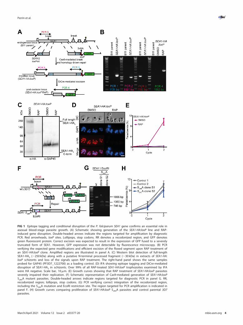
FIG 1 Epitope tagging and conditional disruption of the P. falciparum SEA1 gene confirms an essential role inasexual blood-stage parasite growth. (A) Schematic showing generation of the SEA1-HA:loxP line and RAP-induced gene disruption. Double-headed arrows indicate the regions targeted for amplification by diagnosticPCR. Red arrowheads, loxP sites. Lollipops, stop codons. RR denotes a recodonized region, and GFP denotesgreen fluorescent protein. Correct excision was expected to result in the expression of GFP fused to a severelytruncated form of SEA1. However, GFP expression was not detectable by fluorescence microscopy. (B) PCRverifying the expected gene modifications and efficient excision of the floxed segment upon RAP treatment ofan SEA1-HA:loxP clone. Amplified regions are illustrated in panel A. (C) Western blot detection of full-lengthSEA1-HA3 (;250 kDa) along with a putative N-terminal processed fragment (;30 kDa) in extracts of SEA1-HA:loxP schizonts and loss of the signals upon RAP treatment. The right-hand panel shows the same samplesprobed for GAP45 (PF3D7_1222700) as a loading control. (D) IFA showing epitope tagging and DiCre-mediateddisruption of SEA1-HA3 in schizonts. Over 99% of all RAP-treated SEA1-HA:loxP trophozoites examined by IFAwere HA negative. Scale bar, 10mm. (E) Growth curves showing that RAP treatment of SEA1-HA:loxP parasitesseverely impaired their replication. (F) Schematic representation of Cas9-mediated generation of SEA1-HA:loxPS280A mutant parasites. Double-headed arrows indicate regions targeted for diagnostic PCR in panel G. RR,recodonized region; lollipops, stop codons. (G) PCR verifying correct integration of the recodonized region,including the S280A mutation and EcoRI restriction site. The region targeted for PCR amplification is indicated inpanel F. (H) Growth curves comparing proliferation of SEA1-HA:loxP S280A parasites and control parental 3D7parasites.
Perrin et al. ®
March/April 2021 Volume 12 Issue 2 e03377-20 mbio.asm.org 4

Analysis by IFA showed an SEA1-HA3 signal closely associated with the nucleus (Fig. 1Dand 2A to C), as previously observed in one study (7) (but in contrast to another [4]).This strong, punctate signal was intense in SEA1-HA:loxP trophozoites and early schiz-onts (Fig. 1D and 2A to C) (in which nuclear replication is actively progressing) but wasmuch reduced in very mature segmented schizonts (in which nuclear replication hasceased) that expressed the microneme protein AMA1 (Fig. 2B). Further double-staininganalysis using superresolution imaging showed that the SEA1-HA3 signal comprised asingle focus within each nucleus that was often flanked by twinned signals corre-sponding to components of the nuclear division apparatus (Fig. 2C to E and 3A and B).The SEA1-HA3 foci localized most closely to a-tubulin, which is a component of MTOCsand a marker for mitotic spindles. The signals for centrin, a key component of the cen-trosome, flanked the SEA1-HA3 foci more distally (Fig. 2C to E and 3A and B). We inter-preted these results as strongly indicative of a subcellular localization of SEA1 in thedividing nucleus at a position that could be consistent with the previously proposedcentromere association (7).
A previous study used an anti-SEA1 antibody to immunoprecipitate a-tubulin fromasexual blood-stage parasites (7), indicating that these two proteins associate in vivo.We performed similar immunoprecipitation experiments using anti-HA antibodies.Mass spectrometric analysis of the pulldowns detected over 100 proteins associatingspecifically with the tagged SEA1 (see Table S1 in the supplemental material). Whilewe did not detect a-tubulin in the set of SEA1-HA3-associated proteins, this list wasparticularly enriched with proteins annotated as being localized to the nucleus(Table S1). These data support our localization results, indicating that SEA1 is presentin a nuclear or perinuclear location and interacts with other nuclear proteins.
Loss of SEA1 results in defective schizogony. To understand the essential func-tion of SEA1, we examined the development of RAP-treated SEA1-HA:loxP (SEA1-null)parasites. SEA1-null ring-stage parasites were able to progress through the trophozoitestage, replicate their DNA, and form schizonts (Fig. 3A to D). Close inspection of matureSEA1-null schizonts by IFA showed the presence of plasma membranes and underlyinginner membrane complexes (IMCs), indicated by MSP1 and GAP45 staining, respec-tively, delineating each merozoite (Fig. 4A and Fig. S1A). However, the distribution ofthe DNA in the mutant parasites was markedly different from that in wild-type schiz-onts, with Giemsa- or 49,6-diamidino-2-phenylindole (DAPI)-stained nuclear materialaccumulating in large diffuse aggregates rather than the defined, punctate signal nor-mally observed in mature wild-type merozoites (Fig. 3D and 4A and B and Fig. S1A).Additionally, a large proportion of the DNA appeared to be present within anexpanded food vacuole rather than within daughter merozoites in SEA1-null schizonts(Fig. 4A and B and Fig. S1A).
To investigate these cellular features in more detail, we carried out electron tomog-raphy on high-pressure-frozen, freeze-substituted plastic sections of mature, seg-mented SEA1-HA:loxP schizonts (Fig. 4C and Fig. S1B and Movie S1). Control schizontstypically comprised a set of fully-formed merozoites, each with a C-shaped nucleus,positioned proximal to the site of attachment to the central food vacuole (Fig. 4Ci toiii). This was in stark contrast to mature SEA1-null schizonts, where merozoites wereequipped with all the organelles typical of mature schizonts, but, strikingly, most werenonnucleated (Fig. 4Civ and v and Fig. S1). The misplaced nuclei, bounded by intactnuclear envelopes and coated with ribosomes, were observed clustered together withthe hemozoin crystal within or adjacent to the food vacuole in a ribosome-filled cyto-plasm (Fig. 4Civ to ix). In this region, we also observed IMC structures outside thedefined merozoites (Fig. 4Cvi to viii), indicating that the absence of SEA1 leads todelayed, impaired, or aborted segmentation. Together with the IFA evidence, theseobservations showed a severe defect in the localization and segregation of nuclei inthe SEA1-null mutants.
SEA1-null merozoites egress aberrantly and are not viable. To assess the effectsof the SEA1-null defect on merozoite egress, we monitored the fate of highly synchron-ized schizont populations using established approaches to measure schizont rupture
P. falciparum SEA1 Is Essential during Schizogony ®
March/April 2021 Volume 12 Issue 2 e03377-20 mbio.asm.org 5
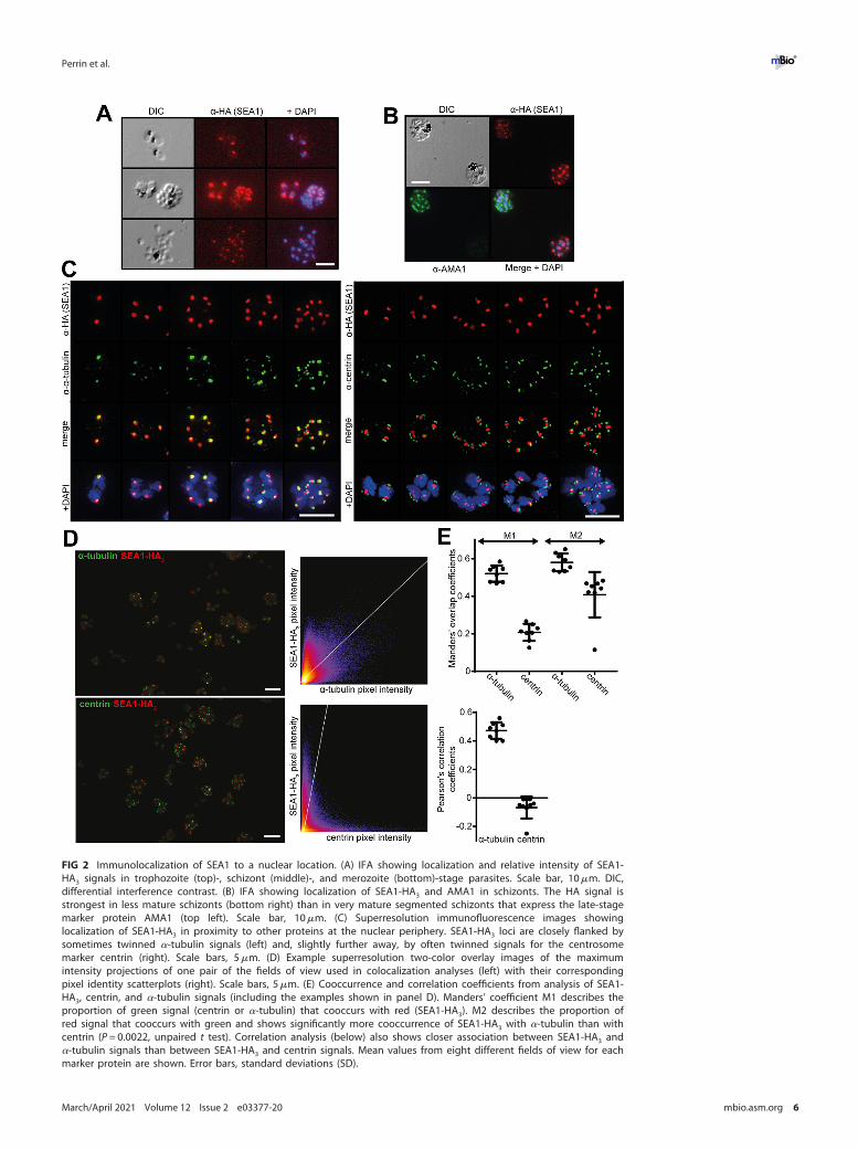
FIG 2 Immunolocalization of SEA1 to a nuclear location. (A) IFA showing localization and relative intensity of SEA1-HA3 signals in trophozoite (top)-, schizont (middle)-, and merozoite (bottom)-stage parasites. Scale bar, 10mm. DIC,differential interference contrast. (B) IFA showing localization of SEA1-HA3 and AMA1 in schizonts. The HA signal isstrongest in less mature schizonts (bottom right) than in very mature segmented schizonts that express the late-stagemarker protein AMA1 (top left). Scale bar, 10mm. (C) Superresolution immunofluorescence images showinglocalization of SEA1-HA3 in proximity to other proteins at the nuclear periphery. SEA1-HA3 loci are closely flanked bysometimes twinned a-tubulin signals (left) and, slightly further away, by often twinned signals for the centrosomemarker centrin (right). Scale bars, 5mm. (D) Example superresolution two-color overlay images of the maximumintensity projections of one pair of the fields of view used in colocalization analyses (left) with their correspondingpixel identity scatterplots (right). Scale bars, 5mm. (E) Cooccurrence and correlation coefficients from analysis of SEA1-HA3, centrin, and a-tubulin signals (including the examples shown in panel D). Manders’ coefficient M1 describes theproportion of green signal (centrin or a-tubulin) that cooccurs with red (SEA1-HA3). M2 describes the proportion ofred signal that cooccurs with green and shows significantly more cooccurrence of SEA1-HA3 with a-tubulin than withcentrin (P = 0.0022, unpaired t test). Correlation analysis (below) also shows closer association between SEA1-HA3 anda-tubulin signals than between SEA1-HA3 and centrin signals. Mean values from eight different fields of view for eachmarker protein are shown. Error bars, standard deviations (SD).
Perrin et al. ®
March/April 2021 Volume 12 Issue 2 e03377-20 mbio.asm.org 6
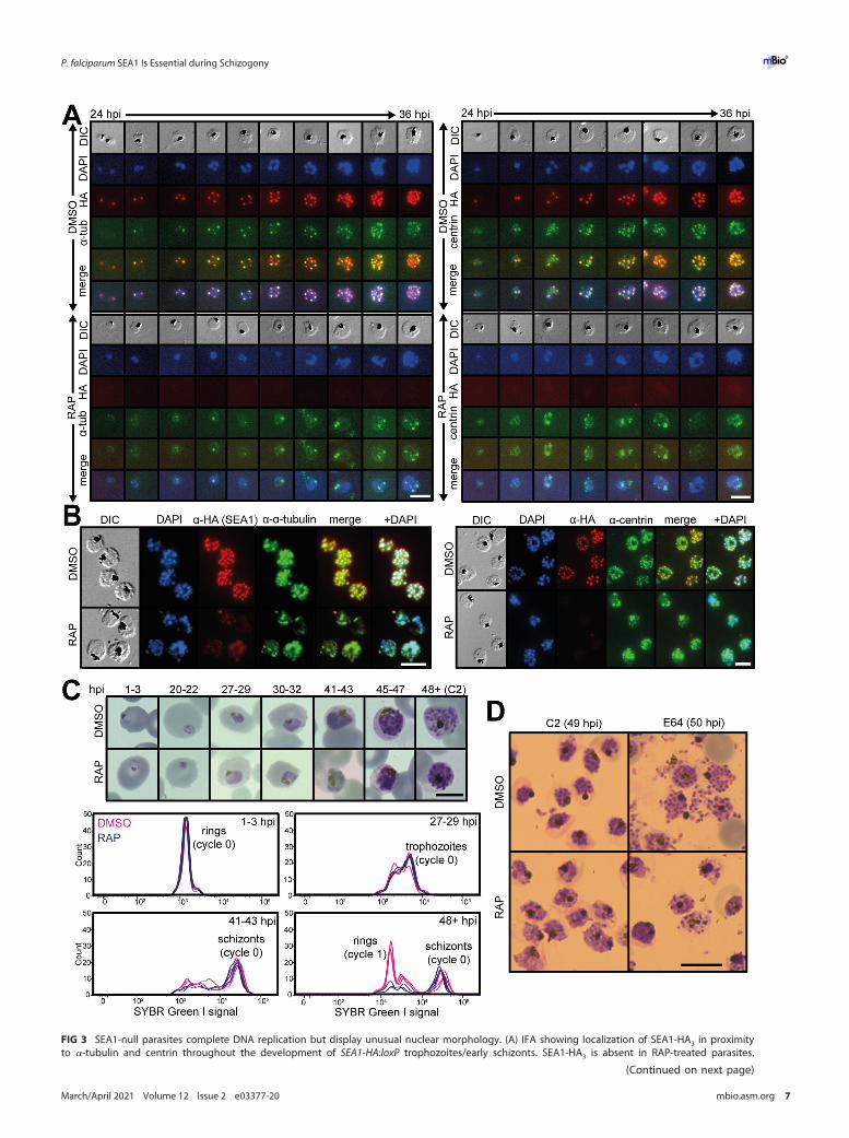
FIG 3 SEA1-null parasites complete DNA replication but display unusual nuclear morphology. (A) IFA showing localization of SEA1-HA3 in proximityto a-tubulin and centrin throughout the development of SEA1-HA:loxP trophozoites/early schizonts. SEA1-HA3 is absent in RAP-treated parasites.
(Continued on next page)
P. falciparum SEA1 Is Essential during Schizogony ®
March/April 2021 Volume 12 Issue 2 e03377-20 mbio.asm.org 7
and RBC invasion by the released merozoites. Despite the morphological defects inSEA1-deficient schizonts, they did undergo rupture, with overall proportions of SEA1-null schizonts undergoing egress at around 90% of those observed in controls (Fig. 5Aiand 5B). We observed proteolytically processed SERA5 in cell culture supernatants(Fig. 5C), indicating that SUB1 discharge and activity occur and that the PV and RBCmembranes break in both DMSO- and RAP-treated SEA1-HA:loxP schizonts. Schizontrupture in the RAP-treated SEA1-HA:loxP parasites was slightly delayed compared tothat of controls (Fig. 5Ai, C, and D), consistent with observations reported for SEA1-knockdown parasites (4). However, close inspection by time-lapse video microscopyshowed that the SEA1-null schizonts ruptured atypically, often failing to release mero-zoites effectively (Fig. 5D and 5E and Movies S2, S3, and S4). The residual bodies thatremained following egress of the SEA1-null parasites were significantly larger (on aver-age, ;1.8-fold greater in diameter) than those from control parasites (Fig. 5F) and con-tained DNA (Movies S2 and S3), consistent with the observations of DNA and nuclearmislocalization we made by light microscopy, IFA, and electron tomography (Fig. 3Dand 4A to C). Invasion was severely impaired in the absence of SEA1 (Fig. 5Aii and B),and most of the SEA1-null ring-stage parasites that formed subsequently containedsome detectable DNA (Fig. 5G and H). However, these rings were unable to developfurther (Fig. 5I and J). These observations indicate that correct partitioning of DNA intoindividual merozoites is critical to their subsequent development. We concluded thatimpaired development of SEA1-null parasites leads indirectly to atypical egress and therelease of defective merozoites that cannot complete another cycle of replication.
DISCUSSION
Our results demonstrate that SEA1 is an essential P. falciparum asexual blood-stageprotein that plays an important role in orchestrating the correct partitioning of DNAinto merozoites. Our finding that SEA1 is an essential P. falciparum gene is consistentwith previous results of others showing that knockdown of SEA1 expression severelyimpairs parasite replication (4). A recent high-throughput transposon-based insertionalmutagenesis screen for essential P. falciparum genes suggested that it was possible tomutate the SEA1 gene without impairment of parasite growth (19). In this particularcase, however, the insertion observed was close to the 39 end of the gene (its positioncorresponding to residues 1766 to 2248), such that at least 78% of the gene could stillbe translated. Taken together, these results indicate that while SEA1 is an essential pro-tein, residues close to the C terminus are not critical for its function.
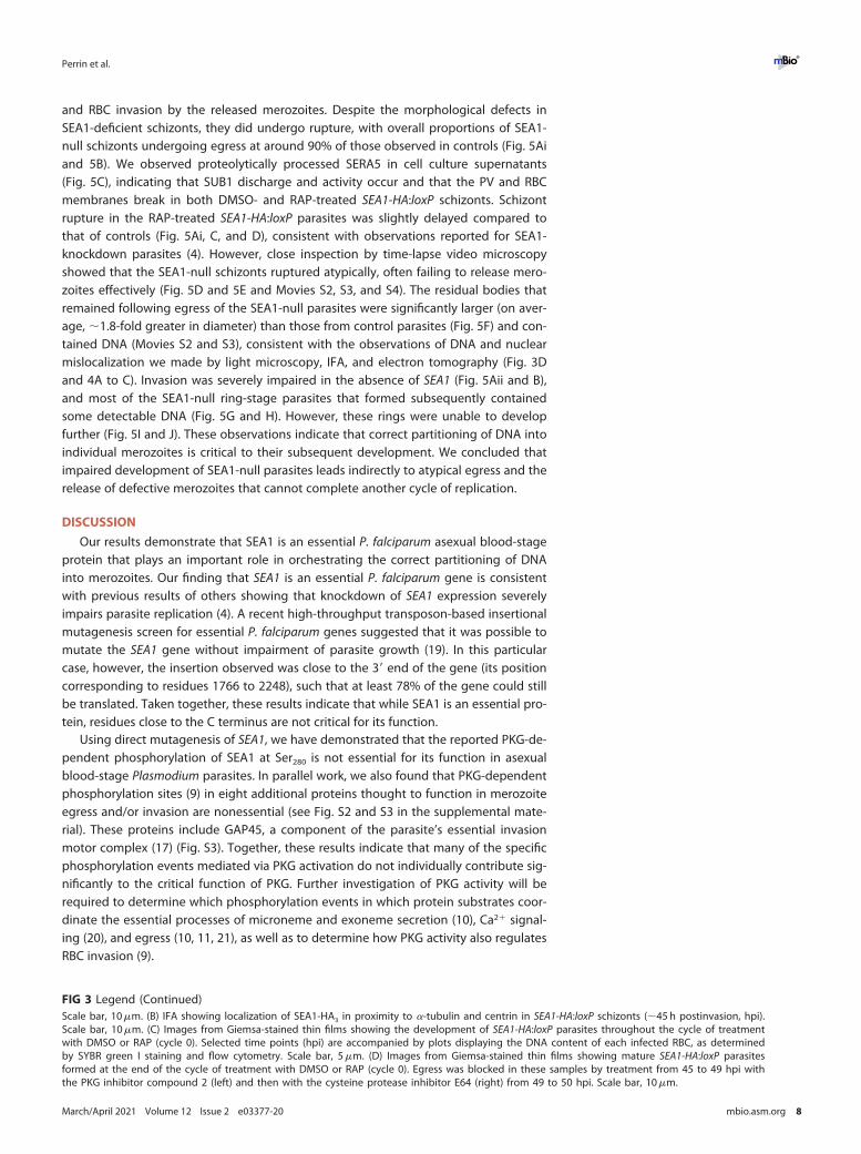
Using direct mutagenesis of SEA1, we have demonstrated that the reported PKG-de-pendent phosphorylation of SEA1 at Ser280 is not essential for its function in asexualblood-stage Plasmodium parasites. In parallel work, we also found that PKG-dependentphosphorylation sites (9) in eight additional proteins thought to function in merozoiteegress and/or invasion are nonessential (see Fig. S2 and S3 in the supplemental mate-rial). These proteins include GAP45, a component of the parasite’s essential invasionmotor complex (17) (Fig. S3). Together, these results indicate that many of the specificphosphorylation events mediated via PKG activation do not individually contribute sig-nificantly to the critical function of PKG. Further investigation of PKG activity will berequired to determine which phosphorylation events in which protein substrates coor-dinate the essential processes of microneme and exoneme secretion (10), Ca21 signal-ing (20), and egress (10, 11, 21), as well as to determine how PKG activity also regulatesRBC invasion (9).
FIG 3 Legend (Continued)Scale bar, 10mm. (B) IFA showing localization of SEA1-HA3 in proximity to a-tubulin and centrin in SEA1-HA:loxP schizonts (;45 h postinvasion, hpi).Scale bar, 10mm. (C) Images from Giemsa-stained thin films showing the development of SEA1-HA:loxP parasites throughout the cycle of treatmentwith DMSO or RAP (cycle 0). Selected time points (hpi) are accompanied by plots displaying the DNA content of each infected RBC, as determinedby SYBR green I staining and flow cytometry. Scale bar, 5mm. (D) Images from Giemsa-stained thin films showing mature SEA1-HA:loxP parasitesformed at the end of the cycle of treatment with DMSO or RAP (cycle 0). Egress was blocked in these samples by treatment from 45 to 49 hpi withthe PKG inhibitor compound 2 (left) and then with the cysteine protease inhibitor E64 (right) from 49 to 50 hpi. Scale bar, 10mm.
Perrin et al. ®
March/April 2021 Volume 12 Issue 2 e03377-20 mbio.asm.org 8
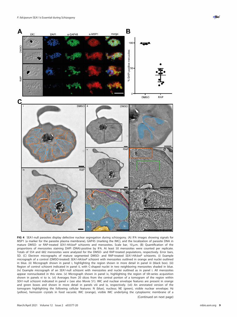
FIG 4 SEA1-null parasites display defective nuclear segregation during schizogony. (A) IFA images showing signals forMSP1 (a marker for the parasite plasma membrane), GAP45 (marking the IMC), and the localization of parasite DNA inmature DMSO- or RAP-treated SEA1-HA:loxP schizonts and merozoites. Scale bar, 10mm. (B) Quantification of theproportions of merozoites staining DAPI (DNA)-positive by IFA. At least 50 merozoites were counted per replicate.Totals of 554 and 682 merozoites were analyzed for the DMSO- and RAP-treated populations, respectively. Error bars,SD. (C) Electron micrographs of mature segmented DMSO- and RAP-treated SEA1-HA:loxP schizonts. (i) Examplemicrograph of a control (DMSO-treated) SEA1-HA:loxP schizont with merozoites outlined in orange and nuclei outlinedin blue. (ii) Micrograph shown in panel i, highlighting the region shown in more detail in panel iii (black box). (iii)Region of control schizont indicated in panel ii, with C-shaped nuclei in two neighboring merozoites shaded in blue.(iv) Example micrograph of an SEA1-null schizont with merozoites and nuclei outlined as in panel i. All merozoitesappear nonnucleated in this view. (v) Micrograph shown in panel iv, highlighting the region of tilt-series acquisitionshown in panels vi to ix. (vi) Averages from 20 slices from the central portion of a tomogram of the region withinSEA1-null schizont indicated in panel v (see also Movie S1). IMC and nuclear envelope features are present in orangeand green boxes and shown in more detail in panels viii and ix, respectively. (vii) An annotated version of thetomogram highlighting the following cellular features: N (blue), nucleus; NE (green), visible nuclear envelope; Hz(yellow), hemozoin crystals in food vacuole; IMC (orange), visible IMC underlying the cytoplasmic membrane of a
(Continued on next page)
P. falciparum SEA1 Is Essential during Schizogony ®
March/April 2021 Volume 12 Issue 2 e03377-20 mbio.asm.org 9
SEA1 was previously designated an egress antigen based on in vitro data, indicatingthat egress was delayed upon protein knockdown and that anti-SEA1 antibodiesappeared to block schizont rupture (4). However, a more recent study did not replicatethe growth-inhibitory effects of anti-SEA1 antibodies (22), and our present data dem-onstrate that, despite severe morphological defects, SEA1-null schizonts do undergoegress. Vaccination studies appear to indicate that an anti-SEA1 immune response canelicit some protection from parasitemia (4, 6), but based on our data, we suggest thatthis is unlikely to be explained by any egress-blocking capacity of anti-SEA1 antibodies.
SEA1 was previously proposed to be a functional homologue of the mammaliancentromere-binding protein CENP-C, a component of the kinetochore complex thatlinks centromeres to microtubules as chromosomes are segregated during mitosis (7).CENP-C knockout phenotypes in vertebrate cells include delayed mitosis (23) and chro-mosome mis-segregation (23, 24). Our data showing that (i) SEA1 localizes in closeproximity to MTOC components within each parasite nucleus, (ii) that the SEA1 foci of-ten lie between twin puncta obtained with antibodies expected to highlight microtu-bule spindles (a-tubulin) and centrosomes (centrin), (iii) that SEA1 expression appearsto be highest in nuclei during the actively dividing trophozoite and early schizontstages, and (iv) that SEA1-null parasites fail to properly segregate replicated DNA intomerozoites are collectively consistent with SEA1 playing a role similar to that of CENP-C during schizogony. The small proportion of merozoites released from SEA1-knockoutschizonts that successfully invade a new RBC subsequently fail to progress from ring totrophozoite stage; we estimate that around 20% of these invasive merozoites did notcontain nuclei, and it is quite possible that those that were nucleated did not containthe normal complement of chromosomes, in line with what has been observed inCENP-C-deficient cells in other systems (23, 24). SEA1 has a relatively low level ofsequence homology with known CENP-C proteins, but together with the previous dataindicating that regions of the P. falciparum SEA1 protein can complement CENP-C defi-ciencies in yeast (7), our data support the hypothesis that SEA1 plays an essential roleat or near P. falciparum kinetochores during schizogony.
The divergent mechanisms by which Plasmodium parasites control daughter cell for-mation and how this is coordinated with DNA replication and nuclear division remainlargely unclear. These processes have been much more intensively investigated inToxoplasma gondii tachyzoites, which reproduce by endodyogeny rather than by schiz-ogony (25). In these parasites, the initiation of daughter cell formation relies on struc-tures emanating from centrosomes, thereby linking nuclear replication to segmentation(26). Similar mechanisms operating in P. falciparum schizonts could explain how the lossof a potential centromere-associated protein could have a downstream effect on the for-mation of merozoites, although there is a growing body of evidence suggesting thatcytoplasmic division in P. falciparum schizonts does not depend on the completion ofnuclear division. In particular, recent detailed electron microscopy studies describe theinitiation of membrane invagination while nuclei are uncompacted and not fully divided(15), and a very recent study has shown that there may be no mechanism that preventscytokinesis from taking place in the absence of normal nuclear division (27).
The aberrant morphology of segmented SEA1-null schizonts, typified by incom-pletely formed merozoites that fail to effectively separate from an expanded residualbody, is strikingly similar to that observed by others upon knockdown of a small num-ber of other parasite proteins, including cyclin homologue Cyc1 (PF3D7_1463700) (28),a basal complex component named CINCH (P. falciparum coordinator of nascent cell detach-ment, PF3D7_0407800) (29), and merozoite organizing protein (MOP, PF3D7_0917000),which has been proposed to be important in defining the apical end of developing mero-
FIG 4 Legend (Continued)partially formed merozoite; PVM (purple), parasitophorous vacuole membrane; and RBCM (red), host RBC membrane.(viii) Region from the tomogram in panel vi showing more detail of the IMC formed in the SEA1-null schizont. (ix)Region from the tomogram in panel vi showing more detail of nuclear envelopes surrounding the SEA1-null nuclei thathave not segregated into merozoites. Scale bars, 500 nm.
Perrin et al. ®
March/April 2021 Volume 12 Issue 2 e03377-20 mbio.asm.org 10
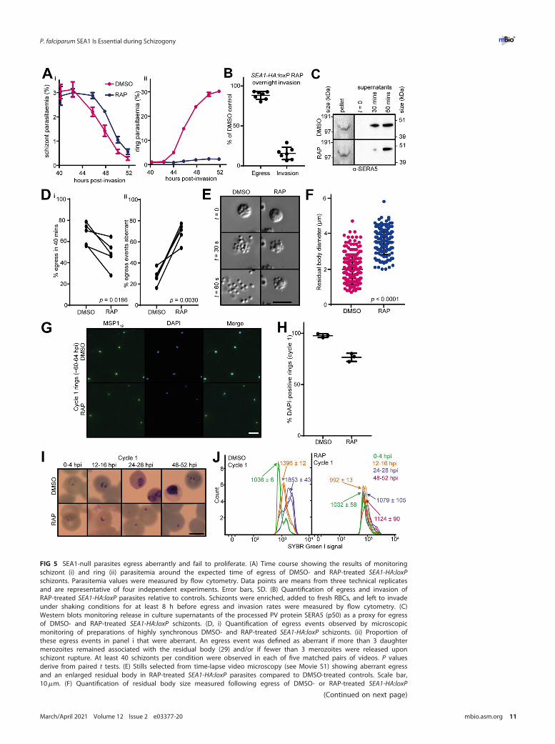
FIG 5 SEA1-null parasites egress aberrantly and fail to proliferate. (A) Time course showing the results of monitoringschizont (i) and ring (ii) parasitemia around the expected time of egress of DMSO- and RAP-treated SEA1-HA:loxPschizonts. Parasitemia values were measured by flow cytometry. Data points are means from three technical replicatesand are representative of four independent experiments. Error bars, SD. (B) Quantification of egress and invasion ofRAP-treated SEA1-HA:loxP parasites relative to controls. Schizonts were enriched, added to fresh RBCs, and left to invadeunder shaking conditions for at least 8 h before egress and invasion rates were measured by flow cytometry. (C)Western blots monitoring release in culture supernatants of the processed PV protein SERA5 (p50) as a proxy for egressof DMSO- and RAP-treated SEA1-HA:loxP schizonts. (D, i) Quantification of egress events observed by microscopicmonitoring of preparations of highly synchronous DMSO- and RAP-treated SEA1-HA:loxP schizonts. (ii) Proportion ofthese egress events in panel i that were aberrant. An egress event was defined as aberrant if more than 3 daughtermerozoites remained associated with the residual body (29) and/or if fewer than 3 merozoites were released uponschizont rupture. At least 40 schizonts per condition were observed in each of five matched pairs of videos. P valuesderive from paired t tests. (E) Stills selected from time-lapse video microscopy (see Movie S1) showing aberrant egressand an enlarged residual body in RAP-treated SEA1-HA:loxP parasites compared to DMSO-treated controls. Scale bar,10mm. (F) Quantification of residual body size measured following egress of DMSO- or RAP-treated SEA1-HA:loxP
(Continued on next page)
P. falciparum SEA1 Is Essential during Schizogony ®
March/April 2021 Volume 12 Issue 2 e03377-20 mbio.asm.org 11
zoites (30). We used affinity purification-mass spectrometry to attempt to obtain insightsinto SEA1-associated proteins, and the results strongly suggested an association with nuclearproteins. However, it is worth noting that generation of parasite extracts led to degradationof the full-length SEA1-HA3 protein to an;30-kDa HA-tagged C-terminal fragment (Fig. 1C),which was the predominant species captured by the anti-HA antibodies and identified bymass spectrometry. We suspect that as a result, our experiments did not capture the full pro-file of proteins potentially interacting with SEA1 in vivo. Nonetheless, our data indicate thatSEA1 physically associates with CINCH in schizonts, suggesting a functional link betweenthese two proteins. However, differences in the established subcellular localizations of thesetwo proteins (29) and the lack of detection of SEA1 in a published CINCH affinity purifica-tion-mass spectrometry study (29) means these data should be interpreted with caution.Similar to our SEA1-null schizonts, MOP and CINCH knockdown mutants can undergo egressdespite severe morphological defects (29, 30). Taken together, these phenotypes show thatproper merozoite segmentation is not a prerequisite for egress and suggest that the devel-opmental cues that govern merozoite formation and egress are quite distinct.
In conclusion, this work has sought to address inconsistencies arising from previouspublications regarding the suggested localization and function of SEA1 and, as such,has established that (i) SEA1 is an essential parasite gene, (ii) SEA1 localizes to the nu-cleus of trophozoites and immature schizonts in a manner consistent with it being acentromere-associated protein, (iii) SEA1 function is critically important for nuclear seg-regation and schizont development prior to egress, and (iv) neither SEA1 nor the com-pletion of segregation are required for merozoite egress, making it unlikely that anti-SEA1 antibody responses could block egress as part of a protective immune response.
MATERIALS ANDMETHODSP. falciparum culture and transfection. Transgenic P. falciparum erythrocytic stages were cultured
and synchronized in human erythrocytes, as described previously (31). Schizonts were enriched for alltransfections, which were performed using an AMAXA 4D Nucleofector and P3 reagent (Lonza).Transfected parasites were selected with WR99210 as described previously (17).
Generation and conditional disruption of SEA1-HA:loxP. The SEA1-HA:loxP parasite line was gener-ated by Cas9-mediated genome editing of P. falciparum B11 parasites that constitutively express thecomponents of the DiCre system (17). A synthetic loxP-containing intron was added close to the 59 endof the gene using a commercially synthesized repair template (GeneArt; Thermo) comprising a 59 homol-ogy region (bp 201 to 950), a SERA2loxPint module (18), approximately 400 bp of recodonized sequence(Table 1), and a 39 homology region (bp 2642 to 3391). The linearized repair template was cotrans-fected with a pDC2-based plasmid encoding Cas9 and a single guide RNA (sgRNA) targeted toATTGTTGAAGAAGAACAATG. The 39 end of the gene was modified using the same methodology with ansgRNA targeted to GTTGATCCTATAGATGATGG and another synthetic construct comprising a 59 homologyarm (bp 5143 to 5892), a 39 30-bp recodonized region corresponding to the final 110 amino acids of codingsequence, an HA3 tag sequence followed by a stop codon and loxP site, an enhanced GFP reporter gene,and a 994-bp homology region corresponding to the intergenic region downstream of SEA1. Clones wereobtained by limiting dilution, and successful double modification was confirmed by PCR (Table 2 and Fig. 1Aand B) and capillary sequencing.
To induce DiCre activity and excise the majority of the SEA1 gene, ring-stage SEA1-HA:loxP parasiteswere treated with 10 nM RAP (Sigma) for 8 to 12 h. Control parasites were treated with vehicle only (1%,vol/vol, dimethyl sulfoxide [DMSO]).
Generation of SEA1 S280A mutants. SEA1 S280A mutants were generated by Cas9-mediated genomeediting of P. falciparum 3D7 parasites, using methods similar to those described above. pDC2-based
FIG 5 Legend (Continued)schizonts. More than 150 residual bodies from a total of five videos per condition were measured, and each pointrepresents an individual residual body. Mean values and standard errors of the means are indicated, and P values werederived from Student's t test. (G) IFA showing control and SEA1-null ring-stage parasites from the cycle followingDMSO/RAP treatment (cycle 1). Ring-stage parasites were identified by staining with an antibody against the C-terminalfragment of MSP1 (MSP119). Scale bar, 10mm. (H) Quantification of the proportions of MSP119-positive cycle 1 ringspossessing detectable DNA by IFA (DAPI positive). Totals of 549 and 301 merozoites were analyzed for the DMSO- andRAP-treated populations, respectively. Error bars, SD. (I) Images from Giemsa-stained thin films showing the fate ofSEA1-HA:loxP parasites that successfully invade following the cycle of treatment with DMSO or RAP. Rings derived fromthe RAP-treated (SEA1-null) merozoites failed to develop. Scale bar, 5mm. (J) Flow cytometry plots showing SYBR greenI fluorescence (indicating DNA content) associated with parasite-infected RBC over the course of the erythrocytic cyclefollowing treatment of SEA1-HA:loxP parasites with DMSO or RAP (cycle 1). Labels indicate the mean fluorescenceintensity of each population 6 SD from three replicates. Parasites derived from the RAP-treated (SEA1-null) populationfailed to develop in cycle 1, consistent with the microscopic images shown in panel H.
Perrin et al. ®
March/April 2021 Volume 12 Issue 2 e03377-20 mbio.asm.org 12
plasmids carrying sgRNA target ATTGTTGAAGAAGAACAATG (clone 1) or ATAGATTAAGAGATAAAAGG(clone 2) were cotransfected with a linear repair construct comprising a 59 homology arm (bp 286 to768) followed by a recodonized region (Table 1) and a 39 homology arm (bp 1345 to 1844). Clonesobtained by limiting dilution were validated via the detection of the novel EcoRI site present in therecodonized region (Tables 1 and 2 and Fig. 2) and by capillary sequencing.
Flow cytometry. P. falciparum-infected RBC samples were fixed with 0.2% glutaraldehyde in phos-phate-buffered saline (PBS) and then stained with SYBR green prior to analysis using a BD Fortessainstrument.
IFA. Thin blood films were fixed with 4% paraformaldehyde (PFA) for 30min and then permeabilizedwith PBS containing 0.1% Triton X-100 for 10min. Slides were then blocked for 1 h in PBS containing 4%bovine serum albumin (BSA) before staining with the relevant antibodies and conjugates. These were3F10 rat anti-HA monoclonal antibody (MAb) (diluted 1:500; Roche), goat anti-rat biotin conjugate(1:1,000), Alexa 594-streptavidin conjugate (1:1,000; Invitrogen), rabbit anti-AMA1 polyclonal antibody(32) (1:500), rabbit anti-GAP45 polyclonal antibody (1:1,000), goat anti-rabbit Alexa 488 conjugate(1:1,000; Invitrogen), mouse anti-a tubulin MAb (1:500; Sigma), 20H5 mouse anti-centrin MAb (1:500;Millipore), 5.2 mouse anti-MSP119 monoclonal antibody (1:1,000), highly adsorbed goat anti-mouseAlexa 488 conjugate (1:1,000; Invitrogen), X509 human anti-MSP1 antibody (1:1,000), and goat anti-human Alexa 594 conjugate (1:1,000; Invitrogen). Slides were mounted with ProLong gold antifade
TABLE 1 Synthetic sequences used in modification of the SEA1 gene
SEA1 region Synthetic sequencea
SEA1-HA:loxP 59 recodonized amino acids L318–D450 TTACGCGACAAGCGCGGTAAGTATCACAAGCTTGGAGACTACCAGAACATCGAGAACTACCGCAAGACAGGAGAGCACTCGTTCGACTGCATGAACATGAGCGATATTATGCACAGCAACAAGATGAGTCACGTAAACATTATGGACCACATGATCTACAAGGACAACAATAACATGTCTAAGTTAGTGGACACTATCAACTCACGCGAGAAAGACGTCAAGAACTACGATGACAATTTCGAGTCCTACAACAACTTCTTTAAAAACAACAATGACGAGCAGCACATTTGCTTAGAATACGATGACACCTACAATCTTAAGGACACAGTAAAGAACATCATCGTAGAAGAGGAGCAGTGTGGAAAAGGAGTAGCATGCATTTGCGACAAAAATGAGGAC
SEA1-HA:loxP 39 recodonized amino acids P1965–I2074 CCAATCAACAAGTTGGCAGTTTCATCTAACTTGGGTCCACCTAGCAGTATTGGCTCTACTGAGATCCAACCAATCCAGAAAAACTTCAATGACTTTAAGATGAACATCAATGTTTATTGTATCCGTATGGAACCACACGAGAAGTATTCGAGCTACAGCCACAAGAACAACCTTGTGGTTTATATCGACAAAGGTGAGAAGATCAACATCATCATCAATATGAGCAAAACCTACGAGAAGGGCGACTTCTTCTATATTCCCCGCTTCAGCAATTTTCAGATTATTAACGACTCCCGCTGCGACTGCGTCCTGTACGTCTGCCCGTTGATT
SEA1 S280A recodonized amino acids E257–N448 withEcoRI site
GAGAACCAGAAGGATATCATCTATCTGAACAACCTGAACAATATCATGATGGACAAGTACAGCAACTGCGCGGACTCGCGCAAGAAAGAGTACTCTCACTTTAACTCCCAAGAATTCTCGTACGACAAGTACAGCATGAAGGATAGGATGTTCCTGAAGAACTTATACATGAAGCAGAACCGCCTGCGCGACAAGCGCGGCAAGTACCATAAGTTAGGAGACTACCAGAACATCGAGAATTACCGCAAGACAGGAGAGCACAGCTTCGACTGCATGAACATGTCCGACATCATGCACTCCAACAAGATGAGTCACGTTAACATAATGGACCATATGATCTACAAGGATAACAACAACATGAGTAAGCTAGTAGACACTATTAACTCACGCGAGAAAGACGTAAAGAACTATGATGACAATTTCGAGAGTTACAACAACTTCTTTAAAAACAATAACGACGAGCAGCATATTTGCTTAGAGTATGATGACACTTACAACTTAAAGGATACTGTAAAGAACATTATCGTGGAGGAGGAGCAGTGCGGTAAAGGAGTTGCTTGCATCTGCGACAAGAAT
aThe SEA1 S280A codon substitution is shown in boldface italic, and the resulting new EcoRI site is in boldface.
TABLE 2 PCR primers used in the validation of the integration and excision of genesequences at the SEA1 locus
PCR Primer sequencea
SEA1-HA:loxP-39’ integration (Fig. 1, PCR1) F, GGACAAACATGAAATGGATTTGAACR, GCCCATGGCATAGTCCGGGACGTC
SEA1-HA:loxP 59 integration (Fig. 1, PCR2) F, GATGGAAAATAAATACCCAAATGAR, CATATATAATAACTTCGTATAATGTATGC
SEA1 wt specific (Fig. 1, PCR3) F, GATGGAAAATAAATACCCAAATGAR, CATATGATCCATGATATTAACATGGCTC
SEA1-HA:loxP excision (Fig. 1, PCR4) F, GATGGAAAATAAATACCCAAATGAR, CAATTTATACAAAAATTGTCCTATTTTC
SEA1 bp 135–1940 (Fig. 2) F, GAATGAAAACGATGGTATATGTGAAR, TCACGTAGCTCATTACTAAGATCCA
aF, forward; R, reverse.
P. falciparum SEA1 Is Essential during Schizogony ®
March/April 2021 Volume 12 Issue 2 e03377-20 mbio.asm.org 13
mountant containing DAPI (Thermo). Imaging was performed using a Nikon Eclipse Ni fluorescencemicroscope fitted with a Hamamatsu C11440 camera.
Superresolution imaging and colocalization analysis. To analyze the colocalization of SEA1-HA3
with a-tubulin and centrin, Z-stacks (125-nm Z-step) were acquired on an VT-iSIM superresolution imag-ing system (Visitech International), using an Olympus IX83 microscope, 150�/1.45 Apochromat objective(UAPON150XOTIRF), ASI motorized stage with piezo Z, and Prime BSI Express scientific complementarymetal oxide semiconductor camera (Teledyne Photometrics). The microscope was controlled with Micro-Manager v2.0 gamma software (33). Huygens Deconvolution Software (SVI) was used to enhance the sig-nals shown in Fig. 2C. Colocalization analysis was performed on the original images (without deconvolu-tion) using the Coloc 2 plugin in Fiji. A mask image was generated by autothresholding of the DAPIchannel (using Otsu’s autothresholding method). After background subtraction, colocalization of SEA1-HA3 with a-tubulin and centrin was assessed using Costes’ autothresholding method to calculateManders’ overlap coefficients M1 and M2 and Pearson’s correlation coefficient (PCC).
Time-lapse video microscopy. Egress videos were carried out as described previously (17). Videoswere analyzed using Image J.
SEA1-HA3 immunoprecipitation and proteomic analysis by mass spectrometry. SynchronousSEA1-HA:loxP parasite lines were treated at ring stage with DMSO or 10 nM RAP and allowed to developto early schizont stage (;40 h). Parasites were extracted from RBCs with 0.15% saponin and washed sev-eral times with PBS. Ten matched sample pairs were used to produce two pooled samples of ;200mlschizonts (DMSO and RAP treated), from which lysates were prepared in radioimmunoprecipitation assay(RIPA) buffer (Thermo). Triplicate samples of lysates were loaded onto anti-HA-coated magnetic beads(Pierce) by incubation at 4°C with rotation for 1 h. Following wash steps, proteins bound to the beadswere eluted by incubation in SDS sample buffer containing 100mM dithiothreitol (DTT) at 95°C for10min.
In-gel tryptic digestion was used to produce peptides for analysis by mass spectrometry. Thesepeptides were dried by vacuum centrifugation and resuspended in 40ml 0.1% formic acid beforebeing loaded onto prepared Evosep tips and injected on a 15-cm column for 44 min using the HCDIT UM method on an Orbitrap Lumos Fusion instrument. Raw files were analyzed using MaxQuant(34) v1.6.12.0 using the iBAQ algorithm and integrated Andromeda peptide search engine (35).Variable modifications of methionine residues (oxidation) and the protein N terminus (acetylation)were permitted, along with fixed modification of cysteine residues (carbamidomethylation). The esti-mated false discovery rate was set to 1%. The PlasmoDB v28 P. falciparum and Swiss-Prot H. sapiensprotein databases were searched to identify peptides. Further data analyses were performed inPerseus v1.4.0.2 and Microsoft Excel.
Electron microscopy. Mature SEA1-knockout and control schizonts were treated with the PKGinhibitor 4-[7-[(dimethylamino)methyl]-2-(4-fluorphenyl)imidazo[1,2-a]pyridine-3-yl]pyrimidin-2-amine (compound 2) for 4 h to synchronize the parasites at a highly mature developmental stage,Percoll purified, washed with PBS, and fixed for 5 min at 37°C with 1% glutaraldehyde and 3%formaldehyde. Schizonts were washed again, pelleted, and mixed with 20% (wt/vol) dextran inphenol red-free RPMI containing baker’s yeast before freezing with a Leica HPM100 high-pressurefreezer. Vitrified cells were freeze-substituted using a Leica electron micrograph (EM) AFS2 intoLowicryl HM20 resin (EMS) with 0.2% (wt/vol) uranyl acetate. One hundred twenty-nanometer sec-tions were cut using a Leica UC7 microtome and mounted onto glow-discharged, carbon-coated,copper London finder grids (EMS). Sections were poststained with 0.2% (wt/vol) uranyl acetateand 4% (wt/vol) lead citrate (EMS). Micrographs and tilt series were acquired using a Model 2040dual-axis tomography holder (Fischione Instruments) on a Tecnai T12 120-kV transmission electronmicroscope (FEI) equipped with a 4K-by-4K Ultrascan 4000 charge-coupled device camera (Gatan).Overview images were acquired using a digital micrograph (Gatan). Dual-axis tilt series wereacquired from 260° to 160° with an increment of 2° using SerialEM (36) and processed usingeTomo (part of IMOD) (37) with fiducial-less alignment by patch tracking.
SUPPLEMENTAL MATERIAL
Supplemental material is available online only.MOVIE S1, MPG file, 5.6 MB.MOVIE S2, MPG file, 6 MB.MOVIE S3, MPG file, 1.6 MB.MOVIE S4, MPG file, 1.9 MB.TEXT S1, DOCX file, 0.01 MB.FIG S1, TIF file, 1 MB.FIG S2, TIF file, 0.4 MB.FIG S3, TIF file, 1 MB.TABLE S1, XLSX file, 0.04 MB.TABLE S2, DOCX file, 0.02 MB.
Perrin et al. ®
March/April 2021 Volume 12 Issue 2 e03377-20 mbio.asm.org 14
ACKNOWLEDGMENTSWe thank Rita Tewari, along with everyone in the Blackman, Deu, Holder, and Treeck
laboratories at the Crick Institute, for helpful conversations and technical advice. We areparticularly grateful to Ellen Knuepfer and Judith Green for sharing antibody reagents.All experiments were designed and carried out by A.J.P., with the exception of the EMimaging, which was performed by C.B. and R.A.L. under the instruction of C.B., and themass spectrometry, which was carried out by P.A.F. Instructions for using the iSIMinstrument and analysis of the data were provided by M.J.R., while H.R.S. and R.A.F.provided electron microscopy resources. Overall supervision was provided by M.J.B.,D.A.B., and A.P.S.
This research was funded in whole, or in part, by the Wellcome Trust. For thepurpose of Open Access, the author has applied a CC BY public copyright license to anyAuthor Accepted Manuscript version arising from this submission. This work was alsosupported by funding to M.J.B. from the Francis Crick Institute (https://www.crick.ac.uk/),which receives its core funding from Cancer Research UK (FC001043; https://www.cancerresearchuk.org), the UK Medical Research Council (FC001043; https://www.mrc.ac.uk/), and the Wellcome Trust (FC001043; https://wellcome.ac.uk/). The work was alsosupported by Wellcome Trust grant 106239/Z/14/A (A.J.P. and M.J.B.), Wellcome Trustgrant 106240/Z/14/Z (D.A.B.), and Medical Research Council grant MR/P010288/1 (C.B., H.R.S., R.A.F., and M.J.B.).
REFERENCES1. Proietti C, Krause L, Trieu A, Dodoo D, Gyan B, Koram KA, et al. 2020.
Immune signature against Plasmodium falciparum antigens predicts clini-cal immunity in distinct malaria endemic communities. Mol Cell Proteo-mics 19:101–113. https://doi.org/10.1074/mcp.RA118.001256.
2. Obiero JM, Campo JJ, Scholzen A, Randall A, Bijker EM, Roestenberg M,Hermsen CC, Teng A, Jain A, Davies DH, Sauerwein RW, Felgner PL. 2019.Antibody biomarkers associated with sterile protection induced by con-trolled human malaria infection under chloroquine prophylaxis. mSphere4:e00027-19. https://doi.org/10.1128/mSphereDirect.00027-19.
3. Osier FHA, Fegan G, Polley SD, Murungi L, Verra F, Tetteh KKA, et al. 2008.Breadth and magnitude of antibody responses to multiple Plasmodium fal-ciparum merozoite antigens are associated with protection from clinicalmalaria. Infect Immun 76:2240–2248. https://doi.org/10.1128/IAI.01585-07.
4. Raj DK, Nixon CP, Nixon CE, Dvorin JD, DiPetrillo CG, Pond-Tor S, Wu H-W,Jolly G, Pischel L, Lu A, Michelow IC, Cheng L, Conteh S, McDonald EA,Absalon S, Holte SE, Friedman JF, Fried M, Duffy PE, Kurtis JD. 2014. Anti-bodies to PfSEA-1 block parasite egress from RBCs and protect againstmalaria infection. Science 344:871–877. https://doi.org/10.1126/science.1254417.
5. Kurtis JD, Raj DK, Michelow IC, Park S, Nixon CE, McDonald EA, et al. 2018.Maternally derived antibodies to Schizont Egress Antigen-1 and protec-tion of infants from severe malaria. Clin Infect Dis 68:1718–1724. https://doi.org/10.1093/cid/ciy728.
6. Nixon CE, Park S, Pond-Tor S, Raj D, Lambert LE, Orr-Gonzalez S, BarnafoEK, Rausch KM, Friedman JF, Fried M, Duffy PE, Kurtis JD. 2017. Identifica-tion of protective B-cell epitopes within the novel malaria vaccine candi-date Plasmodium falciparum schizont egress antigen 1. Clin VaccineImmunol 24:e00068-17. https://doi.org/10.1128/CVI.00068-17.
7. Verma G, Surolia N. 2014. The dimerization domain of Pf CENP-C isrequired for its functions as a centromere protein in human malaria para-site Plasmodium falciparum. Malar J 13:475. https://doi.org/10.1186/1475-2875-13-475.
8. Verma G, Surolia N. 2013. Plasmodium falciparum CENH3 is able to function-ally complement Cse4p and its, C-terminus is essential for centromere func-tion. Mol Biochem Parasitol 192:21–29. https://doi.org/10.1016/j.molbiopara.2013.11.002.
9. Alam MM, Solyakov L, Bottrill AR, Flueck C, Siddiqui FA, Singh S, Mistry S,Viskaduraki M, Lee K, Hopp CS, Chitnis CE, Doerig C, Moon RW, Green JL,Holder AA, Baker DA, Tobin AB. 2015. Phosphoproteomics reveals malariaparasite protein kinase G as a signaling hub regulating egress and inva-sion. Nat Commun 6:7285. https://doi.org/10.1038/ncomms8285.
10. Collins CR, Hackett F, Strath M, Penzo M, Withers-Martinez C, Baker DA,Blackman MJ. 2013. Malaria parasite cGMP-dependent protein kinase
regulates blood stage merozoite secretory organelle discharge andegress. PLoS Pathog 9:e1003344. https://doi.org/10.1371/journal.ppat.1003344.
11. Koussis K, Withers-Martinez C, Baker DA, Blackman MJ. 2020. Simultaneousmultiple allelic replacement in the malaria parasite enables dissection of PKGfunction. Life Sci Alliance 3(4):e201900626. https://doi.org/10.26508/lsa.201900626.
12. Thomas JA, Tan MSY, Bisson C, Borg A, Umrekar TR, Hackett F, Hale VL,Vizcay-Barrena G, Fleck RA, Snijders AP, Saibil HR, Blackman MJ. 2018. Aprotease cascade regulates release of the human malaria parasite Plasmo-dium falciparum from host red blood cells. Nat Microbiol 3:447–455.https://doi.org/10.1038/s41564-018-0111-0.
13. Read M, Sherwin T, Holloway SP, Gull K, Hyde E. 1993. Microtubular orga-nization visualized by immunofluorescence microscopy during erythro-cytic schizogony in Plasmodium falciparum and investigation of post-translational modifications of parasite tubulin. Parasitology 106:223–232.https://doi.org/10.1017/s0031182000075041.
14. Gerald N, Mahajan B, Kumar S. 2011. Mitosis in the human malaria parasitePlasmodium falciparum. Eukaryot Cell 10:474–482. https://doi.org/10.1128/EC.00314-10.
15. Rudlaff RM, Kraemer S, Marshman J, Dvorin JD. 2020. Three-dimensionalultrastructure of Plasmodium falciparum throughout cytokinesis. PLoSPathog 16:e1008587. https://doi.org/10.1371/journal.ppat.1008587.
16. Verma G, Surolia N. 2018. Centromere and its associated proteins-whatwe know about them in Plasmodium falciparum. IUBMB Life 70:732–742.https://doi.org/10.1002/iub.1878.
17. Perrin AJ, Collins CR, Russell MRG, Collinson LM, Baker DA, Blackman MJ.2018. The actinomyosin motor drives malaria parasite red blood cell invasionbut not egress. mBio 9:e00905-18. https://doi.org/10.1128/mBio.00905-18.
18. Jones ML, Das S, Belda H, Collins CR, Blackman MJ, Treeck M. 2016. A ver-satile strategy for rapid conditional genome engineering using loxP sitesin a small synthetic intron in Plasmodium falciparum. Sci Rep 6:srep21800.https://doi.org/10.1038/srep21800.
19. Zhang M, Wang C, Otto TD, Oberstaller J, Liao X, Adapa SR, Udenze K,Bronner IF, Casandra D, Mayho M, Brown J, Li S, Swanson J, Rayner JC,Jiang RHY, Adams JH. 2018. Uncovering the essential genes of the humanmalaria parasite Plasmodium falciparum by saturation mutagenesis. Sci-ence 360:eaap7847. https://doi.org/10.1126/science.aap7847.
20. Brochet M, Collins MO, Smith TK, Thompson E, Sebastian S, Volkmann K,Schwach F, Chappell L, Gomes AR, Berriman M, Rayner JC, Baker DA,Choudhary J, Billker O. 2014. Phosphoinositide metabolism links cGMP-dependent protein kinase G to essential Ca21 signals at key decision
P. falciparum SEA1 Is Essential during Schizogony ®
March/April 2021 Volume 12 Issue 2 e03377-20 mbio.asm.org 15
points in the life cycle of malaria parasites. PLoS Biol 12:e1001806.https://doi.org/10.1371/journal.pbio.1001806.
21. Taylor HM, McRobert L, Grainger M, Sicard A, Dluzewski AR, Hopp CS, etal. 2010. The malaria parasite cyclic GMP-dependent protein kinase playsa central role in blood-stage schizogony. Eukaryot Cell 9:37–45. https://doi.org/10.1128/EC.00186-09.
22. Illingworth JJ, Alanine DG, Brown R, Marshall JM, Bartlett HE, Silk SE, et al.2019. Functional comparison of blood-stage Plasmodium falciparummalaria vaccine candidate antigens. Front Immunol 10:1254. https://doi.org/10.3389/fimmu.2019.01254.
23. Kwon MS, Hori T, Okada M, Fukagawa T. 2007. CENP-C is involved in chro-mosome segregation, mitotic checkpoint function, and kinetochore as-sembly. Mol Biol Cell 18:2155–2168. https://doi.org/10.1091/mbc.e07-01-0045.
24. Giunta S, Funabiki H. 2017. Integrity of the human centromere DNArepeats is protected by CENP-A, CENP-C, and CENP-T. Proc Natl Acad SciU S A 114:1928–1933. https://doi.org/10.1073/pnas.1615133114.
25. Francia ME, Striepen B. 2014. Cell division in apicomplexan parasites. NatRev Microbiol 12:125–136. https://doi.org/10.1038/nrmicro3184.
26. Francia ME, Jordan CN, Patel JD, Sheiner L, Demerly JL, Fellows JD, deLeon JC, Morrissette NS, Dubremetz J-F, Striepen B. 2012. Cell division inapicomplexan parasites is organized by a homolog of the striated rootletfiber of algal flagella. PLoS Biol 10:e1001444. https://doi.org/10.1371/journal.pbio.1001444.
27. Absalon S, Dvorin JD. 2020. Depletion of the mini-chromosome mainte-nance complex binding protein allows the progression of cytokinesis de-spite abnormal karyokinesis during the asexual development of Plasmo-dium falciparum. Cell Microbiol 2020:e13284. https://doi.org/10.1111/cmi.13284.
28. Robbins JA, Absalon S, Streva VA, Dvorin JD. 2017. The malaria parasitecyclin H homolog PfCyc1 is required for efficient cytokinesis in blood-stage Plasmodium falciparum. mBio 8:e00605-17. https://doi.org/10.1128/mBio.00605-17.
29. Rudlaff RM, Kraemer S, Streva VA, Dvorin JD. 2019. An essential contractilering protein controls cell division in Plasmodium falciparum. Nat Commun10 https://doi.org/10.1038/s41467-019-10214-z.
30. Absalon S, Robbins JA, Dvorin JD. 2016. An essential malaria proteindefines the architecture of blood-stage and transmission-stage parasites.Nat Commun 7:11449. https://doi.org/10.1038/ncomms11449.
31. Patel A, Perrin AJ, Flynn HR, Bisson C, Withers-Martinez C, Treeck M,Flueck C, Nicastro G, Martin SR, Ramos A, Gilberger TW, Snijders AP,Blackman MJ, Baker DA. 2019. Cyclic AMP signaling controls key compo-nents of malaria parasite host cell invasion machinery. PLoS Biol 17:e3000264. https://doi.org/10.1371/journal.pbio.3000264.
32. Collins CR, Withers-Martinez C, Hackett F, Blackman MJ. 2009. An inhibi-tory antibody blocks interactions between components of the malarialinvasion machinery. PLoS Pathog 5:e1000273. https://doi.org/10.1371/journal.ppat.1000273.
33. Edelstein AD, Tsuchida MA, Amodaj N, Pinkard H, Vale RD, Stuurman N.2014. Advanced methods of microscope control using mManager soft-ware. J Biol Methods 1:10. https://doi.org/10.14440/jbm.2014.36.
34. Cox J, Mann M. 2008. MaxQuant enables high peptide identification rates,individualized p.p.b.-range mass accuracies and proteome-wide proteinquantification. Nat Biotechnol 26:1367–1372. https://doi.org/10.1038/nbt.1511.
35. Cox J, Neuhauser N, Michalski A, Scheltema RA, Olsen JV, Mann M. 2011. An-dromeda: a peptide search engine integrated into the MaxQuant environ-ment. J Proteome Res 10:1794–1805. https://doi.org/10.1021/pr101065j.
36. Mastronarde DN. 2005. Automated electron microscope tomographyusing robust prediction of specimen movements. J Struct Biol 152:36–51.https://doi.org/10.1016/j.jsb.2005.07.007.
37. Kremer JR, Mastronarde DN, McIntosh JR. 1996. Computer visualization ofthree-dimensional image data using IMOD. J Struct Biol 116:71–76. https://doi.org/10.1006/jsbi.1996.0013.
Perrin et al. ®
March/April 2021 Volume 12 Issue 2 e03377-20 mbio.asm.org 16
Related Documents