SEPTEMBER 2015 | WWW.THE-SCIENTIST.COM EVOLVING EARS DRUGS FOR HEARING LOSS AND TINNITUS HAIR-CELL DYNAMICS PLUS AUDITORY BRAINSTEM IMPLANTS MAKING SENSE OF HEARING

Welcome message from author
This document is posted to help you gain knowledge. Please leave a comment to let me know what you think about it! Share it to your friends and learn new things together.
Transcript

SEPTEMBER 2015 | WWW.THE-SCIENTIST.COM
EVOLVING EARS
DRUGS FOR HEARING LOSS AND TINNITUS
HAIR-CELL DYNAMICS
PLUS AUDITORY
BRAINSTEM IMPLANTS
MAKING SENSE OF
HEARING
www.biotek.com
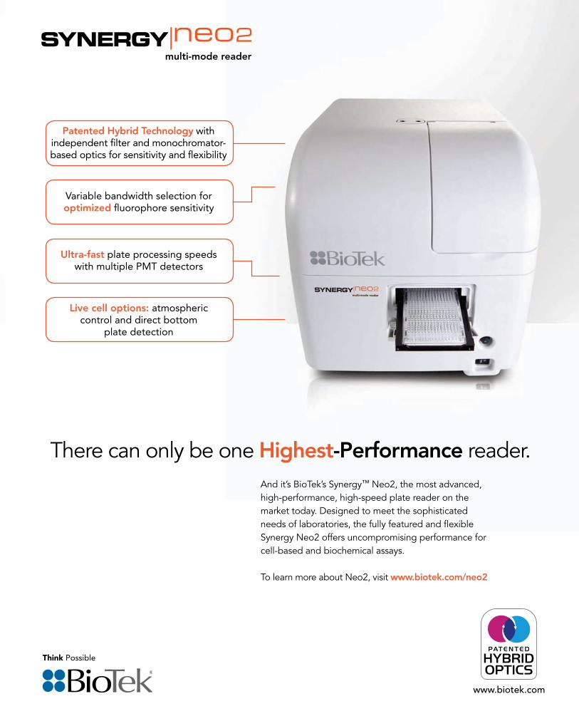
There can only be one Highest-Performance reader.And it’s BioTek’s Synergy™ Neo2, the most advanced, high-performance, high-speed plate reader on the market today. Designed to meet the sophisticated needs of laboratories, the fully featured and flexible Synergy Neo2 offers uncompromising performance for cell-based and biochemical assays.
To learn more about Neo2, visit www.biotek.com/neo2
Patented Hybrid Technology with independent filter and monochromator-based optics for sensitivity and flexibility
Variable bandwidth selection for optimized fluorophore sensitivity
Live cell options: atmospheric control and direct bottom
plate detection
Ultra-fast plate processing speeds with multiple PMT detectors

Toll-Free Tel: (US & Canada): 1.877.BIOLEGEND (246.5343)Tel: 858.768.5800
biolegend.com
08-0052-10
The MojoSort™ Magnetic Cell Separation System is designed for the separation of target populations using positive or negative selection. MojoSort™ nanoparticles deliver excellent purity and yield at an unmatched, a� ordable price. Magnetically sorted cells can be used for multiple downstream applications.
A suspension of single cells from pooled C57BL/6 mouse spleen and lymph nodes was prepared to isolate CD4+ T cells using the MojoSort™ Mouse CD4 T Cell Isolation Kit. Cells were stained with PE anti-mouse CD4 (clone RM4-4), APC anti-mouse CD3ε (145-2C11), and 7-AAD. Grateful Dead cells were excluded from the analysis.
Before Isolation After MojoSort™ Isolation After Competitor Isolation
MojoSort™ advantages:• Small and large test size formats to meet research needs
• Robust performance
• Preserved cell functionality after sorting
• Excellent price
Add some Mojo to your experiment and explore the possibilities!
To learn more, visit: biolegend.com/mojosort
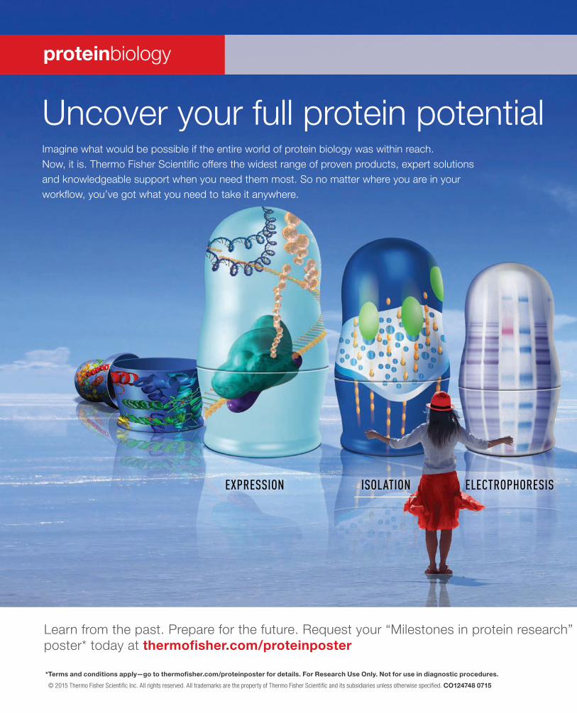
ELECTROPHORESISISOLATIONEXPRESSION
Uncover your full protein potentialImagine what would be possible if the entire world of protein biology was within reach. Now, it is. Thermo Fisher Scientifi c offers the widest range of proven products, expert solutions and knowledgeable support when you need them most. So no matter where you are in your workfl ow, you’ve got what you need to take it anywhere.
proteinbiology
Learn from the past. Prepare for the future. Request your “Milestones in protein research” poster* today at thermofi sher.com/proteinposter
* Terms and conditions apply—go to thermofi sher.com/proteinposter for details. For Research Use Only. Not for use in diagnostic procedures.
© 2015 Thermo Fisher Scientifi c Inc. All rights reserved. All trademarks are the property of Thermo Fisher Scientifi c and its subsidiaries unless otherwise specifi ed. CO124748 0715
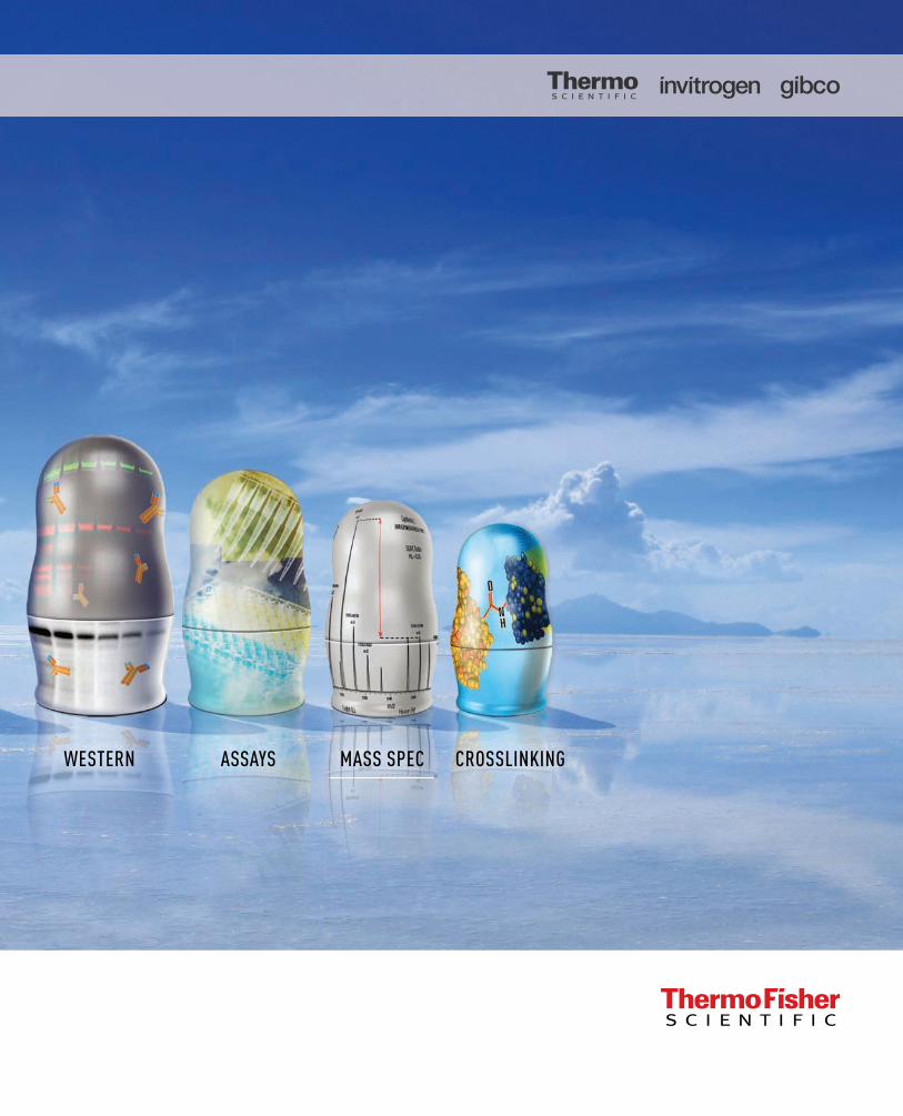
CROSSLINKINGMASS SPECASSAYSWESTERN
Introducing the NEBNext® Library Quant Kit for Illumina®
NEW ENGLAND BIOLABS®, NEB® and NEBNEXT® are registered trademarks of New England Biolabs, Inc.ILLUMINA® and MISEQ® are registered trademarks of Illumina, Inc.
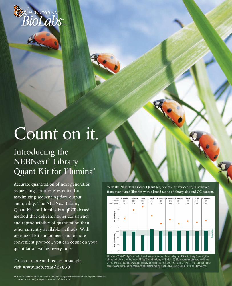
Accurate quantitation of next generation sequencing libraries is essential for maximizing sequencing data output and quality. The NEBNext Library Quant Kit for Illumina is a qPCR-based method that delivers higher consistency and reproducibility of quantitation than other currently available methods. With optimized kit components and a more convenient protocol, you can count on your quantitation values, every time.
To learn more and request a sample, visit www.neb.com/E7630
Count on it.
Libraries of 310–963 bp from the indicated sources were quantitated using the NEBNext Library Quant Kit, then diluted to 8 pM and loaded onto a MiSeq® (v2 chemistry; MCS v2.4.1.3). Library concentrations ranged from 7–120 nM, and resulting raw cluster density for all libraries was 965–1300 k/mm2 (ave. =1199). Optimal cluster density was achieved using concentrations determined by the NEBNext Library Quant Kit for all library sizes.
With the NEBNext Library Quant Kit, optimal cluster density is achieved from quantitated libraries with a broad range of library size and GC content.
0
50
100
0
400
800
1,200
Input:
Library Size (bp):
H. influenzae
311
E. coli
341
Jurkat
633
R. palustris
638
H. influenzae
656
R. palustris
900
Jurkat
946
E. coli
958
H. influenzae
963
Target Density
qPCR
Qua
nt (n
M)
Clus
ter D
ensi
ty (k
/mm
2 )
R. palustris
310
GC Content: 38% 51% 41% 65% 38% 65% 41% 51% 38%65%
509.2015 | THE SCIENTIST
ContentsTHE SCIENTIST THE-SCIENTIST.COM VOLUME 29 NUMBER 9
SEPTEMBER 2015
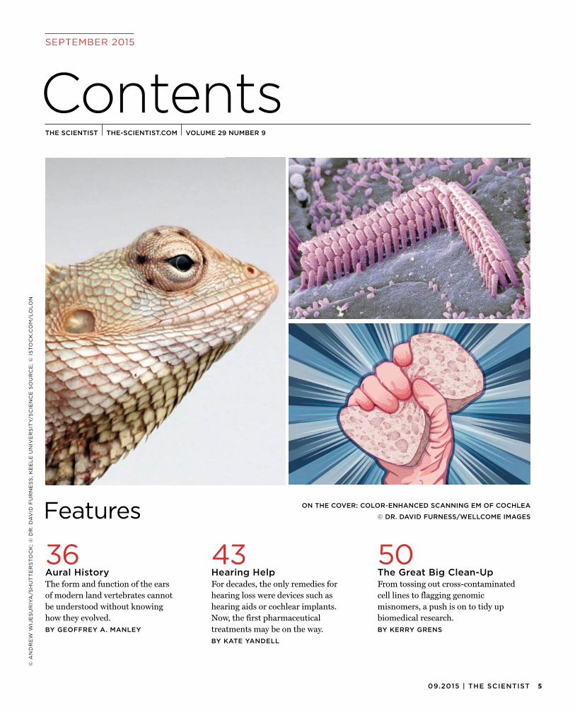
Features ON THE COVER: COLOR-ENHANCED SCANNING EM OF COCHLEA
© DR. DAVID FURNESS/WELLCOME IMAGES
© A
ND
RE
W W
IJE
SU
RIY
A/S
HU
TT
ER
ST
OC
K;
© D
R.
DA
VID
FU
RN
ES
S,
KE
EL
E U
NIV
ER
SIT
Y/S
CIE
NC
E S
OU
RC
E;
© I
ST
OC
K.C
OM
/LO
LO
N
50The Great Big Clean-UpFrom tossing out cross-contaminatedcell lines to flagging genomic misnomers, a push is on to tidy up biomedical research.BY KERRY GRENS
43Hearing HelpFor decades, the only remedies forhearing loss were devices such as hearing aids or cochlear implants. Now, the first pharmaceutical treatments may be on the way.BY KATE YANDELL
36Aural HistoryThe form and function of the earsof modern land vertebrates cannot be understood without knowing how they evolved.BY GEOFFREY A. MANLEY
BioResearch
www.lonza.com/stem
Take the trial and error out of your stem cell workflow with optimized toolkits from Lonza. Whether you work with iPSC or adult stem cells, your direct route to biologically relevant results starts here.
– NEW! L7™ Reprogramming and Culture System – robust system for generation and maintenance of hPSCs– Nucleofector™ Technology – non-viral transfection for reliable reprogramming and genome editing– Clonetics™ Primary Cells and Poietics™ Stem Cells – extensive range available Streamline your stem cell workflow today.
It All Stems from Here
Simplify Your Reprogramming, Proliferation and Differentiation
© 2014 Lonza Walkersville, Inc
709.2015 | THE SCIENTIST
SEPTEMBER 2015
Department Contents
CO
CH
LE
AR
IN
C.;
© S
CO
TT
GO
LD
; ©
EN
CY
CL
OP
ED
IA B
RIT
AN
NIC
A/U
IG/G
ET
TY
IM
AG
ES
CORRECTION:In Capsule Reviews (The Scientist, August 2015), the review of Gods of the Morning mistakenly mentioned that blackcap chickadees inhabit Scotland instead of blackcaps (Sylvia atricapilla).
The Scientist regrets the error.
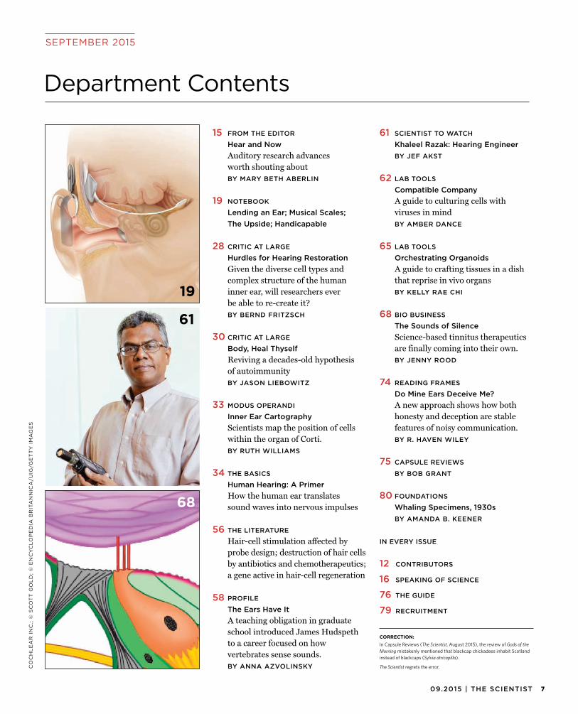
15 FROM THE EDITOR
Hear and NowAuditory research advancesworth shouting aboutBY MARY BETH ABERLIN
19 NOTEBOOK
Lending an Ear; Musical Scales;The Upside; Handicapable
28 CRITIC AT LARGE
Hurdles for Hearing RestorationGiven the diverse cell types andcomplex structure of the human inner ear, will researchers ever be able to re-create it?BY BERND FRITZSCH
30 CRITIC AT LARGE
Body, Heal ThyselfReviving a decades-old hypothesisof autoimmunityBY JASON LIEBOWITZ
33 MODUS OPERANDI
Inner Ear CartographyScientists map the position of cellswithin the organ of Corti.BY RUTH WILLIAMS
34 THE BASICS
Human Hearing: A PrimerHow the human ear translatessound waves into nervous impulses
56 THE LITERATURE
Hair-cell stimulation affected byprobe design; destruction of hair cells by antibiotics and chemotherapeutics; a gene active in hair-cell regeneration
58 PROFILE
The Ears Have ItA teaching obligation in graduateschool introduced James Hudspeth to a career focused on how vertebrates sense sounds.BY ANNA AZVOLINSKY
61 SCIENTIST TO WATCH
Khaleel Razak: Hearing EngineerBY JEF AKST
62 LAB TOOLS
Compatible CompanyA guide to culturing cells withviruses in mindBY AMBER DANCE
65 LAB TOOLS
Orchestrating OrganoidsA guide to crafting tissues in a dishthat reprise in vivo organsBY KELLY RAE CHI
68 BIO BUSINESS
The Sounds of SilenceScience-based tinnitus therapeuticsare finally coming into their own.BY JENNY ROOD
74 READING FRAMES
Do Mine Ears Deceive Me?A new approach shows how bothhonesty and deception are stable features of noisy communication.BY R. HAVEN WILEY
75 CAPSULE REVIEWS
BY BOB GRANT
80 FOUNDATIONS
Whaling Specimens, 1930sBY AMANDA B. KEENER
IN EVERY ISSUE
12 CONTRIBUTORS
16 SPEAKING OF SCIENCE
76 THE GUIDE
79 RECRUITMENT
19
61
68
8 THE SCIENTIST | the-scientist.com
Coming in October
SEPTEMBER 2015
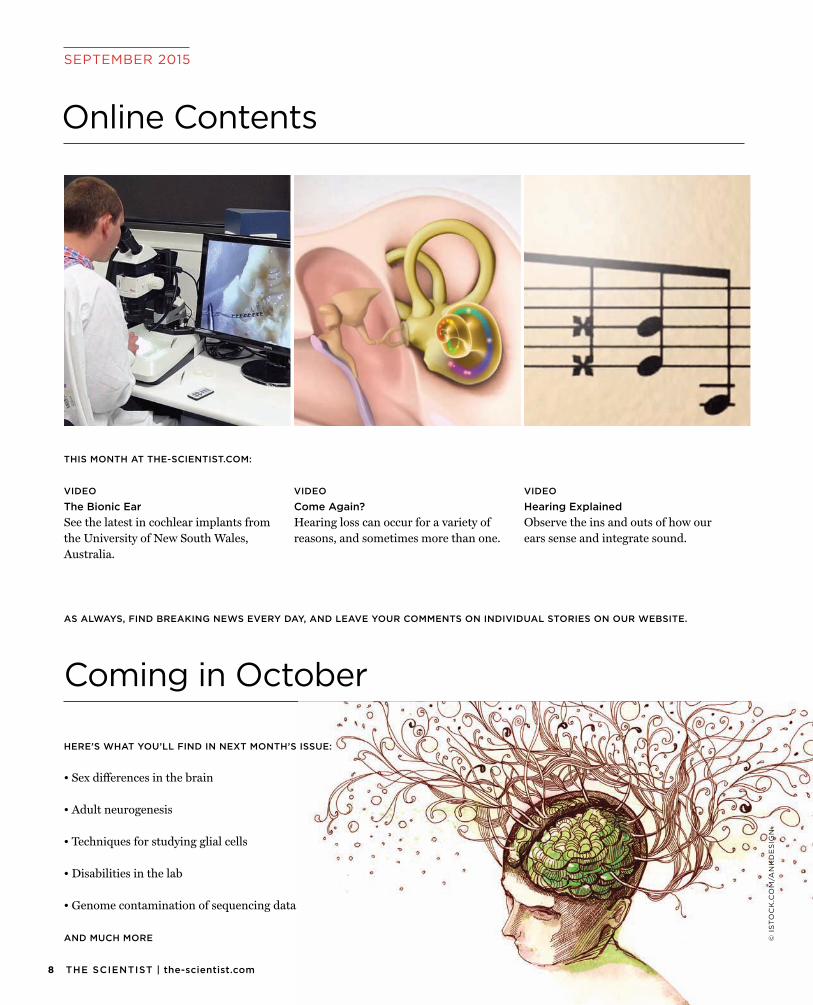
Online Contents
AS ALWAYS, FIND BREAKING NEWS EVERY DAY, AND LEAVE YOUR COMMENTS ON INDIVIDUAL STORIES ON OUR WEBSITE.
© I
ST
OC
K.C
OM
/AN
KD
ES
IGN
HERE’S WHAT YOU’LL FIND IN NEXT MONTH’S ISSUE:
• Sex differences in the brain
• Adult neurogenesis
• Techniques for studying glial cells
• Disabilities in the lab
• Genome contamination of sequencing data
AND MUCH MORE
VIDEO
Come Again?Hearing loss can occur for a variety ofreasons, and sometimes more than one.
VIDEO
Hearing ExplainedObserve the ins and outs of how ourears sense and integrate sound.
VIDEO
The Bionic EarSee the latest in cochlear implants fromthe University of New South Wales, Australia.
THIS MONTH AT THE-SCIENTIST.COM:

Do You Suffer from Panel Envy?
analytes in 1 Luminex® assay.
6Ckine, Adiponectin, Adpisin, A-FABP, AFP, Aggrecan, Ang-1, Ang-2, Angiogenin, ANGPTL3, ANGPTL4, APP, AREG, ATIII, Autotaxin, β2M, B7-H1, BAFF, BCMA, BDNF, BLC, BMP-2, BMP-4, BMP-9, BRAK, C5a, CA125, CA15-3, Calbindin D, CCL14, CD14, CD27, CD30, CD40, CD40L, CD44, CD93, CD163, Chemerin, CINC-2, CINC-3, Clusterin, COL4A1, C-Peptide, Cripto-1, CRP, CTACK, cTNI, CXCL16, Cys-tatin C, DcR3, Dkk-1, DPPIV, DR3, EGF, EMMPRIN, ENA-78, Endoglin, Endostatin, Eotaxin, Eotaxin-2, Eotaxin-3, EphA2, Epo, ESAM, E-Selectin, ET-1, Fas, FasL, Fetuin A, FGF acidic, FGF basic, FGF-21, FLRG, Flt-3L, FST, Gal-3, Gal-3BP, Gal-9, GCP-2, G-CSF, GDF-15, GDNF, Glucagon, GM-CSF, gp130, GRO-α, HB-EGF, Her2, H-FABP, HGF, hGH, ICAM-1, IFN-γ, IFN-γ R1, IGFBP-1, IGFBP-3, IGFBP-rp1, IL-1 ra, IL-1 RI, IL-1 RII, IL-1α, IL-1β, IL-2, IL-2 Rα, IL-3, IL-4, IL-5, IL-6, IL-6 Rα, IL-7, IL-8, IL-9, IL-10, IL-12 p70, IL-13, IL-15, IL-17A, IL-17F, IL-18 BPa, IL-19, IL-22, IL-23, IL-25, IL-27, IL-28A, IL-31, IL-33, IL-34, IL-36β, IGF-I, Insulin, IP-10, I-TAC, ITIH4, KC KIM-1, KLK5, Leptin, L-FABP, LIF, L-Selectin, MIP-2, LIX, Lumican, MCP-1, MCP-2, MCP-3, MCP-4, M-CSF, MDC, MFG-E8, MICA, MICB, MIF, MIG, MIP-1α, MIP-1β, MIP-3α, MMP-1, MMP-2, MMP-3, MMP-7, MMP-8, MMP-9, MMP-10, MMP-12, MMP-13, MPO, MSP, Myo-globin, NCAM-1, Nephrin, NGAL, NRG1-β1, NT-4, NT-Pro-ANP, OPN, OSM, PAI-1, PAPP-A, PARC, PCSK9, PDGF-AA, PDGF-BB, Periostin, PF4, PlGF, PGRN, Prolactin, Protein C, PRTN3, PSA, P-Selectin, PTX3, RAGE, RANK L, RANTES, RBP4, Renin, Resistin, ROBO4, S100A8, S100A9, S100B, SCF, SDF-1α, SHBG, SOST, SPARC, SP-D, ST2, TACI, TARC, Tenascin C, TFF3, TFPI, TfR, TGF-β1, TGF-β2, TGF-β3, THBS2, THBS4, Tie-1, Tie-2, TIMP-1, TIMP-2, TIMP-3, TIMP-4, TNF-α, TNF RI, TNF RII, Tpo, TRAIL, TRAIL R2, TRAIL R3, ULBP-1, ULBP-2/5/6, ULBP-3, ULBP-4, uPA, uPAR, Uteroglobin, VAP-1, VCAM-1, VEGF, VEGF-C, VEGF-D, VEGF R2, VEGF R3, Visfatin, Vit DBP, vWF-A2, YKL-40
EXPLORE NOWrndsystems.com/100Plex
#PanelEnvy #100Plex

It takes sophisticated tools to reveal the secrets of biology. So when quality can’t be compromised, scientists turn to Biosearch Technologies to synthesize the exact oligonucleotides they need. At our new world-class facilities, we build our products as if lives depend on it, because they often do. Our proprietary BHQ® probes enable reliable qPCR testing for the most consequential applications – whether that’s detection of cancer cells, genetic mutations, wine-spoiling microbes, or bio-threat agents. And that’s just today. Who knows what we’ll help you discover tomorrow?
A NEW PERSPECTIVE ON DNA
© 2013 Biosearch Technologies, Inc. Products and technologies appearing in this ad may have trademark or patent restrictions associated with them. Please see www.biosearchtech.com/legal for full legal disclosure.
with accurate high throughput TEER
measurement
Monitor growth
• Save time • Cost effective• Reduce errors • Intuitive software• Superior repeatability • PC-controlled robotic arm• Works with major manufacturers of HTS culture plates
For more information please visit us at www.wpiinc.com/hts.
tissue culturesof epithelial
EDITORIAL ADVISORY BOARD
Roger Beachy Donald Danforth Plant Science Center
Steven A. Bent Foley and Lardner LLP
Deborah Blum University of Wisconsin
Annette Doherty Pfizer Global Research and Development
Kevin Horgan GE Healthcare
Steve Jackson University of Cambridge
Elizabeth Kerr Life Technologies/Applied Biosystems
Simon Levin Princeton University Center for BioComplexity
Edison Liu Genome Institute of Singapore
Peter Raven Missouri Botanical Garden
Joseph Schlessinger Yale University School of Medicine
J. Craig Venter J. Craig Venter Institute
Marc Vidal Dana Farber Cancer Institute Harvard University
H. Steven Wiley Biomolecular Systems Pacific Northwest National Laboratory
Alastair J.J. Wood Symphony Capital
SUBSCRIPTION RATES & SERVICESIn the United States & Canada individual subscriptions: $39.95. Rest of the world: air cargo add $25.
For assistance with a new or existing subscription please contact us at:
Phone: 847.763.9519 Fax: 847.763.-9674 E-mail: [email protected] Mail: The Scientist, PO Box 2015 , Skokie, Illinois 60076
For institutional subscription rates and services visit www.the-scientist.com/info/subscribe or e-mail [email protected]
LIST RENTALS Contact Statlistics, Jennifer Felling at 203-778-8700 or [email protected]
REPRINTS Contact Lee Denton at [email protected]
PERMISSIONS For photocopy and reprint permissions, contact Copyright Clearance Center at www.copyright.com
415 Madison Avenue, Suite 1508, New York, NY 10017E-mail: [email protected]
POSTMASTER: Send address changes to The Scientist, PO Box 2015, Skokie, Illinois 60076. Canada Publications Agreement #40641071 The Scientist is indexed in Current Contents, Science Citation Index, BasicBIOS IS, and other databases. Articles published in The Scientist reflect the views of their authors and are not the official views of the publication, its editorial staff, or its ownership. The Scientist is a registered trademark of LabX Media Group Inc. The Scientist® (ISSN 0890-3670) is published monthly.
Advertising Office: The Scientist, 415 Madison Avenue, Suite 1508, New York, NY 10017. Periodical Postage Paid at New York, NY, and at additional mailing offices.
EDITORIAL
Editor-in-Chief Mary Beth Aberlin [email protected]
Senior Editors Jef Akst [email protected]
Bob Grant [email protected]
Associate Editor Kerry Grens [email protected]
News Editor Tracy Vence [email protected]
Contributing Editor Alla Katsnelson
Copy Editor Annie Gottlieb
Correspondents Anna Azvolinsky Ruth Williams
Interns Amanda B. Keener
DESIGN AND PRODUCTION
Art Director Lisa Modica [email protected]
Graphic Designer Erin Lemieux [email protected]
MANAGEMENT AND BUSINESS
President Bob Kafato [email protected]
General Manager Ken Piech [email protected]
Managing Partner Mario Di Ubaldi [email protected]
Publisher Robert S. D’Angelo [email protected]
ADMINISTRATION
Customer Service [email protected]
Administrative Assistant Lee Denton [email protected]
ADVERTISING AND MARKETING
Account Executive Northeast U.S.
Anita Bell [email protected]
Account Executive Mid-West U.S., Eastern Canada
Melanie Dunlop [email protected]
Account Executive Southeast U.S., Europe, TS Careers
Nicole Dupuis [email protected]
Senior Account Executive West U.S. and Western Canada,
Pacific Rim
Ashley Haire (Munro) [email protected]
Engagement Manager, Life Sciences
Susan Harrison Uy [email protected]
Audience Development Manager Brian McGann [email protected]
Creative Services Director Vince Navarro [email protected]
Career Recruitment and Circulation Coordinator
Lee Denton [email protected]
12 THE SCIENTIST | the-scientist.com
Contributors
GE
OF
FR
EY
MA
NL
EY
; U
NIV
ER
SIT
Y O
F I
OW
A;
HA
VE
N W
ILE
Y
Although his first love in science was animal research, retired zoology profes-sor and hearing researcher Geoffrey Manley was quickly drawn into neurobiol-ogy as a graduate student at Princeton University, earning his PhD in 1970. After all, he says, “neurobiology is just a branch of zoology.” At Princeton, Manley began studying how hearing evolved. “At the time we knew almost nothing about hear-ing in lizards or how hearing in birds worked.” He began to investigate the middle ear biomechanics of lizards and mammals as a postdoc at the University of West-ern Australia in Perth, while in the midst of his assistant professorship at McGill University in Montreal. His postdoctoral work exposed him for the first time to biomedical engineering and tools used to analyze hearing structures at the sub-micron level. “That certainly broadened my perspective,” he says. Manley moved to Munich for a sabbatical in 1978 and remained to serve as the first chair of the Technical University in Munich’s newly minted zoology department, a position he says suited him well until his retirement at the end of 2011. “Zoology was my natu-ral home.” In his article, “Aural History” (page 36), Manley discusses what compar-ative biology has taught us about the evolution of hearing.
University of Iowa neuroscientist Bernd Fritzsch has spent his entire career studying ears. As a zoology PhD student at the Technical University (TU)Darmstadt in Germany, he focused on how the ear and brain connect in mice and in chickens. As a postdoctoral researcher at TU Darmstadt and the Uni-versity of Bielefeld, he focused on lateral line, electroreception, and hearing in amphibians. Later, Fritzsch received an award from the Heisenberg Program, which gave him five years of unfettered research funding and brought him to the Scripps Research Institute in La Jolla, California. “I liked America so much that I stayed here,” he says. In 2008, Fritzsch joined the faculty at Iowa where he studies the mouse cochlear and vestibular system and the molecular basis of hair cell development. In “Hurdles for Hearing Restoration” (page 28), Fritzsch draws upon his decades of experience to discuss what it will take for research-ers to restore the complicated structure of the organ of Corti. In science, he says, “you have to have a prepped mind to know what is new and put it into perspective of what is known.”
As a graduate student at Rockefeller University, Haven Wiley spent many weeks each spring studying the social behavior of grouse on the sagebrush plains of Wyoming and Montana. Since then, he and his graduate students have con-tinued studying animal societies and communication in the field throughout the Americas. “It gives us a chance to study animals outdoors—at least part of the time,” he says. Early in his career, Wiley became interested in how noise influ-ences the evolution of animals’ signals for communication. After teaching and doing research at the University of North Carolina at Chapel Hill for 40 years, he retired to collect his ideas in his first book, which discusses how signal detec-tion theory has far-reaching implications for every level of signaling from mol-ecules to language. In his essay “Do Mine Ears Deceive Me?” (page 74), Wiley focuses on how signal detection theory provides a way to understand how hon-esty becomes the norm in communication.
Although retired, Wiley says he is still analyzing results and writing as much as he ever did. “I’ve always enjoyed studying animal communication, and I still do,” he says.
SEPTEMBER 2015

PASS
ION
VISION
INSPIRAT
I ON
YEARS20
Celebrating
mirusbio.comProviding gene delivery expertise since 1995
Facebook “f ” Logo CMYK / .ai Facebook “f ” Logo CMYK / .ai
©2015 All rights reserved Mirus Bio LLC. CHOgro is a trademark of Mirus Bio LLC.
At Mirus Bio, we know it’s all about expression. Introducing the new CHOgro™ Expression System, a transient transfection platform that �nally gets high protein titers with robust cell growth in the most relevant CHO cells.
• Ef�cient – Enables high protein titers with simple work�ow• Convenient – Quick adaptation to CHO cell line lineages• Optimized – High density growth with minimal clumping post-transfection• Worry-free – No commercial license required; animal origin free
Visit www.mirusbio.com/chogro to qualify for a CHOgro™ Expression System Trial Kit.
Available as a complete system or components sold separately.
NEW! CHOgro™ Expression System
For Research Use Only. Not for use in diagnostic procedures. © 2015 Thermo Fisher Scientific Inc. All rights reserved. All trademarks are the property of Thermo Fisher Scientific and its subsidiaries unless otherwise specified. CO125174 0715
Discovery. Applied.Identifying meaningful biological variants is what you do. Genetic analysis is how you get there. Robust chemistries, reliable instruments – and scalable informatics – your research demands nothing less. And it all must work together. Seamlessly and consistently. Because when the right tools are applied, discovery is not far behind.
Explore your path to discovery at thermofisher.com/appliedbiosystems
1509.2015 | THE SCIENTIST
FROM THE EDITOR
Auditory research advances worth shouting about
BY MARY BETH ABERLIN
Hear and Now
AN
DR
ZE
J K
RA
UZ
E
This issue devoted to hearing researchcompletes our five-year tour of the “classical” Aristotelian senses: taste, touch, smell,
sight, and hearing. Each year, a different sense commanded our immediate attention; we saved hearing for last because we thought hearing research might be less interesting. Boy oh boy, were we wrong. The more we surveyed the current state of the field, the more excited we got.
So here (hear!) you have it. Our own behind-the-scenes need for a primer to consult on how a sound turns into a nerve impulse led to a beautiful two-page infographic of the auditory pathway (page 34). Hid-den deep in the human inner ear, encased in bone, is the amazing organ of Corti, a spiral staircase nearly an inch long, studded with sensory cells that deliver sound to our brains in a frequency-specific fashion. To get a fuller sense of auditory dynamics, check out the online offerings selected to enhance this issue, including an animated tour of the middle and inner ear responding to bars from a Beethoven symphony.
In “Aural History” (page 36), Geoffrey Manley lays out how the middle and inner ears of terrestrial vertebrates evolved. Despite branching off from a common ancestor some 300 million years ago, before the evolution of a dedicated hearing organ, the three extant lineages of amniotes—lepidosaurs (lizards and snakes), archosaurs (crocodilians and birds), and mammals—all process sound in very similar ways at the physiological level. It’s a remarkable example of convergent evolution, with each lineage now possessing a bony middle ear to amplify sound and a delicate auditory papilla outfitted with sensory cells topped with what look like weird haircuts, appropriately dubbed hair cells.
Studying the actual workings of inner-ear hair cells in vivo is hampered by the cells’ inaccessibility, and the complicated anatomical arrangement of the organ of Corti makes in vitro studies challenging. Researchers have devised a number of clever techniques to align in vitro studies with in vivo reality and to search for hints on how to restore function to damaged cells. “Inner Ear Cartography” (page 33) describes an elegant spatial mapping strategy based on gene expression in nine cell types of the hearing organ. Two recent research reports
use hair cells from the vestibular system (from which hearing sensory cells evolved) to get a bead on hair-cell regeneration, a phenomenon that has been lost in mammalian hearing organs but that researchers hope could be reactivated (page 57). A longer literature report describes discrepancies that arise as a result of probing function under in vitro conditions (page 56).
Encouragingly, basic research is edging closer to translation into new therapies for hearing disorders. “The Sounds of Silence” (page 68) reports on treatments for tinnitus, a persistent ringing in the ears for which no drug currently exists, and whose maddening quality Edgar Allan Poe captured so well in his lines about “the tintinnabulation of the bells, bells, bells.” And in “Hearing Help” (page 43), Kate Yandell explores the small molecules and gene therapies on the horizon for patients suffering from hearing loss. Some researchers are even hoping to regenerate inner-ear hair cells or to enhance their connections with sensory neurons. In an opinion piece (page 28), Bernd Fritzsch sounds a cautionary note, however: building a hearing organ from scratch may never be possible.
The entire Notebook section (page 19) is devoted to behind-the-scenes stories about hearing research in the lab and in the clinic. “The Ears Have It” (page 58) profiles the prolific career of James Hudspeth, and the issue is peppered with quotes and research from scientists who spent time in his lab.
One sense per year means updates are in order, but before revisiting the classical five, we have something “extra” in store for 2016. g
Basic research is edging closer to translation into new therapies for hearing disorders.
16 THE SCIENTIST | the-scientist.com
QUOTES
Speaking of Science
NA
SA
/AM
ES
/JP
L-C
ALT
EC
H/T
. P
YL
EIf you’re a scientist who refuses to open up your dataset and your detailed methodology, people increasingly start looking at you funny, because that attitude is deeply at odds with the scientific method. If you want to find the truth, you need multiple people all looking critically at the same data, and having an open and transparent debate about how to interpret it.—Fusion financial journalist Felix Salmon on the lack of replication in
science (July 27)
It is my feeling that the lawyers and embassies should fight it out amongst themselves and just let the scientists get on with plying their trade. I don’t care a jot where the specimen is . . . so long as it is in a safe and accessible place for future scientists to research it.
—University of Portsmouth paleobiologist David Martill, responding toquestions that arose about the possible illegal exportation of a Brazilian
four-legged snake fossil he recently found in a German museum (August 4)
At this point it is unethical to teach any other way.
—Clarissa Dirks, microbiologist at the Evergreen State College in Olympia, Washington, and cochair of the US National Academies Scientific Teaching Alliance, on the use of active learning in STEM
teaching (July 15)
I worry about the breadth of the Indian scientific enterprise. I think there’s not enough investment in training people properly. There’s a divorce between research and education, by putting research in central institutes and leaving universities to fend for themselves.
—Venkatraman Ramakrishnan, structural biologist who won the 2009 Nobel Prize in Chemistry, discussing the state of science in his
native India (August 6)
If the only alternatives for scientific publishing are either inhabiting the gated communities of the 1 percent of the world population, which concentrates wealth at the cost of exploiting the other 99 percent, or being with the people in a favela, long live the favela. —Brazilian Forum of Public Health Journals Editors and the Associação
Brasileira de Saúde Coletiva, responding to librarian Jeffrey Beall’s comparison of some open-access publishers to low-income
neighborhoods in Brazil known as favelas (August 2)
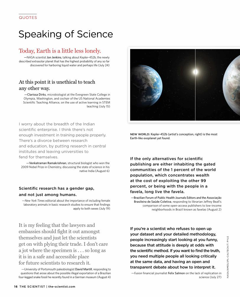
Today, Earth is a little less lonely. —NASA scientist Jon Jenkins, talking about Kepler-452b, the newly
described extrasolar planet that has the highest probability of any so far discovered for harboring liquid water and perhaps life (July 24)
NEW WORLD: Kepler-452b (artist’s conception, right) is the most Earth-like exoplanet yet found.
Scientific research has a gender gap,and not just among humans.
—New York Times editorial about the importance of including female laboratory animals in basic research studies to ensure that findings
apply to both sexes (July 19)

DISCOVER AN EPIGENETICS ASSAY
PORTFOLIO YOU’LL NEVER OUTGROW
EPIGENEOUS™ EPIGENETICS ASSAYS.
From universal to specific assays, biochemical to cell-based, no one gives you a broader range of tools to research enzymatic targets in epigenetics than Cisbio. With a comprehensive and ever-expanding portfolio, as well as complete custom assay services, we can give you exactly what you need today and in the future.
• Research faster and more effectively with optimized andtargeted assays.
• Get robust, biologically relevant results with proven HTRF® technology.
• Enhance throughput with easily miniaturized homogeneous assaysrequiring no wash steps.
• Assays available for: Demethylases, Methyltransferases, Deacetylases,Kinases and Ubiquitinases.
For more information on our complete line or to discuss your specific custom requirements, please email us at [email protected] or visit www.htrf.com/epigenetic-screening
Cop
yrig
ht C
isbi
o B
ioas
says
. All
right
s re
serv
ed. A
ll tr
adem
arks
are
pro
pert
y of
Cis
bio
Bio
assa
ys.
www.cisbio.com

See what we can do for you at www.idtdna.com.
Custom oligos from IDT provide the best quality and
most consistent foundation for your life science projects.
Our oligos can be used in a wide variety of applications
from research and diagnostics to crop science to
personalized medicine.
Genotyping NextGenerationSequencing
GeneExpression
RNAi
SyntheticBiology
CUSTOMOLIGOS for
1909.2015 | THE SCIENTIST
Notebook SEPTEMBER 2015
NEWS AND ANALYSISC
OC
HL
EA
R I
NC
.
Lending an Ear
Shortly after giving birth to her firstchild, Julie Lopez found out her daughter was deaf. Angelica failed
her newborn hearing screen. Lopez and her family, who live in Big Spring, Texas, drove five hours to Dallas for a second evaluation, and the test result was the same. “We were devastated,” says Lopez. “We did not expect it.”
No one in Lopez’s or her husband’s family is deaf, and they wanted their daughter to have a chance to hear. So they opted to try cochlear implants (CIs)—devices that involve threading an elec-trode array through the inner ear to stim-ulate the auditory nerve upon input from an external microphone and speech pro-
cessor. But Angelica’s auditory nerve was too severely underdeveloped to make use of the implants, and after six months she could still hear nothing. Then her doctor presented another option: a clinical trial in Los Angeles that was offering an implant that bypasses the ear completely and goes straight to the brainstem.
Auditory brainstem implants (ABIs) are much more invasive than cochlear
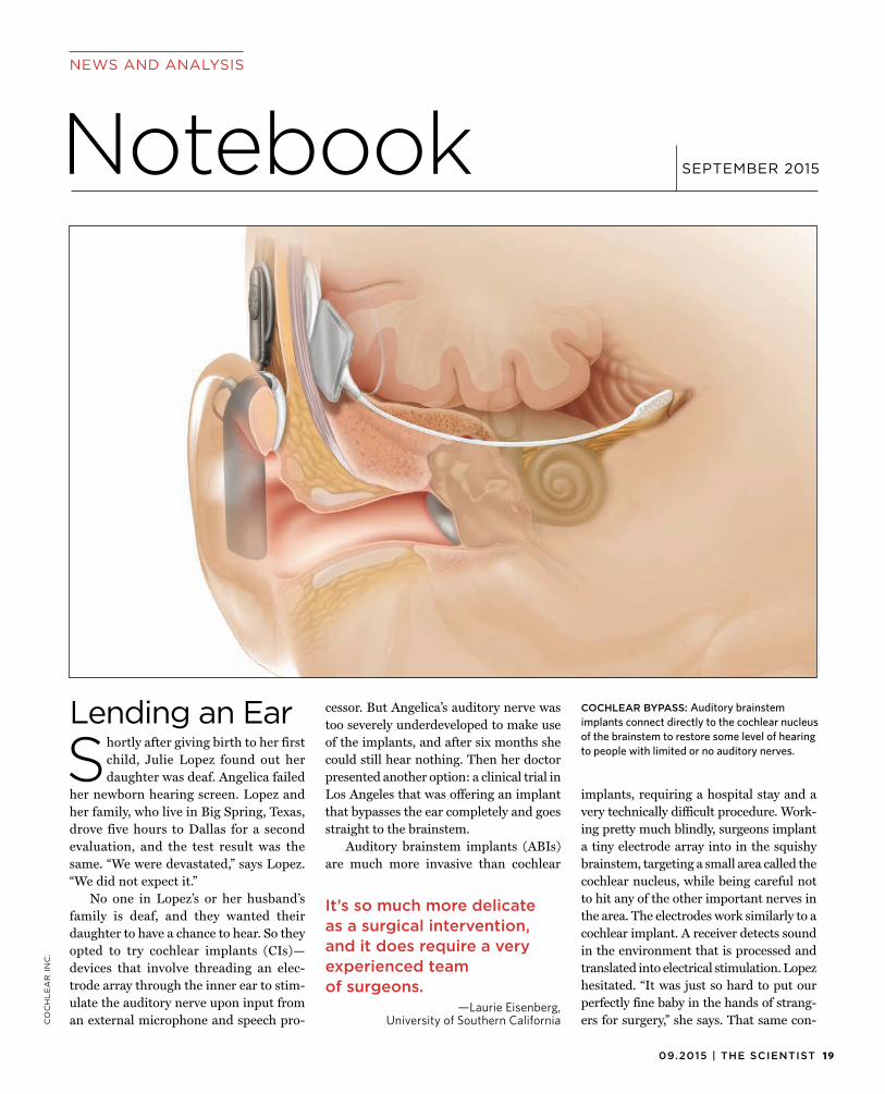
implants, requiring a hospital stay and a very technically difficult procedure. Work-ing pretty much blindly, surgeons implant a tiny electrode array into in the squishy brainstem, targeting a small area called the cochlear nucleus, while being careful not to hit any of the other important nerves in the area. The electrodes work similarly to a cochlear implant. A receiver detects sound in the environment that is processed and translated into electrical stimulation. Lopez hesitated. “It was just so hard to put our perfectly fine baby in the hands of strang-ers for surgery,” she says. That same con-
It’s so much more delicate as a surgical intervention, and it does require a very experienced team of surgeons.
—Laurie Eisenberg,University of Southern California
COCHLEAR BYPASS: Auditory brainstem implants connect directly to the cochlear nucleus of the brainstem to restore some level of hearing to people with limited or no auditory nerves.
NOTEBOOK
CO
CH
LE
AR
IN
C.
cern—subjecting a child to a craniotomywhen she wouldn’t otherwise need one to survive—for decades kept US regulators from approving the implant for people who weren’t already undergoing brain surgery. But now, four institutions are testing the waters in clinical trials.
The first ABI was handmade and implanted in 1979 by William House, inven-tor of the cochlear implant, in Los Angeles. Laurie Eisenberg, a pediatric audiologist at the University of Southern California, was a member of the team who worked with that first patient. At that time—and still now, excepting the four clinical trials—only patients with neurofibromatosis, a condition that can lead to tumor growth on the auditory nerves, were considered candidates for ABIs. In these cases, the patients need brain surgery to remove the tumors and save their lives.
After reports of success with ABI, fam-ilies with deaf children who did not have neurofibromatosis and who had failed with cochlear implants became interested. But Eisenberg and others could not offer it to them. The US Food and Drug Administra-tion first wanted evidence in adults that ABI in non-neurofibromatosis patients was safe, says Eisenberg. But her team could find no adults to enroll in a trial. So fami-lies were left without an option for decades.
Then, in 1996, Vittorio Colletti of the University of Verona in Italy implanted ABIs in adults without neurofibromatosis. “Surprisingly, these patients obtained high levels of open-set speech recognition with-out visual cues, many with more than 50 percent sentence recognition and a few able to converse on the phone like CI patients,”
Colletti writes in an email. He says he was motivated to expand the patient popu-lation for ABI because “we observed sev-eral children with cochlear nerve defi-ciency that were fitted with [CIs] and were not obtaining significant benefit from this device.” Just before Christmas in 2005, the first American child was implanted. “More and more [young patients], every year, and from all over the world” now travel to Verona to receive implants. About 130 non-neurofibromatosis pediatric patients have been fitted with ABIs so far.
The outcomes are encouraging. For children with cochlear deficiency who are fitted with ABIs, Colletti says, 85.7 percent can identify sounds and respond to speech; about 62 percent can understand some speech without lip reading; and roughly 14 percent can talk on the phone. Only about eight percent don’t respond to the implant.
“For many years, the comments of the neurotologists from U.S.A. were that there was no moral justification for an invasive procedure that was placing electrodes in the brain of a child for restoring hearing with no guarantee of an outcome better then those obtainable with a single-channel [CI],” says Colletti. Cochlear implants are generally now multichannel, meaning they stimulate nerve fibers at different parts of the cochlea to confer pitch to the listener. Although ABI design has improved since the 1970s, the devices continue to be single-channel, like the earliest cochlear implants.
But some physicians in the field, including Daniel Lee of Massachusetts Eye and Ear Infirmary, say it’s disappoint-ing when cochlear implants fail and he
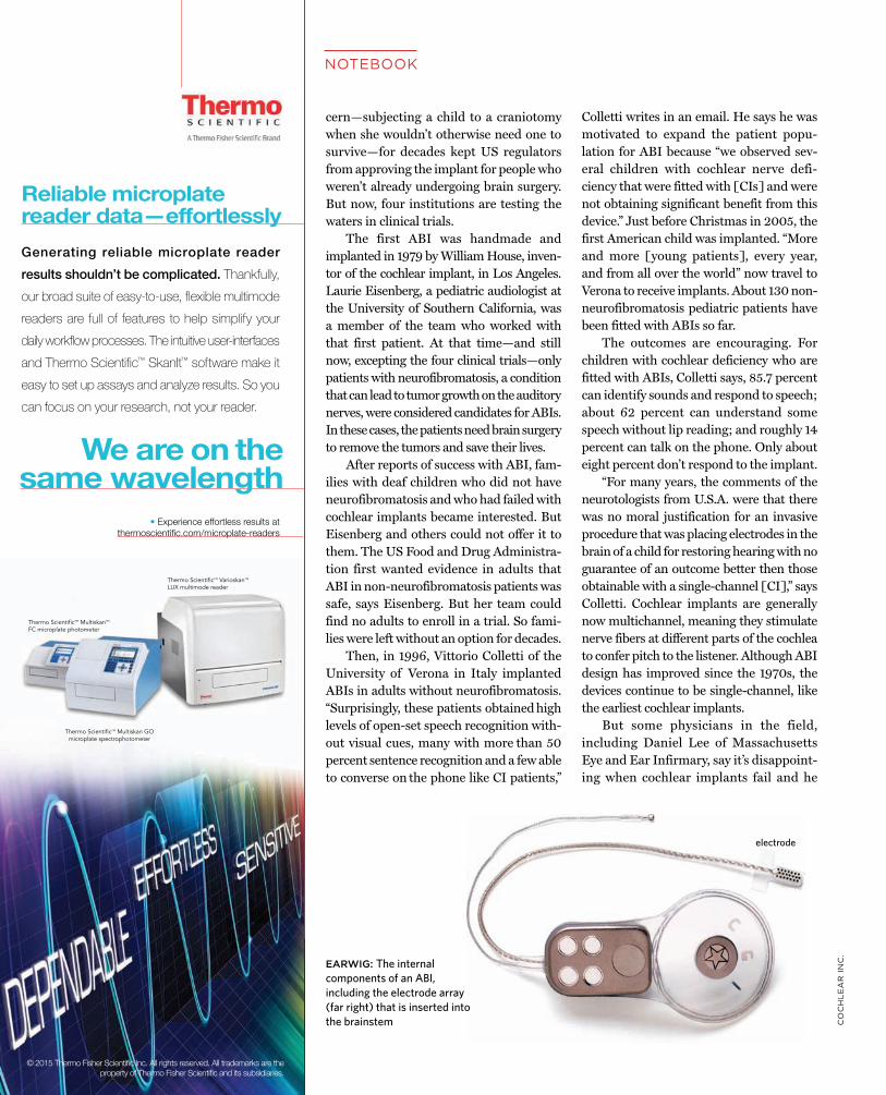
EARWIG: The internalcomponents of an ABI,including the electrode array(far right) that is inserted intothe brainstem
electrode
• Experience effortless results at thermoscientific.com/microplate-readers
Reliable microplate reader data—effortlessly
We are on the same wavelength
Generating reliable microplate reader results shouldn’t be complicated. Thankfully, our broad suite of easy-to-use, flexible multimode readers are full of features to help simplify your daily workflow processes. The intuitive user-interfaces and Thermo Scientific™ SkanIt™ software make it easy to set up assays and analyze results. So you can focus on your research, not your reader.
© 2015 Thermo Fisher Scientific Inc. All rights reserved. All trademarks are the property of Thermo Fisher Scientific and its subsidiaries.
Thermo ScientificTM MultiskanTM FC microplate photometer
Thermo ScientificTM Multiskan GO microplate spectrophotometer
Thermo ScientificTM VarioskanTM LUX multimode reader
2109.2015 | THE SCIENTIST
AN
DR
ZE
J K
RA
UZ
E
can’t offer patients another option. Col-letti’s work is now changing that. “It put ABI back on the map,” Lee says.
Lee is now leading a clinical trial to enroll 10 children who have been deaf since birth and five who have developed deafness. Lee traveled to Verona to train for the surgery with Colletti, and Colletti attended Lee’s first implant. So far, Lee has implanted ABIs in five children with congenital deafness. One had to have it removed after he fell and hit his head, but the other four have had no trouble—in fact, they all have sound detection with their implants and are making progress toward pattern perception. One child is showing the first signs of oral language abilities, and it’s still early days, says Lee.
In Eisenberg’s trial at USC, four children have undergone the surgery, and the goal is to enroll ten. Although it’s been a long time since Eisenberg worked with the first ABI patient, she says she doesn’t necessarily want the procedure to gain widespread approval in the U.S. “It’s so much more delicate as a surgical intervention [than cochlear implants], and it does require a very experi-enced team of surgeons . . . and support from pediatric specialists.” She says she’d rather see it offered only within centers that have developed expertise with the implant tech-nique and the therapy that follows.
After much prayer, Julie Lopez agreed to enroll Angelica in Eisenberg’s study. “My concern was safety,” says Lopez. “I didn’t want her to be walking down the street and not hear a car or a dog coming up behind her.” Just after Angelica turned three last year, she underwent the surgery. Months later, nothing seemed different, until one day last winter when Lopez took her daugh-ter to the bathroom during an audiology therapy visit. Someone out of sight flushed a toilet and Angelica signed to her mother, “You listen, flush potty.” Lopez was thrilled. “I thought, ‘Oh my God, you heard that!’ ”
—Kerry Grens
Musical ScalesSeveral years ago, ichthyologist Eric Par-mentier met a French marine biologist
and filmmaker, Laurent Ballesta, who wasorganizing an expedition to South Africa to produce a documentary film on the coelacanth. This ancient fish—one whose fossil record dates back at least 350 mil-lion years—has an almost mythical legacy. Although it was widely assumed to have gone extinct 65 million years ago, a live specimen was found in 1938, and scien-tists have identified two extant species of coelacanth. Both species move in a pecu-liar way, waggling four lobe-like fins in an alternating pattern, as we do our arms and legs. Their anatomy is also unusual: a tiny brain, a joint at the back of the head that allows the animal to open its jaws widely, and only rudimentary vertebrae. Ballesta’s trip inspired Parmentier, who studies fish acoustics, to collaborate with the team. “I hoped to be the first guy to record [sounds of] the coelacanth.”
Parmentier, a morphologist at the University of Liège in Belgium, has trav-eled the world to understand fish sounds. When I spoke with him, he was between trips to Taiwan, Corsica, and Chile. But for this 2013 South African expedition to cap-ture images and audio of coelacanth, he was going to leave the recording to the film crew’s divers, as it involved installing equip-ment in a cave whose mouth lies more than 100 meters below the water’s surface.
In the spring of 2013, the divers suc-cessfully planted a hydrophone inside the cave and also shot video footage of a coel-acanth. (The resulting documentary by Ballesta is available on YouTube. Although it is in French, the footage obviates the need for fluency to enjoy the film.) Day and night, for weeks, the hydrophone dutifully recorded the sounds within the cave. When Parmentier retrieved the files and went to analyze the recordings, there was one big problem: it was filled with dozens of dif-ferent fish calls. “Maybe the coelacanth is in these sound files, but it’s completely masked by the other sounds,” he says.
Nonetheless, the tape captured cease-less, never-before-heard chatter among the aquatic organisms within the cave (PNAS, 112:6092-97, 2015). To make some sense of it, Parmentier’s team undertook the laborious task of characterizing the sounds recorded over 19 nonconsecu-tive days (to make this feasible, the group pared down its analysis to the first nine minutes of every hour). The research-ers assigned more than 2,700 sounds to 17 groups, most of which sounded to Par-mentier like fish (one group was clearly dolphin, based on its high frequency, he says). These included frog-like croaks, grunts that sounded like a creaking door, a moan, and one that sounded like a whis-
22 THE SCIENTIST | the-scientist.com
NOTEBOOK
tle blown under water. “It’s fair to say,based on the characteristics of the sounds they were hearing, they are probably fish sounds,” says Erica Staaterman, a postdoc at the Smithsonian who studies fish acous-tic communication.
Fish have come up with a variety of ways to make sounds. They may rub their bones together, grind their teeth, or rap-idly contract muscles connected to their swim bladder. Staaterman says fish can use these sounds to defend a territory or attract a mate. Craig Radford, a fish biol-ogist at the University of Auckland, stud-ies a bigeye species (Pempheris adspersa) in waters near New Zealand that makes a click sound similar to one Parmentier recorded in the South African cave. Rad-ford’s group has found that when the big-eyes leave their daytime hideouts to feed on plankton at night, they produce the click sounds to maintain the structure of their schools. When the calls are frequent, the school is tight-knit, and when there are fewer clicks, the school disbands (J Exp Biol, 218:940-48, 2015). “Soundscapes are important for fish to be able to com-municate with each other,” says Radford, but also important for them to understand their environment.
The function of all the sounds in the South African cave is anyone’s guess, but it’s not pure chaos. Parmentier found some organization to the noises. In particular, the frequencies overlap less at night than during the day (despite its being an under-water cave, he says, there is still light avail-able). In other words, the night sounds are more distinct from one another than those produced during the day. “The hypothesis is that during the day, the songs could be there just to support the visual displays,” says Parmentier. “Of course, during the night it is not possible.” Without light, sounds become more important.
David Mann, the president of Logger-head Instruments, which makes underwa-ter recording equipment, says there have been plenty of studies on acoustic niche partitioning among other animals, birds in particular, but such partitioning in the acoustic environment of fishes is a new observation. “I don’t think anyone had seen
that before,” he says, adding that it will beimportant to test whether this also occurs in other settings, such as coral reefs.
The day/night structuring of the acous-tic soundscape in the South African cave is a good hypothesis, says Arthur Popper, a pro-fessor emeritus at the University of Mary-land, but there’s just one thing missing: the hydrophone didn’t actually capture the type of sounds fish hear. Unlike our ears and hydrophones, fish ears don’t detect sound pressure, which is the compression of mol-ecules. Instead, they perceive something called particle motion, the tiny back-and-forth movements of particles in response to sound waves. “What [the group] should have done is also measure particle motion to get a better sense of what the soundscape is for local fish,” says Popper, adding that to do so is complicated and extremely expen-sive. Radford says it’s likely the fish are able to hear the sounds picked up by the hydro-phone. Laboratory studies have shown that fish can hear calls in these frequencies.
Popper says there are a great number of unknowns about the underwater sound-scape—little is documented about what’s out there, let alone how sounds, both nat-ural and man-made, affect aquatic animal behavior. Staaterman, for one, is working on the basics, measuring acoustic habitats off the coast of Panama and in the Chesa-peake Bay to get baseline soundscapes and to observe broad patterns over time. She says there’s so much to explore. “You don’t know what you’re going to find when you put your hydrophone in the water.”
—Kerry Grens
The UpsideImagine a continuous ringing in yourears that you know will probably never stop. Or, imagine you are one of the 360
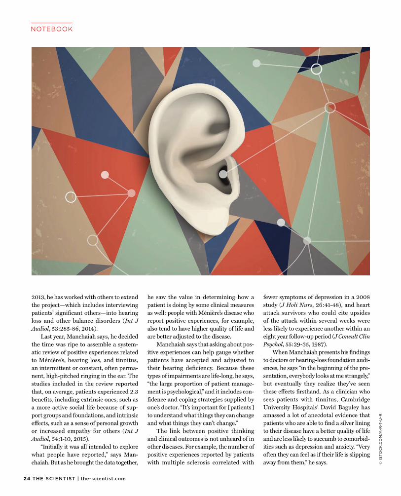
million people worldwide who are los-ing their hearing. Now consider the many ways those changes will affect your life. Do you see any upsides? This is the bold question Vinaya Manchaiah, an audiology researcher at Lamar Univer-sity in Beaumont, Texas, has been ask-ing patients with hearing loss and other related impairments for several years. It’s a question, he says, that has real clinical value, and daring to ask it may possibly lead to positive effects.
Manchaiah is aware of the counterintui-tive nature of his research. He says the first question doctors ask their patients is, “What’s the problem?” not “What are the benefits of the problem?” Even Manchaiah was skepti-cal when he was a PhD student at Linköping University in Sweden and his advisor, Dafydd Stephens, an audiologist who died in 2012, presented him with early results indicating the value of positive experience reporting.
In 2004, using questionnaires, Stephens found that nearly half of family members sur-veyed, especially children and grandchildren, reported one or more positive effects result-ing from their loved one’s hearing impair-ment, including improved communication skills and being able to do noisy activities without their hearing (Audiological Medi-cine, 2:134-38, 2004). Stephens wanted to find quantitative ways to study such experi-ences and their clinical effects. “When I heard this in the beginning, I thought it was a really strange idea,” says Manchaiah.
Manchaiah became convinced that this was a worthwhile research topic only after spending some time at hear-ing-loss support group meetings, where he saw that often the most well-adjusted patients were those who reported posi-tive experiences as simple as using their declining hearing as an excuse to pretend not to hear someone speaking to them. “When I go and talk to people in that set-ting, they’re a lot more open to talking about these things.”
Manchaiah worked with Stephens to catalogue positive experiences among patients with Ménière’s disease, an inner-ear disorder that causes random episodes of vertigo, ringing in the ears, and hear-ing loss. Since completing his doctorate in
Soundscapes are important for fish to be able to com-municate with each other.
—Craig Radford, University of Auckland
There’s only one Millex® filter.Don’t be fooled.
CAUTION:• Syringe filters are posing as high-quality Millex®
filters, but are rather thinly disguised.
• These filters may burst unpredictably and lose membrane integrity.
• Victims may experience noisy baselines and unreliable data.
Failu
re R
ate
(> 4
0 pa
rtic
les
pass
ing)
Competitor A Competitor B Millex® Filter
4%
5%
6%
7%
8%
9%
3%
2%
1%
0%
4%
8%
0%
Device integrity test: what percent of devices still allow particles to pass through? SHOULD BE ZERO
EMD Millipore is a division of Merck KGaA, Darmstadt, Germany
EMD Millipore, the M logo and Millex are registered trademarks of Merck KGaA, Darmstadt, Germany.
© 2015 EMD Millipore Corporation, Billerica, MA, USA. All rights reserved. BS GEN-15-11097 03/2015
See the difference for yourself! Request a free sample of the only true Millex® filter at www.emdmillipore.com/oneMillex
Don’t be fooled – EMD Millipore’s history of membrane technology and filter device engineering is unmatched.
24 THE SCIENTIST | the-scientist.com
© I
ST
OC
K.C
OM
/A-R
-T-U
-R
NOTEBOOK
2013, he has worked with others to extendthe project—which includes interviewing patients’ significant others—into hearing loss and other balance disorders (Int J Audiol, 53:285-86, 2014).
Last year, Manchaiah says, he decided the time was ripe to assemble a system-atic review of positive experiences related to Ménière’s, hearing loss, and tinnitus, an intermittent or constant, often perma-nent, high-pitched ringing in the ear. The studies included in the review reported that, on average, patients experienced 2.3 benefits, including extrinsic ones, such as a more active social life because of sup-port groups and foundations, and intrinsic effects, such as a sense of personal growth or increased empathy for others (Int J Audiol, 54:1-10, 2015).
“Initially it was all intended to explore what people have reported,” says Man-chaiah. But as he brought the data together,
he saw the value in determining how a patient is doing by some clinical measures as well: people with Ménière’s disease who report positive experiences, for example, also tend to have higher quality of life and are better adjusted to the disease.
Manchaiah says that asking about pos-itive experiences can help gauge whether patients have accepted and adjusted to their hearing deficiency. Because these types of impairments are life-long, he says, “the large proportion of patient manage-ment is psychological,” and it includes con-fidence and coping strategies supplied by one’s doctor. “It’s important for [patients] to understand what things they can change and what things they can’t change.”
The link between positive thinking and clinical outcomes is not unheard of in other diseases. For example, the number of positive experiences reported by patients with multiple sclerosis correlated with
fewer symptoms of depression in a 2008 study (J Holi Nurs, 26:41-48), and heart attack survivors who could cite upsides of the attack within several weeks were less likely to experience another within an eight year follow-up period (J Consult Clin Psychol, 55:29-35, 1987).
When Manchaiah presents his findings to doctors or hearing-loss foundation audi-ences, he says “in the beginning of the pre-sentation, everybody looks at me strangely,” but eventually they realize they’ve seen these effects firsthand. As a clinician who sees patients with tinnitus, Cambridge University Hospitals’ David Baguley has amassed a lot of anecdotal evidence that patients who are able to find a silver lining to their disease have a better quality of life and are less likely to succumb to comorbid-ities such as depression and anxiety. “Very often they can feel as if their life is slipping away from them,” he says.
There’s only one Stericup® filter.Don’t be fooled.
CAUTION:• Sterile filters are posing as high-quality Stericup®
filters, but are rather thinly disguised.
• These filters may exhibit clogging, loss of volume and unwanted binding of serum proteins and other additives.
• Victims report anxiety due to potential damage to cell health.
Protein Binding Performance
Stericup® filter Competitor filters
Tota
l Pro
tein
Los
s (µ
g/cm
2 )
Stericup®filter
B C D E F G
40
80
120
160
200
240
0
EMD Millipore is a division of Merck KGaA, Darmstadt, Germany
EMD Millipore, the M logo and Stericup are registered trademarks of Merck KGaA, Darmstadt, Germany.
© 2015 EMD Millipore Corporation, Billerica, MA, USA. All rights reserved. BS GEN-15-11097 03/2015
View more data and place an order for the one and only true Stericup® filter at www.emdmillipore.com/oneStericup
Don’t be fooled – EMD Millipore’s history of membrane technology and filter device engineering is unmatched.
26 THE SCIENTIST | the-scientist.com
NOTEBOOK
Baguley was happy to coauthor thereview with Manchaiah to bring some hard data to the attention of other clini-cians. Although their analysis found that associations between positive experiences and increased quality of life have thus far only been reported for Ménière’s, Baguley says it still “fits very well with the obser-vations I’ve made over the last 30 years. Some patients will find that experiences with hearing loss, and tinnitus in particu-lar, are a stimulus for growth. People will say, ‘I always wish it hadn’t happened to me, but in a way, I’m a stronger, kinder person because of that.’”
Manchaiah says he’d like to extend those correlative studies to include hearing loss and tinnitus, and also find out whether there are ways that doctors can encourage positive thinking and hence any clinical benefits that go along with it. According to the review, when asked open-ended ques-tions about their positive experiences, only 40 to 45 percent of the respondents in the studies wrote of any, but that number sur-passed 90 percent when surveys contained more-structured questions. If given the explicit option of reporting positive experi-ences, he says, most people will agree there is at least one benefit to their situation, and he plans to study whether just being asked by their doctor to consider the upsides to their hearing loss could impact quality of life, acceptance, and other outcomes.
“I think it could guide our work with patients in these areas and look at how we can build upon their resilience and try to help them improve and grow in these cir-cumstances,” says Baguley.
—Amanda B. Keener
HandicapableDominic Pisano hadn’t even arrived oncampus to start his freshman year at Johns Hopkins University when he got his first email from biomedical engi-neer Tilak Ratnanather. He had heard Pisano was deaf and wanted to meet with him. Ratnanather, who has been deaf since birth, showed up for the meet-ing accompanied by a second deaf stu-
dent who would later become a doctor. “He was, like: ‘Here’s my deaf army,’” Pisano recalls.
Soon, Pisano, a soccer enthusiast from Ohio, was interpreting magnetic reso-nance imaging (MRI) in Ratnanather’s department. When Pisano decided he wanted to go to medical school, Rat-nanather was ready to introduce him to his wide network of friends in the otolar-yngology department at Hopkins. Pisano assisted in MRI research at Hopkins for a year before attending Tufts University School of Medicine in Boston.
“I’ll be honest with you, if it weren’t for Tilak I probably wouldn’t have gone to medical school,” says Pisano, now a resi-dent in anesthesiology at Tufts Medical Center. “I probably wouldn’t have done biomedical engineering research. Most importantly, I probably wouldn’t have the kind of network I have.”
It was this kind of service that won Ratnanather the Presidential Award for Excellence in Science, Mathematics, and Engineering Mentoring this past March. Over the years, Ratnanather has lobbied for better resources for deaf attendees at conferences, organized annual dinners for deaf researchers, helped award schol-arships to hearing-impaired students through the Alexander Graham Bell Asso-ciation for the Deaf and Hard of Hear-ing (AG Bell), and mentored more than a dozen hearing-impaired students.
“He’s by nature the most gregarious and extroverted individual,” says Howard Francis, a professor of otolaryngology at Hopkins who has known Ratnanather for 23 years. “He has a sense of mission and is committed to making it possible for others to achieve what he has achieved.”
“A lot of people have a hard time under-standing him [due to his deafness-related difficulties with speech],” says Pisano, “but despite that, they still enjoy his company, and they want to be connected with him.”
Ratnanather was born in 1963 in Sri Lanka with profound hearing loss of unknown origin. His family moved to Lon-don when he was 18 months old, and he grew up wearing hearing aids and attending the Mary Hare School for Deaf Children.
Ratnanather’s parents, a pediatrician and a computer systems programmer, had high hopes for their son. “My father and I would talk about mathematics and would go through some problems at home,” he says. “I had an aptitude, and then, of course, I would go to the science museum and learn about famous mathematicians.”
Ratnanather enrolled at University College London, where he met mathema-tician Keith Stewartson, who immediately made the young undergrad comfortable about his hearing loss and the assistive technologies he needed to use in the class-room. “I knew he would make my life easy,” says Ratnanather. “I didn’t have to worry about my deafness.”
Tragically, Stewartson died suddenly at the end of Ratnanather’s first year at uni-versity. But the young student forged ahead, and after doing some reading about Stew-artson’s research on fluid dynamics, Rat-nanather went on to study the subject in graduate school at the University of Oxford, receiving his DPhil in mathematics in 1989.
Up until that point, Ratnanather had only had occasional opportunities to learn about an area near to his heart: hearing research. This changed after he attended a research symposium at the 1990 AG Bell Conven-tion in Washington, D.C. Fascinated by the work of William Brownell, Ratnanather approached the Johns Hopkins researcher after Brownell had given a talk about outer hair cell electromotility—the process by which these sensory cells shorten or lengthen in response to electrical impulses.
When outer hair cells change shape, they transmit mechanical force to the cochlea, amplifying the ear’s sensitivity to soft sounds at specific frequencies. Forces transmitted through pressurized fluids in outer hair cells make electromotility pos-sible, explains Brownell, who is now at Baylor College of Medicine in Houston, Texas. He needed someone who could
I’ll be honest with you, if it weren’t for Tilak I probably wouldn’t have gone to medical school. —Dominic Pisano, Tufts Medical Center
© I
ST
OC
K.C
OM
/TO
DO
R T
SV
ET
KO
V
model the dynamics of fluid within thesetiny spaces. “Tilak had the computational tools to begin to study this,” Brownell says.
Ratnanather began a postdoc in Brownell’s lab in 1991. During his postdoc, he realized he could bestow upon students the confidence his mentors fostered in him. The Internet helped him reach out to other deaf people through newsgroups. Lina Reiss, who had severe hearing loss by age two, first met Ratnanather when she was an under-graduate at Princeton University and he replied to an online post in which she intro-duced herself to one of these newsgroups.
The daughter of two PhDs, Reiss had always known that she wanted to go into the sciences. But she was not sure what career would be possible with her hear-ing loss. “I didn’t have any role models of what it was like to be a deaf faculty mem-ber,” she recalls. “Until I met [Tilak and some of his deaf friends], I couldn’t imag-ine becoming a professor.”
Ratnanather helped get Reiss a sum-mer internship in the hearing-research lab of a colleague at Johns Hopkins, where she studied how neurons in the brain stem encode and process sound. Enthralled with the research, she went on to do her PhD in biomedical engineering in the same lab. She is now an assistant profes-sor at Oregon Health & Science University
in Portland researching how hearing loss, hearing aids, and cochlear implants influ-ence the way people perceive sound.
Ratnanather now primarily does brain-mapping research focused on understanding how brain structures are altered in people with diseases such as schizophrenia, Alzheimer’s, and bipolar disorder. But hearing science continues to influence his work. He has published several recent studies on fluid dynamics and hair cell function and has upcoming papers on imaging the auditory regions of the brain in deaf adults and babies.
And, spurred partly by his own cochlear implant surgery in 2012, Ratnanather has created an app for adults learning how to hear following the surgery. Called Speech Banana, the app is named after the banana-shaped region in an audiogram that con-tains human speech.
More than just providing professional connections, Ratnanather has influenced how his former students navigate the world. Being deaf can make it scary to think out-side the box or challenge opinions, Pisano says. Ratnanather encourages his mentees to keep an open mind and engage with oth-ers—hearing and nonhearing alike. “That helped shape my mentality about life in gen-eral today,” Pisano says.
—Kate Yandell
© 2015 Bethyl Laboratories, Inc. All rights reserved.
For really good antibodies,
visit bethyl.com/trialsize
This ad won’t work.
Our antibodies will.
28 THE SCIENTIST | the-scientist.com
CRITIC AT LARGE
© O
NE
SM
AL
LS
QU
AR
E/S
HU
TT
ER
ST
OC
K
Hurdles for Hearing Restoration
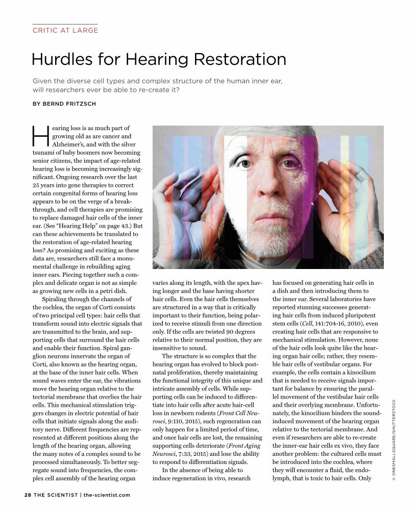
Hearing loss is as much part ofgrowing old as are cancer and Alzheimer’s, and with the silver
tsunami of baby boomers now becoming senior citizens, the impact of age-related hearing loss is becoming increasingly sig-nificant. Ongoing research over the last 25 years into gene therapies to correct certain congenital forms of hearing loss appears to be on the verge of a break-through, and cell therapies are promising to replace damaged hair cells of the inner ear. (See “Hearing Help” on page 43.) But can these achievements be translated to the restoration of age-related hearing loss? As promising and exciting as these data are, researchers still face a monu-mental challenge in rebuilding aging inner ears. Piecing together such a com-plex and delicate organ is not as simple as growing new cells in a petri dish.
Spiraling through the channels of the cochlea, the organ of Corti consists of two principal cell types: hair cells that transform sound into electric signals that are transmitted to the brain, and sup-porting cells that surround the hair cells and enable their function. Spiral gan-glion neurons innervate the organ of Corti, also known as the hearing organ, at the base of the inner hair cells. When sound waves enter the ear, the vibrations move the hearing organ relative to the tectorial membrane that overlies the hair cells. This mechanical stimulation trig-gers changes in electric potential of hair cells that initiate signals along the audi-tory nerve. Different frequencies are rep-resented at different positions along the length of the hearing organ, allowing the many notes of a complex sound to be processed simultaneously. To better seg-regate sound into frequencies, the com-plex cell assembly of the hearing organ
varies along its length, with the apex hav-ing longer and the base having shorter hair cells. Even the hair cells themselves are structured in a way that is critically important to their function, being polar-ized to receive stimuli from one direction only. If the cells are twisted 90 degrees relative to their normal position, they are insensitive to sound.
The structure is so complex that the hearing organ has evolved to block post-natal proliferation, thereby maintaining the functional integrity of this unique and intricate assembly of cells. While sup-porting cells can be induced to differen-tiate into hair cells after acute hair-cell loss in newborn rodents (Front Cell Neu-rosci, 9:110, 2015), such regeneration can only happen for a limited period of time, and once hair cells are lost, the remaining supporting cells deteriorate (Front Aging Neurosci, 7:33, 2015) and lose the ability to respond to differentiation signals.
In the absence of being able to induce regeneration in vivo, research
has focused on generating hair cells in a dish and then introducing them to the inner ear. Several laboratories have reported stunning successes generat-ing hair cells from induced pluripotent stem cells (Cell, 141:704-16, 2010), even creating hair cells that are responsive to mechanical stimulation. However, none of the hair cells look quite like the hear-ing organ hair cells; rather, they resem-ble hair cells of vestibular organs. For example, the cells contain a kinocilium that is needed to receive signals impor-tant for balance by ensuring the paral-lel movement of the vestibular hair cells and their overlying membrane. Unfortu-nately, the kinocilium hinders the sound-induced movement of the hearing organ relative to the tectorial membrane. And even if researchers are able to re-create the inner-ear hair cells ex vivo, they face another problem: the cultured cells must be introduced into the cochlea, where they will encounter a fluid, the endo-lymph, that is toxic to hair cells. Only
Given the diverse cell types and complex structure of the human inner ear,will researchers ever be able to re-create it?
BY BERND FRITZSCH
2909.2015 | THE SCIENTIST
Submit your cutting-edge, life-sciences technology innovation for consideration by a panel of expert judges. The winners will be the subject of a feature article in the December 2015 issue of The Scientist.
• An “innovation” is defined as any product that researchers use in a lab: machines, instruments, tools, cell lines, custom-made molecular probes and labels, software, apps, etc.
• Products released on or after October 1, 2014 are eligible.
• Entries accepted from April 13 to September 15, 2015.
For further information, contact us at: [email protected]
Announcing The Scientist’s annual Top 10 Innovations Competition
online at: www.the-scientist.com/top10enter
the hair cells’ stereocilia are normallyexposed to the endolymph; the cell body is surrounded by perilymph that is ioni-cally equivalent to the cerebrospinal fluid. Cells injected into the endolymph need to insert quickly into the surround-ing membranes to avoid being killed by the endolymph.
But assuming all such hurdles can be overcome, there is still the chief obstacle to inner-ear regeneration: namely, guid-ing the insertion of a suitable number of appropriate cell types at the precise position and in the correct orientation to generate a functional hearing organ. Mice are deaf even when all hair cells are formed, if those cells are disorganized (Development, doi:10.124/dev123091, 2015). And an organ with hair cells that have an altered polarity or are in the wrong position would certainly not help for hearing at all. Finally, assuming a patient lacks all inner-ear hair cells, how many new ones are needed for a clinically
functional outcome? The healthy organ of Corti contains some 3,500 inner hair cells, which convert the sound signals to electrical impulses, and 15,000 outer hair cells, which help amplify the sound. Could just a few hundred new cells restore hearing that’s been lost?
Clearly, building a hearing organ from scratch is not an easy task. Nev-ertheless, auditory scientists must con-tinue to explore effective ways to restore hair cells and supporting cells, to assem-ble these cells into the complex mosaic of their microenvironment, and to ensure their functionality (BioEssays, doi:10.1002/bies.201500044, 2015). But
it’s also important that efforts extend beyond hair-cell restoration and replace-ment to focus on the mitigation of dam-age as perhaps a more easily attainable goal to treat age-related hearing loss. To quote Benjamin Franklin, “An ounce of prevention is worth a pound of cure.” Deciphering how genetic predisposition is compounded by a lifetime of exposure to sound could hold relevant information to prevent hearing loss, currently the bet-ter alternative. g
Bernd Fritzsch is the department chairand codirector of the Center on Aging at the University of Iowa in Iowa City. He recently coauthored articles on the challenges of regenerating hearing with Israt Jahan and Ning Pan, both associate research scientists in his lab, as well as with Richard J. Smith, Sterba Hearing Research Professor and director of the Iowa Institute of Human Genetics.
Piecing together such a complex and delicate organ is not as simple as growing new cells in a petri dish.
LAST CHANCE!
30 THE SCIENTIST | the-scientist.com
CRITIC AT LARGE
© I
ST
OC
K.C
OM
/WIL
LS
IE
Generations of in-depth research into human anatomy,histology, and basic physiology have largely explained the physical manifestations of diseases affecting nearly
every organ of the body. From cardiology to gastroenterology and pulmonology, form implies function. It is no mystery, for example, why a blood clot between the heart and lungs causes shortness of breath, problems with oxygenation, and strain on the muscles of the heart.
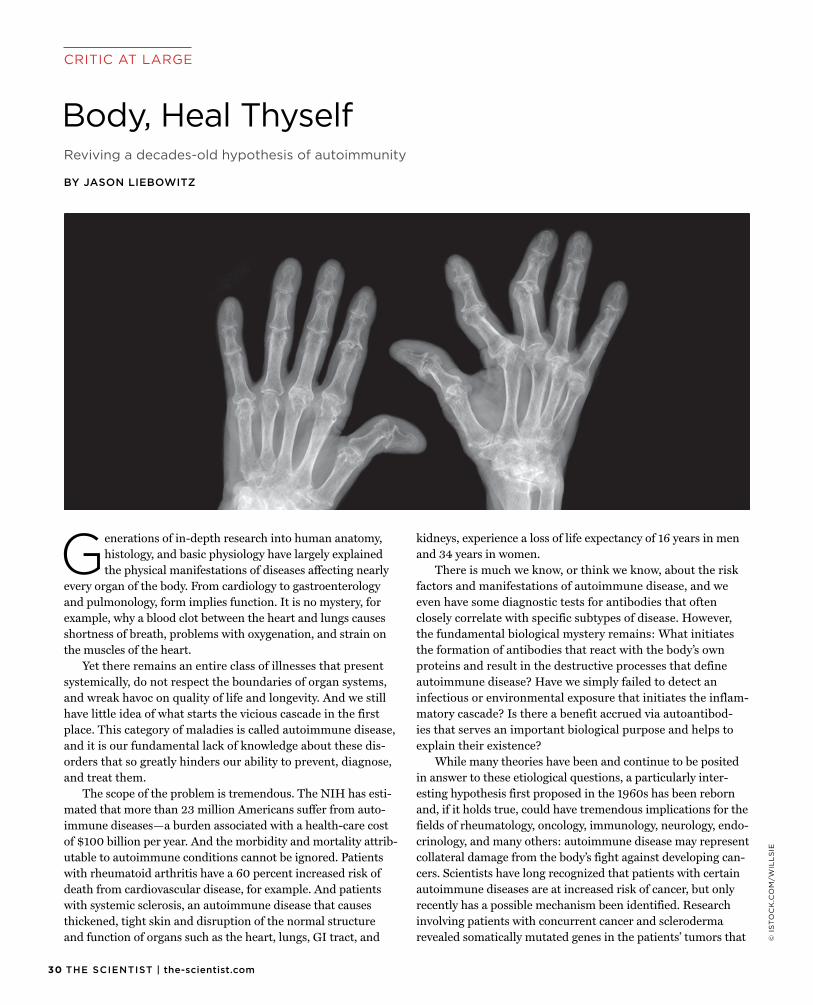
Yet there remains an entire class of illnesses that present systemically, do not respect the boundaries of organ systems, and wreak havoc on quality of life and longevity. And we still have little idea of what starts the vicious cascade in the first place. This category of maladies is called autoimmune disease, and it is our fundamental lack of knowledge about these dis-orders that so greatly hinders our ability to prevent, diagnose, and treat them.
The scope of the problem is tremendous. The NIH has esti-mated that more than 23 million Americans suffer from auto-immune diseases—a burden associated with a health-care cost of $100 billion per year. And the morbidity and mortality attrib-utable to autoimmune conditions cannot be ignored. Patients with rheumatoid arthritis have a 60 percent increased risk of death from cardiovascular disease, for example. And patients with systemic sclerosis, an autoimmune disease that causes thickened, tight skin and disruption of the normal structure and function of organs such as the heart, lungs, GI tract, and
kidneys, experience a loss of life expectancy of 16 years in men and 34 years in women.
There is much we know, or think we know, about the risk factors and manifestations of autoimmune disease, and we even have some diagnostic tests for antibodies that often closely correlate with specific subtypes of disease. However, the fundamental biological mystery remains: What initiates the formation of antibodies that react with the body’s own proteins and result in the destructive processes that define autoimmune disease? Have we simply failed to detect an infectious or environmental exposure that initiates the inflam-matory cascade? Is there a benefit accrued via autoantibod-ies that serves an important biological purpose and helps to explain their existence?
While many theories have been and continue to be posited in answer to these etiological questions, a particularly inter-esting hypothesis first proposed in the 1960s has been reborn and, if it holds true, could have tremendous implications for the fields of rheumatology, oncology, immunology, neurology, endo-crinology, and many others: autoimmune disease may represent collateral damage from the body’s fight against developing can-cers. Scientists have long recognized that patients with certain autoimmune diseases are at increased risk of cancer, but only recently has a possible mechanism been identified. Research involving patients with concurrent cancer and scleroderma revealed somatically mutated genes in the patients’ tumors that
Reviving a decades-old hypothesis of autoimmunity
BY JASON LIEBOWITZ
Body, Heal Thyself

initiated cellular immunity and cross-reactive humoral immune responses, producing antibodies that reacted to the cancer and are known to play an important role in scleroderma itself (Sci-ence, 343:152-57, 2014). The finding implies that the autoim-mune disease may arise as an unintended consequence of the body’s own immune response to a developing cancer, which in certain patients will never become clinically evident.
This idea is not far-fetched considering that certain syn-dromes of this type have been recognized for quite some time. Lambert-Eaton syndrome, in which damage to motor syn-apses can cause weakness, and limbic encephalitis—inflam-mation of the brain and resultant confusion and neurologic symptoms—are two examples of conditions known to occur in reaction to a cancer in the body. But the novel findings sug-gest a much larger possibility that all autoimmune diseases are due to the immune system’s response to cancer. If this is true, detection of specific autoantibodies would help predict which patients will manifest a clinically-apparent cancer. Indeed, researchers have found that specific autoantibodies found in patients with autoimmune myositis—muscle inflamma-tion and symptoms in the joints, skin, lungs, and other body parts—are not only associated with specific subtypes of these diseases, but can also be used to predict which patients will develop the types of myositis associated with cancer (Arthritis Rheum, 67:317-26, 2015).
Such support for the idea that the immune system’s response to cancer cells may trigger autoimmunity has opened a proverbial Pandora’s box of scientific questions. Why do some patients with tumor cells develop autoantibod-ies while others do not? Can autoimmune diseases be pre-vented, instead of waiting for them to develop and treating the aftermath? Perhaps most importantly, can we harness the power of the immune system to effectively fight cancer with-out the resultant production of autoantibodies that cause autoimmune disease? In 2013, the editors of Science named cancer immunotherapy the “Breakthrough of the Year,” fore-seeing a new paradigm in medicine. Understanding the ori-gins of autoimmunity is critical to ensuring the safety of these therapies, which represent newfound hope for millions of patients worldwide.
The time is ripe to tackle this most challenging problem of understanding autoimmune etiology. By investing time and effort in these fundamental questions of biology, we can hopefully one day declare with conviction and clarity: body, heal thyself. g
Jason Liebowitz is a third-year internal medicine resident at Johns Hopkins Bayview Medical Center in Baltimore, Maryland.
Our fundamental lack of knowledge about autoimmune diseases greatly hinders our ability to prevent, diagnose, and treat them.
sAVE TIME
sAVE MONEY
Antibodies ELISA Kits Biochemicals Primary Cells Cell Lines Growth Media Proteins Cytokines Inhibitors/Compounds
… and much, much more!
www.cedarlanelabs.com/SAVE
Search by manufacturer product code and view live USD pricing when shop-ping for research kits and reagents at
www.cedarlanelabs.com/SAVE

A Dream Come True, Without the Unicorn.A Dream Come True, Without the Unicorn.
Compatible with both Mac® and PC systems.
Download Your FREE Copy of Image Studio™ Lite atwww.licor.com/islite
Flaunt your Legendary Western blot images!
Export High Quality Images for Publication
FREE WESTERN BLOT ANALYSIS SOFTWARE
3309.2015 | THE SCIENTIST
AT A GLANCE
MODUS OPERANDI
Medial Lateral
Basa
l
© G
EO
RG
E R
ET
SE
CK
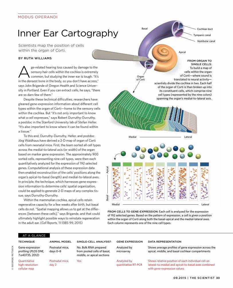
SINGLE-CELL ANALYSIS?
No. Bulk RNA prepared from pooled cells of basal, middle, or apical sections
Yes
GENE EXPRESSION
Analyzed by microarray
Analyzed by quantitative RT-PCR
DATA REPRESENTATION
Shows average profiles of gene expression across the apical, middle, and basal cochlear compartments
Shows relative position of each individual cell on lateral-to-medial and apical-to-basal axes combined with gene-expression values
ANIMAL MODEL
Postnatal mice, days 0–8
Postnatal mice, day 2
TECHNIQUE
Gene-expression profiling (PLOS ONE, 7:e40735, 2012)
Quantitative high-resolution cellular map
Age-related hearing loss caused by damage to thesensory hair cells within the cochlea is extremely common, but studying the inner ear is tough. “It’s
in the densest bone in the body, so you don’t have access,” says John Brigande of Oregon Health and Science Univer-sity in Portland. Even if you can extract cells, he says, “there are so darn few of them.”
Despite these technical difficulties, researchers have gleaned gene-expression information about different cell types within the organ of Corti—home to the sensory cells within the cochlea. But “it’s not only important to know what a cell expresses,” says Robert Durruthy-Durruthy, a postdoc in the Stanford University lab of Stefan Heller. “It’s also important to know where it can be found within a tissue.”
To this end, Durruthy-Durruthy, Heller, and postdoc Jörg Waldhaus have derived a 2-D map of organ of Corti cells from neonatal mice. First, the team sorted all cell types across the medial-to-lateral axis (or width) of the organ based on marker gene expression. The approximately 900 sorted cells, representing nine cell types, were then each quantitatively analyzed for the expression of 192 selected genes. Computational analysis of these expression data then enabled reconstruction of the cells’ positions along the organ’s apical-to-basal (length) and medial-to-lateral axes. In principle, the technique, which harnesses gene-expres-sion information to determine cells’ spatial organization, could be applied to generate 2-D maps of any complex tis-sue, says Durruthy-Durruthy.
Within the mammalian cochlea, apical cells retain regenerative capacity for a few weeks after birth, but basal cells do not. “Spatial mapping allows us to get at the differ-ences [between these cells],” says Brigande, and that could ultimately highlight possible ways to reinstate regeneration in the adult ear. (Cell Reports, 11:1385-99, 2015)
Scientists map the position of cellswithin the organ of Corti.
BY RUTH WILLIAMS
Inner Ear Cartography
FROM ORGAN TO SINGLE CELLS:
To build a map of cells within the organ
of Corti—where sound is translated to neural activity—
scientists divide the cochlea in two. Each half of the organ of Corti is then broken up into
its constituent cells, which comprise nine cell types (represented by the nine colors)
spanning the organ’s medial-to-lateral axis.
FROM CELLS TO GENE-EXPRESSION: Each cell is analyzed for the expressionof 192 selected genes. Based on the pattern of expression, a cell is given a positionwithin the organ of Corti along both the basal-apical and the medial-lateral axes.Each column represents one of the nine cell types.
Medial Lateral
Basal
Tympanic canal
Vestibular canal
Cochlear duct
Apical
Organ of Corti
Api
cal
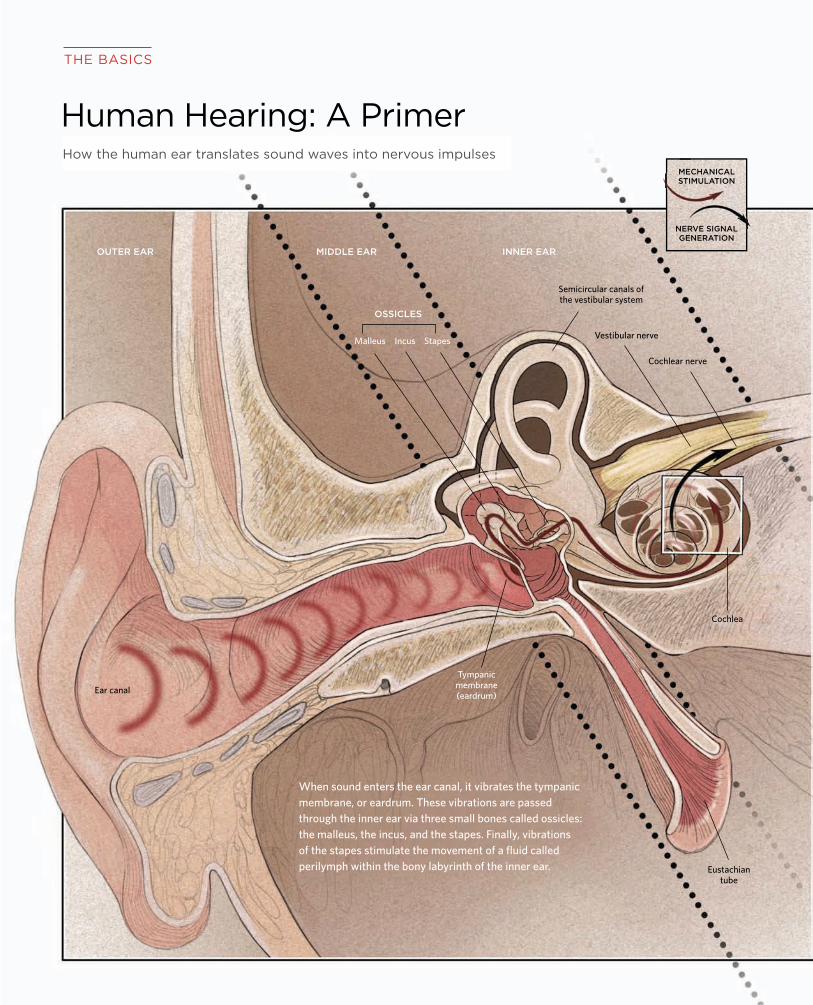
34 THE SCIENTIST | the-scientist.com
OUTER EAR MIDDLE EAR INNER EAR
Malleus Incus Stapes Vestibular nerve
Cochlear nerve
Eustachian tube
THE BASICS
Human Hearing: A Primer
Semicircular canals ofthe vestibular system
OSSICLES
Ear canal
Tympanic membrane(eardrum)
When sound enters the ear canal, it vibrates the tympanic membrane, or eardrum. These vibrations are passed through the inner ear via three small bones called ossicles: the malleus, the incus, and the stapes. Finally, vibrations of the stapes stimulate the movement of a fluid called perilymph within the bony labyrinth of the inner ear.
Cochlea
How the human ear translates sound waves into nervous impulses
NERVE SIGNAL GENERATION
MECHANICAL STIMULATION
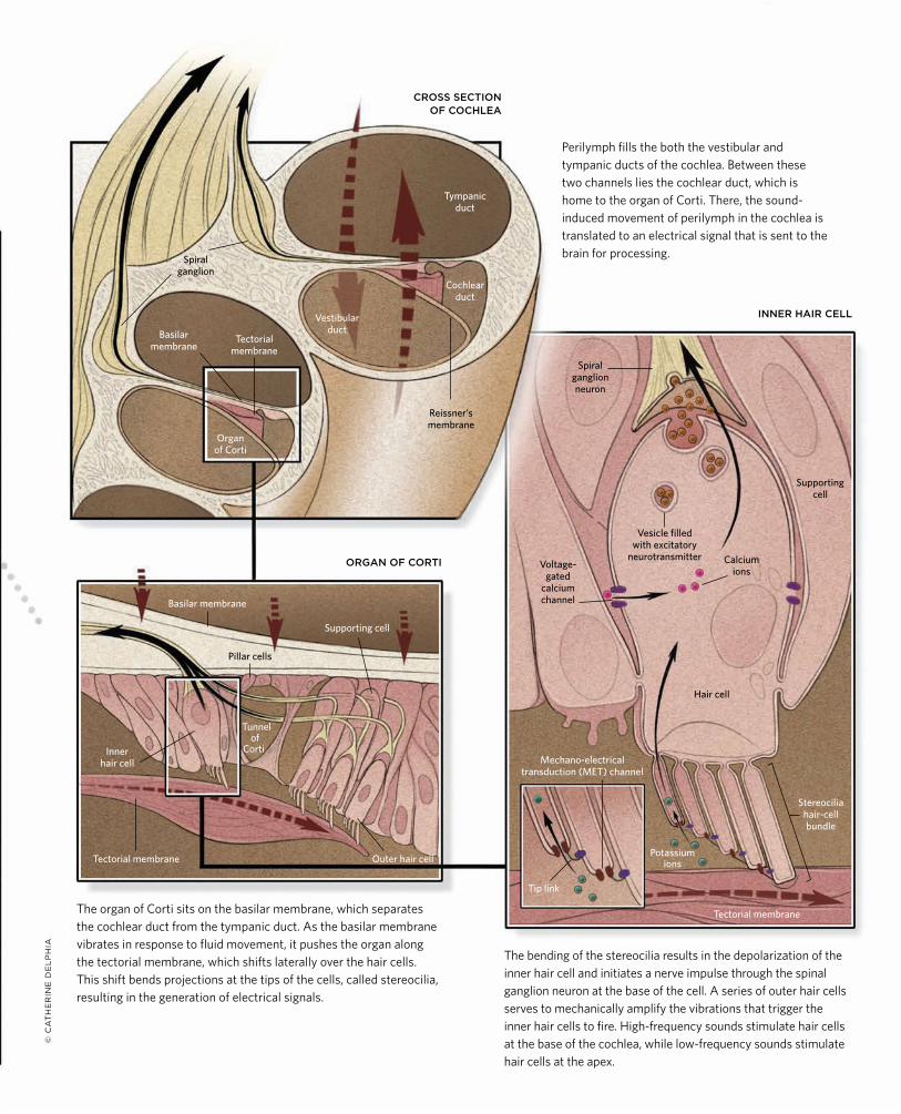
3509.2015 | THE SCIENTIST
Spiral ganglion neuron
Tympanic duct
Vestibular duct
Cochlear duct
Inner hair cell
Outer hair cellTectorial membrane
Tectorial membrane
Tectorial membrane
Organ of Corti
Supporting cell
Reissner’s membrane
CROSS SECTION OF COCHLEA
INNER HAIR CELL
Potassium ions
Hair cell
Supporting cell
Stereocilia hair-cell bundle
Mechano-electrical transduction (MET) channel
Voltage- gated
calcium channel
Vesicle filled with excitatory
neurotransmitter Calcium ions
Tunnel of
Corti
Pillar cells
Spiral ganglion
Basilar membrane
Basilar membrane
Perilymph fills the both the vestibular and tympanic ducts of the cochlea. Between these two channels lies the cochlear duct, which is home to the organ of Corti. There, the sound-induced movement of perilymph in the cochlea is translated to an electrical signal that is sent to the brain for processing.
The organ of Corti sits on the basilar membrane, which separates the cochlear duct from the tympanic duct. As the basilar membrane vibrates in response to fluid movement, it pushes the organ along the tectorial membrane, which shifts laterally over the hair cells. This shift bends projections at the tips of the cells, called stereocilia, resulting in the generation of electrical signals.
The bending of the stereocilia results in the depolarization of the inner hair cell and initiates a nerve impulse through the spinal ganglion neuron at the base of the cell. A series of outer hair cells serves to mechanically amplify the vibrations that trigger the inner hair cells to fire. High-frequency sounds stimulate hair cells at the base of the cochlea, while low-frequency sounds stimulate hair cells at the apex.
Tip link
ORGAN OF CORTI
© C
AT
HE
RIN
E D
EL
PH
IA

36 THE SCIENTIST | the-scientist.com
Aural HistoryThe form and function of the ears of modern land vertebrates
cannot be understood without knowing how they evolved.
BY GEOFFREY A. MANLEY
PH
OT
O C
RE
DIT
S S
EE
PA
GE
42
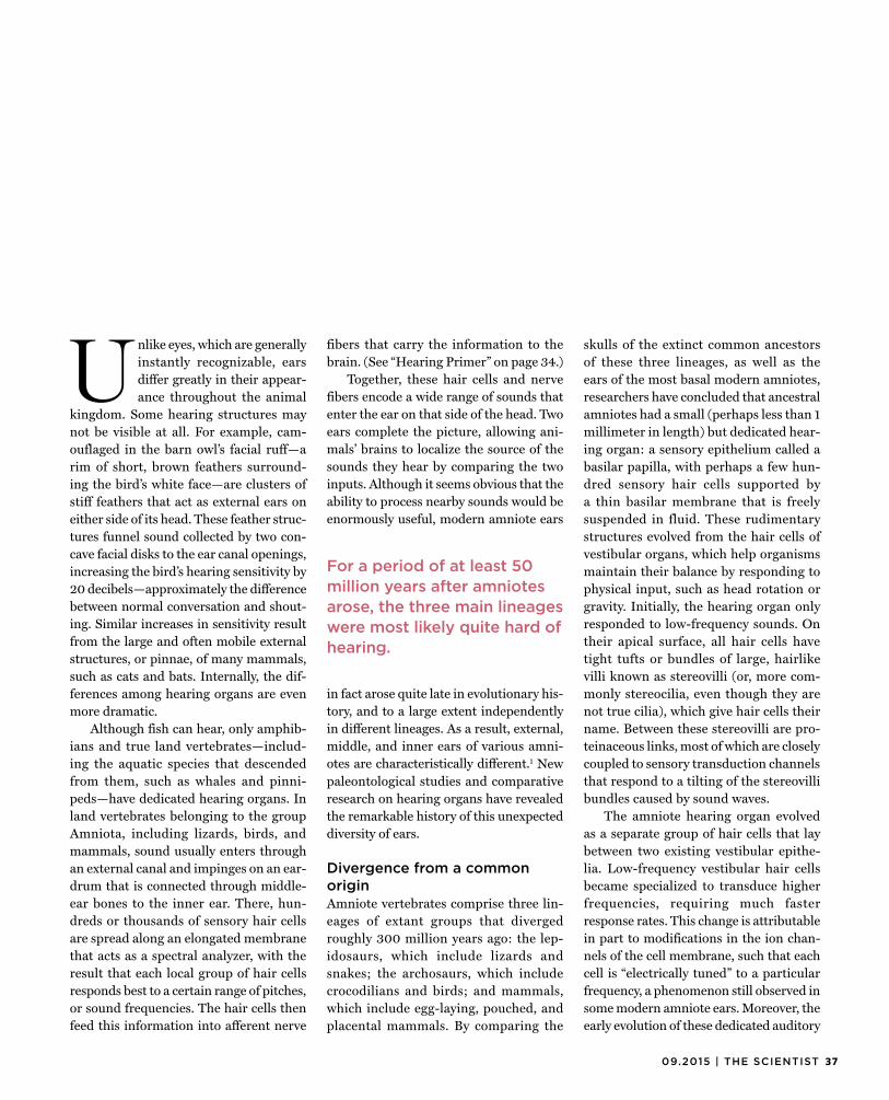
3709.2015 | THE SCIENTIST
Unlike eyes, which are generallyinstantly recognizable, ears differ greatly in their appear-ance throughout the animal
kingdom. Some hearing structures may not be visible at all. For example, cam-ouflaged in the barn owl’s facial ruff—a rim of short, brown feathers surround-ing the bird’s white face—are clusters of stiff feathers that act as external ears on either side of its head. These feather struc-tures funnel sound collected by two con-cave facial disks to the ear canal openings, increasing the bird’s hearing sensitivity by 20 decibels—approximately the difference between normal conversation and shout-ing. Similar increases in sensitivity result from the large and often mobile external structures, or pinnae, of many mammals, such as cats and bats. Internally, the dif-ferences among hearing organs are even more dramatic.
Although fish can hear, only amphib-ians and true land vertebrates—includ-ing the aquatic species that descended from them, such as whales and pinni-peds—have dedicated hearing organs. In land vertebrates belonging to the group Amniota, including lizards, birds, and mammals, sound usually enters through an external canal and impinges on an ear-drum that is connected through middle-ear bones to the inner ear. There, hun-dreds or thousands of sensory hair cells are spread along an elongated membrane that acts as a spectral analyzer, with the result that each local group of hair cells responds best to a certain range of pitches, or sound frequencies. The hair cells then feed this information into afferent nerve
fibers that carry the information to the brain. (See “Hearing Primer” on page 34.)
Together, these hair cells and nerve fibers encode a wide range of sounds that enter the ear on that side of the head. Two ears complete the picture, allowing ani-mals’ brains to localize the source of the sounds they hear by comparing the two inputs. Although it seems obvious that the ability to process nearby sounds would be enormously useful, modern amniote ears
in fact arose quite late in evolutionary his-tory, and to a large extent independently in different lineages. As a result, external, middle, and inner ears of various amni-otes are characteristically different.1 Newpaleontological studies and comparative research on hearing organs have revealed the remarkable history of this unexpected diversity of ears.
Divergence from a commonoriginAmniote vertebrates comprise three lin-eages of extant groups that diverged roughly 300 million years ago: the lep-idosaurs, which include lizards and snakes; the archosaurs, which include crocodilians and birds; and mammals, which include egg-laying, pouched, and placental mammals. By comparing the
skulls of the extinct common ancestors of these three lineages, as well as the ears of the most basal modern amniotes, researchers have concluded that ancestral amniotes had a small (perhaps less than 1 millimeter in length) but dedicated hear-ing organ: a sensory epithelium called a basilar papilla, with perhaps a few hun-dred sensory hair cells supported by a thin basilar membrane that is freely suspended in fluid. These rudimentary structures evolved from the hair cells of vestibular organs, which help organisms maintain their balance by responding to physical input, such as head rotation or gravity. Initially, the hearing organ only responded to low-frequency sounds. On their apical surface, all hair cells have tight tufts or bundles of large, hairlike villi known as stereovilli (or, more com-monly stereocilia, even though they are not true cilia), which give hair cells their name. Between these stereovilli are pro-teinaceous links, most of which are closely coupled to sensory transduction channels that respond to a tilting of the stereovilli bundles caused by sound waves.
The amniote hearing organ evolved as a separate group of hair cells that lay between two existing vestibular epithe-lia. Low-frequency vestibular hair cells became specialized to transduce higher frequencies, requiring much faster response rates. This change is attributable in part to modifications in the ion chan-nels of the cell membrane, such that each cell is “electrically tuned” to a particular frequency, a phenomenon still observed in some modern amniote ears. Moreover, the early evolution of these dedicated auditory
For a period of at least 50 million years after amniotes arose, the three main lineages were most likely quite hard of hearing.

38 THE SCIENTIST | the-scientist.com
COMMON ANCESTOR OF AMNIOTES
CONVERGING ON THE EARThe hearing organ of vertebrates arose from vestibular organs, which containedhundreds of hair cells that could detect very low-frequency physical input, such as head rotation or gravity. Starting around 250 million years ago, the three amniote lineages—lepidosaurs (lizards and snakes), archosaurs (crocodilians and birds), and mammals—separately evolved a tympanic middle ear, followed by evolution of the inner ear, both of which served to increase hearing sensitivity. Despite the independent origin of hearing structures in the three lineages, the outcomes were functionally quite similar, serving as a remarkable example of convergent evolution.
MODERN MIDDLE EARSArose 250–200 million years ago
In mammals, three bony ossicles evolved from an old jaw joint (malleus, incus) together with the hyomandibula (stapes).
In both cases, the middle-ear bones formed a lever system that greatly magnified sound input.
LEPIDOSAURS
Columella
Extracolumella
Tympanic membrane (eardrum)
ARCHOSAURS
In archosaurs and lepidosaurs, a long and thin middle-ear bone called the columella evolved from the hyomandibula, a bone that earlier had formed a large strut connecting the braincase to the outer skull.
MAMMALS
Stapes
Incus
Malleus
Tympanic membrane (eardrum)
Ear canal Footplate
Ear canal
organs in land vertebrates led to the loss ofthe heavy otolithic membrane that overlies the hair-cell bundles of vestibular organs and is responsible for their slow responses. What remains is the watery macromo-lecular gel known as the tectorial mem-brane, which assures that local groups of hair cells move synchronously, resulting in greater sensitivity.
Good high-frequency hearing did not exist from the start, however. For a period of at least 50 million years after amniotes arose, the three main lineages were most likely quite hard of hearing. They had not yet evolved any mechanism for absorbing sound energy from air; they lacked the middle ear and eardrum that are vital for the function of modern hearing organs. As such, ancestral amniotes most likely per-ceived only sounds of relatively low fre-quency and high amplitude that reached the inner ear via the limbs or, if the skull were rested on the ground, through the tis-sues of the head. It is unclear what kind of stimuli could have existed that would have led to the retention of such hearing organs for such a long time.
The magnificent middle earDuring the Triassic period, some 250 to200 million years ago, a truly remark-able thing happened. Independently, but within just 20 million to 30 million years of one another, all three amniote lineages evolved a tympanic middle ear from parts of the skull and the jaws.2
The tympanic middle ear is the assem-blage of tiny bones that connects at one end to an eardrum and at the other end to the oval window, an aperture in the bone of the inner ear. Despite the tem-poral coincidence in the evolution of these structures in the three amniote lineages and the functional similarities of the adaptations, the groups were by this time so far separated that the mid-dle ears evolved from different structures into two different configurations. The single middle-ear bone, the columella, of archosaurs and lepidosaurs derived from the hyomandibular, a bone that ear-lier had formed a large strut connecting the braincase to the outer skull. In mod-
Footplate covering the oval window
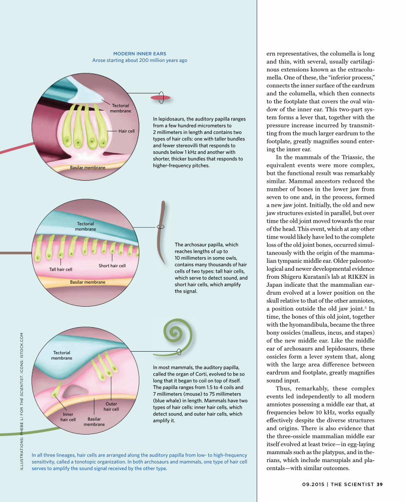
3909.2015 | THE SCIENTIST
MODERN INNER EARSArose starting about 200 million years ago
In all three lineages, hair cells are arranged along the auditory papilla from low- to high-frequency sensitivity, called a tonotopic organization. In both archosaurs and mammals, one type of hair cell serves to amplify the sound signal received by the other type.
In lepidosaurs, the auditory papilla ranges from a few hundred micrometers to 2 millimeters in length and contains two types of hair cells: one with taller bundles and fewer stereovilli that responds to sounds below 1 kHz and another with shorter, thicker bundles that responds to higher-frequency pitches.
The archosaur papilla, which reaches lengths of up to 10 millimeters in some owls, contains many thousands of hair cells of two types: tall hair cells, which serve to detect sound, and short hair cells, which amplify the signal.
In most mammals, the auditory papilla, called the organ of Corti, evolved to be so long that it began to coil on top of itself. The papilla ranges from 1.5 to 4 coils and 7 millimeters (mouse) to 75 millimeters (blue whale) in length. Mammals have two types of hair cells: inner hair cells, which detect sound, and outer hair cells, which amplify it.
Tectorial membrane
Tall hair cellShort hair cell
Basilar membrane
Inner hair cell
Outer hair cell
ern representatives, the columella is longand thin, with several, usually cartilagi-nous extensions known as the extracolu-mella. One of these, the “inferior process,” connects the inner surface of the eardrum and the columella, which then connects to the footplate that covers the oval win-dow of the inner ear. This two-part sys-tem forms a lever that, together with the pressure increase incurred by transmit-ting from the much larger eardrum to the footplate, greatly magnifies sound enter-ing the inner ear.
In the mammals of the Triassic, the equivalent events were more complex, but the functional result was remarkably similar. Mammal ancestors reduced the number of bones in the lower jaw from seven to one and, in the process, formed a new jaw joint. Initially, the old and new jaw structures existed in parallel, but over time the old joint moved towards the rear of the head. This event, which at any other time would likely have led to the complete loss of the old joint bones, occurred simul-taneously with the origin of the mamma-lian tympanic middle ear. Older paleonto-logical and newer developmental evidence from Shigeru Kuratani’s lab at RIKEN in Japan indicate that the mammalian ear-drum evolved at a lower position on the skull relative to that of the other amniotes, a position outside the old jaw joint.3 Intime, the bones of this old joint, together with the hyomandibula, became the three bony ossicles (malleus, incus, and stapes) of the new middle ear. Like the middle ear of archosaurs and lepidosaurs, these ossicles form a lever system that, along with the large area difference between eardrum and footplate, greatly magnifies sound input.
Thus, remarkably, these complex events led independently to all modern amniotes possessing a middle ear that, at frequencies below 10 kHz, works equally effectively despite the diverse structures and origins. There is also evidence that the three-ossicle mammalian middle ear itself evolved at least twice—in egg-laying mammals such as the platypus, and in the-rians, which include marsupials and pla-centals—with similar outcomes. IL
LU
ST
RA
TIO
NS
: P
HE
BE
LI
FO
R T
HE
SC
IEN
TIS
T.
ICO
NS
: IS
TO
CK
.CO
M
Tectorial membrane
Tectorial membrane
Basilar membrane
Hair cell
Basilar membrane
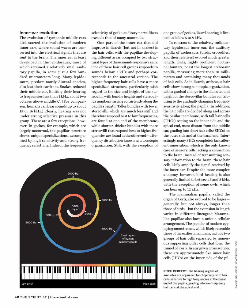
40 THE SCIENTIST | the-scientist.com
BA
SE
D O
N M
ED
-EL
WW
W.M
ED
EL
.CO
M
Inner-ear evolutionThe evolution of tympanic middle earskick-started the evolution of modern inner ears, where sound waves are con-verted into the electrical signals that are sent to the brain. The inner ear is least developed in the lepidosaurs, most of which retained a relatively small audi-tory papilla, in some just a few hun-dred micrometers long. Many lepido-saurs, predominantly diurnal species, also lost their eardrum. Snakes reduced their middle ear, limiting their hearing to frequencies less than 1 kHz, about two octaves above middle C. (For compari-son, humans can hear sounds up to about 15 or 16 kHz.) Clearly, hearing was not under strong selective pressure in this group. There are a few exceptions, how-ever. In geckos, for example, which are largely nocturnal, the papillar structure shows unique specializations, accompa-nied by high sensitivity and strong fre-quency selectivity. Indeed, the frequency
selectivity of gecko auditory nerve fibers exceeds that of many mammals.
One part of the inner ear that did improve in lizards (but not in snakes) is the hair cells, with the papillae develop-ing different areas occupied by two struc-tural types of these sound-responsive cells. One of these hair cell groups responds to sounds below 1 kHz and perhaps cor-responds to the ancestral version. The higher-frequency hair cells have a more specialized structure, particularly with regard to the size and height of the ste-reovilli, with bundle heights and stereovil-lus numbers varying consistently along the papilla’s length. Taller bundles with fewer stereovilli, which are much less stiff and therefore respond best to low frequencies, are found at one end of the membrane, while shorter, thicker bundles with more stereovilli that respond best to higher fre-quencies are found at the other end—a fre-quency distribution known as a tonotopic organization. Still, with the exception of
one group of geckos, lizard hearing is lim-ited to below 5 to 8 kHz.
In contrast to the relatively rudimen-tary lepidosaur inner ear, the auditory papilla of archosaurs (birds, crocodiles, and their relatives) evolved much greater length. Owls, highly proficient noctur-nal hunters, boast the longest archosaur papilla, measuring more than 10 milli-meters and containing many thousands of hair cells. As in lizards, archosaur hair cells show strong tonotopic organization, with a gradual change in the diameter and height of the stereovillar bundles contrib-uting to the gradually changing frequency sensitivity along the papilla. In addition, the hair cells are divided along and across the basilar membrane, with tall hair cells (THCs) resting on the inner side and the apical end, most distant from the middle ear, grading into short hair cells (SHCs) on the outer side and at the basal end. Inter-estingly, many SHCs completely lack affer-ent innervation, which is the only known case of sensory cells lacking a connection to the brain. Instead of transmitting sen-sory information to the brain, these hair cells likely amplify the signal received by the inner ear. Despite the more complex anatomy, however, bird hearing is also generally limited to between 5 and 8 kHz, with the exception of some owls, which can hear up to 12 kHz.
The mammalian papilla, called the organ of Corti, also evolved to be larger—generally, but not always, longer than those of birds—but the extension in length varies in different lineages.4 Mamma-lian papillae also have a unique cellular arrangement. The papillae of modern egg-laying monotremes, which likely resemble those of the earliest mammals, include two groups of hair cells separated by numer-ous supporting pillar cells that form the tunnel of Corti. In any given cross section, there are approximately five inner hair cells (IHCs) on the inner side of the pil-
PITCH PERFECT: The hearing organs of amniotes are organized tonotopically, with hair cells sensitive to high frequencies at the basal end of the papilla, grading into low-frequency hair cells at the apical end.
8000 Hz
4000 Hz
2000 Hz
1000 Hz
500 Hz 250 Hz
Low pitch High pitch
Apical region
Basal region of human
auditory papilla
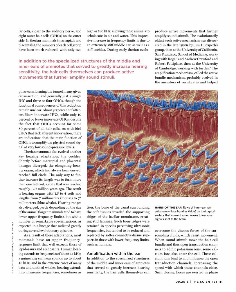
4109.2015 | THE SCIENTIST
© S
TE
VE
GS
CH
ME
ISS
NE
R/S
CIE
NC
E S
OU
RC
E
lar cells, closer to the auditory nerve, andeight outer hair cells (OHCs) on the outer side. In therian mammals (marsupials and placentals), the numbers of each cell group have been much reduced, with only two
pillar cells forming the tunnel in any given cross-section, and generally just a single IHC and three or four OHCs, though the functional consequences of this reduction remain unclear. About 90 percent of affer-ent fibers innervate IHCs, while only 10 percent or fewer innervate OHCs, despite the fact that OHCs account for some 80 percent of all hair cells. As with bird SHCs that lack afferent innervation, there are indications that the main function of OHCs is to amplify the physical sound sig-nal at very low sound-pressure levels.
Therian mammals also evolved another key hearing adaptation: the cochlea. Shortly before marsupial and placental lineages diverged, the elongating hear-ing organ, which had always been curved, reached full circle. The only way to fur-ther increase its length was to form more than one full coil, a state that was reached roughly 120 million years ago. The result is hearing organs with 1.5 to 4 coils and lengths from 7 millimeters (mouse) to 75 millimeters (blue whale). Hearing ranges also diverged, partly depending on the size of the animal (larger mammals tend to have lower upper-frequency limits), but with a number of remarkable specializations, as expected in a lineage that radiated greatly during several evolutionary episodes.
As a result of these adaptations, most mammals have an upper frequency-response limit that well exceeds those of lepidosaurs and archosaurs. Human hear-ing extends to frequencies of about 15 kHz; a guinea pig can hear sounds up to about 45 kHz; and in the extreme cases of many bats and toothed whales, hearing extends into ultrasonic frequencies, sometimes as
high as 180 kHz, allowing these animals to echolocate in air and water. This impres-sive increase in frequency limits is due to an extremely stiff middle ear, as well as a stiff cochlea. During early therian evolu-
tion, the bone of the canal surrounding the soft tissues invaded the supporting ridges of the basilar membrane, creat-ing stiff laminae. Such bony ridges were retained in species perceiving ultrasonic frequencies, but tended to be reduced and replaced by softer connective-tissue sup-ports in those with lower-frequency limits, such as humans.
Amplification within the earIn addition to the specialized structuresof the middle and inner ears of amniotes that served to greatly increase hearing sensitivity, the hair cells themselves can
produce active movements that further amplify sound stimuli. The evolutionarily oldest such active mechanism was discov-ered in the late 1980s by Jim Hudspeth’s group, then at the University of California, San Francisco, School of Medicine, work-ing with frogs,5 and Andrew Crawford andRobert Fettiplace, then at the University of Cambridge, working with turtles.6 Theamplification mechanism, called the active bundle mechanism, probably evolved in the ancestors of vertebrates and helped
overcome the viscous forces of the sur-rounding fluids, which resist movement. When sound stimuli move the hair-cell bundle and thus open transduction chan-nels to admit potassium ions, some cal-cium ions also enter the cell. These cal-cium ions bind to and influence the open transduction channels, increasing the speed with which these channels close. Such closing forces are exerted in phase
In addition to the specialized structures of the middle andinner ears of amniotes that served to greatly increase hearing sensitivity, the hair cells themselves can produce active movements that further amplify sound stimuli.
HAIRS OF THE EAR: Rows of inner-ear hair cells have villous bundles (blue) on their apical surface that convert sound waves to nervous signals sent to the brain.
42 THE SCIENTIST | the-scientist.com
with the incoming sound waves, increas-ing the distance that the hair cells move in response, and thereby increasing their sensitivity. It is likely that this mechanism operates in all vertebrate hair cells.5 In liz-ards, my group provided evidence that this bundle mechanism really does operate in the living animal.7
In 1985, a second mechanism of hair cell–driven sound amplification was dis-covered in mammalian OHCs by Bill Brownell’s group, then at the University of Florida School of Medicine. Brownell and his colleagues showed that mammalian OHCs, but not IHCs, changed their length very rapidly in phase with the signal if exposed to an alternating electrical field.8
Such fields occur when hair cells respond to sound. Subsequent experiments showed that the change in cell length is due to changes in the molecular configu-ration of a protein, later named prestin, which occurs in high density along the lateral cell membrane of OHCs. In mam-mals, the force produced by the OHCs is so strong that the entire organ of Corti, which includes all cell types that surround the hair cells and the basilar membrane itself, is driven in an up-and-down motion. This movement can amplify sounds by at least 40dB, allowing very quiet noises to be detected. There is evidence for the inde-pendent evolution of specific molecular configurations of prestins that allow for the amplification of very high ultrasonic frequencies in bats and whales.9
Bird ears also appear to produce activeforces that amplify sound. The SHCs have bundles comprising up to 300 stereovilli (about three times as many as the bundles of mammalian OHCs),10 and the move-ment of these bundles probably drives the movement of THCs indirectly via the tec-torial membrane. Also, very recent data from the lab of Fettiplace, now at the Uni-versity of Wisconsin–Madison, suggests that in birds, prestin (albeit in a different molecular form) may work in the plane across the hearing organ (i.e., not up and down as in mammals), perhaps reinforc-ing the influence of the bundle active mechanism on the THCs via the tectorial membrane.11
In addition to amplifying hair-cell activity, these active mechanisms manifestas spontaneous movements of the hear-ing organ, oscillating even in the absence of sound stimuli. Such spontaneous move-ments actually produce sound that is emit-ted through the middle ear to the outside world and can be measured in the ear canal. These spontaneous otoacoustic emissions (SOAEs) enable remote sensing of what is going on within the inner ear and have permitted increasingly important research on inner-ear mechanisms and new clinical diagnostic methods to monitor the health
of the ear’s sensory epithelium. We recently showed that spectral patterns of SOAEs in lizards, birds, and mammals are remark-ably similar, despite up to 70-fold differ-ences in the size of the hearing organs, suggesting that there are profound com-monalities among the inner ears of amni-otes that we still do not really understand.12
Remarkable convergenceThree hundred million years of evolutionhave resulted in a fascinating variety of ear configurations that, despite their struc-tural diversity, show remarkably similar physiological responses. There are hardly any differences in sensitivity between the hearing of endothermal birds and mam-mals, and the frequency selectivity of responses is essentially the same in most lizards, birds, and mammals. The com-bined research efforts of paleontologists, anatomists, physiologists, and develop-mental biologists over several decades have clarified the major evolutionary steps in all lineages that modified the mallea-ble middle and inner ears into their pres-ent-day kaleidoscopic variety of form, yet a surprising consensus in their function. g
Geoffrey A. Manley is a retired professorfrom the Institute of Zoology at the Tech-nical University in Munich, Germany. He is currently a guest scientist in the labora-tory of his wife, Christine Köppl, at Olden-burg University in Germany.
References1. G.A. Manley, C. Köppl, “Phylogenetic
development of the cochlea and its innervation,” Curr Opin Neurobiol, 8:468-74, 1998.
2. J.A. Clack, “Patterns and processes in the early evolution of the tetrapod ear,” J Neurobiol, 53:251-64, 2002.
3. T. Kitazawa et al., “Developmental genetic bases behind the independent origin of the tympanic membrane in mammals and diapsids,” Nat Commun, 6:6853, 2015.
4. G.A. Manley, “Evolutionary paths to mammalian cochleae,” JARO, 13:733-43, 2012.
5. A.J. Hudspeth, “How the ear’s works work: Mechanoelectrical transduction and amplification by hair cells,” C R Biol, 328:155-62, 2005.
6. A.C. Crawford, R. Fettiplace, “The mechanical properties of ciliary bundles of turtle cochlear hair cells,” J Physiol, 364:359-79, 1985.
7. G.A. Manley et al., “In vivo evidence for a cochlear amplifier in the hair-cell bundle of lizards,” PNAS, 98:2826-31, 2001.
8. W.E. Brownell et al., “Evoked mechanical responses of isolated cochlear outer hair cells,” Science, 227:194-96, 1985.
9. Y. Liu et al., “Convergent sequence evolution between echolocating bats and dolphins,” Curr Biol, 20:R53-R54, 2010.
10. C. Köppl et al., “Big and powerful: A model of the contribution of bundle motility to mechanical amplification in hair cells of the bird basilar papilla,” in Concepts and Challenges in the Biophysics of Hearing, ed. N.P. Cooper, D.T. Kemp (Singapore: World Scientific, 2009), 444-50.
11. M. Beurg et al., “A prestin motor in chicken auditory hair cells: Active force generation in a nonmammalian species,” Neuron, 79:69-81, 2013.
12. C. Bergevin et al., “Salient features of otoacoustic emissions are common across tetrapod groups and suggest shared properties of generation mechanisms,” PNAS, 112:3362-67, 2015.
Three hundred million years of evolution have resulted in a fascinating variety of ear configurations that, despite their struc tural diversity, show remarkably similar physiological responses.
Images on page 36: Sea Lion: © iStock/LFStewart;Squirrel: © Erik Mandre/Shutterstock; Frog: © Frank B. Yuwono/Shutterstock; Owl: © XNature.Photography/Shutterstock; Lizard: © Andrew Wijesuriya/Shutterstock; Bat: © iStock/GlobalP; Ostrich: © Jamen Percy/Shutterstock; Dog: © Annette Shaff/Shutterstock; Lynx: © Dmitri Gomon/Shutterstock
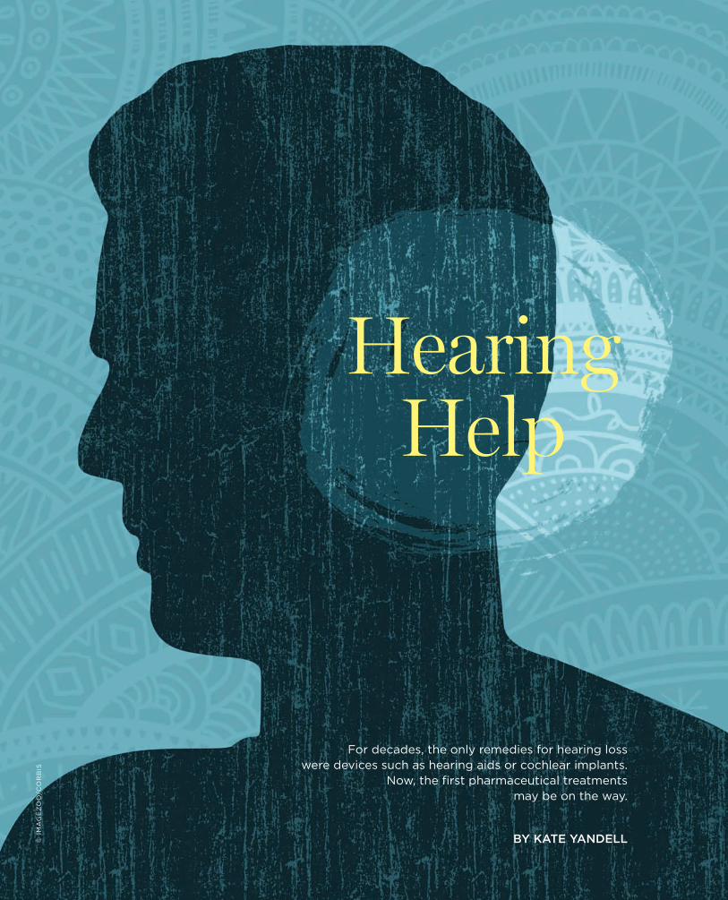
4309.2015 | THE SCIENTIST
Hearing Help
For decades, the only remedies for hearing losswere devices such as hearing aids or cochlear implants.
Now, the first pharmaceutical treatmentsmay be on the way.
BY KATE YANDELL© I
MA
GE
ZO
O/C
OR
BIS
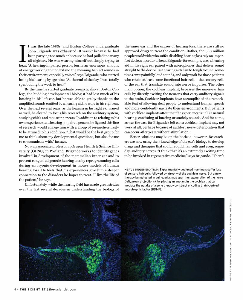
44 THE SCIENTIST | the-scientist.com
IMA
GE
BY
JE
RE
MY
PIN
YO
N A
ND
GA
RY
HO
US
LE
Y U
NS
W A
US
TR
AL
IA.
I t was the late 1980s, and Boston College undergraduateJohn Brigande was exhausted. It wasn’t because he had been partying too much or because he had pulled too many all-nighters. He was wearing himself out simply trying to
hear. “A hearing-impaired person burns an enormous amount of energy working to understand the meaning behind sounds in their environment, especially voices,” says Brigande, who started losing his hearing by age nine. “At the end of the day, I was totally spent doing the work to hear.”
By the time he started graduate research, also at Boston Col-lege, the budding developmental biologist had lost much of his hearing in his left ear, but he was able to get by thanks to the amplified sounds emitted by a hearing aid he wore in his right ear. Over the next several years, as the hearing in his right ear waned as well, he elected to focus his research on the auditory system, studying chick and mouse inner ears. In addition to relating to his own experience as a hearing-impaired person, he figured this line of research would engage him with a group of researchers likely to be attuned to his condition. “That would be the best group for me to think about my developmental questions, but also for me to communicate with,” he says.
Now an associate professor at Oregon Health & Science Uni-versity (OHSU) in Portland, Brigande works to identify genes involved in development of the mammalian inner ear and to prevent congenital genetic hearing loss by reprogramming cells during embryonic development in mouse models of human hearing loss. He feels that his experiences give him a deeper connection to the disorders he hopes to treat. “I live the life of the patient,” he says.
Unfortunately, while the hearing field has made great strides over the last several decades in understanding the biology of
the inner ear and the causes of hearing loss, there are still no approved drugs to treat the condition. Rather, the 360 million people worldwide who suffer disabling hearing loss rely on imper-fect devices in order to hear. Brigande, for example, uses a hearing aid in his right ear paired with microphones that deliver sound straight to the device. But hearing aids can be tough to tune, some-times emit painfully loud sounds, and only work for those patients who retain at least some functional hair cells—the sensory cells of the ear that translate sound into nerve impulses. The other main option, the cochlear implant, bypasses the inner-ear hair cells by directly exciting the neurons that carry auditory signals to the brain. Cochlear implants have accomplished the remark-able feat of allowing deaf people to understand human speech and more confidently navigate their environments. But patients with cochlear implants attest that the experience is unlike natural hearing, consisting of buzzing or staticky sounds. And for some, as was the case for Brigande’s left ear, a cochlear implant may not work at all, perhaps because of auditory nerve deterioration that can occur after years without stimulation.
Better solutions may be on the horizon, however. Research-ers are now using their knowledge of the ear’s biology to develop drugs and therapies that could rebuild hair cells and even, some-day, auditory nerves. “I think that it’s an extremely exciting time to be involved in regenerative medicine,” says Brigande. “There’s
NERVE REGENERATION: Experimentally deafened mammals suffer lossof sensory hair cells followed by atrophy of the cochlear nerve. But a newtherapy being tested in guinea pigs may spur the regeneration of the nerve(left, green projections), by placing an implant in the cochlea that canmediate the uptake of a gene therapy construct encoding brain-derivedneurotrophic factor (BDNF).
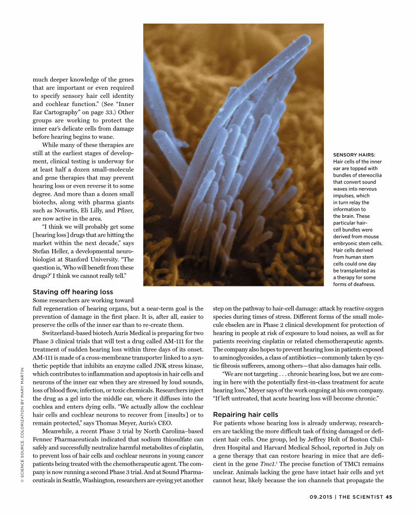
4509.2015 | THE SCIENTIST
© S
CIE
NC
E S
OU
RC
E.
CO
LO
RIZ
AT
ION
BY
MA
RY
MA
RT
IN
much deeper knowledge of the genesthat are important or even required to specify sensory hair cell identity and cochlear function.” (See “Inner Ear Cartography” on page 33.) Other groups are working to protect the inner ear’s delicate cells from damage before hearing begins to wane.
While many of these therapies are still at the earliest stages of develop-ment, clinical testing is underway for at least half a dozen small-molecule and gene therapies that may prevent hearing loss or even reverse it to some degree. And more than a dozen small biotechs, along with pharma giants such as Novartis, Eli Lilly, and Pfizer, are now active in the area.
“I think we will probably get some [hearing loss] drugs that are hitting the market within the next decade,” says Stefan Heller, a developmental neuro-biologist at Stanford University. “The question is, ‘Who will benefit from these drugs?’ I think we cannot really tell.”
Staving off hearing lossSome researchers are working towardfull regeneration of hearing organs, but a near-term goal is the prevention of damage in the first place. It is, after all, easier to preserve the cells of the inner ear than to re-create them.
Switzerland-based biotech Auris Medical is preparing for two Phase 3 clinical trials that will test a drug called AM-111 for the treatment of sudden hearing loss within three days of its onset. AM-111 is made of a cross-membrane transporter linked to a syn-thetic peptide that inhibits an enzyme called JNK stress kinase, which contributes to inflammation and apoptosis in hair cells and neurons of the inner ear when they are stressed by loud sounds, loss of blood flow, infection, or toxic chemicals. Researchers inject the drug as a gel into the middle ear, where it diffuses into the cochlea and enters dying cells. “We actually allow the cochlear hair cells and cochlear neurons to recover from [insults] or to remain protected,” says Thomas Meyer, Auris’s CEO.
Meanwhile, a recent Phase 3 trial by North Carolina–based Fennec Pharmaceuticals indicated that sodium thiosulfate can safely and successfully neutralize harmful metabolites of cisplatin, to prevent loss of hair cells and cochlear neurons in young cancer patients being treated with the chemotherapeutic agent. The com-pany is now running a second Phase 3 trial. And at Sound Pharma-ceuticals in Seattle, Washington, researchers are eyeing yet another
step on the pathway to hair-cell damage: attack by reactive oxygen species during times of stress. Different forms of the small mole-cule ebselen are in Phase 2 clinical development for protection of hearing in people at risk of exposure to loud noises, as well as for patients receiving cisplatin or related chemotherapeutic agents. The company also hopes to prevent hearing loss in patients exposed to aminoglycosides, a class of antibiotics—commonly taken by cys-tic fibrosis sufferers, among others—that also damages hair cells.
“We are not targeting . . . chronic hearing loss, but we are com-ing in here with the potentially first-in-class treatment for acute hearing loss,” Meyer says of the work ongoing at his own company. “If left untreated, that acute hearing loss will become chronic.”
Repairing hair cellsFor patients whose hearing loss is already underway, research-ers are tackling the more difficult task of fixing damaged or defi-cient hair cells. One group, led by Jeffrey Holt of Boston Chil-dren Hospital and Harvard Medical School, reported in July on a gene therapy that can restore hearing in mice that are defi-cient in the gene Tmc1.1 The precise function of TMC1 remainsunclear. Animals lacking the gene have intact hair cells and yet cannot hear, likely because the ion channels that propagate the
SENSORY HAIRS:Hair cells of the innerear are topped withbundles of stereociliathat convert soundwaves into nervousimpulses, whichin turn relay theinformation tothe brain. Theseparticular hair-cell bundles werederived from mouseembryonic stem cells.Hair cells derivedfrom human stemcells could one daybe transplanted asa therapy for someforms of deafness.
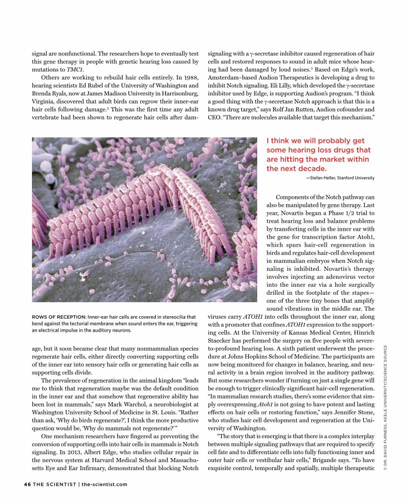
46 THE SCIENTIST | the-scientist.com
© D
R.
DA
VID
FU
RN
ES
S,
KE
EL
E U
NIV
ER
SIT
Y/S
CIE
NC
E S
OU
RC
E
signal are nonfunctional. The researchers hope to eventually testthis gene therapy in people with genetic hearing loss caused by mutations to TMC1.
Others are working to rebuild hair cells entirely. In 1988, hearing scientists Ed Rubel of the University of Washington and Brenda Ryals, now at James Madison University in Harrisonburg, Virginia, discovered that adult birds can regrow their inner-ear hair cells following damage.2 This was the first time any adultvertebrate had been shown to regenerate hair cells after dam-
age, but it soon became clear that many nonmammalian speciesregenerate hair cells, either directly converting supporting cells of the inner ear into sensory hair cells or generating hair cells as supporting cells divide.
The prevalence of regeneration in the animal kingdom “leads me to think that regeneration maybe was the default condition in the inner ear and that somehow that regenerative ability has been lost in mammals,” says Mark Warchol, a neurobiologist at Washington University School of Medicine in St. Louis. “Rather than ask, ‘Why do birds regenerate?’, I think the more productive question would be, ‘Why do mammals not regenerate?’ ”
One mechanism researchers have fingered as preventing the conversion of supporting cells into hair cells in mammals is Notch signaling. In 2013, Albert Edge, who studies cellular repair in the nervous system at Harvard Medical School and Massachu-setts Eye and Ear Infirmary, demonstrated that blocking Notch
signaling with a γ-secretase inhibitor caused regeneration of haircells and restored responses to sound in adult mice whose hear-ing had been damaged by loud noises.3 Based on Edge’s work,Amsterdam–based Audion Therapeutics is developing a drug to inhibit Notch signaling. Eli Lilly, which developed the γ-secretase inhibitor used by Edge, is supporting Audion’s program. “I think a good thing with the γ-secretase Notch approach is that this is a known drug target,” says Rolf Jan Rutten, Audion cofounder and CEO. “There are molecules available that target this mechanism.”
Components of the Notch pathway can also be manipulated by gene therapy. Last year, Novartis began a Phase 1/2 trial to treat hearing loss and balance problems by transfecting cells in the inner ear with the gene for transcription factor Atoh1, which spurs hair-cell regeneration in birds and regulates hair-cell development in mammalian embryos when Notch sig-naling is inhibited. Novartis’s therapy involves injecting an adenovirus vector into the inner ear via a hole surgically drilled in the footplate of the stapes—one of the three tiny bones that amplify sound vibrations in the middle ear. The
viruses carry ATOH1 into cells throughout the inner ear, along with a promoter that confines ATOH1 expression to the support-ing cells. At the University of Kansas Medical Center, Hinrich Staecker has performed the surgery on five people with severe-to-profound hearing loss. A sixth patient underwent the proce-dure at Johns Hopkins School of Medicine. The participants are now being monitored for changes in balance, hearing, and neu-ral activity in a brain region involved in the auditory pathway. But some researchers wonder if turning on just a single gene will be enough to trigger clinically significant hair-cell regeneration. “In mammalian research studies, there’s some evidence that sim-ply overexpressing Atoh1 is not going to have potent and lasting effects on hair cells or restoring function,” says Jennifer Stone, who studies hair cell development and regeneration at the Uni-versity of Washington.
“The story that is emerging is that there is a complex interplay between multiple signaling pathways that are required to specify cell fate and to differentiate cells into fully functioning inner and outer hair cells or vestibular hair cells,” Brigande says. “To have exquisite control, temporally and spatially, multiple therapeutic
I think we will probably get some hearing loss drugs that are hitting the market within the next decade.
—Stefan Heller, Stanford University
ROWS OF RECEPTION: Inner-ear hair cells are covered in stereocilia that bend against the tectorial membrane when sound enters the ear, triggering an electrical impulse in the auditory neurons.
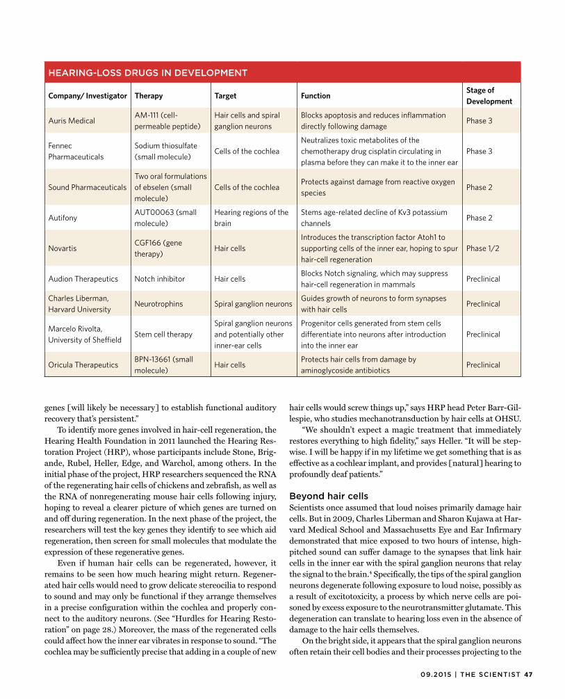
4709.2015 | THE SCIENTIST
genes [will likely be necessary] to establish functional auditoryrecovery that’s persistent.”
To identify more genes involved in hair-cell regeneration, the Hearing Health Foundation in 2011 launched the Hearing Res-toration Project (HRP), whose participants include Stone, Brig-ande, Rubel, Heller, Edge, and Warchol, among others. In the initial phase of the project, HRP researchers sequenced the RNA of the regenerating hair cells of chickens and zebrafish, as well as the RNA of nonregenerating mouse hair cells following injury, hoping to reveal a clearer picture of which genes are turned on and off during regeneration. In the next phase of the project, the researchers will test the key genes they identify to see which aid regeneration, then screen for small molecules that modulate the expression of these regenerative genes.
Even if human hair cells can be regenerated, however, it remains to be seen how much hearing might return. Regener-ated hair cells would need to grow delicate stereocilia to respond to sound and may only be functional if they arrange themselves in a precise configuration within the cochlea and properly con-nect to the auditory neurons. (See “Hurdles for Hearing Resto-ration” on page 28.) Moreover, the mass of the regenerated cells could affect how the inner ear vibrates in response to sound. “The cochlea may be sufficiently precise that adding in a couple of new
hair cells would screw things up,” says HRP head Peter Barr-Gil-lespie, who studies mechanotransduction by hair cells at OHSU.
“We shouldn’t expect a magic treatment that immediately restores everything to high fidelity,” says Heller. “It will be step-wise. I will be happy if in my lifetime we get something that is as effective as a cochlear implant, and provides [natural] hearing to profoundly deaf patients.”
Beyond hair cellsScientists once assumed that loud noises primarily damage haircells. But in 2009, Charles Liberman and Sharon Kujawa at Har-vard Medical School and Massachusetts Eye and Ear Infirmary demonstrated that mice exposed to two hours of intense, high-pitched sound can suffer damage to the synapses that link hair cells in the inner ear with the spiral ganglion neurons that relay the signal to the brain.4 Specifically, the tips of the spiral ganglionneurons degenerate following exposure to loud noise, possibly as a result of excitotoxicity, a process by which nerve cells are poi-soned by excess exposure to the neurotransmitter glutamate. This degeneration can translate to hearing loss even in the absence of damage to the hair cells themselves.
On the bright side, it appears that the spiral ganglion neurons often retain their cell bodies and their processes projecting to the
HEARING-LOSS DRUGS IN DEVELOPMENT
Company/ Investigator Therapy Target FunctionStage of Development
Auris MedicalAM-111 (cell-permeable peptide)
Hair cells and spiral ganglion neurons
Blocks apoptosis and reduces inflammation directly following damage
Phase 3
Fennec Pharmaceuticals
Sodium thiosulfate (small molecule)
Cells of the cochlea Neutralizes toxic metabolites of the chemotherapy drug cisplatin circulating in plasma before they can make it to the inner ear
Phase 3
Sound Pharmaceuticals Two oral formulations of ebselen (small molecule)
Cells of the cochleaProtects against damage from reactive oxygen species
Phase 2
AutifonyAUT00063 (small molecule)
Hearing regions of the brain
Stems age-related decline of Kv3 potassium channels
Phase 2
NovartisCGF166 (gene therapy)
Hair cellsIntroduces the transcription factor Atoh1 to supporting cells of the inner ear, hoping to spur hair-cell regeneration
Phase 1/2
Audion Therapeutics Notch inhibitor Hair cellsBlocks Notch signaling, which may suppress hair-cell regeneration in mammals
Preclinical
Charles Liberman, Harvard University
Neurotrophins Spiral ganglion neuronsGuides growth of neurons to form synapses with hair cells
Preclinical
Marcelo Rivolta, University of Sheffield
Stem cell therapySpiral ganglion neurons and potentially other inner-ear cells
Progenitor cells generated from stem cells differentiate into neurons after introduction into the inner ear
Preclinical
Oricula TherapeuticsBPN-13661 (small molecule)
Hair cellsProtects hair cells from damage by aminoglycoside antibiotics
Preclinical
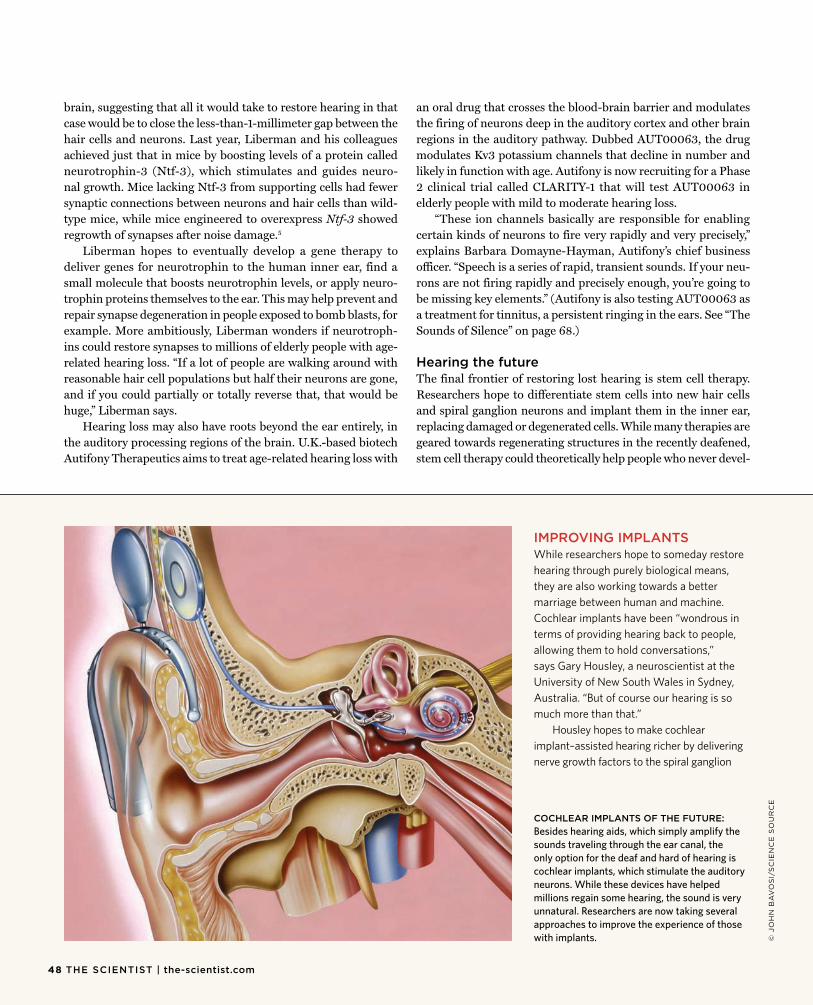
brain, suggesting that all it would take to restore hearing in thatcase would be to close the less-than-1-millimeter gap between the hair cells and neurons. Last year, Liberman and his colleagues achieved just that in mice by boosting levels of a protein called neurotrophin-3 (Ntf-3), which stimulates and guides neuro-nal growth. Mice lacking Ntf-3 from supporting cells had fewer synaptic connections between neurons and hair cells than wild-type mice, while mice engineered to overexpress Ntf-3 showed regrowth of synapses after noise damage.5
Liberman hopes to eventually develop a gene therapy todeliver genes for neurotrophin to the human inner ear, find a small molecule that boosts neurotrophin levels, or apply neuro-trophin proteins themselves to the ear. This may help prevent and repair synapse degeneration in people exposed to bomb blasts, for example. More ambitiously, Liberman wonders if neurotroph-ins could restore synapses to millions of elderly people with age-related hearing loss. “If a lot of people are walking around with reasonable hair cell populations but half their neurons are gone, and if you could partially or totally reverse that, that would be huge,” Liberman says.
Hearing loss may also have roots beyond the ear entirely, in the auditory processing regions of the brain. U.K.-based biotech Autifony Therapeutics aims to treat age-related hearing loss with
an oral drug that crosses the blood-brain barrier and modulates the firing of neurons deep in the auditory cortex and other brain regions in the auditory pathway. Dubbed AUT00063, the drug modulates Kv3 potassium channels that decline in number and likely in function with age. Autifony is now recruiting for a Phase 2 clinical trial called CLARITY-1 that will test AUT00063 in elderly people with mild to moderate hearing loss.
“These ion channels basically are responsible for enabling certain kinds of neurons to fire very rapidly and very precisely,” explains Barbara Domayne-Hayman, Autifony’s chief business officer. “Speech is a series of rapid, transient sounds. If your neu-rons are not firing rapidly and precisely enough, you’re going to be missing key elements.” (Autifony is also testing AUT00063 as a treatment for tinnitus, a persistent ringing in the ears. See “The Sounds of Silence” on page 68.)
Hearing the futureThe final frontier of restoring lost hearing is stem cell therapy.Researchers hope to differentiate stem cells into new hair cells and spiral ganglion neurons and implant them in the inner ear, replacing damaged or degenerated cells. While many therapies are geared towards regenerating structures in the recently deafened, stem cell therapy could theoretically help people who never devel-
COCHLEAR IMPLANTS OF THE FUTURE:Besides hearing aids, which simply amplify thesounds traveling through the ear canal, theonly option for the deaf and hard of hearing iscochlear implants, which stimulate the auditoryneurons. While these devices have helpedmillions regain some hearing, the sound is veryunnatural. Researchers are now taking severalapproaches to improve the experience of thosewith implants.
IMPROVING IMPLANTSWhile researchers hope to someday restorehearing through purely biological means, they are also working towards a better marriage between human and machine. Cochlear implants have been “wondrous in terms of providing hearing back to people, allowing them to hold conversations,” says Gary Housley, a neuroscientist at the University of New South Wales in Sydney, Australia. “But of course our hearing is so much more than that.”
Housley hopes to make cochlear implant–assisted hearing richer by delivering nerve growth factors to the spiral ganglion
© J
OH
N B
AV
OS
I/S
CIE
NC
E S
OU
RC
E
48 THE SCIENTIST | the-scientist.com
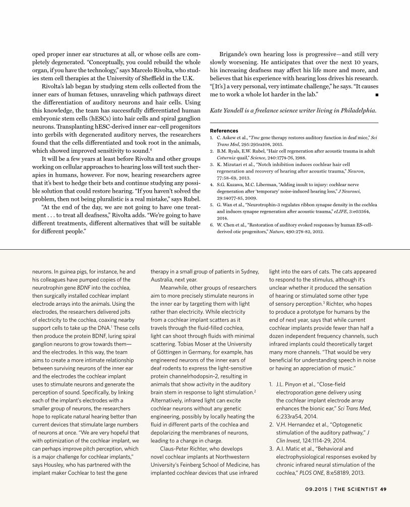
oped proper inner ear structures at all, or whose cells are com-pletely degenerated. “Conceptually, you could rebuild the whole organ, if you have the technology,” says Marcelo Rivolta, who stud-ies stem cell therapies at the University of Sheffield in the U.K.
Rivolta’s lab began by studying stem cells collected from the inner ears of human fetuses, unraveling which pathways direct the differentiation of auditory neurons and hair cells. Using this knowledge, the team has successfully differentiated human embryonic stem cells (hESCs) into hair cells and spiral ganglion neurons. Transplanting hESC-derived inner ear–cell progenitors into gerbils with degenerated auditory nerves, the researchers found that the cells differentiated and took root in the animals, which showed improved sensitivity to sound.6
It will be a few years at least before Rivolta and other groups working on cellular approaches to hearing loss will test such ther-apies in humans, however. For now, hearing researchers agree that it’s best to hedge their bets and continue studying any possi-ble solution that could restore hearing. “If you haven’t solved the problem, then not being pluralistic is a real mistake,” says Rubel.
“At the end of the day, we are not going to have one treat-ment . . . to treat all deafness,” Rivolta adds. “We’re going to have different treatments, different alternatives that will be suitable for different people.”
Brigande’s own hearing loss is progressive—and still veryslowly worsening. He anticipates that over the next 10 years, his increasing deafness may affect his life more and more, and believes that his experience with hearing loss drives his research. “[It’s] a very personal, very intimate challenge,” he says. “It causes me to work a whole lot harder in the lab.” g
Kate Yandell is a freelance science writer living in Philadelphia.
References1. C. Askew et al., “Tmc gene therapy restores auditory function in deaf mice,” Sci
Trans Med, 295:295ra108, 2015.2. B.M. Ryals, E.W. Rubel, “Hair cell regeneration after acoustic trauma in adult
Coturnix quail,” Science, 240:1774-76, 1988.3. K. Mizutari et al., “Notch inhibition induces cochlear hair cell
regeneration and recovery of hearing after acoustic trauma,” Neuron, 77:58-69, 2013.
4. S.G. Kuzawa, M.C. Liberman, “Adding insult to injury: cochlear nerve degeneration after ‘temporary’ noise-induced hearing loss,” J Neurosci, 29:14077-85, 2009.
5. G. Wan et al., “Neurotrophin-3 regulates ribbon synapse density in the cochlea and induces synapse regeneration after acoustic trauma,” eLIFE, 3:e03564, 2014.
6. W. Chen et al., “Restoration of auditory evoked responses by human ES-cell-derived otic progenitors,” Nature, 490:278-82, 2012.
neurons. In guinea pigs, for instance, he andhis colleagues have pumped copies of the neurotrophin gene BDNF into the cochlea,then surgically installed cochlear implant electrode arrays into the animals. Using the electrodes, the researchers delivered jolts of electricity to the cochlea, coaxing nearby support cells to take up the DNA.1 These cellsthen produce the protein BDNF, luring spiral ganglion neurons to grow towards them—and the electrodes. In this way, the team aims to create a more intimate relationship between surviving neurons of the inner ear and the electrodes the cochlear implant uses to stimulate neurons and generate the perception of sound. Specifically, by linking each of the implant’s electrodes with a smaller group of neurons, the researchers hope to replicate natural hearing better than current devices that stimulate large numbers of neurons at once. “We are very hopeful that with optimization of the cochlear implant, we can perhaps improve pitch perception, which is a major challenge for cochlear implants,” says Housley, who has partnered with the implant maker Cochlear to test the gene
therapy in a small group of patients in Sydney, Australia, next year.
Meanwhile, other groups of researchers aim to more precisely stimulate neurons in the inner ear by targeting them with light rather than electricity. While electricity from a cochlear implant scatters as it travels through the fluid-filled cochlea, light can shoot through fluids with minimal scattering. Tobias Moser at the University of Göttingen in Germany, for example, has engineered neurons of the inner ears of deaf rodents to express the light-sensitive protein channelrhodopsin-2, resulting in animals that show activity in the auditory brain stem in response to light stimulation.2
Alternatively, infrared light can excite cochlear neurons without any genetic engineering, possibly by locally heating the fluid in different parts of the cochlea and depolarizing the membranes of neurons, leading to a change in charge.
Claus-Peter Richter, who develops novel cochlear implants at Northwestern University’s Feinberg School of Medicine, has implanted cochlear devices that use infrared
light into the ears of cats. The cats appeared to respond to the stimulus, although it’s unclear whether it produced the sensation of hearing or stimulated some other type of sensory perception.3 Richter, who hopesto produce a prototype for humans by the end of next year, says that while current cochlear implants provide fewer than half a dozen independent frequency channels, such infrared implants could theoretically target many more channels. “That would be very beneficial for understanding speech in noise or having an appreciation of music.”
1. J.L. Pinyon et al., “Close-field electroporation gene delivery using the cochlear implant electrode array enhances the bionic ear,” Sci Trans Med, 6:233ra54, 2014.
2. V.H. Hernandez et al., “Optogenetic stimulation of the auditory pathway,” J Clin Invest, 124:1114-29, 2014.
3. A.I. Matic et al., “Behavioral and electrophysiological responses evoked by chronic infrared neural stimulation of the cochlea,” PLOS ONE, 8:e58189, 2013.
09.2015 | THE SCIENTIST 49

From tossing out cross-contaminated cell lines to flagging genomic misnomers,a push is on to tidy up biomedical research.
BY KERRY GRENS
The Great BigClean-Up
© D
AN
PA
GE
CO
LL
EC
TIO
N/T
HE
ISP
OT
5109.2015 | THE SCIENTIST
S everal years ago, a manuscript characterizing a cell line called RGC-5, which was derived from rat retina, came across the desk
of Thomas Yorio, then an associate edi-tor at Investigative Ophthalmology and Visual Science (IOVS). The line was com-monly used in vision research; Yorio had used it in his own work at the University of North Texas Health Science Center, and researchers across the field had by then published more than 200 studies involv-ing the cells. But the authors of the new paper had found that RGC-5 cells were not retinal ganglion cells after all. RGC-5 cells hadn’t even come from a rat.1 Sud-denly, all of those published studies were called into question.
“They were the first to bring it to my attention,” says Yorio, now editor in chief of IOVS. “That got me to say . . . ‘If this is true, we have to identify [this cell line] and how the heck it’s mouse and not rat.’ ”
A University of North Texas lab first derived the RGC-5 cell line in 2001. By the time Yorio read the manuscript ques-tioning the line, the principal investiga-tor of the originating lab had since left for a position at another institution, so Yorio took it upon himself to investigate. He assembled a forensic team to gather notebooks and frozen, early-passage sam-ples of RGC-5 cells and sent the cells to various independent groups for analysis. The results confirmed that RGC-5 was not what everyone thought it was. Rather, it appeared to be a mouse cone photore-ceptor line.2 In addition to the fact thatthe cells came from a different species, the functional differences between the
cell types are vast: photoreceptors detect light, while ganglion cells transmit this information to the brain via their axons, which make up the optic nerve.
“In our little community, the RGC-5 story was extremely embarrassing to everyone,” says John Nickerson, an Emory University retina researcher and an edi-tor in chief of Molecular Vision, a jour-nal that had published 18 papers using
RGC-5 as an in vitro model. Because so many researchers had used the cell line in their own work, “everyone got burned a lit-tle bit,” he adds.
To deal with the problem, the vision field did something unorthodox. The edi-tors of IOVS, Molecular Vision, and another leading vision journal, Experimental Eye Research, joined forces to blackball RGC-5. In August 2013, the three journals issued new editorial policies that essentially banned publications that used the cell line.
“We declared from that point forward that papers stating they were using RGC-5 would not be accepted,” says Emory Uni-versity vision researcher Jeffrey Boatright, another editor in chief at Molecular Vision. His journal and IOVS also went further,
requiring authors to validate the authen-ticity of all cell lines used in their work, rather than rely on information from sup-pliers or on prior characterizations.
Although cell line misidentification has plagued science for more than half a century (see “Seeded by Weeds,” The Sci-entist, May 2015), the journals’ stance was progressive, reflecting a growing appreci-ation for the magnitude of the problem and its contribution to irreproducible research. In a recent analysis of preclini-cal science, Leonard Freedman, president of the Global Biological Standards Insti-tute (GBSI), and a pair of economists esti-mated that about half of all studies cannot be replicated because of flaws in design, procedure, analysis, or materials—to the tune of about $28 billion.3 And more than a third of those studies could be attributed to “contaminated, mishandled, or misla-beled biological reagents like antibodies or cell lines,” they wrote in their report.
The vision field isn’t the only scien-tific discipline making efforts to clean up its mess. From authenticating cell lines to identifying mislabeled genomic sequences and developing purer reagents, researchers across the life sciences are engaging in the fight against irreproduc-ible research caused by contaminants. “There are more people involved trying to improve the situation, at all levels: sci-entists at the bench who are more aware, funders, people trying to do advocacy or build data sets to work out how signifi-cant the problem is,” says Amanda Capes-Davis, chair of the International Cell Line Authentication Committee (ICLAC). “I think things are improving.”
If we’re not using what we think were using, we’re not testing our hypotheses. We’re just gumming up the literature. I’m not sure what we’re doing, but that’s not science.
—Jeffrey Boatright, Emory Uni versity
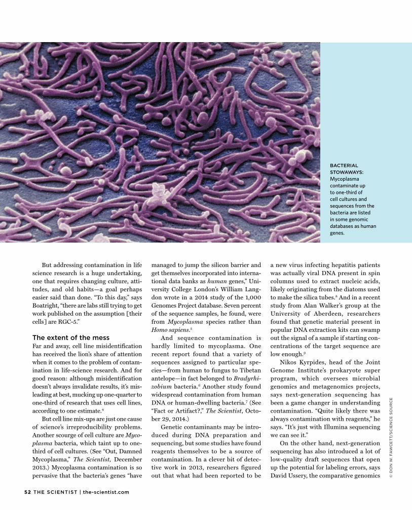
52 THE SCIENTIST | the-scientist.com
© D
ON
W.
FA
WC
ET
T/S
CIE
NC
E S
OU
RC
E
But addressing contamination in lifescience research is a huge undertaking, one that requires changing culture, atti-tudes, and old habits—a goal perhaps easier said than done. “To this day,” says Boatright, “there are labs still trying to get work published on the assumption [their cells] are RGC-5.”
The extent of the messFar and away, cell line misidentificationhas received the lion’s share of attention when it comes to the problem of contam-ination in life-science research. And for good reason: although misidentification doesn’t always invalidate results, it’s mis-leading at best, mucking up one-quarter to one-third of research that uses cell lines, according to one estimate.4
But cell line mix-ups are just one cause of science’s irreproducibility problems. Another scourge of cell culture are Myco-plasma bacteria, which taint up to one-third of cell cultures. (See “Out, Damned Mycoplasma,” The Scientist, December 2013.) Mycoplasma contamination is so pervasive that the bacteria’s genes “have
managed to jump the silicon barrier and get themselves incorporated into interna-tional data banks as human genes,” Uni-versity College London’s William Lang-don wrote in a 2014 study of the 1,000 Genomes Project database. Seven percent of the sequence samples, he found, were from Mycoplasma species rather than Homo sapiens.5
And sequence contamination ishardly limited to mycoplasma. One recent report found that a variety of sequences assigned to particular spe-cies—from human to fungus to Tibetan antelope—in fact belonged to Bradyrhi-zobium bacteria.6 Another study foundwidespread contamination from human DNA or human-dwelling bacteria.7 (See“Fact or Artifact?,” The Scientist, Octo-ber 29, 2014.)
Genetic contaminants may be intro-duced during DNA preparation and sequencing, but some studies have found reagents themselves to be a source of contamination. In a clever bit of detec-tive work in 2013, researchers figured out that what had been reported to be
a new virus infecting hepatitis patients was actually viral DNA present in spin columns used to extract nucleic acids, likely originating from the diatoms used to make the silica tubes.8 And in a recentstudy from Alan Walker’s group at the University of Aberdeen, researchers found that genetic material present in popular DNA extraction kits can swamp out the signal of a sample if starting con-centrations of the target sequence are low enough.9
Nikos Kyrpides, head of the Joint Genome Institute’s prokaryote super program, which oversees microbial genomics and metagenomics projects, says next-generation sequencing has been a game changer in understanding contamination. “Quite likely there was always contamination with reagents,” he says. “It’s just with Illumina sequencing we can see it.”
On the other hand, next-generation sequencing has also introduced a lot of low-quality draft sequences that open up the potential for labeling errors, says David Ussery, the comparative genomics
BACTERIALSTOWAWAYS:Mycoplasmacontaminate upto one-third ofcell cultures andsequences from thebacteria are listedin some genomicdatabases as humangenes.
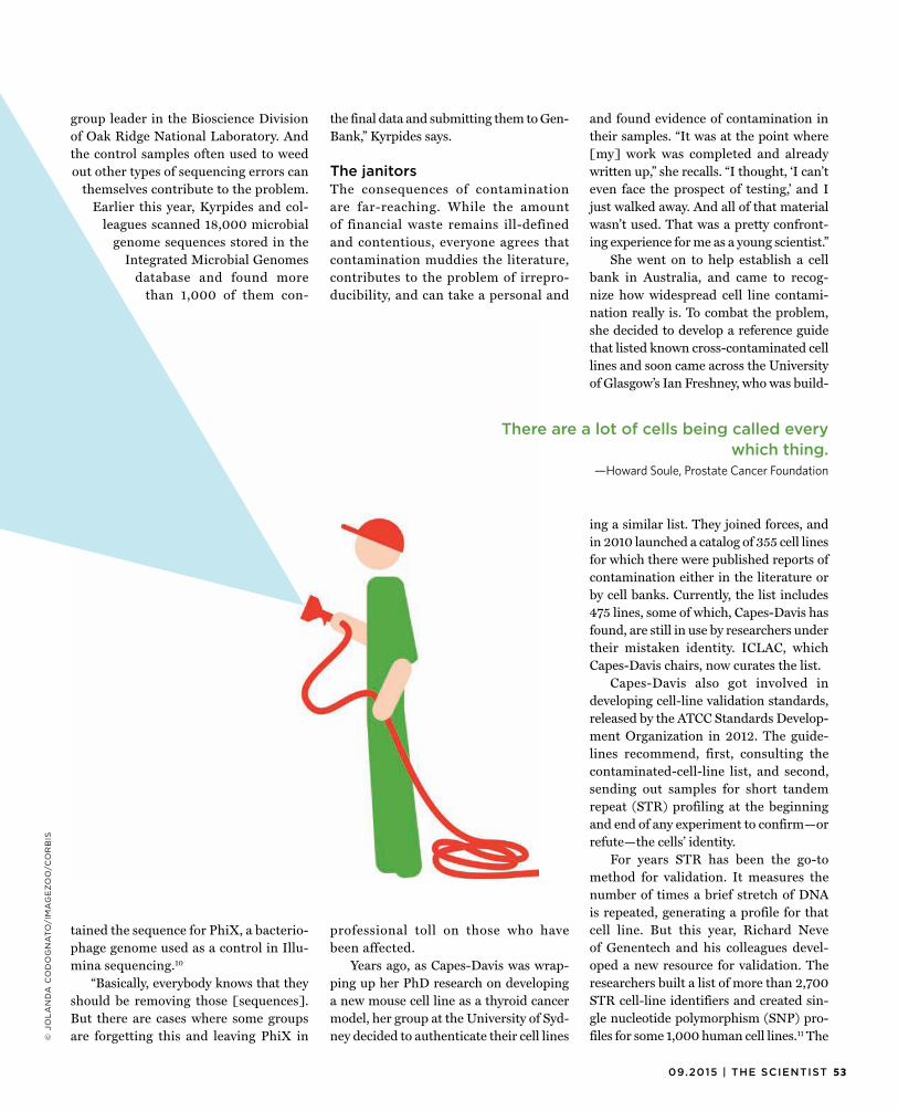
5309.2015 | THE SCIENTIST
group leader in the Bioscience Divisionof Oak Ridge National Laboratory. And the control samples often used to weed out other types of sequencing errors can
themselves contribute to the problem. Earlier this year, Kyrpides and col-
leagues scanned 18,000 microbial genome sequences stored in the
Integrated Microbial Genomes database and found more
than 1,000 of them con-
tained the sequence for PhiX, a bacterio-phage genome used as a control in Illu-mina sequencing.10
“Basically, everybody knows that they should be removing those [sequences]. But there are cases where some groups are forgetting this and leaving PhiX in
the final data and submitting them to Gen-Bank,” Kyrpides says.
The janitorsThe consequences of contaminationare far-reaching. While the amount of financial waste remains ill-defined and contentious, everyone agrees that contamination muddies the literature, contributes to the problem of irrepro-ducibility, and can take a personal and
professional toll on those who have been affected.
Years ago, as Capes-Davis was wrap-ping up her PhD research on developing a new mouse cell line as a thyroid cancer model, her group at the University of Syd-ney decided to authenticate their cell lines
and found evidence of contamination in their samples. “It was at the point where [my] work was completed and already written up,” she recalls. “I thought, ‘I can’t even face the prospect of testing,’ and I just walked away. And all of that material wasn’t used. That was a pretty confront-ing experience for me as a young scientist.”
She went on to help establish a cell bank in Australia, and came to recog-nize how widespread cell line contami-nation really is. To combat the problem, she decided to develop a reference guide that listed known cross-contaminated cell lines and soon came across the University of Glasgow’s Ian Freshney, who was build-
ing a similar list. They joined forces, and in 2010 launched a catalog of 355 cell lines for which there were published reports of contamination either in the literature or by cell banks. Currently, the list includes 475 lines, some of which, Capes-Davis has found, are still in use by researchers under their mistaken identity. ICLAC, which Capes-Davis chairs, now curates the list.
Capes-Davis also got involved in developing cell-line validation standards, released by the ATCC Standards Develop-ment Organization in 2012. The guide-lines recommend, first, consulting the contaminated-cell-line list, and second, sending out samples for short tandem repeat (STR) profiling at the beginning and end of any experiment to confirm—or refute—the cells’ identity.
For years STR has been the go-to method for validation. It measures the number of times a brief stretch of DNA is repeated, generating a profile for that cell line. But this year, Richard Neve of Genentech and his colleagues devel-oped a new resource for validation. The researchers built a list of more than 2,700 STR cell-line identifiers and created sin-gle nucleotide polymorphism (SNP) pro-files for some 1,000 human cell lines.11 The
There are a lot of cells being called every which thing.
—Howard Soule, Prostate Cancer Foundation
© J
OL
AN
DA
CO
DO
GN
AT
O/I
MA
GE
ZO
O/C
OR
BIS
54 THE SCIENTIST | the-scientist.com
National Center for Biotechnology Infor-mation (NCBI) will maintain the catalog as it develops. “There are a massive num-ber of cell lines in academia that have never been deposited in cell banks,” Neve says. “The idea of this paper is to lay down that foundation of good metrics and qual-ity controls.” Capes-Davis agrees: “It’s a great step forward.”
Some journals are also strengthen-ing their resolve to combat cell-line con-taminants at the point of manuscript submission, requiring authors to docu-ment the source and validation of their line. At Nature and its sister journals, such an initiative has been in place since 2013, but most authors have not com-plied. In a recent editorial published in the same issue as Neve’s publication, the editors note: “Out of a sample of around 60 cell-line-based papers published across several Nature journals in the past two years, almost one-quarter did not report the source. Only 10 percent of authors said that they had authenticated the cell line. This is especially problematic given that almost one-third said that they had obtained the cell lines as a gift from another laboratory.” The journals’ editors now plan to be more proactive in asking for authentication data.
At least 30 other journals, includ-ing PLOS ONE, the Journal of Molecular Biology, and the Journal of the National Cancer Institute, have instituted simi-lar polices requiring cell-line verification, according to a list compiled by Capes-Davis, but there’s a long way to go, says Neve. “It’s really a drop in the ocean, and it’s really quite worrying.”
Funding organizations and academic institutions have been slower to put such regulations in place, but there are signs of change. The National Institutes of Health (NIH) is planning to add a question to grant proposal requests about how appli-cants plan to validate materials. “The first reason to do this is really to make both the applicants and the reviewers confront these problems,” says Jon Lorsch, direc-tor of the National Institute of General Medical Sciences. Lorsch hopes the ques-tion will be enough to get people to com-
ply, but if necessary, validation could be a condition of reward. Still, there’s no for-mal method for enforcing such a policy. “There is that balance between adminis-trative burden and efficiency,” Lorsch says.
That answer isn’t good enough for some concerned about the growing irre-producibility problem in the life sciences. “It lacks a stick,” Howard Soule, the execu-tive vice president and chief science officer of the Prostate Cancer Foundation, says of the NIH’s position. “We need enforceable standards.” As an example, the Prostate Cancer Foundation last year implemented a policy that requires grantees to provide
authentication data to receive a second funding check. Soule says that so far, the plan has worked. Not only have funding recipients complied, but the policy has helped root out contaminants, prevent-ing scientists from wasting resources on a line they weren’t intending to study. “If NIH would do what we do, it would prob-ably [save] hundreds of millions of dollars, maybe billions,” says Soule. “There are a lot of cells being called every which thing.”
For their part, laboratory-supply com-panies are responding to contamination with new assays to detect contaminants and with ultrapure, DNA-free reagents. Mike Brewer, director of pharmaceutical analytics at Thermo Fisher Scientific, says that his firm is developing a quantitative PCR product that could detect a wide vari-ety of viruses in a sample.
And in terms of identifying mislabeled sequences in genome databases, database managers and users are leading the charge, developing automated sequence-verifica-tion methods to efficiently sort through the massive number of genome sequences they host. A German group led by Alexis Stamatakis, a bioinformatician at the Hei-delberg Institute for Theoretical Studies, for instance, is developing an automated
method to scan a large number of genetic sequences in a given database, identify sequences that seem unlikely to belong to the labeled organisms based on taxonomic information, and flag those possible rogue sequences for manual validation. Mean-while, Kyrpides’s team at JGI has devel-oped a program called ProDeGe (for Pro-tocol for fully automated Decontamination of Genomes) that automatically removes any sequence foreign to the target organ-ism, with 84 percent accuracy.12
“I have a vision here that over the next few years we have a variety of computa-tional approaches . . . to create curated
subsets [of possible contamination] across all of GenBank,” David Lipman, the direc-tor of NCBI, told The Scientist in January. (See “Mistaken Identities,” The Scientist, January 1, 2015.)
To address the problem of low-quality draft sequences and the labeling errors they can cause, Oak Ridge’s Ussery has teamed up with Kyrpides as part of a working group called the Genomic Standards Consortium. One of their projects, in collaboration with the NCBI and the European and Japanese genetic databases, is to develop standard-ized quality scores for sequences, which could stop mislabeled or poorly assembled sequences from muddying subsequent studies. “Just put quality scores in there, and the user can decide, ‘I want everything above this threshold,’” says Ussery.
Lacking consistencyMuch of the contamination problemmay stem from the “wild west” nature of basic-science laboratories, says the GBSI’s Freedman. “There are no real, formal—or even informal—best practices in place,” he says. “Not just [for] how to handle cells, but from statistical analysis to the best way to validate an antibody. All of these things have been generally ignored.”
There are no real, formal—or even informal—best practices in place. Not just for how to handle cells, but from statistical
analysis to the best way to validate an antibody.—Leonard Freedman, Global Biological Standards Insti tute
5509.2015 | THE SCIENTIST
© I
ST
OC
K.C
OM
/LO
LO
N
GBSI is one of a number of groupsworking to establish and disseminate best practices. Freedman says his team’s focus is on authenticating cell lines because it’s a tractable problem with straightforward solutions. The researchers launched a big #authenticate campaign via social media this year, for example, to raise awareness about the issues and remind investigators to check their cells. GBSI is also working to establish best practices for other pro-tocols—from designing experiments to ensuring the quality of reagents—by devel-oping free, online educational modules for researchers. “I think part of the problem is there’s no formal training when it comes to best practices overall,” Freedman says. GBSI has also applied for grant funding from the NIH to design online training courses for grad students and postdocs.
Sharing experiences with protocols and reagents could also help root out contaminants and establish best prac-tices. Although not specifically designed
to address contamination, protocols.io is a new website that allows users to post their protocols or their tweaks on pub-lished techniques. The Protocol Exchange, hosted by Nature Protocols, has a similar mission. Users of the exchange can share methods and comment on one anoth-er’s techniques in an open-access format. Lenny Teytelman, the cofounder of pro-tocols.io, says the site can’t prevent prob-lems, but it can help expose them. Often if people uncover issues with a technique or a reagent, he says, “there’s no place to com-municate them. They end up in our heads, not on paper.”
Those aiming to clean up science’s con-tamination problem are hopeful that edi-torial policies, social media campaigns, and media coverage of irreproducible research will inspire labs to scrutinize their work for contamination. But even after a contaminant has been exposed, labs don’t always clean up the mess they’ve left behind in the literature. Take RGC-5,
for instance. A PubMed search revealsjust one retraction related to RGC-5—the paper describing the original character-ization—and a PubMed search for RGC-5 and “correction” or “erratum” yields no results. Hundreds of studies that used RGC-5 continue to stand as written in the literature, including dozens that have been published since 2013, after the three lead-ing vision journals banned RGC-5–based studies from their pages.
“If we’re not using what we think were using, we’re not testing our hypotheses. We’re just gumming up the literature,” says Molecular Vision’s Boatright. “I’m not sure what we’re doing, but that’s not science.” g
References1. N.J. Van Bergen et al., “Recharacterization of the
RGC-5 retinal ganglion cell line,” IOVS, 50:4267-72, 2009.
2. R.R. Krishnamoorthy et al., “A forensic path to RGC-5 cell line authentication: Lessons learned,” IOVS, 54:5712-19, 2013.
3. L.P. Freedman et al., “The economics of reproducibility in preclinical research,” PLOS Biology, doi:10.1371/journal.pbio.1002165, 2015.
4. P. Hughes, “The costs of using unauthenticated, over-passaged cell lines: how much more data do we need?” BioTechniques, 43:575, 577-78, 581-82, passim, 2007.
5. W.B. Langdon, “Mycoplasma contamination in the 1000 Genomes Project,” BioData Mining, 7:3, 2014.
6. M. Laurence et al., “Common contaminants in next-generation sequencing that hinder discovery of low-abundance microbes,” PLOS ONE, doi:10.1371/journal.pone.0097876, 2014.
7. R.W. Lusk, “Diverse and widespread contamination evident in the unmapped depths of high throughput sequencing data,” PLOS ONE, doi:10.1371/journal.pone.0110808, 2014.
8. S.N. Naccache et al., “The perils of pathogen discovery: Origin of a novel parvovirus-like hybrid genome traced to nucleic acid extraction spin columns,” J Virol, doi:10.1128/JVI.02323-13, 2013.
9. S.J. Salter et al., “Reagent and laboratory contamination can critically impact sequence-based microbiome analyses,” BMC Biology, 12:87, 2014.
10. S. Mukherjee et al., “Large-scale contamination of microbial isolate genomes by Illumina PhiX control,” Standards in Genomic Sciences, 10:18, 2015.
11. M. Yu et al., “A resource for cell line authentication, annotation and quality control,” Nature, 520:307-11, 2015.
12. K. Tennessen et al., “ProDeGe: A computational protocol for fully automated decontamination of genomes,” The ISME Journal, doi:10.1038/ismej.2015.100, 2015.
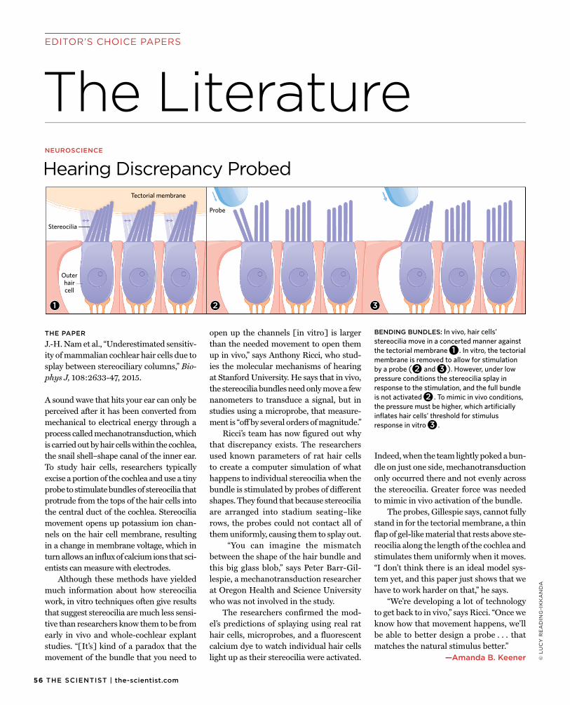
56 THE SCIENTIST | the-scientist.com
The LiteratureEDITOR’S CHOICE PAPERS
© L
UC
Y R
EA
DIN
G-I
KK
AN
DA
THE PAPER
J.-H. Nam et al., “Underestimated sensitiv-ity of mammalian cochlear hair cells due to splay between stereociliary columns,” Bio-phys J, 108:2633-47, 2015.
A sound wave that hits your ear can only be perceived after it has been converted from mechanical to electrical energy through a process called mechanotransduction, which is carried out by hair cells within the cochlea, the snail shell–shape canal of the inner ear. To study hair cells, researchers typically excise a portion of the cochlea and use a tiny probe to stimulate bundles of stereocilia that protrude from the tops of the hair cells into the central duct of the cochlea. Stereocilia movement opens up potassium ion chan-nels on the hair cell membrane, resulting in a change in membrane voltage, which in turn allows an influx of calcium ions that sci-entists can measure with electrodes.
Although these methods have yielded much information about how stereocilia work, in vitro techniques often give results that suggest stereocilia are much less sensi-tive than researchers know them to be from early in vivo and whole-cochlear explant studies. “[It’s] kind of a paradox that the movement of the bundle that you need to
open up the channels [in vitro] is larger than the needed movement to open them up in vivo,” says Anthony Ricci, who stud-ies the molecular mechanisms of hearing at Stanford University. He says that in vivo, the stereocilia bundles need only move a few nanometers to transduce a signal, but in studies using a microprobe, that measure-ment is “off by several orders of magnitude.”
Ricci’s team has now figured out why that discrepancy exists. The researchers used known parameters of rat hair cells to create a computer simulation of what happens to individual stereocilia when the bundle is stimulated by probes of different shapes. They found that because stereocilia are arranged into stadium seating–like rows, the probes could not contact all of them uniformly, causing them to splay out.
“You can imagine the mismatch between the shape of the hair bundle and this big glass blob,” says Peter Barr-Gil-lespie, a mechanotransduction researcher at Oregon Health and Science University who was not involved in the study.
The researchers confirmed the mod-el’s predictions of splaying using real rat hair cells, microprobes, and a fluorescent calcium dye to watch individual hair cells light up as their stereocilia were activated.
Indeed, when the team lightly poked a bun-dle on just one side, mechanotransduction only occurred there and not evenly across the stereocilia. Greater force was needed to mimic in vivo activation of the bundle.
The probes, Gillespie says, cannot fully stand in for the tectorial membrane, a thin flap of gel-like material that rests above ste-reocilia along the length of the cochlea and stimulates them uniformly when it moves. “I don’t think there is an ideal model sys-tem yet, and this paper just shows that we have to work harder on that,” he says.
“We’re developing a lot of technology to get back to in vivo,” says Ricci. “Once we know how that movement happens, we’ll be able to better design a probe . . . that matches the natural stimulus better.”
—Amanda B. Keener
NEUROSCIENCE
Hearing Discrepancy Probed
BENDING BUNDLES: In vivo, hair cells’ stereocilia move in a concerted manner against the tectorial membrane 1 . In vitro, the tectorial membrane is removed to allow for stimulation by a probe (2 and 3 ). However, under low pressure conditions the stereocilia splay in response to the stimulation, and the full bundle is not activated 2 . To mimic in vivo conditions, the pressure must be higher, which artificially inflates hair cells’ threshold for stimulus response in vitro 3 .
Stereocilia
Outer hair cell
Probe
Tectorial membrane
2 3 1
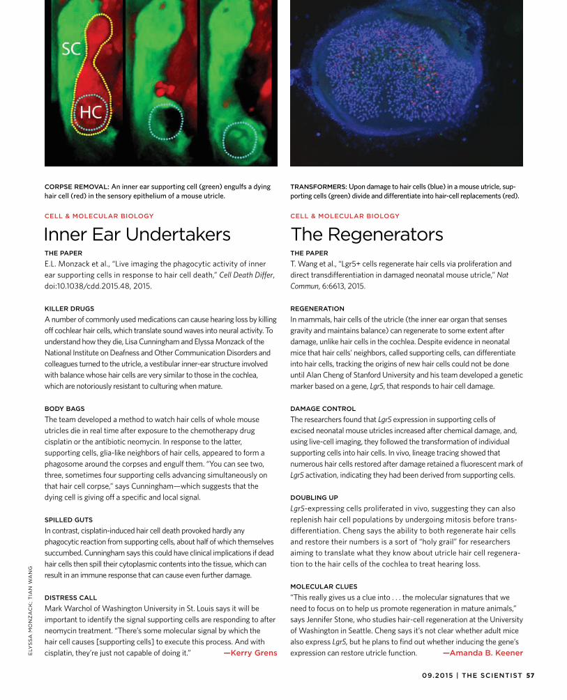
5709.2015 | THE SCIENTIST
CORPSE REMOVAL: An inner ear supporting cell (green) engulfs a dying hair cell (red) in the sensory epithelium of a mouse utricle.
TRANSFORMERS: Upon damage to hair cells (blue) in a mouse utricle, sup-porting cells (green) divide and differentiate into hair-cell replacements (red).
ELY
SS
A M
ON
ZA
CK
; T
IAN
WA
NG
CELL & MOLECULAR BIOLOGY
The RegeneratorsTHE PAPER
T. Wang et al., “Lgr5+ cells regenerate hair cells via proliferation anddirect transdifferentiation in damaged neonatal mouse utricle,” Nat Commun, 6:6613, 2015.
REGENERATION
In mammals, hair cells of the utricle (the inner ear organ that sensesgravity and maintains balance) can regenerate to some extent after damage, unlike hair cells in the cochlea. Despite evidence in neonatal mice that hair cells’ neighbors, called supporting cells, can differentiate into hair cells, tracking the origins of new hair cells could not be done until Alan Cheng of Stanford University and his team developed a genetic marker based on a gene, Lgr5, that responds to hair cell damage.
DAMAGE CONTROL
The researchers found that Lgr5 expression in supporting cells ofexcised neonatal mouse utricles increased after chemical damage, and, using live-cell imaging, they followed the transformation of individual supporting cells into hair cells. In vivo, lineage tracing showed that numerous hair cells restored after damage retained a fluorescent mark of Lgr5 activation, indicating they had been derived from supporting cells.
DOUBLING UP
Lgr5-expressing cells proliferated in vivo, suggesting they can alsoreplenish hair cell populations by undergoing mitosis before trans-differentiation. Cheng says the ability to both regenerate hair cells and restore their numbers is a sort of “holy grail” for researchers aiming to translate what they know about utricle hair cell regenera-tion to the hair cells of the cochlea to treat hearing loss.
MOLECULAR CLUES
“This really gives us a clue into . . . the molecular signatures that weneed to focus on to help us promote regeneration in mature animals,” says Jennifer Stone, who studies hair-cell regeneration at the University of Washington in Seattle. Cheng says it’s not clear whether adult mice also express Lgr5, but he plans to find out whether inducing the gene’s expression can restore utricle function. —Amanda B. Keener
CELL & MOLECULAR BIOLOGY
Inner Ear UndertakersTHE PAPER
E.L. Monzack et al., “Live imaging the phagocytic activity of innerear supporting cells in response to hair cell death,” Cell Death Differ, doi:10.1038/cdd.2015.48, 2015.
KILLER DRUGS
A number of commonly used medications can cause hearing loss by killingoff cochlear hair cells, which translate sound waves into neural activity. To understand how they die, Lisa Cunningham and Elyssa Monzack of the National Institute on Deafness and Other Communication Disorders and colleagues turned to the utricle, a vestibular inner-ear structure involved with balance whose hair cells are very similar to those in the cochlea, which are notoriously resistant to culturing when mature.
BODY BAGS
The team developed a method to watch hair cells of whole mouseutricles die in real time after exposure to the chemotherapy drug cisplatin or the antibiotic neomycin. In response to the latter, supporting cells, glia-like neighbors of hair cells, appeared to form a phagosome around the corpses and engulf them. “You can see two, three, sometimes four supporting cells advancing simultaneously on that hair cell corpse,” says Cunningham—which suggests that the dying cell is giving off a specific and local signal.
SPILLED GUTS
In contrast, cisplatin-induced hair cell death provoked hardly anyphagocytic reaction from supporting cells, about half of which themselves succumbed. Cunningham says this could have clinical implications if dead hair cells then spill their cytoplasmic contents into the tissue, which can result in an immune response that can cause even further damage.
DISTRESS CALL
Mark Warchol of Washington University in St. Louis says it will beimportant to identify the signal supporting cells are responding to after neomycin treatment. “There’s some molecular signal by which the hair cell causes [supporting cells] to execute this process. And with cisplatin, they’re just not capable of doing it.” —Kerry Grens
58 THE SCIENTIST | the-scientist.com
PROFILE
ZA
CH
VE
ILL
EU
X,
DIR
EC
TO
R O
F P
UB
LIC
AT
ION
S &
ED
ITO
R-I
N-C
HIE
F, C
OM
MU
NIC
AT
ION
S A
ND
PU
BL
IC A
FF
AIR
S,
TH
E R
OC
KE
FE
LL
ER
UN
IVE
RS
ITY
A teaching obligation in graduate school introduced James Hudspeth to a career focused on how vertebrates sense sounds.
BY ANNA AZVOLINSKY
The Ears Have It
When he was 11 and 12, James Hudspeth spent his sum-mers working in a Texas law firm, a job arranged by his lawyer father. But Hudspeth already knew he would
never become an attorney. “I’ve been a scientist as long as I can remember,” he says. He pleaded with his parents to find him a job with a science bent and spent the following three summers working for his first scientific mentor—Peter Kellaway, a neurophysiologist at Baylor College of Medicine. “He was a stunning role model for a kid. I was very impressed by his seriousness and his sense of purpose.” Hud-speth worked first as a histology technician and then in an electron-ics shop, soldering EEG electrodes for use in medical school courses.
The jobs also gave Hudspeth a sneak peek at medicine in action. During his lunch break, he would surreptitiously sneak up into the observation dome above the hospital’s surgical the-ater and watch open-heart surgery while he ate his lunch. “It was so grand. This was the hospital where open-heart surgery tech-niques were pioneered,” says Hudspeth. “There were four surgi-cal theaters, and surgeon Michael DeBakey would go clockwise
from room to room doing the critical parts of surgeries. I would tag along with my sandwich. Fourteen-year-olds were not sup-posed to be there, and especially not eating a tuna sandwich!”
This kind of zealous interest and curiosity is what has kept Hudspeth in the laboratory for more than 40 years. Despite an inauspicious beginning that included a string of difficult situ-ations—expulsion from high school, the Vietnam War, and an aborted postdoc—Hudspeth has since devoted his career to understanding how hair cells in the inner ear translate sound into electrical signals transmitted to the brain. His laboratory was the first to show directly that mechanical stimulation of the hair cells causes an electrical response that results in sound perception. More recently, his team built a microscope that takes one million measurements per second, with subnanometer resolution, allow-ing an assessment of the mechanical properties of molecular con-stituents in the hair bundle—an organelle made up of about 60 stereocilia that project from the apical surface of a single hair cell.
Here, Hudspeth talks about how he made his decision to leave an unfortunate work situation after falling into a fjord during an early morning jog in the pitch-black Stockholm winter; how the dean of one university moved him to the basement adjacent to the morgue; and how the structures in our ears are analogous to a public-address system.
HUDSPETH HATCHESA born naturalist. By the time Hudspeth finished high school,he and his younger brother had amassed a veritable zoo—more than 200 animals, including poisonous snakes, a raccoon, opos-sums, armadillos, and a colony of breeding box turtles. To main-tain the impressive turtle colony, the brothers arranged to receive produce discarded by the local supermarket. “We would bring home a bushel of rotting produce. After a while, more turtles would turn up and try to get into our fenced backyard—it was such a good feed,” he says.
Good and bad vibrations. “My high school experience was mostly distinguished by being expelled,” says Hudspeth. “The school was an oppressive Episcopal day school.” To rebel, Hud-speth says, he ran a modest crime ring. He became very good at counterfeiting a key by memorizing its imprint and applied this skill to the school’s master key, giving him and his friends access to rooms off-limits to students. Hudspeth also tapped faculty phone lines and hijacked the school’s PA system to play Beach Boys songs. Thinking phosphorus was analogous to sodium, which is submerged in kerosene to prevent it from bursting into flames, he discovered that mixing kerosene and phosphorus creates “a monstrous liquid that couldn’t be exposed without risk of life.” Although he disposed of the experimental jar in the river, this she-nanigan, on top of the others, got him expelled. He was readmit-ted to school, but as punishment had to remain in study hall after school and was not allowed to participate in extracurricular activi-ties. “My grades went up to the top of the class as a result,” he says.
Peer pressure. Hudspeth entered Harvard University as a fresh-man in 1963, majoring in biochemistry. But the transition was dif-ficult. “I was always very shy. In second grade, because I wouldn’t talk, the teacher thought I was learning-impaired and wanted to send me back a grade. But fortunately, I was tested and got sent a grade ahead instead. I had not been paying attention because I was bored, so the pressure helped me work harder. It was similar in college. I was suddenly terrified. I was with these students from
“The hope is that we can restore hearingin humans by regenerating hair cells in our own ears, basically by hijacking the molecular program that made the hair cells in the first place.”
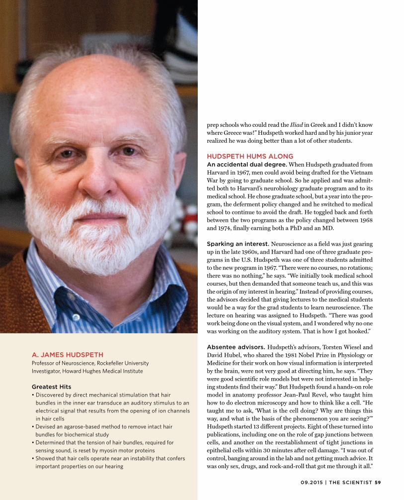
5909.2015 | THE SCIENTIST
CR
ED
IT L
INE
A. JAMES HUDSPETHProfessor of Neuroscience, Rockefeller UniversityInvestigator, Howard Hughes Medical Institute
Greatest Hits• Discovered by direct mechanical stimulation that hair
bundles in the inner ear transduce an auditory stimulus to an electrical signal that results from the opening of ion channels in hair cells
• Devised an agarose-based method to remove intact hair bundles for biochemical study
• Determined that the tension of hair bundles, required for sensing sound, is reset by myosin motor proteins
• Showed that hair cells operate near an instability that confers important properties on our hearing
prep schools who could read the Iliad in Greek and I didn’t knowwhere Greece was!” Hudspeth worked hard and by his junior year realized he was doing better than a lot of other students.
HUDSPETH HUMS ALONGAn accidental dual degree. When Hudspeth graduated fromHarvard in 1967, men could avoid being drafted for the Vietnam War by going to graduate school. So he applied and was admit-ted both to Harvard’s neurobiology graduate program and to its medical school. He chose graduate school, but a year into the pro-gram, the deferment policy changed and he switched to medical school to continue to avoid the draft. He toggled back and forth between the two programs as the policy changed between 1968 and 1974, finally earning both a PhD and an MD.
Sparking an interest. Neuroscience as a field was just gearing up in the late 1960s, and Harvard had one of three graduate pro-grams in the U.S. Hudspeth was one of three students admitted to the new program in 1967. “There were no courses, no rotations; there was no nothing,” he says. “We initially took medical school courses, but then demanded that someone teach us, and this was the origin of my interest in hearing.” Instead of providing courses, the advisors decided that giving lectures to the medical students would be a way for the grad students to learn neuroscience. The lecture on hearing was assigned to Hudspeth. “There was good work being done on the visual system, and I wondered why no one was working on the auditory system. That is how I got hooked.”
Absentee advisors. Hudspeth’s advisors, Torsten Wiesel and David Hubel, who shared the 1981 Nobel Prize in Physiology or Medicine for their work on how visual information is interpreted by the brain, were not very good at directing him, he says. “They were good scientific role models but were not interested in help-ing students find their way.” But Hudspeth found a hands-on role model in anatomy professor Jean-Paul Revel, who taught him how to do electron microscopy and how to think like a cell. “He taught me to ask, ‘What is the cell doing? Why are things this way, and what is the basis of the phenomenon you are seeing?’” Hudspeth started 13 different projects. Eight of these turned into publications, including one on the role of gap junctions between cells, and another on the reestablishment of tight junctions in epithelial cells within 30 minutes after cell damage. “I was out of control, banging around in the lab and not getting much advice. It was only sex, drugs, and rock-and-roll that got me through it all.”
60 THE SCIENTIST | the-scientist.com
PROFILE
Caught in the middle. Hudspeth chose to do a postdoc withÅke Flock at a research institute associated with the Karolin-ska University Hospital in Stockholm, both because Flock was using electron microscopy to study hair cells, and because Sweden could potentially provide a safe haven from the draft situation in the U.S. But upon his arrival, researchers with whom Hudspeth shared lab space were surprisingly cold to him. He subsequently learned that their behavior stemmed from disapproval of Flock’s planned career move to the Karolinska Institute. Any horizon-tal move to a new position did not follow the hierarchical Swed-ish professorship system. Although Hudspeth conducted three months of library research on the auditory system, he performed almost no actual experiments over ten months in Stockholm. In December 1974, while out jogging, he slipped and fell into a fjord. “As I went underwater I reasoned that if I didn’t drown, I would leave the next day. I didn’t drown—and I left.”
Mysterious supporters. Despite his fruitless postdoc, Hud-speth returned to the U.S. to faculty position offers from Rock-efeller University and Caltech. Clearly, Hudspeth had supporters who thought he could do research, but he says he doesn’t know who recommended him for the positions.
Mechanical stimuli. After joining Caltech, Hudspeth began to execute the research plan he had mapped out in Stockholm. With David Corey, his first graduate student, he directly manipulated hair cells in vitro to show, for the first time, that physical pushing of the hair bundle stimulates hair cells to produce an electrical response. The two then showed that the response originates from mechanically sensitive channels that pass potassium ions but are also permeable to other cations. Hudspeth’s lab chose the hear-ing organ of the bullfrog for the experiments because it was large and easy to work with. “I had assumed from the beginning that hair cells of vertebrates all worked similarly. And by and large, the discoveries made in frog and turtle systems have been shown to also apply as well in mammals, and presumably, in humans.”
Hair removal. In 1983, Hudspeth moved to the University of California, San Francisco. “This was a very exciting institution at that point, nearing its peak as the best place for biomedi-cal research, with extremely collaborative professors and stu-dents and minimal departmental boundaries.” Then, in 1989, he accepted an opportunity to build a neuroscience program at the University of Texas Southwestern Medical Center. After almost three years of work to set up the program, which included recruit-ing new professors, Hudspeth was told that the program was not needed. Coinciding with Hudpseth’s acceptance into the National Academy of Sciences, the dean of the university moved him to a basement office next door to the morgue. “It was a cruel act, but the dean was subsequently removed, and the president [of the university] was placed under investigation for deflection of university funds for private use.” Despite the drama, his research continued to go well. Hudspeth and postdoc Peter Gillespie devel-
oped a way to isolate all of the 3,000 hair bundles from the inner ear of the frog at once, for biochemical work. Inspired by the age-old practice of using wax as a depilatory, the two put molten aga-rose over the inner ear hairs, let it set, and then briskly twisted and pulled the agarose, ripping out the hair bundles. The tech-nique has since become standard and has been adapted for use in other vertebrate systems.
HUDSPETH HOMES INPublic-address system. In 1995, when Hudspeth movedto Rockefeller University, his research also took a new turn. “I thought by this time I had exhausted what I wanted to do with hair cells, but then a new idea came to the fore. It turns out that the ear is not just a passive recipient of sound but has a built-in amplifier, something like a biological hearing aid that is part of the hair cell apparatus,” he says. This amplifier has a profound effect on hearing—making it 100- to 1,000-fold more sensitive. When this amplifier wears out or is damaged, we become hard of hearing. There were hints of this amplification, called the active process, for more than 50 years, and in 1999, along with post-doc Pascal Martin, Hudspeth showed that in amphibians the hair bundle itself is the source of this amplification. “This amplifier is based on a particular kind of instability that has interesting math properties. If you turn up a public-address system too far, it finally goes unstable and begins to howl and whine, oscillating sponta-neously. And that is just what happens in our own ears. Each of the sensory hair cells has in it an amplifier that can go unstable so that sound actually comes out of the ear.”
Restoring hearing. Hudspeth’s lab is now working on zebrafish, which, unlike mammals, are able to regenerate their hair cells. Under-standing which genes are activated in cells during regeneration may provide clues about how to turn on this process in mam-mals. The lab recently identified a spectrum of genes that may be involved in the differentiation of progenitor cells to hair cells in zebrafish. “The hope is that we can restore hearing in humans by regenerating hair cells in our own ears, basically by hijacking the molecular program that made the hair cells in the first place.”
Mentorship philosophy. “I try, particularly with postdocs, to give them a start on what they are going to do independently. Peter Gillespie and I started the twist-off biochemistry work on hair bundles, but when he took that direction in his own lab I quit doing the biochemistry as a major effort. And when Joe Howard pursued single-molecule work, I gave up the reins on that to him.”
Common theme. While Hudspeth says he found most of his Harvard courses uninspiring, his physical chemistry profes-sor, George Kistiakowsky, was very committed to undergradu-ate teaching. “This turned out to be the most important thread in my scientific career, applying rigorous physical and chemical principles to biological systems. This course was an inspiration for that and what the rest of [my career] has been about.” g
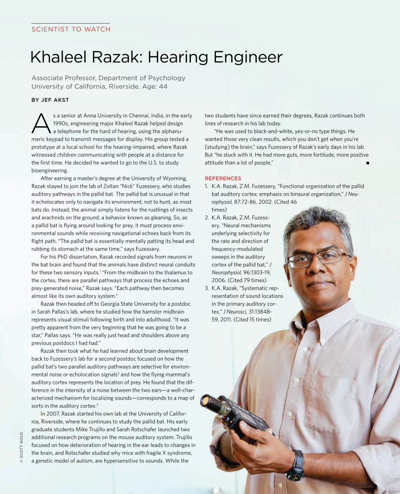
SCIENTIST TO WATCH
Associate Professor, Department of PsychologyUniversity of California, Riverside. Age: 44
BY JEF AKST
Khaleel Razak: Hearing Engineer
As a senior at Anna University in Chennai, India, in the early1990s, engineering major Khaleel Razak helped design a telephone for the hard of hearing, using the alphanu-
meric keypad to transmit messages for display. His group tested a prototype at a local school for the hearing-impaired, where Razak witnessed children communicating with people at a distance for the first time. He decided he wanted to go to the U.S. to study bioengineering.
After earning a master’s degree at the University of Wyoming, Razak stayed to join the lab of Zoltan “Nick” Fuzessery, who studies auditory pathways in the pallid bat. The pallid bat is unusual in that it echolocates only to navigate its environment, not to hunt, as most bats do. Instead, the animal simply listens for the rustlings of insects and arachnids on the ground, a behavior known as gleaning. So, as a pallid bat is flying around looking for prey, it must process envi-ronmental sounds while receiving navigational echoes back from its flight path. “The pallid bat is essentially mentally patting its head and rubbing its stomach at the same time,” says Fuzessery.
For his PhD dissertation, Razak recorded signals from neurons in the bat brain and found that the animals have distinct neural conduits for these two sensory inputs.1 “From the midbrain to the thalamus tothe cortex, there are parallel pathways that process the echoes and prey-generated noise,” Razak says. “Each pathway then becomes almost like its own auditory system.”
Razak then headed off to Georgia State University for a postdoc in Sarah Pallas’s lab, where he studied how the hamster midbrain represents visual stimuli following birth and into adulthood. “It was pretty apparent from the very beginning that he was going to be a star,” Pallas says. “He was really just head and shoulders above any previous postdocs I had had.”
Razak then took what he had learned about brain development back to Fuzessery’s lab for a second postdoc focused on how the pallid bat’s two parallel auditory pathways are selective for environ-mental noise or echolocation signals2 and how the flying mammal’sauditory cortex represents the location of prey. He found that the dif-ference in the intensity of a noise between the two ears—a well-char-acterized mechanism for localizing sounds—corresponds to a map of sorts in the auditory cortex.3
In 2007, Razak started his own lab at the University of Califor-nia, Riverside, where he continues to study the pallid bat. His early graduate students Mike Trujillo and Sarah Rotschafer launched two additional research programs on the mouse auditory system. Trujillo focused on how deterioration of hearing in the ear leads to changes in the brain, and Rotschafer studied why mice with fragile X syndrome, a genetic model of autism, are hypersensitive to sounds. While the
two students have since earned their degrees, Razak continues both lines of research in his lab today.
“He was used to black-and-white, yes-or-no type things. He wanted those very clean results, which you don’t get when you’re [studying] the brain,” says Fuzessery of Razak’s early days in his lab. But “he stuck with it. He had more guts, more fortitude, more positive attitude than a lot of people.” g
REFERENCES1. K.A. Razak, Z.M. Fuzessery, “Functional organization of the pallid
bat auditory cortex: emphasis on binaural organization,” J Neu-rophysiol, 87:72-86, 2002. (Cited 46 times)
2. K.A. Razak, Z.M. Fuzess-ery, “Neural mechanisms underlying selectivity for the rate and direction of frequency-modulated sweeps in the auditory cortex of the pallid bat,” J Neurophysiol, 96:1303-19, 2006. (Cited 79 times)
3. K.A. Razak, “Systematic rep-resentation of sound locations in the primary auditory cor-tex,” J Neurosci, 31:13848-59, 2011. (Cited 15 times)
© S
CO
TT
GO
LD
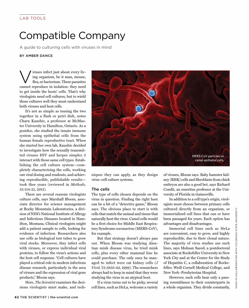
62 THE SCIENTIST | the-scientist.com
LAB TOOLS
NIA
ID I
N C
OL
LA
BO
RA
TIO
N W
ITH
CO
LO
RA
DO
STA
TE
UN
IVE
RS
ITY
A guide to culturing cells with viruses in mind
BY AMBER DANCE
Compatible Company
V iruses infect just about every liv-ing organism, be it man, mouse, flea, or bacterium. These parasites
cannot reproduce in isolation: they need to get inside the hosts’ cells. That’s why virologists need cell cultures, but to wield those cultures well they must understand both viruses and host cells.
It’s not as simple as tossing the two together in a flask or petri dish, notes Charu Kaushic, a professor at McMas-ter University in Hamilton, Ontario. As a postdoc, she studied the innate immune system using epithelial cells from the human female reproductive tract. When she started her own lab, Kaushic decided to investigate how the sexually transmit-ted viruses HIV and herpes simplex 2 interact with those same cell types. Estab-lishing the cell culture system—com-pletely characterizing the cells, working out viral dosing and readouts, and achiev-ing reproducible, publishable results—took thee years (reviewed in Methods, 55:114-21, 2011).
There are several reasons virologists culture cells, says Marshall Bloom, asso-ciate director for science management at Rocky Mountain Laboratories, a divi-sion of NIH’s National Institute of Allergy and Infectious Diseases located in Ham-ilton, Mon tana. Clinical virologists might add a patient sample to cells, looking for evidence of infection. Researchers also use cells as biological test tubes to grow viral stocks. Moreover, they infect cells with viruses, or express individual viral proteins, to follow the virus’s actions and the host cell response. “Cell cultures have played a critical role in modern infectious disease research, particularly in the area of viruses and the expression of viral gene products,” Bloom says.
Here, The Scientist examines the deci-sions virologists must make, and tech-
niques they can apply, as they design virus–cell culture systems.
The cellsThe type of cells chosen depends on thevirus in question. Finding the right host can be a bit of a “detective game,” Bloom says. The obvious place to start is with cells that match the animal and tissue that naturally host the virus. Camel cells would be a first choice for Middle East Respira-tory Syndrome coronavirus (MERS-CoV), for example.
But that strategy doesn’t always pan out. When Bloom was studying Aleu-tian mink disease virus, he tried mink cells, plus every other mammal line he could purchase. The only ones he man-aged to infect were cat kidney cells (J Virol, 73:3835-42, 1999). The researchers always had to keep in mind that they were studying the virus in an atypical host.
If a virus turns out to be picky, several cell lines, such as HeLa, welcome a variety
of viruses, Bloom says. Baby hamster kid-ney (BHK) cells and fibroblasts from chick embryos are also a good bet, says Richard Condit, an emeritus professor at the Uni-versity of Florida in Gainesville.
In addition to a cell type’s origin, virol-ogists must choose between primary cells cultured directly from an organism, or immortalized cell lines that can or have been passaged for years. Each option has advantages and disadvantages.
Immortal cell lines such as HeLa are convenient, easy to grow, and highly reproducible, due to their clonal nature. The majority of virus studies use such lines, says Mohsan Saeed, a postdoctoral associate at Rockefeller University in New York City and at the Center for the Study of Hepatitis C, a collaboration of Rocke-feller, Weill Cornell Medical College, and New York–Presbyterian Hospital.
However, such cells bear only a pass-ing resemblance to their counterparts in a whole organism. They divide constantly,
MERS-CoV particles oncamel epithelial cells.
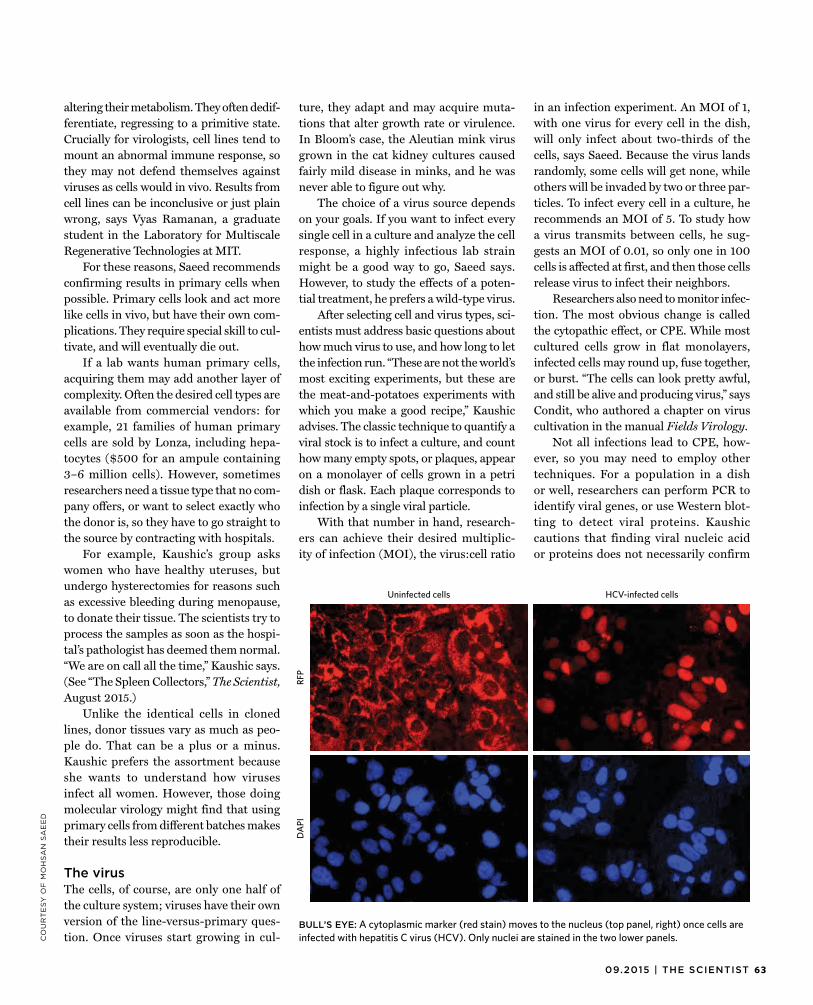
6309.2015 | THE SCIENTIST
CO
UR
TE
SY
OF
MO
HS
AN
SA
EE
D
altering their metabolism. They often dedif-ferentiate, regressing to a primitive state. Crucially for virologists, cell lines tend to mount an abnormal immune response, so they may not defend themselves against viruses as cells would in vivo. Results from cell lines can be inconclusive or just plain wrong, says Vyas Ramanan, a graduate student in the Laboratory for Multiscale Regenerative Technologies at MIT.
For these reasons, Saeed recommends confirming results in primary cells when possible. Primary cells look and act more like cells in vivo, but have their own com-plications. They require special skill to cul-tivate, and will eventually die out.
If a lab wants human primary cells, acquiring them may add another layer of complexity. Often the desired cell types are available from commercial vendors: for example, 21 families of human primary cells are sold by Lonza, including hepa-tocytes ($500 for an ampule containing 3–6 million cells). However, sometimes researchers need a tissue type that no com-pany offers, or want to select exactly who the donor is, so they have to go straight to the source by contracting with hospitals.
For example, Kaushic’s group asks women who have healthy uteruses, but undergo hysterectomies for reasons such as excessive bleeding during menopause, to donate their tissue. The scientists try to process the samples as soon as the hospi-tal’s pathologist has deemed them normal. “We are on call all the time,” Kaushic says. (See “The Spleen Collectors,” The Scientist, August 2015.)
Unlike the identical cells in cloned lines, donor tissues vary as much as peo-ple do. That can be a plus or a minus. Kaushic prefers the assortment because she wants to understand how viruses infect all women. However, those doing molecular virology might find that using primary cells from different batches makes their results less reproducible.
The virusThe cells, of course, are only one half ofthe culture system; viruses have their own version of the line-versus-primary ques-tion. Once viruses start growing in cul-
ture, they adapt and may acquire muta-tions that alter growth rate or virulence. In Bloom’s case, the Aleutian mink virus grown in the cat kidney cultures caused fairly mild disease in minks, and he was never able to figure out why.
The choice of a virus source depends on your goals. If you want to infect every single cell in a culture and analyze the cell response, a highly infectious lab strain might be a good way to go, Saeed says. However, to study the effects of a poten-tial treatment, he prefers a wild-type virus.
After selecting cell and virus types, sci-entists must address basic questions about how much virus to use, and how long to let the infection run. “These are not the world’s most exciting experiments, but these are the meat-and-potatoes experiments with which you make a good recipe,” Kaushic advises. The classic technique to quantify a viral stock is to infect a culture, and count how many empty spots, or plaques, appear on a monolayer of cells grown in a petri dish or flask. Each plaque corresponds to infection by a single viral particle.
With that number in hand, research-ers can achieve their desired multiplic-ity of infection (MOI), the virus:cell ratio
in an infection experiment. An MOI of 1, with one virus for every cell in the dish, will only infect about two-thirds of the cells, says Saeed. Because the virus lands randomly, some cells will get none, while others will be invaded by two or three par-ticles. To infect every cell in a culture, he recommends an MOI of 5. To study how a virus transmits between cells, he sug-gests an MOI of 0.01, so only one in 100 cells is affected at first, and then those cells release virus to infect their neighbors.
Researchers also need to monitor infec-tion. The most obvious change is called the cytopathic effect, or CPE. While most cultured cells grow in flat monolayers, infected cells may round up, fuse together, or burst. “The cells can look pretty awful, and still be alive and producing virus,” says Condit, who authored a chapter on virus cultivation in the manual Fields Virology.
Not all infections lead to CPE, how-ever, so you may need to employ other techniques. For a population in a dish or well, researchers can perform PCR to identify viral genes, or use Western blot-ting to detect viral proteins. Kaushic cautions that finding viral nucleic acid or proteins does not necessarily confirm
RFP
HCV-infected cellsUninfected cells
DA
PI
BULL’S EYE: A cytoplasmic marker (red stain) moves to the nucleus (top panel, right) once cells areinfected with hepatitis C virus (HCV). Only nuclei are stained in the two lower panels.
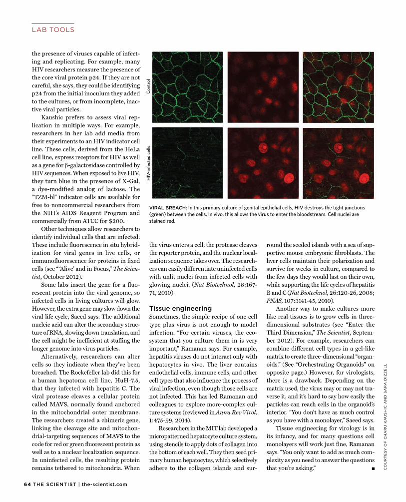
64 THE SCIENTIST | the-scientist.com
LAB TOOLS
CO
UR
TE
SY
OF
CH
AR
U K
AU
SH
IC A
ND
SA
RA
DIZ
ZE
LL
the presence of viruses capable of infect-ing and replicating. For example, many HIV researchers measure the presence of the core viral protein p24. If they are not careful, she says, they could be identifying p24 from the initial inoculum they added to the cultures, or from incomplete, inac-tive viral particles.
Kaushic prefers to assess viral rep-lication in multiple ways. For example, researchers in her lab add media from their experiments to an HIV indicator cell line. These cells, derived from the HeLa cell line, express receptors for HIV as well as a gene for β-galactosidase controlled by HIV sequences. When exposed to live HIV, they turn blue in the presence of X-Gal, a dye-modified analog of lactose. The “TZM-bl” indicator cells are available for free to noncommercial researchers from the NIH’s AIDS Reagent Program and commercially from ATCC for $200.
Other techniques allow researchers to identify individual cells that are infected. These include fluorescence in situ hybrid-ization for viral genes in live cells, or immunofluorescence for proteins in fixed cells (see “ ‘Alive’ and in Focus,” The Scien-tist, October 2012).
Some labs insert the gene for a fluo-rescent protein into the viral genome, so infected cells in living cultures will glow. However, the extra gene may slow down the viral life cycle, Saeed says. The additional nucleic acid can alter the secondary struc-ture of RNA, slowing down translation, and the cell might be inefficient at stuffing the longer genome into virus particles.
Alternatively, researchers can alter cells so they indicate when they’ve been breached. The Rockefeller lab did this for a human hepatoma cell line, HuH-7.5, that they infected with hepatitis C. The viral protease cleaves a cellular protein called MAVS, normally found anchored in the mitochondrial outer membrane. The researchers created a chimeric gene, linking the cleavage site and mitochon-drial-targeting sequences of MAVS to the code for red or green fluorescent protein as well as to a nuclear localization sequence. In uninfected cells, the resulting protein remains tethered to mitochondria. When
the virus enters a cell, the protease cleaves the reporter protein, and the nuclear local-ization sequence takes over. The research-ers can easily differentiate uninfected cells with unlit nuclei from infected cells with glowing nuclei. (Nat Biotechnol, 28:167-71, 2010)
Tissue engineeringSometimes, the simple recipe of one celltype plus virus is not enough to model infection. “For certain viruses, the eco-system that you culture them in is very important,” Ramanan says. For example, hepatitis viruses do not interact only with hepatocytes in vivo. The liver contains endothelial cells, immune cells, and other cell types that also influence the process of viral infection, even though those cells are not infected. This has led Ramanan and colleagues to explore more-complex cul-ture systems (reviewed in Annu Rev Virol, 1:475-99, 2014).
Researchers in the MIT lab developed a micropatterned hepatocyte culture system, using stencils to apply dots of collagen into the bottom of each well. They then seed pri-mary human hepatocytes, which selectively adhere to the collagen islands and sur-
round the seeded islands with a sea of sup-portive mouse embryonic fibroblasts. The liver cells maintain their polarization and survive for weeks in culture, compared to the few days they would last on their own, while supporting the life cycles of hepatitis B and C (Nat Biotechnol, 26:120-26, 2008; PNAS, 107:3141-45, 2010).
Another way to make cultures more like real tissues is to grow cells in three-dimensional substrates (see “Enter the Third Dimension,” The Scientist, Septem-ber 2012). For example, researchers can combine different cell types in a gel-like matrix to create three-dimensional “organ-oids.” (See “Orchestrating Organoids” on opposite page.) However, for virologists, there is a drawback. Depending on the matrix used, the virus may or may not tra-verse it, and it’s hard to say how easily the particles can reach cells in the organoid’s interior. “You don’t have as much control as you have with a monolayer,” Saeed says.
Tissue engineering for virology is in its infancy, and for many questions cell monolayers will work just fine, Ramanan says. “You only want to add as much com-plexity as you need to answer the questions that you’re asking.” g
HIV
-infe
cted
cel
lsCo
ntro
l
VIRAL BREACH: In this primary culture of genital epithelial cells, HIV destroys the tight junctions (green) between the cells. In vivo, this allows the virus to enter the bloodstream. Cell nuclei are stained red.
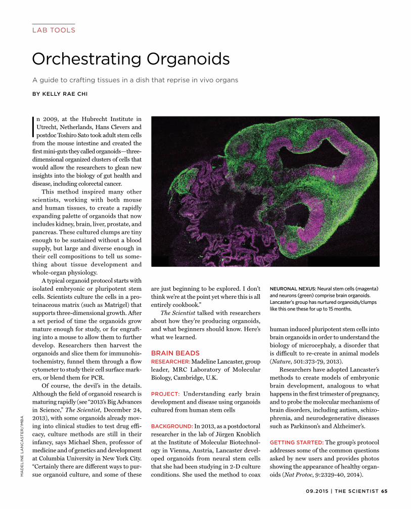
6509.2015 | THE SCIENTIST
LAB TOOLSM
AD
EL
INE
LA
NC
AS
TE
R/I
MB
A
In 2009, at the Hubrecht Institute inUtrecht, Netherlands, Hans Clevers and postdoc Toshiro Sato took adult stem cells
from the mouse intestine and created the first mini-guts they called organoids—three-dimensional organized clusters of cells that would allow the researchers to glean new insights into the biology of gut health and disease, including colorectal cancer.
This method inspired many other scientists, working with both mouse and human tissues, to create a rapidly expanding palette of organoids that now includes kidney, brain, liver, prostate, and pancreas. These cultured clumps are tiny enough to be sustained without a blood supply, but large and diverse enough in their cell compositions to tell us some-thing about tissue development and whole-organ physiology.
A typical organoid protocol starts with isolated embryonic or pluripotent stem cells. Scientists culture the cells in a pro-teinaceous matrix (such as Matrigel) that supports three-dimensional growth. After a set period of time the organoids grow mature enough for study, or for engraft-ing into a mouse to allow them to further develop. Researchers then harvest the organoids and slice them for immunohis-tochemistry, funnel them through a flow cytometer to study their cell surface mark-ers, or blend them for PCR.
Of course, the devil’s in the details. Although the field of organoid research is maturing rapidly (see “2013’s Big Advances in Science,” The Scientist, December 24, 2013), with some organoids already mov-ing into clinical studies to test drug effi-cacy, culture methods are still in their infancy, says Michael Shen, professor of medicine and of genetics and development at Columbia University in New York City. “Certainly there are different ways to pur-sue organoid culture, and some of these
are just beginning to be explored. I don’t think we’re at the point yet where this is all entirely cookbook.”
The Scientist talked with researchers about how they’re producing organoids, and what beginners should know. Here’s what we learned.
BRAIN BEADSRESEARCHER: Madeline Lancaster, groupleader, MRC Laboratory of Molecular Biology, Cambridge, U.K.
PROJECT: Understanding early braindevelopment and disease using organoids cultured from human stem cells
BACKGROUND: In 2013, as a postdoctoralresearcher in the lab of Jürgen Knoblich at the Institute of Molecular Biotechnol-ogy in Vienna, Austria, Lancaster devel-oped organoids from neural stem cells that she had been studying in 2-D culture conditions. She used the method to coax
human induced pluripotent stem cells into brain organoids in order to understand the biology of microcephaly, a disorder that is difficult to re-create in animal models (Nature, 501:373-79, 2013).
Researchers have adopted Lancaster’s methods to create models of embryonic brain development, analogous to what happens in the first trimester of pregnancy, and to probe the molecular mechanisms of brain disorders, including autism, schizo-phrenia, and neurodegenerative diseases such as Parkinson’s and Alzheimer’s.
GETTING STARTED: The group’s protocoladdresses some of the common questions asked by new users and provides photos showing the appearance of healthy organ-oids (Nat Protoc, 9:2329-40, 2014).
A guide to crafting tissues in a dish that reprise in vivo organs
BY KELLY RAE CHI
Orchestrating Organoids
NEURONAL NEXUS: Neural stem cells (magenta) and neurons (green) comprise brain organoids. Lancaster’s group has nurtured organoids/clumps like this one these for up to 15 months.
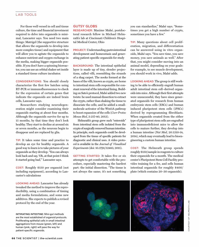
66 THE SCIENTIST | the-scientist.com
LAB TOOLS
CO
UR
TE
SY
OF
HE
LM
RA
TH
LA
B
For those well versed in cell and tissueculture, the time and financial investment required to delve into organoids is mini-mal, Lancaster says. You need two main things: Matrigel (the supportive structure that allows the organoids to develop into more complex tissue) and equipment that will allow you to agitate the organoids to enhance nutrient and oxygen exchange in the media, making bigger organoids pos-sible. If you don’t have a spinning bioreac-tor, you can use an orbital shaker set inside a standard tissue culture incubator.
CONSIDERATIONS: You should closelycharacterize the first few batches using RT-PCR or immunofluorescence to check for the expression of certain genes that indicate the organoids are indeed brain cells, Lancaster says.
Researchers studying neurodegen-eration might consider examining their organoids starting at about four months. Although the organoids survive for up to 15 months, by that time they don’t look healthy. They start to decline at around six or seven months, as the neurons begin to disappear and are replaced by glia.
TIP: It takes some time and practice todevelop an eye for healthy organoids. A good way to learn is to take pictures of your organoids as they develop. “You can always look back and say, ‘Oh, at that point I think it started going bad,’” Lancaster says.
COST: Roughly $150 per organoid (notincluding equipment), according to Lan-caster’s calculations
LOOKING AHEAD: Lancaster has alreadytweaked the method to improve the repro-ducibility, using a combination of timing and media formulations, and some new additives. She expects to publish a revised protocol by the end of the year.
GUTSY GLOBSRESEARCHER: Maxime Mahé, postdoc-toral research fellow in Michael Helm-rath’s lab at Cincinnati Children’s Hospi-tal Medical Center, Ohio
PROJECT: Understanding gastrointestinaldevelopment and homeostasis and gener-ating patient-specific organoids for study
BACKGROUND: The intestinal epitheliallayer is made up of tiny, slender projec-tions, called villi, resembling the strands of a shag carpet. The nooks formed at the bases of the villi, known as crypts, are home to intestinal stem cells responsible for con-stant renewal of the intestinal lining. Build-ing on Sato’s protocol, Mahé added two new twists: he used manual dissection to extract the crypts, rather than shaking the tissue to dissociate the cells; and he added a small-molecule activator of the Wnt3A pathway to boost expansion of the cells (Curr Protoc Mouse Biol, 3:217-40, 2013).
Helmrath’s group grew such “enteroids” from intestinal stem cells isolated from the crypts of surgically removed human intestine. In principle, such organoids could be devel-oped from the tissue of specific patients for diagnostic and clinical uses. A video proto-col is available in the Journal of Visualized Experiments (doi: 10.3791/52483, 2015).
GETTING STARTED: It takes five or sixattempts to get comfortable with the pro-cedure, especially mastering the hardest part: the initial dissection. “The tissue is not always the same; it’s not something
you can standardize,” Mahé says. “Some-times you get a high number of crypts, sometimes you have a few.”
TIP: Many questions about cell prolif-eration, migration, and differentiation can be answered using in vitro organ-oids, Mahé says. “You save time, you save money, you save animals as well.” After that, you might consider moving into an animal model, depending on your goals: for example, to see muscle development, you should work in vivo, Mahé adds.
LOOKING AHEAD: The group is still work-ing to be able to efficiently engraft human adult intestinal stem cell–derived organ-oids into mice. Although their first attempts were unsuccessful, they have since gener-ated organoids for research from human embryonic stem cells (ESCs) and human induced pluripotent stem cells (iPSCs) derived by reprogramming fibroblasts. When organoids created from the either type of pluripotent stem cells are engrafted into immunodeficient mice to allow the cells to mature further, they develop into a human intestine (Nat Med, 20:1310-14, 2014), which may eventually lead to bioen-gineering a custom human intestine.
COST: The Helmrath group spendsroughly $150/sample in reagents to culture their organoids for a month. The medical center’s Pluripotent Stem Cell Facility pro-vides training for a fee, and sells human intestinal organoids for roughly $400/plate (which contains 20–30 organoids).
INTIMATING INTESTINE: Mini-gut methods are the most established of organoid protocols. Proliferating epithelial cells in small intestinal aggregations from mouse (green, left) and human (pink, right) will pave the way for patient-specific organoids.
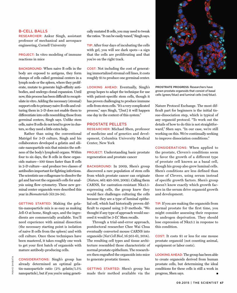
6709.2015 | THE SCIENTIST
MA
HO
SH
IBA
TA
B-CELL BALLSRESEARCHER: Ankur Singh, assistantprofessor of mechanical and aerospace engineering, Cornell University
PROJECT: In vitro modeling of immunereactions in mice
BACKGROUND: When naive B cells in thebody are exposed to antigens, they form clumps of cells called germinal centers in a lymph node or the spleen, where they prolif-erate, mutate to generate high-affinity anti-bodies, and undergo clonal expansion. Until now, this process has been difficult to recapit-ulate in vitro. Adding the necessary (stromal) support cells to primary naive B cells and cul-turing them in 2-D does not enable them to differentiate into cells resembling those from germinal centers, Singh says. Unlike stem cells, naive B cells do not tend to grow in clus-ters, so they need a little extra help.
Rather than using the conventional Matrigel for 3-D culture, Singh and his collaborators developed a gelatin and sili-cate-nanoparticle mix that mimics the soft-ness of the body’s lymphoid organs. Within four to six days, the B cells in these organ-oids mature—100 times faster than B cells in 2-D culture—and produce two classes of antibodies important for fighting infections. The scientists use collagenase to dissolve the gel and harvest the organoid’s cells for anal-ysis using flow cytometry. These new ger-minal center organoids were described this year in Biomaterials (63:24-34).
GETTING STARTED: Making the gela-tin-nanoparticle mix is as easy as making Jell-O at home, Singh says, and the ingre-dients are commercially available. You’ll need experience with animal dissection (the necessary starting point is isolation of naive B cells from the spleen) and with cell culture. Once these techniques have been mastered, it takes roughly one week to get your first batch of organoids with mature antibody-producing cells.
CONSIDERATIONS: Singh’s group has already determined an optimal gela-tin-nanoparticle ratio (2% gelatin/1.5% nanoparticle), but if you you’re using geneti-
cally mutated B cells, you may need to tweak the ratios. “It can be easily tuned,” Singh says.
TIP: After four days of incubating the cellswith gel, you will see dark spots—a sign that the cells are proliferating and that you’re on the right track.
COST: Not including the cost of generat-ing immortalized stromal cell lines, it costs roughly $1 to produce one germinal center.
LOOKING AHEAD: Eventually, Singh’sgroup hopes to adapt the technique for use with patient-specific stem cells, though it has proven challenging to produce immune cells from stem cells. “It’s a very complicated process,” says Singh, “[but] it will happen one day in the context of this system.”
PROSTATE PELLETSRESEARCHER: Michael Shen, professorof medicine and of genetics and devel-opment, Columbia University Medical Center, New York
PROJECT: Understanding basic prostateregeneration and prostate cancer
BACKGROUND: In 2009, Shen’s groupdiscovered a rare population of stem cells from which prostate cancer can originate (Nature, 461:495-500, 2009). Calling them CARNS, for castration-resistant Nkx3.1-expressing cells, the group knew they would face challenges culturing the cells because they are a type of luminal epithe-lial cell, which had historically proven dif-ficult to expand using 2-D methods. “We thought if any type of approach would suc-ceed it would be 3-D,” Shen recalls.
Through a trial-and-error approach, postdoctoral researcher Chee Wai Chua eventually converted mouse CARNS into organoids (Nat Cell Biol, 16:951-61, 2014). The resulting cell types and tissue archi-tecture resembled those characteristic of normal prostate epithelium. The research-ers then engrafted the organoids into mice to generate prostatic tissues.
GETTING STARTED: Shen’s group has made their method available via the
Nature Protocol Exchange. The most dif-ficult part for beginners is the initial tis-sue-dissociation step, which is typical of any organoid protocol. “To work out the details of how to do this is not straightfor-ward,” Shen says. “In our case, we’re still working on this. We’re continually seeking to improve dissociation conditions.”
CONSIDERATIONS: When applied tothe prostate, Clevers’s conditions seem to favor the growth of a different type of prostate cell known as a basal cell, though his group also grew luminal cells. Shen’s conditions are less defined than those of Clevers, using serum instead of specific growth factors. Shen’s group doesn’t know exactly which growth fac-tors in the serum drive organoid growth and development.
TIP: If you are making the organoids fromnormal prostate for the first time, you might consider assessing their response to androgen deprivation. They should lose expression of Nkx3.1 in response to this condition.
COST: It costs $1 or less for one mouseprostate organoid (not counting animal, equipment or labor costs).
LOOKING AHEAD: The group has been ableto create organoids derived from human prostate cells, but determining the ideal conditions for these cells is still a work in progress, Shen says. g
PROSTRATE PROGRESS: Researchers have grown prostate organoids that consist of basal cells (green/blue) and luminal cells (red/blue).
68 THE SCIENTIST | the-scientist.com
BIO BUSINESS
Science-based tinnitus therapeutics are finally coming into their own.
BY JENNY ROOD
The Sounds of Silence
It often starts off with a bang. Many asoldier, construction worker, concert-goer, or innocent passerby exposed to
a loud noise walks away with the telltale symptom of tinnitus, a persistent ringing in the ears. The condition can also arise from other ear traumas, such as middle-ear infections or exposure to high pressure while scuba diving, and begins with dam-age to the hair cells in the cochlea of the inner ear or to the auditory nerve. Until recently, such damage was thought to be the cause of the phantom sounds that plague tinnitus sufferers. Now, research-ers are realizing that it’s much more com-plex than that.
“Damage to hair cells and auditory nerve fibers sets the stage for the develop-ment of tinnitus,” says Jennifer Melcher of the Massachusetts Eye and Ear Infirmary. But the true culprit is really the brain, which eventually begins to compensate for the loss of input from the ear by “turning up the volume” on the sound signals it is trying to pick up, she adds. Navzer Engi-neer, chief scientific officer of Dallas-based MicroTransponder, which is developing a neurostimulative treatment for tinnitus, agrees: “Cells in the brain don’t stay dor-mant” even though they have lost input from the ear, he says.
It’s unclear when the condition transi-tions from the ear to the brain. Research-ers also do not yet know whether the brain or peripheral nerves are primarily respon-sible for amplifying the spontaneous neu-ral activity in the auditory pathway. But in the end the effect is the same: the brain begins to capture sounds of its own cre-ation. “The pathology is in the ear . . . but the sounds are generated by the brain,” says Engineer.
The University of Regensburg’s Ber-thold Langguth, chairman of the executive committee of the Tinnitus Research Ini-tiative, likens the compensatory sound to
the phantom limb sensation experienced by amputees. And like the phenomenon of phantom limbs, there’s not just a sin-gle brain region at fault. In addition to the auditory cortex, the limbic cortex—partic-ularly the amygdala, the brain’s emotional center—as well as the temporal, pari-etal, and sensorimotor cortex areas have all been implicated in tinnitus percep-tion (Curr Biol, 25:1208-14, 2015; eLife, 4:e06576, 2015).
A better scientific understanding of tinnitus could be key to developing an effective treatment. One in five Ameri-
cans has tinnitus, including more than a million veterans who experienced loud noises in the line of duty, and many suf-fer a severe form of the disorder. Yet treat-ment options are largely limited to cog-nitive behavioral therapy to learn to tune out the sound and physical exercises such as contracting the head and neck mus-cles (by clenching their jaw, for example) to adjust the rogue sound’s pitch or loud-ness. For those who continue to suffer sig-nificant psychological and emotional con-sequences of tinnitus, there has been no pharmaceutical treatment or cure. “It’s a very desperate group,” Engineer says.
Inside the earThe most advanced treatment in devel-opment for tinnitus targets the auditory neurons that connect the hair cells of the inner ear to the auditory cortex. In the mid-1990s, researchers at Inserm in Montpellier, France, found that chemi-cally inducing tinnitus in rats was asso-
ciated with upregulated N-methyl-D-aspartate (NMDA) receptors on the animals’ cochlear neurons (J Neurosci, 23:3944-52, 2003). NMDA receptors play a role in forming new synapses at these neurons, and regulate the levels of other neuronal receptors. In 2003, team-ing up with Swiss entrepreneur Thomas Meyer and his company Auris Medical, the Inserm researchers also observed such increased levels of NMDA receptors in rodents suffering from noise-induced tinnitus. Prior to noise trauma, the ani-mals had been trained to jump onto a pole in response to a sound, and after trauma, rodents with tinnitus continued these behaviors, even in the absence of an external tone.
To treat the condition, the group set about designing a drug that would block NMDA receptors. These days, Auris is test-ing the small-molecule drug S-ketamine in two Phase 3 trials of trauma-induced tin-nitus patients. The treatment, delivered directly into the inner ear via three injec-tions over three days, must catch the dis-order while the problem is still within the ear, before the brain has begun overcom-pensating for the loss of hearing. Once that happens, no amount of adjustment to the receptors on the auditory nerves will do any good.
Because it is not known when that transition from ear to brain occurs, one of the current trials, of 300 European patients, is specifically testing tinnitus sufferers who have developed the con-dition no more than three months prior to treatment. The other, a study of 330 North American patients, is investigating a therapy within one year post-trauma. Preliminary results suggest that S-ket-amine is effective beyond three months, but declines in effectiveness within a year of the initial trauma, so later stages of the trial are being refocused on the four- to
The pathology of tinnitus is in the ear but the sounds are generated by the brain.
—Navzer Engi neer, MicroTransponder
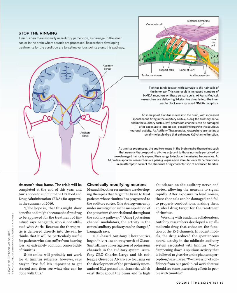
6909.2015 | THE SCIENTIST
six-month time frame. The trials will becompleted at the end of this year, and Auris hopes to submit to the US Food and Drug Administration (FDA) for approval in the summer of 2016.
“[The hope is] that this might show benefits and might become the first drug to be approved for the treatment of tin-nitus,” says Langguth, who is not affili-ated with Auris. Because the therapeu-tic is delivered directly into the ear, he thinks that it will be particularly useful for patients who also suffer from hearing loss, an extremely common comorbidity of tinnitus.
S-ketamine will probably not work for all tinnitus sufferers, however, says Meyer. “We feel it’s important to get started and then see what else can be done with this.”
Chemically modifying neuronsMeanwhile, other researchers are develop-ing therapies that target the brain to treat patients whose tinnitus has progressed to the auditory cortex. One strategy currently under investigation is the manipulation of the potassium channels found throughout the auditory pathway. “[Using] potassium channel modulators, the activity in the central auditory pathway can be changed,” Langguth says.
U.K.-based Autifony Therapeutics began in 2011 as an outgrowth of Glaxo-SmithKline’s investigation of potassium channels in the auditory system. Auti-fony CEO Charles Large and his col-league Giuseppe Alvaro are focusing on the development of the previously unex-amined Kv3 potassium channels, which exist throughout the brain and in high
abundance on the auditory nerve and cortex, allowing the neurons to signal rapidly. After exposure to loud noises, these channels can be damaged and fail to properly conduct ions, making them an ideal drug target for the treatment of tinnitus.
Working with academic collaborators, Autifony researchers developed a small-molecule drug that enhances the func-tion of the Kv3 channels. In rodent mod-els, the drug reduced the spontaneous neural activity in the midbrain auditory system associated with tinnitus. “We’re dampening down a spurious activity that is believed to give rise to the phantom per-ception,” says Large. “We have a lot of con-fidence from our preclinical work that we should see some interesting effects in peo-ple with tinnitus.”
six-month time frame. The trials will be Chemically modifying
STOP THE RINGINGTinnitus can manifest early in auditory perception, as damage to the inner ear, or in the brain where sounds are processed. Researchers developing treatments for the condition are targeting various points along this pathway.
At some point, tinnitus moves into the brain, with increased spontaneous firing in the auditory cortex. Along the auditory nerve
and in the auditory cortex, Kv3 potassium channels can be damaged after exposure to loud noises, possibly triggering the spurious
neuronal activity. At Autifony Therapeutics, researchers are testing a small-molecule drug that enhances Kv3 channel function.
As tinnitus progresses, the auditory maps in the brain rewire themselves such that neurons that respond to pitches adjacent to those normally perceived by
now-damaged hair cells expand their range to include the missing frequencies. At MicroTransponder, researchers are pairing vagus nerve stimulation with certain tones
in an attempt to correct the abnormal firing characteristic of advanced tinnitus.
Tinnitus tends to start with damage to the hair cells of the inner ear. This can result in increased numbers of
NMDA receptors on these sensory cells. At Auris Medical,researchers are delivering S-ketamine directly into the inner
ear to block overexpressed NMDA receptors.
© M
AR
IE S
CH
MIT
T/S
CIE
NC
E S
OU
RC
E;
© E
NC
YC
LO
PE
DIA
BR
ITA
NN
ICA
/UIG
/GE
TT
Y I
MA
GE
S
Inner hair cell
Outer hair cellTectorial membrane
Basilar membrane
Support cells Tunnel of Corti
Auditory neurons
Auditory nerve
Auditory cortex
70 THE SCIENTIST | the-scientist.com
BIO BUSINESS
Autifony researchers are currentlyrecruiting patients for Phase 2 trials in the U.K. In contrast to Auris Medical’s target patient population, Autifony focuses on people whose tinnitus is established in the brain and who have had the disorder for at least six months (but no more than 18 months). The treatment is currently taken as a daily oral pill for 28 days, although the length of the treatment course is still under investigation.
“Autifony is really quite unique in hav-ing a drug treatment that’s been ratio-nally designed around the idea that we can dampen down the hyperexcitability that we see in the nervous system,” Large says.
Retraining the brainFor patients with chronic tinnitus beyondthe 18-month window being targeted by Autifony, a third potential treatment is making its way through clinical tri-als. MicroTransponder’s therapy is a riff on a decades-old treatment for epilepsy and depression called vagus-nerve stim-ulation. More than 90,000 patients have undergone such treatment.
MicroTransponder was started out of Michael Kilgard’s lab at the Univer-sity of Texas, Dallas, where Engineer conducted his postdoctoral research. In 1998, Kilgard’s group published a rat study demonstrating that direct stimu-lation of the nucleus basalis of the fore-brain could be paired with the playing of a particular tone to change how sounds map to the brain’s auditory cortex (Sci-ence, 279:1714-18). The researchers were later able to accomplish the same sound remapping in the rat brain by stimu-lating the more-accessible vagus nerve, which projects to the nucleus basalis (Nature, 470:101-04, 2011).
The auditory maps in the brains of tin-nitus sufferers rewire themselves without external stimulation. In the human inner ear, the cochlea contains more than 3,500 inner hair cells, each of which is tuned to a single frequency. As these cells are dam-aged by loud noise, infection, or other insults, the brain is deprived of normal input from the ear at particular frequen-cies. As a result, neurons that represent
adjacent frequencies expand their range to include the missing frequencies. These neighboring neurons begin to fire sponta-neously, sending phantom signals to create the perceived sound of tinnitus. Kilgard’s work suggests that retraining the audi-tory cortex by pairing tones with electri-
cal stimulation could correct such abnor-mal firing. “There was the idea that maybe there could be specific forms of auditory stimulation which could have a beneficial effect,” Langguth says.
Engineer’s stimulation therapy has successfully stemmed tinnitus in a rat model, in which the animals were exposed to a loud noise that impaired their hear-ing. The treatment, now in human tri-als, involves two incisions in the neck and chest wall to insert a helical electrode, which winds around the left vagus nerve in the neck, and wires to connect the elec-trode to a pacemaker-like pulse genera-tor in the chest. The researchers deter-mine the pitch of a patient’s tinnitus by playing various tones until the patient reports a match with the perceived sound, then pair tones near but not at the tinni-tus pitch with vagus-nerve stimulation in half-second pulses. The idea is to train the brain regions that have begun to fire spontaneously—and cause tinnitus—to respond only to the non-tinnitus frequen-cies that the ear actually hears. “[It] actu-ally reverts the auditory cortex map down to normal,” Engineer says. Vagus-nerve stimulation or the tones by themselves don’t work, he noted. “The key is the pair-ing.” The course of treatment is a 2.5-hour daily listening session for six weeks.
In a preliminary 10-patient study in Belgium, about half of patients with chronic tinnitus improved (Neuromod-ulation, 17:170-79, 2014). However, the researchers noted decreased efficacy if the patients were on antidepressants. Stimulating the vagus nerve causes the release of the neurotransmitters nor-
epinephrine and acetylcholine. Antide-pressant medications can interfere with this release, suggesting that these natu-ral chemicals are required for the vagus-nerve stimulation treatment for tinnitus to work. The proof-of-concept trial was followed up by a larger-scale study of
30 patients at four sites in the U.S. that concluded this April. The most common side effect was a hoarse voice, but oth-erwise the treatment is considered safe. Results from the trial will be published this autumn, but Engineer says that the data look promising.
Looking aheadWhile there is still no approved drug totreat tinnitus, Meyer of Auris Medical is optimistic that the future for patients suf-fering from the disorder is bright. “We have learned a tremendous amount over the last few years. We know things we absolutely had no idea about 10 years ago,” he says. In addition to the therapies cur-rently in trials for acute tinnitus, “I believe that long-term there will be also solutions for chronic tinnitus,” he adds.
Meanwhile, further research into the pathophysiology of the disease will be critical to develop targeted treatments. “There’s not one tinnitus,” Langguth says. “There are probably many forms, which differ in their mechanisms and differ in their best possible treatment.” Studies that help scientists better delineate these different forms of tinnitus into clinically meaningful subgroups will likely inform future drug targets, he adds.
“The hearing space is where ophthal-mology was 10 or 12 years ago,” says Auti-fony executive Barbara Domayne-Hay-man. At that time, the basic research community was not that interested in cer-tain eye disorders, “whereas now it’s an extremely hot and active space. We think that hearing is going to go in exactly the same way,” she adds. g
In tinnitus, the auditory maps in the brain rewire themselves without external stimulation.

TS Webinars
Thinking Outside the Brain: Interactions Beyond the CNS
Development proceeds in a tightly controlled manner regulated by diverse, but intersecting signaling pathways. A growing area of research examines the role of communication between the brain and surrounding systems to regulate development and function. The Scientist brings together a panel of experts to discuss the role of these synergistic interactions. Topics to be covered include signaling between the neural and vascular systems during development, as well as the role of cerebrospinal fluid in regulating neurogenesis. Attendees will have an opportunity to interact with the experts, ask questions, and seek advice on topics that are related to their research.
MARIA LEHTINEN, PhD Assistant ProfessorDepartment of Pathology Boston Children’s HospitalHarvard Medical School
THURSDAY, SEPTEMBER 10, 20152:30–4:00 PM EDT
REGISTER NOW! www.the-scientist.com/InteractionsBeyondtheCNS The webinar video will also be available at this link.
COMINGSOON
ZHEN HUANG, PhD Associate ProfessorDepartments of Neurology & NeuroscienceUniversity of Wisconsin–Madison
Pluripotent stem cells provide a powerful and physiologically relevant system to characterize the regulatory mechanisms that control cellular differentiation. Innovative technologies and strategies are being employed to decipher the mechanisms that control the directed differentiation of pluripotent stem cells. Our panel of experts will share their perspectives on stem cell differentiation, including techniques for optimizing lineage specific differentiation. Attendees can interact with the panel during the live webinar by asking questions and sharing their experiences using stem cells.
NIRUMPAMA PIKE, PhD Directory of Scientific PartnershipsStem Cell Theranostics, Inc.
WEBINAR SPONSORED BY:
WEBINAR SPONSORED BY:
Tackling the Challenges Involved in Stem Cell DifferentiationCOMINGSOON
TOPICS TO BE COVERED:• Advances in cardiomyocyte differentiation that enable
disease modeling
WEDNESDAY, SEPTEMBER 16, 20152:30–4:00 PM EDT
REGISTER NOW! www.the-scientist.com/stemcelldifferentation The webinar video will also be available at this link.
Additional speakers to be confirmed.

TS
Web
inar
s
Cancer stem cells represent an attractive model for in vitro tumor modeling, especially for closer, physiological screening of new anticancer compounds. Although much progress has been made in culturing cancer stem cells, technical challenges persist in the identification and characterization of cell types in a heterogeneous cellular population. In this webinar, our panelist will discuss emerging tools and strategies for the characterization of stem cells derived from heterogeneous populations and applications of this model system for drug discovery. Following the presentation, attendees will have an opportunity to ask questions concerning their specific applications and receive answers in real-time.
WEBINAR SPONSORED BY:
Humanized mouse models, where human tumor tissue is engrafted into immunodeficient mouse models, are now being used in oncology and infectious disease studies to provide valuable translational insight. In this webinar, our panel of experts will discuss the successes and caveats of using humanized mouse models to understand disease biology and evaluate therapeutic strategies. Topics to be covered include the use of humanized mice to study tumor biology and to evaluate therapies for infectious disease, including HIV. Attendees will have an opportunity to interact with the experts, ask questions, and seek advice on topics that are unique to their research.
RICHARD BANKERT, PhDProfessor, Department of Microbiology and Immunology,The State University of New York at Buffalo School of Medicine and Biomedical Sciences
WEBINAR SPONSORED BY:
STEVEN BRADFUTE, PhD Research Assistant ProfessorCenter for Global Health & Department of Internal MedicineUniversity of New Mexico
PRITI KUMAR, PhD Assistant ProfessorDepartments of Infectious Diseases and Microbial PathogenesisYale School of Medicine
Cancer Stem Cells: Challenges and OpportunitiesCOMINGSOON
TOPICS TO BE COVERED:• An overview of 2-D versus 3-D cancer
stem cell culture models• Screening of anticancer drug
treatments using a cancer stem cell-derived model
• Approaches for characterizing and verifying cell type over time based on intracellular biomarker detection REGISTER NOW!
www.the-scientist.com/cancerstemcellsThe webinar video will also be available at this link.
THURSDAY, SEPTEMBER 24, 20152:30–4:00 PM EDT
Humanized Mouse Models: Applications in Oncology and Infectious DiseaseCOMINGSOON
WEDNESDAY, SEPTEMBER 30, 20152:30–4:00 PM EDT
REGISTER NOW! www.the-scientist.com/humanizedmousemodelsThe webinar video will also be available at this link.
CHRISTIAN D. MULLER, PhDFaculty MemberFaculté de PharmacieUniversité de Strasbourg

TS Webinars
ONDEMAND Advances in Clinical Immunoassays: Applications in Oncology and Ophthalmology
There is a critical need for sensitive, highly specific immunoassays of clinical samples using minimal sample volume. Advances in microfluidic systems enable the sensitive quantitation of proteins in a small sample volume by using parallel detection systems. In contrast to multiplexing, this parallel approach prevents cross-reactivities of detection antibodies and negative effects on sensitivity of the assays. In this webinar our panel of experts discuss how they are validating immunoassays for clinical use.
WATCH NOW! www. the-scientist.com/clinicalimmunoassayswebinar
MARIANNE MANCHESTER, PhD Head, Immunoassay and Metabolites LabRoche Innovation Center Basel
MARTINA THEIR, PhD Head, Immunoassay LabRoche Innovation Center Basel
WEBINAR SPONSORED BY:
ISABELLE WEY Technician, Immunoassay LabRoche Innovation Center Basel
ONDEMAND New Perspectives on microRNA in Disease and Therapy
MicroRNAs play an important role in modulating gene expression. Characterizing the mechanism of action of microRNAs and identifying microRNA targets is critical for understanding the function of microRNA in development and disease. In this webinar our panel of experts discuss tenmicroRNA functions, including the role of microRNA in cancer and viral infection, and the therapeutic targeting of microRNA.
WATCH NOW! www. the-scientist.com/microRNAwebinar
ANDREA KASINSKI, PhD William and Patty Miller Assistant Professor of Biological SciencesDepartment of Biological SciencesPurdue University
BENJAMIN TENOEVER, PhD Fishberg Professor of MedicineProfessor of MicrobiologyDepartment of MicrobiologyIcahn School of Medicine at Mount Sinai
WEBINAR SPONSORED BY:
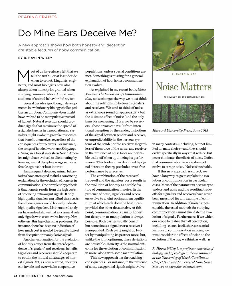
74 THE SCIENTIST | the-scientist.com
READING FRAMES
Most of us have always felt that wetell the truth—or at least decide when to or not. Linguists, engi-
neers, and most biologists have also always taken honesty for granted when studying communication. At one time, students of animal behavior did so, too.
Several decades ago, though, develop-ments in evolutionary biology challenged this assumption. Communication might have evolved to be manipulative instead of honest. Natural selection should pro-duce signals that maximize the spread of a signaler’s genes in a population, so sig-nalers might evolve to provoke responses that benefit themselves regardless of the consequences for receivers. For instance, the songs of hooded warblers (Setophaga citrina) in a forest in eastern North Amer-ica might have evolved to elicit mating by females, even if deceptive songs seduce a female against her best interest.
In subsequent decades, animal behav-iorists have attempted to find a convincing explanation for the evolution of honesty in communication. One prevalent hypothesis is that honesty results from the high costs of producing extravagant signals. If only high-quality signalers can afford these costs, then these signals would honestly indicate high-quality signalers. Mathematical analy-ses have indeed shown that as a general rule only signals with costs evolve honesty. Nev-ertheless, this hypothesis has problems. For instance, there has been no indication of how much cost is needed to separate honest from deceptive or manipulative signals.
Another explanation for the evolution of honesty comes from the interdepen-dence of signalers’ and receivers’ benefits. Signalers and receivers should cooperate to obtain the mutual advantages of hon-est signals. Yet, as now realized, cheaters can invade and overwhelm cooperative
populations, unless special conditions are met. Something is missing for a general explanation of how honest communica-tion evolves.
As explained in my recent book, Noise Matters: The Evolution of Communica-tion, noise changes the way we must think about the relationship between signalers and receivers. We tend to think of noise as extraneous sound or spurious data but the ultimate effect of noise (and the only basis for measuring it) is error by receiv-ers. Those errors can result from inten-tional deception by the sender, distortions of the signal between sender and receiver, or unpredictability in the nervous sys-tems of the sender or the receiver. Regard-less of the source of the noise, any receiver in the presence of noise faces an inevita-ble trade-off when optimizing its perfor-mance. This trade-off, as described by sig-nal detection theory, precludes error-free performance by a receiver.
The combination of the receivers’ trade-off and the signalers’ costs results in the evolution of honesty as a stable fea-ture of communication in noise. In the presence of noise, signalers and receiv-ers evolve to a joint optimum, an equilib-rium at which each does the best it can, provided the other does so also. At this point, communication is usually honest, but deception or manipulation is always possible. Both parties usually benefit, but sometimes a signaler or a receiver is manipulated. Each party might do bet-ter by manipulating its partner more, but, unlike the joint optimum, these deviations are not stable. Honesty is the normal out-come for the evolution of communication in noise, along with some manipulation.
This new approach has far-reaching consequences. For instance, in the presence of noise, exaggerated signals might evolve
in many contexts—including, but not lim-ited to, mate choice—and they should evolve specifically in ways that reduce, but never eliminate, the effects of noise. Notice that communication in noise does not evolve to escape noise. Noise is inevitable.
If this new approach is correct, we have a long way to go to explain the evo-lution of communication in particular cases. Most of the parameters necessary to understand noise and the resulting trade-offs for signalers and receivers have never been measured for any example of com-munication. In addition, if noise is ines-capable, the usual methods for studying communication cannot elucidate the evo-lution of signals. Furthermore, if we widen our scope to realize that all perception, including science itself, shares essential features of communication in noise, we must consider the effects of noise on the evolution of the way we think as well. g
R. Haven Wiley is a professor emeritus ofbiology and of ecology and environment at the University of North Carolina at Chapel Hill. Read an excerpt from Noise Matters at www.the-scientist.com.
A new approach shows how both honesty and deceptionare stable features of noisy communication.
BY R. HAVEN WILEY
Do Mine Ears Deceive Me?
Harvard University Press, June 2015
7509.2015 | THE SCIENTIST
CAPSULE REVIEWS
Brain Storms: The Race to Unlockthe Mysteries of Parkinson’s DiseaseJon PalfremanScientific American / Farrar, Straus and Giroux, September 2015
Parkinson’s is personal for award-winning television producer and journalist Jon Palfreman: he was diagnosed with the disease in 2011. In
his latest book, Brain Storms, he invites readers along as he seeks to understand the neurodegenerative disorder through the eyes of the researchers working every day to uncover vulnerabilities in the disease’s rapacious progression.
“In a profound sense, understanding Parkinson’s disease and finding a cure are now my journalistic beat,” Palfreman writes in the book’s prologue.
The author rewinds the clock all the way back to the early 19th century, when the disease was first described by James Parkinson, and brings the story all the way forward to the present day, as scientists and drug developers seek to extinguish the neurodegenerative disorder. And the journey is not a dispassionate one; it is imbued with the sense of urgency that Palfreman feels as Parkinson’s erodes his brain, and as 60,000 new cases are reported in the U.S. every year.
Orphan: The Quest to Save Children with Rare Genetic DisordersPhilip R. ReillyCold Spring Harbor Laboratory Press, August 2015
Medical geneticist and lawyer Philip Reilly takes on the tough topic of childhood genetic diseases in Orphan, a book about scientific efforts to stamp out the
hundreds of individually rare disorders that collectively afflict children and
adolescents all over the world. “About120,000 (3%) [of the babies born in the U.S. every year] will be diagnosed with a genetic disorder that is caused (or heavily influenced) by a mutation in a single gene,” he writes. Some, however, go undiagnosed for decades.
Reilly, who has long been involved in the ethics of biotechnology and in nonprofit patient advocacy groups, gives a comprehensive overview of what we’re learning about several such maladies, from the latest research into their genetic bases to new diagnostic tools for early detection and cutting-edge treatments.
Although diseases such as alkaptonuria, dystrophic epidermolysis bullosa, and Friedreich’s ataxia are far from household names and are considered “rare” by science, their impact is far-reaching, and, because they usually manifest early in life, the disorders impose a particularly wrenching burden.
Maize for the Gods: Unearthing the 9,000-Year History of CornMichael BlakeUniversity of California Press, August 2015
Corn, humble and unassuming, is one of the most important plants in human history, not to mention biological science. Maize, which is what much of the
world calls corn, also serves as grist for a new book by University of British Columbia anthropologist Michael Blake. In Maize for the Gods, Blake traces the history of corn, from modest grassland plant through millennia of agricultural experimentation and migration to modern global staple.
Blake lays out a fine and factual feast, visiting the work of researchers cracking into corn’s biology as well as its impact on human culture. “We see today, all around us, the transformation of both maize and people as it has spread to almost every place on the planet where crops are
grown—and even where they aren’t,maize still manages to find a way there,” he writes.
Only one important kernel is missing from Blake’s history of maize: it would have been nice to have mentioned the plant’s seminal role in Nobel Prize–winning biologist Barbara McClintock’s pioneering genetics research.
Paranoid: Exploring Suspicion from the Dubious to the DelusionalDavid J. LaPortePrometheus Books, September 2015
Our post-9/11 world is the perfect breeding ground for runaway paranoia. So now, more than ever, it’s important to recognize and treat such overexpres-sions of what was once
an evolutionarily useful emotion. Enter psychologist David LaPorte and his new book, Paranoid, which gives a good over-view of paranoia and the real harm it can do when it distorts one’s thinking.
The most extreme modern expressions of paranoia come in the form of the mass shootings and bombings by obsessed loners that seem to make headlines ever more frequently. Social change, intrusive technology, and post-9/11 security make people feel increasingly insecure, it seems. “The deadly violence perpetrated by individuals such as Seung-Hui Cho (Virginia Tech), Timothy McVeigh, Jared Loughner, and most recently, Aaron Alexis (Washington Navy Yard) is a chilling testament to the effects of that paranoia,” LaPorte writes.
The fact that the author is three or four mass shootings behind (due to the publication cycle, no doubt) is a further testament that he’s on to something. But as with so many other problems faced by society today, science offers a way out. By understanding paranoia in the context of mental health and illness, we can attempt to calm the frazzled nerves of this and future generations.
—Bob Grant
76 THE SCIENTIST | the-scientist.com
The
Gui
de RAFT™ 3D Cell Culture System
The RAFT™ 3D Kit is designed to closely mimic the native environment of cells. It offers optimized reagents, consumables and protocols to develop 3D cultures in under one hour. These robust RAFT™ Cultures can be analyzed using many imaging, biochemical and histological techniques. Along with Lonza’s human primary cells, the RAFT™ Kit empowers researchers to create physiologically relevant cell culture models.
LONZA WALKERSVILLE, INC.1-(800) 521-0390E-mail: [email protected]/raft
Papain (Neural) Dissociation SystemWorthington’s Papain Neural Cell Isolation System is a complete kit for the convenient, consistent isolation of morphologically intact neural single cells for culture, flow cytometry or other applications. The method is based upon published techniques using papain to dissociate CNS tissues providing higher yields and viability than trypsin procedures. Each kit includes five single-use Enzyme Vials, EBSS, Inhibitor and is use-tested to assure performance.
WORTHINGTON BIOCHEMICAL CORP.730 Vassar AveLakewood, NJ 08701Ph: 800-445-9603 • 732-942-1660Fax: 800-368-3108 • 732-942-9270www.worthington-biochem.com
Barrier Function of Endothelial Cells When endothelial cell monolayers are grown in tissue culture, ECIS (Electric Cell-substrate Impedance Sensing) can be used to electrically probe changes in the paracellular pathway between the cells (red arrows in the diagram). ECIS has been used to monitor the effects of molecules effecting barrier function including VEGF, thrombin, TNF alpha, histamine and sphingosine-1-phosphate. These measurements are carried out in real time and without the use of labels.
APPLIED BIOPHYSICS Inc518-880-6860www.biophysics.com
The RAFT™ 3D Cell Culture System
Real-time Budget Control & Analytics Complete accounting software for multi-fund, multi-employee expense and budget tracking and analysis. Calculate overhead, salaries and fringe benefits automatically. Review burn rate and trend graphs. Allocate individual employees’ salaries and effort between multiple accounts/grants over time. Assign expenses to a cost center across many accounts. Export and import data between other systems. Available as public or private cloud versions and local installations.
NORTHERN LIGHTS SOFTWARE+1-(415)453-0481Fax: +1-(415)453-498sales@northernlightssoftware.comwww.northernlightssoftware.com
Laser Autofocus Option Cytation• For Cytation cell imaging multi-mode readers to go along with the current image-based autofocus method• Patent pending laser autofocus design puts all the required components into a single, user-installable cube that can be added at any time to a new or existing system• Offers speed, reproducibility, and lowered risk of phototoxicity and photobleaching
BIOTEKwww.biotek.com
Pathology FISH Probes Cytocell®
• Expand current portfolio with the addition of eight new FISH probes for 1q25, 1p36, 19p13, 19q13, ROS1-GOPC, and RET• Provide bright signals for accurate scoring leading to consistent results, backed up by full evaluation support from OGT• In addition to pathology probes, a large number of other probes are available
OXFORD GENE TECHNOLOGYwww.ogt.com
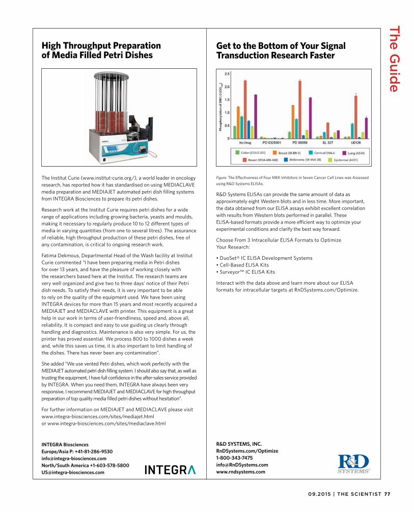
7709.2015 | THE SCIENTIST
The Guid
e
The Institut Curie (www.institut-curie.org/), a world leader in oncology research, has reported how it has standardised on using MEDIACLAVE media preparation and MEDIAJET automated petri dish filling systems from INTEGRA Biosciences to prepare its petri dishes.
Research work at the Institut Curie requires petri dishes for a wide range of applications including growing bacteria, yeasts and moulds, making it necessary to regularly produce 10 to 12 different types of media in varying quantities (from one to several litres). The assurance of reliable, high throughput production of these petri dishes, free of any contamination, is critical to ongoing research work.
Fatima Dekmous, Departmental Head of the Wash facility at Institut Curie commented “I have been preparing media in Petri dishes for over 13 years, and have the pleasure of working closely with the researchers based here at the Institut. The research teams are very well organized and give two to three days' notice of their Petri dish needs. To satisfy their needs, it is very important to be able to rely on the quality of the equipment used. We have been using INTEGRA devices for more than 15 years and most recently acquired a MEDIAJET and MEDIACLAVE with printer. This equipment is a great help in our work in terms of user-friendliness, speed and, above all, reliability. It is compact and easy to use guiding us clearly through handling and diagnostics. Maintenance is also very simple. For us, the printer has proved essential. We process 800 to 1000 dishes a week and, while this saves us time, it is also important to limit handling of the dishes. There has never been any contamination”.
She added “We use vented Petri dishes, which work perfectly with the MEDIAJET automated petri dish filling system. I should also say that, as well as trusting the equipment, I have full confidence in the after-sales service provided by INTEGRA. When you need them, INTEGRA have always been very responsive. I recommend MEDIAJET and MEDIACLAVE for high throughput preparation of top quality media filled petri dishes without hesitation”.
For further information on MEDIAJET and MEDIACLAVE please visit www.integra-biosciences.com/sites/mediajet.html or www.integra-biosciences.com/sites/mediaclave.html
INTEGRA BiosciencesEurope/Asia P: +41-81-286-9530 [email protected] North/South America +1-603-578-5800 [email protected]
High Throughput Preparation of Media Filled Petri Dishes
Figure: The Effectiveness of Four MEK Inhibitors in Seven Cancer Cell Lines was Assessed using R&D Systems ELISAs.
R&D Systems ELISAs can provide the same amount of data as approximately eight Western blots and in less time. More important, the data obtained from our ELISA assays exhibit excellent correlation with results from Western blots performed in parallel. These ELISA-based formats provide a more efficient way to optimize your experimental conditions and clarify the best way forward.
Choose From 3 Intracellular ELISA Formats to Optimize Your Research:
• DuoSet® IC ELISA Development Systems• Cell-Based ELISA Kits• Surveyor™ IC ELISA Kits
Interact with the data above and learn more about our ELISA formats for intracellular targets at RnDSystems.com/Optimize.
Get to the Bottom of Your Signal Transduction Research Faster
R&D SYSTEMS, INC.RnDSystems.com/[email protected]

78 THE SCIENTIST | the-scientist.com
The
Gui
de
Read The Scientist on your iPad!
iPadAds_2015.indd 6
Biosearch Technologies recently launched qPCRdesign.com, a comprehensive online resource for assay design, oligo analysis, webinars, and references.
You can design, analyze, experiment, and play with a variety of interactive tools at your disposal. This online resource provides quick navigation to all the information you need to perform real-time PCR assays in no time:
• Explore qPCR Design Lab’s Toolbox that consists of six different tools and calculators, including:
° OligoSpecTM Calculator - Determine the physical properties of your oligo° Spectral Overlay Tool for Multiplexed qPCR - Visualize which dyes and quenchers are most compatible with your qPCR instrument for multiplex applications° Oligo Resuspension Calculator - Easily find the volume to create oligo stock solutions° Oligo Concentration Calculator - Utilize your OD260 spectrophotometer measurement to determine molarity
• Design assays using our free online RealTimeDesign™ software for qPCR applications including gene expression analysis, SNP genotyping, and multiplexed qPCR.• Read about the history of PCR and the origins of BHQ® probes.• Explore qPCR Design Lab’s library of technical application notes, publications, and protocols to help fuel your research and expand your knowledge base.• Watch educational animations and webinars to stay up-to-date with assay design’s best practices or learn more about MIQE guidelines.• Take a break and play the qPCR CrushTM game and find out how many amplicons you can score!
Stop wasting time searching through multiple websites to get the details you need. qPCRdesign.com is created for quick and easy access to all things related to real-time PCR.
qPCR Design Lab, a New Online Tool for Real-time PCR
BIOSEARCH TECHNOLOGIES, INC.1-800-436-6631qPCRdesign@biosearchtech.comwww.biosearchtech.com
Read The Scientist on your iPad!

Go Nowwww.labx.com
Got old equipment sittinG around takinG up space? SELL ITwww.labx.com
jpG jpG
jpGTakE a PhoToA picture is worth a 1,000 words… Adding images attracts online buyers and makes you money.
1260,000+average monthly visits
labx.com
PoST oN Labx.comPost your equipment where lab professionals shop. List your items for sale via auctions or classified ads.
2coLLEcT Your moNEYIf you’ve followed steps 1 & 2, this should be easy! Visit LabX.com to list your used or surplus lab products.
$ $ $
3 SoLD
www.labx.comSince 1995, LabX is your online marketplace to buy and sell lab equipment and supplies. Have equipment to sell? Use our online auctions or classified ads to sell your lab equipment. If you are a lab equipment manufacturer and have new, demo, or surplus lab products contact us today to promote your items with a customized selling package: 705-528-6888 or email [email protected].
Go Nowwww.labx.com
13th Annual
September 21 - 25, 2015Westin Waterfront Boston | Boston, MA
The Industry’s Preeminent Event on Novel Drug Targets
DiscoveryOnTarget.com
SEPTEMBER 22 - 23
n Targeting Epigenetic Readers and Chromatin Remodelers n Targeting the Ubiquitin Proteasome Systemn Targeting the Microbiome n GPCR-Based Drug Discovery - Part 1 n Antibodies Against Membrane Protein Targets - Part 1 n RNAi for Functional Genomics Screening n Gene Therapy Breakthroughs n Targeting Ocular Disorders
SEPTEMBER 23 - 24
n Targeting Histone Methyltransferases and Demethylasesn Targeting the Unfolded Protein Response n Kinase Inhibitor Discoveryn GPCR-Based Drug Discovery - Part 2 n Antibodies Against Membrane Protein Targets - Part 2 n New Frontiers in Gene Editingn Quantitative Systems Pharmacology
SYMPOSIA
SEPTEMBER 21n Next-Generation Histone Deacetylase Inhibitors n Strategies for Rare Diseases
SEPTEMBER 22n Developing CRISPR-Based Therapies
SAVE $100 OFF Reference key code O75 upon registeringTHE CURRENT REGISTRATION RATE
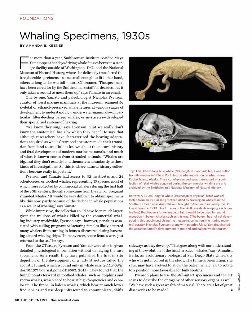
80 THE SCIENTIST | the-scientist.com
FOUNDATIONS
For more than a year, Smithsonian Institute postdoc MayaYamato spent her days driving whale fetuses between a stor-age facility outside of Washington, D.C., and the National
Museum of Natural History, where she delicately transferred the irreplaceable specimens—some small enough to fit in her hand, others as long as she was tall—into a CT scanner. “The specimens have been cared for by the Smithsonian’s staff for decades, but it only takes a second to mess them up,” says Yamato in an email.
One by one, Yamato and paleobiologist Nicholas Pyenson, curator of fossil marine mammals at the museum, scanned 56 skeletal or ethanol-preserved whale fetuses at various stages of development to understand how underwater mammals—in par-ticular, filter-feeding baleen whales, or mysticetes—developed their specialized systems of hearing.
“We know they sing,” says Pyenson. “But we really don’t know the anatomical basis by which they hear.” He says that although researchers have characterized the hearing adapta-tions acquired as whales’ tetrapod ancestors made their transi-tion from land to sea, little is known about the natural history and fetal development of modern marine mammals, and much of what is known comes from stranded animals. “Whales are big, and they don’t exactly lend themselves abundantly to these kinds of investigations. So this is where natural history collec-tions become really important.”
Pyenson and Yamato had access to 32 mysticetes and 24 odontocetes, or toothed whales, representing 15 species, most of which were collected by commercial whalers during the first half of the 20th century, though some came from bycatch or pregnant stranded whales. “It would be very difficult to obtain specimens like this now, partly because of the decline in whale populations as a result of whaling,” says Yamato.
While impressive, the collection could have been much larger, given the millions of whales killed by the commercial whal-ing industry worldwide, Pyenson says; however, penalties asso-ciated with culling pregnant or lactating females likely deterred many whalers from turning in fetuses discovered during harvest-ing aboard whaling ships. “In many cases, those fetuses were just returned to the sea,” he says.
From the CT scans, Pyenson and Yamato were able to glean detailed physiological information without damaging the rare specimens. As a result, they have published the first in situ depiction of the development of a fatty structure called the acoustic funnel, which is found only in whale ears (PLOS ONE, doi:10.1371/journal.pone.0118582, 2015). They found that the funnel points forward in toothed whales, such as dolphins and sperm whales, which need to hear at high frequencies and echo-locate. The funnel in baleen whales, which hear at much lower frequencies and use deep infrasound to communicate, shifts
sideways as they develop. “That goes along with our understand-ing of the evolution of the head in baleen whales,” says Annalisa Berta, an evolutionary biologist at San Diego State University who was not involved in the study. The funnel’s orientation, she says, may have evolved to allow the baleen whale jaw to rotate to a position more favorable for bulk feeding.
Pyenson plans to use the still-intact specimens and the CT scans to describe the ontogeny of other sensory organs as well. “We have such a great wealth of material. There are a lot of great discoveries to be made.”
Top: This 39-cm-long blue whale (Balaenoptera musculus) fetus was culled from its mother in 1936 at Port Hobron whaling station on what is now Kodiak Island, Alaska. The alcohol-preserved specimen is part of a col-lection of fetal whales acquired during the commercial whaling era and archived by the Smithsonian’s National Museum of Natural History. Bottom: A 65-cm-long fin whale (Balaenoptera physalus) fetus was col-lected from an 18.3-m-long mother killed by Norwegian whalers in the Southern Ocean near Australia and brought to the Smithsonian by the US Coast Guard in 1939. This CT scan of the skull reveals developing ear bones (yellow) that house a funnel made of fat, thought to be used for sound reception in baleen whales such as this one. (The baleen has not yet devel-oped in this specimen.) Using the museum’s collection, the marine mam-mal curator, Nicholas Pyenson, along with postdoc Maya Yamato, charted the acoustic funnel’s development in toothed and baleen whale fetuses.
BY AMANDA B. KEENER
Whaling Specimens, 1930s
MA
YA
YA
MA
TO

Psssttt!
In case you haven’t heard, there’s a new “it” girl on the block. She runs Simple Plex assays like you wouldn’t believe! Four immunoassays, 25 μL of sample, 5 minute setup and data ready-to-publish in 60 minutes. Meet Ella.
proteinsimple.com/ella.html

052.A1.0101.D © 2015 Eppendorf AG.
ccc.eppendorf.com • 800-645-3050
Impress YourselfThe new Eppendorf Cell Culture Consumables
> Unsurpassed quality, clarity, purity and sterility, providing reliable cell culture conditions
> Significantly improved design for more safety and consistency
> Maximum safety and confidence during storage and transportation
The all new line of Eppendorf Cell Culture Consumables will truly delight your cells. The outstanding design, reliability and purity is based on more than 50 years of experience. Products created by experts, developed for perfectionists. Impress yourself!
Related Documents