MAKERERE UNIVERSITY PREVALENCE AND GENOTYPING OF AFRICAN SWINE FEVER VIRUS IN APPARENTLY HEALTHY PIGS IN MASAKA, MUKONO AND KAMULI DISTRICTS IN UGANDA BY Joyce Akol (BBLT, Mak) 2011/HD17/2777U A DISSERTATION SUBMITTTED TO THE DIRECTORATE OF RESEARCH AND GRADUATE TRAINING AS PARTIAL FULFILMENT OF THE REQUIREMENTS FOR THE AWARD OF THE DEGREE OF MASTERS OF SCIENCE IN MOLECULAR BIOLOGY OF MAKERERE UNIVERSITY SEPTEMBER 2015

Welcome message from author
This document is posted to help you gain knowledge. Please leave a comment to let me know what you think about it! Share it to your friends and learn new things together.
Transcript

MAKERERE UNIVERSITY
PREVALENCE AND GENOTYPING OF AFRICAN SWINE FEVER VIRUS IN
APPARENTLY HEALTHY PIGS IN MASAKA, MUKONO AND KAMULI
DISTRICTS IN UGANDA
BY
Joyce Akol (BBLT, Mak)
2011/HD17/2777U
A DISSERTATION SUBMITTTED TO THE DIRECTORATE OF RESEARCH AND
GRADUATE TRAINING AS PARTIAL FULFILMENT OF THE REQUIREMENTS
FOR THE AWARD OF THE DEGREE OF MASTERS OF SCIENCE IN
MOLECULAR BIOLOGY OF MAKERERE UNIVERSITY
SEPTEMBER 2015

DECLARATION
I Joyce Akol declare that this work is my original work and has never been submitted to this
university or any other institution of higher learning for any award.
Signature……………………………………Date ……………………………
This work has been done under the supervision and approval of my supervisors,
Associate Professor Charles Masembe (BVM, MSc, PhD),
Makerere University, College of Natural Sciences
P.O.BOX 7062 Kampala, Uganda
Signature………………………………….. Date………………………………
Dr. Michel Dione (DVM, MSc, PhD)
International Livestock Research Institute
P.O.BOX 24384 Kampala, Uganda
Signature…………………………………. Date………………………………..
Dr. Denis Muhangi (BVM, MSc, PhD)
Makerere University, College of Veterinary Medicine, Animal Resources and Biosecurity,
P.O.BOX 7062 Kampala, Uganda
Signature…………………………………Date…………………………………

ii
DEDICATION
This work is dedicated to my parents, Mr. John Francis Esegu and Mrs. Mary Akello Esegu, my
brothers Olinga C, Otule M, Omulala S, Esegu M, sisters Adeke B, Alupo J, my sister-in-law
Kongai J and my dear son Aginya E.J, for the encouragement, spiritual, financial and moral support
accorded to me during my studies.

iii
ACKNOWLEDGEMENT
I would like to express my gratitude to the International Livestock Research Institute-Smallholder
Pig Value Chain Development (ILRI-SPVCD) project Uganda for funding my research work. I
am forever grateful to my supervisors; Associate Professor. Charles Masembe, Dr. Michel Dione
and Dr. Denis Muhangi for their patience; tireless guidance, professional commitment, influence
and guidance that has helped me finish my work. I would like to express my thanks to Dr. Yona
Baguma at the National Crops Resources Research Institute (NaCRRI) for granting me the
opportunity to study and work concurrently. I also appreciate the Madhvani group for financially
supporting me with tuition fees for my first year. I am entirely indebted to all my lecturers for the
knowledge imparted which has improved me as a whole. I also extend my thanks to the District
Veterinary Officers(DVOs) of Masaka, Mukono and Kamuli districts together with the local
council members and field support staff for their contribution to the study and Dr. Edward Okoth
who guided me with data analysis. To my friends Martin Chamai, Joseph Kungu and Doreen
Buhwa thank you so much for the academic, financial and moral support. Last but not least, I
appreciate my parents, brothers; sister and son for bearing with my absence, you all are my biggest
inspiration. Finally to God for His provident love and mercy that has enabled me to complete this
work.

iv
TABLE OF CONTENTS
Contents DECLARATION ........................................................................................................................................... i
DEDICATION .............................................................................................................................................. ii
ACKNOWLEDGEMENT ........................................................................................................................... iii
LIST OF TABLES ....................................................................................................................................... vi
LIST OF ABBREVIATIONS .................................................................................................................... viii
ABSTRACT ................................................................................................................................................. ix
CHAPTER ONE ........................................................................................................................................... 1
INTRODUCTION ........................................................................................................................................ 1
1.1Background .......................................................................................................................................... 1
1.2 Statement of the problem .................................................................................................................... 2
1.3.1 Specific objectives ....................................................................................................................... 3
1.4 Research questions .............................................................................................................................. 3
1.5 Justification of the study ..................................................................................................................... 3
CHAPTER TWO .......................................................................................................................................... 4
LITERATURE REVIEW ............................................................................................................................. 4
2.1 Pig production systems in Uganda ...................................................................................................... 4
2.2 African swine fever ............................................................................................................................. 5
2.2.1 Aetiology ...................................................................................................................................... 5
2.2.3 Pathology ..................................................................................................................................... 7
2.2.4 Clinical Signs ............................................................................................................................... 8
2.2.5 Epidemiology of ASF .................................................................................................................. 8
2.3 Burden of ASFV in Uganda .............................................................................................................. 11
2.4 The ASFV Genome and Genotyping .................................................................................................... 11
2.5 Laboratory diagnosis of ASFV ......................................................................................................... 12
2.5.1 Haemadsorption virus isolation (HAD) ..................................................................................... 12
2.5.2 Detection of antibodies against ASFV ....................................................................................... 13
2.5.3 Detection of viral particle .......................................................................................................... 13

v
2.6 Risk factors for African swine fever transmission in Uganda .......................................................... 14
2.7 ASF control measures ....................................................................................................................... 15
CHAPTER THREE .................................................................................................................................... 17
MATERIALS AND METHODS ................................................................................................................ 17
3.1 Study area.......................................................................................................................................... 17
3.3 Study sites ......................................................................................................................................... 17
3.4 Sample size determination ................................................................................................................ 19
3.5 House-hold and pig selection ............................................................................................................ 20
3.6 Sample collection .............................................................................................................................. 20
3.7 ASF DNA detection, characterization and antibody detection ......................................................... 21
3.7.1 The ASFV antibody detection .................................................................................................... 21
3.8 Ethical consideration ......................................................................................................................... 27
CHAPTER FOUR ....................................................................................................................................... 28
RESULTS ................................................................................................................................................... 28
4.1 Demographic characteristics ............................................................................................................. 28
4.1.2 Characteristics of pig production systems .................................................................................. 28
4.2 Antibody detection of ASF ............................................................................................................... 29
4.3 Molecular detection of ASFV ........................................................................................................... 29
4.3.1 Detection of ASFV using Real time PCR .................................................................................. 29
4.3.2 Conventional amplification of ASFV ........................................................................................ 30
4.4 Sequencing ........................................................................................................................................ 31
4.4.1 Sequence Alignment of the p72 and p54 ................................................................................... 31
4.4.2 Phylogenetic analysis of the p72 and P54 regions ..................................................................... 34
CHAPTER FIVE ........................................................................................................................................ 36
DISCUSSION ............................................................................................................................................. 36
CHAPTER SIX ........................................................................................................................................... 39
CONCLUSIONS AND RECOMMENDATIONS ..................................................................................... 39
6.1 Conclusions ....................................................................................................................................... 39
6.2 Recommendations ............................................................................................................................. 39
REFERENCES ........................................................................................................................................... 40

vi
LIST OF TABLES
Table 1; Districts, sub counties and villages visited ..................................................................... 18
Table 2; Shows the Real time PCR reaction mix .......................................................................... 24
Table 3; Sets of primer sequences used for base pair amplification of ASFV DNA .................... 25
Table 4; Characteristics of production system .............................................................................. 29
Table 5; Sequences downloaded from NCBI, region amplified, country of origin and genotype 32

vii
LIST OF FIGURES
Figure 1 Transmission cycles of the African swine fever virus ................................................... 10
Figure 2 Map of Uganda showing study sites, with districts and Sub-counties .......................... 19
Figure 3 shows each sample ran with the primers amplifying different regions of the ASFV
genome.. ........................................................................................................................................ 30
Figure 4 Sequence alignment of the P54 region of the different isolates. ................................... 33
Figure 5 Phylogenetic tree base of the C-terminal end of the p72 sequences ............................. 34
Figure 6 Phylogenetic tree base of the full length P54 protein sequences ................................... 35

viii
LIST OF ABBREVIATIONS
ASF African swine fever
ASFV African swine fever virus
CVR Central variable region
DVO District Veterinary Officer
EDTA Ethylene Diamine Tetra acetic Acid
ELISA Enzyme Linked Immunosorbent Assay
GIS Geographical Information System
ILRI International Livestock Research Institute
LC Local council
NAADS National Agricultural Advisory Services
NGO Non-Governmental Organization
OD Optical Density
OR Odds Ratio
PC Positive Control
RR Rural-Rural
RT-PCR Real time Polymerase Chain Reaction
SPVCD Smallholder Pig Value Chain Development

ix
ABSTRACT
African swine fever (ASF) is a viral hemorrhagic disease associated with death in infected pigs.
African swine fever virus (ASFV) is a DNA virus that circulates in blood and lymphoid system of
the pigs causing disease. There are various reports on ASF outbreaks in the country with a few
confirmed in apparently healthy pigs which pigs show no signs of infection. Therefore a survey of
apparently healthy pigs was undertaken to show the extent they habour the antibodies and antigen
of ASFV and later determine the genetic diversity of the virus ASF in Kamuli, Mukono and
Masaka districts of Uganda using serological, molecular and genotyping techniques. In total 1,192
blood and sera samples were collected and analyzed. All the pigs tested except one (1/1192) were
negative for (ASFV) and none for antibodies indicating that ASFV causes a paracute / acute
infection in Ugandan pigs with rare detection of virus or antibodies in apparently healthy pigs.
Therefore chronically infected pigs are unlikely to be important in the epidemiology of ASF. The
positive pig in Kamuli district was infected with genotype IX, the most common circulating ASFV
genotype in Uganda. With one positive pig for ASFV, it was not possible to authoritatively
associate predictors of infection with disease in tested pig farms. It is thus recommended that these
predictors of infection with ASFV are studied in future ASF outbreak areas where the virus or
antibodies in pigs may occur in high prevalence.
Key words: African swine fever virus, apparently healthy pigs, Serology, PCR, Sequencing

CHAPTER ONE
INTRODUCTION
1.1 Background
Uganda has the largest and rapidly growing pig production industry in Eastern Africa and its pig
population has risen from 0.19 to 3.2 million in the past 3 decades (UBOS, 2013). This increase is
estimated to be about 70% in Uganda that has the highest per capita pork consumption in East Africa
(UBOS, 2008). Pig production is mainly dominated by the rural free-range smallholder systems ;
however, other systems such as the intensive and semi-intensive also exist but are more common in
the urban and peri-urban areas (Muhanguzi et al., 2012).
Despite the increase in pig production in the country, this industry faces a lot of constraints like: poor
housing, high feeding costs and disease burden (Dione et al., 2014). ASF is important socio-
economically to the farmers; because it can lead to very high mortality and morbidity in infected pigs.
These “apparently healthy pigs” on farms are rarely tested for presence of antibodies and ASFV
because these tests are quite expensive. The apparently healthy pigs are pigs which show no signs of
disease but may harbor disease causing pathogens.
Epidemiological studies undertaken to understand the disease in Uganda have hinted at possible
presence of the virus in domestic pigs without clinical signs of ASF (Tejler, 2012: Atuhaire et al.,
2013). These chronically infected pigs may spread the virus for long since they may have acquired
resistance or adapted to the condition, hence the continued outbreaks that lead to income losses and
unemployment in the country (Atuhaire et al., 2013). However, other factors like: consumption of
contaminated swill, concentrate, pasture, sharing feeding utensils and the use of the same breeding

2
boars may precipitate viral infection amongst pigs (Fasina et al., 2012). Exchange of pigs among
farmers is another avenue for viral spread in the pig population communities (Costard et al., 2012).
These pigs need to be investigated for pathogen presence using rapid and reliable diagnostic
techniques.
This study was therefore designed to determine the sero and antigen status of ASFV in “apparently
healthy” pigs in Masaka, Mukono and Kamuli districts in Uganda.
1.2 Statement of the problem
Smallholder pig farmers face a serious challenge of ASF. Recent studies have shown presence of the
virus in some apparently healthy pigs mainly at the slaughter slabs (Tejler, 2012:Muwonge et al.,
2012:Atuhaire et al., 2013). Inaddition, various activities along the pig value chain like; movement
of pigs from farm to farm and from farm to market; movement of extension service providers from
farm to farm without protective wear and disinfection expose the pigs to viral infection (Nantima et
al., 2015). At the farm level, there is lack of proper disease surveillance, quarantine, protective wear
and disinfection of personnel and equipment (Fasina et al., 2012). Subsequently, farmers normally
borrow boars for mating and restock their farms with pigs from within the community without
determining their health status also poses a risk of infection (Muhangi et al.,2015). With the lack of
routine diagnostics in the smallholder farmers and high disease prevalence as reported by farmers
without confirmation in Masaka, Mukono and Kamuli districts (Dione et al., 2014), there was need
to detect presence of antibodies and ASFV in the apparently healthy pigs on farm in Mukono, Masaka
and Kamuli districts.
3
1.3 General objective
The general objective was to assess the healthy status of the apparently healthy pigs to ASF antibodies
and antigen in Masaka, Mukono and Kamuli districts.
1.3.1 Specific objectives
1. To determine the antibody and ASFV DNA status in apparently healthy pigs in the districts of
Mukono, Masaka and Kamuli.
2. To genotype the detected ASFV from the apparently healthy pigs.
1.4 Research questions
1. What is the sero and antigen status of ASFV in apparently healthy pigs in the above districts?
2. What are the circulating genotypes of ASFV in apparently healthy pigs in the above districts?
1.5 Justification of the study
Several households depend on the livestock industry like piggery for improved livelihood. ASF poses
a major challenge to the pig industry in Uganda where the disease is endemic. With the disease being
ranked first in Masaka, Mukono and Kamuli districts (Dione et al., 2014), there was need to confirm
presence of ASFV and antibodies in the apparently healthy pigs in these same districts. Determining
the sero and antigen status of the ASFV in apparently healthy pigs and further characterization of the
virus will help understand the molecular epidemiology of the disease in Uganda. This information
once generated, will be communicated and used to better inform the currently available prevention
and control strategies for ASF.

4
CHAPTER TWO
LITERATURE REVIEW
2.1 Pig production systems in Uganda
The pig production industry in Uganda has experienced a population increment of about 70% in the
last decade (UBOS, 2013). The livestock sector contributes 3% GDP to the government of Uganda
(MAAIF, 2011). Pig farming provides improved livelihood to the farmers mainly in rural areas since
the costs incurred are not high. The pigs are easily sold off by farmers to cater for school fees, medical
bills and other family requirements (Ouma et al.,2014). The pig feces are used as manure to improve
soil fertility; further improving agriculture which is the backbone of this country. The breeds of pigs
reared are dependent on the farmers’ perceptions and include, land race, yorkshire, large white,
cambrough (exotics), cross breed (hybrid) and local breed (indigenous) (Tatwangire, 2013). The
exotic and cross breeds mature fast. On the contrary, the local breeds are assumed to be disease
resistant and easier to manage since minimal attention is required.
Uganda generally has three pig management systems which are; intensive, semi-intensive and
extensive systems (Tatwangire, 2013). The intensive pig system involves housing the pigs in pens
and providing them with feeds and water (Mutetikka, 2009; Pezo & Waiswa, 2012; Ouma et al.,
2013; Dione et al., 2014). These enclosed houses are constructed depending on the availability of
resources as raised or flat cemented floor. In the semi-intensive system pigs are housed but
occasionally left to roam outside posing a risk to viral infection (Mutetikka, 2009). Finally, in the
extensive system the pigs are left to roam around the area looking for food and water, with little effort
required for management hence such pigs are at a very high risk of viral infection.
5
Farmers in the rural area mainly practice the extensive system since the majority have limited
resources for developing structure and maintaining the pigs (Dione et al., 2014). Although the
extensive system is inexpensive, other farmers complain of crop destruction by roaming pigs (Pezo,
D. & Waiswa, 2012). Most of the peri-urban and urban pig farmers construct pig shelters using
timber, concrete, galvanized iron and partition the herd for easy management (Tatwangire, 2013). In
the rural areas however, majority of farmers construct pig shelters using local materials like mud,
bamboo, wood, grass and banana leaves which are less durable and expensive since these materials
need to be replaced with time.
2.2 African swine fever
African swine fever is a haemorrhagic viral disease in pigs which is responsible for high mortality
rates thus affecting the pig industry in Sub-Saharan Africa (Dixon & Takamatsu, 2012). The disease
was first described in Kenya in 1920’s, and ever since then, it has spread to other parts of the world
(Montgomery, 1921).
2.2.1 Aetiology
The causative agent of ASF is a double stranded, icosahedral DNA virus (Dixon et al., 2000). This
disease was first described by Montgomery 1921 in Kenya and there are 22 known genotypes ( I-
XXII) of ASFV circulating world-wide (Boshoff et al., 2007 and Bastos et al., 2003). Although some
studies suggest both genotypes IX and X to be present in Uganda, most recent studies indicate
genotype IX is more prevalent (Atuhaire et al., 2014).

6
2.2.2. Viral Structure and Replication
The ASFV is organized in complex multi-layer structures consisting of an 80 nm core structure, a
30nm nucleiod, surrounded by a 50nm lipid layer and protein icosahedral capsid (Carrascosa et al.,
1985). The diameter is estimated to be about 170-190nm and has an estimated size of 175-215nm.
The Viral DNA genome is about 170-193kbp in length. The variations in length are due to losses or
gain of members of multi-gene families which include 100, 110, 300, 360, 505/530 and p22 (Dixon
et al., 2012).The external envelope, although not necessary for infectivity, contains CD2v (EP402R),
the only glycoprotein involved in virus haemadsorption (Ruiz-Gonzalvo et al., 1996; Rowlands et
al., 2009) . Viral entry into the porcine host mainly is through the tonsils to the closest lymph nodes,
blood where it is carried to tissue organs. The pig may be bitten by infected ticks during feeding and
introduce the virus directly into the blood stream of the pig. Upon entry into the soft tick, the virus
replicates in different cell types, and in the mononuclear phagocytic system of pigs and bush pigs
(Carriloo et al., 1994; Karalyan et al., 2012). Viral entry into the host cell is through receptor–
mediated endocytosis although recent studies suggest that viral entry may be through clathrin
dependent endocytosis and micropinocytosis (Sánchez et al., 2012; Alonso et al., 2013). The major
proteins involved in macropinocytosis are capsid protein p72, p54 and p12. These proteins are
responsible for binding and entry into the cytoplasm (Etter et al., 2011). Although immature viral
DNA is observed in the nucleus and mature viral DNA in the cytoplasm, both DNAs are responsible
for the mature cross-linked viral DNA present in infected cells (Dixon et al., 2012). The early phase
of viral replication occurs in the nucleus with peak DNA replication occurring for the first six hours
and then decreases to zero in the next 12hours (Dixon et al., 2012). Viral assembly takes place in the
cytoplasm, since the cytoplasm contains viral structural proteins, viral DNA and many membranous
7
materials required for ASFV assembly (Rojo et al., 1999). Attachment proteins p12 and p24 are found
on the external membrane of the extracellular particles, while p150, p37, p34 and p14 proteins are
localized in the virus core (Alcami et al., 1990; Suárez, Salas, & Rodríguez, 2010). The Vp72 protein
(B646L gene) is the main component of the viral capsid while p54 (E183L gene) is the most important
integral membrane protein and both proteins are used in virus genotyping (Cobbold&Wileman,
1998). The inner viral envelope is very complex and contains many viral proteins with trans-
membrane domains (Sun et al., 1996). The ASFV particles contain different enzymes for replication,
mRNA polyadenylation, methylation and capping. Clumping of DNA polymerase to DNA is
facilitated by DNA polymerase type B and protein E301R. The C962R gene may be important for
DNA repair at the replication fork, although more studies should be undertaken to verify this (Dixon
et al., 2012). Synthesis of mRNA in the cytoplasm is possible without host RNA polymerase II,
because the virus possesses all enzymes and factors required for transcription and translation process.
2.2.3 Pathology
Pigs infected with ASFV show varying forms of the disease; peracute, acute, subacute and chronic
forms. The variations in the forms of the disease are associated with virulence of the viral strain and
immunological status of the pigs (Wilkinson, 2000). The peracute form is linked with
thrombocytopenia mainly in the stomach resulting in prolonged bleeding and death of the pig. Acute
and subacute forms of the disease are characterized with severe vascular changes, haemorrhage in
different organs, mucosal nasal discharge and nasal haemorrhage, renal petechiae, diffuse
haemorrhage in the lymph nodes and erythema. Erythema is easily seen in the white pigs. Oedema of
the gall bladder, perirenal oedema and pulmonary oedema is evident in infected pigs.

8
2.2.4 Clinical Signs
The appearance of symptoms in pigs depends on the incubation period of the virus, the viral genotype,
animal breed, environment, pig production system and viral load in the pig (Jori & Bastos, 2009).
There are a number of clinical symptoms observed amongst which include; the typical and acute
forms that affect the lympho-reticular endothelial cells, high fever, anorexia, dullness, reddened skin
particularly on the ears, lower legs and ventral abdomen (mainly observed in exotic pigs), abdominal
pain, nasal discharge, vomiting, constipation, bloody diarrhea and abortion in pregnant sows. Pigs
that present with these signs normally die between three to 14 days after infection (Mebus, 1988).
Unfortunately, pigs that usually recover from this state become carriers of the virus and spread the
virus to naïve pigs through contaminated faeces, fluids and exhalation. Sub-acute forms of the disease
caused by less virulent strains of the virus display with wavering fever, swollen and painful joints,
cardiac damage, moist cough and difficulty in breathing(Costard et al., 2012). Subsequently, these
pigs continue to exist for weeks to several months, with their role in disease transmission not fully
investigated. The chronic form of the disease may be seen in survivors and is characterized by
emaciation, stunted growth, and haemorrhagic necrosis of the skin, bony protuberances and deep
ulceration.
2.2.5 Epidemiology of ASF
The transmission cycle of ASF Mainly occurs in three cycles: the sylvatic, tick and pig –pig cycle,
with few studies showing the existence of transmission from the wild life to the domestic pigs in
Africa mainly in Uganda (Jori et al., 2013).

9
2.2.5.1 Sylvatic cycle
Warthogs, possibly bush pigs and forest hogs are known vertebrate hosts of ASFV. The virus is
transmitted to the ticks as they feed on these vertebrate hosts. Eventually, the infected ticks transmit
the virus through bites to naïve warthogs and pigs. The ASFV replicates in the bush pigs, although
their involvement in viral transmission to the pigs and soft ticks is not well understood (Jori & Bastos,
2009). A recent study by Ståhl et al.,(2014) gives an insight of the probable involvement of bush pigs
in the transmission of ASFV to pigs and warthogs. Studies on bush pigs are limited since bush pigs
are not easily monitored as a result of their few numbers and their nocturnal nature.
2.2.5.2 Tick-pig cycle
The soft ticks are reservoirs and the only known arthropod vectors for the ASFV. The ASFV
replicates in the midgut and spreads to the coxal and salivary gland in the ticks. The virus is then
transmitted to the pigs as the ticks feed (Kleibeker et al., 1999; Ticks et al., 2007). Notably, these
ticks can remain infectious for about 15months without any blood meal and some studies have
detected presence of the virus in soft ticks found in pig pens emptied four years ago (Ravaomanana
et al., 2010).
2.2.5.3 Pig to pig cycle
This cycle sometimes is referred to as the indirect transmission of ASFV in pigs. The virus is spread
to pigs through contaminated feeds, fomites, vehicles, clothes and poor farming systems (Penrith,
2009). Transmission of ASFV by aerosol may occur since the virus normally occurs during the dry
season. The contaminated aerosol may facilitate the spread of the virus between herds that are near
each other. The ASFV can remain viable in pig secretions, products, bi-products and decayed blood

10
for about 11 days which may aid in the viral spread (Penrith, 2009). Outbreaks in Tanzania were
previously linked to movement of infected pigs and pork products (Misinzo et al., 2009),which
factors were responsible for disease outbreaks in Uganda (Muhangi et al., 2014). Important to note is
the presence of ASFV without presence of a sylvatic cycle, mainly in West Africa and some parts of
Uganda (Jori et al., 2013).The pig cycle could be the main attribute of ASFV transmission in Uganda,
although pigs grazing close to forests are at a high risk of infection because wildlife and ticks colonize
them when looking for food (Björnheden, 2011).
Sylivatic cycle Tick cycleDomestic cycle
Warthog
Bush pig
Domestic pigHuman activities
O. moubata
Ms Akol Joyce & Dr. Denis Muhangi (MUK)
Figure 1; Transmission cycles of the African swine fever virus

11
2.3 Burden of ASFV in Uganda
ASF is endemic in the country with over 300 outbreaks reported between 2001 and 2012 (Atuhaire
et al., 2013) which were responsible for several pig deaths hence affecting the farmers and economy
at large. There are continuous rampant outbreaks in Uganda with a number of serological and
molecular epidemiology studies on ASFV undertaken (Atuhaire et al., 2013). In Uganda, a study
carried out in Mubende showed a 0.2% seroprevalence at slaughter slabs (Muwongeet al., 2012) and
in Gulu a 50% presence of ASFV was observed (Tejler, 2012) during outbreak investigations.
However, in Busia there was no presence of ASFV antibodies using the OIE serological prescribed
tests during the 2007 outbreak since the virus has no neutralizing antibodies (Gallardo et al., 2011).
2.4 The ASFV Genome and Genotyping
The ASFV genome is encoded by five multigene families and about 50 polypeptides. Structural or
function proprieties were identified in 113 proteins after complete sequencing of one Spanish isolate
(Yanez et al., 1995). The presence /absence of some of these proteins in some viral isolates are
responsible for the heterogeneity in ASFV. Classification and understanding the virus isolates can be
determined through genetic studies. The PCR-sequencing method used for genetic studies provides
accurate explanation of major genotype nucleotide sequences of the viral genome (Bastos et al.,
2003). In depth genetic characterization of the central variable region (CVR) of the 9RL open reading
frame (ORF) shows intra-genotypic relation of the various isolates (Bastos et al., 2004). The
molecular epidemiology of ASF in East African showed sixteen vp72 genotypes existed after
phylogenetic analysis of the conserved 404bp region of the C-terminal end of the vp72 gene (Lubisi
et al., 2005). Phylogenetic analysis of four variable regions of 41 isolates of the vp72 genotype

12
produced 16 new sub-groups (Nix et al., 2006). Phylogenetic analysis of the CRV which varies in
size from 300-500bp and has a 132-bp direct repeat, analyses the number and composition of tandem
tetramers in a given isolate (Irusta et al., 1996). Combined vp72 and CVR phylogenetic analysis
permits intra genotyping of ASFV isolates in pigs. Molecular characterization of the p72, p54 and the
CRV of the identified viral isolates showed genotype IX to be circulating in Uganda (Gallardo et al.,
2011; Atuhaire et al., 2013). The genotype IX is present both in pigs and wild suids in areas around
national parks, forests, neighboring communities and areas far away from wildlife (Jori et al., 2013).
However, regions far away from these national parks complain of pig deaths due to ASFV, therefore
it is important to identify other factors linked to disease transmission apart from the ticks and wild
suids which are known to be reservoirs.
2.5 Laboratory diagnosis of ASFV
Isolation and diagnosis of ASFV is possible using various techniques; both serological and molecular
methods as recommended by OIE, 2008.
2.5.1 Haemadsorption virus isolation (HAD)
The test involves the adherence of pig erythrocytes to surface infected pig monocytes or macrophages
with ASFV (Malmquist& Hay 1960). This test is suitable for only virus adsorbing isolates and not
the non-adsorbing isolates that lack the CD2v protein responsible for absorbing red blood cells to
infected virus cell. The suspected blood or tissue suspension is cultured in primary porcine bone
marrow (PBM) cells, primary leukocyte cultures or into alveolar macrophage cells as described by
Malmquist& Hay (1960). Although the test is sensitive, it is laborious, time consuming and not

13
suitable for ‘non- haemadsorbing’ ASFV (OIE, 2008). Therefore, the test is normally performed as a
confirmatory test on positive samples from ELISA and PCR.
2.5.2 Detection of antibodies against ASFV
There are a variety of tests available for the detection of antibodies against ASFV. These tests are
able to detect antibodies; produced immediately after infection and also antibodies in pigs that have
recovered normally referred to as “apparently healthy”. These apparently healthy pigs are normally
infected with less or non-virulent isolates and positive pigs are easily detected through serological
tests (OIE, 2012). Antibody ELISAs detect IgM for recent infection and IgG in chronically infected
or apparently healthy pigs (Reis et al., 2007). These antibodies are present and detected in serum and
fluids from tissues. The commonly used ELISA techniques are immunofluorescence antibody test
and immunoblotting. Immunofluorescence involves the use of cell cultures or tissue sections from
infected pigs stained and viewed under a light microscope (OIE, 2012). Immunoblotting is able to
detect weak positive samples since viral proteins that induce specific antibodies are placed on antigen
strips. The ELISAs are more effective than HAD because both virulent and avirulent strains can be
detected using non-infectious soluble antigens (Wardley et al., 1979). Pigs infected with virulent
ASFV normally do not produce antibodies but kill the pigs within a short time before an immune
response is activated. Therefore these tests are performed in combination with tests that detect viral
presence in the infected pigs.
2.5.3 Detection of viral particle
The virus in body fluid mainly in blood can be detected after DNA extraction. A wide range of
isolates, both haemadsorbing and non haemadsorbing can be amplified. There are various viral
detection techniques like: polymerization chain reaction (PCR), Real-time PCR, and the loop

14
amplification mediated polymorphism (LAMP) (OIE, 2008). These techniques involve the use of
species specific primer sets to amplify a conserved region of the ASFV genome, thermal stable
enzyme, dNTPs, Magnesium chloride, distilled water and the extracted DNA template. The LAMP
method is very sensitive, specific and requires a single temperature for the reaction as compared to
the other amplification method with pre-set condition reactions at different temperatures (Hjertner et
al., 2005). This makes LAMP cheaper and ideal for viral detection in developing countries and in
areas where the disease is endemic and a single temperature required for amplification (Notomi et
al., 2000). The Realtime PCR is more sensitive than the other techniques because very small
quantities of viral DNA in a sample can be detected. Realtime PCR requires less time to attain results
as compared to the others. In addition both qualitative and quantitative results are determined on a
computer, thus no need of running a gel (Gallardo et al., 2012). On the other hand, conventional PCR
is advantageous in that viral DNA can be amplified, size of the genotype determined and the purified
products sent for sequencing (Aguero et al., 2003; Steiger et al., 1992; Bastos et al., 2003).
2.6 Risk factors for African swine fever transmission in Uganda
The pig farmers, pig traders and family members normally do not adhere to biosecurity methods of
decontamination of personal protective wear with a disinfectant before and after visiting the farm
hence a risk for the disease transmission. The pig traders/middle men move from farm to farm looking
for pigs to sale; and transport these pigs on motorcycles and lorries without decontamination (Aliro
et al.,2012) . The pigs are sold very fast during or when farmers suspect disease oubreak (Muwonge
et al., 2012: Muhangi et al., 2014). They are sold cheaply or slaugherted immediately without
inspection. At the slaughter slab, there are insufficient or no facilities for waste disposal of blood,
offals and used water. Therefore, the pork obtained may be contaminated and is sold in the butcheries

15
or roasted/fried by the road sides, restaurants and bars (Costard et al., 2012). The risk factors for
disease transmission need to be investigated, understood and communicated to farmers, stakeholders
and policy markers to contain and prevent disease outbreaks in the country.
2.7 ASF control measures
ASF being endemic in Southern and East Africa has proved difficult to eradicate. Most of the farmers
in the pig sector are subsistence farmers with limited finances available to invest on sanitary biosafety
measures; hence they expose themselves to high risks of infection (Tatwangire A., 2013). These risks
predispose farmers to ASFV outbreaks and cause enormous losses in the house-hold and country at
large. The lack of restriction of live animal movement within a country, transboundary and
transcontinental, free-ranging and farm visits, facilitates disease spread (Costard et al., 2009).
Training of farmers, veterinary and livestock producers on cause and transmission of disease creates
disease awareness; thus controlling the spread (OIE, 2010). Regular spraying with acaricides on tick
infested herds or complete abandonment of previously infested places prevents survival and breeding
ground for ticks that may harbor the virus (Costard et al., 2012). In areas with frequent outbreaks
contact of infected faeces with other animals, birds, pigs, chicken and humans can result in
transmission to healthy pigs, therefore proper disposal of this waste prevents viral spread since the
virus remains viable for 60 days in the excreta (Penrith, 2009). Quarantine for a minimum of 30 days
is important in addition to cleaning, disinfecting clothes, foot wear and isolation units since animals
excreta are regarded infectious (Penrith, 2009). Restaurant waste and kitchen scraps that provide high
energy and protein as growth requirements for pigs, are heated to 100ºC for 1 hour to destroy the
virus before feeding the pigs (OIE, 2010). Improperly buried infected carcasses, when exhumed are
potential sources of infection. The control strategy for ASFV transmission largely relies on early

16
rapid disease diagnosis, implementing strict biosecurity measures and good communication among
all parties involved during and after outbreaks (Oura et al., 2013).

17
CHAPTER THREE
MATERIALS AND METHODS
3.1 Study area
A cross-sectional survey was conducted from April to August 2013 in the districts of Masaka,
Mukono and Kamuli. These districts were selected based on Step 1: Geographical targeting using
GIS characterization and spatial analysis to select potential sites, pig population density of >20
heads/Km2 and poverty (people living on <1.25$/day) levels of >50%. The population density in
Masaka and Mukono was > 50 head/ Km2 while Kamuli was between 5-10 head/ Km2. All the above
districts had a 60-70% population of people living on <1.25$/day.
3.3 Study sites
The study sub-counties and villages were selected using pig population data at sub-county level from
the Livestock Census data of 2008. For each district, 4-6 sub-counties were purposively selected
basing on pig population density. Within each selected sub-county, two to three villages were
randomly selected. In this study, 23 villages out of the 35 were selected purposively across the three
districts (Table 1). The number and choice of villages was based on financial resources availability
and other activities taking place in the same villages, to minimise farmer fatigue (Ouma, et al., 2014).

18
Table 1; Districts, sub counties and villages visited
District Sub-county Village
Mukono Kkingo Kisoso
Ssenya
Kimanya-kyabakuza Kijjabwemi
Kyabakuza
Katwe-Butego Butego
Kyamuyimbwa
Nyendo-Ssenyange Ssenyange A
Kabonera Kikalala
Kyanamukaka Kanoni-Bukunda
Lukindu
Kamuli Bugabula Baluboinewa
Butansi Bukyonza
Kitayunjwa Ntansi
Namwendwa Isingo A
IsingoB
Mukono Goma Misindye
Mukono TC Kitete
Joggo
Kyampisi Ddundu
Kyoga
Ntenjeru Bugoye/Kabira
Kazo/Kalagala
Nsanja/Gonve

19
The map of Uganda with the study sites in different colours
Figure 2 Map of Uganda showing study sites, with districts and Sub-counties
3.4 Sample size determination
The sample size was calculated using the formula adopted from Thrusfield (2007) as follows: n =
[Z2P(1-P)]/d2 Where: n is the required sample size; Z is the multiplier from a standard normal
distribution (1.96) at a probability level of 0.05; P is the estimated prevalence which is most
conservatively estimated to be 50% considering that there is no reliable prevalence data for African
swine fever per district and d is the desired precision for the estimate (+/- 5%). A sample size of 384

20
pigs was required for the study in each district. A total of 375, 408, and 402 pigs were sampled in
Masaka, Kamuli and Mukono, respectively. The actual number of pigs sampled in Masaka was lower
than the required sample size because of the harsh working conditions experienced due to heavy rain
on some days. More pigs in Kamuli and Mukono were sampled because they were available.
3.5 House-hold and pig selection
A list of all pig keeping house-holds was generated in each village. The study house-holds were then
randomly selected using computer-generated random numbers. The inclusion criteria were as
follows: pigs older than three months since we assumed they were exposed, not weak or emaciated,
not a pregnant sow or a sow with a litter under two months old to were not considered to avoid any
losses of pigs and at less than 2 months, it was difficult to collect the blood sample. In each house-
hold, one pig fulfilling the inclusion criteria was randomly selected for blood collection. A pig bio-
data form and a structured questionnaire were administered to the owner of the pigs.
3.6 Sample collection
The farmers were first briefed on the background of the study before signing the consent forms. One
pig randomly selected from each farm was restrained using a snare. Subsequently, whole blood was
collected from the jugular vein of the pig using sterile vacutainer needles into sterile vacutainer tubes.
The plain vacutainers were used to obtain serum after centrifugation at 3000rpm for 10 minutes and
used for ASFV antibody detection. Whole blood was collected in EDTA-coated vacutainers and used
for PCR. These tubes were labeled immediately with the farmer (identity) ID; the district and house-
hold number e.g. M001. The tubes then were put in a cold box before transportation to the laboratory
for analysis. Both whole blood and serum were put into two separate cryovials and stored at -80ºC

21
until analyses. At the farm, biosecurity measures like: disinfection of all personnel, materials used,
gumboots and vehicles with 70% Virkon diluted in a pump sprayer was done before movement to the
next farm to prevent virus spread.
3.7 ASF DNA detection, characterization and antibody detection
This study used both antibody and viral DNA detection methods simultaneously as recommended by
the OIE for diagnosis of ASF. Antibody analysis on the harvested serum and DNA extraction from
whole blood was performed at the Molecular Genetics Laboratory at Makerere University, College
of Agriculture and Environmental Sciences. Molecular detection of the virus on the extracted DNA
samples was performed using UPL Realtime PCR at the International Livestock Research Institute
(ILRI) Nairobi.
3.7.1 The ASFV antibody detection
Blood collected in the plain vacutainer tubes was centrifuged at 3000 rmp for 10 minutes to separate
the blood clot from serum. The supernatant (serum) was then harvested into two cryovials and stored
at -80ºC. A commercial blocking enzymatic immunoassay kit from Ingenasa (Ingezim 11.PPA.K3,
ingenasa, Madrid, Spain) was used for ASFV antibody detection. This test is very sensitive and
specific (OIE, 2008). The wells in the plates were pre-coated with a purified protein extract from the
virus (VP73). Protein VP73 is a major structural protein from the ASFV which is the most antigenic
and suitable for diagnosis (OIE,2008). Before use, the serum stored at -80ºC was left to thaw to room
temperature together with the ELISA kit reagents (INGEZIM COMPAC 11.PPA.K3) except the
enzyme conjugate. All reagents used in this process were purchased with the kit, except for absolute
alcohol used to dilute some reagents. Both samples and controls were diluted directly in the 96 well
22
plates in 1:1(50μl of sample serum+50μl of diluent) with the diluent supplied in the kit. The plates
were then sealed and incubated at 37ºC for 1 hour. The plate contents were later emptied and washed.
The plates were then washed 4 times by pipetting 300μl of washing solution (supplied in the kit) to
each well. The washing solution used was diluted according to the manufacturer’s protocol, and after
the last wash, the plate was turned over on an absorbent paper. Specific conjugate of 100μl was
prepared according to the manufacturer’s protocol and added to each well, sealed and incubated for
30 minutes at 37ºC. After incubation, the plates were once more washed 5 times using the wash
solution. Then 100μl of substrate was added to each well and the plate was left at room temperature
for 15 minutes. Finally, 100μl of stop solution was added to each well. The optical density (OD) value
was read at 450nm using an ELISA reader (Multiskan EX, Vantaa Finland) with the help of Ascent
software (www.ascent.software.html). The negative and positive cut off values were obtained from
both the mean OD values of the negative (NC) and positive (PC) controls. The test was valid if the
OD of the NC was at least 4 times higher than the OD of the PC according to the manufacturer’s
protocol:
OD NC= ≥ 4
OD PC
The calculation of the cut off was important in the classification of samples as negative, ambiguous
and positive. The cut off value was calculated using the equation:
Positive Cut Off= NC-[(NC-PC X0.5]
Negative Cut Off=NC-[(NC-PC X0.4]
Interpretation of results:

23
Sera with optical density lower than positive Cut Off were considered as positive sera to ASFV
antibodies while, sera with optical density higher than Negative Cut Off were considered as negative
sera. Sera with optical density between the positive and negative Cut Off were considered as doubtful
sera and was re-run to ascertain the true status of the sample.
3.7.2 Total genomic extraction
Total genomic DNA was extracted from whole blood samples. The whole blood samples were left to
thaw to room temperature before extraction using the DNAeasy Blood and Tissue Purification kit
(QIAGEN, Hilden, Germany). A volume of 200μl whole blood was pipetted into a 2ml eppendorf
tube. The proceeding steps were performed according to the manufacturer’s instructions. Later, 5μl
of the DNA extract was run on a 2% agarose gel containing ethidium bromide which intercalates with
DNA in an electrophoresis tank for 20 minutes. The product of electrophoresis was visualized under
ultra violet light
3.7.3 Real time PCR
A commercial real time PCR kit was purchased which contained all the reagents required to detect
presence of ASFV from the extracted DNA samples. The kit that was used constituted: Universal
Probe Library (UPL#162), specifically designed primer set ASF-VP72 F1
(CCCAGGRGATAAAATGACTG) and ASF-VP72-R-1(CACTRGTTCCCTCCACCGATA) to
detect a conserved sequence of ASFV DNA, and Taqman polymerase enzyme. The UPL ASFV Real
time PCR master mix was constituted according to the manufacture’s protocol as shown in table 2
below. A volume of 22.5μl of the master mix was drawn into each SmartCycler tube
(www.cepheid.com). Afterward, 2.5μl of the extracted DNA from each sample was added to make a

24
25μl volume for real-time PCR. Besides this, positive and negative controls were set before running
the samples in an automated real-time SmartCycler (Cepheid Inc., Sunnyvale, California). The
programme was run according to the set conditions; activation of DNA polymerase at 95ºC for
3minutes, DNA denaturation at 95ºC for 10 seconds, primer annealing and elongation at 58ºC for
30seconds. The SmartCycler was set for 45 cycles with a fluorescence collection in the FAM channel
at the end of each cycle.
Table 2; Shows the Real time PCR reaction mix
Master mix reagents Volume 1x(Reaction 25μl) Reactions N Final Concentration
PCR water 7.75μl
Master mix 2x 12.5μl 1X
VP72-F 20μM 1μl 0.8μM
VP72-R 20μM 1μl 0.8μM
Taqman probe10μM 0.25μl 0.25Μm
Master mix volume 22.5μl
2.5μl of template DNA was added to each smart cycler tube
3.7.2.3 Genotyping of ASFV
Conventional PCR was performed on the single sample that was positive in the real-time PCR. This
PCR reaction constituted primers amplifying different regions of the viral DNA as shown in table 3
as previously defined (Bastos et al., 2003). Before the amplification process, 2𝜇l of the DNA template
was added to 23𝜇l of prepared PCR master mix in a 0.2ml reaction tube to make 25𝜇l as previously
descried by (Bastos et al., 2003). The master mix contained 16.375 𝜇l ddH2O, 2.5𝜇l of PCR buffer
10x, 2.5𝜇l of Mgcl2 25Mm, 0.5𝜇l dNTP's10Mm, 0.5𝜇l primer forward and primer reverse for each
set, 0.125𝜇l of Taq Gold 5U/𝜇l and 2 𝜇l of template DNA for a single reaction. The reaction was run
as follows; activation of Taq Gold DNA polymerase for 10 min at 95ºC, DNA denaturation for 30

25
seconds at 95ºC, annealing of primer at 52ºC for 1 minute and elongation for 1 minute at 72ºC for 35
cycles and one final cycle of elongation for 10 minute at 72ºC and hold at 4ºC. The PCR products
were then run on a 2% agarose gel using a 1 kb lambda DNA/ EcoRI + Hind III marker and visualized
under UV light to obtain a 478bp, 676bp and 400-600bp region (Bastos et al., 2003) .
Table 3; Sets of primer sequences used for base pair amplification of ASFV DNA
Primer Sequence Expected amplicon (bp)
p72-U 5’-GGCACAAGTTCGGACATGT-3’ 478
p72-D 5’GTACTGTAACGCAGCACAG3’
PPA89 5’TGTAATTTCATTGCGCCACAAC3’ 676
PPA722 5’CGAAGTGCATGTAATAAACGTC3’
CVR1 5’ACTTTGAAACAGGAAACWAATGATG3’ 400-600
CVR2 5’ATATTTTGTAATATGTGGGCTGCTG3’
3.7.5 Purification of PCR Products
The PCR products were purified to remove excess oligonucleotides primers, dNTPs and enzymes.
The process involved addition of 5 volumes of Buffered PBS to 1 volume of PCR sample. The
mixture was then placed in a 2ml QIA quick spin column provided in the kit and centrifuged for I
minute to bind the DNA in sample to the column. The flow-through was discarded and the QIA quick
column was placed back into the same tube. Buffer PE 750μl was added to the QIA quick column
and centrifuged for 1 minute to wash DNA bound on the column. The flow through was discarded
and the QIA quick column placed in a clean 1.5ml microcentrifuge tube. The bound DNA was eluted
after addition of 50μl of elution buffer to the center of the QIA quick membrane and centrifugation
for 1 minute. The purified PCR product was analyzed on a 2% gel, where 1 volume of loading dye
26
was added to 5 volumes of purified DNA. This solution was mixed by pipetting up and down before
loading the gel and then electrophoresis was run.
3.7.6 Sequencing of purified PCR Products
The purified PCR products from the gel were sent for sequencing to determine the base sequence
arrangement of the p72 and p54 coding regions of the amplified ASFVs’ genome. Specific sets of
primers both forward and reverse targeting the p72 and p54 regions were used for amplification,
dNTPs, ddNTPs and DNA polymerase. The cycle sequencing involved the use of the Big Dye
Terminator version 3.1 kit (Applied Bio system) which later was run on an automated DNA sequencer
(ABI Prism® 3700) by Macrogen in the United Kingdom.
3.7.7 Analysis of sequences
Sequences in ABI format were imported to CLC main work bench 6.8.1 for editing. The forward and
reverse sequences of each region were visualized and later aligned to generate consensus sequences
in Bioedit (Hall, 1999). Before production of a consensus sequence, quality control was performed
which involved trimming of the first 10 bases and the last 300 bases of both the forward and reverse
sequences. The trimming was done to improve on the quality of reads. Consensus sequences
generated were used as query (sequences of interest) and subjected to BLAST (Basic Local
Alignment Search Tool) on (http://blast.ncbi.nlm.nih.gov) to produce homologous sequences to
ASFV from previous studies available in the gene bank. The default settings in the gene bank were
used to compare the query sequence against the sequences in the gene bank. Similar sequences were
downloaded into MEGA 6 (www.megasoftware.net) and aligned with MUSCLE in the same software
as described by Tamura et al., (2013).

27
3.7.8 Phylogenetic analysis
The current isolate together with downloaded sequences from the gene bank
(http://blast.ncbi.nlm.nih.gov) were aligned using MEGA 6 .The Kimura 2-parameter model which
had the lowest BIC number (Bayesian Information Criterion) was determined in MEGA 6 and used
to estimate the best phylogenetic trees (Tamura et al, 2013).
3.8 Ethical consideration
This study was part of a large funded program implemented by the International Livestock Research
Institute and permission was sought from the Uganda National Council of Science and Technology(
approval is 1477, appendix 2 ). Permission before sample collection from owners and managers of
the facilities was attained after signing the consent form. This study was approved by the College of
Veterinary Medicine, Animal production and Biosecurity COVAB Higher degrees committee
(VAB/REC/13/102).

28
CHAPTER FOUR
RESULTS
4.1 Demographic characteristics
A total of 1192 pigs were sampled in the three districts of Uganda. Majority of the respondents in the
survey were male (68%) as compared to female (32%).
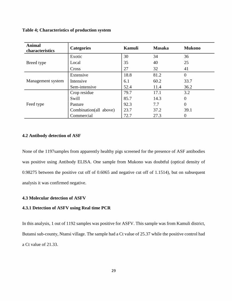
4.1.2 Characteristics of pig production systems
The pig production system in the three districts varied. More farmers kept the cross breed of pigs
(mixed colours), while the highest percentage of the local breed pigs (black in colour) was in Mukono
as shown in table 5.The extensive management system characterized by free movement of pigs was
practiced in Masaka and Kamuli districts, with Masaka having the highest (81.2%) number of
farmers. Regarding the feed type, most farmers fed their pigs on all feed types with exception of
Mukono who never use swill, pasture and commercial feeds as shown in table 5
29
Table 4; Characteristics of production system
Animal
characteristics Categories Kamuli Masaka Mukono
Breed type
Exotic 30 34 36
Local 35 40 25
Cross 27 32 41
Management system
Extensive 18.8 81.2 0
Intensive 6.1 60.2 33.7
Sem-intensive 52.4 11.4 36.2
Feed type
Crop residue 79.7 17.1 3.2
Swill 85.7 14.3 0
Pasture 92.3 7.7 0
Combination(all above) 23.7 37.2 39.1
Commercial 72.7 27.3 0
4.2 Antibody detection of ASF
None of the 1197samples from apparently healthy pigs screened for the presence of ASF antibodies
was positive using Antibody ELISA. One sample from Mukono was doubtful (optical density of
0.98275 between the positive cut off of 0.6065 and negative cut off of 1.1514), but on subsequent
analysis it was confirmed negative.
4.3 Molecular detection of ASFV
4.3.1 Detection of ASFV using Real time PCR
In this analysis, 1 out of 1192 samples was positive for ASFV. This sample was from Kamuli district,
Butansi sub-county, Ntansi village. The sample had a Ct value of 25.37 while the positive control had
a Ct value of 21.33.

30
4.3.2 Conventional amplification of ASFV
The p72, p54 and the central variable region of ASFV were amplified using different sets of primers
as shown in the gel picture below figure 1.
Figure 3 shows each sample ran with the primers amplifying different regions of the ASFV genome.P1-P3 is
the positive control with primers amplifying different size segments; P54 (678bp) and P72 (478bp). The
samples are 1,2,3,4 and NC is the negative control and M the ladder 1 Kb.

31
4.4 Sequencing
4.4.1 Sequence Alignment of the p72 and p54
Sequences that were downloaded using BLAST (htt://blast.st-vancbi.nl.gov/Blast.cgi) (Tamur et al.,
2013) were between 80-100% similarities with the query sequence and had E values between 0 and
3e-137. The query sequence was (ASF KAMULI 2013) obtained from this study as shown in table
5 below.

32
Table 5; Sequences downloaded from NCBI, region amplified, country of origin and genotype
Accession numbers and abbreviation Region Country of origin Genotype
KC112561 Ken10/KAKFAI p72 Kenya IX
KC112563.1 Ken10/Kis028 p72 Kenya IX
KC909904.1 Ug12Kampala4 p72 Uganda IX
KC990902.1 Ug10 Amuru p72 Uganda IX
KC990898.1 Ug10Moyo2 p72 Uganda IX
KC990895.1 Ug11 Mpigi p72 Uganda IX
KC990892.1 Ug10Kumi p72 Uganda IX
KC990890.1 Ug12Kabale p72 Uganda IX
ASF KAMULI 2013 p72 Uganda IX
FJ154429.1 Ug03H.2 p72 Uganda IX
FJ154433.1 Ug03P.6 p72 Uganda IX
GQ477138.1 UG07.Wak p72 Uganda IX
GQ477140.1 UG07Wak3 p72 Uganda IX
AY351549.1 MWHOG/3 p72 Georgia X
AY351565.1 MWHOG/9 p72 Georgia X
FJ174383.1 ug64 p72 Uganda X
AF449472.1 BUR/90/1 p72 Burundi X
AY538726.1 MOZ-77/98 p72 Mozambique II
KC990889.1 Ug12 Kampala4 p54 Uganda IX
ASF KAMULI 2013 p54 Uganda IX
KC990886.1 Ug10 Amuru p54 Uganda IX
KC990882.1 Ug10 Moyo 2 p54 Uganda IX
KC990880.1 Ug10 Tororo p54 Uganda IX
KC112574.1Ken11/Kaksp p54 Kenya IX
KC112573.1 Ken11/Thik P06 p54 Kenya IX
KC112570.1Ken10/Kis028 p54 Kenya IX
KC112568.1Ken10 KAKFA1 p54 Kenya IX
GQ477149.1 UG 07 Mukono p54 Uganda IX
GQ477148.1 UG 07.Wak4 p54 Uganda IX
GQ477146.1 UG07 Wak2 p54 Uganda IX
FJ174432.1 Ug03H.1 p54 Uganda IX
KC990888.1 Ug13.Busia2 p54 Uganda IX
FJ174430.1 Ug64 p54 Uganda X
JN590915.1 Ken08DP/Ndhiwa p54 Kenya X
JN590916.1 Ken08DP/Nyarongi p54 Kenya X
GQ410767.1 TAN98MAZIMBU p54 Tanzania XV
GQ410763.1 TAN08MABIBO p54 Tanzania XV

33
Sequence alignment of the downloaded p72 and p54 had gaps at various regions, when compared
with the ASF KAMULI 2013 obtained from this study as shown in fig 4 below.
Figure 4; Sequence alignment of the P54 region of the different isolates. The gaps indicate missing bases
which could be associated with mutations in those areas over time.

34
4.4.2 Phylogenetic analysis of the p72 and P54 regions
The downloaded p72 sequences after alignment with the query (ASF KAMULI 2013) and
comparison with 14 isolate sequences representing genotype II, X and IX showed the ASF KAMULI
2013 belonged to genotype IX. The query sequence clustered with previous isolates from Uganda
and some from Kenya responsible for outbreaks as seen in fig—below.
Figure 5; Phylogenetic tree base of the C-terminal end of the p72 sequences (ASF KAMULI-p-72) and other
isolates. The Neighbor-joining model was used for evolutionary history; the evolutionary distances were
computed using kimura 2-parameter and the bootstrap test was set at 1000 replicates. The analysis was
comprised of 14 nucleotide sequence.
GENOTYPE II
GENOTYPE X
GENOTYPE IX
35
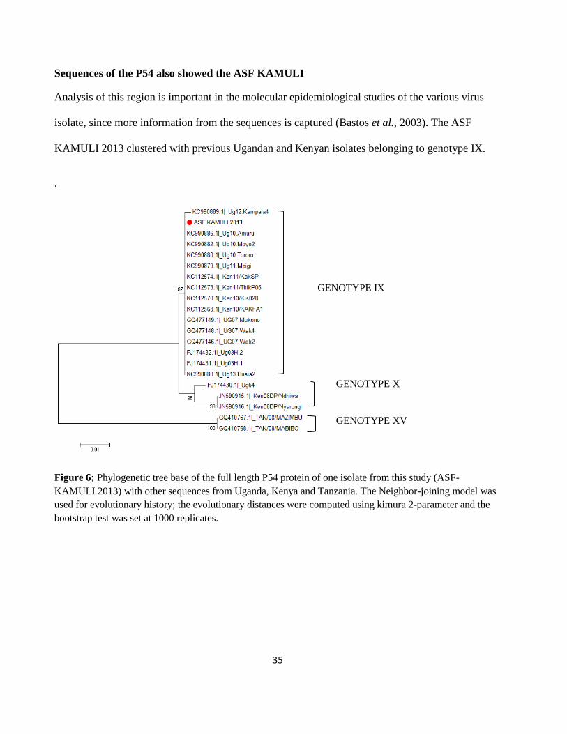
Sequences of the P54 also showed the ASF KAMULI
Analysis of this region is important in the molecular epidemiological studies of the various virus
isolate, since more information from the sequences is captured (Bastos et al., 2003). The ASF
KAMULI 2013 clustered with previous Ugandan and Kenyan isolates belonging to genotype IX.
.
Figure 6; Phylogenetic tree base of the full length P54 protein of one isolate from this study (ASF-
KAMULI 2013) with other sequences from Uganda, Kenya and Tanzania. The Neighbor-joining model was
used for evolutionary history; the evolutionary distances were computed using kimura 2-parameter and the
bootstrap test was set at 1000 replicates.
GENOTYPE IX
GENOTYPE X
GENOTYPE XV

36
CHAPTER FIVE
DISCUSSION
This was a cross-sectional study carried out in the districts of Masaka, Mukono and Kamuli to
determine the antibody and antigen status in apparently healthy pigs. A large sample size of 1192
blood and serum samples was collected from pigs in the different geographical locations. Only 1
sample out of 1192 was positive for ASFV using the molecular diagnostic tests.
The production systems practiced in these districts were mainly the extensive system involving
tethering and free ranging highest in Kamuli. This finding is in conformity with (Dione et al., 2014),
where the extensive system is thought to be the cheapest since little effort in terms of time, feed and
labour is required from the farmer.
All serum samples screened in the study were non-reactive to indirect ELISA indicating absence of
detectable levels of serum antibodies to ASFV. In comparison to previous studies, these findings are
different from Atuhaire et al.,(2013), who reported 11.5% presence of ASFV antibodies in slaughter
slabs in Kampala and also with Tejler, (2012) who reported 0.5% presence in Gulu district. However,
the finding from this study are similar to Muhangi et al. (2014) from Masaka and Rakai districts who
also never detected ASF antibodies in apparently healthy pigs.
This may be attributed to the fact that farmers sell off their animals immediately they hear of ASF
outbreaks, but suspected case are collected, sold, poled together for slaughter hence increased chance
of the antibody detection in the slaughter slabs (Atuhaire et al., 2013). There is also a possibility that
there was no recent infection and outbreaks when the sampling was done. These results are in
agreement with findings from Gallardo et al.,(2011) and Okoth et al., (2012) who also did not detect
antibodies using the indirect ELISA kit (Ingezim 14HSK3, ingenasa, Madrid, Spain). This failure to

37
detect circulating antibodies could be as a result of the pigs dying before seroconverting as this could
have been an early stage infection (OIE, 2012). Other factors like disease tolerance due to different
host factors, and presence of avirulent strains may not induce antibody responses to the virus thus the
absence of antibodies (Costard et al., 2009). The samples in this study might have been collected
during post infection or after outbreaks when pigs were wiped out with disease or sickly pigs sold for
slaughter.
Molecular diagnosis using UPL # 162-Real-time PCR showed viral presence in one of the samples
collected in this study. The UPL # 162 Real-time PCR is a more sensitive and specific method for
detection of minute volumes of the virus in domesticated pigs, wild porcine and ticks (Fernández-
Pinero et al., 2012). Isolates from previous studies on apparently healthy pigs were sequenced and
genotype IX was confirmed to be circulating in Uganda (Atuhaire et al., 2013; Gallardo et al., 2011).
Coincidently, the isolate sequenced from this study belongs to genotype IX. This confirms persistence
of genotype IX in Uganda which is implicated in previous and recent outbreaks in the country
(Atuhaire et al., 2013; Gallardo et al., 2011).
These findings also suggest possible seasonality of ASF occurrence. Sampling was conducted during
the rainy season, though it has been observed that majority of the outbreaks occur during the dry
season Atuhaire et al., (2013) possibly associated with times pigs are allowed to roam freely. In
addition, the ASF KAMULI 2013 isolate clusters with previous isolates of genotype IX responsible
for 2010-2012 outbreaks in Uganda and 2010 -2011 Kenya (Atuhaire et al., 2013; Gallardo et al.,
2011) . This shows there is a possibility of pig movements between borders (Kenya-Uganda) and
within country (Uganda) since this ASF KAMULI 2013 was introduced because farmer had not
experienced any outbreaks before (Atuhaire et al., 2013). Most of the farmers interviewed were

38
knowledgeable of the clinical signs and symptom, hence they would sell off their pigs immediately
on hearing or noticing these clinical signs and symptoms thus a further explanation of the low
prevalence of ASF observed (Chenais et al., 2015). Finally from this study, where one sample was
positive for ASFV indicates ASFV is highly pathogenic and is associated with acute and paracute
disease, thus the chronic carriers are unlikely to be important in the epidemiology of ASF. This one
sample was positive for ASFV but seronegative which implies long time carriers may not be
associated with disease transmission in these apparently healthy pigs in areas with no reports on
previous outbreaks. From this study, only one pig was positive for ASFV and so it was not possible
to identify predicators for ASF in the three districts.
Limitations
There were various limitations encountered during the survey which included the:
Most of the farmers often got bored when answering the questionnaires, because they were long,
consuming the farmers' time to perform other tasks.
Some farmers had sold their animals to traders and this resulted in looking for other interested
farmers having animals.
There was also a problem of farmer identification which was time consuming, because most
female farmers were known by their children’s names and not by their own or husband’s names.
The CVR of the virus was not amplified in this study because the sample was exhausted, since
a lot of optimization of the PCR was required. Therefore difference in the tetrameric repeats in
the p72 region was not determined as this provides high level resolution for viral discrimination.

39
CHAPTER SIX
CONCLUSIONS AND RECOMMENDATIONS
6.1 Conclusions
The findings from this study show a very low prevalence of ASFV like those in the literature which
indicates that ASFV is paracute / acute and is rarely detected in apparently healthy pigs. This indicates
that chronically infected pigs are not likely to be important in ASFV epidemiology.
The commonly circulating virus in Uganda is genotype IX.
6.2 Recommendations
The following recommendations are made from the study
1. Longitudinal surveillance systems should be designed to understand the epidemiology of ASFV
which is dynamic. These systems should be periodically monitored and evaluated to identify
risk factors responsible for the disease’s continued existence.
2. A risk-based approach should be formulated on biosecurity measures to check on farmers’
adherence and implementation on these measures in the country during and outside outbreaks.

40
REFERENCES
Aguero, M., Fernandez, J., Romero, L., Sanchez Mascaraque, C., Arias, M., & Sanchez-Vizcaino, J.
M. (2003). Highly sensitive PCR assay for routine diagnosis of African swine fever virus in
clinical samples. Journal of Clinical Microbiology, 41, 4431–4434.
Alcami, A., Carrascosa, A.L., Vinuela, E. (1990). Interaction of African swine fever virus with
macrophages. Virus Research, 17, 93–104.
Aliro, T., Tejler, E., Muhangi, D., Nyanabo, S., Boqvist, A., Ademun Okurut, A., Masembe, C.,
Emanuelson, U., Stahl, K. (2012). Spatio-temporal dynamics of African swine fever in Gulu
district, northern Uganda. EPIZONE 6th Annual Meeting “viruses on the move”.
Alonso, C., Galindo, I., Cuesta-Geijo, M. A., Cabezas, M., Hernaez, B., & Muñoz-Moreno, R.
(2013). African swine fever virus-cell interactions: from virus entry to cell survival. Virus
Research, 173(1), 42–57. https://doi.org/10.1016/j.virusres.2012.12.006
Atuhaire, D. K., Afayoa, M., Ochwo, S., Mwesigwa, S., Mwiine, F. N., Okuni, J. B., … Ojok, L.
(2013). Prevalence of African swine fever virus in apparently healthy domestic pigs in
Uganda. BMC Veterinary Research, 9, 263. https://doi.org/10.1186/1746-6148-9-263
Atuhaire, D. K., Afayoa, M., Ochwo, S., Mwesigwa, S., Okuni, J. B., Olaho-Mukani, W., & Ojok,
L. (2013). Molecular characterization and phylogenetic study of African swine fever virus
isolates from recent outbreaks in Uganda (2010-2013). Virology Journal, 10(1), 247.
https://doi.org/10.1186/1743-422X-10-247
Bastos, A.D., Penrith, M.L, Cruciere, C., Edrich, J.L., Hutchings, G., Roger, F., Couacy-Hyman, E.,
Thomson, G. . (2003). Genotyping field strains of African swine fever virus by partial p72
gene characterisation. Archives of Virology, 148, 693–706.
Bastos, A.D., Penrith, M.L., Macome.F., Pinto, F., Thomson, G. (2004). Co-circulation of two
genetically distinct viruses in an outbreak of African swine fever in Mozambique: no evidence
for individual co-infection. Veterinary Microbiology, 103, 169–182.
Björnheden, L. (2011). A study of domestic pigs , wild suids and ticks as reservoirs for African
swine fever virus in Uganda Sveriges lantbruksuniversitet A study of domestic pigs , wild
suids and ticks as reservoirs for African swine fever virus in Uganda.
Boshoff, C. I., Bastos, a D. S., Gerber, L. J., & Vosloo, W. (2007). Genetic characterisation of
African swine fever viruses from outbreaks in southern Africa (1973-1999). Veterinary
Microbiology, 121(1–2), 45–55. https://doi.org/10.1016/j.vetmic.2006.11.007
Carrascosa, A.L., Val, M.D., Santaaren, J.F., Vinuela, E. (1985). Purification and properties of
African swine fever virus. Journal of Virology, 54, 337–344.
Carriloo, C., Borca, M.V., Afonso, C.L., Onisk, D.V., Rock, D. L. (1994). Long-term persistent
infection of swine monocytes/macrophages with African swine fever virus. Virology Journal,
68, 580–583.
Chenais, E., Boqvist, S., Emanuelson, U., Ouma, E., Dione, M., & Aliro, T. (2015). Knowledge ,
Attitudes and Practices Related to African Swine Fever Within Smallholder Pig Production in
Northern Uganda, 2011, 1–15. https://doi.org/10.1111/tbed.12347

41
Cobbold,C &Wileman, T. (1998). The major structural protein of African swine fever virus p73, is
packaged into large structures,indicative of viral capsid or matrix precursors, on the
endoplasmic reticulum. Journal of Virology, 72(6), 5215–5223.
Costard, S., Mur, L., Lubroth, J., Sanchez-Vizcaino, J. M., & Pfeiffer, D. U. (2012). Epidemiology
of African swine fever virus. Virus Research, 1–7.
https://doi.org/10.1016/j.virusres.2012.10.030
Costard, S., Wieland, B., de Glanville, W., Jori, F., Rowlands, R., Vosloo, W., … Dixon, L. K.
(2009). African swine fever: how can global spread be prevented? Philosophical Transactions
of the Royal Society of London. Series B, Biological Sciences, 364(1530), 2683–96.
https://doi.org/10.1098/rstb.2009.0098
Dione, M. M., Ouma, E. a, Roesel, K., Kungu, J., Lule, P., & Pezo, D. (2014). Participatory
assessment of animal health and husbandry practices in smallholder pig production systems in
three high poverty districts in Uganda. Preventive Veterinary Medicine, 117, 565–576.
https://doi.org/10.1016/j.prevetmed.2014.10.012
Dixon, L.K., Costa, J.V., Escribano, J.M., Rock, D.L., Vinuela, E., Wilkinson, P. J. (2000). Family
Asfarviridae. In Virus Taxonomy,7th Report of the ICTV, 159–165.
Dixon, L. K., Chapman, D. a G., Netherton, C. L., & Upton, C. (2012). African swine fever virus
replication and genomics. Virus Research, 173(1), 3–14.
https://doi.org/10.1016/j.virusres.2012.10.020
Dixon, L. K., & Takamatsu, H. (2012). African swine fever virus: current situation and prospects
for control. Pig Journal, 67, 11–17. Retrieved from http://www.pigjournal.co.uk
Etter, E. M. C., Seck, I., Grosbois, V., Jori, F., Blanco, E., Vial, L., … Roger, F. L. (2011).
Seroprevalence of African swine fever in Senegal, 2006. Emerging Infectious Diseases, 17(1),
49–54. https://doi.org/10.3201/eid1701.100896
Fasina, F. O., Agbaje, M., Ajani, F. L., Talabi, O. A., Lazarus, D. D., Gallardo, C., … Bastos, A. D.
S. (2012). Risk factors for farm-level African swine fever infection in major pig-producing
areas in Nigeria, 1997-2011. Preventive Veterinary Medicine, 107(1–2), 65–75.
https://doi.org/10.1016/j.prevetmed.2012.05.011
Fernández-Pinero, J., Gallardo, C., Elizalde, M., Robles, A., Gómez, C., Bishop, R., & Heath, L.,
Couacy-Hymann, E., Fasina, F.O., Pelayo, V., Soler, A., Arias, M. (2012). Molecular
diagnosis of African swine fever by a new real-time PCR using Universal Probe Library.
Transboundary and Emerging Diseases. Retrieved from http://dx.doi.org/10.1111/j.1865-
1682.2012.01317
Gallardo, C., Ademun, A. R., Nieto, R., Nantima, N., Arias, M., Pelayo, V., & Bishop, R. P. (2011).
Genotyping of African swine fever virus ( ASFV ) isolates associated with disease outbreaks
in Uganda in 2007, 10(17), 3488–3497. https://doi.org/10.5897/AJB10.1439
Gallardo, C., Anchuelo, R., Pelayo, V., Poudevigne, F., Leon, T., Nzoussi, J., … Arias, M. (2011).
African swine fever virus p72 genotype IX in domestic pigs, Congo, 2009. Emerging
Infectious Diseases, 17(8), 1556–8. https://doi.org/10.3201/eid1708.101877
Gallardo, C., Elizalde, M., Robles, A., & Go, C. (2012). Molecular Diagnosis of African Swine
Fever by a New Real-Time PCR Using Universal Probe Library, 1–11.

42
https://doi.org/10.1111/j.1865-1682.2012.01317.x
Gallardo, C., Okoth, E., Pelayo, V., Anchuelo, R., Llorente, A., Nieto, R., … Bishop, R. P. (2011).
African swine fever viruses with two different genotypes , both of which occur in domestic
pigs , are associated with ticks and adult warthogs , respectively , at a single geographical site,
432–444. https://doi.org/10.1099/vir.0.025874-0
Hall, T. A. (1999). Bioedit a user friendly biological sequence alignment editor and analysis
program for windows 95/98/NT. In Nucleic Acids Symposium.
Hjertner, B., Meehan, B., McKillen, J., McNeilly, F., Belák, S. (2005). Adaptation of an Invader
assay for the detection of African swine fever virus DNA. Journal of Virological Methods 124,
1–10.
Irusta, P. M., Borca, M. V, Kutish, G. F., Lu, Z., Caler, E., Carrillo, C., & Rock, D. L. (1996).
Amino acid tandem repeats within a late viral gene define the central variable region of
African swine fever virus. Virology, 220(1), 20–27. https://doi.org/10.1006/viro.1996.0281
Jori, F., & Bastos, A. D. S. (2009). Role of wild suids in the epidemiology of African swine fever.
EcoHealth, 6(2), 296–310. Retrieved from http://www.ncbi.nlm.nih.gov/pubmed/19915917
Jori, F., Vial, L., Penrith, M. L., Pérez-Sánchez, R., Etter, E., Albina, E., … Roger, F. (2013).
Review of the sylvatic cycle of African swine fever in sub-Saharan Africa and the Indian
ocean. Virus Research, 173(1), 212–27. https://doi.org/10.1016/j.virusres.2012.10.005
Karalyan, Z., Zakaryan, H., Sargsyan, K., Voskanyan, H., Hakobyan, L., Abroyan, L., … Karalova,
E. (2012). Pathology of porcine peripheral white blood cells during infection with African
swine fever virus. BMC Veterinary Research, 8, (28 February 2012)-(28 February 2012).
Retrieved from <Go to ISI>://CABI:20123095929
Kleibeker, S.B., Scoles, G.A., Burrage, T.G., Sur, J. (1999). African swine fever virus replication in
the midgut epithelium is required for infection of Ornithodoros ticks. Journal of Virology,
73(10), 8587–8598.
Lubisi, B. A., Bastos, A. D. S., Dwarka, R. M., & Vosloo, W. (2005). Molecular epidemiology of
African swine fever in East Africa, 2439–2452. https://doi.org/10.1007/s00705-005-0602-1
Malmquist, W.A & Hay, D. (1960). Hemaadsorption and cytopathic effect produced by African
swine fever virus in swine bone marrow and buffy coat cultures. AM J Vet Res, 21, 104–108.
Mebus, C. A. (1988). Afircan swine fever. Advances in Virus Research, 35:, 251–269.
Misinzo, G., Jumapili, F., Ludosha, M., Mafie, E., & Silialis, J. (2009). Genotyping of African
swine fever virus from a 2009 outbreak in Tanzania. Research Opinions in Animal
&Veterinary Sciences, 334–338.
Montgomery, R. E. (1921). On a form of African swine fever occuring in the British East Africa
(Kenya colony). Journal of Comparative Pathology and Therapeutics, 34, 159–191, 243–265.
Muhangi, D., Masembe, C., Berg, M., Ståhl, K., & Ocaido, M. (2014). Practices in the pig value
chain in Uganda; implications to African swine fever transmission. Livestock Research for
Rural Development, 26((5).).
Muhangi, D., Masembe, C., Emanuelson, U., Boqvist, S., Mayega, L., Ademun, R. O., … Ståhl, K.
(2015). A longitudinal survey of African swine fever in Uganda reveals high apparent disease

43
incidence rates in domestic pigs , but absence of detectable persistent virus infections in blood
and serum. 1–11. https://doi.org/10.1186/s12917-015-0426-5
Muhanguzi, D., Lutwama, V., & Mwiine, F. (2012). Factors that influence pig production in
Central Uganda - Case study of Nangabo Sub-County, Wakiso district. Veterinary World, 5(6),
346. https://doi.org/10.5455/vetworld.2012.346-351
Mutetikka, D. (2009). A Guide to pig Production at Farm level,Makerere University, Kampala-
Uganda. Fountain Publishers, Kampala.
Muwonge, A., Munang’andu, H. M., Kankya, C., Biffa, D., Oura, C., Skjerve, E., & Oloya, J.
(2012). African swine fever among slaughter pigs in Mubende district, Uganda. Tropical
Animal Health and Production, 44(7), 1593–8. https://doi.org/10.1007/s11250-012-0112-6
Nantima, N., Ocaido, M., Ouma, E., Davies, J., Okoth, E., & Bishop, R. (n.d.). Risk factors
associated with occurrence of African swine fever outbreaks in smallholder pig farms in four
districts along the Uganda-Kenya border. https://doi.org/10.1007/s11250-015-0768-9
Nix, R.J., Gallardo, C., Hutchings, G., Blanco, E., Dixon, L. K. (n.d.). Molecular epidemiology of
African swine fever virus studied by analysis of four variable genome regions. Archives of
Virology, 151, 2475–24.
Notomi, T., Okayama, H., Masubuchi, H., Yonekawa, T., Watanabe, K., Amino, N., & Hase, T.
(2000). Loop-mediated isothermal amplification of DNA. Nucleic Acids Research, 28(12),
E63. https://doi.org/10.1093/nar/28.12.e63
O.I.E. (2010). Early detection and contingency plan for African swine fever, 139–147.
OIE. (2008). Chapter 2.8.1. African swine fever. In Manual of diagnostic tests and vaccines for
terrestrial animals 2008. Office International des Epizooties, Paris, France.
http://www.oie.int/eng/ normes/mmanual/2008/pdf/2.08.01_ASF.pdf.
OIE. (2012). African swine fever. In: Manual of Diagnostic Tests and vaccine for Terrestrial
Animals. Office Internatinal des Epizooties, Paris, France. (Chapter 2.8.1,NB: Version adopted
in May 2012).
Okoth,E.,Gallardo,C.,Macharia,J.M.,Omore,A.,Pelay,V.,Bulimo,D.W.,Arias,M.,Kitala,P.,Baboon,
K.,Lekolol, I.,Mijele, D.,Bishop, R. . (n.d.). Comparison of African swine fever virus
prevalence and risk in two constrating pig-farming systems in South-West and Central Kenya.
Preventive Veterinary Medicine, 110(2), 198–205.
Ouma, E., Dione, M., Lule, P., Pezo, D., Marshall, K., Roesel, K., Mayega, L., Kiryabwire, D.,
Nadiope, G and Jagwe, J. (2014). Smallholder pig value chain assessment in Uganda : results
from producer focus group discussions and key informant interviews. Nairobi, Kenya.
Ouma, E., Dione, M., Lule, P., Roesel, K., & Pezo, D. (2013). Characterization of smallholder pig
production systems in Uganda: Constraints and opportunities for engaging with market
systems. Livestock Research for Rural Development, 26(3).
Oura, C. a L., Edwards, L., & Batten, C. a. (2013). Virological diagnosis of African swine fever--
comparative study of available tests. Virus Research, 173(1), 150–8.
https://doi.org/10.1016/j.virusres.2012.10.022
Penrith, M.-L. (2009). African swine fever. The Onderstepoort Journal of Veterinary Research,

44
76(1), 91–95. Retrieved from http://www.ncbi.nlm.nih.gov/pubmed/19967933
Pezo, D. & Waiswa, C. (2012). Farming Systems Perspectives:Lessons for managing health risks in
smallholder pig systems.Paper presented in the South-South Symposium“Managing Risks in
Emerging Pork Markets”. Hano, Viet Nam 23-25 Apirl 2012; International Livestock Research
Institute(ILRI) and Makerere University Kampala, Uganda.
Ravaomanana, J., Jori, f., Andriatsimahavandy, A.,Roger,F., Albina, E., Vial, L. (2010). First
detection of African swine fever virus in Ornithodoros porcinus in Madagascar and new
insights into tick distribution and taxonomy. Parasites and Vectors, 3, 115.
Reis, A.L., Parkhouse, R.M., Penedos, A.R., Martins, C., Leitao, A. (2007). Systematic analysis of
longitudinal serological responses of pigs infected experimentally with African swine fever
virus. General Virology, 88:, 2426–2534.
Rojo, G., Chamorro, M., Salas, M.L., Vinuela, E., Cuezva, J.M., Salas, J. (1999). Replication of
African swine fever DNA in infected cells. Virology, 257, 524–536.
Rowlands, R. J., Duarte, M. M., Boinas, F., Hutchings, G., & Dixon, L. K. (2009). The CD2v
protein enhances African swine fever virus replication in the tick vector, Ornithodoros
erraticus. Virology, 393(2), 319–328. https://doi.org/10.1016/j.virol.2009.07.040
Ruiz-Gonzalvo, F., Rodriguez, F., Escribano, J. M. (1996). Functional and Immunological
properties of the baculovirus-expressed hemagglutinin of African swine fever virus. Virology,
218, 285–289.
Ståhl.M.K., Ogweng.P., Okoth. E., Aliro.T, Muhangi.D., LeBlanc.N, Atimnedi.P., B. M., &
Bishop.R.P., Rasmussen.H.B, Masembe. C. (2014). Understanding the dynamics and spread of
African swine fever virus at the wildlife-livestock interface: insights into the potential role of
the bushpig, Potamochoerus larvatus. Newsletter of the IUCN / SSC Wild Pig, Peccary and
Hippo Specialist Groups, 13(1).
Steiger Y., Ackermann M., M. C., & U., K. (1992). Rapid and biologically safe diagnosis of
African swine fever virus infection by using polymerase chain reaction. Journal of Clinical
Microbiology, 30, 1–8.
Suárez, C., Salas, M. L., & Rodríguez, J. M. (2010). African swine fever virus polyprotein pp62 is
essential for viral core development. Journal of Virology, 84(1), 176–87.
https://doi.org/10.1128/JVI.01858-09
Sun, H., Jenson, J., Dixon, L.K., Parkhouse, M. E. (1996). Characterisation of the African swine
fever virion protein,j18L. Virology Journal, 77(5), 941–946.
Tamura, K., Stecher, G., Peterson, D., Filipski, A., & Kumar, S. (2013). Molecular Evolutionary
Genetics Analysis version 6.0. Molecular Biology and Evolution, 30:, 2725–2729.
Tatwangire A. (2013). Sucesses and failures of institutional innovations to improve access to
services,input and output markets for smallholder pig production system and value chains in
Uganda. Retrieved from Review Report. www.livestockfish.cgiar.org
Tejler, E. (2012). Outbreaks of African swine fever in domestic pigs in Gulu district , Uganda
Outbreaks of African swine fever in domestic pigs in Gulu district , Uganda.
Thrusfield, M. (2007). Veterinary Epidemiology, 3rd edition.

45
Ticks, S., Vial, L., Wieland, B., Jori, F., Etter, E., Dixon, L., & Roger, F. (2007). Fever Virus DNA
in, 13(12), 1928–1931.
UBOS. (2008). A summary report of the national livestock census. Entebbe, Uganda.In
Anonymus(eds.) Kampala.
Uganda Bureau of Statistics. (2013). 2013 Statistical Abstract, 1–264. Retrieved from
www.ubos.org
Wardley, R.C., Abu, Elzein, E.M.E., Crowther, P.J. (1979). A solid- phase enzyme linked
immunosorbent assay for the detection of African swine fever virus antigen and antibody.
Journal of Hygiene (Cambridge), 83, 363–369.
Wilkinson, P. J. (2000). African swine fever In: Manual of Standards for Diagnostic Tests and
Vaccines, fourth ed. Office International des Epizooties, Paris.
Yanez, R.J., Rodriguez, J.M., Nogal, M.L., Yuste, L., Enriquez, C., Rodriguez, J.M., Vinuela, E.
(1995). Analysis of the complete nucleotide sequence of African swine fever virus. Virology,
208(1), 249–278.
Related Documents