MAJOR ACHIEVEMENTS IN NUCLEAR CARDIOLOGY Advances in technical aspects of myocardial perfusion SPECT imaging Piotr J. Slomka, PhD, a James A. Patton, PhD, b Daniel S. Berman, MD, a and Guido Germano, PhD a Although myocardial perfusion SPECT (MPS) imaging is widely used in current clinical practice, it suffers from some fundamental limitations including long image acquisition, low image resolution, and patient radiation dose. In the last two decades, MPS was performed most commonly by standard dual-head scin- tillation cameras with parallel-hole collimators, typically configured in a 90° detector geometry and image reconstruction based on standard filtered-back projection algorithms. The required scan times were as along as 15-20 minutes for each stress and rest MPS acquisition to provide adequate imaging statistics, resulting in long overall test times and frequent artifacts caused by patient motion during the scan as well as compromised patient comfort. Recently, it has become very important to address these limitations, since MPS has new competi- tors in the non-invasive imaging arena most notably coronary CT angiography (CCTA), which allow diag- nostic imaging in a very short time. In addition, a practice of combining MPS with other modalities such as CCTA for better diagnostic certainty 1 has intensified concerns regarding total radiation dose delivered to the patient. 2 The radiation dose and acquisition time are intrinsically linked with each other, as longer acquisition times could be used with lower injected doses and higher doses could be used to shorten acquisition times. There have been significant recent efforts by industry and academia to develop new imaging systems with increased sensitivity and new methods of image reconstruction optimizing image quality, which will simultaneously allow higher photon sensitivity and improve both image quality and resolution. These efforts address the main limitations of MPS by combining several approaches such as changing the detector geometry and optimizing tomographic sampling of the field of view for myocardial imaging, improving the detector material and collimator design, and optimizing the image reconstruction algorithms. In this review article we summarize these developments. NEW HARDWARE FOR OPTIMIZED MPS IMAGING Several new dedicated hardware camera systems with optimized acquisition geometry, collimator design, and associated reconstruction software have been recently introduced by various vendors. Innovative designs of the gantry and detectors have been proposed which allow increased sampling of the myocardial region, and thus allow better local sensitivity. These systems combine an improvement in spatial resolution and sensitivity. By faster imaging times due to increased sensitivity and by eliminating the need to position the patient’s arms above the head by imaging in an upright or reclining position, patient comfort is dramatically improved. As a conse- quence of faster imaging times and more comfortable patient positioning, these systems have the additional benefit of reducing patient motion during a scan. Fur- thermore, claustrophobic effects are reduced and the floor space requirements are more flexible since the new detectors and the associated mechanical are significantly smaller in comparison to standard equipment. DIGIRAD CARDIUS 3 XPO Digirad, Inc. (Poway, CA) has developed a Cardius XPO camera dedicated to fast cardiac imaging. This system can be configured in 2- or 3-detector configura- tions. 3 The Cardius 3 camera and its geometry (triple- head configuration) is shown in Figure 1. These models use indirect, solid-state detectors consisting of pixilated CsI(Tl) and photodiodes to configure detector heads that are more compact than conventional cameras, equipped with photomultipliers. Each detector head is 21.2 9 15.8 cm and contains an array of 768 6.1 9 6.1 9 6 mm thick CsI(Tl) crystals, coupled to individual sili- con photodiodes, which are used to convert the light From the Departments of Imaging and Medicine, a AIM Program, Cedars-Sinai Medical Center, Los Angeles, CA; Department of Radiology and Radiological Sciences, b Vanderbilt University Medical Center, Nashville, TN. Received for publication Dec 30, 2008; final revision accepted Jan 6, 2009. Reprint requests: Piotr J. Slomka, PhD, Departments of Imaging and Medicine, AIM Program, Cedars-Sinai Medical Center, Los Angeles, CA, USA; [email protected]. J Nucl Cardiol 2009;16:255–76. 1071-3581/$34.00 Copyright Ó 2009 by the American Society of Nuclear Cardiology. doi:10.1007/s12350-009-9052-6 255

Welcome message from author
This document is posted to help you gain knowledge. Please leave a comment to let me know what you think about it! Share it to your friends and learn new things together.
Transcript

MAJOR ACHIEVEMENTS IN NUCLEAR CARDIOLOGY
Advances in technical aspects of myocardialperfusion SPECT imaging
Piotr J. Slomka, PhD,a James A. Patton, PhD,b Daniel S. Berman, MD,a and
Guido Germano, PhDa
Although myocardial perfusion SPECT (MPS)
imaging is widely used in current clinical practice, it
suffers from some fundamental limitations including
long image acquisition, low image resolution, and
patient radiation dose. In the last two decades, MPS was
performed most commonly by standard dual-head scin-
tillation cameras with parallel-hole collimators, typically
configured in a 90� detector geometry and image
reconstruction based on standard filtered-back projection
algorithms. The required scan times were as along as 15-20
minutes for each stress and rest MPS acquisition to
provide adequate imaging statistics, resulting in long
overall test times and frequent artifacts caused by patient
motion during the scan as well as compromised patient
comfort. Recently, it has become very important to
address these limitations, since MPS has new competi-
tors in the non-invasive imaging arena most notably
coronary CT angiography (CCTA), which allow diag-
nostic imaging in a very short time. In addition, a
practice of combining MPS with other modalities such
as CCTA for better diagnostic certainty1 has intensified
concerns regarding total radiation dose delivered to the
patient.2 The radiation dose and acquisition time are
intrinsically linked with each other, as longer acquisition
times could be used with lower injected doses and
higher doses could be used to shorten acquisition times.
There have been significant recent efforts by
industry and academia to develop new imaging systems
with increased sensitivity and new methods of image
reconstruction optimizing image quality, which will
simultaneously allow higher photon sensitivity and
improve both image quality and resolution. These efforts
address the main limitations of MPS by combining
several approaches such as changing the detector
geometry and optimizing tomographic sampling of the
field of view for myocardial imaging, improving the
detector material and collimator design, and optimizing
the image reconstruction algorithms. In this review
article we summarize these developments.
NEW HARDWARE FOR OPTIMIZED MPSIMAGING
Several new dedicated hardware camera systems
with optimized acquisition geometry, collimator design,
and associated reconstruction software have been recently
introduced by various vendors. Innovative designs of the
gantry and detectors have been proposed which allow
increased sampling of the myocardial region, and thus
allow better local sensitivity. These systems combine an
improvement in spatial resolution and sensitivity. By
faster imaging times due to increased sensitivity and by
eliminating the need to position the patient’s arms above
the head by imaging in an upright or reclining position,
patient comfort is dramatically improved. As a conse-
quence of faster imaging times and more comfortable
patient positioning, these systems have the additional
benefit of reducing patient motion during a scan. Fur-
thermore, claustrophobic effects are reduced and the floor
space requirements are more flexible since the new
detectors and the associated mechanical are significantly
smaller in comparison to standard equipment.
DIGIRAD CARDIUS 3 XPO
Digirad, Inc. (Poway, CA) has developed a Cardius
XPO camera dedicated to fast cardiac imaging. This
system can be configured in 2- or 3-detector configura-
tions.3 The Cardius 3 camera and its geometry (triple-
head configuration) is shown in Figure 1. These models
use indirect, solid-state detectors consisting of pixilated
CsI(Tl) and photodiodes to configure detector heads that
are more compact than conventional cameras, equipped
with photomultipliers. Each detector head is 21.2 9
15.8 cm and contains an array of 768 6.1 9 6.1 9
6 mm thick CsI(Tl) crystals, coupled to individual sili-
con photodiodes, which are used to convert the light
From the Departments of Imaging and Medicine,a AIM Program,
Cedars-Sinai Medical Center, Los Angeles, CA; Department of
Radiology and Radiological Sciences,b Vanderbilt University
Medical Center, Nashville, TN.
Received for publication Dec 30, 2008; final revision accepted Jan 6,
2009.
Reprint requests: Piotr J. Slomka, PhD, Departments of Imaging and
Medicine, AIM Program, Cedars-Sinai Medical Center, Los Angeles,
CA, USA; [email protected].
J Nucl Cardiol 2009;16:255–76.
1071-3581/$34.00
Copyright � 2009 by the American Society of Nuclear Cardiology.
doi:10.1007/s12350-009-9052-6
255

output of the crystals to electrical pulses. Digital logic
and software is used to process the signals and create
images instead of analog Anger positioning circuits. In
the 3-detector system the detector heads are positioned
at 67.5� between heads, as shown in Figure 1B. Heads
are allowed to be moved in and out (closer to or farther
away from the patient). For imaging, the patient sits on a
chair with his arms placed on an arm rest above the
detectors. Data acquisition is typically accomplished in
7.5 minutes by rotating the patient chair by 67.5�, pro-
ducing a total acquisition arc of 202.5�. With this system
the manufacturer reports a reconstructed spatial resolu-
tion of 8.95 mm (at a 20 cm orbit radius) and a
sensitivity of 234 cpm/lCi, using the system’s cardiac
collimator and a 3D version of the ordered subsets
expectation maximization (OSEM) approach for recon-
struction. These systems are now used clinically in
several sites, and reports have been published comparing
their performance to that of a standard dual-headed
camera when it was found that the similar quality
could be obtained with 38% reduction in the acquisition
time.4
Data have recently shown that the acquisition time
can be further reduced with this scanner by the appli-
cation of optimized image reconstruction protocols,
developed by Digirad. The nSPEED reconstruction5
models the depth-dependent detector spatial response of
the SPECT systems with a 3D version of the OSEM
reconstruction method. In the preliminary results from
the multi-center trial (10 sites) with 448 patients, the
image quality improvement with nSPEED was com-
pared to a conventional 2D-OSEM technique.6 The trial
demonstrated that nSPEED applied to data obtained with
Cardius-3 camera enables the reduction of the acquisi-
tion time by 50%, while maintaining image quality and
information with diagnostically equivalent images for
rest and stress studies as well as reliable quantification
of function and perfusion. The mean imaging times were
4.2 minutes for stress and 4.8 minutes for rest with the
optimized 3D reconstruction. A representative image
from this trial is shown in Figure 2.
CARDIARC
CardiArc (Canton, MI) has developed a dedicated
nuclear cardiology SPECT camera in which the detector
and collimation are redesigned and optimized specifi-
cally for cardiac imaging.7 This device has no visibly
moving parts and has a single internally moving part
which is hidden from the patient.8 Therefore from the
Figure 1. Upright patient position on the Digirad Cardius 3 XPO (C 3 XPO) triple-head, pixilateddetector camera (A), its geometry (B), and a photograph of the camera (C). Detectors remain fixedwhile patient is rotated through 202.5� in a rotating chair. Images courtesy of Digirad, San Diego,CA.
256 Slomka et al Journal of Nuclear Cardiology
Advances in technical aspects of myocardial perfusion SPECT imaging March/April 2009

outside the detector appears motionless, and for comfort
the patient is positioned upright. Scan times reported by
the company are as short as 2 minutes.7 The camera
system and a typical patient position are shown in Figure 3.
This system was originally designed to use arrays
of CZT crystals as detectors. However, due to high cost
of CZT material and potential long-term stability issues
with CZT,9 the detector material was changed in order to
enable commercial production. Figure 4 illustrates the
design and the principle of operation of the current
model. The system incorporates a high high-definition,
high-spatial resolution detector utilizing 3 curved
NaI(Tl) crystals with graduated grooving technology and
an array of 60 photomultiplier tubes arranged in three
rows (Figure 4A). The detector uses a proprietary digital
process developed by CardiArc that replaces the con-
ventional Anger logic. Horizontal photon collimation in
each slice is accomplished by using a thin, curved, lead
sheet with 6 narrow vertical slots (Aperture Arc)
(Figure 4A). Vertical collimation is accomplished using
a series of stationary lead vanes that are stacked verti-
cally between the aperture arc and the NaI(Tl) crystals. In
this way, data are collected in 1-mm-thick slices using
the 6 vertical apertures to collimate photons so that they
are detected continuously across the detector surface
with no overlap of data from different apertures. During
acquisition, the aperture arc rotates to acquire data from
multiple projections providing 1,280 angular samples in
0.14� increments over 180�, which is a factor of 21
greater than a conventional camera angular sampling
(typically 3�). All detector pixels are active simulta-
neously while photons are detected from multiple angles
(Figure 4B), which provides high-efficiency imaging.
The movement of the aperture arc is synchronized elec-
tronically with the areas of the NaI(Tl) crystals that are
imaging the photons passing through the individual slots.
The aperture arc’s weight of 35 lbs is much lighter than
traditional moving gantries, which facilities precise
2D Full
3D Half
2D Full
2D Full
3D Half
3D Half
Figure 2. Images at full-time stress (6.7 minutes) and half-time (3.3 minutes) obtained with Cardius 3 XPO and n-speed reconstruction in Tc-99m sestamibi study of a male patient age: 55, stress type: exercise, height: 5090 0 andweight: 183 lbs. The full-time acquisition times were 6.7 minutes for stress (20 seconds/proj) with 31 mCi Tc-99msestamibi dose. Images courtesy of Dr. Jashmid Maddahi and Digirad, San Diego, CA6.
Journal of Nuclear Cardiology Slomka et al 257
Volume 16, Number 2;255–76 Advances in technical aspects of myocardial perfusion SPECT imaging

motion control. The aperture arc movement ranges *9
inches in order to cover the entire cardiac field of view,
and each traverse of the arc takes 10 seconds.
SPECT reconstructed spatial resolution values (full
width half maximum) quoted by CardiArc range from
3.6 mm (at 82 mm source to aperture arc distance) to
7.8 mm (at 337 mm source to aperture arc distance). An
independent evaluation concluded that the CardiArc
system appears to gain image quality by a factor of 5-10
when compared to the conventional dual-head camera.10
A comparison of patient imaging capabilities is shown
in Figure 5.
SPECTRUM DYNAMICS
Spectrum Dynamics, Haifa, Israel, has manufac-
tured a system called D-SPECT. The design and the
principle of its operation are shown in Figure 6. The
patient is imaged in a semi-upright position with the left
arm placed on top of the camera (Figure 6A) or in the
supine position. Acquisition time as short as 2 minutes
has been reported.11 This system uses pixilated CZT
detector arrays (Figure 6B) mounted in 9 vertical col-
umns and placed in a 90� gantry geometry (Figure 6C).
While CZT detectors are higher in cost, they have
advantages of superior energy resolution (by a factor of
approximately 1.7 at 140 keV) and compact size as
compared to the combination of NaI(Tl) with photo-
multiplier tubes of the conventional Anger camera. With
D-SPECT, each detector column is fixed in a mechanical
mounting, and the data acquisition is performed by
rotating these multiple columns in synchrony. The
photons from a given location are detected at multiple
angles by multiple columns as the fields of view of the
detectors are swept through the region of interest. Each
column (as shown in Figure 6B) consists of an array of
1024 CZT elements (2.46 9 2.46 9 5 mm thick),
arranged in a 16 9 64 element array with an approxi-
mate size of 40 9 160 mm. Each column is fitted with
square, parallel hole, high sensitivity collimators, such
that the dimensions of each hole are matched to the size
of a single detector element. The collimators are fabri-
cated from tungsten to eliminate the production of lead
x-rays that might interfere with 201Tl imaging. The
Figure 3. Photograph of the CardiArc SPECT-HD demon-strating patient positioning for optimal cardiac imaging and thetechnologist in the operating position, taking advantage of thebuilt-in radiation shielding. Images courtesy of CardiArc, Inc.
Figure 4. Design and principle of operation of CardiArccamera. The camera uses three stationary NaI(Tl) crystals andcorresponding photomultiplier tubes for photon detection (A).The aperture arc has six slots (apertures) for horizontalcollimation, and continuously rotates while imaging. Alldetector pixels are utilized simultaneously, allowing imagingmultiple angles (B). Image courtesy from CardiArc, Inc., withpermission.
258 Slomka et al Journal of Nuclear Cardiology
Advances in technical aspects of myocardial perfusion SPECT imaging March/April 2009

collimators have a larger effective diameter than con-
ventional LEHR collimators used with scintillation
cameras, yielding a significant gain in their geometric
efficiency. The collimator has a hole length of 24.5 mm
with a 2.46 mm pitch and 2.26 mm hole diameter. The
compensation for the loss in geometric spatial resolution
that results from this design is accomplished by the use
of CZT, with its superior energy resolution, and software
compensation methods. All data are collected in list
mode. A proprietary BroadviewTM iterative reconstruc-
tion algorithm, based on the ML-EM approach, with
resolution recovery and use of the cardiac shape priors,
has been developed and patented by the manufacturer.12
The overall system resolution is 5 mm in line source
experiments, superior to that of the standard Anger
camera systems.
Data acquisition is accomplished in a two-step pro-
cess. First, a 10-second pre-scan is performed to identify
the location of the region of interest. Scan limits and
timings are then set for each detector column, and the final
scan is performed with each detector column rotating
within the limits set from the pre-scan data. This process is
shown diagrammatically in Figure 7. This process is
termed region-of-interest (ROI)-Centric scanning by the
manufacturer because the scan field is limited only to the
myocardial region. It is not possible to measure an abso-
lute value of sensitivity for this system as prescribed by
the NEMA quality control standards13 because the sen-
sitivity is significantly dependent on the field-of-view,
defined individually for each patient by the pre-scan
process. However, the most centrally located point has
been reported to demonstrate a sensitivity of 1407 counts/
lCi/minute compared to the 160-240 counts/lCi/minute
range generally observed with standard cameras.14 A case
example showing image quality on both D-SPECT and
A-SPECT, obtained with the same isotope injection, is
Figure 5. Stress images of a 55-year-old male, 6050 0 235 lbs with substernal chest pain andshortness of breath. Cholesterol = 240. Developed 1.5-2.0 mm ST depression in inferior leads atstress, injected with 31.2 mCi Tc-99m sestamibi at peak stress. Image set A, was acquired with aconventional dual-head scintillation camera using LEHR collimators for 10.6 minutes anddemonstrated an infero-basal defect. Image set B, was acquired with CardiArc for 4.7 minutes,and correctly demonstrated a more severe and more extensive defect in the inferior wall to the apex.Angiography revealed high-grade, proximal PDA stenosis. Image courtesy from CardiArc, Inc.,with permission.
Journal of Nuclear Cardiology Slomka et al 259
Volume 16, Number 2;255–76 Advances in technical aspects of myocardial perfusion SPECT imaging

shown in Figure 8. In a recently published study, when
D-SPECT was compared to A-SPECT, the myocardial
count rate (with the same injection of the isotope) was
7 to 8 times higher for D-SPECT (Figure 9).11 Pre-
liminary work has shown that simultaneous dual isotope
SPECT MPI with this camera is feasible using Tl-201 and
Tc-99m, taking advantage of the improved energy reso-
lution of CZT.15
The higher sensitivity of this system has been
exploited to develop new clinical protocols. Cedars-
Sinai Medical Center has reported the routine clinical
use of this scanner in over 400 patients using a stress201Tl (2 mCi)/rest tetrofosmin or sestamibi (8-10 mCi)
protocol. Using half of the radioactivity associated with
standard dual isotope procedures, this protocol includes
upright and supine immediate post-stress images of
6 minutes each followed by rest injection and immediate
4-minute rest imaging. The total imaging time of this
protocol is 19 minutes. Good to excellent image quality
without significant extracardiac interference was
observed in over 96% of the cases.16 A multi-center
study conducted at Cedars-Sinai Medical Center, Miami
Baptist Hospital, Vanderbilt University Medical Center
and the Brigham and Women’s Hospital has confirmed a
close correlation and diagnostic equivalence to standard
acquisition techniques using objective quantitative
measures.17
Figure 6. D-SPECT camera. Photograph of the D-SPECT camera showing patient position (A). Adiagram of a single detector column from the D-SPECT camera (B), and a photograph of 9 detectorcolumns configuration (C). Photograph courtesy of Spectrum Dynamics, Haifa, Israel.
Figure 7. The ROI-centric technique utilized by the D-SPECTcamera to optimize data collection from the myocardium.
260 Slomka et al Journal of Nuclear Cardiology
Advances in technical aspects of myocardial perfusion SPECT imaging March/April 2009

MULTI-PINHOLE COLLIMATION APPROACH
Some vendors have explored image collimation
using multi-pinhole design. Multi-pinhole collimation
provides an alternative approach to parallel-hole
rotational tomography. Previously, useful results have
been demonstrated in small animal imaging with multi-
pinhole SPECT providing improved spatial resolution
and detection efficiency in comparison to parallel-hole
collimation.18-20 The multi-pinhole approach allows
many views to be acquired simultaneously throughout
the entire image acquisition period without the need for
motion of the detector, collimator or patient. This
capability allows image acquisition to be accomplished
without the need for any electro-mechanical hardware,
potentially reducing the manufacturing and servicing
costs. In addition to the increase in detection sensitivity,
the use of stationary detectors equipped with multi-
pinhole collimation provides coincident sets of raw
images eliminating view-to-view inconsistencies and
thereby reducing artifacts induced by patient motion.
Therefore, by the multi-pinhole approach, all views are
active for the entire acquisition period providing a
compatible dataset for input to iterative SPECT recon-
struction algorithms.
However, multi-pinhole design potentially suffers
from limitations which will need to be addressed. The
approach may be prone to greater formation of artifacts
Figure 8. A study with standard dual head SPECT camera (A-SPECT) and D-SPECT. Gender:male, age: 61 years, weight: 200 lbs. Patient had history of coronary disease LAD stent, atypicalangina, shortness of breath, diabetes, hypertension, or current smoking. The rest/stress MIBIprotocol was performed with rest 8.2 mCi, stress 37 mCi dose. X-ray angiography found proximalto mid LAD 70% long lesion correlation. Both A-SPECT and D-SPECT correlate to coronaryangiography but D-SPECT shows more ischemia correlating better to coronary angiography. Theacquisition times for both stress and rest were: A-SPECT—rest 17 min, stress 15 min; D-SPECT—rest 4 min, stress 2 min. Images courtesy of Dalia Dickman (Spectrum Dynamics, Haifa, Israel).
136
47
962
384
0
200
400
600
800
1000
1200
1400
Stress Rest
A-SPECT
D-SPECT
CPM*103
*
*
* p<0.0001
Figure 9. The higher system sensitivity of the D-SPECTsystem is demonstrated by a significantly higher myocardialcount rate (7 to 8 times), compared with conventional single-photon emission tomography (SPECT) at stress and restimages. CPM, Counts/minute. Reproduced with permission.
Journal of Nuclear Cardiology Slomka et al 261
Volume 16, Number 2;255–76 Advances in technical aspects of myocardial perfusion SPECT imaging

because it inherently produces an incomplete tomo-
graphic dataset and it acquires images from only limited
views.21 Background activity from other organs may not
be seen by all of the views, which could lead to incon-
sistencies in the reconstructed data. It is also known that
the resolution and sensitivity of pinhole collimators
decreases with the distance from the collimator 22;
however, there is the potential that resolution recovery
can be applied during the reconstruction to compensate
for this spatial variation.
Clinical multi-pinhole imaging systems for opti-
mized MPS imaging have been developed recently by
Eagle Heart Imaging (Westminster, Colorado) as an
add-on to standard cameras and by General Electric
combined with dedicated solid-state detectors and are
described in the following sections.
EAGLE HEART IMAGING
Existing SPECT systems with one or more large-
area detectors are potentially adaptable to the stationary
multi-pinhole SPECT approach. Eagle Heart Imaging
(Westminster, Colorado) has integrated multi-pinhole
methodology with the Emory Reconstruction Toolbox
(Syntermed, Atlanta, GA) to provide a commercial
multi-pinhole upgrade product called MP-SPECTTM for
existing dual-head SPECT gamma cameras.
Figure 10 illustrates an approach for upgrading a
standard dual-detector SPECT camera for imaging the
human heart by multi-pinhole SPECT technique. The
performance characteristics for a multi-pinhole SPECT
system applicable to cardiac imaging were reported by
Funk et al21 using a 9-pinhole collimator design applied
using a 1, 2, or 4 detector configuration. In experiments
with an initial prototype system, the authors found that
the spatial resolution of the 9-pinhole collimator using
8-mm-diameter pinholes was 30% less than that
achieved by standard parallel-hole collimation. How-
ever, the detection efficiency was increased 10-fold.
These data predict a 5-fold increase in sensitivity and
would provide comparable resolution to that of a stan-
dard gamma camera. A similar increase in detection
sensitivity has been reported by the same group for a full
ring, small animal multi-pinhole SPECT system.23
Another issue addressed by Funk et al21 concerns
the minimal number of views and the optimal viewing
geometry required for clinical cardiac SPECT. There are
both advantages and disadvantages to having fewer
views. As the number of views present in the SPECT
data set is decreased, the geometric appearance of the
heart is visibly altered. This aspect must be weighed
against the fact that the increased statistical content and
simultaneity of these views improves the comparability
of the stress versus rest data sets acquired on the same
Figure 10. Illustration of a multi-pinhole upgrade to an existing commercially available dual-headgamma camera system. A, Diagram looking longitudinally toward the patient’s feet. B, Photographtaken from the patient’s perspective showing the pair of 9-pinhole collimators of the dual-detectorMP-SPECTTM upgrade. Images courtesy of Dennis Kirch (Eagle Heart Imaging, Westminster, CO).
262 Slomka et al Journal of Nuclear Cardiology
Advances in technical aspects of myocardial perfusion SPECT imaging March/April 2009

patient. The improved statistical content of multi-pin-
hole SPECT images is also a key factor in supporting the
ability to image multiple isotopes simultaneously. The
clinical utility of this approach has been reported by
Steele et al24 who demonstrated clinical comparability
between a three detector 18-pinhole SPECT system and
a conventional rotational SPECT gamma camera.
Reconstructed images in slice format developed by
Eagle Heart Imaging are shown in Figure 11A and the
corresponding polar perfusion maps are displayed in
Figure 11B. The results of breath-by-breath motion
correction are shown in Figure 12.
GE HEALTHCARE ULTRA FAST CARDIAC (UFC)CAMERA
General Electric Healthcare recently (SNM 2008)
introduced the UFC camera based on the multi-pinhole
design and an array of cadmium zinc telluride (CZT)
pixilated detectors. The camera has received 510(k)
Figure 11. Reconstructed short- and long-axis slices for an MP-SPECTTM study in which theresting Tc-99m images and the stress Tl-201 images were acquired simultaneously. Imageacquisition time was 15 minutes. The black-and-white images in the upper right-hand corner are theraw 9-pinhole projections for detector 2 for each isotope (Tc-99m for rest and Tl-201 for stress) (A).Stress and rest polar perfusion maps developed from the MP SPECTTM reconstructions (B) do notshow significant ([10%) differences as seen in the lower right-hand map. Images courtesy ofDennis Kirch (Eagle Heart Imaging, Westminster, CO).
Journal of Nuclear Cardiology Slomka et al 263
Volume 16, Number 2;255–76 Advances in technical aspects of myocardial perfusion SPECT imaging

clearance from the U.S. Food and Drug Administration
(FDA) and is manufactured by GE Healthcare. The use
of CZT improves the energy and spatial resolution while
the use of simultaneously acquired views improves the
overall sensitivity and gives complete and consistent
angular data needed for both dynamic studies and for the
reduction of motion artifacts. In this system, the detec-
tors and collimators do not move during acquisition and
all lines of response are acquired simultaneously through
a proprietary multi-pinhole collimator with a large
number of pinholes. Patients are imaged in a supine
position with their arms placed over their heads. The
design of the UFC camera is shown in Figure 13. It has
been shown that for fixed energy acceptance windows,
the asymmetric CZT energy response shape leads to a
30% reduction of the scatter component in measured
data.25 It has been also shown that the combination of
CZT with a pinhole collimator is seen to further enhance
the improved energy resolution available as compared to
from CZT alone,26 which may facilitate new applica-
tions such as simultaneous dual isotope imaging. GE
uses maximum a posteriori (MAP) iterative reconstruc-
tion adapted to the UFC geometry.
Three institutions (Emory University in Atlanta; the
Mayo Clinic in Rochester, MN; and Rambam University
in Haifa, Israel) collaborated in clinical trials reporting
Figure 11. continued.
264 Slomka et al Journal of Nuclear Cardiology
Advances in technical aspects of myocardial perfusion SPECT imaging March/April 2009

preliminary data from 126 patients in which imaging
was done in sequence with the same injected dose with
standard GE Ventri camera (12-17 min stress and 12-
14 minute rest acquisitions) and UFC camera (4 minute
rest 2 minute stress acquisitions) with an equivalent
imaging protocol and reported that 85% of UFC scans
were rated as ‘‘excellent,’’ compared to 63% with the
standard SPECT camera. Also, in a recent preliminary
report, compared to the standard, state-of-the-art SPECT
camera Ventri, UFC demonstrated improvements of
1.65-fold in energy resolution, 1.7-2.5 fold in spatial
resolution and 5-7 fold in sensitivity with UFC energy
resolution of 5.70% and spatial resolution in the
4.3-4.9 mm range.27 Typical clinical images obtained by
UFC camera are shown in Figure 14.
SIEMENS IQ•SPECT
Siemens introduced recently (SNM 2008)
IQ•SPECT, which consists of three components: an
astigmatic collimator, an optimized organ-of-interest
centered acquisition, and iterative reconstruction. The
collimator is based on a previously developed astigmatic
(cardiofocal) collimator concept.28 The collimator is
designed so that the center of the field-of-view magnifies
the heart both in axial as well as in trans-axial direction,
Figure 12. Short-axis reconstructed MP-SPECTTM images before (above) and after (below)correction for respiratory motion. The correction algorithm applied here breaks the list-modeacquisition into segments corresponding to individual R-to-R wave intervals and the image segmentfor each beat is shifted so that the centers-of-mass superimpose. Images courtesy of Dennis Kirch(Eagle Heart Imaging, Westminster, CO).
Figure 13. The photograph of the GE Healthcare UltrafastCardiac camera. Images courtesy of Frank Antsett, GEHealthcare.
Journal of Nuclear Cardiology Slomka et al 265
Volume 16, Number 2;255–76 Advances in technical aspects of myocardial perfusion SPECT imaging

266 Slomka et al Journal of Nuclear Cardiology
Advances in technical aspects of myocardial perfusion SPECT imaging March/April 2009

while the edges sample the entire body to avoid trun-
cation artifacts common to single focal collimators when
imaging the torso. With an appropriate orbit this vari-
able-focus collimator increases the number of detected
events from the heart by more than a factor of two in
each direction compared to that of a parallel-hole col-
limator with equivalent resolution, and magnifying the
heart while imaging the rest of the torso under tradi-
tional conditions.28 The principle of image acquisition
with these collimators is shown in Figure 15. Tradi-
tionally, MPS data are obtained by keeping the detectors
positioned at 90� and as close to the body as possible,
and utilizing mechanically centered detector rotation,
where organ-specific magnification cannot be achieved.
Symbia S and T Siemens gantries allow an organ centric
detector rotation, where the principal ray of the colli-
mators intersect the organ of interest in all views and
maintaining a constant radius of rotation about that
center; in this case, the heart.
In IQ•SPECT this organ centric orbit acquisition
technique is combined with a new proprietary iterative
reconstruction algorithm based on Flash3D29,30 (see also
the section on image reconstruction) which models the
astigmatic geometry of these collimators. IQ•SPECT
reconstruction also includes state-of-the-art distant-
dependent isotropic (3D) resolution recovery, CT-based
attenuation correction, and energy window-based scatter
correction. The reported image acquisition time of this
system can be as short as 4 minutes. These collimators
are offered as an upgrade to the existing Symbia line of
cameras.31 The Symbia T series systems also allow for
obtaining of CT calcium scan in as little as 30 seconds
during the same imaging session, where the CT data
could be also used for attenuation correction. Figure 16
shows an example of a clinical 4-minute stress and
4-minute rest MPS scan with CT attenuation correction
obtained with Symbia T camera equipped with
IQ•SPECT technology.
RECONSTRUCTION ALGORITHMS FOR FASTIMAGING WITH STANDARD MPS SYSTEMS
Faster MPS imaging can be also accomplished by
advanced image reconstruction techniques, which
improve image contrast and reduce noise levels inherent
in images with low counts reconstructed with filtered
Figure 14. Results from a normal patient who underwent rest/stress Tc-99m tetrofosminmyocardial perfusion imaging using 10 mCi for rest and 30 mCi for stress. The figure showsshort, vertical, and horizontal oblique axis slices starting with stress images in the first row andimmediately below the corresponding resting images. Rest and stress acquisitions were 4 and2 minutes, respectively, for UFC camera and 14 and 12 minutes, respectively, for conventionalCardioMD SPECT system. UFC images are shown in (A) and standard images are shown in (B).Images courtesy of Dr. Ernest V. Garcia, Emory University, Atlanta, GA.
b
Figure 15. The photograph (A) and the collimation design (B) of the SMARTZoom Siemenscollimators. The astigmatic collimator is designed to achieve a 29 magnification of the heart in alldirections and thus a 49 sensitivity increase for the heart region without truncation of the torso in aspecified orbit. In the specified orbit, the detector heads are positioned at 76�, keeping a fixed radiusof 28 cm about the ‘‘center of rotation’’ which now is located in the heart region. The scan range is208�, combining views from both detectors. Images courtesy of Siemens Medical Solutions USA,Inc.; Molecular Imaging, Hoffman Estates, IL.
Journal of Nuclear Cardiology Slomka et al 267
Volume 16, Number 2;255–76 Advances in technical aspects of myocardial perfusion SPECT imaging

back-projection (FBP). These developments have cen-
tered on the development of new proprietary algorithms
based on maximum likelihood expectation-maximiza-
tion (MLEM)32,33 and accelerated method of ordered
subsets expectation maximization (OSEM).34
FBP reconstruction assumes that the object is
detected equally in all of the angular projections. This
leads to various artifacts caused by variations in atten-
uation, scatter, resolution, and count density. Iterative
MLEM and OSEM reconstruction methods allow the
geometry of the acquisition to vary for each projection,
greatly enhancing the flexibility in modeling the physi-
cal parameters. MPS is significantly affected by Poisson
noise, scatter, attenuation correction, and variable image
resolution35 but the iterative methods allow incorpora-
tion of accurate corrections for these degrading factors
into the reconstruction process, so that the reconstructed
image is a better representation of the object being
imaged.
Currently, the most widely used iterative technique
is based on the OSEM approach, which is an accelerated
version of the MLEM algorithm. This technique groups
projection data into an ordered sequence of subsets for
efficient computation. One iteration of the OSEM
algorithm is defined as a single pass through all of the
subsets.34 Typically, 2-4 projections per subsets are
used, with 4-12 iterations, which is computationally less
demanding than 1 iteration of standard MLEM algo-
rithm (assuming 64 projections). Even with 1 iteration of
OSEM and 32 subsets it is possible to obtain a reason-
able initial reconstruction. Typically, OSEM results in
an order of magnitude decrease of computing time
without measurable loss of image quality, as demon-
strated by Hudson et al.34 Computational efficiency of
OSEM allows for incorporation of more complicated
modeling during the reconstruction process. In OSEM,
reconstruction image data are updated for each subset
during each iteration. Therefore, the number of updates
is the product of iterations and projections subsets. As
the number of updates increases, the spatial resolution
increases; however, with increasing noise, which
necessitates an optimization process where the noise
smoothing filter, the number of iterations, and the
number of subsets are properly balanced in order to
obtain optimal image quality, i.e. spatial resolution and
uniformity. Therefore, most current algorithms utilize
various forms of noise suppression during iterative
reconstruction.
Figure 16. A 65-year-old male patient with a history of myocardial infarction underwent 99m-TcMIBI MPS scan on Symbia T6 IQ•SPECT with treadmill (stress dose 7 mCi 99m-Tc MIBIfollowed by rest dose 22 mCi after 3 hours). Integrated low-dose CT was performed during freebreathing and used for CT attenuation correction. The study shows a large fixed perfusion defect inthe anterior wall, apex and septum related to previous infarction and a slight amount of reversibleischemia in the peri-infarct zone, distal septum and inferior wall. Stress and rest study wereacquired in 4 minutes each. Images courtesy of Siemens Medical Solutions USA, Inc.; MolecularImaging, Hoffman Estates, IL.
268 Slomka et al Journal of Nuclear Cardiology
Advances in technical aspects of myocardial perfusion SPECT imaging March/April 2009

Scatter and attenuation compensation can be inte-
grated within iterative reconstruction. In addition,
resolution recovery techniques can be incorporated to
correct for losses in spatial resolution due to image
blurring by the collimator.36 The current algorithms
simultaneously address these problems by modeling the
instrumentation and imaging parameters used for a
specific application in order to eliminate the degrading
physical effects and suppress noise in the image recon-
struction process. The resolution recovery aspects of
these algorithms can be emphasized to provide signifi-
cant improvements in spatial resolution and MPS image
quality, and the noise suppression aspects can be
emphasized to decrease imaging times.
PHILIPS ASTONISH
Philips (San Jose, CA) has developed a fast SPECT
reconstruction algorithm (Astonish) that includes cor-
rections for the major factors degrading SPECT image
quality. It is based on the OSEM reconstruction method
with built-in noise reduction methods during the itera-
tive process, and incorporating corrections for photon
scatter, photon attenuation, and variations in spatial
resolution. Correction for Compton scattering in the
patient improves lesion contrast and is required for
accurate attenuation correction. Correction for photon
attenuation provides a more accurate representation of
the counts from lesions that are at different depths inside
the patient. Correction for variations in spatial resolution
with depth allows the preservation of sharper details and
small lesions with greater conspicuity. The company has
developed this approach to shorten the MPI acquisition
time without compromising the image quality.
The corrections for variations in spatial resolution
use measurements of the changes in spatial resolution
with distance from the collimator. Calibrations for each
of the collimators are measured initially by the manu-
facturer. Astonish software incorporates this collimator
information both into the back projection and the for-
ward projection parts of the reconstruction. The
resolution recovery correction in Astonish can be per-
formed with or without attenuation and scatter
corrections.
In Astonish, the corrections for the photon scatter
are performed by the ESSE method described by
Kadrmas et al.37 The corrections for the photon scatter
are performed prior to the attenuation correction in each
iterative OSEM step. Corrections for attenuation are
performed during the forward projection process.
Attenuation correction requires knowledge of both the
photon attenuation coefficient and the density of each
pixel that the ‘‘counts’’ are forward projected through.
The density information is accessed in an attenuation
map, modified from a previously acquired density
image, either with a scanning line source or with a CT
scanner.
To avoid amplification of statistical noise during the
reconstruction process, Astonish uses a proprietary
(patent pending) noise reduction method of smoothing
both the estimated projection data and the measured
projection data internally during the reconstruction
process38,39 using an optional Hanning filter. This
modification of OSEM allows for optimized control of
Poisson image noise while maintaining higher image
resolution. The estimated projections are also smoothed
with the same filter prior to the measured/estimated
comparison being taken during each subset. This
approach can be compared with other methods that
smooth the image data after the reconstruction process.40
Astonish technique has been tested in a multi-center
trial consisting of 221 patients, and preliminary results
have been reported.41 The half-time Astonish data were
obtained by simulation from the full-time data by using
half of the original projections. Interpretative certainty
and diagnostic accuracy (Figure 17) were the same for
standard FBP reconstruction, full-time Astonish and
half-time Astonish. An example of MPS image quality
achievable with Astonish is shown in Figure 18. In
addition, this technique has been applied to perform
stress only fast imaging with attenuation correction and
preliminary study confirmed equivalent diagnostic
results to the standard stress/rest scans reconstructed
with FBP.42
GENERAL ELECTRIC HEALTHCARE—EVOLUTIONSOFTWARE
General Electric Healthcare, Waukesha, WI, has
developed a modification of the OSEM algorithm
which incorporates resolution recovery or OSEM-RR
Figure 17. The sensitivity, specificity, and normalcy weredetermined for filtered back projection (FBP), full-timeAstonish (FTA), and half-time Astonish (HTA). Figurecourtesy of Gary Heller University of Connecticut School ofMedicine CT42.
Journal of Nuclear Cardiology Slomka et al 269
Volume 16, Number 2;255–76 Advances in technical aspects of myocardial perfusion SPECT imaging

270 Slomka et al Journal of Nuclear Cardiology
Advances in technical aspects of myocardial perfusion SPECT imaging March/April 2009

(Evolution for Cardiac). Their approach includes mod-
eling of the integrated collimator and detector response
function (CDR) in an iterative reconstruction algorithm
and performs image resolution recovery43 based on
these parameters. This technique has been described in
detail by DePuey et al.44 The OSEM-RR modeling
includes basic collimator geometric response function
for round-hole-shaped collimators36,45 which can be
applied with good approximation to hexagonal holes.
The CDR compensation technique utilized in
OSEM-RR was developed at the University of North
Carolina Chapel Hill and Johns Hopkins University by
Tsui et al.43,45,46 It is accomplished by convolving the
projected photon ray with the corresponding line spread
function (LSF) during iterative projection and back-
projection. The following parameters are accounted for
and compensated: collimator hole and septa dimensions,
intrinsic detector resolution, crystal thickness, collimator
to detector gap, and projection-angle specific center-of-
rotation to collimator face distances. These collimator-
specific data are embedded in the software in the form of
look-up tables. Some of the relevant acquisition
parameters (such as object to collimator distance) are
obtained directly from the raw projection data.
Additionally, similar to other optimized recon-
struction methods, OSEM-RR incorporates noise
suppression, which is required since the resolution
recovery during iterative reconstruction process ampli-
fies noise which can lead to the formation of hot spots in
the final image. An MAP technique47 is incorporated to
control image noise in the OSEM-RR design. A modi-
fied one-step-late algorithm with a Green prior48 is
utilized. The specific parameters in these reconstructions
are optimized separately for each clinical protocol, and
separately for gated and attenuation corrected images.
The last iteration is performed using a Median root
prior.49
SIEMENS FLASH 3D
Siemens has developed software (Flash3D) incor-
porating iterative fast OSEM reconstruction with 3D
resolution recovery, 3D Collimator and Detector
Response Correction, and attenuation and scatter com-
pensation.50 SPECT cardiac acquisition protocols
(CardioFlash) have been developed utilizing Flash3D,
where the acquisition time can be reduced to between
33% and 50%, as compared to the standard acquisition
protocols with FBP reconstruction. An example of the
image quality obtained with CardioFlash is shown in
phantoms (Figure 19) and in clinical images (Fig-
ure 20). To date, it has been shown, in phantom data in
combination with few clinical scans, that Flash3D
allows faster acquisition protocols but still provides
sufficient myocardial uniformity and lesion detectabil-
ity.50 A preliminary report of a study of myocardial
Figure 18. Example of image quality with Astonish. Stress (A) and rest (B) images arereconstructed with standard FBP image reconstruction (FBP), Half-time Astonish reconstruction(HTA) and half-time Astonish reconstruction with attenuation correction (HTA-AC). Images of a209 lbs male were acquired with a Tc/Tc protocol with 64 projections and 25 seconds per view (fulltime), and with 32 projections and 25 seconds per view (half time), acquired on Cardio MD Philipscamera. Images courtesy of Philips Healthcare.
Torso Phantom normal (33kcts/view; 642 4.8 mm)
P (3° step >1.1/mm2)
(B) Flash3D (6° cont >0.8/mm2)
Torso Phantom normal (33kcts/view; 642 4.8 mm)
(A) FBP (3° step >1.1/mm2)
D (6° cont >0.8/mm2)
Figure 19. Example images comparing a standard protocoland a protocol using Flash3D in 36% of the acquisition time.The infero-lateral artifact due to lack of attenuation correctionis well visible in phantoms. Reproduced with permission fromVija et al50.
Figure 20. Example patient data acquired with CardioFlash.For images (A) and (C) the original projection data are used,but reconstructed with FBP and Flash3D. The projection datain (B) are extracted from the original data and represent a‘‘what-if’’ protocol dataset with twice the angular step size and80% dwell time reduction, and reconstructed with Flash3D.Reproduced with permission from Vija et al50.
b
Journal of Nuclear Cardiology Slomka et al 271
Volume 16, Number 2;255–76 Advances in technical aspects of myocardial perfusion SPECT imaging

272 Slomka et al Journal of Nuclear Cardiology
Advances in technical aspects of myocardial perfusion SPECT imaging March/April 2009

perfusion distribution in normals with half-time Flash3D
imaging has been recently presented by the University
of Michigan group.51 They found that, in normal
patients, Flash3D can handle imaging times reduced to
50% with no change in the normal perfusion
distribution.
The computer reconstruction times of the 2007
release of Flash3D (Siemens’ OSEM reconstruction
with 3D distance dependent resolution recovery and
optional scatter and attenuation corrections) have been
improved, and it is now possible to process an entire
clinical gated cardiac dataset in less than one minute on
a standard workstation. High correlation (r2 [ .97) has
been shown between the ejection fractions obtained
from conventional FBP-based protocol and the Cardio-
Flash reconstructions in a preliminary study.52
ULTRASPECT WIDE BEAM RECONSTRUCTION
UltraSpect, Inc. (Haifa, Israel) has developed a
standalone workstation (Xpress.cardiac) which utilizes
the patented wide beam reconstruction (WBRTM) algo-
rithm.53 The WBR reconstruction technique, phantom
validation, and its clinical application have been
recently described by Borges-Neto et al.54 This system is
available as an additional workstation and can recon-
struct data from most existing gamma cameras with
standard collimators. WBR models the physics and
geometry of the emission and detection processes and
attempts resolution recovery. During the iterative
reconstruction it uses the information regarding the
collimator’s geometry (such as the dimensions and
shape of holes or the septa thickness) and the detector’s
Figure 21. SPECT scans of a 56-year-old male hypertensive smoker with no prior history ofcoronary disease. Images obtained with Tc-99m sestamibi (dose: 8 mCi at rest and 32 mCi atstress) protocol and acquired with dual-head scintillation camera without attenuation correction.Images were reconstructed with full-time OSEM (15 minutes rest and 12 minutes stress) (A) andwith separate wide beam reconstruction (WBRTM) (9 minutes rest, 7 minutes stress) acquisitions(B) following the rest and stress OSEM acquisitions, respectively. The actual acquisition time for‘‘half time’’ WBR is slightly longer than 1/2 due to the dead time associated with gantry rotation.However, the WBR acquisition time per camera stop is one-half that for OSEM. Both WBR andOSEM images show the same small apical defect which is likely physiological apical thinning.Images courtesy Dr. Gordon DePuey, Columbia University, New York City.
b
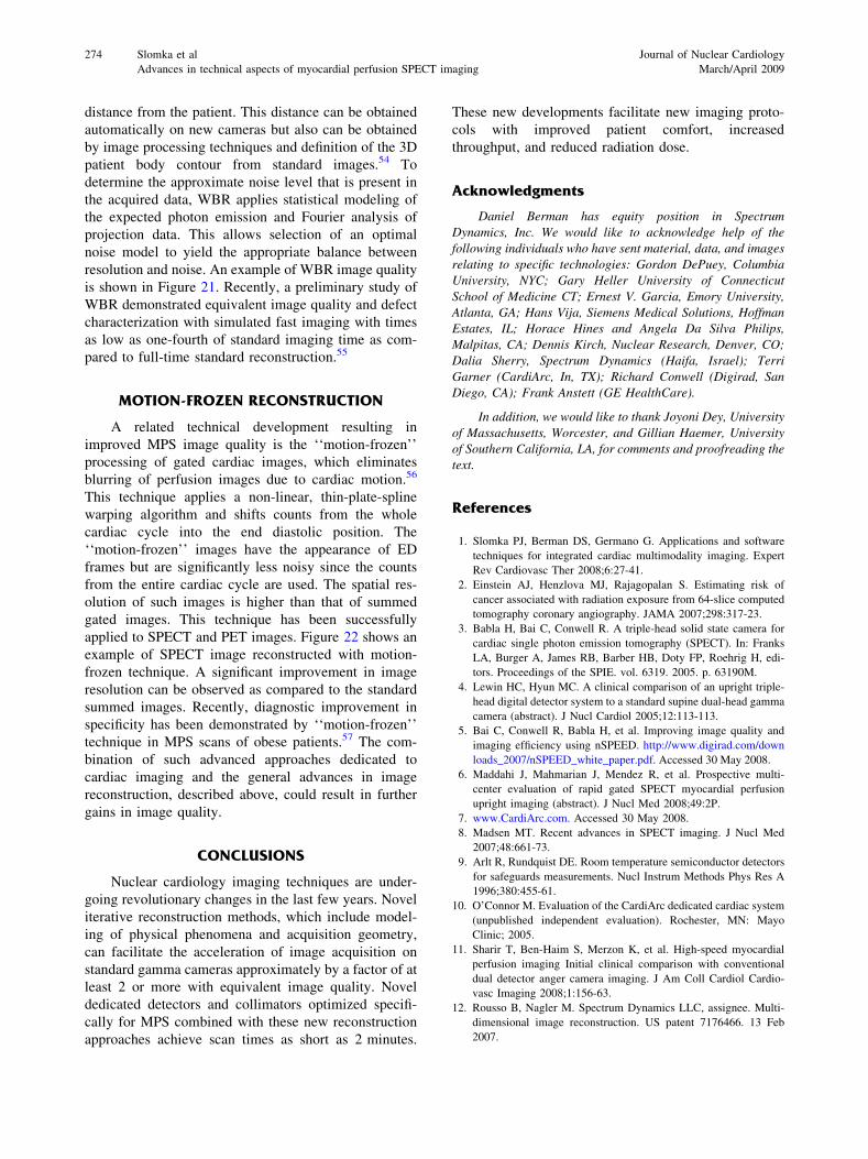
Figure 22. Short axis and vertical long axis of motion frozen (MF) reconstruction and standardsummed reconstruction (SUM) of gated SPECT images. Motion-frozen perfusion images comparedto the summed perfusion images in the case of double vessel disease confirmed by angiography(100% LAD occlusion and 80% LCX occlusion). Both standard quantification technique and visualanalysis of summed data identified only the LAD lesion; the additional LCX lesion was identifiedonly by the ‘‘motion-frozen’’ quantification. Reproduced with permission from Slomka et al56.
Journal of Nuclear Cardiology Slomka et al 273
Volume 16, Number 2;255–76 Advances in technical aspects of myocardial perfusion SPECT imaging
distance from the patient. This distance can be obtained
automatically on new cameras but also can be obtained
by image processing techniques and definition of the 3D
patient body contour from standard images.54 To
determine the approximate noise level that is present in
the acquired data, WBR applies statistical modeling of
the expected photon emission and Fourier analysis of
projection data. This allows selection of an optimal
noise model to yield the appropriate balance between
resolution and noise. An example of WBR image quality
is shown in Figure 21. Recently, a preliminary study of
WBR demonstrated equivalent image quality and defect
characterization with simulated fast imaging with times
as low as one-fourth of standard imaging time as com-
pared to full-time standard reconstruction.55
MOTION-FROZEN RECONSTRUCTION
A related technical development resulting in
improved MPS image quality is the ‘‘motion-frozen’’
processing of gated cardiac images, which eliminates
blurring of perfusion images due to cardiac motion.56
This technique applies a non-linear, thin-plate-spline
warping algorithm and shifts counts from the whole
cardiac cycle into the end diastolic position. The
‘‘motion-frozen’’ images have the appearance of ED
frames but are significantly less noisy since the counts
from the entire cardiac cycle are used. The spatial res-
olution of such images is higher than that of summed
gated images. This technique has been successfully
applied to SPECT and PET images. Figure 22 shows an
example of SPECT image reconstructed with motion-
frozen technique. A significant improvement in image
resolution can be observed as compared to the standard
summed images. Recently, diagnostic improvement in
specificity has been demonstrated by ‘‘motion-frozen’’
technique in MPS scans of obese patients.57 The com-
bination of such advanced approaches dedicated to
cardiac imaging and the general advances in image
reconstruction, described above, could result in further
gains in image quality.
CONCLUSIONS
Nuclear cardiology imaging techniques are under-
going revolutionary changes in the last few years. Novel
iterative reconstruction methods, which include model-
ing of physical phenomena and acquisition geometry,
can facilitate the acceleration of image acquisition on
standard gamma cameras approximately by a factor of at
least 2 or more with equivalent image quality. Novel
dedicated detectors and collimators optimized specifi-
cally for MPS combined with these new reconstruction
approaches achieve scan times as short as 2 minutes.
These new developments facilitate new imaging proto-
cols with improved patient comfort, increased
throughput, and reduced radiation dose.
Acknowledgments
Daniel Berman has equity position in SpectrumDynamics, Inc. We would like to acknowledge help of thefollowing individuals who have sent material, data, and imagesrelating to specific technologies: Gordon DePuey, ColumbiaUniversity, NYC; Gary Heller University of ConnecticutSchool of Medicine CT; Ernest V. Garcia, Emory University,Atlanta, GA; Hans Vija, Siemens Medical Solutions, HoffmanEstates, IL; Horace Hines and Angela Da Silva Philips,Malpitas, CA; Dennis Kirch, Nuclear Research, Denver, CO;Dalia Sherry, Spectrum Dynamics (Haifa, Israel); TerriGarner (CardiArc, In, TX); Richard Conwell (Digirad, SanDiego, CA); Frank Anstett (GE HealthCare).
In addition, we would like to thank Joyoni Dey, Universityof Massachusetts, Worcester, and Gillian Haemer, Universityof Southern California, LA, for comments and proofreading thetext.
References
1. Slomka PJ, Berman DS, Germano G. Applications and software
techniques for integrated cardiac multimodality imaging. Expert
Rev Cardiovasc Ther 2008;6:27-41.
2. Einstein AJ, Henzlova MJ, Rajagopalan S. Estimating risk of
cancer associated with radiation exposure from 64-slice computed
tomography coronary angiography. JAMA 2007;298:317-23.
3. Babla H, Bai C, Conwell R. A triple-head solid state camera for
cardiac single photon emission tomography (SPECT). In: Franks
LA, Burger A, James RB, Barber HB, Doty FP, Roehrig H, edi-
tors. Proceedings of the SPIE. vol. 6319. 2005. p. 63190M.
4. Lewin HC, Hyun MC. A clinical comparison of an upright triple-
head digital detector system to a standard supine dual-head gamma
camera (abstract). J Nucl Cardiol 2005;12:113-113.
5. Bai C, Conwell R, Babla H, et al. Improving image quality and
imaging efficiency using nSPEED. http://www.digirad.com/down
loads_2007/nSPEED_white_paper.pdf. Accessed 30 May 2008.
6. Maddahi J, Mahmarian J, Mendez R, et al. Prospective multi-
center evaluation of rapid gated SPECT myocardial perfusion
upright imaging (abstract). J Nucl Med 2008;49:2P.
7. www.CardiArc.com. Accessed 30 May 2008.
8. Madsen MT. Recent advances in SPECT imaging. J Nucl Med
2007;48:661-73.
9. Arlt R, Rundquist DE. Room temperature semiconductor detectors
for safeguards measurements. Nucl Instrum Methods Phys Res A
1996;380:455-61.
10. O’Connor M. Evaluation of the CardiArc dedicated cardiac system
(unpublished independent evaluation). Rochester, MN: Mayo
Clinic; 2005.
11. Sharir T, Ben-Haim S, Merzon K, et al. High-speed myocardial
perfusion imaging Initial clinical comparison with conventional
dual detector anger camera imaging. J Am Coll Cardiol Cardio-
vasc Imaging 2008;1:156-63.
12. Rousso B, Nagler M. Spectrum Dynamics LLC, assignee. Multi-
dimensional image reconstruction. US patent 7176466. 13 Feb
2007.
274 Slomka et al Journal of Nuclear Cardiology
Advances in technical aspects of myocardial perfusion SPECT imaging March/April 2009

13. Hines H, Kayayan R, Colsher J, et al. Recommendations for
implementing SPECT instrumentation quality control. Eur J Nucl
Med Mol Imaging 1999;26:527-32.
14. Patton J, Sandler M, Berman D, et al. D-SPECT: A new solid state
camera for high speed molecular imaging. Soc Nuclear Med
2006;47:189-189.
15. Ben-Haim S, Hutton B, Van Gramberg D, et al. Simultaneous dual
isotope myocardial perfusion scintigraphy (DI MPS)—Initial
experience with fast D-SPECT (abstract). J Nucl Cardiol
2008;15:S2.
16. Berman D, SW H, Wolak A, et al. Stress thallium-201/rest Tc-99m
sequential dual isotope high-speed myocardial perfusion imaging.
Circulation 2008;118:S1010.
17. Sharir T, Ben Haim S, Slomka PJ, et al. Validation of quantitative
analysis of high-speed myocardial perfusion imaging: Comparison
to conventional SPECT imaging (abstract). J Nucl Cardiol
2008;15:S4.
18. Jaszczak RJ, Li J, Wang H, Zalutsky MR, Coleman RE. Pinhole
collimation for ultra-high-resolution, small-field-of-view SPECT.
Phys Med Biol 1994;39:425-37.
19. Schramm NU, Ebel G, Engeland U, Schurrat T, Behe M, Behr
TM. High-resolution SPECT using multipinhole collimation. IEEE
Trans Nucl Sci 2003;50:315-20.
20. Beekman FJ, Vastenhouw B. Design and simulation of a high-
resolution stationary SPECT system for small animals. Phys Med
Biol 2004;49:4579-92.
21. Funk T, Kirch DL, Koss JE, Botvinick E, Hasegawa BH. A novel
approach to multipinhole SPECT for myocardial perfusion imag-
ing. J Nucl Med 2006;47:595-602.
22. Metzler SD, Bowsher JE, Smith MF, Jaszczak RJ. Analytic
determination of pinhole collimator sensitivity with penetration.
IEEE Trans Med Imaging 2001;20:730-41.
23. Funk T, Despres P, Barber WC, Shah KS, Hasegawa BH. A
multipinhole small animal SPECT system with submillimeter
spatial resolution. Med Phys 2006;33:1259-68.
24. Steele PP, Kirch DL, Koss JE. Comparison of simultaneous dual-
isotope multipinhole SPECT with rotational SPECT in a group of
patients with coronary artery disease. J Nucl Med 2008;49:1080.
25. Volokh L, Hugg J, Blevis I, Asma E, Jansen F, Manjeshwar R.
Effect of detector energy response on image quality of myocardial
perfusion SPECT. Paper presented at IEEE nuclear science sym-
posium and medical imaging conference, 19–26, 2008; Dresden.
26. Blevis I, Tsukerman L, Volokh L, Hugg J, Jansen F, Bouhnik J.
CZT gamma camera with pinhole collimator: Spectral measure-
ments. Paper presented at IEEE 2008 nuclear science and medical
imaging conference, 2008; Dreseden, Germany.
27. Garcia EV, Tsukerman L, Keidar Z. 2.05: A new solid state, ultra
fast cardiac multi-detector SPECT system. J Nucl Cardiol
2008;15:S3-S3.
28. Hawman PC, Haines EJ. The cardiofocal collimator: A variable
focus collimator for cardiac SPECT. Phys Med Biol 1994;39:439-
50.
29. Vija A, Hawman E, Engdahl J. Analysis of a SPECT OSEM
reconstruction method with 3D beam modeling and optional
attenuation correction: Phantom studies. Paper presented at IEEE
nuclear science symposium and medical imaging conference,
2003.
30. Romer W, Reichel N, Vija HA, et al. Isotropic reconstruction of
SPECT data using OSEM3D: Correlation with CT. Acad Radiol
2006;13:496-502.
31. Vija H, Chapman J, Ray M. IQ•SPECT technology white paper.
Siemens Medical Solutions, USA. Mol Imaging 2008;1–7.
32. Shepp LA, Vardi Y. Maximum likelihood reconstruction for
emission tomography. IEEE Trans Med Imaging 1982;1:113-22.
33. Lange K, Carson R. EM reconstruction algorithms for emission
and transmission tomography. J Comput Assist Tomogr
1984;8:306-16.
34. Hudson HM, Larkin RS. Accelerated image reconstruction using
ordered subsets of projection data. IEEE Trans Med Imaging
1994;13:601-9.
35. El Fakhri G, Buvat I, Benali H, Todd-Pokropek A, Di Paola R.
Relative impact of scatter, collimator response, attenuation, and
finite spatial resolution corrections in cardiac SPECT. J Nucl Med
2000;41:1400-8.
36. Metz CE. The geometric transfer function component for scintil-
lation camera collimators with straight parallel holes. Phys Med
Biol 1980;25:1059-70.
37. Kadrmas DJ, Frey EC, Karimi SS, Tsui BMW. Fast implementa-
tion of reconstruction-based scatter compensation in fully 3D
SPECT image reconstruction. Phys Med Biol 1998;43:857-73.
38. Ye J, Song X, Zhao Z, Da Silva AJ, Wiener JS, Shao L. Iterative
SPECT reconstruction using matched filtering for improved image
quality. IEEE Nucl Sci Symp Conf Rec 2006;4.
39. Ye J, Shao L, Zhao Z, Durbin M. Iterative reconstruction with
enhanced noise control filtering. WO patent WO/2007/034,342;
2007.
40. Van Laere K, Koole M, Lemahieu I, Dierckx R. Image filtering in
single-photon emission computed tomography: Principles and
applications. Comput Med Imaging Graph 2001;25:127-33.
41. Venero CV, Ahlberg AW, Bateman TM, et al. Enhancement of
nuclear cardiac laboratory efficiency—Multicenter evaluation of a
new post-processing method with depth-dependent collimator
resolution applied to full and half-time acquisitions. J Nucl Cardiol
2008;15:S4.
42. Bateman TM, Heller GV, McGhie AI, et al. 2.04: Multicenter
investigation comparing a highly efficient half-time stress-only
attenuation correction approach against standard rest-stress
Tc-99m SPECT imaging. J Nucl Cardiol 2008;15:S3-S3.
43. Tsui BMW, Hu HB, Gilland DR, Gullberg GT. Implementation of
simultaneous attenuation and detector response correction in
SPECT. IEEE Trans Nucl Sci 1988;35:778-83.
44. DePuey E, Gadiraju R, Clark J, Thompson L, Anstett F, Shwartz S.
OSEM and wide beam reconstruction (WBR) ‘‘half-time’’ gated
myocardial perfusion SPECT functional imaging: A comparison to
‘‘full-time’’ filtered back projection. J Nucl Cardiol 2008;15:547-
63.
45. Tsui BMW, Gullberg GT. The geometric transfer-function for
cone and fan beam collimators. Phys Med Biol 1990;35:81-93.
46. Tsui BMW, Frey EC, Zhao X, Lalush DS, Johnston RE,
McCartney WH. The importance and implementation of accurate
3D compensation methods for quantitative SPECT. Phys Med Biol
1994;39:509-30.
47. Bruyant PP. Analytic and iterative reconstruction algorithms in
SPECT. J Nucl Med 2002;43:1343-58.
48. Green PJ. Bayesian reconstructions from emission tomography
data using a modified EM algorithm. IEEE Trans Med Imaging
1990;9:84-93.
49. Alenius S, Ruotsalainen U. Bayesian image reconstruction for
emission tomography based on median root prior. Eur J Nucl Med
Mol Imaging 1997;24:258-65.
50. Vija AH, Zeintl J, Chapman JT, Hawman EG, Hornegger J.
Development of rapid SPECT acquisition protocol for myocardial
perfusion imaging. IEEE Nucl Sci Symp Conf Rec 2006;3:1811-6.
51. Ficaro EP, Kritzman JN, Corbett JR. 15.34: Effect of reconstruc-
tion parameters and acquisition times on myocardial perfusion
distribution in normals. J Nucl Cardiol 2008;15:S20-S20.
52. Zeintl J, Ding X, Vija AH, Hawman EG, Hornegger J, Kuwert T.
Estimation accuracy of ejection fraction in gated cardiac SPECT/
Journal of Nuclear Cardiology Slomka et al 275
Volume 16, Number 2;255–76 Advances in technical aspects of myocardial perfusion SPECT imaging

CT imaging using iterative reconstruction with 3D resolution
recovery in rapid acquisition protocols. 2007 NSS ‘07 IEEE Nucl
Sci Symp Conf Rec 2007;6:4491-6.
53. Ultraspect. www.UltraSPECT.com. Accessed 6 Sept 2008.
54. Borges-Neto SPR, Shaw LK, et al. Clinical results of a novel wide
beam reconstruction method for shortening scan time of Tc-99m
cardiac SPECT perfusion studies. J Nucl Cardiol 2007;14:555-65.
55. DePuey EG, Bommireddipalli S, Beletsky I, et al. 2.01: Quarter-
time myocardial perfusion SPECT wide beam reconstruction. J
Nucl Cardiol 2008;15:S2-S2.
56. Slomka PJ, Nishina H, Berman DS, et al. ‘‘Motion-frozen’’ dis-
play and quantification of myocardial perfusion. J Nucl Med
2004;45:1128-34.
57. Suzuki Y, Slomka PJ, Wolak A, et al. Motion-frozen myocardial
perfusion SPECT improves detection of coronary artery disease in
obese patients. J Nucl Med 2008;49:1075-9.
276 Slomka et al Journal of Nuclear Cardiology
Advances in technical aspects of myocardial perfusion SPECT imaging March/April 2009
Related Documents