HETEROCYCLES, Vol. 79, 2009, pp. 1007 - 1017. © The Japan Institute of Heterocyclic Chemistry Received, 4th November, 2008, Accepted, 8th December, 2008, Published online, 12th December, 2008. DOI: 10.3987/COM-08-S(D)78 MAITOTOXIN-PHOTOACTIVE PROBE BINDS TO MEMBRANE PROTEINS IN BLOOD CELLS Keiichi Konoki, a,b, * Masaki Hashimoto, a Kaori Honda, a Kazuo Tachibana, a Rie Tamate, b Futoshi Hasegawa, b Tohru Oishi, b and Michio Murata b, * a Department of Chemistry, School of Science, The University of Tokyo, Hongo, Bunkyo-ku, Tokyo 113-0033, Japan b Department of Chemsitry, Graduate School of Science, Osaka University, 1-1 Machikaneyama, Toyonaka, Osaka 560-0043, Japan FAX: (+81)-6-6850-5774, E-mail: [email protected] (K. K.) and [email protected] (M. M.) Abstract – The photoactive and biotinylating ligand was prepared from MTX and maleimide-conjugated Hatanaka reagent with use of Diels-Alder reaction. Blood cells were subjected to affinity labelling experiments using the ligand thus obtained. The labelled band on SDS-PAGE was replaced not by MTX but by brevetoxin B (PbTx2), which suggested the presence of binding proteins in blood cells. Screening of polyether compounds for MTX inhibitory activity using Ca 2+ flux assays in C6 cells disclosed that a synthetic fragment of the hydrophilic portion of MTX inhibited the MTX activity. INTRODUCTION Maitotoxin (MTX, 1) was first discovered as one of the toxins responsible for ciguatera 1 , seafood poisoning caused by ingestion of coral reef fish, and was later shown to be a metabolite of the dinoflagellate Gambierdiscus toxicus. 2 The chemical structure of 1 was determined by spectroscopic methods 3 and by chemical synthesis. 4,5 The striking structural feature is the presence of 32 ether rings encompassing hydroxy and sulfate groups, which comprise a remarkably large heterocyclic molecule. In addition to these unique chemical features, MTX has extremely potent bioactivities exemplified by LD 50 against mice (50 ng/kg, ip.). 3 MTX elicits Ca 2+ influx in virtually all cells and tissues so far tested as reviewed by John Daly and Fabian Gusovsky. 6 This elevation in intracellular calcium concentration leads Dedication to the great contribution of Dr. John Daly to natural product chemistry. HETEROCYCLES, Vol. 79, 2009 1007

Welcome message from author
This document is posted to help you gain knowledge. Please leave a comment to let me know what you think about it! Share it to your friends and learn new things together.
Transcript
HETEROCYCLES, Vol. 79, 2009, pp. 1007 - 1017. © The Japan Institute of Heterocyclic Chemistry Received, 4th November, 2008, Accepted, 8th December, 2008, Published online, 12th December, 2008. DOI: 10.3987/COM-08-S(D)78
MAITOTOXIN-PHOTOACTIVE PROBE BINDS TO MEMBRANE
PROTEINS IN BLOOD CELLS
Keiichi Konoki,a,b,* Masaki Hashimoto,a Kaori Honda,a Kazuo Tachibana,a
Rie Tamate,b Futoshi Hasegawa,b Tohru Oishi,b and Michio Muratab,*
aDepartment of Chemistry, School of Science, The University of Tokyo, Hongo,
Bunkyo-ku, Tokyo 113-0033, Japan bDepartment of Chemsitry, Graduate School of Science, Osaka University, 1-1
Machikaneyama, Toyonaka, Osaka 560-0043, Japan
FAX: (+81)-6-6850-5774, E-mail: [email protected] (K. K.) and
[email protected] (M. M.)
Abstract – The photoactive and biotinylating ligand was prepared from MTX and
maleimide-conjugated Hatanaka reagent with use of Diels-Alder reaction. Blood
cells were subjected to affinity labelling experiments using the ligand thus
obtained. The labelled band on SDS-PAGE was replaced not by MTX but by
brevetoxin B (PbTx2), which suggested the presence of binding proteins in blood
cells. Screening of polyether compounds for MTX inhibitory activity using Ca2+
flux assays in C6 cells disclosed that a synthetic fragment of the hydrophilic
portion of MTX inhibited the MTX activity.
INTRODUCTION
Maitotoxin (MTX, 1) was first discovered as one of the toxins responsible for ciguatera1, seafood
poisoning caused by ingestion of coral reef fish, and was later shown to be a metabolite of the
dinoflagellate Gambierdiscus toxicus.2 The chemical structure of 1 was determined by spectroscopic
methods3 and by chemical synthesis.4,5 The striking structural feature is the presence of 32 ether rings
encompassing hydroxy and sulfate groups, which comprise a remarkably large heterocyclic molecule. In
addition to these unique chemical features, MTX has extremely potent bioactivities exemplified by LD50
against mice (50 ng/kg, ip.).3 MTX elicits Ca2+ influx in virtually all cells and tissues so far tested as
reviewed by John Daly and Fabian Gusovsky.6 This elevation in intracellular calcium concentration leads
Dedication to the great contribution of Dr. John Daly to natural product chemistry.
HETEROCYCLES, Vol. 79, 2009 1007
to secondary events; e.g., phosphoinositide breakdown,7 arachidonic acid release,8 muscle contraction,9
and secretion of dopamine,10 norepinephrine,11 and insulin,12 some of which are elicited by depolarization
of the membrane potential.13 Electrophysiological studies have revealed that channels activated by MTX
are non-selective and voltage-independent.14 On the other hand, some studies indicated that
MTX-induced membrane depolarization is essentially independent of Ca2+ and due mainly to influx of
Na+, which suggest the possibility that the direct action of MTX on biomembranes does not always
involve Ca2+ influx, but can be due to the entry of other cations. Many hydrophobic amines, which
potentially interact with cation channel proteins to alter their conductivity, have been reported to block
the actions of MTX, such as nifedipine, verapamil, SK&F 96365, tetrahexylammonium salt,15,16 and so on.
On the other hand, some non-amine inhibitors of MTX activity, which possess higher probability to
interact directly with the MTX target rather than ion channels that are indirectly activated by MTX, are
reported so far. One of examples is glycosphingolipid GM1, which has been reported to be a potent
blocker of MTX-induced Ca2+ influx in bovine aortic endothelial cells.17 Our group has recently
demonstrated that synthetic fragments of MTX and structural mimics such as PbTx2 and other
ladder-shaped polyethers potentially inhibited the biological activities of MTX.18 The establishment of
the complete chemical structure of MTX allowed us to deduce the three-dimensional shape of the
molecule,4,18 which implies possible interactions with biomembranes. The hydrophobic polycyclic ethers
from rings R through F' in MTX (1) could penetrate into a plasma membrane, as suggested for other
ladder-shaped polyethers,19-22 whereas the polyhydroxy groups residing on rings A through Q along with
two anionic sulfate esters keeping this hydrophilic half outside the membrane.
Photoaffinity labeling 23 is one of the most frequently used methods to identify the target proteins of
drugs and other bioactive compounds. However, this methodology is usually applied for small molecules
or small peptides, thus leaving its applicability to larger non-peptidic compounds like MTX. In this study
we prepared a photoactive and biotinylated ligand of MTX and attempted to identify the binding proteins.
On the competitive inhibition of MTX-binding, small fragmental structures and structure mimics were
used in lieu of MTX.24
RESULTS
Preparation of Photoaffinity Probe of MTX.
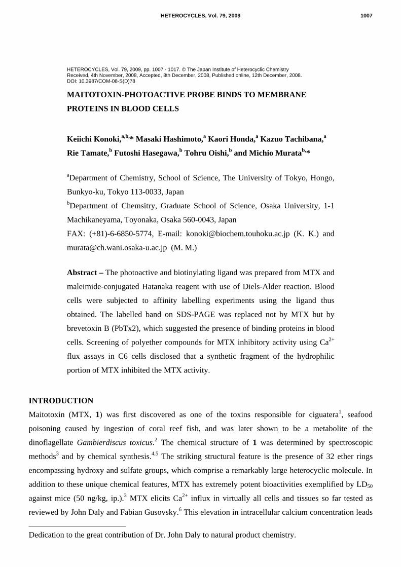
Photoactive-biotinylating agent (2, Hatanaka Reagent) was kindly gifted by Prof. Y. Hatanaka.23 As
shown in the Scheme 1, the reagent was treated with mono-N-(t-Boc)-ethylenenediamine in the presence
of EDC and HOBt followed by deprotection by TFA to provide monoamine amide 3. The coupling with
maleimide-containing activated carboxylic acid proceeded to give rise to photoactive-biotinylating
dienophile 4. The crucial Diels-Alder reaction with the diene group of MTX was carried out under high
1008 HETEROCYCLES, Vol. 79, 2009
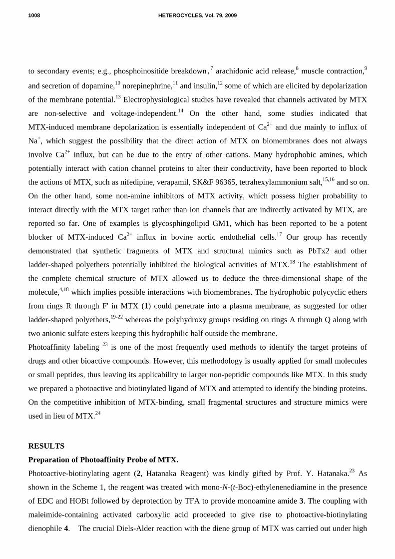
pressure of 1.0 GPa for 3 days. Production of the Dieal-Alder adduct 5 was confirmed by HPLC and mass
spectrometry as shown in Figure 1. Although an attempt to measure NMR was unsuccessful due to a
small sample amount and large molecular weight, the mass number obtained in ESIMS completely agreed
with that of intended product 5. The biological activity of the product was examined by Ca2+ assays using
erythrocytes where the product showed significantly Ca2+ influx into blood cells (data not shown).
CO2H
NN
F3C
O
O
O
HNO
SNH
HN
O
H
HH
NN
F3C
R
a, b c
NN
F3C
R
NH
NH2
O NH
HN
O
N
O
O
O
d
2 3
4
5
Photo-Biotin-MTX
Scheme 1. (a) Mono-N-(t-Boc)-ethylenenediamine, EDC, HOBt, 1:1 THF-MeOH, rt, 3 h; (b) 1:1
TFA-CH2Cl2, rt, 30 min; (c) Succinimidyl-4-(N-maleimidomethyl) cyclohexane-1-carboxylate, Et3N,
DMF, rt, 4 h (35% in 3 steps); (d) MTX, 10:46:44 MeOH-H2O-MECN, 1 x 1.0 GPa, rt, 3 days
Me Me Me
Me
Me
Me
MeMe
OH
O
OH
OH
HO OH
H
Me
HHO OH
OH
OMe
OH
OH
H H
H H H H H H
HO Me Me
OH
NaO3SOOH OH
OH
OH
OHOH
HO
OH H H
O
O H
H
H
H
H
O
O
O
O
O
H H
H
H
H
H
H
H
H
H
O
Me
Me
Me
O
Me
Me
O
HO
HO
H
H
H
H
H
OH
OH
OH
H
H
H H
Me
OSO3Na
OH
MeOH
H H H H
H
H
HO
O
O
OO
OO
OO
H
H
HH
H
Me
Me OH
OH
O
O
O
O
OO
O O
O
O
OO
HN
OS
NHHN
O
HH
H
NN
F3C
O
NH
HN
O
N
O
O
O
5
Me
HO
OSO3Na
OH
MeOH1: Maitotoxin
A
QR
F'
W
C'
HETEROCYCLES, Vol. 79, 2009 1009
Figure 1. Mass spectrum of Diels-Alder adduct 5. The Diels-Alder product after HPLC purification was
subjected to ESI-TOF mass spectrometer. The ion peaks at 1414.3 and 2121.92 correspond to
[M-Na2-H]3- and [M-Na2]2-, respectively. Their isotopic patterns (inset figures) for those at m/z 1414 and
2121 depict that these ions are divalent and trivalent, respectively.
NaO3SO
O
O
O
O
O
O
OO
OO
O
MeHO
Me
MeMe
NaO3SO MeOH
Me
HH
HH H H
H
H
H
HH
H
H
HH
HH
H
HO
Me
MeMe
HH H
H HH
H
HO
O
O
O
O
O
O
O
O
O
O
O
O
O
OO
CHO
O
MeH
MeH H Me
MeH H
HMe
HO
HH
HMe
HHHH
HH
Me
O
O
O
OR
OR
Me H H
H Me Me
7: YTX
6: PbTx2
8: FH2OH, R = Bn
9: FH4OH, R = H
1010 HETEROCYCLES, Vol. 79, 2009
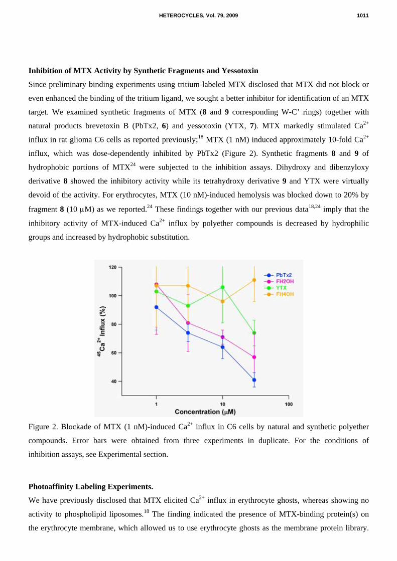
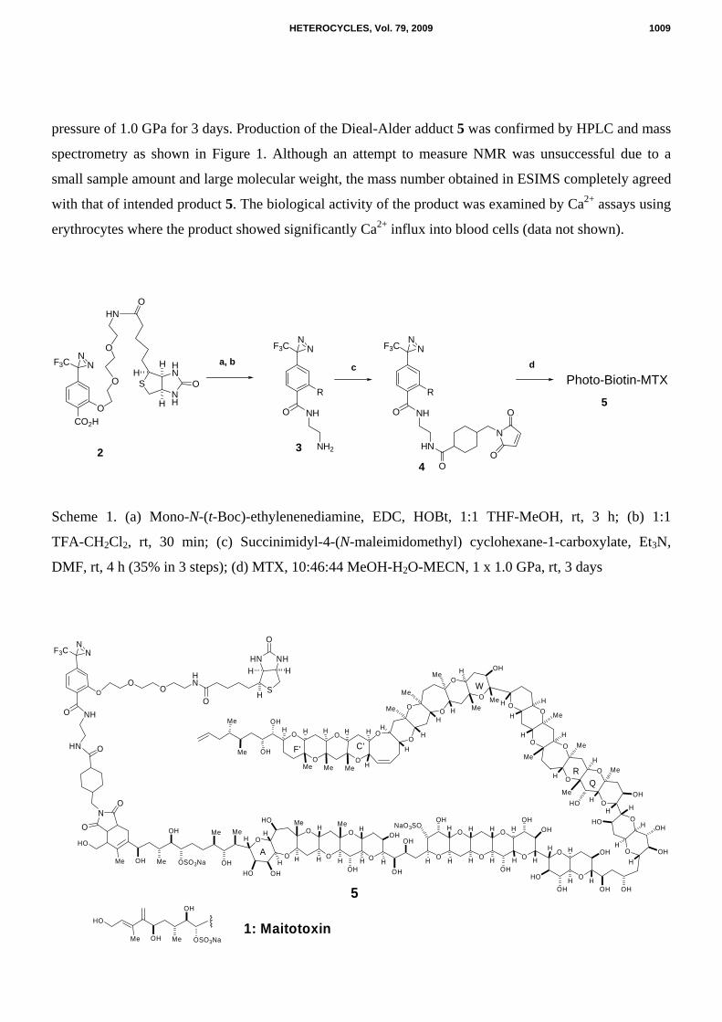
Inhibition of MTX Activity by Synthetic Fragments and Yessotoxin
Since preliminary binding experiments using tritium-labeled MTX disclosed that MTX did not block or
even enhanced the binding of the tritium ligand, we sought a better inhibitor for identification of an MTX
target. We examined synthetic fragments of MTX (8 and 9 corresponding W-C’ rings) together with
natural products brevetoxin B (PbTx2, 6) and yessotoxin (YTX, 7). MTX markedly stimulated Ca2+
influx in rat glioma C6 cells as reported previously;18 MTX (1 nM) induced approximately 10-fold Ca2+
influx, which was dose-dependently inhibited by PbTx2 (Figure 2). Synthetic fragments 8 and 9 of
hydrophobic portions of MTX24 were subjected to the inhibition assays. Dihydroxy and dibenzyloxy
derivative 8 showed the inhibitory activity while its tetrahydroxy derivative 9 and YTX were virtually
devoid of the activity. For erythrocytes, MTX (10 nM)-induced hemolysis was blocked down to 20% by
fragment 8 (10 µM) as we reported.24 These findings together with our previous data18,24 imply that the
inhibitory activity of MTX-induced Ca2+ influx by polyether compounds is decreased by hydrophilic
groups and increased by hydrophobic substitution.
Figure 2. Blockade of MTX (1 nM)-induced Ca2+ influx in C6 cells by natural and synthetic polyether
compounds. Error bars were obtained from three experiments in duplicate. For the conditions of
inhibition assays, see Experimental section.
Photoaffinity Labeling Experiments.
We have previously disclosed that MTX elicited Ca2+ influx in erythrocyte ghosts, whereas showing no
activity to phospholipid liposomes.18 The finding indicated the presence of MTX-binding protein(s) on
the erythrocyte membrane, which allowed us to use erythrocyte ghosts as the membrane protein library.
HETEROCYCLES, Vol. 79, 2009 1011
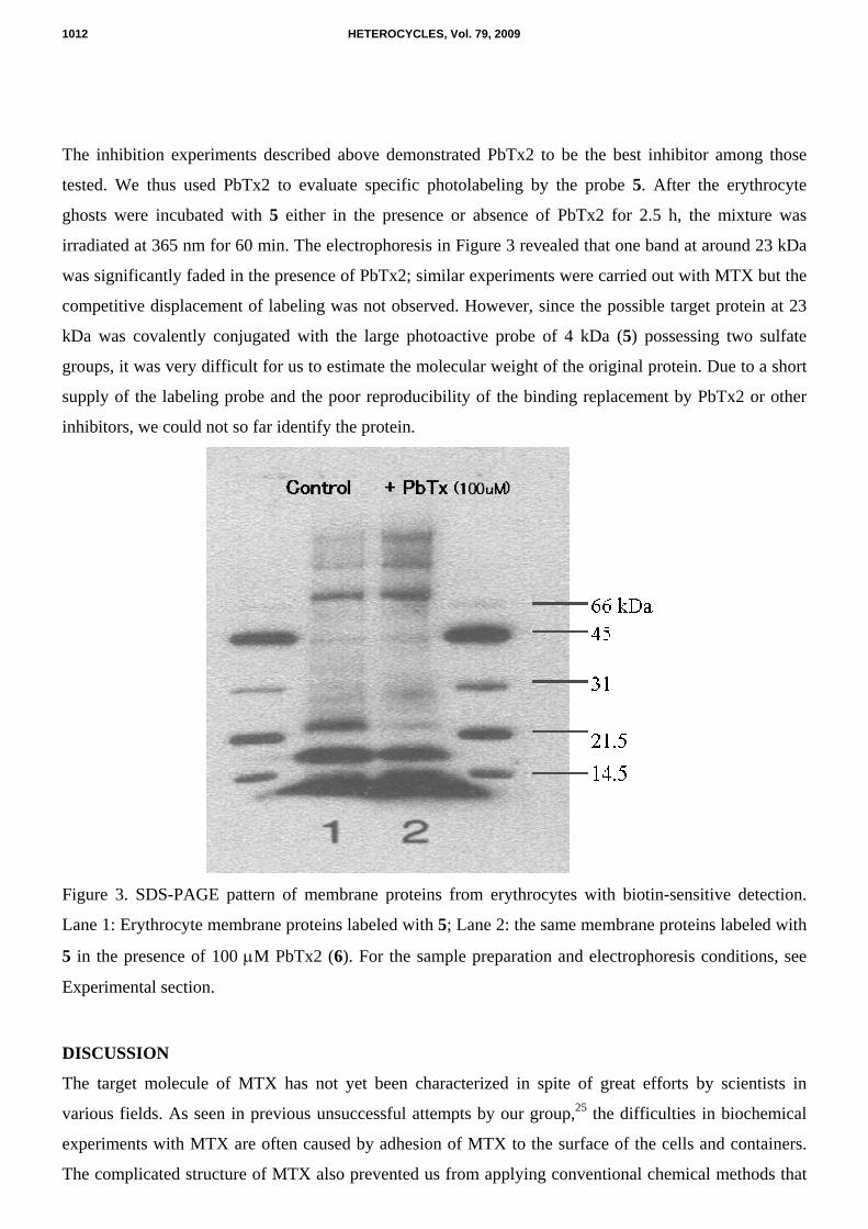
The inhibition experiments described above demonstrated PbTx2 to be the best inhibitor among those
tested. We thus used PbTx2 to evaluate specific photolabeling by the probe 5. After the erythrocyte
ghosts were incubated with 5 either in the presence or absence of PbTx2 for 2.5 h, the mixture was
irradiated at 365 nm for 60 min. The electrophoresis in Figure 3 revealed that one band at around 23 kDa
was significantly faded in the presence of PbTx2; similar experiments were carried out with MTX but the
competitive displacement of labeling was not observed. However, since the possible target protein at 23
kDa was covalently conjugated with the large photoactive probe of 4 kDa (5) possessing two sulfate
groups, it was very difficult for us to estimate the molecular weight of the original protein. Due to a short
supply of the labeling probe and the poor reproducibility of the binding replacement by PbTx2 or other
inhibitors, we could not so far identify the protein.
Figure 3. SDS-PAGE pattern of membrane proteins from erythrocytes with biotin-sensitive detection.
Lane 1: Erythrocyte membrane proteins labeled with 5; Lane 2: the same membrane proteins labeled with
5 in the presence of 100 µM PbTx2 (6). For the sample preparation and electrophoresis conditions, see
Experimental section.
DISCUSSION
The target molecule of MTX has not yet been characterized in spite of great efforts by scientists in
various fields. As seen in previous unsuccessful attempts by our group,25 the difficulties in biochemical
experiments with MTX are often caused by adhesion of MTX to the surface of the cells and containers.
The complicated structure of MTX also prevented us from applying conventional chemical methods that
1012 HETEROCYCLES, Vol. 79, 2009
were originally designed for drugs or smaller molecules. These fruitless efforts using MTX itself
prompted us to seek for an alternative agent with a smaller molecular weight.16 The chemical structure of
MTX implied us that PbTx2 might be an appropriate substitute for MTX because of their close
resemblance in the hydrophobic polyether structures. In addition to PbTx2, synthetic fragment 8 with
hydrophobic benzyloxy sidechains revealed the comparable inhibitory activity (Figure 2) despite of its
fewer ether rings than those of PbTx2. As reported previously,25 we examined inhibition of MTX binding
to rat brain synaptosomes by PbTx2 using 3H-labeled hydrogenated MTX, which was prepared in John
Daly’s laboratory.25 The binding of tritiated-MTX to synaptosomes was often enhanced by unlabeled
MTX, which was particularly unusual in binding replacement tests. This may be accountable by the mode
of binding of MTX to biomembranes; one possible explanation is formation of dimers or oligomers of
MTX upon binding to the target, where a mixed assembly of labeled and unlabeled MTX enhanced the
total binding. When the same experiment was carried out with PbTx2 as an inhibitor, binding replacement
was not observed.18 This unexpected results can be explained by the manner of MTX binding to
biomembranes. MTX was not completely washed out from rat brain synaptosomes even after repeated
perfusions,16 which suggests that its amphiphilicity stabilizes the molecule and results in abundant
nonspecific binding of 3H-labeled hydrogenated MTX. Thus, photolabeling experiments were carried out
in this study to confirm the binding replacement of MTX by PbTx2. We successfully prepared the ligand
from MTX and observed the promising band, for which binding of MTX-derivative was blocked by
PbTx2. Further examinations to identify the protein have been hampered due to difficulties in preparing
the ligand from MTX and in effectively labeling the protein. Rapid progress in proteomics technology
will soon enable us to identify the target protein in an extremely small quantity.
The present results confirmed that the synthetic fragments prominently inhibit MTX-induced Ca2+ influx,
and further support the idea that the hydrophobic portion of MTX plays an important role in recognizing
the target molecule. If the hydrophobic portion of MTX binds to the transmembrane α-helix domain of its
target proteins in a manner similar to PbTx2 binding to sodium channels,18 it is plausible that a relatively
high concentration of polyether compounds could mask the MTX-binding site despite their low affinities
to the site.
The present experimental results together with previous findings allow us to propose a hypothetical
mechanism for the biological activities of MTX:18 MTX binds to a plasma membrane with its
hydrophobic tail (R-F’rings), whereas its hydrophilic portion staying outside. MTX interacts to a
transmembrane α-helix domain of the target proteins with its hydrophobic portion to modify the protein
function to allow ions to pass across membrane, which causes large Ca2+ influx. Further investigations
using synthetic fragments are underway in the laboratories to gain a better understanding of the action
mechanism of this unique marine toxin.
HETEROCYCLES, Vol. 79, 2009 1013
EXPERIMENTAL
Materials
MTX was isolated from the French Polynesian strain of the dinoflagellate Gambierdiscus toxicus
collected off Gambier Island.2 The toxin was dissolved in 50 % aqueous methanol (290 nM) and kept at -
30 ºC until use. Rat glioma C6 cells, RPMI 1640, a penicillin-streptomycin solution, fetal bovine serum,
and trypsin (tissue culture grade) were purchased from Dainippon Pharmaceutical (Osaka, Japan). 45CaCl2 and [3H]-leucine were from New England Nuclear (Boston, USA).
Preparation of Photoactive-Biotinylating MTX
Synthesis of dienophile. Hatanaka Reagent23 (20.0 mg, 0.033 mmol) in 1:1 THF-MeOH was reacted with
EDC (7.0 mg, 1.1 eq), HOBt (5.4 mg, 1.2 eq) and mono-N-(t-Boc)-ethylenenediamine (12.8 mg, 2.4 eq)
at rt for 3 h. The mixture was evaporated, and partitioned between AcOEt (15 mL) and H2O (5.0 mL).
The organic layer was washed with brine and dried over MgSO4. After evaporated, the crude mixture
primarily containing t-Boc protected amine was directly used for the following reaction. The crude
mixture dissolved in 250 µL of CH2Cl2 was added dropwise to 250 µL of trifluoroacetic acid. After 30
min, the mixture was evaporated and the concentrate was dissolved in 500 µL of DMF. The solution was
treated with Et3N (4.6 µL, 0.033 mmol) and succinimidyl-4-(N-maleimidomethyl)
cyclohexane-1-carboxylate (12.1 mg, 0.0362 mmol, Thermo Fisher Scientific, Rockford, USA) at rt for 4
h. The mixture was concentrated and subjected to ODS column chromatography (1.0 g, YMC ODS-AM,
YMC, Kyoto, Japan). The fraction eluted with 1:1 MeCN-H2O was concentrated to give the dienophile
(14.3 mg, 45% in 3 steps). A part of the product was further purified with HPLC (YMC ODS AM323, φ
10 x 250 mm, 1.0 mL/min, 50% MeCN, 400 nm) for the following Diels-Alder Reaction.
Diels-Alder Reaction. Maitotoxin (50 µg, 15 nmol) dissolved in 36 µL of 83% aqueous MeOH was
added to a specified reaction container for high-pressure reaction. The dienophile (639 µg, 740 nmol)
dissolved in 250 µL of 50% aqueous MeCN was added to the container. The mixture was covered with
hexane to fill the rest of the space in the container, which was then tightly capped. The mixture was
gradually pressurized up to 1.0 x 1.0 GPa and maintained at 25 ºC for 3 days. The whole mixture was
transferred to a vial and the lower layer (not the hexane layer) was transferred to another vial. The
mixture was dried under stream of N2. To remove the excess dienophile, the concentrate was treated with
H2O (100 µL) and washed with AcOEt (100 µL x 2), CHCl3 (100 µL), and then hexane (100 µL x 2). The
aqueous layer was concentrated and subjected to HPLC analysis (Develosil TMS-5, φ4.6 x 150 mm,
35% MeCN/10 mM NH4OAc, 1.0 mL/min, 230 nm). The fractions for Diels-Alder adduct were estimated
by comparison of HPLC profile with that for the mock reaction without MTX. ESI-TOF measurements
1014 HETEROCYCLES, Vol. 79, 2009
using Q-Tof 2 (Micromass, UK) to identify the fraction containing Diels-Alder reaction adduct was first
carried out at Jasco International Corp (Tokyo, Japan).
Photoaffinity Labeling
Two micro litters of human red blood cells were added to 96 µL of a buffer consisting of 150 mM NaCl,
5 mM KCl, 2 mM CaCl2, 5 mM D-glucose, and 10 mM Hepes/Tris (pH 7.4). The suspension was mixed
at 0 ºC for 2.5 h with 1.0 µL of the photoactive biotinylated maitotoxin either in the presence or absence
of 1.0 µL of intact maitotoxin or PbTx2. The mixture was irradiated with a black light lamp (365 nm) for
1 h and then heated at 95 ºC for 10 min.
SDS-PAGE
The Sample Buffer consists of 1.0 mL of 0.5 M Tris/HCl (pH 6.8), 2.0 mL of 10% SDS, 0.6 mL of 10%
DTT, 1.0 mL of glycerol, 0.4 mL of mili-Q grade water and 3 drops of 1% aqueous Bromophenol Blue.
After photolabeling, the mixture was mixed with the Sample Buffer and heated at 95 ºC for 10 min. The
denatured sample was loaded to a polyacrylamide gel (Ready Gel J 5-20%, Biorad, Hercules, USA) as
well as biotinylated protein markers (Biorad, Hercules, USA). The acryl amide gel was electroblotted to a
nitrocellulose membrane. The membrane was blocked with 5% skim milk/0.1% Tween 20/PBS (T-PBS),
washed 5 times with 0.1% T-PBS for 2, 2, 15, 5 and 5 min and reacted with horse raddish
peroxidase-conjugated streptavidin for 1 h. ECL western blotting reagent (GE, Chalfont St. Giles, UK)
was used for detection. The chemiluminescence was recorded with Hyperfilm ECL (GE, Chalfont St.
Giles, UK).
Cell culture
Rat glioma C6 cells were cultured at 37 ºC in a humidified atmosphere of 5% CO2/95% air with a CO2
incubator. The culture media consisted of RPMI 1640 medium supplemented with 10% fetal bovine
serum, 50 units/mL penicillin, and 50 µg/mL streptomycin. C6 cells were grown for 2 days (the cell
density usually reached about 2.4 x 10⁶ cells/mL) and inoculated onto new media to a cell density of 1.2 x
106 cells/mL.
45Ca2+ influx assays with inhibitors
After passage, rat glioma cells were grown for 2 days, harvested by treatment of trypsin, and diluted to
6.7 x 10⁵ cells/mL. The detached cells were transferred to 12 well plates and [3H]-leucine (0.1 µCi/mL)
was added. After overnight incubation, the medium was replaced with 238 µL of buffer A containing 150
mM NaCl, 5 mM KCl, 2 mM CaCl2, 5 mM glucose, and 50 mM Hepes (pH 7.4 adjusted by Tris), along
HETEROCYCLES, Vol. 79, 2009 1015
with an inhibitor tested. After 12 min of preincubation, 50 µL of buffer A containing 45CaCl2 (1.5
µCi/mL), and 12 µL of MTX solution in 50% aqueous methanol were added to the media. The cells were
incubated for 12 min and then washed three times with buffer A followed by solubilization with 250 µL
of 1% sodium dodecyl sulfate (SDS) in 0.5 M NaOH at 37ºC for 20 min. The solution was transferred to
a scintillation vial (20 mL), neutralized with 250 µL of 0.5 M HCl, and mixed with 5 mL of scintillation
cocktail. The radioactivity was measured with a scintillation counter set for simultaneous counting of 3H
and 45Ca. Differences in the growth of the cells among microplate wells were normalized by the 3H-count
for incorporated [3H]-leucine.
ACKNOWLEDGEMENTS
This work was supported by Grant-In-Aids for Scientific Research (A) (No. 15201048) and (S) (No.
18101010), for Priority Area (A) (No. 16073211) from MEXT, Japan, and by a grant from the CREST,
Japan Science and Technology Corporation. We are grateful to late Dr. John W. Daly for discussion and
collaboration, and to Prof. Takeshi Yasumoto for encouragements of maitotoxin research. We also thank
Prof. Masaharu Nakamura, Kyoto University, for Diels-Alder reaction, and Dr. Nobuaki Matsumori in
our group for discussion.
REFERENCES
1. T. Yasumoto, R. Bagnis, and J. P. Vernoux, Bull. Jpn. Soc. Fish., 1976, 42, 359.
2. A. Yokoyama, M. Murata, Y. Oshima, T. Iwashita, and T. Yasumoto, J. Biochem., 1988, 104, 184.
3. M. Murata, H. Naoki, S. Matsunaga, M. Satake, and T. Yasumoto, J. Am. Chem. Soc., 1994, 116,
7098.
4. M. Sasaki, N. Matsumori, T. Maruyama, T. Nonomura, M. Murata, K. Tachibana, and T. Yasumoto,
Angew. Chem., Int. Ed. Engl., 1996, 35, 1672.
5. W. Zheng, J. A. DeMattei, J.-P. Wu, J. J.-W. Duan, L. R. Cook, H. Oinuma, and Y. Kishi, J. Am.
Chem. Soc., 1996, 118, 7946.
6. F. Gusovsky and J. W. Daly, Biochem. Pharmacol., 1990, 39, 1633.
7. F. Gusovsky, T. Yasumoto, and J. W. Daly, FEBS Lett., 1989, 243, 307.
8. O. H. Choi, W. L. Padgett, Y. Nishizawa, F. Gusovsky, T. Yasumoto, and J. W. Daly, Mol.
Pharmacol., 1989, 37, 222.
9. Y. Ohizumi, A. Kajiwara, and T. Yasumoto, J. Pharmacol. Exp. Ther., 1983, 227, 199.
10. M. Taglialatela, S. Amoroso, T. Yasumoto, G. D. Renzo, and L. Annunziato, Brain Res.,1986, 381,
356.
11. M. Takahashi, M. Tatsumi, Y. Ohizumi, and T. Yasumoto, J. Biol. Chem., 1983, 258, 10944.
1016 HETEROCYCLES, Vol. 79, 2009
12. D. Soergel, F. Gusovsky, T. Yasumoto, and J. W. Daly, J. Pharmacol. Exp. Ther.,1990, 255, 1360.
13. D. Xi., F. M. Van Dolah, and J. S. Ramsdell, J. Biol. Chem., 1992, 267, 25025.
14. L. I. Escobar, C. Salvador, M. Martinez, and L. Vaca, J. Neurobiol., 1998, 6, 59.
15. J. W. Daly, J. Lueder, W. L. Padgett, S. Yangmee, and F. Gusovsky, Biochem. Parmacol., 1995, 50,
1187.
16. K. Konoki, M. Hashimoto, M. Murata, K. Tachibana, and T. Yasumoto, J. Natur. Toxins, 1996, 5,
209.
17. J. P. Bressler, L. Belloni-Olivi, and S. Forman, Life Sci., 1993, 54, 49.
18. K. Konoki, M. Hashimoto, T. Nonomura, M. Sasaki, M. Murata, and K. Tachibana, J.
Neurochem.,1998, 70, 409.
19. V. L. Trainer, D. G. Baden, and W. A. Catterall, J. Biol. Chem., 1994, 269, 19904.
20. R. E. Gawley, K. S. Rein, G. Jeglitsch, D. J. Adams, E. A. Theodorakis, J. Tiebes, K. C. Nicolaou,
and D. G. Baden, Chem. Biol., 1995, 2, 533.
21. S. Matile, N. Berova, and K. Nakanishi, Chem. Biol., 1996, 3, 379.
22. S. Matile and K. Nakanishi, Angew. Chem., Int. Ed. Engl., 1996, 35, 757.
23. T. Tomohiro, M. Hashimoto, and Y. Hatanaka, Chem. Rec., 2005, 5, 385; Y. Hatanaka, M.
Hashimoto, and Y. Kanaoka, J. Am. Chem. Soc., 1998, 120, 453; Y. Hatanaka, M. Hashimoto, and Y.
Kanaoka, Bioorg. Med. Chem., 1994, 2, 1367; Y. Hatanaka, M. Hashimoto, H. Kurihara, H.
Nakayama, and Y. Kanaoka, J. Org. Chem., 1994, 59, 383.
24. T. Oishi, F. Hasegawa, K. Torikai, K. Konoki, N. Matsumori, and M. Murata, Org. Lett., 2008, 10,
3599.
25. M. Murata, F. Gusovsky, T. Yasumoto, and J. W. Daly, Eur. J. Pharmacol., 1992, 227, 43; M.
Murata, M. Sasaki, A. Yokoyama, T. Iwashita, F. Gusovsky, J. W. Daly, and T. Yasumoto, Bull. Soc.
Path. Exot., 1992, 85, 470.
26. M. Estacion, H. B. Nguyen, and J. J. Gargus, Am. J. Physiol., 1994, 270, C1145.
HETEROCYCLES, Vol. 79, 2009 1017
Related Documents