Maintenance of muscle stem cell quiescence by microRNA-489 Tom H. Cheung 1,2 , Navaline L. Quach 1,2 , Gregory W. Charville 1,2,3 , Ling Liu 1,2 , Lidia Park 1,2 , Abdolhossein Edalati 1,2 , Bryan Yoo 1,2 , Phuong Hoang 1,2 , and Thomas A. Rando 1,2,4,5 1 Paul F. Glenn Laboratories for the Biology of Aging, Stanford University School of Medicine, Stanford, California 94305, USA 2 Department of Neurology and Neurological Sciences, Stanford University School of Medicine, Stanford, California 94305, USA 3 Department of Developmental Biology, Stanford University School of Medicine, Stanford, California 94305, USA 4 Neurology Service, Veterans Affairs Palo Alto Health Care System, Palo Alto, CA 94304, USA 5 Rehabiliitation Research and Development Center of Excellence, Veterans Affairs Palo Alto Health Care System, Palo Alto, CA 94304, USA Abstract Among the key properties that distinguish adult mammalian stem cells from their more differentiated progeny is the ability of stem cells to remain in a quiescent state for prolonged periods of time 1,2 . However, the molecular pathways for the maintenance of stem cell quiescence remain elusive. Using adult muscle stem cells (“satellite cells” (SCs)) as a model system, we show that the microRNA (miRNA) pathway is essential for the maintenance of the quiescent state. SCs lacking a functional miRNA pathway spontaneously exit quiescence and enter the cell cycle. We identified quiescence-specific miRNAs in the SC lineage by microarray analysis. Among these, microRNA-489 (miR-489) is highly expressed in quiescent SCs and quickly down-regulated during SC activation. Further analysis revealed that miR-489 functions as a regulator of SC quiescence by post-transcriptionally suppressing the oncogene DEK, a protein that localizes to the more differentiated daughter cell during asymmetric division of SCs and promotes the transient proliferative expansion of myogenic progenitors. Our results provide the first evidence of the miRNA pathway in general, and a specific miRNA, miR-489, in actively maintaining the quiescent state of an adult stem cell population. Keywords satellite cell; DEK; microRNA-489; myogenesis; stem cell quiescence The miRNA pathway has been shown to be essential for stem cell pluripotency, proliferation, and differentiation 3,4 . To understand whether adult quiescent stem cells are Correspondence and requests for materials should be addressed to T.A.R., [email protected]. Contributions T.H.C. and T.A.R. conceived the study. T.H.C., N.L.Q., G.W.C., L.L. and T.A.R. designed the experiments. T.H.C, B.Y. and L.L. performed all FACS analyses. T.H.C., N.L.Q., G.W.C., L.P., A.E., B.Y. and P.H. performed and analyzed the experimental data. T.H.C. and T.A.R. wrote the manuscript. Competing financial interests The authors declare that they have no competing financial interests. NIH Public Access Author Manuscript Nature. Author manuscript; available in PMC 2012 August 23. Published in final edited form as: Nature. ; 482(7386): 524–528. doi:10.1038/nature10834. NIH-PA Author Manuscript NIH-PA Author Manuscript NIH-PA Author Manuscript

Welcome message from author
This document is posted to help you gain knowledge. Please leave a comment to let me know what you think about it! Share it to your friends and learn new things together.
Transcript
Maintenance of muscle stem cell quiescence by microRNA-489
Tom H. Cheung1,2, Navaline L. Quach1,2, Gregory W. Charville1,2,3, Ling Liu1,2, LidiaPark1,2, Abdolhossein Edalati1,2, Bryan Yoo1,2, Phuong Hoang1,2, and Thomas A.Rando1,2,4,5
1Paul F. Glenn Laboratories for the Biology of Aging, Stanford University School of Medicine,Stanford, California 94305, USA2Department of Neurology and Neurological Sciences, Stanford University School of Medicine,Stanford, California 94305, USA3Department of Developmental Biology, Stanford University School of Medicine, Stanford,California 94305, USA4Neurology Service, Veterans Affairs Palo Alto Health Care System, Palo Alto, CA 94304, USA5Rehabiliitation Research and Development Center of Excellence, Veterans Affairs Palo AltoHealth Care System, Palo Alto, CA 94304, USA
AbstractAmong the key properties that distinguish adult mammalian stem cells from their moredifferentiated progeny is the ability of stem cells to remain in a quiescent state for prolongedperiods of time1,2. However, the molecular pathways for the maintenance of stem cell quiescenceremain elusive. Using adult muscle stem cells (“satellite cells” (SCs)) as a model system, we showthat the microRNA (miRNA) pathway is essential for the maintenance of the quiescent state. SCslacking a functional miRNA pathway spontaneously exit quiescence and enter the cell cycle. Weidentified quiescence-specific miRNAs in the SC lineage by microarray analysis. Among these,microRNA-489 (miR-489) is highly expressed in quiescent SCs and quickly down-regulatedduring SC activation. Further analysis revealed that miR-489 functions as a regulator of SCquiescence by post-transcriptionally suppressing the oncogene DEK, a protein that localizes to themore differentiated daughter cell during asymmetric division of SCs and promotes the transientproliferative expansion of myogenic progenitors. Our results provide the first evidence of themiRNA pathway in general, and a specific miRNA, miR-489, in actively maintaining thequiescent state of an adult stem cell population.
Keywordssatellite cell; DEK; microRNA-489; myogenesis; stem cell quiescence
The miRNA pathway has been shown to be essential for stem cell pluripotency,proliferation, and differentiation3,4. To understand whether adult quiescent stem cells are
Correspondence and requests for materials should be addressed to T.A.R., [email protected]. and T.A.R. conceived the study. T.H.C., N.L.Q., G.W.C., L.L. and T.A.R. designed the experiments. T.H.C, B.Y. and L.L.performed all FACS analyses. T.H.C., N.L.Q., G.W.C., L.P., A.E., B.Y. and P.H. performed and analyzed the experimental data.T.H.C. and T.A.R. wrote the manuscript.Competing financial interestsThe authors declare that they have no competing financial interests.
NIH Public AccessAuthor ManuscriptNature. Author manuscript; available in PMC 2012 August 23.
Published in final edited form as:Nature. ; 482(7386): 524–528. doi:10.1038/nature10834.
NIH
-PA Author Manuscript
NIH
-PA Author Manuscript
NIH
-PA Author Manuscript
under active post-transcriptional control by miRNAs, we conditionally ablated the miRNAprocessing enzyme Dicer in adult muscle stem cells, or “satellite cells” (SCs), using a mousestrain that both expresses a SC-specific, Tamoxifen (Tmx)-inducible Cre/loxP system5
(Supplementary Fig. 1) and is homozygous for a floxed Dicer allele6 and a Cre-dependentYFP reporter7. Six days after the first Tmx injection to this conditional knockout (cKO)strain, Dicer protein and miRNA levels were significantly down-regulated in YFP+ve SCs(Supplementary Fig. 2, 3). Strikingly, in cKO mice we detected YFP+ve SCs that hadspontaneously exited quiescence and entered the cell cycle (Fig. 1a, b). In control mice, lessthan 1% of YFP+ve SCs were Ki67+ve at this time. These observations suggest that an intactmiRNA pathway is essential for the maintenance of SC quiescence. Deletion of Dicer alsoled to apoptosis of proliferating SC progeny (Fig. 1c, d; Supplementary Fig. 4). Together,these experiments demonstrate the essential role of miRNAs in the maintenance of SCquiescence and survival of proliferating myogenic progenitors.
To assess the impact of miRNA pathway disruption on SC homeostasis, we quantified SCnumber using single fiber explants and mononuclear cells isolated from uninjured musclesof cKO mice two weeks after Tmx injections. We observed a dramatic reduction in SCnumber in the absence of Dicer (Fig. 1e, f). To confirm the functional loss of SCs, hindlimbmuscles of Tmx-injected cKO animals were injured to induce SC-mediated regeneration.Seven days after injury, virtually no regenerated fibers were observed in the cKO mice,indicating severely impaired regeneration (Fig. 1g). Further analysis six months after injuryrevealed a dramatic reduction in the mass of injured muscles compared to the contralateral,uninjured muscles. In comparison, control mice exhibited a hypertrophic response aftermuscle injury (Fig. 1h). Consistent with the finding that adult muscle SCs have a lowturnover rate8, uninjured muscle appeared to be grossly normal six months after disruptionof the Dicer gene (Supplementary Fig. 5a). However, the loss of SCs resulted in mild musclefiber atrophy in cKO animals over time (Supplementary Fig. 5b).
As the disruption of Dicer caused SCs to break quiescence and enter the cell cycle, we wereinterested in defining the role of specific miRNAs in maintaining the quiescent state.Quantitative RT-PCR-based (qRT-PCR) miRNA microarray analysis of highly purifiedQSCs and activated SCs (ASCs) (Supplementary Fig. 6) revealed that 351 miRNAs weredifferentially regulated during SC activation (Supplementary Table 1). Of these, 22 werehighly expressed in the quiescent state and markedly down-regulated upon SC activation(Fig. 2a). Among the 22 quiescence-specific miRNAs, we focused on miR-489 because it isbroadly conserved among species9 and because it resides in intron 4 of the gene encodingCalcitonin Receptor (CT-R) (Supplementary Fig. 7a), a gene that is highly expressed inQSCs (Supplementary Fig. 7b, c) and has been previously shown to regulate SCquiescence10. Previous reports have suggested that intronic miRNAs co-express with hostgenes to co-regulate similar pathways11. The quiescence-specific expression of miR-489 andCT-R was verified by qRT-PCR analysis (Fig. 2b, c). To determine if miR-489 isspecifically expressed in QSCs, we performed qRT-PCR analysis of isolated SCs and othermononuclear cell populations from uninjured muscle. As expected from the expressionpattern of CT-R (Supplementary Fig. 7c), miR-489 was highly enriched in QSCs relative tomultinucleate muscle fibers or other mononuclear cells in the muscle (Fig. 2d, e).
To test if a sustained expression of miR-489 could lead to an impairment of muscleregeneration by suppressing SC activation, a miR-489 expression plasmid waselectroporated into hindlimb muscles in vivo. qRT-PCR analysis revealed a high level ofmiR-489 expression in TA muscles electroporated with miR-489 plasmid compared tocontrols (Supplementary Fig. 8b). Six days after electroporation, control muscles exhibitednormal regeneration whereas muscles expressing miR-489 exhibited a severe defect inregeneration (Fig. 3a; Supplementary Fig. 8a).
Cheung et al. Page 2
Nature. Author manuscript; available in PMC 2012 August 23.
NIH
-PA Author Manuscript
NIH
-PA Author Manuscript
NIH
-PA Author Manuscript
To test the hypothesis that overexpression of miR-489 suppresses muscle regeneration bymaintaining SC quiescence and suppressing activation, we overexpressed miR-489 or anti-miR-489 in fiber-associated QSCs ex vivo. Using Syndecan-4 as a SC marker on fiberexplants12–13, we quantified the number of SCs on fibers three days after transfection. SCstreated with anti-miR-489 exhibited similar proliferative activity as control SCs, whereasSCs treated with miR-489 exhibited markedly reduced proliferation (and no evidence ofapoptosis) (Fig. 3b). Furthermore, fewer than 50% of the cells treated with miR-489progressed through a single round of cell division over the course of the experiment asdetermined by 5-ethynyl-2′-deoxyuridine (EdU) labeling (Fig. 3c). To further test thatmiR-489 regulates SC quiescence in a cell autonomous manner, we used MyoD expressionas an indicator of SC activation14 and quantified the percentage of SCs expressing MyoD 48hours after miR-489 transfection. Consistent with the fiber explant experiment, miR-489suppressed SC activation (Fig. 3d). Together, these experiments demonstrate that miR-489regulates SC quiescence in a cell-autonomous manner and that overexpression of a singlemiRNA is sufficient to prolong the quiescent state and delay QSC activation, resulting in animpairment of regeneration in vivo.
We next tested whether inhibition of miR-489 could result in the spontaneous activation ofQSCs, which rarely divide in the absence of any activating stimuli8,. Cholesterol-conjugated“antagomirs” 15 specifically targeting miR-489 or control scrambled antagomirs weredelivered systemically to adult mice. Four days after a single antagomir injection, miR-489transcript levels decreased precipitously (Supplementary Fig. 9). In contrast to the controlmice injected with scramble antagomirs, mice injected with anti-miR-489 antagomirexhibited spontaneous activation of QSCs which incoproated EdU (Fig. 3e). Strikingly,inhibition of one quiescence-specific miRNA, miR-489, was sufficient to induce QSCs tobreak quiescence and progress through the cell cycle in uninjured muscle.
The observation that inhibition of miR-489 induced SC activation and proliferationprompted us to hypothesize that miR-489 functions to suppress one or more key regulatorsof proliferation, thereby maintaining the quiescent state. To this end, we used thebioinformatics tool TargetScan to search for miR-489 target genes that contain putativemiR-489 target sites in their 3′ untranslated regions (3′UTR)9. Among the 86 targetspredicted by TargetScan, the transcript with the highest context score16 was the oncogeneDEK (Supplementary Fig. 10), which has been shown to be induced in tumor cells and toregulate cell proliferation and mRNA splicing17,18. We analyzed the temporal expression ofDEK mRNA and protein during SC activation. Using Pax7 as a marker of QSCs and MyoDas a marker of ASCs19–20, we found that DEK protein was not expressed in QSCs butstrongly up-regulated following SC activation both in fiber explant studies ex vivo and inregeneration studies in vivo (Fig. 4a, Supplementary Fig. 11a, b, c). Likewise, DEK mRNAlevels were higher in ASCs compared to QSCs (Supplementary Fig. 11d).
DEK protein was down-regulated when QSCs or myoblasts were transfected with miR-489(Supplementary Fig. 12), suggesting that DEK is a direct target of miR-489. To test thisdirectly, wild-type and mutant versions of the 3′UTR of DEK were cloned downstream of aluciferase reporter, and these reporter constructs were co-transfected with a miR-489expression construct into 293T cells. The wild-type DEK 3′UTR was effectively down-regulated by miR-489 (Fig. 4b). Although TargetScan analysis revealed three potentialtarget sites for miR-489, a single site (m2) was sufficient to account for the suppression ofreporter expression by miR-489 (Fig. 4b).
We next examined the role of DEK in SC quiescence and activation using a loss-of-functionapproach. DEK knock-down reduced SC proliferation (Fig. 4c) and prevented SC activationto the same degree as miR-489 overexpression (Fig. 4d). The ability of DEK knockdown to
Cheung et al. Page 3
Nature. Author manuscript; available in PMC 2012 August 23.
NIH
-PA Author Manuscript
NIH
-PA Author Manuscript
NIH
-PA Author Manuscript
phenocopy the effect of miR-489 overexpression suggests a central role of DEK inregulating SC exit from quiescence. To understand whether miR-489 overexpressionsuppresses proliferation by regulating DEK expression, we overexpressed miR-489 ormiR-489 mutant along with a DEK cDNA construct that lacks its 3′UTR in proliferatingmyoblasts. While miR-489 overexpression alone reduced cell proliferation, overexpressionof DEK significantly increased cell proliferation independent of the expression of miR-489or miR-489 mutant (Supplementary Fig. 13). Together, these experiments suggest that DEKis an important target of miR-489 involved in the regulation of SC quiescence andactivation.
Although DEK was highly induced upon SC activation, consistent with its role inproliferative expansion of the transit-amplifying myogenic progenitors, it was absent in self-renewed SCs following muscle injury in vivo (Supplementary Fig. 11c). We thus studied SCself-renewal in fiber explants ex vivo, in which the asymmetric expression of MyoD bydaughter cells heralds a divergent cell fate where the MyoD+ve daughter progresses alongthe myogenic lineage and the MyoD−ve daughter renews the SC population 19. Intriguingly,in such pairs, we observed asymmetric DEK expression, where DEK expression coincidedwith MyoD expression in the same daughter cell (Fig. 4e). This co-localization suggests thatthe DEK+ve daughter is destined for proliferative amplification as a progenitor and that theDEK−ve daughter is destined for self-renewal. To test whether the process of self-renewal isassociated with the absence of DEK, we examined cells undergoing asymmetric division byanalyzing non-random chromosome segregation, a process that we and others havepreviously shown to distinguish the differentiating progenitor from the self-renewing stemcell21–22. Consistent with the MyoD asymmetry, we found that DEK was absent in thedaughter cell inheriting chromosomes bearing older template DNA strands, an inheritancepattern characteristic of the self-renewing cell, whereas DEK was expressed in the daughtercell destined for proliferative amplification and differentiation (Fig. 4f, g; SupplementaryFig. 14).
The finding that DEK is a key target of miR-489 in maintaining quiescence provides insightinto the molecular pathways that regulate the quiescent state. These data demonstrate thatthe molecular regulation of quiescence is dependent upon the expression of specificmiRNAs and integrated in the signaling network that regulates divergent fates of stem cellprogeny during asymmetric cell division.
Method SummarySingle fiber explants
EDL muscles were excised and digested in Collagenase II (500 unit /ml in Ham’s F10medium) as previously described23. Fibers were then washed extensively and cultured inmedium containing Ham’s F10, 10% horse serum and 0.05% chick embryo extract. Every24 hours, 50% of the medium was replaced with Ham’s F10 medium with 20% FBS. EDLfibers were cultured in suspension. Fixed fibers were stained and the number of satellitecells was quantified on a per fiber basis.
Satellite cell isolation and FACSHindlimb muscles were dissected and dissociated to yield a muscle suspension and digestedwith Collagenase II (500 unit/ml; invitrogen) in Ham’s F10 medium with 10% horse serum(Invitrogen) for 90 minutes. Digested fiber suspension were washed and further digestedwith Collagenase II (100unit /ml) and Dispase (2 unit/ml; invitrogen) for 30 minutes.Digested fiber suspension were triturated and washed further to yield a mononuclear cellsuspension for cell surface staining for FACS. Mononuclear cells were stained with Vcam-
Cheung et al. Page 4
Nature. Author manuscript; available in PMC 2012 August 23.
NIH
-PA Author Manuscript
NIH
-PA Author Manuscript
NIH
-PA Author Manuscript
biotin (clone 429; BD Bioscience), CD31-APC (clone MEC 13.3; BD Bioscience), CD45-APC (clone 30-F11; BD Bioscience) and Sca-1-Pacific-Blue (clone D7; Biolegend) at 1:75.Streptavidin-PE-cy7 was used to amplify the Vcam signal (BD Biosciences, 1:75). Cellsorting was performed using a BD FACSAria II or BD FACSAria III cell sorter equippedwith 488 nm, 633 nm and 405 nm lasers. The machine was carefully optimized for purityand viability, and sorted cells were subjected to FACS analysis right after sorting to ensurepurity. A small fraction of sorted cells was plated and stained for Pax7 and MyoD to assessthe purity of the sorted population purity.
MethodsAnimals
C57BL/6, ROSAeYFP/eYFP and DcrloxP/loxP were obtained from Jackson Laboratory6–7.Pax7CreER Cre mouse was kindly provided by Charles Keller at the Oregon Health andScience University. Tamoxifen injection for Cre recombinase activation was performed asdescribed previously5. Unless indicated, all control animals used in this study carried thegenotype Pax7+/+; DcrloxP/loxP; ROSA26+/+ and all cKO animals carried the genotypePax7CreER/+; DcrloxP/loxP; ROSA26+/+. In Fig. 1A, B, C, D and Fig. S3, control and cKOanimals refer to mice that carry the genotypes Pax7CreER/+; Dcr+/+; ROSA26eYFP/eYFP andPax7CreER/+; DcrloxP/loxP; ROSA26eYFP/eYFP, respectively. To control for tamoxifeninjection toxicity, we injected all mice with tamoxifen. Mice were housed and maintained inthe Veterinary Medical Unit at Veterans Affairs Palo Alto Health Care Systems. Animalprotocols were approved by the Administrative Panel on Laboratory Animal Care ofStanford University.
Satellite cell isolation and FACSHindlimb muscles were dissected and dissociated to yield a fragmented muscle suspensionusing gentleMACS dissociator (Miltenyl Biotec). The muscle suspension was then digestedwith Collagenase II (500 unit/ml; Invitrogen) in Ham’s F10 medium containing 10% horseserum (Invitrogen) for 90 minutes. Fragmented myofibers were washed and further digestedin Collagenase II (100 unit/ml) and Dispase (2 unit/ml; Invitrogen) for 30 minutes. Digestedfiber suspensions were triturated and washed to yield a mononuclear cell suspension.Mononuclear cells were stained with Vcam-biotin (clone 429; BD Bioscience), CD31-APC(clone MEC 13.3; BD Bioscience), CD45-APC (clone 30-F11; BD Bioscience) and Sca-1-Pacific-Blue (clone D7; Biolegend) at 1:75. Streptavidin-PE-cy7 was used to amplify theVcam signal (BD Biosciences, 1:75). Cell sorting was performed using a BD FACSAria IIor BD FACSAria III cell sorter equipped with 488 nm, 633 nm and 405 nm lasers. Themachine was carefully optimized for purity and viability, and sorted cells were subjected toFACS analysis right after sorting to ensure purity. A small fraction of sorted cells was platedand stained for Pax7 and MyoD to assess the purity of the sorted population purity.
Injections and electroporationMice were anesthetized using isoflurane through a nose cone. Muscle injury was induced byinjecting 1–2 μl of 1.2% BaCl2 into approximately 25 sites in the lower hindlimb muscles.Electroporation of plasmid DNA into the TA muscle was performed as describedpreviously24 using a two-needle electrode array at a setting of five pulses of 50 ms durationat 150 V/cm. Antagomir molecules were tail-vein injected into 8 week old mice at a dose of8 mg/kg body weight.
Cheung et al. Page 5
Nature. Author manuscript; available in PMC 2012 August 23.
NIH
-PA Author Manuscript
NIH
-PA Author Manuscript
NIH
-PA Author Manuscript
Antagomir synthesisPAGE purified RNAs were synthesized with modifications (Dharmacon). Sequences ofsingle stranded RNAs used in this study are as follows (m represents 2′OMe-modifiednucleotides; * represents phosphorothioate backbone at given position; ‘Chl’ representscholesterol linked through a hydroxyprolinol linkage):
Antagomir-4895′mG*mC*mUmGmCmCmAmUmAmUmAmUmGmUmGmGmUmGmUmC*mA*mU*mU*3′-Chl
Scramble5′mU*mU*mUmCmUmAmAmUmCmAmAmGmGmGmUmCmUmGmUmG*mG*mC*mU*3′-Chl
Histology and immunohistochemistyFor H&E staining, TA muscles were dissected and directly frozen in O.C.T. (Tissue-Tek).For immunohistology, TA muscles were fixed for 5 hours using 0.5% EM gradeparaformaldehyde and subsequently transferred to 20% sucrose overnight. Muscles werethen frozen in O.C.T., cryosectioned at 6μm thickness and stained using an M.O.M kit(Vectorlabs) or Zenon labeling kit (Invitrogen) according to manufacturers’ instructions.
miRNA and siRNA transfectionsApproximately 40 fibers were placed in each well of a 6 well plate containing 1 ml of Ham’sF10, 10% horse serum, 0.5% chicken embryo extract (US Biological). 100nM of miR-489 oranti-miR-489 synthetic molecules (Ambion) were transfected into either freshly isolatedsingle fiber explants or C2C12 cells using Lipofectamine 2000 (Invitrogen). Cells wereharvested for western blot at 48 hours after transfection. Control (Cyclophilin B) and DEKsiRNAs were dissolved and diluted as suggested by the manufacturer (Dharmacon).Lipofectamine 2000 was used for the transfection of DEK siRNA according to themanufacturer’s instruction (Invitrogen).
Single fiber explantsEDL muscles were excised and digested in Collagenase II (500 unit /ml in Ham’s F10medium) as previously described23. Fibers were then washed extensively and cultured inmedium containing Ham’s F10, 10% horse serum and 0.05% chick embryo extract. Every24 hours, 50% of the medium was replaced with Ham’s F10 medium with 20% FBS. EDLfibers were cultured in suspension. Fixed fibers were stained and the number of satellitecells was quantified on a per fiber basis.
RT-PCR and miRNA microarrayTotal RNA was isolated using Trizol (Invitrogen). For individual RT-PCR, Taqman probeswere used for detecting miR-17, miR-27b, miR-206, miR-489, sno420, GAPDH, CT-R,Pax7 and MyoG mRNA expression (Applied Biosystems). For miRNA microarrays, reversetranscription and amplification was performed as described by the manufacturer (AppliedBiosystems). Diluted cDNAs were loaded onto the Taqman Array Rodent MicroRNA A+BCards Set v2.0 and qRT-PCR analysis was performed using an ABI 7900HT Fast Real-TimePCR System. miRNA gene expression was normalized to U6 snRNA. Relative quantitationof miRNA gene expression was performed using the delta delta CT method25. Data isavailable at NCBI Gene Expression Omnibus under the accession number GSE26780.
Cheung et al. Page 6
Nature. Author manuscript; available in PMC 2012 August 23.
NIH
-PA Author Manuscript
NIH
-PA Author Manuscript
NIH
-PA Author Manuscript
DNA cloning and luciferase assayA 300 bp genomic fragment flanking pre-miR-489 was cloned from mouse genomic DNAwith the 5′ primer CCCCATGAGGGCAGAAACCAT and the 3′ primerTTATGATGCAACAAATATAT. The fragment was then sub-cloned into pGEM-T-Easy(Promega) and inserted into pcDNA3.1 plasmid to generate CMV-miR-489 plasmid. Togenerate the miR-489 mutant plasmid, four point mutations were introduced into themiR-489 plasmid using the following primers:
489_m1-5′ primer:CTGCAGTGGCAGCTTGGTTTTCATATCTGTAATGATACTTTCTAAAGTCTTCCAG; 3′ primer:CTGGAAGACTTTAGAAAGTATCATTACAGATATGAAAACCAAGCTGCCACTGCAG.
489_m2-5′ primer:CTTTCTAAAGTCTTCCAGAATAACACTACAGATATGGAAGCTAAACTGTTACATGGAACAAC; 3′ primerGTTGTTCCATGTAACAGTTTAGCTTCCATATCTGTAGTGTTATTCTGGAAGAC TTTAGAAAG.
These inserts (CMV-miR-489 and CMV-miR489-mutant) were then subcloned into pMR-Zsgreen1 to generate plasmids containing a ZsGreen reporter.
The DEK 3′UTR was cloned by amplifying the region of the DEK 3′UTR that containsmiR-489 binding sites from mouse genomic DNA using the 5′ primerAAGTGACAGATGTTATTTTT and the 3′primer AACATTGATTTATTCTTTAT. TheDEK UTR luciferase construct was generated by inserting this fragment into pMIR-reportplasmid (Ambion). DEK mutants (m1, m2 and m3) were generated using the QuikChange IIsite directed mutagenesis kit (Stratagene). For each putative miR-489 site, two point mutantswere introduced to the seed sequence using the following primers:
m1-5′ primer: GTTCTGCTTTGCCCTCAAAGTATAATCAATGTGGTTGTG; 3′primer: CACAACCACATTGATTATACTTTGAGGGCAAAGCAGAAC.
m2-5′ primer:GTCATCAATGTGGTTGTGTTAACTCTAAGTATAATAGAAATTTTATAATGAGG ; 3′ primer:CCTCATTATAAAATTTCTATTATACTTAGAGTTAACACAACCACATTGATGAC
m3-5′ primer:GTTGGCCTTTAAGCAATTTATAATAAATCTTCACAATAAAGAATAAATC; 3′primer:GATTTATTCTTTATTGTGAAGATTTATTATAAATTGCTTAAAGGCCAAC.
Luciferase assays were performed by seeding 5×10⁵ cells per well in 6 well plates. Cellswere then transfected with 0.25 μg of 3′DEK UTR constructs, 0.75 μg of the miR-489expression construct and 50 ng of the pRL-TK Renilla luciferase control vector. Cells weretransfected using FuGENE 6 according to the manufacturer’s instructions. 48 hours aftertransfection, cells were lysed and luciferase activities were measured using the DualLuciferase Assay System (Promega) with a 20/20n luminometer (Turner Biosystems).
The mouse pCMV-Sport6 DEK plasmid was purchased from Open Biosystems. The pCMV-Sport6 DEKdeltaUTR construct was made by excising the DEK 3′UTR using restrictionenzymes Bgl II and Not I and re-ligated to generate a DEK expression plasmid without its3′UTR.
Cheung et al. Page 7
Nature. Author manuscript; available in PMC 2012 August 23.
NIH
-PA Author Manuscript
NIH
-PA Author Manuscript
NIH
-PA Author Manuscript
Template strand analysisAnalysis of non-random template strand segregation was performed as described withseveral modifications21. Briefly, muscles of 8 week old mice were injured as described and200 μg of 5-ethynyl-2′-deoxyuridine (EdU; Invitrogen) were injected intraperitoneally 48and 52 hours post-injury. Satellite cells were then sorted using the scheme as described andplated on poly-L-lysine-treated chamber slides (BD Biosciences) coated with ECM gel(Sigma) diluted at 1:100. To facilitate the analysis of sister cell pairs, sorted cells wereplated at very low density (~10 cells/mm2). After allowing cells to adhere for 1 hour,cultures were treated with cytochalasin D (5 μM; Sigma) to prevent cytokinesis. Cells werefixed and stained using the Click-iT EdU Imaging Kit (Invitrogen) and antibodiesrecognizing DEK or MyoD. Sister cell pairs were identified as two nuclei with less than onecell diameter apart with contiguous cytoplasm evident by brightfield microscopy. Between200 and 250 cell pairs were scored per experiment and all experiments were performed intriplicate.
Western blot analysisMuscle tissues and cells were extracted in lysis buffer (50 mM Tris-HCl, pH 7.5, 0.5% SDS,20 μg/ml aprotinin, 20 μg/ml leupeptin, 10 μg/ml phenylmethylsulfonyl fluoride, 1 mMsodium orthovanadate, 10 mM sodium pyrophosphate, 10 mM sodium fluoride, and 1 mMdithiothreitol). Protein extracts were electrophoresed on 4–15% polyacrylamide gradientgels and then transferred to nitrocellulose membranes. The membranes were incubated inblocking buffer (PBS and 5% milk) prior to overnight incubation with primary antibodies.After incubation with corresponding fluorescent secondary antibodies (Invitrogen), themembranes were analyzed using the Odyssey imaging system (LI-COR). GAPDH or actinwas used as a loading control.
Statistical AnalysisAll statistical analyses were performed using GraphPad Prism 5 (GraphPad Software).Unless noted, all error bars represent standard error of the mean (SEM).
Immunofluorescence and antibodiesImmunofluorescence was performed using a Zeiss Observer Z1 fluorescent microscope(Zeiss) equipped with a Hamamatsu Orca-ER camera or a Zeiss confocal system LSM710(Zeiss). Data acquisition and fiber diameter measurements were performed usingImprovision Volocity software (Perkin Elmer) or Zeiss LSM ZEN software (Zeiss).
The following antibodies were used in this study:
Pax7 (DSHB, 1:100)
Ki67 (Abcam, 1:100 and BD Bioscience, 1:50)
Laminin (Sigma, 1:1000)
Cleaved Caspase3 (Cell signaling, 1:100)
MyoD (Dako, 1:1000)
GFP (Invitrogen, 1:250 and Abcam, 1:250)
DEK (Proteintech Group, 1:2000)
Syndecan-4 (gift from Bradley Olwin, 1:1000).
Cheung et al. Page 8
Nature. Author manuscript; available in PMC 2012 August 23.
NIH
-PA Author Manuscript
NIH
-PA Author Manuscript
NIH
-PA Author Manuscript
Supplementary MaterialRefer to Web version on PubMed Central for supplementary material.
AcknowledgmentsWe thank the members of the Rando lab for comments and discussions. We thank Dr. Bradley Olwin (University ofColorado at Boulder) for providing the Syndecan-4 antibody. This work was supported by the Glenn Foundation forMedical Research and by grants from the NIH (P01 AG036695, R37 AG23806 (MERIT Award), R01 AG23806,and DP1 OD000392 (an NIH Director’s Pioneer Award)) and the Department of Veterans Affairs (Merit Review)to T.A.R.
References1. Li L, Clevers H. Coexistence of quiescent and active adult stem cells in mammals. Science.
327:542–545. 327/5965/542 [pii]. 10.1126/science.1180794 [PubMed: 20110496]2. Fuchs E. The tortoise and the hair: slow-cycling cells in the stem cell race. Cell. 2009; 137:811–819.
S0092-8674(09)00523-6 [pii]. 10.1016/j.cell.2009.05.002 [PubMed: 19490891]3. Yi R, Poy MN, Stoffel M, Fuchs E. A skin microRNA promotes differentiation by repressing
‘stemness’. Nature. 2008; 452:225–229. nature06642 [pii]. 10.1038/nature06642 [PubMed:18311128]
4. Tiscornia G, Izpisua Belmonte JC. MicroRNAs in embryonic stem cell function and fate. GenesDev. 24:2732–2741. 24/24/2732 [pii]. 10.1101/gad.1982910 [PubMed: 21159814]
5. Nishijo K, et al. Biomarker system for studying muscle, stem cells, and cancer in vivo. FASEB J.2009; 23:2681–2690. fj.08-128116 [pii]. 10.1096/fj.08-128116 [PubMed: 19332644]
6. Harfe BD, McManus MT, Mansfield JH, Hornstein E, Tabin CJ. The RNaseIII enzyme Dicer isrequired for morphogenesis but not patterning of the vertebrate limb. Proc Natl Acad Sci U S A.2005; 102:10898–10903. 0504834102 [pii]. 10.1073/pnas.0504834102 [PubMed: 16040801]
7. Srinivas S, et al. Cre reporter strains produced by targeted insertion of EYFP and ECFP into theROSA26 locus. BMC Dev Biol. 2001; 1:4. [PubMed: 11299042]
8. Morgan JE, Partridge TA. Muscle satellite cells. Int J Biochem Cell Biol. 2003; 35:1151–1156.S1357272503000426 [pii]. [PubMed: 12757751]
9. Friedman RC, Farh KK, Burge CB, Bartel DP. Most mammalian mRNAs are conserved targets ofmicroRNAs. Genome Res. 2009; 19:92–105. gr.082701.108 [pii]. 10.1101/gr.082701.108 [PubMed:18955434]
10. Fukada S, et al. Molecular signature of quiescent satellite cells in adult skeletal muscle. Stem Cells.2007; 25:2448–2459. 2007-0019 [pii]. 10.1634/stemcells.2007-0019 [PubMed: 17600112]
11. van Rooij E, et al. Control of stress-dependent cardiac growth and gene expression by amicroRNA. Science. 2007; 316:575–579. 1139089 [pii]. 10.1126/science.1139089 [PubMed:17379774]
12. Olguin HC, Olwin BB. Pax-7 up-regulation inhibits myogenesis and cell cycle progression insatellite cells: a potential mechanism for self-renewal. Dev Biol. 2004; 275:375–388.S0012-1606(04)00552-4 [pii]. 10.1016/j.ydbio.2004.08.015 [PubMed: 15501225]
13. Tanaka KK, et al. Syndecan-4-expressing muscle progenitor cells in the SP engraft as satellite cellsduring muscle regeneration. Cell Stem Cell. 2009; 4:217–225. S1934-5909(09)00019-8 [pii].10.1016/j.stem.2009.01.016 [PubMed: 19265661]
14. Zammit PS, Partridge TA, Yablonka-Reuveni Z. The skeletal muscle satellite cell: the stem cellthat came in from the cold. J Histochem Cytochem. 2006; 54:1177–1191. jhc.6R6995.2006 [pii].10.1369/jhc.6R6995.2006 [PubMed: 16899758]
15. Krutzfeldt J, et al. Silencing of microRNAs in vivo with ‘antagomirs’. Nature. 2005; 438:685–689.nature04303 [pii]. 10.1038/nature04303 [PubMed: 16258535]
16. Grimson A, et al. MicroRNA targeting specificity in mammals: determinants beyond seed pairing.Mol Cell. 2007; 27:91–105. S1097-2765(07)00407-8 [pii]. 10.1016/j.molcel.2007.06.017[PubMed: 17612493]
Cheung et al. Page 9
Nature. Author manuscript; available in PMC 2012 August 23.
NIH
-PA Author Manuscript
NIH
-PA Author Manuscript
NIH
-PA Author Manuscript
17. Khodadoust MS, et al. Melanoma proliferation and chemoresistance controlled by the DEKoncogene. Cancer Res. 2009; 69:6405–6413. 69/16/6405 [pii]. 10.1158/0008-5472.CAN-09-1063[PubMed: 19679545]
18. Soares LM, Zanier K, Mackereth C, Sattler M, Valcarcel J. Intron removal requires proofreadingof U2AF/3′ splice site recognition by DEK. Science. 2006; 312:1961–1965. 312/5782/1961 [pii].10.1126/science.1128659 [PubMed: 16809543]
19. Zammit PS, et al. Muscle satellite cells adopt divergent fates: a mechanism for self-renewal? J CellBiol. 2004; 166:347–357. jcb.200312007 [pii]. 10.1083/jcb.200312007 [PubMed: 15277541]
20. Zammit PS, et al. Pax7 and myogenic progression in skeletal muscle satellite cells. J Cell Sci.2006; 119:1824–1832. jcs.02908 [pii]. 10.1242/jcs.02908 [PubMed: 16608873]
21. Conboy MJ, Karasov AO, Rando TA. High incidence of non-random template strand segregationand asymmetric fate determination in dividing stem cells and their progeny. PLoS Biol. 2007;5:e102. 06-PLBI-RA-2054R2 [pii]. 10.1371/journal.pbio.0050102 [PubMed: 17439301]
22. Shinin V, Gayraud-Morel B, Gomes D, Tajbakhsh S. Asymmetric division and cosegregation oftemplate DNA strands in adult muscle satellite cells. Nat Cell Biol. 2006; 8:677–687. ncb1425[pii]. 10.1038/ncb1425 [PubMed: 16799552]
23. Rosenblatt JD, Lunt AI, Parry DJ, Partridge TA. Culturing satellite cells from living single musclefiber explants. In Vitro Cell Dev Biol Anim. 1995; 31:773–779.10.1007/BF02634119 [PubMed:8564066]
24. Bertoni C, et al. Enhancement of plasmid-mediated gene therapy for muscular dystrophy bydirected plasmid integration. Proc Natl Acad Sci U S A. 2006; 103:419–424. 0504505102 [pii].10.1073/pnas.0504505102 [PubMed: 16387861]
25. Pfaffl MW. A new mathematical model for relative quantification in real-time RT-PCR. NucleicAcids Res. 2001; 29:e45. [PubMed: 11328886]
Cheung et al. Page 10
Nature. Author manuscript; available in PMC 2012 August 23.
NIH
-PA Author Manuscript
NIH
-PA Author Manuscript
NIH
-PA Author Manuscript
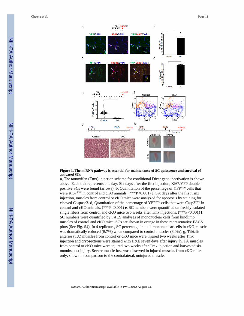
Figure 1. The miRNA pathway is essential for maintenance of SC quiescence and survival ofactivated SCsa, The tamoxifen (Tmx) injection scheme for conditional Dicer gene inactivation is shownabove. Each tick represents one day. Six days after the first injection, Ki67/YFP doublepositive SCs were found (arrows). b, Quantitation of the percentage of YFP+ve cells thatwere Ki67+ve in control and cKO animals. (***P<0.001) c, Six days after the first Tmxinjection, muscles from control or cKO mice were analyzed for apoptosis by staining forcleaved Caspase3. d, Quantitation of the percentage of YFP+ve cells that were Casp3+ve incontrol and cKO animals. (***P<0.001) e, SC numbers were quantified on freshly isolatedsingle fibers from control and cKO mice two weeks after Tmx injections. (***P<0.001) f,SC numbers were quantified by FACS analyses of mononuclear cells from hindlimbmuscles of control and cKO mice. SCs are shown in orange in these representative FACSplots (See Fig. S4). In 4 replicates, SC percentage in total mononuclear cells in cKO muscleswas dramatically reduced (0.7%) when compared to control muscles (3.0%). g, Tibialisanterior (TA) muscles from control or cKO mice were injured two weeks after Tmxinjection and cryosections were stained with H&E seven days after injury. h, TA musclesfrom control or cKO mice were injured two weeks after Tmx injection and harvested sixmonths post injury. Severe muscle loss was observed in injured muscles from cKO miceonly, shown in comparison to the contralateral, uninjured muscle.
Cheung et al. Page 11
Nature. Author manuscript; available in PMC 2012 August 23.
NIH
-PA Author Manuscript
NIH
-PA Author Manuscript
NIH
-PA Author Manuscript
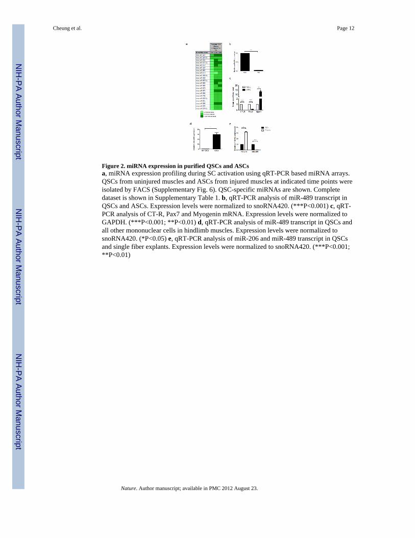
Figure 2. miRNA expression in purified QSCs and ASCsa, miRNA expression profiling during SC activation using qRT-PCR based miRNA arrays.QSCs from uninjured muscles and ASCs from injured muscles at indicated time points wereisolated by FACS (Supplementary Fig. 6). QSC-specific miRNAs are shown. Completedataset is shown in Supplementary Table 1. b, qRT-PCR analysis of miR-489 transcript inQSCs and ASCs. Expression levels were normalized to snoRNA420. (***P<0.001) c, qRT-PCR analysis of CT-R, Pax7 and Myogenin mRNA. Expression levels were normalized toGAPDH. (***P<0.001; **P<0.01) d, qRT-PCR analysis of miR-489 transcript in QSCs andall other mononuclear cells in hindlimb muscles. Expression levels were normalized tosnoRNA420. (*P<0.05) e, qRT-PCR analysis of miR-206 and miR-489 transcript in QSCsand single fiber explants. Expression levels were normalized to snoRNA420. (***P<0.001;**P<0.01)
Cheung et al. Page 12
Nature. Author manuscript; available in PMC 2012 August 23.
NIH
-PA Author Manuscript
NIH
-PA Author Manuscript
NIH
-PA Author Manuscript
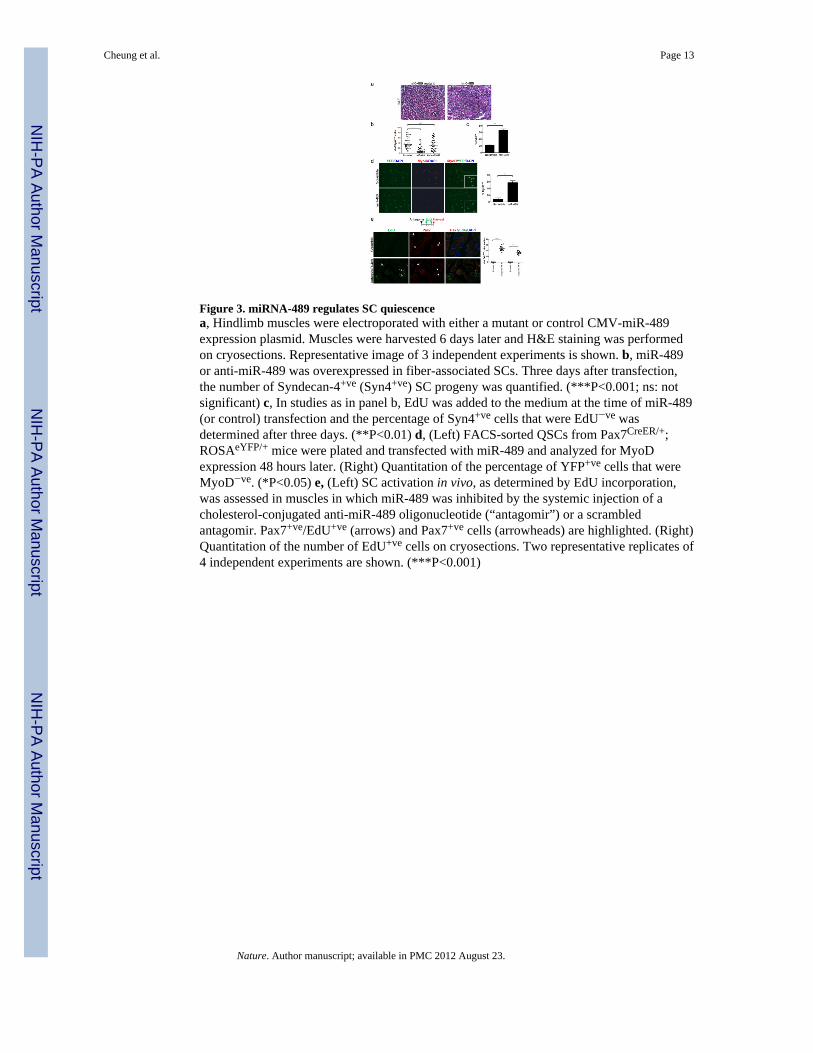
Figure 3. miRNA-489 regulates SC quiescencea, Hindlimb muscles were electroporated with either a mutant or control CMV-miR-489expression plasmid. Muscles were harvested 6 days later and H&E staining was performedon cryosections. Representative image of 3 independent experiments is shown. b, miR-489or anti-miR-489 was overexpressed in fiber-associated SCs. Three days after transfection,the number of Syndecan-4+ve (Syn4+ve) SC progeny was quantified. (***P<0.001; ns: notsignificant) c, In studies as in panel b, EdU was added to the medium at the time of miR-489(or control) transfection and the percentage of Syn4+ve cells that were EdU−ve wasdetermined after three days. (**P<0.01) d, (Left) FACS-sorted QSCs from Pax7CreER/+;ROSAeYFP/+ mice were plated and transfected with miR-489 and analyzed for MyoDexpression 48 hours later. (Right) Quantitation of the percentage of YFP+ve cells that wereMyoD−ve. (*P<0.05) e, (Left) SC activation in vivo, as determined by EdU incorporation,was assessed in muscles in which miR-489 was inhibited by the systemic injection of acholesterol-conjugated anti-miR-489 oligonucleotide (“antagomir”) or a scrambledantagomir. Pax7+ve/EdU+ve (arrows) and Pax7+ve cells (arrowheads) are highlighted. (Right)Quantitation of the number of EdU+ve cells on cryosections. Two representative replicates of4 independent experiments are shown. (***P<0.001)
Cheung et al. Page 13
Nature. Author manuscript; available in PMC 2012 August 23.
NIH
-PA Author Manuscript
NIH
-PA Author Manuscript
NIH
-PA Author Manuscript
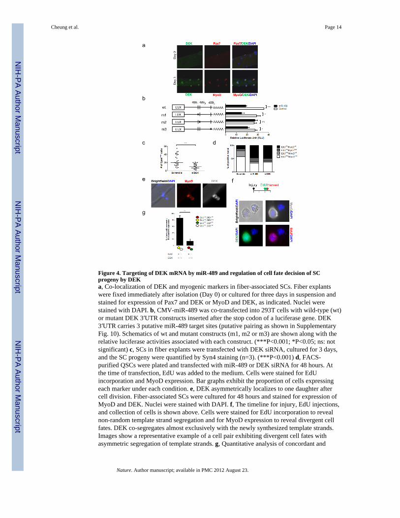
Figure 4. Targeting of DEK mRNA by miR-489 and regulation of cell fate decision of SCprogeny by DEKa, Co-localization of DEK and myogenic markers in fiber-associated SCs. Fiber explantswere fixed immediately after isolation (Day 0) or cultured for three days in suspension andstained for expression of Pax7 and DEK or MyoD and DEK, as indicated. Nuclei werestained with DAPI. b, CMV-miR-489 was co-transfected into 293T cells with wild-type (wt)or mutant DEK 3′UTR constructs inserted after the stop codon of a luciferase gene. DEK3′UTR carries 3 putative miR-489 target sites (putative pairing as shown in SupplementaryFig. 10). Schematics of wt and mutant constructs (m1, m2 or m3) are shown along with therelative luciferase activities associated with each construct. (***P<0.001; *P<0.05; ns: notsignificant) c, SCs in fiber explants were transfected with DEK siRNA, cultured for 3 days,and the SC progeny were quantified by Syn4 staining (n=3). (***P<0.001) d, FACS-purified QSCs were plated and transfected with miR-489 or DEK siRNA for 48 hours. Atthe time of transfection, EdU was added to the medium. Cells were stained for EdUincorporation and MyoD expression. Bar graphs exhibit the proportion of cells expressingeach marker under each condition. e, DEK asymmetrically localizes to one daughter aftercell division. Fiber-associated SCs were cultured for 48 hours and stained for expression ofMyoD and DEK. Nuclei were stained with DAPI. f, The timeline for injury, EdU injections,and collection of cells is shown above. Cells were stained for EdU incorporation to revealnon-random template strand segregation and for MyoD expression to reveal divergent cellfates. DEK co-segregates almost exclusively with the newly synthesized template strands.Images show a representative example of a cell pair exhibiting divergent cell fates withasymmetric segregation of template strands. g, Quantitative analysis of concordant and
Cheung et al. Page 14
Nature. Author manuscript; available in PMC 2012 August 23.
NIH
-PA Author Manuscript
NIH
-PA Author Manuscript
NIH
-PA Author Manuscript

discordant asymmetries of DEK and EdU in asymmetric SC divisions in studies as in panel(f). (**P<0.001)
Cheung et al. Page 15
Nature. Author manuscript; available in PMC 2012 August 23.
NIH
-PA Author Manuscript
NIH
-PA Author Manuscript
NIH
-PA Author Manuscript
Related Documents